N. A. S. Josse,J., andA.Kornberg,J. Biol. Chem.,237, 1968(1962). 20 Monod, J., and F. Jacob, Cold...
9
VOL. 49, 1963 MICROBIOLOGY: VOGT AND DULBECCO 171 12Flaks, J. F., J. Lichtenstein, and S. S. Cohen, J. Biol. Chem., 234, 1507 (1959). ' Delihas, N., Virology, 13, 242 (1961). 14Wiberg, J. S., M. L. Dirksen, R. H. Epstein, S. E. Luria, and J. M. Buchanan, these PRO- CEEDINGS, 48,293 (1962). 15 Dirksen, M. L., J. S. Wiberg, J. F. Koerner, and J. M. Buchanan, these PROCEEDINGS, 46, 1425(1960). 16Aposhian, H. V., and A. Kornberg, J. Biol. Chem., 237, 519 (1962). 17 Kornberg, A., in Enzymatic Synthesis of DNA (New York: John Wiley & Sons, Inc., 1961), pp.98-100. 18 Lehman, I. R., J. Biol. Chem., 235, 1479 (1960). 19 Josse, J., and A. Kornberg, J. Biol. Chem., 237, 1968 (1962). 20 Monod, J., and F. Jacob, Cold Spring Harbor Symposia on Quantitative Biology, vol. 26, 389 (1961). 21 Lwoff, A., Cold Spring Harbor Symposia on Quantitative Biology, vol. 27, in press (1962). 22 Dunnebacke, T. H., and H. B. Reaume, Virology, 6, 8 (1958). 23 Levine, S., Nature, 194, 895 (1962). 24 Levintow, L., M. M. Thoren, J. C. Darnell, Jr., and J. L. Hooper, Virology, 16, 220 (1962). 25 Lwoff, A., B. Roizman, and M. Lwoff, to be published. 2 Lwoff, A., B. Roizman, and M. Lwoff, C. R. Acad. Sci. (Paris), 254, 2462 (1962). STEPS IN THE NEOPLASTIC TRANSFORMATION OF HAMSTER EMBRYO CELLS BY POLYOMA V1RUS* BY MARGUERITE VOGT AND RENATO DULBECCO CALIFORNIA INSTITUTE OF TECHNOLOGY Communicated December 10, 1962 Cultures of hamster embryo cells infected with polyoma virus undergo a char- acteristic transformation within several weeks after infection. The transformed cultures are constituted by highly atypical cells which have an abnormal morph- ology, grow rapidly in vitro, and give rise to progressively growing tumors when inoculated subcutaneously into the adult hamster.1 In the previous experiments, the transformed cells arose in mass cultures and their properties were only studied many cell generations after the original virus- cell interaction had taken place. The experiments to be reported in this communi- cation were undertaken to get some information on the properties of transformed cells at earlier stages after infection with polyoma virus. The experiments show that the atypical cells are produced in two main steps; moreover, they reveal some new characteristics of the transformed cells which may contribute to a clari- fication of the mechanism of transformation. Material and Methods.-The polyoma virus used was of the "large-plaque" type. Its origin and the preparation of its stocks have been described.2 Tissue cultures of whole embryos of Golden hamsters were prepared according to a technique already described.3 A rich growth medium especially favorable for the cloning of mouse embryo cells (Weisberg, R., personal communication) and for the titration of polyoma-induced foci (Bayreuther, K., personal communication) was used for all experiments. It consisted of reinforced4 Eagle's medium (40 pts), Puck's6 nutrient solution N16 (40 pts), and medium NCTC 109 (4 pts); all ingredients were dissolved in Earle's saline containing 0.55 per cent glucose and 0.37 per cent NaHCOs. This medium was further supplemented with 0.3 per cent Bacto tryptose phosphate broth (10 pts) and, unless stated differently, with 10 pts of fetal bovine serum.
-
Upload
hoangxuyen -
Category
Documents
-
view
214 -
download
1
Transcript of N. A. S. Josse,J., andA.Kornberg,J. Biol. Chem.,237, 1968(1962). 20 Monod, J., and F. Jacob, Cold...
VOL. 49, 1963 MICROBIOLOGY: VOGT AND DULBECCO 171
12Flaks, J. F., J. Lichtenstein, and S. S. Cohen, J. Biol. Chem., 234, 1507 (1959).' Delihas, N., Virology, 13, 242 (1961).14Wiberg, J. S., M. L. Dirksen, R. H. Epstein, S. E. Luria, and J. M. Buchanan, these PRO-
CEEDINGS, 48,293 (1962).15Dirksen, M. L., J. S. Wiberg, J. F. Koerner, and J. M. Buchanan, these PROCEEDINGS, 46,
1425(1960).16Aposhian, H. V., and A. Kornberg, J. Biol. Chem., 237, 519 (1962).17 Kornberg, A., in Enzymatic Synthesis of DNA (New York: John Wiley & Sons, Inc., 1961),
pp.98-100.18 Lehman, I. R., J. Biol. Chem., 235, 1479 (1960).19 Josse, J., and A. Kornberg, J. Biol. Chem., 237, 1968 (1962).20 Monod, J., and F. Jacob, Cold Spring Harbor Symposia on Quantitative Biology, vol. 26, 389
(1961).21 Lwoff, A., Cold Spring Harbor Symposia on Quantitative Biology, vol. 27, in press (1962).22 Dunnebacke, T. H., and H. B. Reaume, Virology, 6, 8 (1958).23 Levine, S., Nature, 194, 895 (1962).24 Levintow, L., M. M. Thoren, J. C. Darnell, Jr., and J. L. Hooper, Virology, 16, 220 (1962).25 Lwoff, A., B. Roizman, and M. Lwoff, to be published.2 Lwoff, A., B. Roizman, and M. Lwoff, C. R. Acad. Sci. (Paris), 254, 2462 (1962).
STEPS IN THE NEOPLASTIC TRANSFORMATION OF HAMSTEREMBRYO CELLS BY POLYOMA V1RUS*
BY MARGUERITE VOGT AND RENATO DULBECCO
CALIFORNIA INSTITUTE OF TECHNOLOGY
Communicated December 10, 1962
Cultures of hamster embryo cells infected with polyoma virus undergo a char-acteristic transformation within several weeks after infection. The transformedcultures are constituted by highly atypical cells which have an abnormal morph-ology, grow rapidly in vitro, and give rise to progressively growing tumors wheninoculated subcutaneously into the adult hamster.1
In the previous experiments, the transformed cells arose in mass cultures andtheir properties were only studied many cell generations after the original virus-cell interaction had taken place. The experiments to be reported in this communi-cation were undertaken to get some information on the properties of transformedcells at earlier stages after infection with polyoma virus. The experiments showthat the atypical cells are produced in two main steps; moreover, they revealsome new characteristics of the transformed cells which may contribute to a clari-fication of the mechanism of transformation.
Material and Methods.-The polyoma virus used was of the "large-plaque" type. Its originand the preparation of its stocks have been described.2 Tissue cultures of whole embryos ofGolden hamsters were prepared according to a technique already described.3A rich growth medium especially favorable for the cloning of mouse embryo cells (Weisberg, R.,
personal communication) and for the titration of polyoma-induced foci (Bayreuther, K., personalcommunication) was used for all experiments. It consisted of reinforced4 Eagle's medium (40pts), Puck's6 nutrient solution N16 (40 pts), and medium NCTC 109 (4 pts); all ingredients weredissolved in Earle's saline containing 0.55 per cent glucose and 0.37 per cent NaHCOs. Thismedium was further supplemented with 0.3 per cent Bacto tryptose phosphate broth (10 pts) and,unless stated differently, with 10 pts of fetal bovine serum.
172 MICROBIOLOGY: VOGT AND DULBECCO PROC. N. A. S.
Experimental procedure: Sets of secondary hamster embryo cultures, seeded with 8 X 10⁶ cellsper plate, were infected with polyoma virus: an aliquot of 0.1 ml of a virus suspension of a titerof 109 PFU/ml was distributed over the cells after removal of the fluid medium. Adsorption ofthe virus was allowed to proceed for 2 hr at 370C in an incubator flushed with an adequate C02-airmixture; during this time, about 10 per cent of the input virus was adsorbed to the cells, as wasshown by control experiments with purified, P32-labeled, virus (unpublished). The cultures werewashed three times with medium and overlaid with 5 ml of medium. The cultures were main-tained under optimal growth conditions by three weekly fluid changes; the cells were transferredwhen the cell sheets became confluent.At various times (2, 4, 9, 13, 16, and 24 days) after infection, single cultures of the sets were
trypsinized and used to start a group of sparse subcultures. These cultures were seeded with 105cells and grown for seven days in the presence of 20 per cent fetal bovine serum; thereafter, fluidchanges were done with regular medium. The sparse subcultures were scanned on days 10 to 16with a low-power inverted microscope for areas of high mitotic activity and/or abnormal morph-ology. At this time, the subcultures were mostly constituted by a confluent monolayer of cellsshowing almost no mitotic activity. Some cultures contained one or a few foci of cells of unusualmorphology which grew in several layers and showed frequent mitoses. The cells of these fociwere usually markedly refringent and fusiform in shape; they crisscrossed each other and some-times piled up to form nodules or strands.The number of foci obtained on sparse subcultures started within the first week after infection
was about 10-5 of the cells plated; since the multiplicity of the adsorbed virus was 10-20, theefficiency of focus formation was about 10-6 that of plaque formation on mouse embryo cells.A number of foci were picked from the sparse subcultures. To do so and to carry over the least
amount of untransformed cells, small glass cylinders were used to isolate the foci from the surround-ing cells.6 The glass wells were filled with trypsin and the dispersed cells transferred to new petridishes. In certain cases, the foci were aspirated with a capillary pipette to avoid the use oftrypsin and transferred to petri dishes containing coverslips. The cell lines started from individualfoci will be designated as focal lines. All focal lines derived from sparse subcultures started withinthe first two weeks after infection were isolated from cultures containing each a single focus.They can therefore be assumed to derive from a single transformed cell; however, the presence, atthe outset, of a small proportion of contaminant nontransformed cells cannot be excluded.The focal lines were maintained by a regular regime of fluid changes and transfers; where
trypsin was to be avoided, the old coverslips were replaced at regular intervals by fresh coverslipswhich became repopulated by cells derived from the outgrowth surrounding the coverslips. With-out exception, all focal lines could be maintained indefinitely in an actively growing state. Thecells were characterized by their refringence, their high mitotic activity, and their capacity toform multilayered cell sheets with a crisscross pattern in dense cultures.Two types of controls to the focal lines were done. One type consisted of lines started with
cells from areas of the sparse subcultures outside of a focus= nonfocal lines. Usually, the cellsfrom areas delimited by three glass cylinders were compounded to obtain enough cells to starteach nonfocal line. These lines were constituted by cells with the characteristics of normal cells;they could not be maintained for more than a few passages.The other type of control consisted in a repetition of the entire experimental procedure, as
outlined, with the exception that the original sets of cultures were not infected with polyoma virus.In these control sets, no foci comparable to those of the infected sets were found in the sparsesubcultures. Only a few areas of persistent mitotic activity but of normal morphology were seen.These areas were picked and control lines started. These lines could be maintained for only a fewtransfers. One exceptional line could be cultured for five transfers. This line consisted of veryflat, transparent cells. It had other peculiar features to which we shall return.
Results.-The present investigation consisted in a study of the properties of thecells of the focal lines at various times after their isolation. The earliest observa-tions were carried out at the second or third transfers when a sufficient number ofcells were available. At this time, nontransformed cells which might have beenpicked with the focus when the line was started would have been considerably diluted

Voi. 49,14963 MIC'IROBIOLOGY: VOGT .AM) DULBE(CO 1],:3
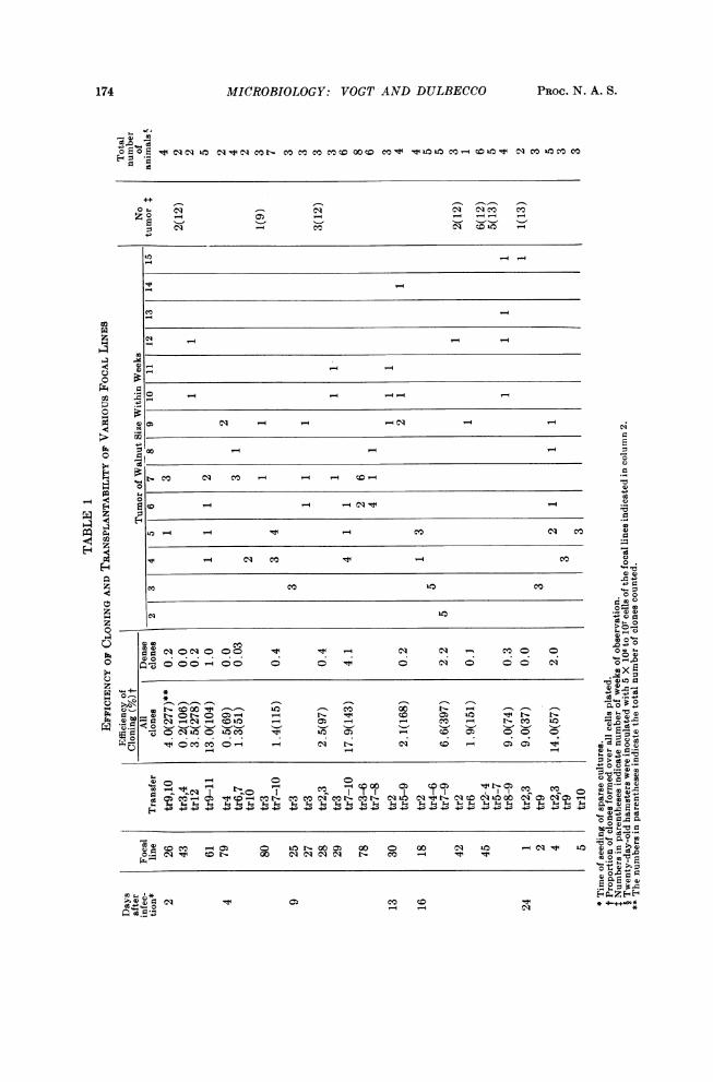
out owing to their limited growth ability, as demonstrated by the poor growth ofcontrol lines. The cells of the focal lines were characterized on the basis of thefollowing properties: morphological types of the clones formed, efficiency of cloning,and transplantability into 20-day-old hamsters. The efficiency of cloning wasmeasured by determining the proportion of cells capable of forming a clone of 200or more cells after seven days of incubation in medium supplemented with 20 percent fetal bovine serum (without feeder layer). The transplantability was deter-mined by inoculating 5 X 10⁶ cells from a trypsinized cell suspension subcutaneouslyinto 20-day-old hamsters. The degree of transplantability was measured by thetime required to form a tumor of the size of a walnut at the site of inoculation.The results of these determinations are summarized in Table 1. The followingpoints should be emphasized.The clones formed by the cells of the focal lines could be divided into two main
morphological groups. One group consisted of "thin" clones in which the cells grewmainly in a single layer; at the early transfers, the thin clones had a very loose struc-ture and were small in size (ca. 200 cells per clone); at later transfers, they becamelarger in size (1,000 and more cells) and showed a uniform crisscross-like pattern.All thin clones were clearly different from normal clones: the cells were refringentand randomly oriented. The thin clones were prevalent at the early transfers;their proportion decreased in later transfers. The second group of clones were"dense" clones, in which the cells piled up in many layers. The morphology ofthese dense clones was variable: some showed a uniform dense center, others hadheavy strands, others nodules. These clones were absent or rare in early transfersand increased in proportion in later transfers.
Clones produced by cells from the nonfocal or control lines were of a strict mono-layer type; the cells were thin and transparent and showed a regular arrangement.They were readily distinguishable from the thin clones just described. We cantherefore conclude that both groups of clones formed by cells from focal lines wereclones of transformed cells.The efficiency of cloning of the cells of the focal lines at the early transfers was
very similar to that of normal cells. At later transfers, this efficiency increased inparallel with the appearance of thin clones of large size and of dense clones.The transplantability of the cells of the focal lines varied markedly from line to
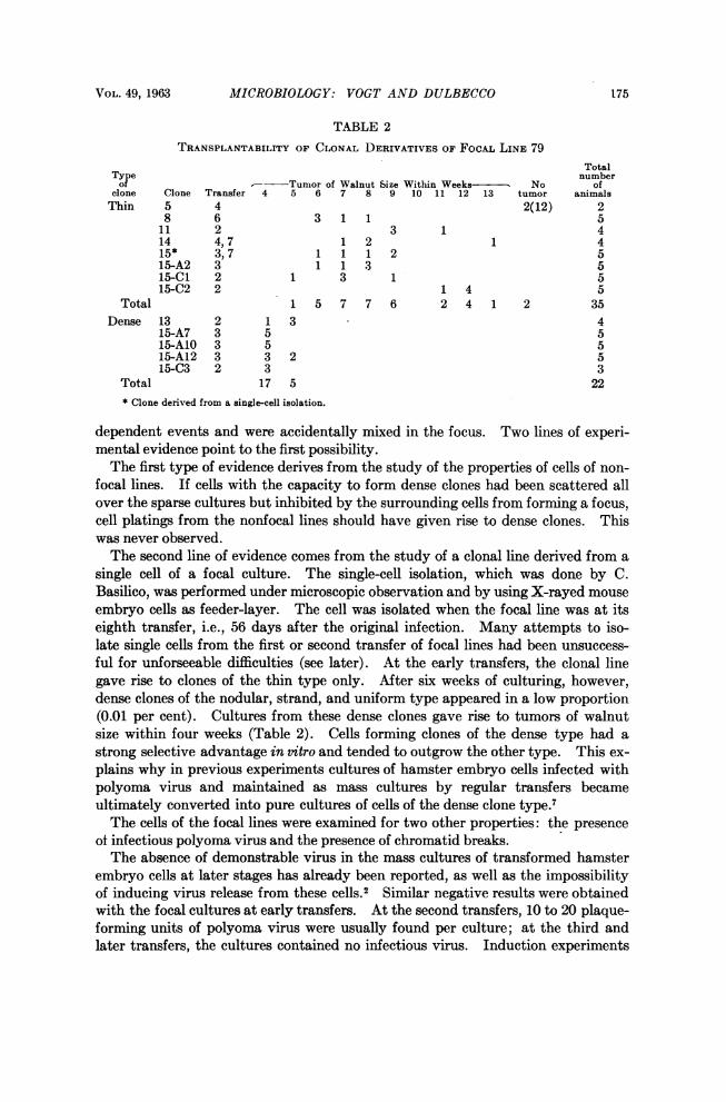
line, and within a given line, with the transfer number. In general, the transplant-ability was very low or absent at early transfers and increased with later transfers.The increase in transplantability proceeded at equal pace with the increase in theproportion of dense clones. These results suggested therefore that the highly trans-plantable cells were the same cells that gave rise to dense clones. This deductionwas confirmed by comparing the transplantability of subcultures derived from eightclones of the thin type, obtained at later transfers when the clones formed wererather large, with that of subcultures of five dense clones. As shown in Table 2,the average time for developing a tumor of walnut size was 8.4 weeks for the culturesof the thin clones (2/35 animals did not develop any tumor) and 4.3 weeks for thecultures of dense clones (23/23 animals developed a tumor).As to the relationship of the two types of cells present in the focal lines, two pos-
sibilities can be visualized: either the cells giving rise to dense clones are variantsof the main cell type which forms thin clones or the two cell types originated in in-
174 MICROBIOLOGY: VOGT AND DULBECCO PROC. N. A. S.
4) W,
Eo E o CI Lo N"N Mt-cor:cMscov-e<com":m:v:L c = mN0 M to M
0Z
_4
tc CT_
0 -._ o _. -l _
° -
~~~~~~~~0)~~~~~~~~~~~~~~~~~~0
Ho Q~~~~~c
044 - '-4
0 -T
,r~~~~~~~~~~~0 co:$ C4-
t 0 W co t-.-i 0Y
04o-4 .v
Itt- 0. 4 to-t- t-
<:Z _~
o 4)
o doyt 0E
O Q Q O00 - 00 0 0 t 0 co 0 0 O- 0
Ps~~~~~~~~~~~~~~~~~~~~~~~~~~~~~~~~~~4&0Xlr. '-_ *
ro40.4) 1>. Ot'-0 0~~'-4 - t- r-4 i r- . 0sOV 4)8-0 b-~ so. - ot - X - oi - _-' _ s 4)4)PeO
X O . O OO O_ _ s -s O 0 0TOO ) X4)W0.=~ 0 0 VO -r-N1 N c N LO O ON*
0 0.
04
_ y LOt0==0 q t ~ ofW r.
CIA"Icli c~~~~~~~l cq N t- VD V ~~~~~a.003.
'S*~~~~~~~~~~~ ~~~~~~~~~~~~~~~~~~~~~~0'
O ~ ~~°~ '-O O~ 0t 0- O ~ - ~
00 - -Oq *
r-4 t C - 00 C SC S t X _ t t0o0
VOL. 49, 1963 MICROBIOLOGY: VOGT AND DULBECCO 175
TABLE 2
TRANSPLANTABILITY OF CLONAL DERIVATIVES OF FoCAL LINE 79Total
Type numberof - Tumor of Walnut Size Within Weeks - No of
clone Clone Transfer 4 5 6 7 8 9 10 11 12 13 tumor animalsThin 5 4 2(12) 2
8 6 3 1 1 511 2 3 1 414 4,7 1 2 1 415* 3, 7 1 1 1 2 515-A2 3 1 1 3 515-C1 2 1 3 1 515-C2 2 1 4 5
Total 1 5 7 7 6 2 4 1 2 35Dense 13 2 1 3 4
15-A7 3 5 515-AlO 3 5 515-A12 3 3 2 515-C3 2 3 3
Total 17 5 22* Clone derived from a single-cell isolation.
dependent events and were accidentally mixed in the focus. Two lines of experi-mental evidence point to the first possibility.The first type of evidence derives from the study of the properties of cells of non-
focal lines. If cells with the capacity to form dense clones had been scattered allover the sparse cultures but inhibited by the surrounding cells from forming a focus,cell platings from the nonfocal lines should have given rise to dense clones. Thiswas never observed.The second line of evidence comes from the study of a clonal line derived from a
single cell of a focal culture. The single-cell isolation, which was done by C.Basilico, was performed under microscopic observation and by using X-rayed mouseembryo cells as feeder-layer. The cell was isolated when the focal line was at itseighth transfer, i.e., 56 days after the original infection. Many attempts to iso-late single cells from the first or second transfer of focal lines had been unsuccess-ful for unforseeable difficulties (see later). At the early transfers, the clonal linegave rise to clones of the thin type only. After six weeks of culturing, however,dense clones of the nodular, strand, and uniform type appeared in a low proportion(0.01 per cent). Cultures from these dense clones gave rise to tumors of walnutsize within four weeks (Table 2). Cells forming clones of the dense type had astrong selective advantage in vttro and tended to outgrow the other type. This ex-plains why in previous experiments cultures of hamster embryo cells infected withpolyoma virus and maintained as mass cultures by regular transfers becameultimately converted into pure cultures of cells of the dense clone type.'The cells of the focal lines were examined for two other properties: the presence
of infectious polyoma virus and the presence of chromatid breaks.The absence of demonstrable virus in the mass cultures of transformed hamster
embryo cells at later stages has already been reported, as well as the impossibilityof inducing virus release from these cells.2 Similar negative results were obtainedwith the focal cultures at early transfers. At the second transfers, 10 to 20 plaque-forming units of polyoma virus were usually found per culture; at the third andlater transfers, the cultures contained no infectious virus. Induction experiments
176 MICROBIOLOGY: VOG7' AND DULBECCO PROC. N. A. S.
were carried out at the third to sixth transfer of the focal lines. No production ofinfectious virus or of infectious viral DNA could be obtained after thymidine star-vation induced by aminopterin as previously described. X-rays and over-crowd-ing were equally ineffective as inducing agents.The search for chromatid breaks was prompted by the presence of an often unu-
sually high number of dead cells in the cultures of the focal lines at a time when nopolyoma virus could be detected. A measure for chromatid breakage was ob-tained by scoring mitotic figures at late anaphase and early telophase for the pres-ence of chromatid bridges. Coverslip cultures were prepared at various transfers
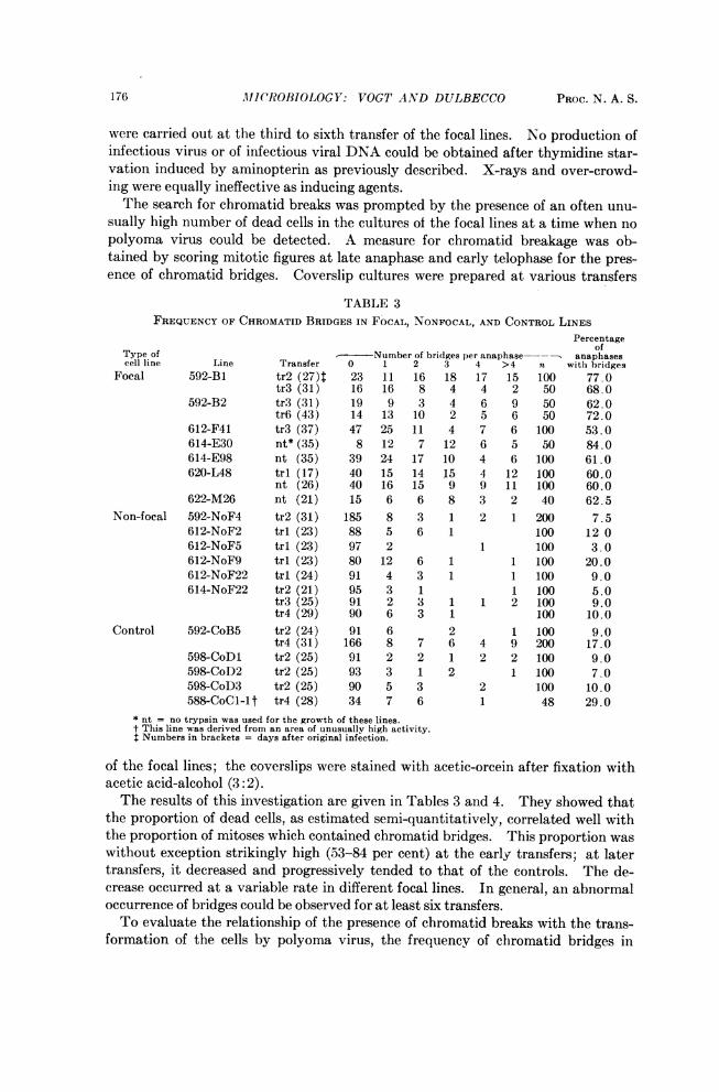
TABLE 3FREQUENCY OF CHROMATID BRIDGES IN FOCAL, NONFOCAL, AND CONTROL LINES
Percentageof
Type of -Number of bridges per anaphase---- anaphasescell line Line Transfer 0 1 2 3 4 >4 n with bridges
Focal 592-Bi tr2 (27)4 23 11 16 18 17 15 100 77.0tr3 (31) 16 16 8 4 4 2 50 68.0
592-B2 tr3 (31) 19 9 3 4 6 9 50 62.0tr6 (43) 14 13 10 2 5 6 50 72.0
612-F41 tr3 (37) 47 25 11 4 7 6 100 53.0614-E30 nt* (35) 8 12 7 12 6 5 50 84.0614-E98 nt (35) 39 24 17 10 4 6 100 61.0620-L48 tri (17) 40 15 14 15 4 12 100 60.0
nt (26) 40 16 15 9 9 11 100 60.0622-M26 nt (21) 15 6 6 8 3 2 40 62.5
Non-focal 592-NoF4 tr2 (31) 185 8 3 1 2 1 200 7.5612-NoF2 tri (23) 88 5 6 1 100 12 0612-NoF5 tri (23) 97 2 1 100 3.0612-NoF9 tri (23) 80 12 6 1 1 100 20.0612-NoF22 tri (24) 91 4 3 1 1 100 9.0614-NoF22 tr2 (21) 95 3 1 1 100 5.0
tr3(25) 91 2 3 1 1 2 100 9.0tr4 (29) 90 6 3 1 100 10.0
Control 592-CoB5 tr2 (24) 91 6 2 1 100 9.0tr4 (31) 166 8 7 6 4 9 200 17.0
598-CoDl tr2 (25) 91 2 2 1 2 2 100 9.0598-CoD2 tr2 (25) 93 3 1 2 1 100 7.0598-CoD3 tr2 (25) 90 5 3 2 100 10.0588-CoCl-1t tr4 (28) 34 7 6 1 48 29.0
* nt = no trypsin was used for the growth of these lines.t This line was derived from an area of unusually high activity.* Numbers in brackets = days after original infection.
of the focal lines; the coverslips were stained with acetic-orcein after fixation withacetic acid-alcohol (3:2).The results of this investigation are given in Tables 3 and 4. They showed that
the proportion of dead cells, as estimated semi-quantitatively, correlated well withthe proportion of mitoses which contained chromatid bridges. This proportion waswithout exception strikingly high (53-84 per cent) at the early transfers; at latertransfers, it decreased and progressively tended to that of the controls. The de-crease occurred at a variable rate in different focal lines. In general, an abnormaloccurrence of bridges could be observed for at least six transfers.To evaluate the relationship of the presence of chromatid breaks with the trans-
formation of the cells by polyoma virus, the frequency of chromatid bridges in
VOL. 49, 1963 MICROBIOLOGY: VOGT AND DULBECCO 177
nonfocal lines and in noninfected control lines is also entered in Table 3. As canbe seen from the table, both types of control lines showed a significantly smallerproportion of mitoses with bridges than the focal lines. In these controls, the high-est frequency of bridges (29 per cent) was found in a clone of normal cells alreadymentioned, which showed an unusually high growth ability. Whether these twoproperties are correlated is unknown.
Since trypsinization is known to induce chromatid breakage,8 some of the focallines were grown without trypsinization. The results showed no difference betweentrypsinized and nontrypsinized cultures.These observations leave therefore no doubt that the production of chromatid
breaks leading to bridge formation is characteristic of the transformation inducedby polyoma virus in this in vitro system.
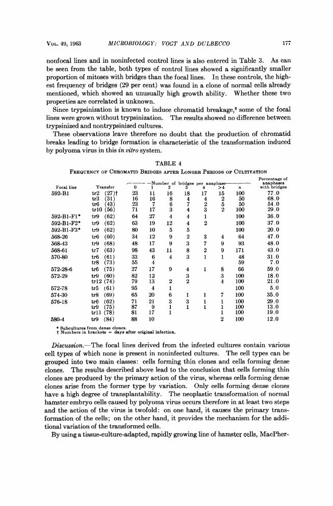
TABLE 4FREQUENCY OF CHROMATID BRIDGES AFTER LONGER PERIODS OF CULTIVATION
Percentage of-Number of bridges per anaphase anaphases
Focal line Transfer 0 1 2 3 4 >4 n with bridges592-Bi tr2 (27)t 23 11 16 18 17 15 100 77.0
tr3 (31) 16 16 8 4 4 2 50 68.0tr6 (43) 23 7 6 7 2 5 50 54.0tr1O (56) 71 17 3 4 3 2 100 29.0
592-B1-F1* tr9 (62) 64 27 4 4 1 100 36.0592-B1-F2* tr9 (62) 63 19 12 4 2 100 37.0592-B1-F3* tr9 (62) 80 10 5 5 100 20.0568-26 tr6 (60) 34 12 9 2 3 4 64 47.0568-43 tr9 (68) 48 17 9 3 7 9 93 48.0568-61 tr7 (63) 98 43 11 8 2 9 171 43.0570-80 tr6 (61) 33 6 4 3 1 1 48 31.0
tr8 (73) 55 4 59 7.0572-28-6 tr6 (75) 27 17 9 4 1 8 66 59.0572-29 tr9 (60) 82 12 3 3 100 18.0
trl2 (74) 79 13 2 2 4 100 21.0572-78 tr5 (61) 95 4 1 100 5.0574-30 tr8 (69) 65 20 6 1 1 7 100 35.0576-18 tr6 (62) 71 21 3 3 1 1 100 29.0
tr9 (75) 87 9 1 1 1 1 100 13.0tr 1 (78) 81 17 1 1 100 19.0
580-4 tr9 (84) 88 10 2 100 12.0* Subcultures from dense clones.t Numbers in brackets = days after original infection.
Discussion.-The focal lines derived from the infected cultures contain variouscell types of which none is present in noninfected cultures. The cell types can begrouped into two main classes: cells forming thin clones and cells forming denseclones. The results described above lead to the conclusion that cells forming thinclones are produced by the primary action of the virus, whereas cells forming denseclones arise from the former type by variation. Only cells forming dense cloneshave a high degree of transplantability. The neoplastic transformation of normalhamster embryo cells caused by polyoma virus occurs therefore in at least two stepsand the action of the virus is twofold: on one hand, it causes the primary trans-formation of the cells; on the other hand, it provides the mechanism for the addi-tional variation of the transformed cells.By using a tissue-culture-adapted, rapidly growing line of hamster cells, MacPher-
178 AIICROBIOLOGY: VOGT AND DULBECCO PROC. N. A. S.
son and Stoker'0 have obtained transformation to the malignant state in a singlestep. This finding need not be at variance with the results of the present investiga-tion. In fact, the cells of the line used by these investigators have already spon-taneously undergone profound changes which allow their unlimited and rapid multi-plication in vitro. Thus, a single additional step, caused by the virus, may be sufficient to transform these cells to a malignant state.The evolution of the focal cultures described in this article is probably one aspect
of the phenomenon of progression' which has been recognized as a constant propertyof cancers in animals. In the present experiments, the progression appears to be,at least to a certain extent, a necessary consequence of the primary transformationand not a superimposed accidental complication.
Chromatid breaks were observed in a large proportion of the cells of most focalcultures, with the highest frequency during the early transfers. This finding raisesthe question whether the progression observed in this system is due to gross chromo-somal alterations caused by the breaks; such a view would be supported by thepreliminary findings of aneuploidy in cells which form dense clones. On the otherhand, the chromatid breaks may not be directly involved in the progression; theycould rather be a symptom of irregularities in the process of DNA replication in thetransformed cells, able to cause several different consequences, including the pro-gression. The inability to find chromatid breaks in one focal line (572-78) may beattributed to a precocious healing of the process causing the breaks, which tendsto heal in all lines at later transfers.The present observations are relevant to the problem of the fate of the viral DNA
in the transformed cells. The results previously obtained, that the cells of thedense clones do not contain the viral nucleic acid in a state detectable by infectivity,has now been extended; it has in fact been shown that this is true also for the cellsof the thin clones. The results still leave the question open whether the virus-cell interaction which leads to transformation is nonproductive from the start orbecomes so after a small number of cell generations.As pointed out in the past,2 these results show that autonomously replicating
polyoma virus DNA is not present in the transformed cells; they do not, however,exclude the presence of an integrated viral DNA. A new point in favor of the latterpossibility is the production of chromatid breaks in the focal lines. These breaks,which are characteristic of the transformed hamster embryo cells, continue to beproduced after infectious virus has disappearaed. A continued production ofchromosomal alterations and of chromatid breaks has also been observed in humancells transformed by the simian SV40 virus,11 12 which is related to polyoma virus,and in cell lines of the Chinese hamster which survived an infection with the DNA-containing herpes virus.'3 "4 The continued production of chromosomal altera-tions suggests that a specific agent-possibly the viral DNA-is continuously pres-ent in these cells after the infection.
Functionally, the primary action of the virus in the transformation of the ham-ster embryo cells appears to be a release of the regulation of cell multiplication;this is revealed by the ability of the cells of the thin-clone type to form foci. In cellsin which this original change has taken place, some of the chromosomal aberra-tions subsequently produced acquire, at least in vitro, a selective value. A step-wise process in this system leads thus finally to the malignant cell.
VOL. 49, 1963 PHYSICS: GURSEY AND LEE 179
We gratefully acknowledge the skillful assistance of Mrs. B. Keeley and Mrs. A. Drew.* This work was aided by grants from the American Cancer Society, Inc., the National Founda-
tion, and the U.S. Public Health Service, Grant No. E5131.1 Vogt, M., and R. Dulbecco, these PROCEEDINGS, 46, 365-370 (1960).2Vogt, M., and R. Dulbecco, Virology, 16, 41-51 (1962).3 Dulbecco, R., and G. Freeman, Virology, 8, 396-397 (1959).4 Fourfold concentration of amino acids and vitamins + 0.4 mM glycine, 0.4 mM serine, and
0.04mM inositol.5 Puck, T. T., S. J. Cieciura, and A. Robinson, J. Exp. Med., 108, 945-955 (1958).6 Puck, T. T., P. I. Marcus, and S. J. Cieciura, J. Exp. Med., 103, 273-284 (1956).7 Dulbecco, R., and M. Vogt, these PROCEEDINGS, 46, 1617-1623 (1960).8 Levan, A., and J. J. Biesele, Ann. N. Y. Acad. Sci., 71, 1022-1053 (1958).9 Foulds, L., Canc. Res., 14, 327-339 (1954).10 Macpherson, I., and M. Stoker, Virology, 16, 147-151 (1962).11 Shein, H. M., and J. F. Enders, these PROCEEDINGS, 48, 1164-1172 (1962).12 Koprowski, H., J. A. Ponten, F. Jensen, R. G. Ravdin, P. Moorhead, and E. Saksela, J.
Cell and Comp. Physiol., 59, 281-292 (1962).13 Hampar, B., and S. A. Ellison, Nature, 192, 145-147 (1961).14 Hampar, B., and S. A. Ellison, Abstr. 40th AMtg. IADR, p. 19 (1962).
SPIN 1/2 WAVE EQUATION IN DE-SITTER SPACE*
BY F. GtRSEY AND T. D. LEEt
MIDDLE EAST TECHNICAL UNIVERSITY, ANKARA, TURKEY, AND COLUMBIA UNIVERSITY, NEW YORK
Communicated by Robert Serber, November 16, 1962
1. Introduction.-The de-Sitter space can be represented by a 4-dimensionalpseudo-sphere embedded in a 5-dimensional space. Let {,,(,u = 1, 2, ..., 5) bethe five real coordinates of the 5-dimensional space. The de-Sitter space is de-scribed by the surface
= R2, (1)
where
{I= cows- (2)7t7 is the (jA, v)th matrix element of the diagonal matrix
37 = (1 1 1),(3)
and the real constant R is the radius of the pseudo-sphere. The totality of allhomogeneous linear transformations among it that leave the surface (1) invariantforms a ten-parameter group. In the limit R -> a, the de-Sitter space becomessimply the usual flat 4-dimensional space, and this ten-parameter group reducesto that of the inhomogeneous Lorentz transformation.