Myoglobin, T Time-Activit Curves for Early...
10
1542 Analysis of Creatine Kinase, CK-MB, Myoglobin, and Troponin T Time-Activit Curves for Early Assessment of Coronary Artery Reperfusion After Intravenous Thrombolysis Markus Zabel, MD; Stefan H. Hohnloser, MD; Wolfgang Koster, MD; Martina Prinz, MD; Wolfgang Kasper, MD; and Hanjorg Just, MD Background. Thrombolysis has become the standard therapeutic approach in patients with acute myocardial infarction. To identify patients who may benefit from early invasive procedures, reliable noninvasive assessment of success or failure of thrombolytic therapy is mandatory. Methods and Results. In a prospective study in 63 consecutive patients undergoing thrombolysis for their first myocardial infarction, serial measurements of creatine kinase (CK), its isoenzyme CK-MB, myoglobin, and troponin T were done to determine their value for noninvasive prediction of coronary artery patency. Blood samples were drawn every 15 minutes during the first 90 minutes, every 30 minutes during the first 4 hours, every 4 hours during the first 24 hours, and every 8 hours during the first 72 hours. The perfusion status of the infarct-related artery was assessed angiographically 90 minutes after initiation of thrombolysis. For each marker, time to its peak concentration and its early initial slope (start of thrombolysis to 90 minutes thereafter) were determined. Areas under receiver operator characteristic (ROC) curves were 0.83, 0.76, 0.82, and 0.80 for maxima of CK, CK-MB, myoglobin, and troponin T, respectively (p=NS by univariate Z test). The corresponding values for early slopes of CK, CK-MB, myoglobin, and troponin T were 0.79, 0.82, 0.89, and 0.80 (p =0.23 for comparison between myoglobin and CK-MB;p=0.07 between myoglobin and CK). Sensitivity, specificity, and positive and negative predictive values regarding noninvasive prediction of coronary artery patency after 90 minutes were 80%o, 82%, 95%, and 61% for time to CK maximum; 91%, 77%, 91%, and 77% for time to myoglobin maximum; 87%, 71%, 89%o, and 67% for early CK slope; and 94%, 88%, 94%, and 82% for myoglobin slope, respectively. When myoglobin slope was assessed together with other clinical reperfusion markers (resolution of chest pain or ST segment elevation, occurrence of reperfusion arrhythmias) by logistic regression analysis, only the myoglobin slope was an independent predictor of coronary artery patency (p<0.0001). Conclusions. With regard to noninvasive prediction of coronary artery patency after thrombolytic therapy, measurement of the early initial slopes of the serum markers within only 90 minutes after the initiation of therapy is as accurate as the determination of the time to their peak concentration. Compared with the other markers examined, myoglobin appears to have advantages because of its earlier rise, yielding a better negative predictive value and a higher area under the ROC curve for determination of its early initial slopes. (Circulation 1993;87:1542-1550) KEY WORDs * myocardial infarction, acute * thrombolysis * reperfusion T he value of coronary artery reperfusion by means of intravenous thrombolysis in patients with evolving myocardial infarction has been rigor- ously evaluated.'-4 An impressive improvement in sur- vival rates has been demonstrated in patients with successful reopening of the infarct-related artery. In addition, left ventricular function has been shown to be superior in patients treated with thrombolysis compared with placebo-treated patients.5 It is evident from both From the University Hospital, Department of Cardiology and Department of Clinical Chemistry (W.K.), Freiburg, Germany. Supported by a grant from the Deutsche Gesellschaft fur Herz- und Kreislaufforschung, Bad Nauheim, Germany (M.Z.). Address for correspondence: Stefan H. Hohnloser, MD, Uni- versity Hospital, Department of Cardiology, Hugstetter Str. 55, 7800 Freiburg, FRG. Received June 15, 1992; revision accepted January 14, 1993. laboratory and clinical studies that the benefits of thrombolytic therapy are conferred by restoration of patency of the infarct-related artery.6-9 Recently, Ab- bottsmith and coworkers10 have reported that salvage angioplasty after failed thrombolysis improves outcome in these patients. Thus, invasive means are valuable in selected patients to establish coronary artery pa- tency.11,2 Rescue thrombolysis with readministration of the same or a different thrombolytic agent may also have a role after failed initial thrombolytic therapy.13 On the other hand, acute routine angiography in all patients receiving thrombolytic therapy for acute infarc- tion is unnecessary and may eventually cause harm to some.14-16 Hence, reliable methods other than angiog- raphy are needed to detect coronary reperfusion early during the hospital course. In previous studies, the normalization of ST segment elevation in the surface by guest on June 19, 2018 http://circ.ahajournals.org/ Downloaded from
Transcript of Myoglobin, T Time-Activit Curves for Early...

1542
Analysis of Creatine Kinase, CK-MB,Myoglobin, and Troponin T Time-Activit
Curves for Early Assessment of Coronary ArteryReperfusion After Intravenous Thrombolysis
Markus Zabel, MD; Stefan H. Hohnloser, MD; Wolfgang Koster, MD; Martina Prinz, MD;Wolfgang Kasper, MD; and Hanjorg Just, MD
Background. Thrombolysis has become the standard therapeutic approach in patients with acutemyocardial infarction. To identify patients who may benefit from early invasive procedures, reliablenoninvasive assessment of success or failure of thrombolytic therapy is mandatory.Methods and Results. In a prospective study in 63 consecutive patients undergoing thrombolysis for their
first myocardial infarction, serial measurements of creatine kinase (CK), its isoenzyme CK-MB,myoglobin, and troponin T were done to determine their value for noninvasive prediction of coronary
artery patency. Blood samples were drawn every 15 minutes during the first 90 minutes, every 30 minutesduring the first 4 hours, every 4 hours during the first 24 hours, and every 8 hours during the first 72hours. The perfusion status of the infarct-related artery was assessed angiographically 90 minutes afterinitiation of thrombolysis. For each marker, time to its peak concentration and its early initial slope (startof thrombolysis to 90 minutes thereafter) were determined. Areas under receiver operator characteristic(ROC) curves were 0.83, 0.76, 0.82, and 0.80 for maxima of CK, CK-MB, myoglobin, and troponin T,respectively (p=NS by univariate Z test). The corresponding values for early slopes of CK, CK-MB,myoglobin, and troponin T were 0.79, 0.82, 0.89, and 0.80 (p =0.23 for comparison between myoglobin andCK-MB;p=0.07 between myoglobin and CK). Sensitivity, specificity, and positive and negative predictivevalues regarding noninvasive prediction of coronary artery patency after 90 minutes were 80%o, 82%, 95%,and 61% for time to CK maximum; 91%, 77%, 91%, and 77% for time to myoglobin maximum; 87%, 71%,89%o, and 67% for early CK slope; and 94%, 88%, 94%, and 82% for myoglobin slope, respectively. Whenmyoglobin slope was assessed together with other clinical reperfusion markers (resolution of chest painor ST segment elevation, occurrence of reperfusion arrhythmias) by logistic regression analysis, only themyoglobin slope was an independent predictor of coronary artery patency (p<0.0001).
Conclusions. With regard to noninvasive prediction of coronary artery patency after thrombolytictherapy, measurement of the early initial slopes of the serum markers within only 90 minutes after theinitiation of therapy is as accurate as the determination of the time to their peak concentration. Comparedwith the other markers examined, myoglobin appears to have advantages because of its earlier rise,yielding a better negative predictive value and a higher area under the ROC curve for determination of itsearly initial slopes. (Circulation 1993;87:1542-1550)KEY WORDs * myocardial infarction, acute * thrombolysis * reperfusion
T he value of coronary artery reperfusion by meansof intravenous thrombolysis in patients withevolving myocardial infarction has been rigor-
ously evaluated.'-4 An impressive improvement in sur-vival rates has been demonstrated in patients withsuccessful reopening of the infarct-related artery. Inaddition, left ventricular function has been shown to besuperior in patients treated with thrombolysis comparedwith placebo-treated patients.5 It is evident from both
From the University Hospital, Department of Cardiology andDepartment of Clinical Chemistry (W.K.), Freiburg, Germany.
Supported by a grant from the Deutsche Gesellschaft fur Herz-und Kreislaufforschung, Bad Nauheim, Germany (M.Z.).Address for correspondence: Stefan H. Hohnloser, MD, Uni-
versity Hospital, Department of Cardiology, Hugstetter Str. 55,7800 Freiburg, FRG.
Received June 15, 1992; revision accepted January 14, 1993.
laboratory and clinical studies that the benefits ofthrombolytic therapy are conferred by restoration ofpatency of the infarct-related artery.6-9 Recently, Ab-bottsmith and coworkers10 have reported that salvageangioplasty after failed thrombolysis improves outcomein these patients. Thus, invasive means are valuable inselected patients to establish coronary artery pa-tency.11,2 Rescue thrombolysis with readministration ofthe same or a different thrombolytic agent may alsohave a role after failed initial thrombolytic therapy.13On the other hand, acute routine angiography in allpatients receiving thrombolytic therapy for acute infarc-tion is unnecessary and may eventually cause harm tosome.14-16 Hence, reliable methods other than angiog-raphy are needed to detect coronary reperfusion earlyduring the hospital course. In previous studies, thenormalization of ST segment elevation in the surface
by guest on June 19, 2018http://circ.ahajournals.org/
Dow
nloaded from

Zabel et al Serum Markers for Prediction of Reperfusion 1543
ECG,17-21 the occurrence of reperfusion arrhyth-mias,19,22-24 the resolution of infarct-related chestpain,25,26 and various cardiac enzymes have been usedfor this purpose. For example, a short time to peakcreatine kinase (CK)27 or CK-MB concentration28,29 hasbeen proposed as an indicator of reperfusion. Morerecently, new serum markers such as myoglobin,28'30CK-MB isoforms,31 and the specific cardiac antigentroponin T32 have become available and have beenexamined in small groups of patients with acute myo-cardial infarction. Up to now, however, the value ofthese new reperfusion markers has not been vigorouslyevaluated, and they have not been compared with eachother in a reasonably large cohort of patients undergo-ing thrombolysis. The aims of the present prospectivestudy, therefore, were 1) to develop simple indexes forsuccess or failure of reperfusion therapy according tothe initial CK and CK-MB time-activity curves and 2)to evaluate whether new reperfusion markers such asmyoglobin and troponin T yield a higher diagnosticaccuracy in prediction of thrombolysis-induced reopen-ing of the infarct-related artery compared with conven-tional serum markers. Particular attention was paid tothe question of whether analysis of early increases inenzyme or protein concentrations (within 120 minutesafter start of thrombolysis) is as accurate regardingprediction of coronary artery patency as the determina-tion of the time to their peak concentration, since thiswould be of marked clinical utility.
MethodsPatient Population
Sixty-three consecutive patients with acute myocar-dial infarction treated with thrombolysis were studiedprospectively between May 1990 and October 1991.Patients with typical chest pain of .30 minutes' dura-tion, unresponsive to nitrates, and with ECG ST seg-ment elevation of .0.1 mV in at least two limb leads or>0.2 mV in at least two precordial (V) leads wereeligible for the study. Patients with cardiogenic shockwere excluded. Thrombolytic therapy had to be initiatedwithin 6 hours after onset of symptoms. The thrombo-lytic agent used was prourokinase in 32 patients com-bined with recombinant tissue plasminogen activator(rt-PA) in 20 patients and anisoylated plasminogenstreptokinase activator complex (APSAC) in 31 pa-tients. After thrombolysis, all patients received a bolusof 7,500 to 10,000 units of heparin followed by acontinuous infusion of 1.250 units/hr for the next 72-96hours (partial thromboplastin time adjusted to twotimes the upper normal). All patients gave their in-formed consent to the study. The usual exclusion crite-ria for thrombolysis were applied.
Coronary AngiographyCoronary angiography was performed within 90 min-
utes after onset of thrombolytic therapy and repeated 24hours later. The angiographic findings were assessedaccording to the Thrombolysis in Myocardial Infarction(TIMI) criteria for reperfusion.4 For this purpose, thefirst injection of contrast was taken as the patencyassessment. TIMI grades 2 and 3 were defined as
patency within the first 90 minutes was consideredsuccessful coronary artery opening.
Serum MarkersBlood samples (10 mL) were taken before initiation
of thrombolysis, every 15 minutes for the first 90 min-utes after start of therapy, every 30 minutes for the first4 hours, every 4 hours up to 24 hours, and every 8 hoursup to 72 hours. The samples were stored at roomtemperature to allow clotting and frozen at -20°C aftercentrifugation until analysis. Samples were analyzed byinvestigators who were unaware of the angiographicresults. Plasma total CK activity was assayed accordingto the method of Rosalki.33 The mass of the CK-MBisoenzyme was determined by means of a rapid fluoro-metric enzyme immunoassay based on a two-site sand-wich immunoassay methodology (Baxter Stratus, Mi-ami, Fla.).34 Units are given in nanograms per milliliter.Myoglobin was determined nephelometrically by aquantitative latex agglutination test developed from anearlier-described semiquantitative test (Behring Werke,Marburg, Germany).35 Assay times are 15-30 minutes.The test was validated against the commonly usedradioimmunoassay determination of myoglobin30 (Beh-ring Werke; data on file). Troponin T was measuredwith a recently developed enzyme immunoassay (Boh-ringer Mannheim, Germany) that has been described indetail elsewhere.3236,37 The upper limits of the normalrange were 70 units/L for creatine kinase, 5.6 ng/mL forCK-MB (mass), 85 ng/mL for myoglobin, and 0.2 ng/mLfor troponin T. For each of these serum markers, timeto peak concentration after start of therapy was calcu-lated. In addition, the slope of each serum markerwithin the first 90 minutes was calculated and expressedin units per liter per hour or nanograms per milliliterper hour by dividing the difference of the correspondingvalues at initiation of therapy and 90 minutes thereafterby 1.5. In Figure 1, raw data from a patient withsuccessful reperfusion and from another without reper-fusion are shown to illustrate the technique of slopedeterminations.
Clinical Reperfusion MarkersResolution of maximal ST segment elevation was
judged from two surface ECGs taken before and 120minutes after start of thrombolysis. In addition, Holtermonitoring was started before injection of the thrombo-lytic agent for assessment of reperfusion arrhythmiaswithin the first 90 minutes. Accelerated idioventricularrhythm (AIVR) was defined by a heart rate of < 100beats per minute, a long coupling interval, a regularconfiguration and rate, and termination by sinus escape.Sinus bradycardia was defined by a heart rate of <55beats per minute. Patients were also interviewed for theseverity of chest pain on a subjective scale ranging from0 to 10 before and 90 minutes after thrombolysis.
Statistical MethodsValues are given as mean±+SD. The calculation of
sensitivity, specificity, positive predictive value, andnegative predictive value was performed according tothe formulas described in our previous report.19 A x2test was used to test the univariate correlation ofnoninvasive markers to patency of the infarct artery aswell as differences of numerical parameters betweencoronary artery patency. For statistical analysis, early
by guest on June 19, 2018http://circ.ahajournals.org/
Dow
nloaded from

1544 Circulation Vol 87, No S May 1993
Minutes after sart of thrombolysis
FIGURE 1. Graphs showing raw data from tworepresentative patients with (TIMI grade 3) andwithout (TIMI grade 0) successful thrombolysis-induced reperfusion. Upper panel: Creatine kinase(CK) values. Lowerpanel: Myoglobin (Myo) values.The two arrows indicate the points of time used fordetermination of the early slopes in CK and myo-globin concentration increases (90-minute value mi-nus 0-minute value divided by 1.5).
0 30 60 90 120 150 180 210 240 400 800 1200 160 2000 2400 2800 3200
Minutes after start of thronmbolysis
groups. Linear regression analysis was performed forrelated parameters, and coefficients of correlationswere calculated and tested for significance. The inde-pendent correlation of serum and clinical markers tocoronary artery patency was determined by means oflogistic regression analysis with coronary artery patencyas the outcome variable. Markers were entered ascategorical variables (positive or negative) by use oftheir respective cutoff values (see below). For statisticalpurposes, the following definitions of positive reperfu-sion markers were applied. According to two previousstudies at our institution,19,38 a maximum of CK activitywithin the first 12 hours after start of thrombolysis wasprospectively defined as a positive marker of coronaryartery patency. For the CK slope, a cutoff of 40units. L`. hr-' was prospectively derived from the re-
port of Lewis et al.39 As reported by Hogg et al17 and aprevious study from our institution,'9 a positive STmarker was defined as a reduction of ST segmentelevation of 250%. With respect to pain score, adecrease of at least two points was considered sugges-tive of reperfusion.26 AIVR19,23 and sinus bradycardia<55 beats per minute19,22 within the first 90 minutes(only in patients with inferior infarctions) were prospec-tively defined as reperfusion arrhythmias. The cutoffs
for the time to peak CK-MB, myoglobin, and troponin Twere retrospectively defined at 10, 3, and 15 hours afterstart of thrombolysis, respectively, since no previouslydescribed cutoff values were available from the litera-ture. Accordingly, the cutoffs for the absolute rise ofeach marker over the first 90 minutes of therapy werechosen as 10, 150, and 0.2 ng* mL-'* hr-' for CK-MB,myoglobin, and troponin T, respectively. To allow com-parison with the CK data, these numbers were chosen torepresent similarly good but not optimal cutoffs. Sincepotential bias concerning the choice of a single cutoffcannot be excluded (even if prospectively defined),sensitivity and specificity data are represented over theentire cutoff range in the applicable figure (Figure 2).Furthermore, receiver operator characteristic (ROC)curve analysis as an objective means for determinationof the utility of the various markers for noninvasiveprediction of coronary artery patency was applied. Themajor advantage of this technique is provided by itsindependence of definitions of cutoff values. This sta-tistical method was initially developed in the field ofsignal detection theory and provides a powerful tool toassess the predictive quality of a given test.40-42 Withthis technique, sensitivity (true positives) is plottedagainst 1-specificity (true negatives) for the possible
CK (U/ll3000 - _~275025002250200017501500
12501000 ;750500250
0
t
Myo [nglmq5500 -
5000.
4500
4000
3500
3000
2500
2000
1500
1000
500 _ Ad
t
by guest on June 19, 2018http://circ.ahajournals.org/
Dow
nloaded from

Zabel et al Serum Markers for Prediction of Reperfusion
CK maximum [cutoff in hi
80
60
40
20
0 100 200 300 400 000 600 700 800CK slope 0-90 min [cutoff In U/l/hi
4 6 10 12 14
Myoglobin maximum [cutoff in hi
426 100 12 14
100 m m
80 %
60
40
20-
v H
CKMB maximum [cutoff In hi
M 20 40 60 80 100 120 140
CKMB slope 0-90 min [cutoff in ng/mil/hl
[%] D
0 10 20 30 40 60 60
Troponin T maximum Icutoff in hi
0 600 1000 1600 2000 2600 3000 3600
Myoglobin slope Icutoff in ng/mil/hl
'- sensitivity + specificity
Do-- ---rf0 2 4 6 8 10 12 14
Troponin T slope Icutoff in ng/ml/hl--& sensitivity + specificity
FIGURE 2. Graphs. PanelsA: Sensitivity and specificityforprediction ofcoronary arterypatency at various cutoffpoints for timeto creatine kinase (CK) maximum and of CK slopes for the first 90 minutes after initiation of thrombolysis. Panels B: CK-MBmaximum and slope. Panels C: Myoglobin maximum and slope. Panels D: Troponin T maximum and slope.
range of cutoff points of a specific marker.40-42 The area
under the ROC curve represents a single number forthe predictive quality of the test, with a maximum of 1.0representing an ideal test (100% accuracy) comparedwith an area of 0.5 indicating that the test is no betterthan chance.40-42 Thus, ROC curve analysis yields a
single value for each marker evaluated. To compare thedifferent areas under the ROC curves of several mark-ers (i.e., to compare the predictive quality of tests), aunivariate Z-test has been described in detail by Hanleyand McNeil.41 Significance was considered at a value ofpS0.05.
so
60
40
20
_160
70
4~ *-^lolp a^^-lone
n 'ff--
1545
*w~ lvv- r - - -
by guest on June 19, 2018http://circ.ahajournals.org/
Dow
nloaded from

1546 Circulation Vol 87, No S May 1993
TABLE 1. Sensitivity, Specificity, Positive and Negative Predictive Values, Univariate x2, and Area of ROC Curvefor Noninvasive Prediction of Coronary Patency by Means of Peak and Slope Analysis of Various Serum Markers
Positive NegativeSensitivity Specificity predictive predictive Area under
Marker (%) (%) value (%) value (%) X2 ROC curve
Time to CK maximum 80 82 95 61 18.5 0.83CK slope 87 71 89 67 17.4 0.79Time to CK-MBmaximum 78 65 86 52 8.5 0.76CK-MB slope 85 71 96 63 15.5 0.79Time to myoglobinmaximum 91 77 91 77 25.6 0.82Myoglobin slope 94 88 94 82 36.7 0.89Time to troponin Tmaximum 74 71 87 50 8.6 0.80Troponin T slope 80 65 93 55 9.7 0.80
ROC, receiver operator characteristics; CK, creatine kinase.
ResultsClinical Data
Sixty-three consecutive patients were included in theprotocol. There were 54 men and nine women with amean age of 59±11 years (range, 33-81 years). Twenty-eight patients suffered from anterior and 35 from infe-rior myocardial infarction. Angiography revealed theleft anterior descending coronary artery in 26, the rightcoronary artery in 32, and the circumflex artery in fivepatients as the corresponding infarct-related artery.The time interval from onset of chest pain to start ofthrombolysis averaged 190±112 minutes. In 46 patients(73%), angiography revealed TIMI grade 2 or 3 reper-fusion of the infarct-related vessel at 90 minutes. In theremaining 17 patients, the infarct-related artery wasfound to be occluded (TIMI grade 0 or 1). Latereperfusion of the infarct vessel between 90 minutes and24 hours occurred in 10 patients, whereas two patientshad reocclusion within this time frame. Among the 17patients with an occluded vessel on the first coronaryangiogram, late reperfusion (within 24 hours) did notsignificantly influence the time elapsed to peak concen-trations of the various serum markers. Thus, thesepatients are referred to as one group throughout the
Time to Peak
paper. Four patients had to be defibrillated within thefirst hours because of the occurrence of ventricularfibrillation, and one other patient had femoral bleedingconsequent to angiography. Since the overall predictivevalue of the markers examined was not significantlyinfluenced by the data of these five patients, they wereincluded in the analysis.
Analysis of CKThe mean peak CK value was 1,041±686 units/L for
patients with a patent versus 942±758 units/L for pa-tients with an occluded infarct artery (p=NS). Time topeak concentration averaged 9.0±6.2 hours in patientswith versus 17.1±6.4 hours for patients without coro-nary artery patency (p<0.001). Sensitivity and specific-ity for prediction of vessel patency at the chosen cutoffwere 80% and 82%, respectively, and they were compa-rable for a range of cutoffs between 10 and 15 hours(Figure 2, Table 1). This resulted in a high area underthe ROC curve of 0.83 and a univariate x2 of 18.5(Figure 3). For the initial CK slope between 0 and 90minutes after start of therapy, sensitivity and specificitywere almost comparable at 87% and 71%, respectively(Figure 1 and Table 1). Accordingly, ROC curve area
Slopes 0 - 90 min
100
60 60 0.0 *~~~~~~~~~~.A CKMB(mass)
Myogiobin40 40 *Troponin T
20 20
00 040 20 40 60 80 100 0 20 40 60 80 100
1-Specificity 1%1 1-Specificity l[l
FIGURE 3. Receiver operator characteristic (ROC) curvesfor noninvasive prediction ofcoronary arterypatency by means ofpeakand slope measurements of creatine kinase (CK), CK-MB, myoglobin, and troponin T (n=63).
by guest on June 19, 2018http://circ.ahajournals.org/
Dow
nloaded from
Zabel et al Serum Markers for Prediction of Reperfusion 1547
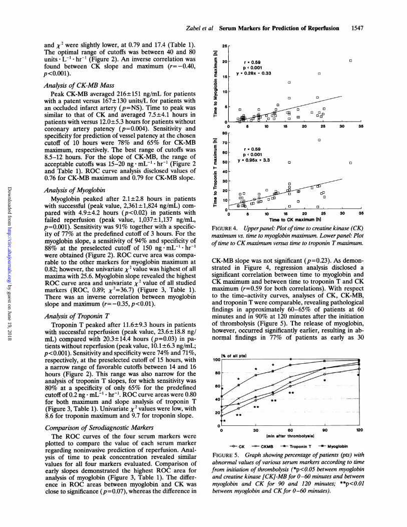
and x2 were slightly lower, at 0.79 and 17.4 (Table 1).The optimal range of cutoffs was between 40 and 80units * - hr-t (Figure 2). An inverse correlation wasfound between CK slope and maximum (r= -0.40,p<O.0Ol).Analysis of CK-MB Mass
Peak CK-MB averaged 216±151 ng/mL for patientswith a patent versus 167±130 units/L for patients withan occluded infarct artery (p=NS). Time to peak wassimilar to that of CK and averaged 7.5±4.1 hours inpatients with versus 12.0±5.3 hours for patients withoutcoronary artery patency (p=0.004). Sensitivity andspecificity for prediction of vessel patency at the chosencutoff of 10 hours were 78% and 65% for CK-MBmaximum, respectively. The best range of cutoffs was8.5-12 hours. For the slope of CK-MB, the range ofacceptable cutoffs was 15-20 ng . mL` * hr-1 (Figure 2and Table 1). ROC curve analysis disclosed values of0.76 for CK-MB maximum and 0.79 for CK-MB slope.
Analysis of MyoglobinMyoglobin peaked after 2.1±2.8 hours in patients
with successful (peak value, 2,361±1,824 ng/mL) com-pared with 4.9±4.2 hours (p<0.02) in patients withfailed reperfusion (peak value, 1,037±1,137 ng/mL,p=0.001). Sensitivity was 91% together with a specific-ity of 77% at the predefined cutoff of 3 hours. For themyoglobin slope, a sensitivity of 94% and specificity of88% at the preselected cutoff of 150 ng. mL- . hr-1were obtained (Figure 2). ROC curve area was compa-rable to the other markers for myoglobin maximum at0.82; however, the univariate X2 value was highest of allmaxima with 25.6. Myoglobin slope revealed the highestROC curve area and univariate x2 value of all studiedmarkers (ROC, 0.89; X2=36.7) (Figure 3, Table 1).There was an inverse correlation between myoglobinslope and maximum (r=-0.35,p<0.01).
Analysis of Troponin TTroponin T peaked after 11.6±9.3 hours in patients
with successful reperfusion (peak value, 23.6±18.8 ng/mL) compared with 20.3±14.4 hours (p=0.03) in pa-tients without reperfusion (peak value, 10.1±6.3 ng/mL;p<0.001). Sensitivity and specificity were 74% and 71%,respectively, at the preselected cutoff of 15 hours, witha narrow range of favorable cutoffs between 14 and 16hours (Figure 2). This range was also narrow for theanalysis of troponin T slopes, for which sensitivity was80% at a specificity of only 65% for the predefinedcutoff of 0.2 ng * mL- * hr-1. ROC curve areas were 0.80for both maximum and slope analysis of troponin T(Figure 3, Table 1). Univariate X2 values were low, with8.6 for troponin maximum and 9.7 for troponin slope.
Comparison of Serodiagnostic MarkersThe ROC curves of the four serum markers were
plotted to compare the value of each serum markerregarding noninvasive prediction of reperfusion. Anal-ysis of time to peak concentration revealed similarvalues for all four markers evaluated. Comparison ofearly slopes demonstrated the highest ROC area foranalysis of myoglobin (Figure 3, Table 1). The differ-ence in ROC areas between myoglobin and CK wasclose to significance (p=O0.07), whereas the difference in
25r
EE
xX
a
2.00
F0
E-
2
I-E.E
E1
c0CL
20-._
20 F
15k
r * 0.59p c 0.001
y * 0.28x - 0.33 0
10F
6O 0
0 on O
01~~~~~~~~-0~~ ~ :11
0 5 10 15 20 25 30 36
80 0
70T
r * 0.59B0 p 0.001
50 y *0.95x +3.3
40
30-so~ ~~~~~ 020
10-~~~~~~~~~10n d
0 5 10 16 20 25Time to CK maximum Ih]
30 35
FIGURE 4. Upperpanel: Plot oftime to creatine kinase (CK)maximum vs. time to myoglobin maximum. Lowerpanel: Plotof time to CK maximum versus time to troponin T maximum.
CK-MB slope was not significant (p=0.23). As demon-strated in Figure 4, regression analysis disclosed asignificant correlation between time to myoglobin andCK maximum and between time to troponin T and CKmaximum (r=0.59 for both correlations). With respectto the time-activity curves, analyses of CK, CK-MB,and troponin T were comparable, revealing pathologicalfindings in approximately 60-65% of patients at 60minutes and in 90% at 120 minutes after the initiationof thrombolysis (Figure 5). The release of myoglobin,however, occurred significantly earlier, resulting in ab-normal findings in 77% of patients as early as 30
12060Imin after thrombolyasil
CK -C- GKMB -* Troponin T Myoglobin
FIGURE 5. Graph showing percentage ofpatients (pts) withabnormal values of various serum markers according to timefrom initiation of thrombolysis (*p<0.05 between myoglobinand creatine kinase [CK]-MB for 0-60 minutes and betweenmyoglobin and CK for 90 and 120 minutes; **p<0.01between myoglobin and CKfor 0-60 minutes).
6
E
9
A
111
11
by guest on June 19, 2018http://circ.ahajournals.org/
Dow
nloaded from

1548 Circulation Vol 87, No S May 1993
TABLE 2. Sensitivity, Specificity, Positive and Negative Predictive Values, Univariate x2, and Area Under the ROCCurve for Noninvasive Prediction of Coronary Patency by Means of Clinical Markers
Positive NegativeSensitivity Specificity predictive predictive Area under
Marker (%) (%) value (%) value (%) X2 ROC curve
Reperfusion arrhythmias 50 94 96 41 8.4 ...
ST segment reduction 76 71 83 42 9.7 0.74Reduction in pain score 83 41 79 47 2.7* 0.67
ROC, receiver operator characteristics. See text for details.*p=NS.
minutes and 91% at 60 minutes after start of therapy(Figure 5).
Comparison With Other ClinicalReperfusion Markers
Table 2 demonstrates that resolution of ST segmentelevation and the occurrence of reperfusion arrhyth-mias were also predictors of coronary artery patency.The calculated x2 values indicate that both markerswere comparable to those serum markers with thelowest predictive values for coronary artery patency(Tables 1 and 2). ROC curve analysis for ST segmentresolution resulted in an area under the ROC curve of0.74 (versus 0.79 for CK slope, p=NS; versus 0.89 formyoglobin slope, p<0.02). Because of the lack of vari-able cutoffs with respect to reperfusion arrhythmias,ROC curve analysis cannot be performed for thismarker. The chest pain score did not yield statisticallysignificant prediction of the reperfusion status (Table2). When myoglobin slope was assessed together withother clinical markers by means of logistic regressionanalysis, only the myoglobin slope was an independentpredictor of coronary artery patency (p<0.0001). Theprobability values for the other markers examined were0.10 for the occurrence of reperfusion arrhythmias, 0.13for ST segment resolution, and 0.28 for resolution ofchest pain.
DiscussionThe present study indicates that noninvasive predic-
tion of coronary artery patency after thrombolytic ther-apy in patients with acute myocardial infarction ispossible by assessment of time-activity curves of serummarkers. Of particular clinical importance is the obser-vation that analysis of the initial slopes of activity curvesover the first 90 minutes after initiation of thrombolysisyields the same and for myoglobin even better predictiveaccuracy than the determination of the time to peakconcentration of each marker. Decision making as tofurther invasive procedures can be improved by use ofanalysis of these early slopes in a substantial proportionof patients. This allows early mechanical interventionssuch as salvage angioplasty in selected individuals inwhom the infarct-related artery remains occluded afterthrombolysis. It has been demonstrated that these pa-tients benefit from invasive interventions,11,12 whereasroutine coronary angiography is unnecessary and mayeven be harmful to some individuals.14-'6
In previous studies from our institution as well asfrom others, a short time to peak CK or CK-MBconcentration has been proposed as an indicator ofsuccessful thrombolysis-induced coronary artery reper-fusion.19'27-29 Only two studies so far have aimed to
examine the usefulness of the initial slopes of time-activity curves of the two serum markers CK andCK-MB concerning assessment of coronary artery pa-tency.29'39 These investigators found that a rapid in-crease in CK and CK-MB concentrations closely re-flected angiographic documentation of reperfusion.However, ROC characteristics were not reported.Other newly developed serum markers such as CK-MBisoforms31 or the specific cardiac antigen troponin T32have also been examined with respect to their utility asindicators of coronary reperfusion. Puleo and Perry-man31 analyzed peaks of MB2/MB, subform ratios in 33patients undergoing thrombolysis and found this ratiohelpful for assessment of coronary artery patency within2 hours after start of therapy. However, their studyappears to be limited by the small number of patientsundergoing thrombolytic therapy and by the fact thatcoronary angiography was performed as late as 5 daysafter thrombolysis. Katus et a132 studied the value of thenew cardiac antigen troponin T in 57 patients andshowed that troponin T peaks significantly earlier if theinfarct-related artery is successfully reperfused. How-ever, cutoff values and initial slopes were not specificallyassessed in this study.To the best of our knowledge, the present study is the
first to compare the serum markers CK, CK-MB, myo-globin, and troponin T systematically regarding theirvalue for noninvasive prediction of reperfusion. Of allmarkers examined, myoglobin appears to exhibit severaladvantages that make this protein particularly suitablefor early noninvasive prediction of patency of the in-farct-related vessel. First, comparison of the ROC curvecharacteristics revealed that analysis of the initial myo-globin concentration increase allows the best overallprediction of success or failure of thrombolytic therapyof all studied markers for analysis of both time to peakand early slopes. Although the differences between theROC areas were small, the difference between myoglo-bin and CK approached significance (p=0.07), indicat-ing potential clinical importance. Furthermore, thehighest univariate X2 values were also obtained for bothmyoglobin slope and time to peak concentration. Thisalso resulted in high sensitivity and positive predictivevalue for prediction of coronary artery patency. Addi-tionally, the negative predictive value and specificity,which reflect identification of patients with occludedvessels, were quite high at 82% and 88%, respectively,numbers that are not reached by the other markersexamined. Second, as already mentioned, quantitativenephelometric analysis of myoglobin, which has beenvalidated against the radioimmunoassay used by Ellis etal,30 provides laboratory results after a short turnovertime of only 15-30 minutes. Furthermore, an even more
by guest on June 19, 2018http://circ.ahajournals.org/
Dow
nloaded from

Zabel et al Serum Markers for Prediction of Reperfusion 1549
rapid assay using turbidimetry has recently been intro-duced into clinical practice.4344 Third, myoglobin con-centration exhibited the earliest rise of all serum mark-ers examined after initiation of therapy. Although thepurpose of the present investigation was not to examinethe accuracy of serum markers with respect to diagnosisof myocardial infarction but rather the efficacy ofthrombolysis, this protein may offer an additional diag-nostic advantage. The usefulness of myoglobin for non-invasive prediction of infarct-artery patency has re-cently been confirmed by other investigators.45However, Clemmensen and coworkers45 reported nocomparison with other serodiagnostic markers in theirstudy. In the present investigation, CK maximum andCK-MB maximum yielded relatively good diagnosticinformation regarding coronary artery patency as well.However, compared with the time elapsed to reach peakmyoglobin levels, it took four to five times as long toreach maximal concentrations of these enzymes. Thistime frame would therefore significantly reduce thechances to salvage jeopardized myocardium by means ofinvasive procedures. It has been shown previously thatcombined analysis of noninvasive markers (i.e., peakCK, resolution of ST segment elevation, occurrence ofreperfusion arrhythmias) can improve assessment ofsuccess or failure of thrombolytic therapy.19 In thepresent study, other clinical reperfusion markers did notadd significantly to the predictive power of myoglobinslope analysis, indicating the particular strength of thismarker.According to the results of the present study, tropo-
nin T offers no advantages for noninvasive prediction ofcoronary reperfusion in patients undergoing thrombol-ysis (Table 1). However, troponin T analysis was helpfulin the four patients suffering from ventricular fibrilla-tion. In these patients, CK values were distorted by theenzyme release from skeletal muscles caused by thedefibrillation shock, whereas troponin T levels were notaffected because of the high specificity of this markerfor myocardial tissue.36
Clinical ImplicationsThe present study demonstrates that analysis of the
early initial rise of several serum markers is as accurateas determination of the time elapsed to reach their peakconcentration with respect to noninvasive prediction ofsuccess or failure of thrombolysis in patients with acuteinfarction. This allows early noninvasive prediction ofcoronary artery patency in the majority of patientswithin the first 2 hours after initiation of thrombolytictherapy. These time limits allow for measures such asrescue angioplasty that aim to salvage myocardium.Early slope analysis of myoglobin appears to be superiorto the other serum markers examined and, in addition,exhibits the most rapid concentration increase of allmarkers evaluated.
References1. Gruppo Italiano per lo Studio della Streptochinasi nell'Infarto
Miocardico (GISSI): Effectiveness of intravenous thrombolytictreatment in acute myocardial infarction. Lancet 1986;1:397-402
2. Wilcox RG, von der Lippe G, Olson CG, Jensen G, Skeine AM,Hampton JR: Trial of tissue plasminogen activator for mortalityreduction in acute myocardial infarction The Anglo-ScandinavianStudy of Early Thrombolysis (ASSET). Lancet 1988;2:525-530
3. ISIS-2 (Second International Study of Infarct Survival) Collabora-tive Group. Randomised trial of intravenous streptokinase, oralaspirin, both, or neither among 17187 cases of suspected acutemyocardial infarction. Lancet 1988;2:349-360
4. The Thrombolysis in Myocardial Infarction (TIMI) Study Group:The thrombolysis in myocardial infarction (TIMI) trial. N Engl JMed 1985;312:932-936
5. White HD, Norris RM, Brown MA, Takayama M, Maslowski A,Bass NM, Ormiston JA, Whitlock T: Effect of intravenous strep-tokinase on left ventricular function and early survival after acutemyocardial infarction. N Engi J Med 1987;317:850-855
6. Tiefenbrunn AJ, Sobel BE: Invited review: Thrombolysis and myo-cardial infarction. Fibrinolysis 1991;5:1-15
7. Kennedy JW, Ritchie JL, Davis KB, Stadius ML, Maynard C, FritzJK: The Western Washington randomized trial of intracoronarystreptokinase in acute myocardial infarction. N Engl J Med 1985;312:1073-1078
8. Califf RM, Topol EJ, George BS, Kereiakes DJ, Aronson LG, LeeKL, Martin L, Candela R, Abottsmith C, O'Neill WW, Stack RS,and the TAMI study group: One-year outcome after therapy withtissue plasminogen activator: Report from the Thrombolysis andAngioplasty in Myocardial Infarction trial. Am Heart J 1990;119:777-785
9. Stack RS, O'Connor CM, Mark DB, Hinohara T, Phillips HR, LeeM, Ramirez NM, O'Callaghan WG, Simonton CA, Carlson EB,Morris KG, Behar VS, Kong Y, Peter RH, Califf RM: Coronaryperfusion during acute myocardial infarction with a combined ther-apy of coronary angioplasty and high-dose intravenous streptoki-nase. Circulation 1988;77:151-161
10. Abbottsmith CW, Topol EJ, George BS, Stack RS, Kereiakes DJ,Candela RJ, Anderson LC, Harrelson-Woodlief SL, Califf RM:Fate of patients with acute myocardial infarction with patency ofthe infarct-related vessel achieved with successful thrombolysisversus rescue angioplasty. JAm Coll Cardiol 1990;16:770-778
11. Topol EJ: Mechanical interventions for acute myocardial infarc-tion, in Topol EJ (ed): Textbook of Interventional Cardiology. Phil-adelphia, Pa, WB Saunders Co, 1990, pp 269-299
12. Muller DWM, Topol EJ: Thrombolytic therapy: Adjuvant mechan-ical intervention for acute myocardial infarction. Am J Cardiol1992;69:60A-70A
13. White HD, Cross DB, Norris RM, Williams BF: Early infarctartery patency after intravenous streptokinase, intracoronary con-trast and intracoronary rt-PA. (abstract) JAm Coll Cardiol 1992;19(suppl A):275A
14. Topol EJ, Califf RM, George BS, Kereiakes DJ, Abbottsmith CW,Candela RJ, Lee KL, Pitt B, Stack RS, O'Neill WW, and theTAMI study group: A randomized trial of immediate versusdelayed elective angioplasty after intravenous tissue plasminogenactivator in acute myocardial infarction. N Engl J Med 1987;317:581-588
15. The TIMI Research Group: Immediate versus delayed catheter-ization and angioplasty following thrombolytic therapy for acutemyocardial infarction. JAMA 1988;260:2849-2858
16. Simoons ML, Arnold AER, Betriu A, de Bono DP, Col J, Dough-erty FC, Essen RV, Lambertz H, Lubsen J, Meier B, Michel PL,Raynaud P, Rutsch W, Sanz GA, Schmidt W, Serruys P, Thery C,Uebis R, Vahanian A, van de Werf F, Willems GM, Wood D,Verstraete M: Thrombolysis with tissue plasminogen activator inacute myocardial infarction: No additional benefit from immediatepercutaneous coronary angioplasty. Lancet 1988;1:197-202
17. Hogg KJ, Hornung RS, Howie CA, Hockings N, Dunn FG, HillisWS: Electrocardiographic prediction of coronary artery patencyafter thrombolytic treatment in acute myocardial infarction: Use ofthe ST segment as a noninvasive marker. Br Heart J 1988;60:275-280
18. Krucoff MW, Green CE, Satler LF, Miller FC, Pallas RS, KentKM, del Negro AA, Pearle DL, Fletcher RD, Rackley CE: Non-invasive detection of coronary artery patency using continuousST-segment monitoring. Am J Cardiol 1986;57:916-922
19. Hohnloser SH, Zabel M, Kasper W, Meinertz T, Just H: Assess-ment of coronary artery patency after thrombolytic therapy: Accu-rate prediction utilizing the combined analysis of three noninvasivemarkers. JAm Coll Cardiol 1991;18:44-49
20. Saran RK, Been M, Furniss SS, Hawkins T, Reid DS: Reduction inST-segment elevation after thrombolysis predicts either coronaryreperfusion or preservation of left ventricular function. Br Heart J1990;64:113-117
21. Clemmensen P, Ohman EM, Sevilla D, Peck S, Wagner NB, Quig-ley PS, Grande P, Lee KL, Wagner GS: Changes in standardelectrocardiographic ST-segment elevation predictive of successful
by guest on June 19, 2018http://circ.ahajournals.org/
Dow
nloaded from

1550 Circulation Vol 87, No S May 1993
reperfusion in acute myocardial infarction. Am J Cardiol 1990;66:1407-1411
22. Gore JM, Ball SP, Corrao JM, Goldberg RJ: Arrhythmias in theassessment of coronary artery reperfusion following thrombolytictherapy. Chest 1988;94:727-730
23. Gorgels APM, Vos MA, Letsch IS, Verschuuren EA, Bar FWHM,Jannsen JHA, Wellens HJJ: Usefulness of the accelerated idioven-tricular rhythm as a marker for myocardial necrosis and reperfu-sion during thrombolytic therapy in acute myocardial infarction.Am J Cardiol 1988;61:231-235
24. Miller FC, Krucoff MW, Satler LF, Green CE, Fletcher RD, delNegro AA, Pearle DL, Kent KM, Rackley CE: Ventriculararrhythmias during reperfusion. Am Heart J 1986;112:928-932
25. Califf RM, O'Neill W, Stack RS, Aronson L, Mark DB, Mantell S,George BS, Candela RJ, Kereiakes DJ, Abbottsmith C, Topol EJ,and the TAMI study group: Failure of simple clinical measure-ments to predict perfusion status after intravenous thrombolysis.Ann Intern Med 1988;108:658-662
26. Kircher BJ, Topol EJ, O'Neill WW, Pitt B: Prediction of infarctcoronary artery recanalization after intravenous thrombolytic ther-apy. Am J Cardiol 1987;59:513-515
27. Gore JM, Roberts R, Ball SP, Montero A, Goldberg RJ, Dalen JE:Peak creatine kinase as a measure of effectiveness of thrombolytictherapy in acute myocardial infarction. Am J Cardiol 1987;59:1234-1238
28. Katus HA, Diederich KW, Scheffold T, Uellner M, Schwarz F,Kubler W: Noninvasive assessment of infarct reperfusion: Thepredictive power of the time to peak value of myoglobin, CKMB,and CK in serum. Eur Heart J 1988;9:619-624
29. Garabedian HD, Gold HK, Yasuda T, Johns JA, Finkelstein DM,Gaivin RJ, Cobbaert C, Leinbach RC, Collen D: Detection ofcoronary artery reperfusion with creatine kinase-MB determina-tions during thrombolytic therapy: Correlation with acute angiog-raphy. JAm Coll Cardiol 1987;11:729-734
30. Ellis AK, Little T, Zaki Masud AR, Liberman HA, Morris DC,Klocke FJ: Early noninvasive detection of successful reperfusion inpatients with acute myocardial infarction. Circulation 1988;78:1352-1357
31. Puleo PR, Perryman MB: Noninvasive detection of reperfusion inacute myocardial infarction based on plasma activity of creatinekinase MB subforms. JAm Coll Cardiol 1991;17:1047-1052
32. Katus HA, Remppis A, Scheffold T, Diederich KW, Kubler W:Intracellular compartmentation of cardiac troponin T and itsrelease kinetics in patients with reperfused and nonreperfusedmyocardial infarction. Am J Cardiol 1991;67:1360-1367
33. Rosalki SB: An improved procedure for creatine phosphokinasedetermination. J Lab Clin Med 1967;69:696-705
34. Giegel JL, Brotherton MM, Cronin P, D'Aquino M, Evans S,Heller ZH, Knight WS, Krishnan K, Sheiman M: Radial partitionimmunoassay. Clin Chem 1982;28:1994-1998
35. Hangaard J, Rasmussen 0, Norregaard-Hansen K, Jorgensen N,Simonsen EE, Norgaard-Pedersen B: Early diagnosis of acute myo-cardial infarction with a rapid latex agglutination test for semi-quantitative estimation of serum myoglobin. Acta Med Scand 1987;221:343-348
36. Katus HA, Remppis A, Looser S, Hallermayer K, Scheffold K,Kubler W: Enzyme linked immunoassay of cardiac troponin T forthe detection of an acute myocardial infarction in patients. J MolCell Cardiol 1989;21:1349-1353
37. Katus HA, Remppis A, Neumann FJ, Scheffold T, Diederich KW,Vinar G, Noe A, Matern G, Kubler W: Diagnostic efficiency oftroponin T measurements in acute myocardial infarction. Circula-tion 1991;83:902-912
38. Meinertz T, Kasper W, Schumacher M, Just H for the APSACMulticenter Trial Study Group: The German multicenter trial ofanisoylated plasminogen streptokinase activator complex versusheparin for acute myocardial infarction. Am J Cardiol 1988;62:347-351
39. Lewis BS, Ganz W, Laramee P, Cercek B, Hod H, Shah PK, LewAS: Usefulness of a rapid initial increase in plasma creatine kinaseactivity as a marker of reperfusion during thrombolytic therapy foracute myocardial infarction. Am J Cardiol 1988;62:20-24
40. Hanley JA, McNeil BJ: The meaning and use of the area under areceiver operator characteristic (ROC) curve. Radiology 1982;143:29-36
41. Hanley JA, McNeil BJ: A method of comparing the areas underreceiver operating characteristic curves derived from the samecases. Radiology 1983;148:839-843
42. Swets JA: Measuring the accuracy of diagnostic systems. Science1988;249:1285-1293
43. Mair J, Smidt J, Artner-Dworzak E, Lechleitner P, Dienstl F,Puschendorf B: Rapid diagnosis of myocardial infarction byimmuno turbidimetric myoglobin measurement. (letter) Lancet1991;337:1343-1344
44. Ishii J, Nomura M, Ando T, Hasegewa H, Kimura M, Tateishi R,Kurokawa H, Kondo T, Hishida H, Mizuno Y: Early detection ofcoronary reperfusion based on serum myoglobin concentration.(abstract) Circulation 1991;84(suppl II):II-217
45. Clemmensen P, Jurlander B, Grande P, Ohman EM, Wagner GS:Monitoring peak serum-myoglobin for non-invasive prediction ofcoronary reperfusion in patients. (abstract) Circulation 1991;84(suppl II):II-116
by guest on June 19, 2018http://circ.ahajournals.org/
Dow
nloaded from

M Zabel, S H Hohnloser, W Köster, M Prinz, W Kasper and H Justassessment of coronary artery reperfusion after intravenous thrombolysis.
Analysis of creatine kinase, CK-MB, myoglobin, and troponin T time-activity curves for early
Print ISSN: 0009-7322. Online ISSN: 1524-4539 Copyright © 1993 American Heart Association, Inc. All rights reserved.
is published by the American Heart Association, 7272 Greenville Avenue, Dallas, TX 75231Circulation doi: 10.1161/01.CIR.87.5.1542
1993;87:1542-1550Circulation.
http://circ.ahajournals.org/content/87/5/1542World Wide Web at:
The online version of this article, along with updated information and services, is located on the
http://circ.ahajournals.org//subscriptions/
is online at: Circulation Information about subscribing to Subscriptions:
http://www.lww.com/reprints Information about reprints can be found online at: Reprints:
document. Permissions and Rights Question and Answer available in the
Permissions in the middle column of the Web page under Services. Further information about this process isOnce the online version of the published article for which permission is being requested is located, click Request
can be obtained via RightsLink, a service of the Copyright Clearance Center, not the Editorial Office.Circulation Requests for permissions to reproduce figures, tables, or portions of articles originally published inPermissions:
by guest on June 19, 2018http://circ.ahajournals.org/
Dow
nloaded from