Myers’ PSYCHOLOGY (7th Ed)
66
Myers’ PSYCHOLOGY (7th Ed) Chapter 2 Neuroscience, Genetics and Behavior James A. McCubbin, PhD Clemson University Worth Publishers
-
Upload
seth-meadows -
Category
Documents
-
view
40 -
download
0
description
Myers’ PSYCHOLOGY (7th Ed). Chapter 2 Neuroscience, Genetics and Behavior James A. McCubbin, PhD Clemson University Worth Publishers. Neural Communication. Biological Psychology branch of psychology concerned with the links between biology and behavior - PowerPoint PPT Presentation
Transcript of Myers’ PSYCHOLOGY (7th Ed)

Myers’ PSYCHOLOGY
(7th Ed)
Chapter 2 Neuroscience, Genetics
and Behavior
James A. McCubbin, PhDClemson University
Worth Publishers


Neural Communication
Biological Psychology branch of psychology concerned with the links
between biology and behavior some biological psychologists call themselves
behavioral neuroscientists, neuropsychologists, behavior geneticists, physiological psychologists, or biopsychologists
Neuron a nerve cell the basic building block of the nervous system

Neural Communication Dendrite
the bushy, branching extensions of a neuron that receive messages and conduct impulses toward the cell body
Axon the extension of a neuron, ending in branching
terminal fibers, through which messages are sent to other neurons or to muscles or glands
Myelin [MY-uh-lin] Sheath a layer of fatty cells segmentally encasing the
fibers of many neurons enables vastly greater transmission speed of
neutral impulses

Neural Communication

Neural Communication
Action Potential a neural impulse; a brief electrical
charge that travels down an axon generated by the movement of
positively charged atoms in and out of channels in the axon’s membrane
Threshold the level of stimulation required to
trigger a neural impulse

Neural Communication
Cell body end of axon
Direction of neural impulse: toward axon terminals

Neural Communication Synapse [SIN-aps]
junction between the axon tip of the sending neuron and the dendrite or cell body of the receiving neuron
tiny gap at this junction is called the synaptic gap or cleft
Neurotransmitters chemical messengers that traverse the synaptic
gaps between neurons when released by the sending neuron, neuro-
transmitters travel across the synapse and bind to receptor sites on the receiving neuron, thereby influencing whether it will generate a neural impulse

Neural Communication

Neural Communication

Neural Communication
Neurotransmitter molecule
Receiving cellmembrane
Receptor site onreceiving neuron
Agonist mimicsneurotransmitter
Antagonistblocksneurotransmitter

End Day 2

The Nervous System Nervous System
the body’s speedy, electro-chemical communication system
consists of all the nerve cells of the body Neurons are the elementary components
– the smallest Lego pieces, if you will There are 2 major divisions of the
nervous system central nervous system peripheral nervous system

Central Nervous System (CNS) the brain and spinal cord
Peripheral Nervous System (PNS) the sensory and motor neurons that
connect the central nervous system (CNS) to the rest of the body (sense receptors, muscles, glands)
Axons carrying PNS info are bundled together in groups called nerves

3 Types of Neurons
Sensory neurons Sends info from body tissues and sensory
organs to the CNS’s brain and spinal cordMotor neurons
CNS send messages to the body’s tissuesInterneurons
CNS internal communication neurons

The Nervous System
Central(brain and
spinal cord)
Nervoussystem
Autonomic (controlsself-regulated action of
internal organs and glands)
Somatic (controlsvoluntary movements of
skeletal muscles)
Sympathetic (arousing)
Parasympathetic (calming)
Peripheral

Further Division
The PNS can be further divided downPNS can be divided into 4 more
branches of the nervous system Somatic nervous system Autonomic nervous system Sympathetic nervous system Parasympathetic nervous system

The Branches of the Peripheral Nervous System
Somatic Nervous System Controls the body’s skeletal muscles Voluntary Autonomic Nervous System Controls the glands and the muscles of the internal
organs (such as the heart) Automatic pilot, but can be overridden
2 sub-branches – sympathetic and parasympathetic Work together to keep a steady internal state Sympathetic Nervous System Arousing – if something alarms, enrages, or challenges
you Mobilizing body’s energy in stressful situations Parasympathetic Nervous System Calms the body, conserving its energy

The Sympathetic Nervous System

The Parasympathetic Nervous System

More on the CNS
Almost unimaginable wiring diagram of a very complex computer
Complex circuits that allow for learning, feeling and thinking
10 of billions of neurons communicate to make all this happen
Spinal cord is the super highway of all “talking” and “communicating” Connects the PNS to the brain Ascending fibers send info up the spinal cord Descending fibers send info down the spinal
cord

The Reflex Reflex
a simple, automatic, inborn response to a sensory stimulus
A reflex involves one simple pathway
A single sensory neuron and a single motor neuron, connected by an interneuron
Knee-jerk reflex for example
Everything goes through spinal cord
Even is spinal cord severed, reflexes can still occur below where it was severed
Skinreceptors
Muscle
Sensory neuron(incoming information)
Motor neuron(outgoing information)
Brain
Interneuron
Spinal cord

Neural Networks
Neural Networks interconnected
neural cells with experience,
networks can learn, as feedback strengthens or inhibits connections that produce certain results
computer simulations of neural networks show analogous learning
Inputs Outputs
Neurons in the brain connect with one
another to form networks
The brain learns by modifyingcertain connections in response to feedback

End Day 3

Endocrine System
The other communication systemGlands secrete chemical messengers
called hormonesHormones start in one tissue and
travel through the bloodstream to affect other tissues, including the brain
They act on the brain and influence behavior

Hormones
Some hormones are chemically identical to neurotransmitters
This makes the nervous system and endocrine systems kindred spirits
Hormones are slower than the zippy messengers of the nervous system
Their effects outlast the effects of the neurotransmitters
Hormones influence many aspects of our lives – growth, reproduction, metabolism and mood

Functions/Locations of Endocrine Glands
Pituitary – many hormones, affect other glands
Hypothalamus – are of the brain, controls pituitary
Thyroid – metabolism Parathyroids – regulate
calcium in blood Adrenal – fight or flight Pancreas – sugar in blood Ovary/Testes – male and
female sex hormones

Specific Examples of Effects
Adrenal Glands Top of kidneys Release epinephrine and norepinephrine
aka adrenaline and noradrenalineIncrease heart rate, blood pressure, blood sugarExtra surge of energy
Pituitary Glands In the core of the brain, pea-sized Controlled by hypothalamus Influence growth Master gland – controls other glands

Endocrine and Brain/Behavior
Brain (hypothalamus) pituitary other glands hormones brain
The 2 systems work so closely together than the line between them can be blurred

End Day 3

Technology and The Brain
With technology – we can now see the brain
We can also lesion (damage) tiny clusters of normal or defective brain cells
All these techniques are to psychology what a microscope was to biology and a telescope was to astronomy.
Electroencephalogram (EEG)
Brain has mental activity that gives off electrical, metabolic, and magnetic signals
These signals can now be read by science
An EEG is an amplified recording of the waves of electrical activity that sweep across the brain’s surface
Waves are measured by electrodes placed on the scalp
We can see, through waves, what wave was caused by what stimulus

“Seeing” the Brain PET (positron emission tomography) Scan
a visual display of brain activity that detects where a radioactive form of glucose goes while the brain performs a given task
Active neurons are glucose hogs MRI (magnetic resonance imaging)
a technique that uses magnetic fields and radio waves to produce computer-generated images that distinguish among different types of soft tissue; allows us to see structures within the brain
fMRI (functional magnetic resonance imaging) Can reveal functioning as well as structure Blood goes where the brain is active Watch the brain light up with bloodflow (see what part of the
brain is working based on certain stimuli) How the brain divides its labor

MRI Scan - Schizophrenia

The Brain Brainstem
the oldest part and central core of the brain, beginning where the spinal cord swells as it enters the skull
responsible for automatic survival functions Crossover point – nerves to and from each side of the brain
crossover to connect with the body’s opposite side. Reticular formation is inside the brainstem
Finger-shaped network of neurons from the spinal cord to the thalamus
Traffic control – tells messages where to go in the brain Arousal
Begins where the spinal cord enters the skull and swells slightly to form the…

The Brain Stem

Brain Stem - Medulla
Medulla [muh-DUL-uh] base of the brainstem
(lower half) controls heartbeat,
breathing, vomiting (thanks a lot), blood pressure, sneezing, coughing, and swallowing
Autonomic functions (involuntary)

Brain Stem - Recticular Formation
Reticular formation Inside the brainstem Finger-shaped network of neurons
from the spinal cord to the thalamus Traffic control – tells messages where
to go in the brain Arousal

Brain Stem - Thalamus Thalamus [THAL-uh-muss]
the brain’s sensory switchboard, located on top of the brainstem
Joined egg-shaped structures Receives all sense messages,
except smell, and routes them the areas of the brain that deal with each sense
transmits replies to the cerebellum and medulla

Cerebellum
Cerebellum [sehr-uh-BELL-um] the “little brain” attached to the rear of the brainstemBaseball-sized, 2 wrinkled halvesit helps coordinate voluntary movement and balanceNonverbal learning and memory enabled by itJudge time, modulate emotions, discriminate sounds and textures

The Limbic System

Limbic System
Limbic System a doughnut-shaped system of neural
structures at the border of the brainstem and cerebral hemispheres
associated with emotions such as fear and aggression and drives such as those for food and sex
includes the hippocampus, amygdala, and hypothalamus.

Limbic - Amygdala Amygdala [ah-MIG-dah-la]
two almond-shaped neural clusters that are components of the limbic system and are linked to emotions
Aggression and fear Be careful though – fear and
aggression are NOT driven by just the amygdala
One link in an integrated and complicated system

Limbic - Hypothalamus Hypothalamus
neural structure lying below (hypo) the thalamus; directs several maintenance activities Eating/hunger Drinking/thirst body temperature sexual behavior
Hypothalamus secretes hormones that control the pituitary, which stimulates other glands to release other hormones that the brain wants/needs

Limbic - HypothalamusReward centers
Pleasure centersMotivates activities that are essential to
survivalReward deficiency syndrome
Alcoholism, drug abuse, and binge eating May stem from a genetically disposed deficiency in
the brain system for pleasure and well-being Leads people to crave whatever provides that
missing pleasure or relieves negative feelings

End Day 5

The Cerebral Cortex
Cerebral Cortex the intricate fabric of interconnected neural
cells that covers the cerebral hemispheres Like the bark of a tree the body’s ultimate control and information
processing center The larger cortex in mammals allow for
increased capacities for learning and thinking
Make us more adaptable It is a “thinking crown”

Glial Cells
Glial Cells cells in the nervous system that support,
nourish, and protect neurons They are like the glue that guide neural
connections, provide nutrients and insulate myelin.
Also mop up ions and neurotransmitters May play a role in learning and thinking Chat with neurons to transmit information and
memory

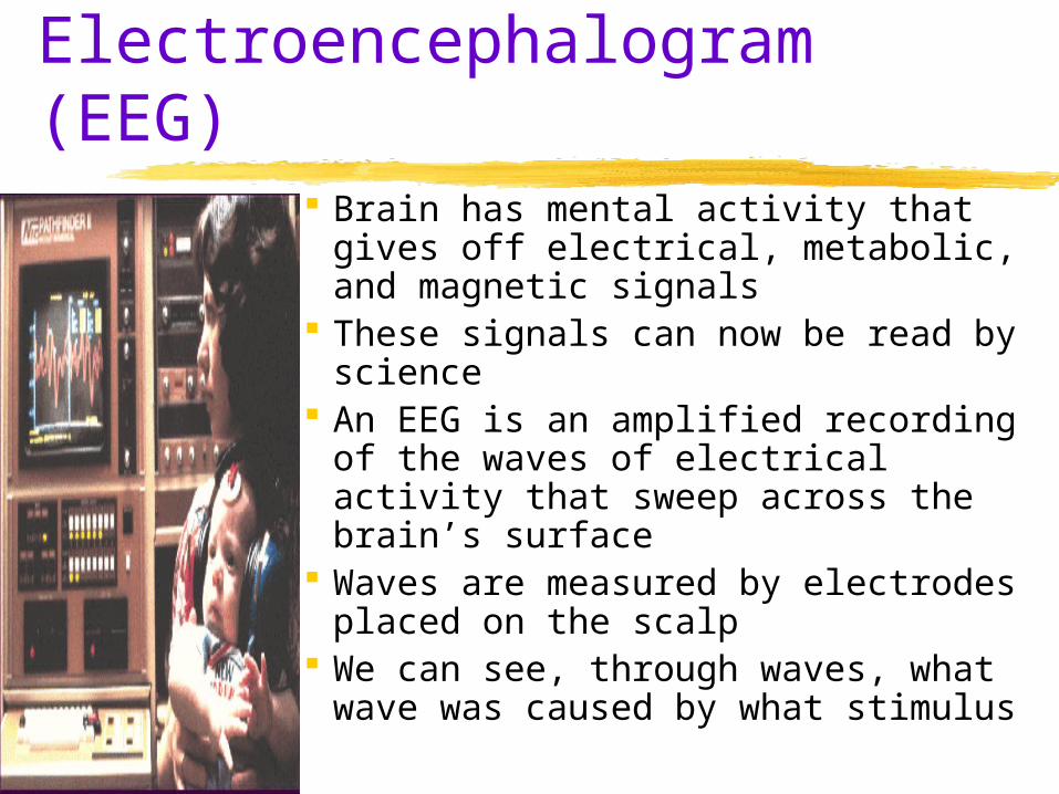
The Cerebral Cortex Wrinkled – the folds increase surface area 4 lobes are divided by fissures (divides)
Many functions require the interplay of several regions/lobes
Frontal Lobes involved in speaking and muscle movements and in
making plans and judgments Parietal Lobes
include the sensory cortex Occipital Lobes
include the visual areas, which receive visual information from the opposite visual field
Temporal Lobes include the auditory areas
The Cerebral Cortex

The Cerebral Cortex
Can make different motor/sensory skills happen by applying stimulation to certain areas of the cerebral cortex
Motor Cortex Runs across the top of the brain Just behind the front lobe Controls voluntary movements
Sensory Cortex Across the top of the brain, behind the motor cortex Receives incoming sensory messages Registers and processes body sensations The more sensitive an are of the body the larger
the area of sensory cortex devoted to it

The Cerebral Cortex

The Cerebral Cortex
Functional MRI scan shows the visual cortex activated as the subject looks at faces

Association Areas
Integrate information Associate various sensory inputs with stored
memories On all 4 lobes Frontal lobe association areas enable us to plan,
judge and process new memories Frontal lobes are also tied to personality Parietal lobes have association areas that enable
mathematical and spatial reasoning An area on the underside of the temporal lobe
allows us to recognize facts

The Cerebral Cortex Aphasia
impairment of language, usually caused by left hemisphere damage either to Broca’s area (impairing speaking) or to Wernicke’s area (impairing understanding)
Broca’s Area an area of the left frontal lobe that directs
the muscle movements involved in speech Wernicke’s Area
an area of the left temporal lobe involved in language comprehension and expression

Specialization and Integration

Specialization and Integration
Brain activity when hearing, seeing, and speaking words

Plasticity at Work
Girl with half a brain!Amazing!http://today.msnbc.msn.com/id/3603
2653/ns/today-today_health/t/meet-girl-half-brain/#
Brain Reorganization
Plasticitythe brain’s capacity for modification, as evident in brain reorganization following damage (especially in children) and in experiments on the effects of experience on brain development

End Day 6
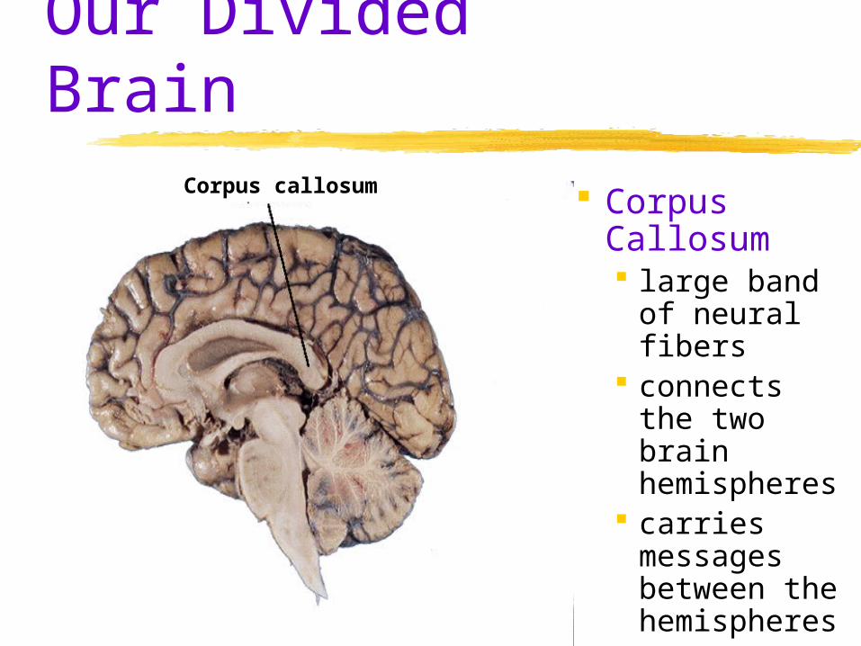
Our Divided Brain
Corpus Callosum large band
of neural fibers
connects the two brain hemispheres
carries messages between the hemispheres
Corpus callosum

Our Divided Brain
The information highway from the eye to the brain

Split Brain
a condition in which the two hemispheres of the brain are isolated by cutting the connecting fibers (mainly those of the corpus callosum) between them

Split Brain
“Look at the dot.” Two words separatedby a dot are momentarily projected.
“What worddid you see?”
or
“Point withyour left hand to theword you saw.”

Disappearing Southpaws
The percentage of left-handers decreases sharply in samples of older people (adapted from Coren, 1993).
The percentage of lefties sharplydeclines with age
10 20 30 40 50 60 70 80 90Age in years
14%
12
10
8
6
4
2
0
Percentage ofleft-handedness

Brain Structures and their Functions