MYELOID TISSUE: MICROANATOMY
81
MYELOID TISSUE
-
Upload
ruthypotpot -
Category
Health & Medicine
-
view
267 -
download
3
Transcript of MYELOID TISSUE: MICROANATOMY

MYELOID TISSUE

Hematopoiesis process specialized blood cells develop from pluripotent stem cells of myeloid tissue in the bone marrow
result of simultaneous, continuous proliferation and differentiation – reduction in the potential of the cells
Site - occurs in myeloid and lymphatic tissue. Myeloid tissue bone marrow Lymphatic tissue lymphatic organs - not a rigidly compartmentalized process; blood cells usually associated with myeloid tissue can arise in lymphoid tissue, and vice versa

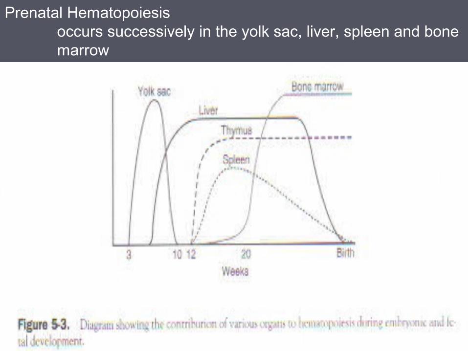
Prenatal Hematopoiesis occurs successively in the yolk sac, liver, spleen and bone marrow


Structural Organization Of Hematopoietic Marrow Cancellous bone – bony spicules or trabeculae lined by endosteum and marrow filled with hematopoietic and non-hematopoietic cells Blood vessels of the marrow compartment 1. Nutrient arteries periosteum pass through the compact bone to enter the marrow space. 2. Longitudinal arteries formed by the division of a nutrient artery - run parallel to the long axis of a bone. 3. Radial arteries spoke-like branches that arise from longitudinal arteries to form thin-walled sinusoids in the hematopoietic tissue.


Bone marrow very large and complex organ cavities of the skeleton total mass, adult – 1600 to 3000 grams ½ - hematopoietically inactive fatty (yellow) marrow few microscopic foci of hematopoietic cells ½ - hematopoietically active (red) marrow function - based on a high degree of structural organization (organization - labile, altering rapidly in response to many stimuli) hematopoietic marrow formation and release - blood cells phagocytosis and degradation - microorganisms and abnormal or senescent rbcs antibody production non-hematopoietic marrow large store of reserve lipids

Sinusoids endothelial cells no basement membrane, overlapping and may interdigitate extensively adventitial layer external discontinuous layer Stroma of the Marrow Cells 3-dimensional meshwork of reticular cells and a delicate web of containing hematopoietic cells, macrophages, mast cells, fat cells, lymphocytes and plasma cells. reticular cells - form a loose net of reticular fibers reinforce the sinusoidal capillaries and internal support for the stroma cytoplasmic processes – lie along the sinusoidal surface and protrude outward in- between hematopoietic cells


Matrix contains collagen types I and III, fibronectin, laminin, and proteoglycans. Laminin, fibronectin, and hemonectin - cell binding substance interact with cell receptors to bind cells to the matrix Other cells osteoblast and osteoclast fat cells newborn - 0% 2 week-old infant – 15% children between 18 months to 11 year-old – 20% to 65% adult – 30% to 70% 70 year-old - >70% Marrow cellularity - proportion of the area occupied by cells other than fat cells normocellular, hypocellular or hypercellular

Macrophages intracytoplasmic inclusion - refractile yellow-brown hemosiderin - iron (+)granules marrow fragments or smear semi-quantitatively to assess total iron store long cytoplasmic processes – protrude into the sinusoids and phagocytozed senescent or damage rbcs, progranulocytes and circulating microorganism present in the erythroblastic islands, plasma cell island and lymphatic nodules but may also occurs elsewhere generate various neutrophilic growth factorsMast cells progenitor cells occur in the marrow, but proliferation and maturation and acquisition of granules occurs in the tissues associated with lymphoid nodule, wall of the arterioles, adjacent to the endothelium of sinusoids and endosteal cells of bone trabeculae

Characterization of Hematopoietic tissue microscopically by differentiating blood cells. 1. Stained smear of bone marrow reveals a complex population comprising several types of blood cells and their precursors 2. These cell types can be sorted into several developmental sequences, each sequence culminating in one of the several types of mature blood cells – M:E ratio 3. Bone marrow biopsy cellularity architecture – structural relationship of the components tumor

Hematopoietic Cords - Cell Associations in Red Bone MarrowHistologic sections of bone marrow show the following relationships.a. Nests of erythroblasts and myelocytes. developing blood cells are often seen clumped into nests or islets - cells clump when mitotic events increase their numbers and the daughter cells remain restricted to the immediate vicinity. b. Normoblasts orthochromatic erythroblasts and macrophages. Macrophages are found in close association with nests of normoblasts, where they phagocytize nuclei expelled by the
normoblasts during erythropoiesis. c. Megakaryocytes and the sinusoidal wall. Megakaryocytes are found in close proximity to the walls of marrow blood capillaries(sinusoids) - facilitates the release of platelets into the blood stream

During preparation of a bone marrow smear, these normal cellular relationships are demolished.


Abnormal Increase in HematopoiesisYoung children – rapid increase in hematpoietic tissue accomodated mainly by a
reduction in the proportion of marrow space occupied by sinusoids
skeletal abnormalities – frontal and parietal bossing, dental deformation, and malocclusion of the teeth, thinning of the cortex – fracture if the increase is substantial - extramedullary hematopoiesAdult initially associated with the replacement of fat cells in the red marrow by hematopoietic cells and also with the spread of red marrow into marrow cavities normally containing yellow marrow if the increase is gross – extramedullary hematopoiesis

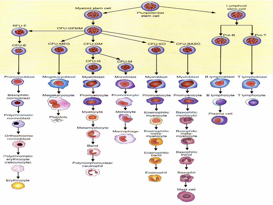
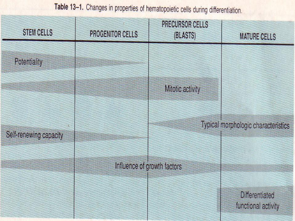
Postnatal Hematopoiesis involved three classes of cells 1. Pluripotent stem cells - primitive hematopoietic two properties – undergo enormous proliferation a. differentiate into multiple cell lineage - ability to mature into several types of blood cells give rise to progenitor cells lymphoid progenitor cells multipotent myeloid progenitor cells b. self- renewal - extensive capacity to generate new stem cells present – blood circulation and BM no identifiable morphologic feature – resemble large lymphocytes

2. Progenitor cells proliferating and differentiating stem cells to form daughter cell with reduced potentiality.
committed to a single cell lineage unipotential or bipotential progenitor cell generate precursor cells (blasts) produce both progenitor and precursor cells, morphologically indistinguishable from stem cells
3. Precursor cells display distinct morphologic characteristics produce only mature blood cells.
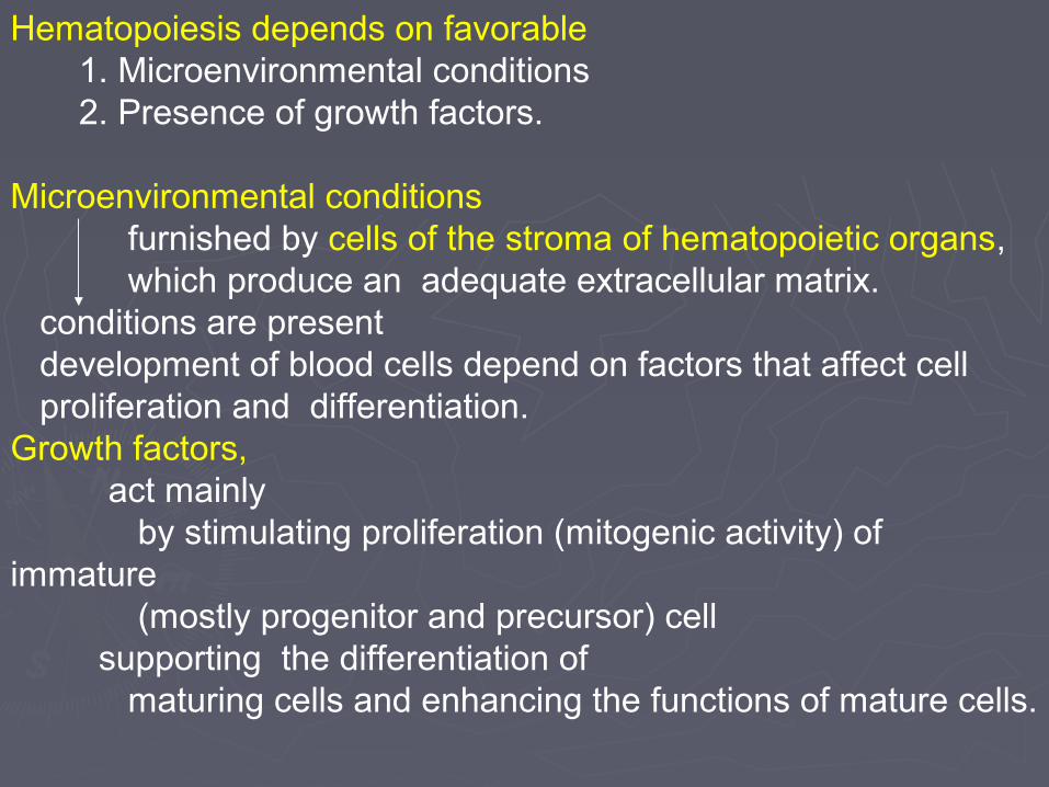
Hematopoiesis depends on favorable 1. Microenvironmental conditions 2. Presence of growth factors.
Microenvironmental conditions furnished by cells of the stroma of hematopoietic organs, which produce an adequate extracellular matrix. conditions are present development of blood cells depend on factors that affect cell proliferation and differentiation. Growth factors, act mainly by stimulating proliferation (mitogenic activity) of immature (mostly progenitor and precursor) cell supporting the differentiation of maturing cells and enhancing the functions of mature cells.

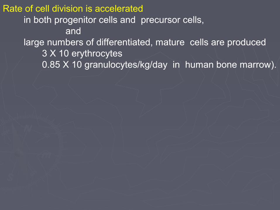
Rate of cell division is accelerated in both progenitor cells and precursor cells, and large numbers of differentiated, mature cells are produced 3 X 10 erythrocytes 0.85 X 10 granulocytes/kg/day in human bone marrow).

Initial steps in Blood Formation Pluripotent hematopoietic stem cells give rise to multipotent hematopoietic stem cells Multipotent hematopoietic stem cells two type - proliferate and differentiate – progenitor cells CFU-S – colony forming unit–spleen erythrocytes, granulocytes, monocytes and platelets CFU-Ly – colony forming unit-lymphocytes T, B, and NK cellsErythropoiesis – yield 1 trillion daily in adult CFU-S begins with the formation of progenitor cells BFU-E – burst forming unit-erythroid high rate of mitotic activity, high conc. of erythropoietin CFU-E – colony forming unit erythroid low conc. of erythropoietin first recognizable – precursor cell - proerythroblast

Granulopoiesis – yields about 1million granulocytes daily in adult CFU-S begins with production of three unipotential or bipotential cells – progenitor cells CFU-Eo – is the progenitor of eosinophil lineage CFU-Ba – is the progenitor of basophils CFU-NM – is the common progenitor of neutrophils and monocytes - give rise to CFU-N and CFU-M give rise to histologically identical - in the early stage of all three lineages myeloblast (precursor) and promyelocytes - develop characteristic granules unique to each cell type during the myelocytes stage and a distinctive nuclear shape during stab stage CFU-NM – give rise to CFU-N – progenitor cells of neutrophils give rise to precursor cells

Monocytopoiesis – yields about 10 trillion daily in adult CFU-NM – progenitor for both neutrophils and monocytes begins with the formation of CFU-M – progenitor of monoocyte
give rise to monoblast – precursor cell
Thrombocytopoiesis CFU-S begins with the progenitor cells CFU-Meg give rise to precursor cells megakaryoblast


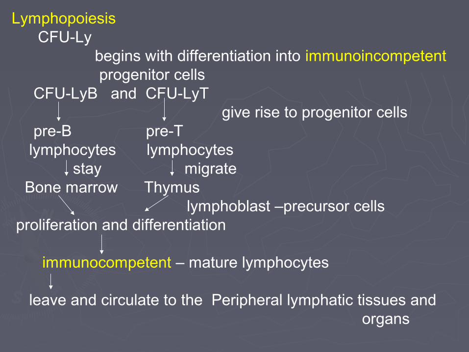
Lymphopoiesis CFU-Ly begins with differentiation into immunoincompetent progenitor cells CFU-LyB and CFU-LyT give rise to progenitor cells pre-B pre-T lymphocytes lymphocytes stay migrate Bone marrow Thymus lymphoblast –precursor cells proliferation and differentiation
immunocompetent – mature lymphocytes
leave and circulate to the Peripheral lymphatic tissues and organs


Release of mature bone cells from the marrow controlled by releasing factors produced in response to the needs of the organism. Substances with releasing activity C3 component of complement, some bacterial toxins. hormones (glucocorticoids and androgens),

Two processes involved in the formation of all types of blood cells Cytodifferentiation (maturation) - all stages of hematopoiesis progressive acquisition of the morphologic , biochemical, and functional characteristics of the particular cell type Cell proliferation stem cells, progenitor cells and immature recognizable precursor cells except megakaryocytic lineageMorphologic criteria of blood cell development.indicators 1.Changes in cell size and nuclear structure, 2.Presence of differentiation products ( cytoplasmic granules and hemoglobin), Cell size. Less mature cells tend to be larger in overall diameter. Nuclear structure 1. Chromatin configuration. Less mature cells have euchromatic (transcriptionally active) nuclei. Nuclei usually become heterochromatic (transcriptionally inactive) later in development. 2. Nuclear lobulation. Granulocytes, during development, acquire characteristically lobed nuclei

3. Nuclear loss. Erythrocytes, during development, extrude the nucleus that is present in an immature cell. 4. Nucleolar loss. intranuclear organelle may be visible in immature blood cells but disappears from cells nearing completion of development.
Differentiation Products Cytoplasmic granules. In granulocytes, presence and staining characteristics of azurophilic granules and specific granules are important developmental criteria. In erythrocytes, gradual changes in cytoplasmic staining caused by accumulating hemoglobin are important developmental criteria


ERYTHROPOIESIS.red blood cells develop through several well-defined stages from a progenitor cell to a mature erythrocyte.
Proerythroblast. first developmental stage not an easily recognized cell Morphologic characteristics Size - large cell (18-25 um) Nucleus - euchromatic and usually has one or two nucleoli. Cytoplasm - exhibits basophilia.

Basophilic erythroblastMorphologic characteristics Size - (15-18 um) smaller than a proerythroblast. Nucleus - spheroidal and becomes increasingly heterochromatic with successive mitoses. Cytoplasm - distinctly basophilic
Significance of cytoplasmic basophilia - large number of polyribosomes, assembled in preparation for the synthesis of hemoglobin.

Polychromatophilic erythroblast. (normoblast)shows evidence of Hgb accumulation. Morphologic characteristics Size - (12-15 um) slightly smaller than a basophilic erythroblast. Nucleus - Coarse heterochromatin and alternating euchromatic regions "checkerboard" arrangement in a spherical nucleus - useful identifying feature. Cytoplasm - acquires a polychromatophilic staining characteristic- acidophilic and basophilic. Significance of cytoplasmic polychromatophilia Polyribosomes- basophilic component Hemoglobin - acidophilic component - accumulates in stainable amounts - Acidophilia of accumulating hemoglobin gradually dilutes the basophilia of the polyribosomes

Orthochromatic erythroblast last stage of erythropoiesis in which a nucleus can be identified. Morphologic characteristics Size - smaller than a polychromatophilic and slightly larger than a mature erythrocyte. Nucleus - pyknotic and is intensely heterochromatic; little evidence of euchromatin is visible. Cytoplasm - acidophilic because of the high concentration of recently synthesized hemoglobin.

Expulsion of the nucleus from the cell a. normoblast stage ends when the condensed, kernel-like nucleus is cast out of the cell. b. nucleus is not viable - phagocytized in the macrophage-rich hematopoietic compartment.
Reticulocyte nearly mature red cells found in circulating blood. Morphologic characteristics Size - is approximately the same size as the mature erythrocyte. Nucleus - does not have a nucleus. Cytoplasm routine blood stains - strongly acidophilic and has essentially the same staining characteristics as the mature erythrocyte. supravital staining by brilliant cresyl blue - stains the remaining polyribosomes of the cell, producing the basophilic reticulum Circulating reticulocytes released into the peripheral blood; therefore, developing red blood cells circulate before erythropoiesis is completed. peripheral blood comprise about 1% to 2% of the circulating red blood cells

Reticulocytes mature into erythrocytes after about 24 hours in circulation. Hemoglobin synthesis continues during this period.

Kinetics of red blood cell development Mitotic and postmitotic phases of erythropoiesis mitosis occurs erythroblasts up to and including the polychromatophilic erythroblast. postmitotic cells orthochromatophilic , reticulocytes, and mature erythrocytes
Distribution of the erythrocyte population Virtually all erythrocytes are released into the circulation as soon as they are formed. Normally released into circulation are reticulocytes, not fully mature cells Erythropoiesis - completed as the cells circulate through the body. Bone marrow - not a site of red blood cell storage Apparently mature erythrocytes observed in routinely stained bone marrow smears are either 1. reticulocytes about to be released into the circulation or 2. intravascular cells that were passing through the marrow at the time of biopsy
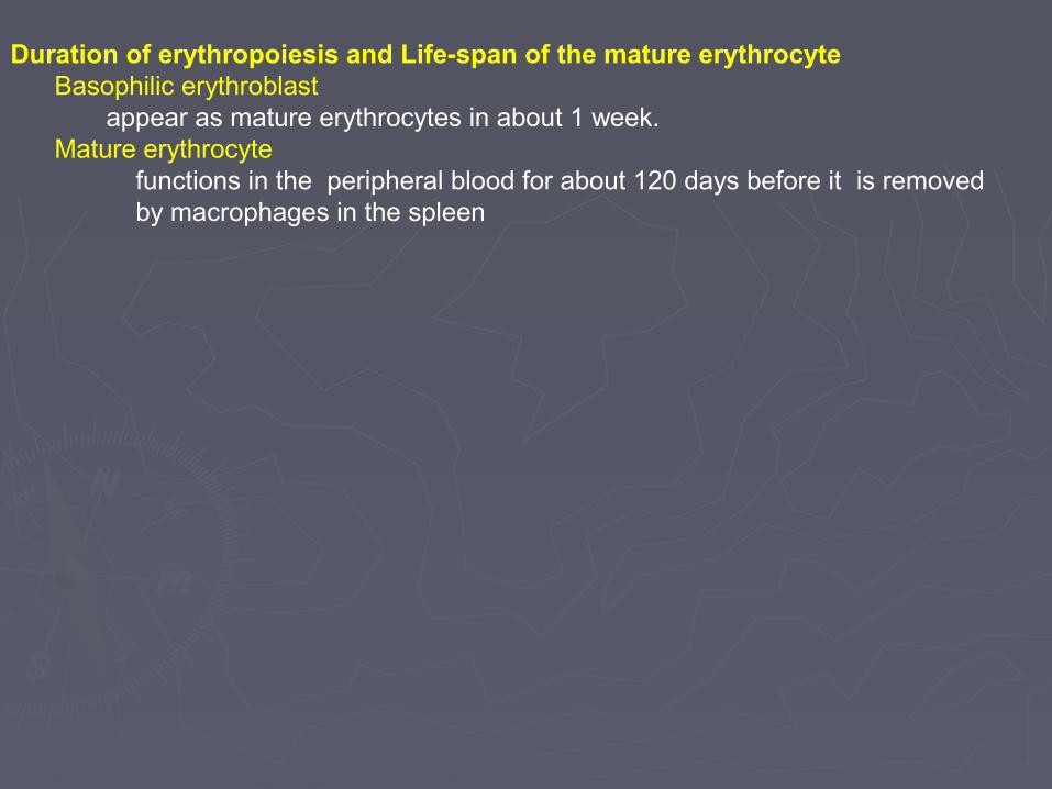
Duration of erythropoiesis and Life-span of the mature erythrocyte Basophilic erythroblast appear as mature erythrocytes in about 1 week. Mature erythrocyte functions in the peripheral blood for about 120 days before it is removed by macrophages in the spleen

Erytrocytes maturation


GRANULOPOIESIS. Dev. of granulocytes (neutrophils, eosinophils, and basophils) passes through several well-defined stages.
Myeloblast first developmental stage not an easily recognized cell Morphologic characteristic Size- large cell (about 14-18 um in diameter), approximately twice
the diameter of an
erythrocyte. Nucleus - euchromatic and nucleoli are usually
visible. Cytoplasm - agranular and slightly basophilic.

Promyelocyte Morphologic characteristics Size - (18-20 um) slightly larger than the myeloblast and is much larger than an RBC Nucleus - large and euchromatic, and nucleoli may be identified. No indentation of the nuclear surface Cytoplasmic granules (1) Azurophilic, or primary, granules are present important indicator of this stage of granulopoiesis. These granules are stained by the azure dye that is one of the components of routinely used blood stains. (2) Specific granules are not present

Characteristics of promyelocyte granules Azurophilic granules (and in later granulocyte stages) Type of lysosome, contain both lysosomal enzymes and peroxidase – myeloperoxidase to emphasize its presence in myeloid cells. synthesized only by promyelocytes, and not by cells in later stages of granulopoiesis. Hence, number of azurophilic granules per developing granulocyte diminishes with each cell division of the promyelocyte and its progeny. Multipotential nature of promyelocytes a. Promyelocytes cannot be divided into neutrophilic, eosinophilic, or basophilic subtypes. become recognizable at the myelocyte stage - specific granules

Myelocyte. commonly encountered cell type in bone marrow. Neutrophilic myelocytes, eosinophilic myelocytes, basophilic myelocytes recognized on the basis of the staining of their specific granules. These secondary granules are first seen at this stage of granulopoiesis.
Morphologic characteristics Size - iapproximately the size of the mature granulocyte (12-15 um). Nucleus acquires an indentation on its surface facing the interior of the cell. becomes more heterochromatic, and nucleoli are usually not visible.

Cytoplasmic granules Two populations of granules - recognized in myelocytes. Specific granules characteristic staining reactions (neutrophilia, acidophilia, or basophilia), first appear in myelocytes. Azurophilic granules form a decreasing fraction of the total number of granules in the developing granulocyte. eosinophils and basophils tend to be obscured by the larger, more numerous, more intensely stained, and more electron-opaque specific granules.
"dawn of neutrophilia" characteristic of the developing neutrophilic myelocyte.

Specificity of secondary granules - Specific granules impart to the developing granulocyte. functional specificity AND morphologic specificity a. Neutrophilic specific granules contain bacteriostatic and bactericidal substances such as lysozyme, lactoferrin, and alkaline phosphatase act in concert with the lysosomal azurophilic granules during the phagocytic function of neutrophils.
b. Eosinophilic specific granules containing a paracrystalline, arginine-rich protein. gives the granule its characteristic acidophilia, its refractility, and its unique fine structure c. Basophilic specific granules contain histamine and heparan sulfate


Metamyelocyte next developmental stage beyond the myelocyte. Nucleus a. indentation of the nucleus deepens and the nucleus becomes kidney-shaped. b. chromatin - slightly more condensed (heterochromatic) than in the myelocyte stage. Cytoplasmic granules a. few hundred granules with specific granules outnumbering azurophilic granules by 3 or 4 to 1. b. No new azurophilic or specific granules are formed


Band cell developmentally closest to the mature neutrophil. stab cell no comparable stage for developing eosinophils and basophils. Morphologic characteristics Nuclear shape - band-like, horseshoe-shaped structure. Nuclear lobulation (1) first indications of nuclear lobe formation (2) Only when nuclear lobulation is complete and when, typically, 3-5 distinct segmented lobes are apparent is the cell considered a mature polymorphonuclear leukocyte (PMN - neutrophil)


KINETIC OF NEUTROPHIL PRODUCTION total time taken for a myeloblast to emerge as a mature neutrophil in the circulation is about 11days. Under normal circumstances, 5 mitotic divisions occur in the myeloblast, promyelocyte, and neutrophilic myelocyte stages of development.
Neutrophils Pass Through Several Functionally and Anatomically Defined Compartments Medullary Formation Compartment subdivided into a 1.Mitotic compartment (-3 days) and a 2.Maturation compartment (-4 days). Remain in this compartment for about 4 days. Medullary Storage Compartment acts as a Buffer System, capable of releasing large numbers of mature neutrophils on demand.

Circulating Compartment consists of neutrophils suspended in plasma and circulating in blood vessels. Marginating Compartment composed of neutrophils that are present in blood but do not circulate, are in capillaries and are temporarily excluded from the circulation by vasoconstriction, or especially in the lungs – they may be at the periphery of vessels, adhering to the endothelium, and not in the main bloodstream.
Marginating and Circulating compartments are of about equal size, and there is a constant interchange of cells between them. half-life of a neutrophil in these two compartments is 6-7 hours.

Medullary Formation and Storage Compartments together are about 10 times as large as the Circulating and Marginating compartments.
Neutrophils and Other Granulocytes enter the connective tissues by passing through intercellular junctions found between endothelial cells of capillaries and postcapilIary venules (diapedesis). Connective Tissues Compartment size is not known. Neutrophils reside here for 1-4 days and then die by apoptosis, whether or not they have performed their major function of phagocytosis.

medullary
MedullaryFormation

Circulating Band Cells. small number of band cells may be found in normal blood smears. number in peripheral blood is elevated under conditions that place demands on the neutrophil population.
Kinetics of neutrophil development 1. Mitotic and postmitotic phases of granulopoiesis a. Cell divisions cease by the late myelocyte stage. b. Postmitotic cells. Metamyelocytes, band cells, and mature neutrophils do not divide. 2. Distribution of the neutrophil population a. Approximately 15 times more mature neutrophils and nearly mature neutrophils (band cells) are found in the Marrow than in the Peripheral blood. b. Large numbers are stored in the marrow and enter the circulation in response to injury and infection. c. leave the circulation to enter the perivascular connective tissue.
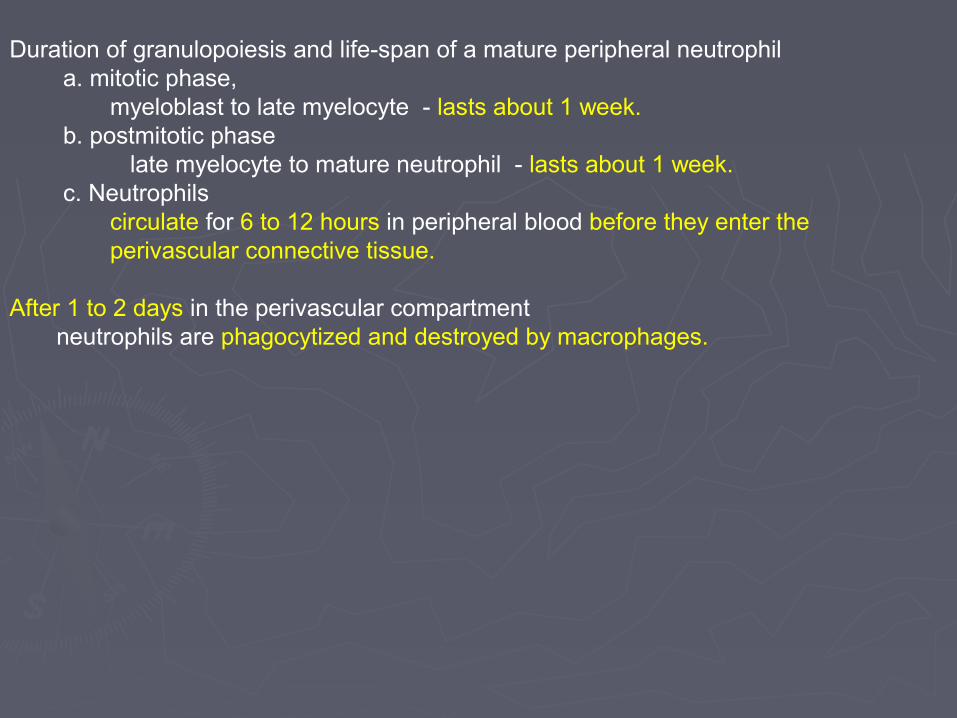
Duration of granulopoiesis and life-span of a mature peripheral neutrophil a. mitotic phase, myeloblast to late myelocyte - lasts about 1 week. b. postmitotic phase late myelocyte to mature neutrophil - lasts about 1 week. c. Neutrophils circulate for 6 to 12 hours in peripheral blood before they enter the perivascular connective tissue.
After 1 to 2 days in the perivascular compartment neutrophils are phagocytized and destroyed by macrophages.

Numbers of neutrophils and erythropoietic cells in bone marrow
1. Neutrophils are the predominant cell type in bone marrow. a. Immature neutrophils (metamyelocytes and band cells) and mature neutrophils account for approximately 50% of the cells in a bone marrow smear. b. Erythropoietic cells, from early basophilic erythroblasts to normoblasts, account for only about 18% of the cells in a marrow smear.
2. Therefore, while erythrocytes vastly outnumber granulocytes in circulating blood, their immature forms are a distinct minority in the marrow compartment.

MEGAKARYOCYTOPOIESIS. Megakaryocytes bone marrow cells that give rise to platelets.
Megakaryoblast immature cell derived from the pluripotential CFC. Morphologic characteristics large cell (about 30 um in diameter) with a nonlobulated nucleus. No evidence is seen of platelet formation by the megakaryoblast.
Megakaryoblast-megakaryocyte transition 1. Successive endomitoses occur in the megakaryoblast. a. DNA replicates and the number of chromosomes increases. b. Neither karyokinesis nor cytokinesis takes place, however, so that the chromosomes remain within one enlarging nucleus and ploidy increases from 2N to 32N or 64N. 2. Once the cell becomes large and polyploid - considered a megakaryocyte.


Megakaryocyte. mature, platelet-forming cell. Chromosome replication does not occur - postendomitotic cell. Morphologic characteristics Size (1) vary in size from 50 to 100 um in diameter. largest cell in normal marrow. (2) Both the cell and its nucleus have increased in size over the megakaryoblast, in proportion to the ploidy of the cell. Cytoplasm. electron microscope, superficial cytoplasm - divided into small compartments by multiple invaginations of the plasma membrane. platelet demarcation channels and they define future platelets.

Cell surface. smears of bone marrow examined by light microscopy, clusters of platelets, about to be released, are often seen at the surface of megakaryocytes.
Platelet formation and release a. Each cytoplasmic compartment defined by platelet demarcation channels in the megakaryocyte, corresponds to a developing platelet. b. platelet is released from the megakaryocyte when its surrounding demarcation channels become continuous with one another. c. Platelets are shed from the surface of the megakaryocyte as small, membrane-bounded cytoplasmic packets.


MONOCYTOPOIESIS. development of the monocyte-macrophage cell line takes place in three sites. Bone Marrow, monocyte develops from the CFC through intermediate stages. monoblasts and promonocytes,
Peripheral Blood, monocytes can be recognized in this location, however, they are not fully differentiated cells. Perivascular Connective Tissue, final differentiation occurs. Monocytes leave the blood by crossing the vessel wall to enter the connective tissue around the blood vessel. differentiate several types of Mononuclear - Phagocytic Cells macrophage - final stage of development of a monocyte.


![[MICROA - 2.1] Myeloid Tissue Histology](https://static.fdocuments.us/doc/165x107/577cc6a61a28aba7119ec582/microa-21-myeloid-tissue-histology.jpg)