Musculoskeletal Program Interventional Pain Management · system into account when determining the...
22
Appropriate.Safe.Affordable © 2017 AIM Specialty Health 2062-0617 Interventional Pain Management Guidelines Musculoskeletal Program Interventional Pain Management EFFECTIVE NOVEMBER 1, 2017 LAST REVIEWED JUNE 13, 2017
-
Upload
hoangtuyen -
Category
Documents
-
view
215 -
download
0
Transcript of Musculoskeletal Program Interventional Pain Management · system into account when determining the...
Appropriate.Safe.Affordable © 2017 AIM Specialty Health
2062-0617
Interventional Pain Management Guidelines
Musculoskeletal Program
Interventional Pain Management
EFFECTIVE NOVEMBER 1, 2017
LAST REVIEWED JUNE 13, 2017

Copyright © 2017. AIM Specialty Health. All Rights Reserved. 2
Table of Contents
Description and Application of the Guidelines ............................................................................................................................................... 3
Epidural Injection Procedures and Diagnostic Selective Nerve Root Blocks .................................................................................................. 4
Description ............................................................................................................................................................................................... 4
Definitions ................................................................................................................................................................................................ 4
Criteria ...................................................................................................................................................................................................... 5
Exclusions ................................................................................................................................................................................................. 7
Selected References ................................................................................................................................................................................ 7
CPT Codes ................................................................................................................................................................................................ 8
Paravertebral Facet Injection/Nerve Block/Neurolysis .................................................................................................................................. 9
Description ............................................................................................................................................................................................... 9
Definitions ................................................................................................................................................................................................ 9
Criteria ................................................................................................................................................................................................... 10
Exclusions .............................................................................................................................................................................................. 11
Selected References ............................................................................................................................................................................. 12
CPT Codes .................................................................................................................................................................................... 12
Regional Sympathetic Nerve Block .............................................................................................................................................................. 13
Description ............................................................................................................................................................................................ 13
Definitions ............................................................................................................................................................................................. 13
Criteria ................................................................................................................................................................................................... 14
Exclusions .............................................................................................................................................................................................. 15
Selected References ............................................................................................................................................................................. 15
CPT Codes .................................................................................................................................................................................... 15
Sacroiliac Joint Injection .............................................................................................................................................................................. 16
Description ............................................................................................................................................................................................ 16
Definitions ............................................................................................................................................................................................. 16
Criteria ................................................................................................................................................................................................... 17
Exclusions .............................................................................................................................................................................................. 18
Selected References ............................................................................................................................................................................. 18
CPT Codes .................................................................................................................................................................................... 19
Spinal Cord Stimulators for Permanent Implantation .................................................................................................................................. 20
Description ............................................................................................................................................................................................ 20
Definitions ............................................................................................................................................................................................. 20
Criteria ................................................................................................................................................................................................... 21
Selected References ............................................................................................................................................................................. 21
CPT Codes .................................................................................................................................................................................... 22

Copyright © 2017. AIM Specialty Health. All Rights Reserved. 3
Description and Application of the Guidelines AIM Clinical Appropriateness Guidelines (hereinafter “AIM Clinical Appropriateness Guidelines” or the
“Guidelines”) are designed to assist providers in making the most appropriate treatment decision for a
specific clinical condition for an individual. As used by AIM, the Guidelines establish objective and
evidence-based, where possible, criteria for medical necessity determinations. In the process, multiple
functions are accomplished:
● To establish criteria for when services are medically necessary
● To assist the practitioner as an educational tool
● To encourage standardization of medical practice patterns
● To curtail the performance of inappropriate and/or duplicate services
● To advocate for patient safety concerns
● To enhance the quality of healthcare
● To promote the most efficient and cost-effective use of services
The AIM guideline development process complies with applicable accreditation standards, including the
requirement that the Guidelines be developed with involvement from appropriate providers with current
clinical expertise relevant to the Guidelines under review and be based on the most up to date clinical
principles and best practices. Relevant citations are included in the “References” section attached to each
Guideline. AIM reviews all of its Guidelines at least annually.
AIM makes its Guidelines publicly available on its website twenty-four hours a day, seven days a week.
Copies of the AIM Clinical Appropriateness Guidelines are also available upon oral or written request.
Although the Guidelines are publicly-available, AIM considers the Guidelines to be important, proprietary
information of AIM, which cannot be sold, assigned, leased, licensed, reproduced or distributed without the
written consent of AIM.
AIM applies objective and evidence-based criteria and takes individual circumstances and the local delivery
system into account when determining the medical appropriateness of health care services. The AIM
Guidelines are just guidelines for the provision of specialty health services. These criteria are designed to
guide both providers and reviewers to the most appropriate services based on a patient’s unique
circumstances. In all cases, clinical judgment consistent with the standards of good medical practice should
be used when applying the Guidelines. Guideline determinations are made based on the information
provided at the time of the request. It is expected that medical necessity decisions may change as new
information is provided or based on unique aspects of the patient’s condition. The treating clinician has
final authority and responsibility for treatment decisions regarding the care of the patient and for justifying
and demonstrating the existence of medical necessity for the requested service. The Guidelines are not a
substitute for the experience and judgment of a physician or other health care professionals. Any clinician
seeking to apply or consult the Guidelines is expected to use independent medical judgment in the context
of individual clinical circumstances to determine any patient’s care or treatment.
The Guidelines do not address coverage, benefit or other plan specific issues. If requested by a health
plan, AIM will review requests based on health plan medical policy/guidelines in lieu of the AIM
Guidelines.

Copyright © 2017. AIM Specialty Health. All Rights Reserved. 4
Epidural Injection Procedures and Diagnostic Selective Nerve Root Blocks
Description
Epidural steroid injection (ESI) involves the administration of corticosteroid via insertion of a needle
into the epidural space surrounding the spinal nerve root. Despite the lack of consistent evidence to
support its efficacy, the procedure is widely used in patients with chronic back, neck and radicular pain.
In 2014, the US Food and Drug Administration issued a drug safety communication about epidural
injection of glucocorticoids, citing the risk for rare but serious adverse effects (loss of vision, stroke,
paralysis, and death). The best evidence supporting its use comes from trials that looked specifically at
patients with radiculopathy due to disc herniation, where short term benefit has been demonstrated.
Injections may performed as part of a diagnostic workup of radicular pain, or as a therapeutic modality
when noninvasive treatment strategies have failed. Injections may be performed via an interlaminar
approach, transforaminal approach, or caudal approach (through the sacral hiatus at the sacral canal)
Selective nerve root block is a related procedure that utilizes a small amount of anesthetic, injected via
transforaminal approach, to anesthetize a specific spinal nerve. Diagnostic selective nerve root blocks
are used to evaluate a patient’s anatomical level and/or source of radicular pain and are often used in
surgical planning and decision making.
Definitions
Conservative management should include a combination of strategies to reduce inflammation,
alleviate pain, and improve function, including but not limited to the following:
Prescription strength anti-inflammatory medications and analgesics
Adjunctive medications such as nerve membrane stabilizers or muscle relaxants
Physician-supervised therapeutic exercise program or physical therapy
Manual therapy or spinal manipulation
Alternative therapies such as acupuncture
Appropriate management of underlying or associated cognitive, behavioral, or addiction
disorders
Documentation of compliance with a plan of therapy that includes elements from these areas is
required. Exceptions may be considered on a case-by-case basis.
Reporting of symptom severity – Severity of pain and its impact on activities of daily living (ADLs) is a
key factor in determining the need for intervention. For purposes of this guideline, significant pain and
functional impairment refers to pain that is at least 3 out of 10 in intensity and is associated with
inability to perform at least two (2) ADLs.

Copyright © 2017. AIM Specialty Health. All Rights Reserved. 5
Imaging studies – All imaging must be performed and read by an independent radiologist. If
discrepancies should arise in the interpretation of the imaging, the radiologist report will supersede.
The results of all imaging studies should correlate with the clinical findings in support of the requested
procedure.
Criteria
Therapeutic Epidural Steroid Injection (ESI) of the cervical or lumbar spine may be indicated when all of
the following criteria are met:
Radicular pain (cervical or lumbar) or neurogenic claudication (lumbar) with associated
functional impairment
Evidence of nerve root compression or spinal stenosis (central or foraminal) is seen on an
advanced imaging study (MRI or CT) and correlates with the clinical findings*
The pain has not responded to at least four (4) weeks of appropriate conservative management,
unless there is evidence of radiculopathy, in which case ESI may be performed following two (2)
weeks of conservative management.
*NOTE: The initial epidural injection for a given episode of pain in the lumbar spine may be performed
without confirmatory advanced imaging if the exam findings are clearly diagnostic of nerve root
compression or spinal stenosis.
Repeat Therapeutic ESI may be indicated when all of the following criteria are met:
The prior injection produced at least a 50% reduction in pain with functional improvement of at
least three (3) weeks’ duration**
The patient has a recurrence of pain with significant functional disability
The patient continues to participate in a conservative treatment between injections
**NOTE: If the initial injection did not result in pain relief, repeat injection may be indicated, provided
that the injection is performed at an adjacent level, OR at the same level utilizing a different approach
or type of steroid.
Diagnostic Selective Nerve Root Block (DSNRB), also known as diagnostic transforaminal injection, is
defined as the injection of anesthetic only, for the purpose of determining the need for surgical
intervention.
DSNRB may be indicated in the evaluation and diagnostic work-up of radicular pain following
consultation with a spine surgeon in any of the following scenarios:
To confirm nerve root compression or spinal stenosis (central or foraminal) noted on an
advanced imaging study (MRI or CT) and that is consistent with, and appears to be contributing
to, the patient’s symptoms.
To determine or confirm the (or most) symptomatic level (i.e., site of compression) in the
presence of multi-level involvement for which the primary symptomatic level is unclear.
When radiculopathy is highly suspected but cannot be confirmed with advanced imaging
studies.

Copyright © 2017. AIM Specialty Health. All Rights Reserved. 6
Procedural Requirements and Restrictions:
Injections must be performed under fluoroscopy or CT guidance.
A maximum of three (3) injections (including diagnostic transforaminal injections) may be
performed in each spinal region (cervical or lumbosacral) in a six (6) month period.
No more than two (2) transforaminal injections may be performed at a single setting (e.g., single
level bilaterally or two levels). Injecting one level bilaterally would be considered two injections.
Injecting two levels, each unilaterally, would also be considered two injections.
For caudal or cervical/lumbar interlaminar injections, only one injection per session may be
performed and NOT in conjunction with a transforaminal injection. A session is defined as all
ESIs or spinal procedures performed on a single day.
After three injections in the same region, the total cumulative dose of steroid must be
documented and may not exceed 240 mg of methylprednisolone or triamcinolone or 36 mg of
betamethasone or 45 mg of dexamethasone.
Contraindications and Risks
The following conditions should prompt further evaluation prior to considering ESI:
New onset of low back pain or neck pain in the setting of established malignancy, or where
there is a suspicion of malignancy based on the clinical presentation
New onset of low back pain or neck pain in persons with risk factors for spinal infection
Comorbid conditions associated with increased risk of bleeding due to coagulopathy or
treatment with anticoagulants
Back pain in the setting of trauma
Additional contraindications include the following conditions:
Cauda equina syndrome
Conus medullaris syndrome
Epidural hematoma
Subarachnoid hemorrhage
Epidural mass
Spinal cord ischemia
Spinal fracture which occurred less than 6 weeks prior to injection
Demyelinating disease or other CNS processes which predispose to transverse myelitis
Systemic infection
Local infection at the injection site
Uncontrolled diabetes

Copyright © 2017. AIM Specialty Health. All Rights Reserved. 7
Exclusions
Indications other than those addressed in this guideline are considered not medically necessary,
including but not limited to the following:
Thoracic level ESI performed for thoracic pathology
Moderate to severe myelopathy on clinical exam
Myelopathy associated with intramedullary cord signal change on T1 or T2 weighted MRI
Isolated axial neck pain or low back pain
Selected References 1 Ammendolia CS, K. J.; Rok, E.; Rampersaud, R.; Kennedy, C. A.; Pennick, V.; Steenstra, I. A.; de Bruin, L. K.; Furlan, A. D. Nonoperative treatment for
lumbar spinal stenosis with neurogenic claudication. Cochrane Database Syst Rev. 2013(8):CD010712.
2 Bicket MC, Horowitz JM, Benzon HT, et al. Epidural injections in prevention of surgery for spinal pain: systematic review and meta-analysis of
randomized controlled trials. The spine journal : official journal of the North American Spine Society. 2015;15(2):348-62.
3 Chou R, Loeser JD, Owens DK, et al. Interventional therapies, surgery, and interdisciplinary rehabilitation for low back pain: an evidence-based clinical
practice guideline from the American Pain Society. Spine. 2009;34(10):1066-77.
4 Cohen SPH, S.; Semenov, Y et al. . Epidural steroid injections, conservative treatment, or combination treatment for cervical radicular pain: a
multicenter, randomized, comparative-effectiveness study. Anesthesiology. 2014;121(5):1045-55.
5 COST B13 Working Group on Guidelines for Chronic Low Back Pain, Airaksinen O, Brox JI, et al. Chapter 4. European guidelines for the management
of chronic nonspecific low back pain. Eur Spine J. 2006;15 Suppl 2:S192-300.
6 COST B13 Working Group on Guidelines for Chronic Low Back Pain, van Tulder M, Becker A, et al. Chapter 3. European guidelines for the
management of acute nonspecific low back pain in primary care. Eur Spine J. 2006;15 Suppl 2:S169-91.
7 Falco FJ, Manchikanti L, Datta S, et al. Systematic review of the therapeutic effectiveness of cervical facet joint interventions: an update. Pain
physician. 2012;15(6):E839-68.
8 Institute for Clinical Systems Improvement GM, Thorson D, et al. . Adult acute and subacute low back pain. 2012:92 pgs. .
9 Institute of Health Economics. Toward Optimized Practice. Guideline for the evidence-informed primary care management of low back pain. 2011:37.
10 Kaye ADM, L.; Abdi, S.; et al. . Efficacy of Epidural Injections in Managing Chronic Spinal Pain: A Best Evidence Synthesis. Pain physician.
2015;18(6):E939-1004.
11 Kreiner DS, Hwang, S. W., North American Spine, Society, et al. An evidence-based clinical guideline for the diagnosis and treatment of lumbar disc
herniation with radiculopathy. Spine J. 2014;14(1):180-91.
12 Kreiner DS, Shaffer WO, Baisden JL, et al. An evidence-based clinical guideline for the diagnosis and treatment of degenerative lumbar spinal
stenosis (update). The spine journal : official journal of the North American Spine Society. 2013;13(7):734-43.
13 Lee CHL, J.; Kang, J. D. et al. . Laminoplasty versus laminectomy and fusion for multilevel cervical myelopathy: a meta-analysis of clinical and
radiological outcomes. J Neurosurg Spine. 2015;22(6):589-95.
14 Lewis RAW, N. H.; Sutton, A. J., et al. . Comparative clinical effectiveness of management strategies for sciatica: systematic review and network meta-
analyses. Spine J. 2015;15(6):1461-77.
15 Liu KL, P.; Liu, R.; Wu, X.; Cai, M. Steroid for epidural injection in spinal stenosis: a systematic review and meta-analysis. Drug Des Devel Ther.
2015;9:707-16.
16 MacVicar JK, W.; Landers, M. H.; Bogduk, N. The effectiveness of lumbar transforaminal injection of steroids: a comprehensive review with systematic
analysis of the published data. Pain Medicine. 2013;14(1):14-28.
17 Manchikanti LB, R. M.; Falco, F. J.; Kaye, A. D.; Hirsch, J. A. Do Epidural Injections Provide Short- and Long-term Relief for Lumbar Disc Herniation? A
Systematic Review. Clin Orthop. 2015;473(6):1940-56.
18 Manchikanti LN, D. E.; Candido, K. D., et al. . Do cervical epidural injections provide long-term relief in neck and upper extremity pain? A systematic
review. Pain physician. 2015;18(1):39-60.
19 Meng HF, Q.; Wang, B.; Yang, Y.; Li, D.; Li, J.; Su, N. Epidural injections with or without steroids in managing chronic low back pain secondary to lumbar
spinal stenosis: a meta-analysis of 13 randomized controlled trials. Drug Des Devel Ther. 2015;9:4657-67.
20 National Institute for Health and Care Excellence,Low back pain and sciatica in over 16s: assessment and management,(2016),London UK,
21 North American Spine Society,Lumbar Epidural Injections - NASS Coverage Policy Recommendations,(May 2014),Burr Ridge IL,12 pgs.
22 North American Spine Society,Cervical Epidural Infections - NASS Coverage Policy Recommendations,(May 2014),Burr Ridge IL 10 pgs.
23 Phillips FMS, P. J.; Youssef, J. A.; Andersson, G.; Papatheofanis, F. Lumbar spine fusion for chronic low back pain due to degenerative disc disease: a
systematic review. Spine. 2013;38(7):E409-22.
Copyright © 2017. AIM Specialty Health. All Rights Reserved. 8
24 Pinto RZM, C. G.; Ferreira, M. L., et al. Epidural corticosteroid injections in the management of sciatica: a systematic review and meta-analysis. Ann
Intern Med. 2012;157(12):865-77.
25 Radcliff KK, C.; Hilibrand, A., et al. . Epidural steroid injections are associated with less improvement in patients with lumbar spinal stenosis: a
subgroup analysis of the Spine Patient Outcomes Research Trial. Spine. 2013;38(4):279-91.
26 Rathmell JP, Benzon HT, Dreyfuss P, et al. Safeguards to Prevent Neurologic Complications after Epidural Steroid InjectionsConsensus Opinions from
a Multidisciplinary Working Group and National Organizations. Anesthesiology. 2015;122(5):974-84.
27 Zhang YL, C.; Tao, Y., et al. Cervical total disc replacement is superior to anterior cervical decompression and fusion: a meta-analysis of prospective
randomized controlled trials. PLoS ONE. 2015;10(3):e0117826.
CPT Codes 62320 Injection(s), of diagnostic or therapeutic substance(s) (eg, anesthetic, antispasmodic, opioid, steroid, other
solution), not including neurolytic substances, including needle or catheter placement, interlaminar epidural
or subarachnoid, cervical or thoracic; without imaging guidance
62321 Injection(s), of diagnostic or therapeutic substance(s) (eg, anesthetic, antispasmodic, opioid, steroid, other
solution), not including neurolytic substances, including needle or catheter placement, interlaminar epidural
or subarachnoid, cervical or thoracic; with imaging guidance (ie, fluoroscopy or CT)
62322 Injection(s), of diagnostic or therapeutic substance(s) (eg, anesthetic, antispasmodic, opioid, steroid, other
solution), not including neurolytic substances, including needle or catheter placement, interlaminar epidural
or subarachnoid, lumbar or sacral (caudal); without imaging guidance
62323 Injection(s), of diagnostic or therapeutic substance(s) (eg, anesthetic, antispasmodic, opioid, steroid, other
solution), not including neurolytic substances, including needle or catheter placement, interlaminar epidural
or subarachnoid, lumbar or sacral (caudal); with imaging guidance (ie, fluoroscopy or CT)
64479 Injection(s), anesthetic agent and/or steroid, transforaminal epidural, with imaging guidance (fluoroscopy or
CT); cervical or thoracic, single level
64480 Injection(s), anesthetic agent and/or steroid, transforaminal epidural, with imaging guidance (fluoroscopy or
CT); cervical or thoracic, each additional level (List separately in addition to code for primary procedure)
64483 Injection(s), anesthetic agent and/or steroid, transforaminal epidural, with imaging guidance (fluoroscopy or
CT); lumbar or sacral, single level
64484 Injection(s), anesthetic agent and/or steroid, transforaminal epidural, with imaging guidance (fluoroscopy or
CT); lumbar or sacral, each additional level (List separately in addition to code for primary procedure)
Copyright © 2017. AIM Specialty Health. All Rights Reserved. 9
Paravertebral Facet Injection/Nerve Block/Neurolysis
Description
Paravertebral facet joints, also referred to as zygapophyseal joints or Z-joints, have been implicated as
a source of chronic neck and low back pain with a prevalence of up to 70% in the cervical spine, and
up to 30% in the lumbar spine. Neither physical exam nor imaging has adequate diagnostic power to
confidently identify the facet joint as a pain source. Facet joint injection techniques have evolved
primarily as a diagnostic tool for pain originating in these joints, but have been widely utilized to treat
chronic pain shown to be of facet origin.
Injections may be performed at one of two sites, either the joint itself (intraarticular injection) or the
nerve that supplies it (medial branch of the dorsal ramus of segmental spinal nerves). Diagnostic
injections are performed with an anesthetic agent alone, while therapeutic injections involve
administration of a corticosteroid, with or without an anesthetic. Following confirmation of facet
pathology using a diagnostic medial branch block (MBB), select patients may undergo a radiofrequency
nerve ablation procedure. Studies have validated the efficacy of this intervention in chronic pain of
facet origin.
Definitions
Conservative management should include a combination of strategies to reduce inflammation,
alleviate pain, and improve function, including but not limited to the following:
Prescription strength anti-inflammatory medications and analgesics
Adjunctive medications such as nerve membrane stabilizers or muscle relaxants
Physician-supervised therapeutic exercise program or physical therapy
Manual therapy or spinal manipulation
Alternative therapies such as acupuncture
Appropriate management of underlying or associated cognitive, behavioral, or addiction
disorders
Documentation of compliance with a plan of therapy that includes elements from these areas is
required. Exceptions may be considered on a case-by-case basis.
Reporting of symptom severity – Severity of pain and its impact on activities of daily living (ADLs) is a
key factor in determining the need for intervention. For purposes of this guideline, significant pain and
functional impairment refers to pain that is at least 3 out of 10 in intensity, and is associated with
inability to perform at least two (2) ADLs.
Imaging studies – All imaging must be performed and read by an independent radiologist. If
discrepancies should arise in the interpretation of the imaging, the radiologist report will supersede.
The results of all imaging studies should correlate with the clinical findings in support of the requested
procedure.
Copyright © 2017. AIM Specialty Health. All Rights Reserved. 10
Criteria
Procedures must be performed with image guidance, either fluoroscopy or CT
Patients must meet all of the following criteria:
o Moderate to severe pain with functional impairment of at least three (3) months’
duration
o Predominant axial pain that is not attributable to radiculopathy**, myelopathy, or
neurogenic claudication
o Physical exam findings which are consistent with the facet joint as the presumed
source of pain
o Absence of non-facet pathology that could explain the source of the patient’s pain,
such as fracture, tumor, or infection.
o Absence of prior surgical fusion at the proposed level
o Lack of improvement or resolution following at least six (6) weeks of conservative
management
**With the exception of synovial cysts
Diagnostic Medial Branch Blocks (MBB)
The primary utility of MBBs is to determine the suitability of the patient for a radiofrequency neurotomy
of painful segmental levels in order to achieve long-term pain management. A positive response is
defined as at least 80% relief of the primary (index) pain, with the onset and duration of relief being
consistent with the agent employed.
Note: The patient must be experiencing pain at the time of the injection (generally rated at least 3 out
of 10 in intensity) in order to determine whether a response has occurred. Provocative maneuvers or
positions which normally exacerbate index pain should also be assessed and documented before and
after the procedure.
Dual MBBs, defined as injections performed in the same location(s) on two (2) separate
occasions at least one week apart, are necessary to confirm the diagnosis due to the
unacceptably high false positive rate of single MBB injections.
A confirmatory injection is indicated only if the first injection results in a positive response. If
the second injection also results in a positive response, the target joint(s) is/are the confirmed
pain generator(s).
If the first session of diagnostic MBBs are negative, a maximum of one additional session may
be performed to determine the primary levels of involvement prior to proceeding with
confirmatory blocks.
A maximum of two (2) levels may be injected during a single session.
Therapeutic Intraarticular (IA) Facet Joint Injections
Therapeutic IA injections should be repeated no more than three times annually and only if the initial
injection results in significant pain relief (at least 50%) for at least three (3) months.

Copyright © 2017. AIM Specialty Health. All Rights Reserved. 11
Thermal Medial Branch Radiofrequency Neurotomy (RFN)
RFN may be offered to patients if dual diagnostic MBB injections each produce ≥ 80% relief of
the primary (index) pain and the onset and minimum duration of relief is consistent with the
agent employed.
RFN may be performed at the same level no more than twice annually and only if the initial
radiofrequency lesion results in significant pain relief (at least 50%) and improvement in patient
specific ADLs for at least six (6) months.
For each covered spinal region (cervical or lumbar), RFN should be performed at no more than
four (4) joints per session (e.g., two [2] bilateral levels or four [4] unilateral levels).
Repeat RFN to treat recurrent facet joint pain in a patient who has failed other conservative
measures may be covered without repeating diagnostic MBB injections if the patient has
experienced significant and prolonged relief of pain (at least 50% reduction for at least five [5]
months) and improvement of function in the past following RF ablation.
RFN may not be performed at C0-C1 or at C1-C2
Procedural Limitations (MBB, IA)
A maximum of six (6) facet joint procedural sessions per region (cervical or lumbar) may be
performed in a 12-month period, regardless of type or indication.
One additional diagnostic block may be indicated prior to a repeat neurotomy when there is
diagnostic uncertainty about the source of pain.
Exclusions
Indications other than those addressed in this guideline are considered not medically necessary,
including but not limited to the following:
Diagnostic intraarticular facet joint injection
Therapeutic medial branch block
Diagnostic or therapeutic IA injection, medial branch block, or radiofrequency ablation in the
thoracic region with the exception of C7-T1 and T12-L1
Use of MBB or RFN in the setting of moderate to severe spondylolisthesis (grade 2 or higher)
Use of MBB or RFN in the setting of an isolated pars defect
Use of MBB or RFN at the level of a posterolateral fusion or posterior instrumentation
Use of non-thermal RF modalities for facet joint denervation, including chemical, low grade
thermal energy (<80 degrees Celsius), and pulsed RF
Any facet joint interventions performed under ultrasound guidance
Intraarticular or extra-articular facet joint prolotherapy
Copyright © 2017. AIM Specialty Health. All Rights Reserved. 12
Selected References 1 Boswell MV, Manchikanti L, Kaye AD, et al. A Best-Evidence Systematic Appraisal of the Diagnostic Accuracy and Utility of Facet (Zygapophysial) Joint
Injections in Chronic Spinal Pain. Pain physician. 2015;18(4):E497-533.
2 Centers for Medicare and Medicaid Services, Local Coverage Determinations: Facet Joint Injections, Medial Branch Blocks, and Facet Joint
Radiofrequency Neurotomy, (2016) Baltimore MD, 62 pgs.
3 Chou R, Loeser JD, Owens DK, et al. Interventional therapies, surgery, and interdisciplinary rehabilitation for low back pain: an evidence-based clinical
practice guideline from the American Pain Society. Spine. 2009;34(10):1066-77.
4 COST B13 Working Group on Guidelines for Chronic Low Back Pain, Airaksinen O, Brox JI, et al. Chapter 4. European guidelines for the management
of chronic nonspecific low back pain. Eur Spine J. 2006;15 Suppl 2:S192-300.
5 Falco FJ, Manchikanti L, Datta S, et al. Systematic review of the therapeutic effectiveness of cervical facet joint interventions: an update. Pain
physician. 2012;15(6):E839-68.
6 Lee CHL, J.; Kang, J. D. et al. . Laminoplasty versus laminectomy and fusion for multilevel cervical myelopathy: a meta-analysis of clinical and
radiological outcomes. J Neurosurg Spine. 2015;22(6):589-95.
7 Manchikanti L, Abdi S, Atluri S, et al. An update of comprehensive evidence-based guidelines for interventional techniques in chronic spinal pain. Part
II: guidance and recommendations. Pain physician. 2013;16(2 Suppl):S49-283.
8 Manchikanti LB, R. M.; Falco, F. J.; Kaye, A. D.; Hirsch, J. A. Do Epidural Injections Provide Short- and Long-term Relief for Lumbar Disc Herniation? A
Systematic Review. Clin Orthop. 2015;473(6):1940-56.
9 North American Spine Society, Facet Joint Interventions - NASS Coverage Policy Recommendations, (August 2016) Burr Ridge IL, 16 pgs.
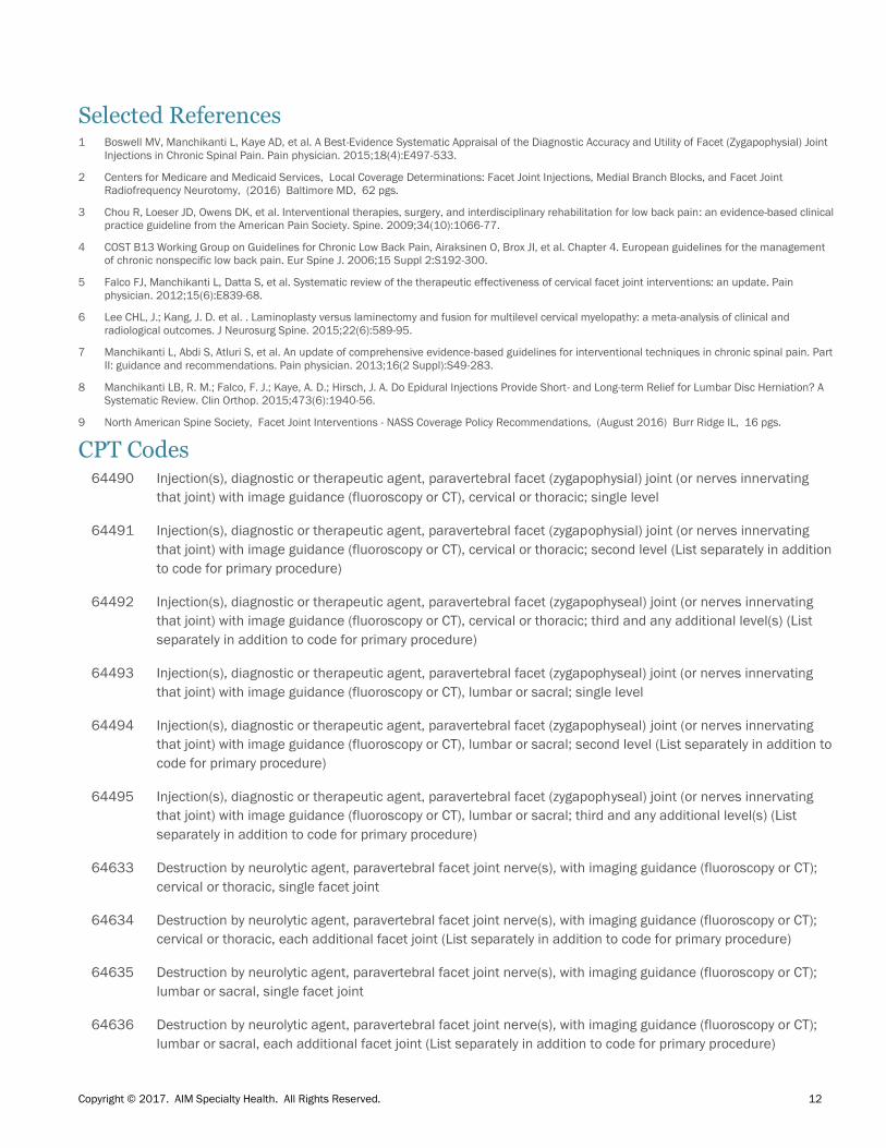
CPT Codes 64490 Injection(s), diagnostic or therapeutic agent, paravertebral facet (zygapophysial) joint (or nerves innervating
that joint) with image guidance (fluoroscopy or CT), cervical or thoracic; single level
64491 Injection(s), diagnostic or therapeutic agent, paravertebral facet (zygapophysial) joint (or nerves innervating
that joint) with image guidance (fluoroscopy or CT), cervical or thoracic; second level (List separately in addition
to code for primary procedure)
64492 Injection(s), diagnostic or therapeutic agent, paravertebral facet (zygapophyseal) joint (or nerves innervating
that joint) with image guidance (fluoroscopy or CT), cervical or thoracic; third and any additional level(s) (List
separately in addition to code for primary procedure)
64493 Injection(s), diagnostic or therapeutic agent, paravertebral facet (zygapophyseal) joint (or nerves innervating
that joint) with image guidance (fluoroscopy or CT), lumbar or sacral; single level
64494 Injection(s), diagnostic or therapeutic agent, paravertebral facet (zygapophyseal) joint (or nerves innervating
that joint) with image guidance (fluoroscopy or CT), lumbar or sacral; second level (List separately in addition to
code for primary procedure)
64495 Injection(s), diagnostic or therapeutic agent, paravertebral facet (zygapophyseal) joint (or nerves innervating
that joint) with image guidance (fluoroscopy or CT), lumbar or sacral; third and any additional level(s) (List
separately in addition to code for primary procedure)
64633 Destruction by neurolytic agent, paravertebral facet joint nerve(s), with imaging guidance (fluoroscopy or CT);
cervical or thoracic, single facet joint
64634 Destruction by neurolytic agent, paravertebral facet joint nerve(s), with imaging guidance (fluoroscopy or CT);
cervical or thoracic, each additional facet joint (List separately in addition to code for primary procedure)
64635 Destruction by neurolytic agent, paravertebral facet joint nerve(s), with imaging guidance (fluoroscopy or CT);
lumbar or sacral, single facet joint
64636 Destruction by neurolytic agent, paravertebral facet joint nerve(s), with imaging guidance (fluoroscopy or CT);
lumbar or sacral, each additional facet joint (List separately in addition to code for primary procedure)

Copyright © 2017. AIM Specialty Health. All Rights Reserved. 13
Regional Sympathetic Nerve Block
Description
Sympathetic blockade includes procedures that temporarily obstruct the local function of the
sympathetic nervous system. Anesthetic is injected directly into sympathetic neural structures that
serve affected limb(s), such as the stellate ganglion or the lumbar sympathetic chain. Radiologic
guidance (fluoroscopy or CT scan) is utilized to ensure accuracy.
Regional sympathetic nerve block has been utilized primarily for treatment of complex regional pain
syndrome. Despite limited evidence supporting its efficacy, it has also been investigated in treating a
number of other pain syndromes thought to be sympathetically mediated.
This and other interventional procedures should be considered only when the full spectrum of
noninvasive management strategies has not provided sufficient relief of symptoms.
Definitions
Conservative management should include a combination of strategies to reduce inflammation,
alleviate pain, and improve function, including but not limited to the following:
Prescription strength anti-inflammatory medications and analgesics
Adjunctive medications such as nerve membrane stabilizers or muscle relaxants
Physician-supervised therapeutic exercise program or physical therapy
Manual therapy or spinal manipulation
Alternative therapies such as acupuncture
Appropriate management of underlying or associated cognitive, behavioral or addiction
disorders
Documentation of compliance with a plan of therapy that includes elements from these areas is
required. Exceptions may be considered on a case-by-case basis.
Reporting of symptom severity -- Severity of pain and its impact on activities of daily living (ADLs) is a
key factor in determining the need for intervention. For purposes of this guideline, significant pain and
functional impairment refers to pain that is at least 3 out of 10 in intensity, and is associated with
inability to perform at least two (2) ADLs.
Imaging studies -- All imaging must be performed and read by an independent radiologist. If
discrepancies should arise in the interpretation of the imaging, the radiologist report will supersede.
The results of all imaging studies should correlate with the clinical findings in support of the requested
procedure.

Copyright © 2017. AIM Specialty Health. All Rights Reserved. 14
Criteria
Complex Regional Pain Syndrome (Type I or Type II)
Diagnostic criteria for Complex Regional Pain Syndrome (CRPS) must be met:
Continuing pain that is disproportionate to any inciting event
At least 1 symptom reported in at least three (3) of the following categories:
o Sensory: Hyperesthesia or allodynia
o Vasomotor: Temperature asymmetry, skin color changes, skin color asymmetry
o Sudomotor/edema: Edema, sweating changes, or sweating asymmetry
o Motor/trophic: Decreased range of motion, motor dysfunction (eg, weakness, tremor,
dystonia), or trophic changes (eg, hair, nail, skin)
At least 1 sign at time of evaluation in at least two (2) of the following categories:
o Sensory: Evidence of hyperalgesia (to pinprick), allodynia (to light touch, temperature
sensation, deep somatic pressure, or joint movement)
o Vasomotor: Evidence of temperature asymmetry (>1°C), skin color changes or
asymmetry
o Sudomotor/edema: Evidence of edema, sweating changes, or sweating asymmetry
o Motor/trophic: Evidence of decreased range of motion, motor dysfunction (eg,
weakness, tremor, dystonia), or trophic changes (eg, hair, nail, skin)
o No other diagnosis better explaining the signs and symptoms
In addition, all of the following are required:
o Level of pain and disability in the moderate to severe range
o Failure of at least two (2) weeks of conservative management
o Documentation of ongoing participation in a comprehensive pain management program
The performance of an initial diagnostic regional sympathetic block is considered medically necessary
to establish the presence or absence of sympathetically mediated complex regional pain syndrome.
A positive response is defined as a significant reduction in pain (at least 80% reduction) and
improvement in function with the duration of relief being consistent with agent employed, and objective
evidence that the block was physiologically effective.
For procedures that target pain in a limb, there must be documentation of a rise in temperature from
baseline of the ipsilateral limb. A sensory exam is required to confirm absence of spread to adjacent
nerve roots.
Following a positive response to the initial diagnostic block,additional diagnostic and therapeutic
regional sympathetic blocks, up to maximum of six (6) total blocks, performed at a frequency of no
more than two (2) per week, may be considered medically necessary when all the following criteria
have been met:

Copyright © 2017. AIM Specialty Health. All Rights Reserved. 15
Benefit has been demonstrated by prior blocks as evidenced by all of the following:
o Decreased use of pain medication
o Improved level of function (e.g., increased range of motion, strength, and use of
extremity in activities of daily living)
o Improved tolerance to touch (e.g., decreased allodynia) or other objective measures
The intervention is being provided as part of a comprehensive pain management program
(physical therapy, patient education, psychosocial support, and oral medication).
If there is no sustained benefit in pain and function after three (3) sympathetic blocks from
baseline (pre block) pain and function, then additional blocks are not warranted.
If there is sustained benefit after the first three (3) sympathetic blocks then up to three (3)
additional blocks may be performed.
Exclusions
Indications other than those addressed in this guideline are considered not medically necessary,
including but not limited to the following:
Use of intravenous phentolamine (Regitine) as a diagnostic test for CRPS
Intravenous regional sympathetic block utilizing guanethidine
Intrapleural analgesia for treatment of CRPS
Selected References
1. Harden RN, Oaklander AL, Burton AW, Perez RS, Richardson K, Swan M, Barthel J, Costa B, Graciosa JR, Bruehl S; Reflex Sympathetic Dystrophy
Syndrome Association. Complex regional pain syndrome: practical diagnostic and treatment guidelines, 4th edition. Pain Med. 2013 Feb;14(2):180-
229. doi: 10.1111/pme.12033. Epub 2013 Jan 17.
2 O'Connell NEW, B. M.; Gibson, W., et al. . Local anaesthetic sympathetic blockade for complex regional pain syndrome. Cochrane Database Syst Rev.
2016;7:CD004598.
3 Zernikow BW, J.; Brehmer, H.; Hirschfeld, G.; Maier, C. Invasive treatments for complex regional pain syndrome in children and adolescents: a scoping
review. Anesthesiology. 2015;122(3):699-707.
CPT Codes 64510 Injection, anesthetic agent; stellate ganglion (cervical sympathetic)
64520 Injection, anesthetic agent; lumbar or thoracic (paravertebral sympathetic)
Copyright © 2017. AIM Specialty Health. All Rights Reserved. 16
Sacroiliac Joint Injection
Description
Non-inflammatory sacroiliac (SI) joint complex pain may be traumatic, degenerative, or due to adjacent
segment disease (after lumbar fusion or total hip replacement). Sacroiliitis is associated with
inflammatory spondyloarthropathies. Pain arising from the SI joint complex typically radiates to the
gluteal area and posterior hip. In addition to localized tenderness over the SI joint, there are additional
examination maneuvers which suggest the diagnosis.
Definitions
Conservative management should include a combination of strategies to reduce inflammation,
alleviate pain, and improve function, including but not limited to the following:
Prescription strength anti-inflammatory medications and analgesics
Adjunctive medications such as nerve membrane stabilizers or muscle relaxants
Physician-supervised therapeutic exercise program or physical therapy
Manual therapy or spinal manipulation
Alternative therapies such as acupuncture
Appropriate management of underlying or associated cognitive, behavioral, or addiction
disorders
Documentation of compliance with a plan of therapy that includes elements from these areas is
required. Exceptions may be considered on a case-by-case basis.
Reporting of symptom severity -- Severity of pain and its impact on activities of daily living (ADLs) is a
key factor in determining the need for intervention. For purposes of this guideline, significant pain and
functional impairment refers to pain that is at least 3 out of 10 in intensity, and is associated with
inability to perform at least two (2) ADLs.
Imaging studies -- All imaging must be performed and read by an independent radiologist. If
discrepancies should arise in the interpretation of the imaging, the radiologist report will supersede.
The results of all imaging studies should correlate with the clinical findings in support of the requested
procedure.
Copyright © 2017. AIM Specialty Health. All Rights Reserved. 17
Criteria
Procedures must be performed with image guidance, either fluoroscopy or CT
Patients must meet all of the following inclusion criteria to be able to proceed with diagnostic
intraarticular SI joint injections, therapeutic intraarticular SI joint injections, or diagnostic lateral
branch blocks.
There is persistent typically unilateral non-radicular pain that is predominantly below the lumbar
spine (L5) and is primarily localized over the region of the sacroiliac joint and has been present
for at least three (3) months.
Examination shows localized tenderness with palpation over the sacral sulcus just inferior to the
posterior superior iliac spine (PSIS) in the absence of tenderness of equal severity elsewhere
(e.g., lumbar spine, greater trochanter, hip, coccyx)
At least one of the following provocative tests is positive: pelvic distraction test, lateral iliac
compression test, sacral compression/thrust test, thigh thrust test, FABER (Patrick’s test), and
Gaenslen’s test.
There is no evidence of acute or subacute radicular pain/radiculopathy or neurogenic
claudication. If there is evidence of radicular pain/radiculopathy or neurogenic claudication the
condition must be fixed and stable and have been maximally addressed through comprehensive
treatment.
Lack of adequate improvement following six (6) weeks of conservative management.
Diagnostic Intraarticular Sacroiliac Joint Injections
The primary utility of diagnostic intraarticular sacroiliac joint injections is to determine if the sacroiliac
joint is the primary pain generator for the patient’s low back pain.
Dual intraarticular sacroiliac joint injections, defined as injections performed in the same joint
on 2 separate occasions, are necessary to confirm the diagnosis due to the unacceptably high
false positive rate of single intraarticular sacroiliac joint injections.
A second confirmatory injection is indicated only if the first injections produces greater than or
equal to 80% relief of the primary (index) pain and the onset and minimum duration of relief is
consistent with the agent employed. This confirmatory block confirms the tested sacroiliac joint
as the source if the index pain is reduced by greater than or equal to 80% and the onset and
minimum duration of relief is consistent with the agent employed.
Anesthetic volume must be limited to 1.5 cc to maximize the anatomic specificity of the
procedure. Concurrent injection of steroid is not appropriate for diagnostic SI joint injection.
The day of the procedure, the patient’s pain must be at least 3/10 severity at rest or during a
consistently provocative maneuver, which will allow accurate monitoring of the response to the
injection.
Copyright © 2017. AIM Specialty Health. All Rights Reserved. 18
Therapeutic Intraarticular (IA) Sacroiliac Joint Injections
Therapeutic IA sacroiliac joint injections are performed with the use of corticosteroid with or
without the use of anesthetic.
Total injection volume should be limited to 2.0 cc to minimize extravasation of the injectate
outside of the SI joint.
Repeat Therapeutic Intraarticular Sacroiliac Joint Injections
Repeat injection is considered medically necessary if symptoms recur and the patient has
demonstrated at least 50% pain relief, and improvement in patient-specific ADLs, for at least 6
weeks after a previous injection.
Injections may not be repeated at intervals of less than three (3) months, with a maximum of
three (3) injections in a 12 month period.
Treatment with therapeutic injections should be accompanied by participation in an ongoing
active rehabilitation program, home exercise program, or functional restoration program.
Ultrasound-guidance
Ultrasound is the only imaging-guidance appropriate for use during pregnancy
Exclusions
Indications other than those addressed in this guideline are considered not medically necessary,
including but not limited to the following:
Intraarticular sacroiliac joint injections performed on the same day as other spine injection
procedures.
Use of corticosteroid with diagnostic intraarticular sacroiliac joint injections
Selected References
1 Chou R, Loeser JD, Owens DK, et al. Interventional therapies, surgery, and interdisciplinary rehabilitation for low back pain: an evidence-based clinical
practice guideline from the American Pain Society. Spine. 2009;34(10):1066-77.
2 COST B13 Working Group on Guidelines for Chronic Low Back Pain, Airaksinen O, Brox JI, et al. Chapter 4. European guidelines for the management
of chronic nonspecific low back pain. Eur Spine J. 2006;15 Suppl 2:S192-300.
3 Hansen H, Manchikanti L, Simopoulos TT, et al. A systematic evaluation of the therapeutic effectiveness of sacroiliac joint interventions. Pain
physician. 2012;15(3):E247-78.
4 Manchikanti L, Abdi S, Atluri S, et al. An update of comprehensive evidence-based guidelines for interventional techniques in chronic spinal pain. Part
II: guidance and recommendations. Pain physician. 2013;16(2 Suppl):S49-283.
5 Muheremu AN, X.; Wu, Z.; et al. . Comparison of the short- and long-term treatment effect of cervical disk replacement and anterior cervical disk
fusion: a meta-analysis. Eur. 2015;25 Suppl 1:S87-100.
6 North American Spine Society,Sacroiliac Joint Injections - NASS Coverage Policy Recommendations, (2015) ,Burr Ridge IL, 11 pgs.
7 Zaidi HAM, A. J.; Dickman, C. A. Surgical and clinical efficacy of sacroiliac joint fusion: a systematic review of the literature. J Neurosurg Spine.
2015;23(1):59-66.

Copyright © 2017. AIM Specialty Health. All Rights Reserved. 19
CPT Codes 27096 Injection procedure for sacroiliac joint, anesthetic/steroid, with image guidance (fluoroscopy or CT) including
arthrography when performed
Copyright © 2017. AIM Specialty Health. All Rights Reserved. 20
Spinal Cord Stimulators for Permanent Implantation
Description
Spinal cord stimulators, also known as dorsal column stimulators, are implantable devices used to
treat chronic pain. Electrodes are surgically placed within the dura mater via laminectomy, or by
percutaneous insertion into the epidural space. Low voltage electrical signals are delivered to the
dorsal column of the spinal cord in order to override or mask sensations of pain.
The patient’s pain distribution pattern determines the level at which the stimulation lead is placed. The
lead may incorporate four (4) to eight (8) electrodes, with 8 electrodes typically used for complex pain
patterns, such as bilateral pain or pain extending from the limbs to the trunk.
Implantation is typically a 2-step process. Initially, the electrode is temporarily implanted in the epidural
space, allowing a trial period of stimulation. Once treatment effectiveness is confirmed (defined as at
least 50% reduction in pain), the electrodes and radio receiver/ transducer are permanently implanted.
Extensive programming of the neurostimulators is often required to achieve optimal pain control.
Definitions
Conservative management should include a combination of strategies to reduce inflammation,
alleviate pain, and improve function, including but not limited to the following:
Prescription strength anti-inflammatory medications and analgesics
Adjunctive medications such as nerve membrane stabilizers or muscle relaxants
Physician-supervised therapeutic exercise program or physical therapy
Manual therapy or spinal manipulation
Alternative therapies such as acupuncture
Appropriate management of underlying or associated cognitive, behavioral, or addiction
disorders
Documentation of compliance with a plan of therapy that includes elements from these areas is
required. Exceptions may be considered on a case-by-case basis.
Reporting of symptom severity -- Severity of pain and its impact on activities of daily living (ADLs) is a
key factor in determining the need for intervention. For purposes of this guideline, significant pain and
functional impairment refers to pain that is at least 3 out of 10 in intensity, and is associated with
inability to perform at least two (2) ADLs.
Imaging studies -- All imaging must be performed and read by an independent radiologist. If
discrepancies should arise in the interpretation of the imaging, the radiologist report will supersede.
The results of all imaging studies should correlate with the clinical findings in support of the requested
procedure.

Copyright © 2017. AIM Specialty Health. All Rights Reserved. 21
Criteria
All of the following criteria are required:
Severe pain and disability with documented pathology or an objective basis for the pain.
Dorsal column stimulation is being used as a late or last resort after documented failure of at
least six (6) consecutive months of physician-supervised conservative management.
Documentation of pain reduction and functional improvement following at least a three (3) day
trial of percutaneous spinal stimulation. This should include at least a 50% reduction of target
pain or analgesic medication use, and specific evidence of improved function.
There is no evidence of existing untreated drug addiction.
The patient has been evaluated by a pain management specialist prior to implantation.
All the facilities, equipment, and professional and support personnel required for the proper
diagnosis, treatment training, and follow-up of the patient must be available.
At least one surgical opinion has been obtained to ensure that the patient does not have a
surgically correctable lesion.
Documentation of an evaluation by a mental health provider (e.g., a face-to-face assessment
with or without psychological questionnaires and/or psychological testing) that confirms no
evidence of an inadequately controlled mental health problem (e.g., alcohol or drug
dependence, depression, psychosis) that would negatively impact the success of a spinal cord
stimulator or contraindicate its placement.
Dorsal column stimulation may be indicated for the relief of chronic intractable neuropathic pain of the
trunk and/or limbs in the following conditions:
Lumbosacral arachnoiditis as documented by high levels of protein in the cerebrospinal fluid
and/or imaging (MRI or myelography)
Nerve root injuries that are post-surgical or post-traumatic, including post-laminectomy
syndrome (failed back syndrome)
Complex regional pain syndrome (CRPS), type I or type II (formerly known as reflex sympathetic
dystrophy or causalgia)
Selected References 1 Centers for Medicare and Medicaid Services, National Coverage Determination for Electrical Nerve Stimulators, (1995) Baltimore MD, 3 pgs.
2 Chou R, Loeser JD, Owens DK, et al. Interventional therapies, surgery, and interdisciplinary rehabilitation for low back pain: an evidence-based clinical
practice guideline from the American Pain Society. Spine. 2009;34(10):1066-77.
3 COST B13 Working Group on Guidelines for Chronic Low Back Pain, Airaksinen O, Brox JI, et al. Chapter 4. European guidelines for the management
of chronic nonspecific low back pain. Eur Spine J. 2006;15 Suppl 2:S192-300.
4 Lewis RAW, N. H.; Sutton, A. J., et al. . Comparative clinical effectiveness of management strategies for sciatica: systematic review and network meta-
analyses. Spine J. 2015;15(6):1461-77.
5 Manchikanti L, Abdi S, Atluri S, et al. An update of comprehensive evidence-based guidelines for interventional techniques in chronic spinal pain. Part
II: guidance and recommendations. Pain physician. 2013;16(2 Suppl):S49-283.

Copyright © 2017. AIM Specialty Health. All Rights Reserved. 22
CPT Codes 63650 Percutaneous implantation of neurostimulator electrode array, epidural
63655 Laminectomy for implantation of neurostimulator electrodes, plate/paddle, epidural
63663 Revision including replacement, when performed, of spinal neurostimulator electrode percutaneous array(s),
including fluoroscopy, when performed
63664 Revision including replacement, when performed, of spinal neurostimulator electrode plate/paddle(s) placed
via laminotomy or laminectomy, including fluoroscopy, when performed
63685 Insertion or replacement of spinal neurostimulator pulse generator or receiver, direct or inductive coupling
63688 Revision or removal of implanted spinal neurostimulator pulse generator or receiver