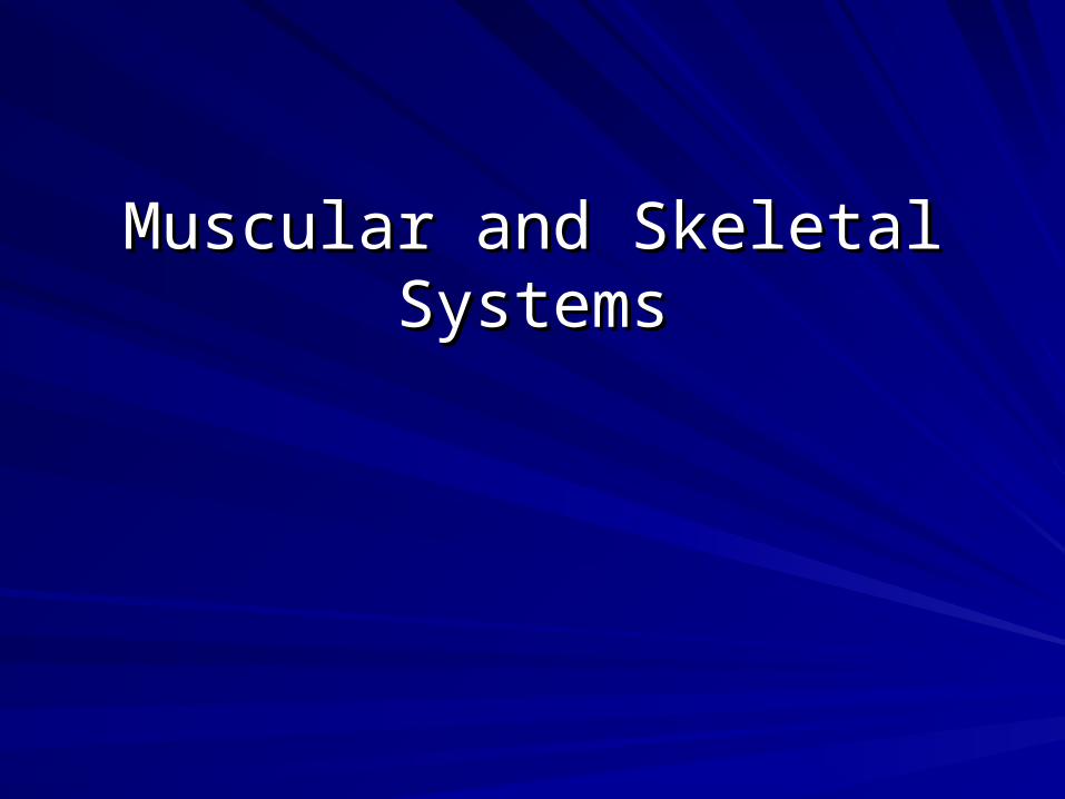
Muscular and Skeletal Systems. I. Muscular System A. Types B. Major Groups.
19
Muscular and Skeletal Muscular and Skeletal Systems Systems
-
Upload
belinda-doris-cannon -
Category
Documents
-
view
223 -
download
1
Transcript of Muscular and Skeletal Systems. I. Muscular System A. Types B. Major Groups.
Muscular and Skeletal SystemsMuscular and Skeletal Systems
I. Muscular SystemI. Muscular System
A.A. TypesTypes
B.B. Major Major GroupsGroups
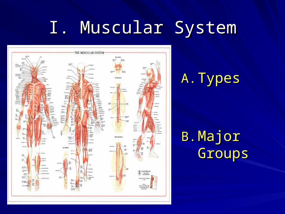
A. TypesA. Types
1)1) SkeletalSkeletal• Provide movement for the Provide movement for the
skeletonskeleton• Most are consciously controlled Most are consciously controlled
by the central nervous systemby the central nervous system• Contain striations Contain striations • Have many nucleiHave many nuclei• Function by nervous stimulationFunction by nervous stimulation
• Motor neurons connected to Motor neurons connected to musclemuscle
• Composition: Composition: many fibers many fibers stacked together in bundlesstacked together in bundles
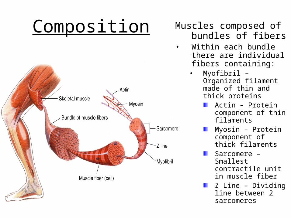
Composition Muscles composed of bundles of fibers
• Within each bundle there are individual fibers containing:
• Myofibril – Organized filament made of thin and thick proteins
Actin – Protein component of thin filamentsMyosin – Protein component of thick filamentsSarcomere – Smallest contractile unit in muscle fiberZ Line – Dividing line between 2 sarcomeres
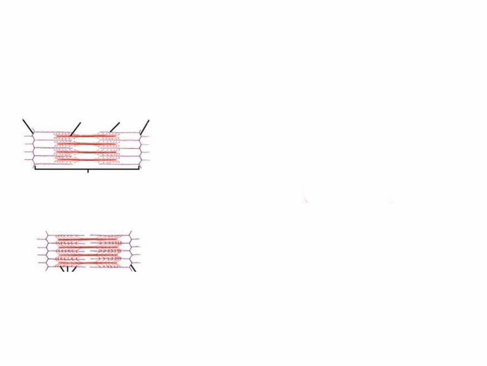
Relaxed Muscle
Contracted Muscle
Z line Myosin Actin Z line
Sarcomore
Cross-bridges Z line
Movement of Actin FilamentActin
Binding sites
Cross-bridge
Myosin
Figure 36-8 Muscle Contraction
Section 36-2
Relaxed Muscle
Contracted Muscle
Z line Myosin Actin Z line
Sarcomore
Cross-bridges Z line
Movement of Actin FilamentActin
Binding sites
Cross-bridge
Myosin
Figure 36-8 Muscle Contraction
Section 36-2
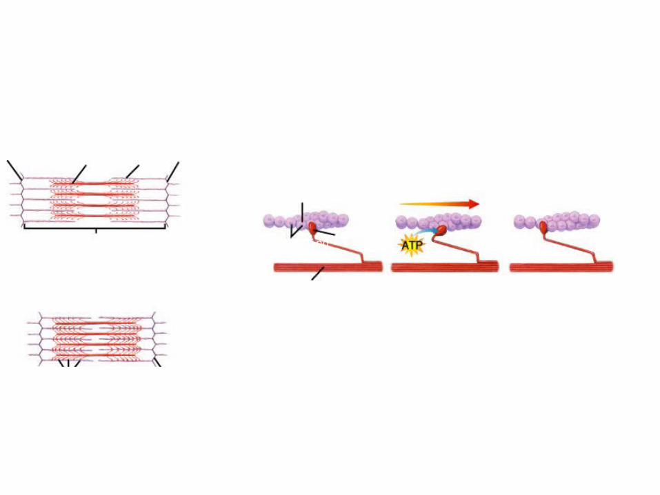
During muscle contraction, the knoblike head of a myosin filament attaches to a binding site on actin, forming a cross-bridge.
Relaxed Muscle
Contracted Muscle
Z line Myosin Actin Z line
Sarcomore
Cross-bridges Z line
Movement of Actin FilamentActin
Binding sites
Cross-bridge
Myosin
Figure 36-8 Muscle Contraction
Section 36-2
During muscle contraction, the knoblike head of a myosin filament attaches to a binding site on actin, forming a cross-bridge.
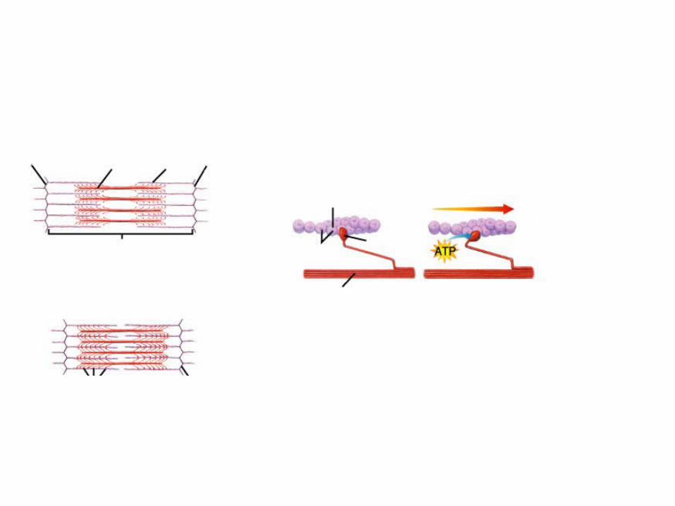
Powered by ATP, the myosin cross-bridge changes shape and pulls the actin filament toward the center of the sarcomere.
Relaxed Muscle
Contracted Muscle
Z line Myosin Actin Z line
Sarcomore
Cross-bridges Z line
Movement of Actin FilamentActin
Binding sites
Cross-bridge
Myosin
Figure 36-8 Muscle Contraction
Section 36-2
During muscle contraction, the knoblike head of a myosin filament attaches to a binding site on actin, forming a cross-bridge.
Powered by ATP, the myosin cross-bridge changes shape and pulls the actin filament toward the center of the sarcomere.
The cross-bridge is broken, the myosin binds to another site on the actin filament, and the cycle begins again.
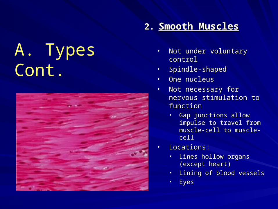
2.2. Smooth MusclesSmooth Muscles
• Not under voluntary controlNot under voluntary control• Spindle-shaped Spindle-shaped • One nucleusOne nucleus• Not necessary for nervous Not necessary for nervous
stimulation to functionstimulation to function• Gap junctions allow impulse Gap junctions allow impulse
to travel from muscle-cell to to travel from muscle-cell to muscle-cellmuscle-cell
• Locations:Locations:• Lines hollow organs (except Lines hollow organs (except
heart)heart)• Lining of blood vesselsLining of blood vessels• EyesEyes
A. Types Cont.
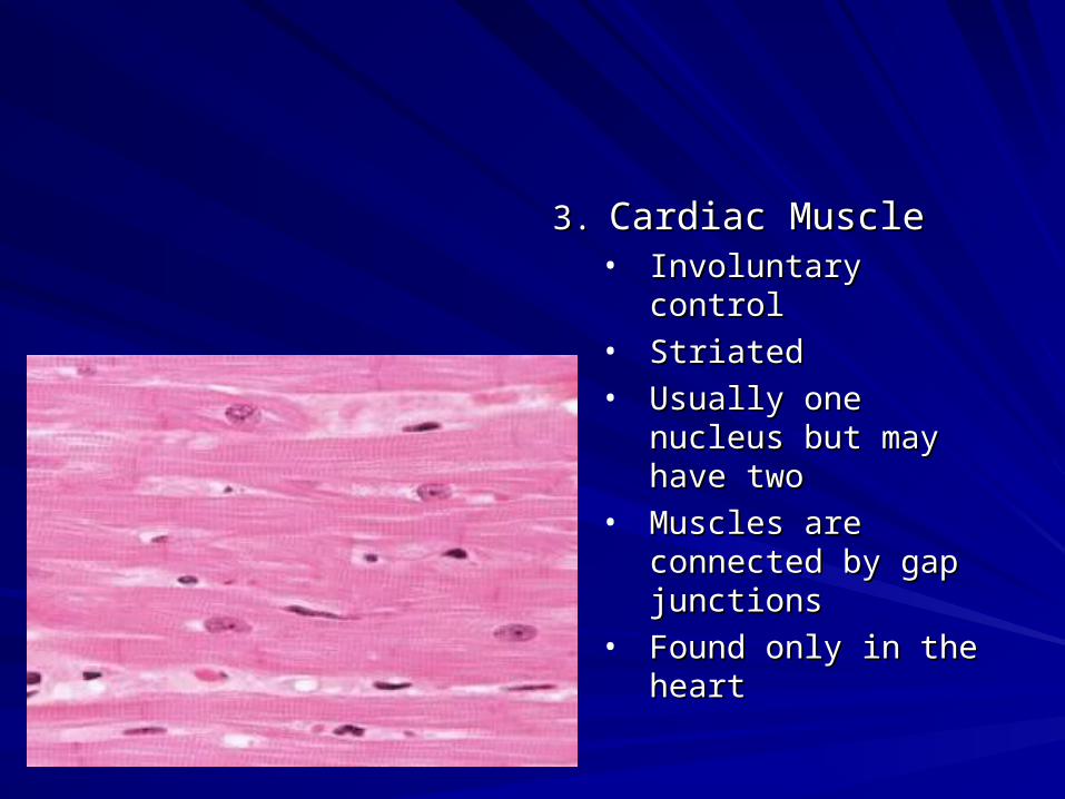
3.3. Cardiac MuscleCardiac Muscle• Involuntary controlInvoluntary control• StriatedStriated• Usually one nucleus Usually one nucleus
but may have twobut may have two• Muscles are Muscles are
connected by gap connected by gap junctionsjunctions
• Found only in the Found only in the heartheart
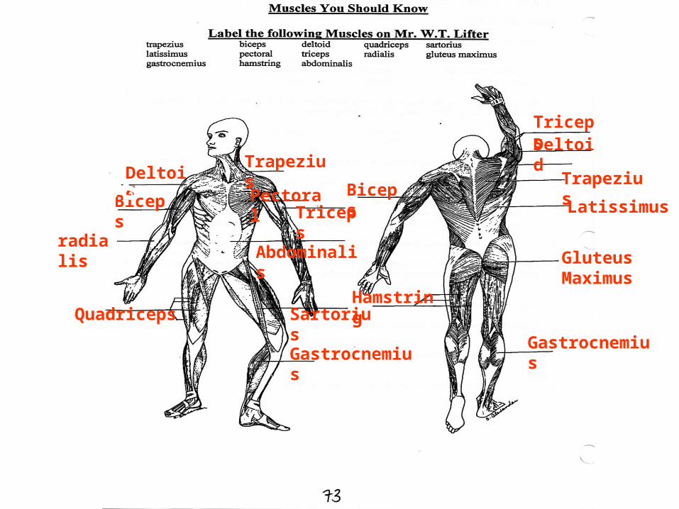
BicepsDeltoid
Trapezius
PectoralTriceps
Abdominalis
TricepsDeltoid
Quadriceps
Gluteus Maximus
Gastrocnemius
Sartorius
Gastrocnemius
radialis
BicepsTrapezius
Hamstring
Latissimus
B. Major Groups
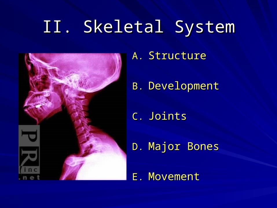
II. Skeletal SystemII. Skeletal System
A.A. StructureStructure
B.B. DevelopmentDevelopment
C.C. JointsJoints
D.D. Major BonesMajor Bones
E.E. MovementMovement

A. A. StructureStructure1.1. BonesBones
• Living cells and protein surrounded by calcium depositionLiving cells and protein surrounded by calcium deposition• Direct blood supplyDirect blood supply
• Periosteum – connective tissue surrounding bonePeriosteum – connective tissue surrounding bone• Bone Marrow Bone Marrow – cavity in bone containing soft tissue– cavity in bone containing soft tissue
• Red Marrow – Produces R.B.C.’s some W.B.C.’s and plateletsRed Marrow – Produces R.B.C.’s some W.B.C.’s and platelets• Yellow Marrow – Made of fat cellsYellow Marrow – Made of fat cells
• 2 Types2 Types• SpongySpongy
• Inner Layer of boneInner Layer of bone• Contains Red MarrowContains Red Marrow
• CompactCompact• Outer layerOuter layer
• Osteocytes – Mature Bone cellsOsteocytes – Mature Bone cells• Osteoclasts – Breakdown boneOsteoclasts – Breakdown bone• Osteoblasts – Create boneOsteoblasts – Create bone
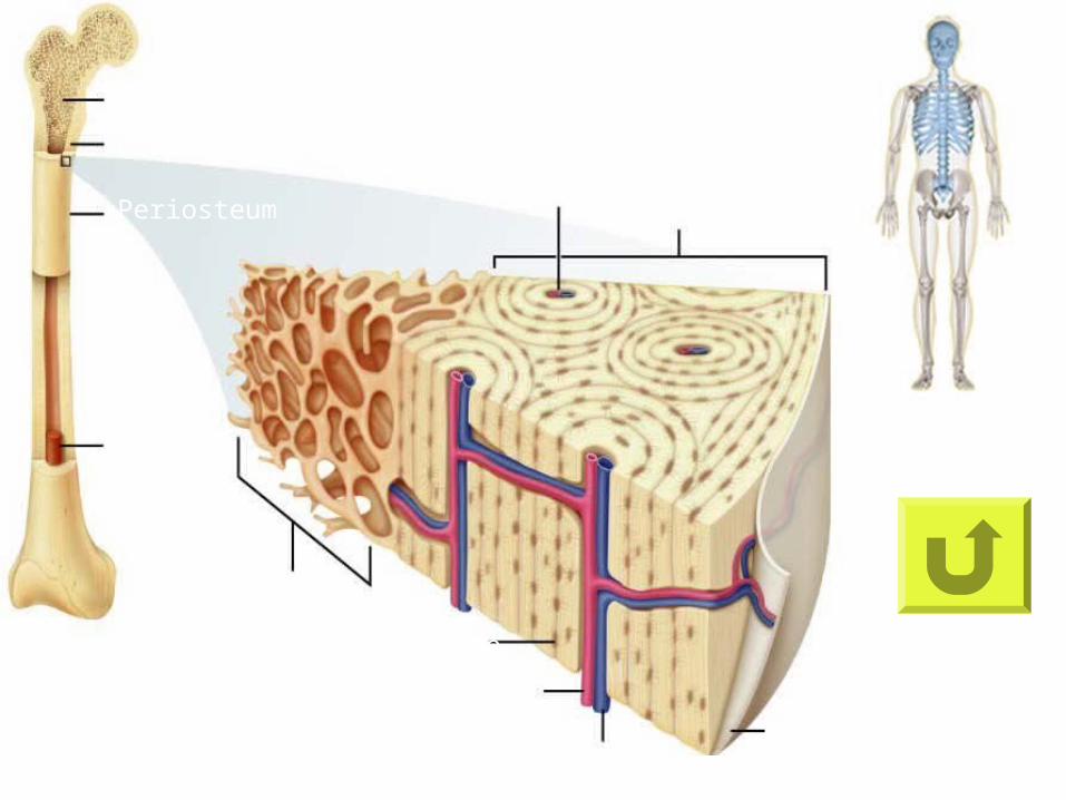
Spongy Bone
Compact Bone
Periosteum
Bone
Marrow
Spongy Bone
Compact Bone
HaversianCanal
Osteocyte
Artery
VeinPeriosteum
B. DevelopmentB. Development
1.1. As We GrowAs We Grow• Embryonic Skeleton - Embryonic Skeleton - Primarily composed of cartilage (elastic Primarily composed of cartilage (elastic
tissue that does not contain blood vessels)tissue that does not contain blood vessels)
• Cartilage is replace by bone through a process called Cartilage is replace by bone through a process called ossificationossification
• Occurs up to 7 months before birthOccurs up to 7 months before birth• The body does not completely ossify to facilitate child birthThe body does not completely ossify to facilitate child birth
• Osteoblasts secrete minerals deposits (forming bone)Osteoblasts secrete minerals deposits (forming bone)• As the osteoblasts become surrounded by bone tissue the mature As the osteoblasts become surrounded by bone tissue the mature
into ostocytesinto ostocytes
• Growth PlatesGrowth Plates• Sites each end of the bone that contains growing cartilageSites each end of the bone that contains growing cartilage• As the cartilage gets replaced by bone we stop growingAs the cartilage gets replaced by bone we stop growing
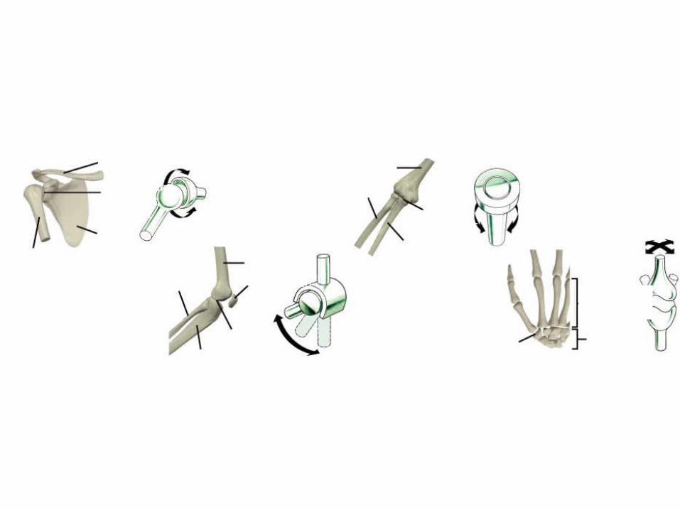
Ball-and-Socket Joint
Hinge Joint
Pivot JointClavicle
Ball-and-socket joint
Scapula
HumerusFemur
Patella
Hinge joint
Tibia
Fibula
Humerus
Radius
Pivot joint
Ulna
Metacarpals
CarpalsSaddle joint
Saddle Joint
C. Joints
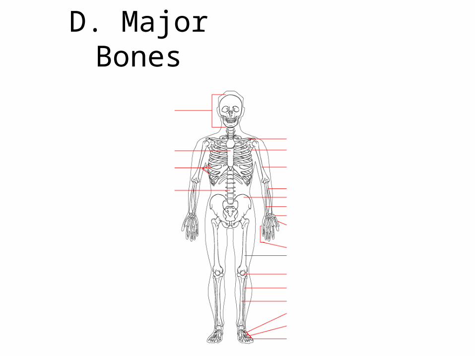
D. Major Bones
Skull
Sternum
Ribs
Vertebral column
Metatarsals
Metacarpals
Phalanges
Clavicle
Scapula
Humerus
RadiusPelvisUlnaCarpals
Femur
Patella
Fibula
TibiaTarsals
Phalanges
Appendicular Skeleton
E. MovementE. Movement
Movement occurs between the coordination Movement occurs between the coordination of the skeletal and muscular systemsof the skeletal and muscular systems
1.1. TendonsTendons• Bundles of connective tissue that link muscle to boneBundles of connective tissue that link muscle to bone• Damage is usually irreversibleDamage is usually irreversible
2.2. LigamentsLigaments• Bundles of connective tissue that link bone to boneBundles of connective tissue that link bone to bone• Common ailment is a torn ACLCommon ailment is a torn ACL