Muscle Tissue Functions of Muscle Tissue zMovement zHeat production zMaintenance of posture zMuscle...
41
Muscle Tissue
-
Upload
mary-creaser -
Category
Documents
-
view
217 -
download
0
Transcript of Muscle Tissue Functions of Muscle Tissue zMovement zHeat production zMaintenance of posture zMuscle...

Muscle Tissue

Functions of Muscle Tissue
MovementHeat productionMaintenance of postureMuscle contraction produces 85% of
body heat
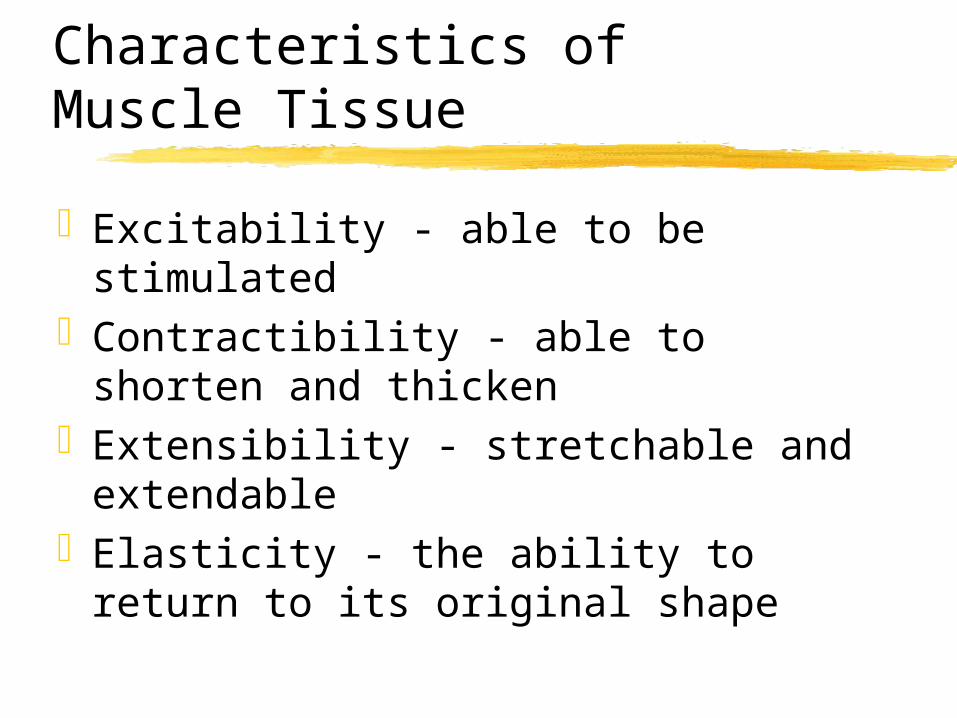
Characteristics of Muscle Tissue
Excitability - able to be stimulatedContractibility - able to shorten and
thickenExtensibility - stretchable and
extendableElasticity - the ability to return to its
original shape

Muscle Tissue Types
Skeletal - found attached to bone, striated, voluntary
Cardiac - forms the walls of heart, striated, involuntary
Smooth - found in viscera, non-striated, involuntary

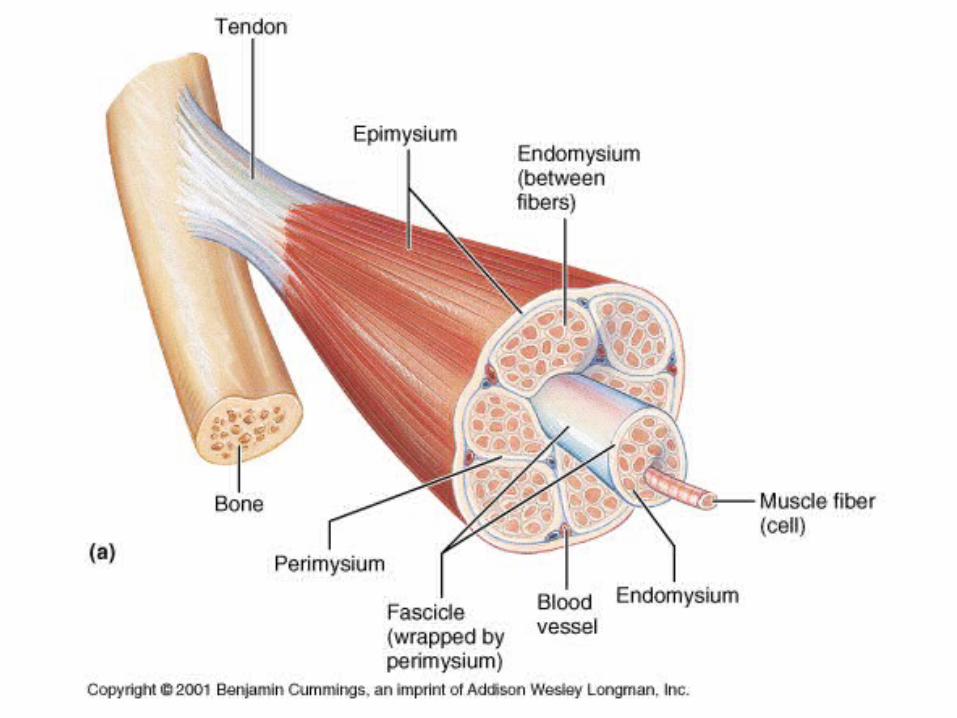
Gross Anatomy of Skeletal Muscle
Each muscle is an organ, containing muscle, blood vessels, nerves, & connective tissue
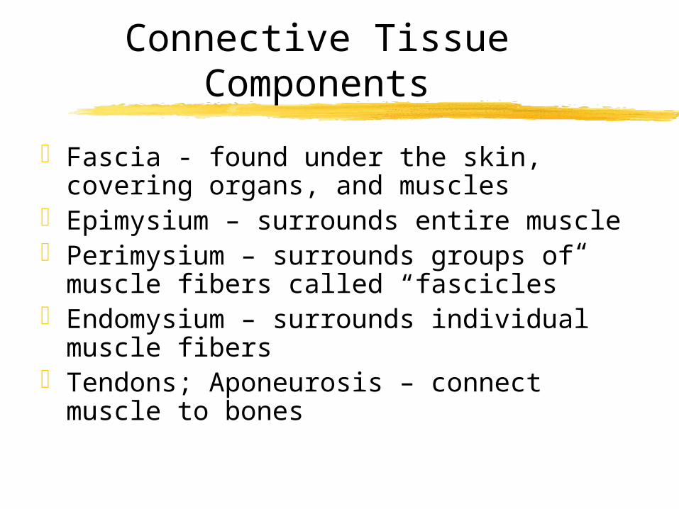
Connective Tissue Components
Fascia - found under the skin, covering organs, and muscles
Epimysium – surrounds entire musclePerimysium – surrounds groups of muscle
fibers called “fascicles”Endomysium – surrounds individual
muscle fibersTendons; Aponeurosis – connect muscle to
bones
Cell Structure
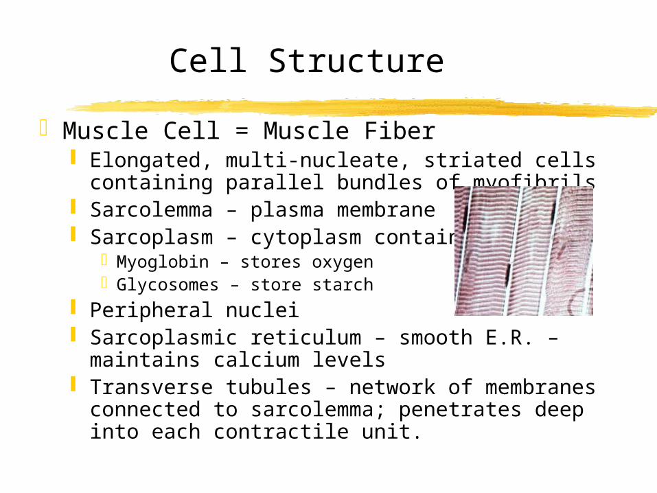
Muscle Cell = Muscle Fiber Elongated, multi-nucleate, striated cells
containing parallel bundles of myofibrils Sarcolemma – plasma membrane Sarcoplasm – cytoplasm containing:
Myoglobin – stores oxygenGlycosomes – store starch
Peripheral nuclei Sarcoplasmic reticulum – smooth E.R. –
maintains calcium levels Transverse tubules – network of membranes
connected to sarcolemma; penetrates deep into each contractile unit.

Specialized contractile organelles
Myofibrils - thread-like structures 100’s to 1000’s in each muscle fiber
(cell) Actin - thin filaments containing actin
protein, 2 strands of tropomyosin, & troponin
Myosin - thick protein filaments composed of myosin molecules

Myofibrils
Actin
Myosin

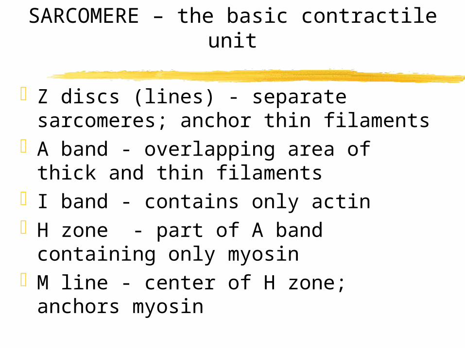
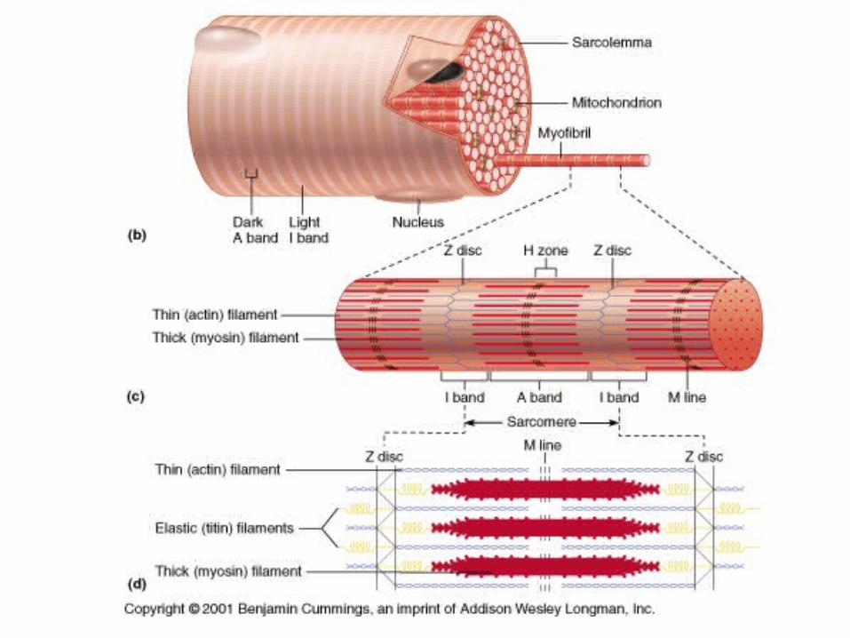
SARCOMERE – the basic contractile unit
Z discs (lines) - separate sarcomeres; anchor thin filaments
A band - overlapping area of thick and thin filaments
I band - contains only actinH zone - part of A band containing
only myosinM line - center of H zone; anchors
myosin


Sliding Filament Mechanism

Skeletal Muscle Contraction
Muscle contraction occurs when actin and myosin are allowed to interact with each other and form crossbridges
The binding sites on actin are blocked by the troponin/tropomyosin complex
Calcium ions in the sarcoplasm will bind to troponin
Muscle Contraction
This binding will cause the troponin / tropomyosin complex to pull away from the active binding site on actin, thus allowing myosin to bind
The myosin head pivots, pulling the thin filaments toward the center of the sarcomere thus shortening the sarcomere
Repeated cycles of attachment, pivoting, detach and release occurs
Muscle Contraction
Successive interaction causes “sliding” of the filament, shortening of the sarcomere, thus shortening of the entire muscle
Calcium is removed from the troponin molecule and returned to the S.R.
Relaxation occurs

What Role Does Calcium Play?
What triggers the release of calcium?

ACTION POTENTIALS
The sudden change in the transmembrane potential
Action Potentials
Resting Membrane PotentialPolarized - positive charge outside,
negative charge insideDepolarized - positive charge inside,
negative charge outside Repolarized - positive charge
reestablished outside, negative charge inside

Resting Potential/ Polarized
When muscle is relaxed, the sarcolemma is polarized having a charge difference between the inside /outside of the cell
When a stimulus is received opening a channel gate , Na+ ions will flow into the cell changing the polarity of the cell

Depolarized
The net charge of the sarcolemma becomes negative in regards to the inside of the cell which is now positive.
The cell is said to be depolarized and the muscle contracted
Repolarized
Membrane pumps quickly restore the original status or condition
The positive charge outside is reestablished once again and resting membrane potential is restored

Neuromuscular Junction
Each fiber is controlled by a motor neuron at a neuromuscular junction
Motor neurons stimulate muscle fibersAcetylcholine (ACh ) is released into the
synaptic cleft with the arrival of an action potential
ACh diffuses across the cleft, binding to receptors on the motor end plate, initiating a muscle action potential
Once initiated, the action potential is unstoppable and self-propagating

RELAXATION
Resting membrane potential is restored by: Acetylcholinesterase active transport pumps that pump Ca+2
ions back into the sarcoplasmic reticulum
Calsequestrin – binds calcium
Role of ATP
Used to activate the myosin head in order to bind to actin
After power stroke, ATP used to break the bond between actin and myosin
ATP used to pump calcium back into the SR
Production of ATP for muscles
Direct phosphorylation Creatine phosphate couples with ADP to form ATP Provides about 15 sec of energy
Glycolysis Glucose broken down anaerobically Produces lactic acid as waste product Provides about 30-60 sec of energy
Aerobic Respiration Glucose broken down with oxygen Hours of energy

Muscle Fatigue
Insufficient oxygenBuild-up of lactic acidDepletion of glycogen
RECOVERY OXYGEN CONSUMPTION OXYGEN DEBT

ALL - or -None Principle
Muscle fibers will contract fully OR not at all once they are stimulated
Threshold stimulus minimal level of stimulation needed to
cause the muscle to contract
Motor Units
Motor units - motor neuron and all the muscle fibers it controls
Number of muscle fibers in motor unit will vary
The fewer the number of fibers per motor unit, the more precise the contraction
The number of motor units being stimulated will determine the strength of contraction of the entire muscle

Muscle Contraction
Twitch contraction rapid, jerky contraction to a single stimuli phases: latent, contraction,
relaxation,refractoryWave summation
increase in the strength of muscle contraction due to rapid successive stimulation
Tetany continuous, smooth, sustained contraction

Muscle Contraction
Treppe repeated stimulation following stimulation
causing a staircase effectIsotonic
tone or tension remains constant-muscle shortens
Isometric tension increases - muscle length remains
same

Muscle Fiber TypesRed oxidative fibers
more myoglobin more capillaries more mitochondria long, slow
contraction
sustained energy aerobic respiration non-fatiguable fibers
White glycolytic fibers less myoglobin less capillaries fewer mitochondria rapid,powerful
contraction quick energy anaerobic respiration fatigue easily
Benefits of Exercise
Increase the size of size and strength of each fiber
Increase muscle toneIncreases the blood supply, thus
increasing the number of red blood cellsIncreased respiratory and
cardiovascular functionLowers blood pressure

Cardiac Muscle
InvoluntaryIntercalated discsForms syncytiumLong refractory periodLong contraction rateMore mitochondria than skeletal
muscle
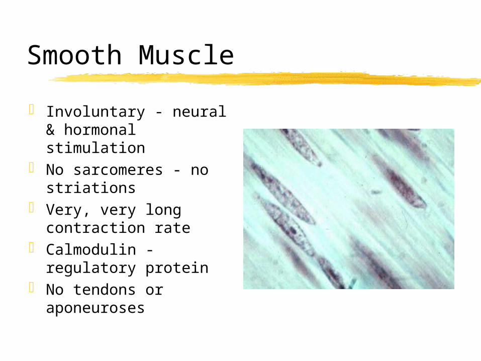
Smooth Muscle
Involuntary - neural & hormonal stimulation
No sarcomeres - no striations
Very, very long contraction rate
Calmodulin - regulatory protein
No tendons or aponeuroses

Muscle / Bone Interaction
Origin – attachment of a muscle to a stationary bone
Insertion – attachment of a muscle to a movable bone
Prime mover – provides major force for specific movement
Antagonist – opposes prime moverSynergist – assists the prime mover
(secondary muscle)

Muscle / Bone Interactions
Levers – rigid bar (bones) moving on fixed point
Fulcrum = fixed point (joints)Effort = applied forceResistance = load

Levers
First class Fulcrum in center = seesaw Lifting head off chest
Second class Load (resistance) in center = wheelbarrow Least common Standing on tiptoes
Third class Effort in center = tweezers Biceps brachii Most common