Muscle damage and inflammation following a three-day trail run
100
Muscle damage and inflammation following a three-day trail run Dr E C Denissen M.Tech Hom (DUT) SUPERVISOR: Prof. E. M. Peters-Futre Submitted to the Discipline of Physiology, School of Laboratory Science and Medical Sciences, College of Health Sciences, University of KwaZulu-Natal, in partial fulfillment of the requirements for the Degree of Master of Medical Sciences in Sports Medicine February 2012
Transcript of Muscle damage and inflammation following a three-day trail run
Muscle damage and inflammation following a three-day trail run
Dr E C Denissen
M.Tech Hom (DUT)
SUPERVISOR: Prof. E. M. Peters-Futre
Submitted to the Discipline of Physiology, School of Laboratory Science and Medical
Sciences, College of Health Sciences, University of KwaZulu-Natal, in partial fulfillment of
the requirements for the Degree of Master of Medical Sciences in Sports Medicine
February 2012
ii
Declaration
I, Emmerentia C. Denissen, student number 210553532, declare that the work on which this
project is based is original and my own work (except where acknowledgements indicate to the
contrary) and that neither the whole work nor part thereof has been, is presently or is to be
submitted for another degree at this or any other university.
I empower the University of KwaZulu-Natal to reproduce for the purpose of research either the
whole or any part of the content in any manner whatsoever.
___________________________
Durban
February 2012

iii
Acknowledgements
It is with sincere gratitude and appreciation that I would like to thank and acknowledge the
following people, without whom this research would not have been possible:
1. My supervisor, Professor Edith Peters-Futre, for her dedication, expert guidance and endless
patience during the planning and orchestration of this challenging study to completion.
2. Ms Heidi Mocke and the Wildlands Conservation Trust are thanked for permitting the
collection of these data at the 2011 Three Cranes Challenge multiday trail run and for providing
the research team with accommodation in the "race village" located in the Bushwillow Campsite.
3. Mr. Navin Singh, Dr. Anton de Waard, Prof Andrew McKune and Mr Ronnie Naicker are
thanked for their support with the field side collection of data reported in this report.
4. Ampath Laboratories for its assistance with phlebotomy and chemical pathology.
5. Mr. Asokaran Rajh for his help with the preparation of graphic material for the manuscripts
prepared for publication.
6. Ergonomics Technologies, Pretoria and the MRC Research Unit for Exercise Science and
Sports Medicine are thanked for the loan of Cor Temp data recorders which made the continuous
monitoring of HR and Tintest possible.
7. The SA Weather Office is thanked for provision of precise data regarding the environmental
conditions during the different stages of the race.
8. Everyone at UKZN who sent messages of support following my husband’s accident – without
your kind words and understanding, the completion of this course would not have been possible.
9. My husband, for his unwavering support, patience and encouragement throughout this project.
10. All the participants of this study without whom it would not have materialised.

iv
Thesis Abstract
Introduction
The physiological effects of single and multiday road running races have been studied extensively
and include the occurrence of rhabdomyolysis, reflected by significantly increased urinary
myoglobin (uMb), as well as increased concentrations of serum creatine phosphokinase (CPK),
high sensitivity C-reactive protein (hsCRP), cortisol and cardiac troponin-T (cTnT), dehydration
and compromised renal function. Furthermore, in hyperthermic athletes, a positive relationship
has been noted between hyperthermia, muscle damage, dehydration and pacing. The
physiological effects of a multiday trail run of similar duration to single day road races, however,
are unknown.
The side-effects of the use of statin medication for hypercholesterolaemia include muscle fatigue,
cramping and increased muscle damage. These have been found to be aggravated in endurance
athletes and it has been reported that females, especially when being medicated from a young age,
are more susceptible to these side-effects.
Objectives
1. To investigate the effect of a three-day trail run on systemic and urinary markers of muscle
damage and inflammation in recreational runners and to establish the association of dehydration
and hyperthermia with these markers.
2. To observe the effect of the three day trail run on systemic and urinary markers of muscle
damage and inflammation on an additional hypercholesterolaemic female athlete using statin
medication in combination with a lipid uptake inhibitor.
Method
Firstly, an observational cohort study was conducted on 19 recreational male (n=6) and female
(n=13) athletes during a 95km trail run over three days.
Pre-and post-stage and 24 and 72 h post-race concentrations of serum CPK, hsCRP, cortisol,
cTnT, and osmolality (sOsm) as well as uMb, changes in body mass, delayed onset muscle

v
soreness (DOMS) and thigh circumference (TC) were measured. Continuous recordings of heart
rate (HR) and intestinal temperature (Tintest ) were made throughout each stage.
In addition, a case report is included on one trained female endurance athlete currently being
treated for familial hypercholesterolaemia with 20 mg Aspavor and 10 mg Ezetrol daily and not
included in the above cohort, to investigate the degree of muscle damage and inflammation she
experienced as a result of participation in the three-day event.
Results:
Heart rate ranged between 77 and 83% age-predicted-maximum (APmax) and Tintest between
36.1 and 40.2 ºC during the three stages. Significant rises in mean serum CPK, hsCRP, sOsm and
blood neutrophil count reached peak concentrations of 1 488U/L, 8.91mg/l, 298mosm/L and
10.21 10^9/L (p≤0.001), respectively. No evidence of elevations in uMb and cTnT were detected.
The stage-induced increments in DOMS correlated positively with CPK, r=0.71; 95% CI [0.62,
0.78]. TC decreased significantly post S1post and S2post (p≤0.05) and a maximum mean body mass
loss of 3.09% (±1.04%) occurred during S2. There was no significant difference between non-
steroidal anti-inflammatory drug (NSAID) users and non-users in terms of serum CPK, hsCRP,
cortisol, post race DOMS scores, running times, TC or sOsm (p>0.05). The post-pre change in
sOsm during each stage correlated inversely with the changes in % body mass, r = -0.36, 95% CI
[-0.57,-0.094] and the pooled data examining the relationship between the change of sOsm and
change in serum CPK for the three stages (n=57), revealed an insignificant positive correlation
(r= 0.034, 95% CI [-0.228, 0.291].
The maximum Tintest ranged between 38.3 º C and 40.2 º C and only exceeded 40º C in two of the
12 athletes monitored. The relationship between the change in Tintest and serum CPK was
insignificant (p>0.05) for the 11 individuals from whom complete sets of data were available (r=
0.24, 95% CI [-0.42, 0.734].
In the hypercholesterolaemic athlete, the maximum serum CPK (665U/L), hsCRP (1.9mg/Ll) and
cortisol (845nmol/L) concentrations corresponded with undetected uMb despite a maximum body
mass loss of 4.5%

vi
Conclusion:
Three consecutive days of 95km trail running resulted in low markers of muscle damage and
inflammation, when compared to results obtained in previous single day road races of similar
duration despite the maintenance of a heart rate above 77% APmax, Tintest rising above 39o C and
mean body mass decrement of >2.0%. The unchanged concentrations of serum cTnT and uMb
confirmed the low values of the markers of muscle damage and inflammation. An insignificant
positive correlation between muscle damage and dehydration was noted.
Furthermore the daily use of 0.4 mg/kg Atorvastatin in combination with 10mg Ezetrol did not
result in the subject experiencing subjective myalgia, cramps, fatigue or increased markers of
muscle damage following her participation in the trail run.

vii
Table of Contents
Page
DECLARATION ii
ACKNOWLEDGEMENTS iii
THESIS ABSTRACT iv
TABLE OF CONTENTS vii
LIST OF ABBREVIATIONS x
CHAPTER ONE: INTRODUCTION
1.1 INTRODUCTION 1
1.2 AIMS AND OBJECTIVES OF STUDY 3
1.3 HYPOTHESES 3
1.4 SCOPE OF THE STUDY 4
CHAPER TWO: REVIEW OF RELATED LITERATURE
2.1 MECHANICAL DAMAGE 6
2.2 PRESENTATION 7
2.3 HISTOLOGICAL APPEARANCE OF DAMAGED MUSCLE CELLS 9
2.4 SYSTEMIC MARKERS OF CELL DAMAGE IN SKELETAL AND 10
CARDIAC MUSCLE
2.4.1 Creatine Phosphokinase 10
2.4.2 Alpha-Actin 11
2.4.3 Lactate Dehydrogenase 11
2.4.4 Plasma Fatty Acid Binding Protein 11
2.4.5 Troponin 12
2.5 INFLAMMATORY RESPONSE 12
2.5.1 Neutrophils, macrophages and cytokines 12
2.5.2 C-Reactive Protein 13
2.5.3 Cortisol 13
2.6 FACTORS AFFECTING MUSCLE DAMAGE AND INFLAMMATION 14
2.6.1 Repeated Bouts 14
2.6.2 Dehydration, hyperthermia and muscle damage 15

viii
2.6.3 Anti-inflammatory drugs 16
2.6.4 Lipid lowering medication 17
2.6.5 Genetics 19
2.6.6 Chromosome telomere erosion 20
2.6.7 Oxidative stress 20
2.6.8 Age 21
2.6.9 Gender 21
2.6.10 Other 22
2.7 CONCLUSION 23
CHAPTER THREE: MANUSCRIPT 1 24
LOW MARKERS OF MUSCLE DAMAGE AND INFLAMMATION FOLLOWING
A THREE-DAY TRAIL RUN
Abstract 25
Introduction 26
Method 27
Results 32
Discussion 37
Conclusion 40
References 41
Acknowledgements 43
CHAPTER FOUR: MANUSCRIPT 2 45
USE OF THE STATIN, ASPAVOR, IN COMBINATION WITH EZETROL, IN A
HYPERCHOLESTEROLAEMIC ENDURANCE ATHLETE COMPLETING A
95KM MULTI-DAY TRAIL RUN
Abstract 46
Introduction 47
Case Description 47
Results 48
Discussion 51
Acknowledgements 54
References 55

ix
CHAPTER FIVE: CONCLUSION AND RECOMMENDATIONS 57
5.1 CONCLUSION 57
5.2 RECOMMENDATIONS 58
REFERENCES 60
APPENDICES 68
APPENDIX A: LETTER OF ACCEPTANCE OF PUBLICATION 69
APPENDIX B: EXAMPLE OF FEEDBACK GIVEN TO EACH SUBJECT 70
APPENDIX C: SUBJECT INFORMATION SHEET 79
APPENDIX D: INFORMED CONSENT FORM 82
APPENDIX E: PRE RACE QUESTIONNAIRE 84
APPENDIX F: POST RACE DOMS QUESTIONNAIRE 88
x
List of Abbreviations
ALT Alanine aminotransferase
APmax Age predicted maximum
AST Aspartate aminotransferase
BMI Body mass index
BP Blood pressure
Bpm
Ca2+
Beats per minute
Calcium
cAMP Cyclic Adenosine Monophosphate
CPK Creatine Phosphokinase
cTnT
DNA
Cardiac troponin T
Deoxyribonucleic acid
DOMS Delayed onset muscle soreness
FABP Plasma fatty acid binding protein
FDA United States Food And Drug Administration
HB Haemoglobin
HCT Haematocrit
HMG-CoA 3-hydroxy-3-methylglutaryl co-enzyme A
HR Heart rate
hsCRP High sensitivity C-Reactive Protein
HSP Heat shock protein
IL-1ra Interleukin-1 receptor antagonist
IL-6 Interleukin-6
IL-10 Interleukin-10
LDL Low density lipoprotein
LDH Lactate dehydrogenase
NSAID Non-steroidal anti-inflammatory drugs
RBC Red blood cell
ROS Reactive oxygen species
S1 Stage 1
S2 Stage 2

xi
S3 Stage 3
sOsm Serum osmolality
TC Thigh circumference
Tintest Intestinal temperature
uMb Urinary myoglobin
UOsm Urinary osmolality
USG Urinary specific gravity
24PR 24 hours post race
72PR 72 hours post race

1
CHAPTER 1
INTRODUCTION 1.1 BACKGROUND TO THE STUDY
Multiday ultra distance events are becoming increasingly popular among professional and
amateur athletes and may encompass running, cycling and/or other disciplines. Various studies
have been published on the physiological changes that occur during single and multiday
endurance road running and cycling events (Knechtle et al., 2008; Skenderi et al., 2006;
Nieman et al., 2005), but few addressed the changes that occur during multiday endurance trail
running races, which have been found to be different to single day ultra distance or marathon
foot races (Millet et al., 2011).
Running as an activity involves eccentric contraction of a large muscle mass including the hip
and knee extensor muscles and muscles of the anterior and posterior tibial compartments
during every stride (Eston et al., 1995). This eccentric component is accentuated whilst
running downhill when the muscle is forced to lengthen while contracting (Proske and Morgan,
2001; Eston et al., 1995).
Clarkson and Hubal (2002) attribute the muscle damage to an increased contractile load per
unit, which occurs as fewer motor units are recruited. This damage results in changes in the
excitation-contraction (E-C) coupling, inflammation, swelling, decreased mobility and delayed
onset muscle soreness (DOMS), which would increase in relation to the duration of the event
(Clarkson and Hubal, 2002). After eccentric exercise there is a presence of sarcoplasmic
enzymes and proteins, including creatine phosphokinase (CPK) in the serum and myoglobin in
the urine, when there is leakage of these enzymes and proteins through the damaged
membranes of muscle cells into the circulation (Martinez-Amat et al., 2005; Clarkson and
Hubal, 2002). These findings are, however, not always reflective of structural muscle damage,
as demonstrated by histological studies (Clarkson and Hubal, 2002; Lieber and Friden, 2002)
due to the specificity of serum CPK that might originate from cardiac muscle damage or might
2
be reduced by other kinases, such as mitochondrial CPK and CPK-immunoglobulin complexes
(Martinez-Amat et al., 2005). In order to differentiate between skeletal and cardiac muscle
damage, the concentration of cardiac troponin-T (cTnT) in the serum has been used as a
reliable marker of cardiac, but not skeletal muscle damage (Martinez-Amat et al., 2005).
Increased levels of markers of skeletal muscle damage in the urine including the presence of
urinary myoglobin (uMb), have been shown to reflect asymptomatic exertional rhabdomyolysis
that has been found to occur during prolonged endurance exercise (Knechtle et al., 2008;
Skenderi et al., 2006; Clarkson and Hubal, 2002) and possibly be related to inflammation,
DOMS and skeletal and cardiac muscle damage (Skenderi et al., 2006; Clarkson and Hubal,
2002). Knechtle et al.(2008) suggest that runners in a 1 200km multiday road running event
experienced decreased skeletal muscle mass and increased total body water due to
rhabdomyolysis and impaired renal function.
The effect of muscle damage on inflammatory response is evident in the circulation as
concentrations of neutrophils and the hepatic acute - phase protein, C-reactive protein (CRP)
are raised (Robson-Ansley et al., 2009, Nieman et al., 2005). Muscle damage during
prolonged exercise is associated with an acute phase response, reflected by changes in pro-
inflammatory cytokine concentrations which, are in turn, correlated significantly with serum
CRP (Robson-Ansley et al., 2009, Nieman et al., 2005).
The use of non-steroidal anti-inflammatory drugs (NSAID) has been found to affect the
inflammatory response, but not skeletal muscle damage during exercise (Nieman et al., 2005).
It would therefore be interesting to compare the results of subgroups of athletes who used
NSAID to non-users to investigate the relationship between the use of NSAID and markers of
muscle damage and inflammation.
Extensive research has also been conducted on the effects of exercise-induced dehydration and
the physiological effects thereof (Casa et al., 2010; Stearns et al., 2009; Cleary et al., 2006;
Maresh et al., 2004; Cheuvront and Haymes, 2001). Recent studies have found that the
concomitant occurrence of dehydration, hyperthermia, increased metabolic rate and the

3
environment augments muscle damage (Cleary et al., 2005), but it is not known to what extent
this would be affected by three consecutive days of trail running.
The use of 3-hydroxy-3-methylglutaryl co-enzyme A (HMG-CoA) reductase inhibitors or
statins, the primary lipid-lowering medication currently prescribed for hypercholesterolaemia
has been associated with muscle cramping, myalgia, fatigue and in rare cases, rhabdomyoloysis
and death (Di Stasi et al., 2010; Thompson et al., 2010; Seachrist et al., 2005, Pasternak et
al., 2002; Evans and Rees, 2002). It is, however, not known how the simultaneous intake of
ezetemibe, a lipid uptake inhibitor, affects the augmented muscle damage, which has been
found to occur when hypercholesterolaemic athletes on statin medication compete in endurance
running events. A case study of an athlete who uses these two medications concurrently, was
therefore included to investigate her reaction to multiday ultra-distance racing.
1.2 AIMS AND OBJECTIVES OF THE STUDY
This observational cohort study firstly set out to determine the effect of a multiday trail running
race over 95km on systemic markers of muscle damage and inflammation using blood
neutrophil and serum CPK, hsCRP and cortisol concentrations, thigh circumference (TC),
DOMS and uMb as markers of muscle damage and inflammation.
Secondly, the effect of increased intestinal temperature (Tintest) and of dehydration, measured
by serum osmolality (sOsm) and body mass changes, was also monitored to determine the
relationship between Tintest, dehydration and muscle damage and inflammation.
Thirdly, the effect of statin medication, at a dosage of 0.4mg/kg, combined with a lipid uptake
inhibitor, on the systemic and urinary markers of muscle damage was investigated in a case
study.

4
1.3 HYPOTHESES
During trail running the environmental demands, challenging terrain underfoot and steep
ascends and descents are added stressors to contend with while running at least three hours per
day (Millet et al., 2011). It is hypothesized that, due to these added stressors, the eccentric
component of skeletal muscle contraction during trail running will be exaggerated causing
increases in systemic and urinary markers of muscle damage and inflammation, DOMS and
TC, which will be augmented by dehydration and hyperthermia and reflected in a change in
anthropometric values (Casa et al., 2010; Cleary et al., 2005, Sawka et al., 1998).
It is furthermore hypothesized that the hypercholesterolaemic athlete included in the case study
will experience muscle cramps and fatigue, which will be reflected in elevated systemic and
urinary markers of muscle damage and augmented by dehydration.
1.4 SCOPE OF THE STUDY
This observational cohort field study was conducted during and after a three day trail run over
95km. It was restricted to a convenience sample of twenty-one apparently healthy subjects of
which nineteen (6 males, 13 females) completed all three stages of the race and 15 runners (4
males, 11 females) completed all within and post- race assessments.
Pre-race assessments included the recording of body mass and height, TC and four-site
skinfold for the determination of % body fat. Self-reported data regarding the medical and
training status, hydration and food intake and use of NSAIDS and chemical stimulants was
obtained from questionnaires.
During the race, urine and blood sampling were done before and after every stage and at 24
and 72PR (post-race) and uMb, sOsm, cortisol, CPK, hsCRP, cTnT and blood neutrophil
concentrations were determined. These results were correlated with sOsm and body mass
changes as markers of dehydration, and Tintest, TC reflecting inflammation, were also measured
and subjects were requested to complete a DOMS questionnaire before and after each stage of

5
the race as well as for the five days following the race to record subjective perceptions of post-
race muscle soreness.
6
CHAPTER TWO
REVIEW OF THE RELATED LITERATURE
EXERCISE INDUCED MUSCLE DAMAGE
2.1 MECHANICAL DAMAGE
Acute bouts of prolonged running, involving eccentric contractions, generate high levels of
strain on the muscle because fewer motor units are recruited, putting more strain on those
motor units (Friden and Lieber, 2001). Clarkson and Hubal (2002) propose that this results in a
higher load per unit which eventually may lead to injury. Lieber and Friden (2002) suggest
that the initial damage to muscle is mechanical in nature, based on sarcomere strain. The
subsequent damage occurs due to inflammation and changes in the E-C coupling in the muscle
(Clarkson and Hubal, 2002).
With regard to mechanical damage, Proske and Morgan (2001) suggest that sarcomere
disruption occurs at the sites of the weakest half-sarcomeres, which lengthen uncontrollably
until the actin/myosin cross bridges are forcibly detached (Morgan, 1990) when the actin and
myosin filaments slide past each other, according to the Huxley model (Huxley, 1974). With
repetition of the eccentric contraction, more of the next-weakest sarcomeres are strained and
lengthened (Proske and Morgan, 2001). These stretched sarcomeres are distributed randomly
along the muscle fibre. When the muscle relaxes, the overstretched sarcomeres may re-
integrate with the undamaged fibres and resume function (Lieber and Friden, 2002). However,
with repeated eccentric contraction during exercise, weaker sarcomeres lengthen and become
disrupted. This leads to an extension in the optimum length of the muscle and at greater
muscle lengths, membrane damage and tearing of the T-tubules occur, leading to inactivation
of some sarcomeres (Proske and Morgan, 2001). At this stage, before the necrosis of fibres, a
fall in muscle tension would be reversible by the administration of caffeine which, in animal
models, causes the release of calcium (Ca2+) from the sarcoplasmic reticulum and subsequent
7
contraction of the muscle (Proske and Morgan, 2001). The continued eccentric contraction
leads to an increased number of disrupted sarcomeres, an increased local contraction and in
return the passive tension in the muscle increases (Clarkson and Hubal, 2002). With extensive
damage and extensive local contraction, parts of, or the whole fibre dies and an irreversible
secondary delayed fall in tension occurs about 24 hours after exercise which is not reversible
with administration of caffeine due to the extensive damage that has occurred (Proske and
Morgan, 2001). Thereafter breakdown products of tissue necrosis cause a local inflammatory
reaction, leading to oedema and soreness (Proske and Morgan, 2001).
According to Huxley (1957), in his sliding filament theory of muscle contraction, the myosin
filament head attaches to a single actin binding site, flexes and contraction of the fibre occurs
in millions of sarcomeres in a single muscle. The Japanese scientist, Toshio Yanagida,
proposed that the actin and myosin are involved in a series of loose couplings (Yanagida,
2007) which explains eccentric contraction, but does however not explain the damage that
takes place when the actin-myosin bond is forcibly detached, according to Huxley’s model.
2.2 PRESENTATION
Generally, there is a loss of strength of the affected muscle and a drop in active tension due to
the mechanical damage and tissue necrosis (McHugh, 2003; Proske and Morgan, 2001),
swelling, soreness and an increase in passive tension (or stiffness) leading to a reduced range
of motion. Proske and Morgan (2001) postulate that stiffness occurs due to the local
contraction and increased release of Ca2+ following the membrane damage. Low frequency
fatigue, the ability to generate force at a lower frequency, lasting up to a week post-exercise,
has also been described (Clarkson and Hubal, 2002).
DOMS, which has been reported to be unique to eccentric exercise, sets in several hours after
eccentric exercise and peaks 24 – 72 hours later (Lambert and Dennis, 1994). Swelling and
soreness appearing within 6 hours after exercise are ascribed to the local inflammatory
response (Proske and Morgan, 2001) and to noxious chemicals such as histamines, bradykinins
and prostaglandins that are released (Clarkson and Hubal, 2002). These chemicals sensitize the
8
Group IV afferent sensory nociceptors and the muscle becomes tender to touch, stretch and
contraction. An accumulation of fluid in the muscle fibre causes increased pressure (Clarkson
and Hubal, 2002) with swelling that may last up to 10 days post exercise.
Some research questions whether inflammation is a cause or consequence of muscle damage.
When comparing the time course of changes in the inflammatory response and the
development of DOMS, Malm et al. (2004) found no correlation between these variables and
concluded that DOMS is a result of muscle adaptation or of the activation of leukocytes that
are present in the epimysium before exercise (Malm et al., 2004). Other studies, using radio-
labeling of neutrophils, found that neutrophil infiltration into damaged muscle increased up to
24 hours post-exercise, coinciding with DOMS and a secondary decrease in eccentric torque
(Raastad et al., 2003).
The elevated calcium levels also trigger the release of endogenous proteases such as calpain
(Feasson et al., 2002; Friden and Lieber, 2001), causing further muscle damage. Calpain does
not affect actin and myosin, but has an affinity for desmin and alpha-actinin (Feasson et al.,
2002; Friden and Lieber, 2001) contributing to the damage at the intermediate filament
sections.
The loss of the structural protein desmin, which links adjacent Z-disks (Bennett et al., 2005),
has been noted in fibres that show signs of damage (Friden and Lieber, 2001). Desmin also
helps to maintain proper alignment of sarcomeres within and between myofibrils (Boriek et al.,
2001) Immunostaining of animal tissue has shown that muscle fibres lose “staining” for
desmin within minutes after the initiation of eccentric contractions. In addition, the number of
fibres that lack desmin increase with time following an eccentric exercise bout and those fibres
that lose desmin staining demonstrate accumulation of plasma fibronectin, indicating a loss of
membrane integrity in these fibres (Boriek et al., 2001).
Dystrophin is a large cytoskeletal protein associated within the muscle sarcolemma and is
thought to help maintain the integrity of the membrane during repeated mechanical loading that
muscle cells experience through everyday contractions (Blake et al., 2002; Hawke and Garry,
9
2001). Within six hours following eccentric exercise, dystrophin staining has been reported to
be completely missing in some fibres, with accompanying loss of desmin and another
structural protein, alpha-actinin (Hawke and Garry, 2001). To compound the loss of membrane
stability, other members of the complex of proteins associated with dystrophin, specifically
beta-spectrin and alpha-sarcoglycan, show signs of early damage following eccentric
contractions (Blake et al., 2002). The number of affected fibres has been shown to increase for
up to 2 days after exercise, indicating a progression of damage over time. It has been suggested
that this rapid loss of membrane-stabilizing function may render the muscle fibres fragile and
more susceptible to damage by further contractions (Hawke and Garry, 2001).
The loss of titin, a component in the A-band that plays an important part in the storage and use
of elastic energy (Donovan, 2004) and links the myosin filament to the Z-disk, is a further
possibility due to the overstretching of one half-sarcomere (Proske and Morgan, 2001) and the
contraction of the other half.
2.3 HISTOLOGICAL APPEARANCE OF DAMAGED MUSCLE CELLS
The morphological findings indicate variable muscle fibre size (Grobler et al., 2004; Friden
and Lieber, 2001), hyper contraction of the fibres, loss of the A-band, distortion of the A-band
and I-band alignment (Friden and Lieber, 2001), Z-disk streaming in mild damage and Z-disk
smearing in severe cases, with dispersion of Z-disk materials into the adjacent sarcomeres
(Clarkson and Hubal, 2002; Friden and Lieber, 2001). In the damaged areas focal loss of the
Z-disk may occur as well as loss of lateral registration of myofilaments (Martinez-Amat et al.,
2005). Enlarged mitochondria, sub-sarcolemmal mitochondria (Grobler et al., 2004) and lipid
and glycogen accumulations are also present in the damaged areas.
According to Lieber and Friden (2002) and Friden and Lieber, (2001), mainly type II fibres are
affected because of their short fibre length and higher tension developed and their lack of
oxidative capacity. Proske and Morgan (2001), however, found that both fibre-types
experience damage after eccentric exercise confirmed by a shift in the length-tension relation
of both types, although the shift in Type 1 fibres was less. The difference in the shift in
10
length-tension relation of the two fibre types was not statistically significant (Proske and
Morgan, 2001).
Activated satellite cells aid in muscle regeneration by proliferating and fusing to each other or
existing damaged myofibres to form new fibres (Hawke and Garry, 2001), remodeling the
damaged areas. Factors affecting satellite cell activity include muscle fibre type, age and the
secretion of multiple growth factors, a timeous immune response, neurotransmitters and
neurotrophic factors (Hawke and Garry, 2001). Without macrophage activity and their
subsequent cytokine factor secretion, no muscle regeneration occurs (Hawke and Garry, 2001)
2.4 SYSTEMIC MARKERS OF CELL DAMAGE IN SKELETAL AND CARDIAC
MUSCLE
Haematological findings after eccentric exercise include increased concentrations of the
following muscle proteins in serum: CPK, alpha-actin, myoglobin and troponin (Martinez-
Amat et al., 2005), CRP, lactate dehydrogenase (Lambert and Dennis, 1994) aspartate
aminotransferase and carbonic anhydrase iso-enzyme II (Clarkson and Hubal, 2002). The use
of muscle proteins as indicators of muscle damage, however, reflect the difference between
what is released from the tissue and cleared from the blood and can have large inter-subject
variability in the response (Clarkson and Hubal, 2002).
2.4.1 Creatine Phosphokinase (CPK): Most researchers determine the extent of damage by
using serum CPK concentration because of the low cost of the assay and because the increase
in CPK concentration is high in comparison to that of other proteins (Clarkson and Hubal,
2002). It also is used clinically to diagnose myositis and rhabdomyolysis and to predict renal
failure (Clarkson et al., 2006). Exertional rhabdomyolysis may occur after strenuous exercise
when serum CPK concentration is >500U/L and urine dipstick is positive for Mb and blood
without haematuria (Knechtle et al., 2008; Skenderi et al., 2006; Clarkson et al., 2006). Renal
failure has been associated with serum CPK concentrations > 20 000U/L, although Clarkson et
al. (2006) found that marked CPK and Mb elevations alone, are not sufficient to result in renal
failure in healthy athletes in response to exercise. Other factors including underlying disease,
11
environmental heat stress, sickle cell trait or drug use also contribute to this condition
(Clarkson et al., 2006).
CPK often shows a large standard deviation and it should be taken into consideration that it is
affected by other kinases such mitochondrial CPK, CPK-immunoglobulin complexes and CPK
derived from cardiac muscle (Martinez-Amat et al., 2005). According to Knechtle and Kohler
(2007), a significant decrease of skeletal muscle mass has been noted during a running race
over 338km within five days, although the biggest change in muscle mass occurred after the
first stage.
2.4.2 Alpha(α)-actin: A major constituent of the contractile apparatus in skeletal muscle is α-
actin which, when it leaks into the serum, has been found to be a reliable marker of muscle
damage and to have a high diagnostic specificity especially within the first few hours after
exercise (Martinez-Amat et al., 2005). The use of α-actin as a marker of skeletal muscle
damage has a high sensitivity (63-100%), represent more than 20% of all muscle cell proteins,
is detected within 1 hour after the onset of muscle damage and can be detected in the serum for
up to 72 hours after its release, indicating greater stability over time (Martinez-Amat et al.,
2005).
2.4.3 Lactate dehydrogenase (LDH): LDH is a commonly used indicator of exercise induced
muscle damage. It reaches peak concentrations within six hours post exercise and returns to
pre-exercise levels within 48 hours after exercise (Maughan et al., 1989). However, elevations
of serum LDH concentrations occur after any tissue damage, including cardiac muscle damage,
hence the specific iso-enzyme must be measured. Furthermore, large intra- and inter-
individual differences of serum LDH changes have been reported, reducing its specificity and
sensitivity as a marker of exercise induced skeletal muscle damage (Martinez-Amat et al.,
2005).
2.4.4 Plasma fatty acid binding protein (FABP): FABP and myoglobin have been found to
increase and decrease more rapidly than CPK inferring that it is possibly more useful than CPK
for the early detection and monitoring of exercise-induced muscle damage (Sorichter et al.,
1998). Due to its rapid plasma clearance, it also is more suited to the assessment of recurrent

12
injury and the separate monitoring of skeletal muscle injury during repeated exercise bouts
(Sorichter et al., 1998). Pelsers et al. (2005), however, recommend that the clinical application
of using FABP will require further commercialization of automated and rapid assays.
2.4.5 Troponin: The effect of prolonged strenuous exercise on markers of cardiac muscle
damage has been studied extensively. Troponin markers are used as reliable markers of
cardiac muscle damage and can be used to differentiate between cardiac and skeletal muscle
damage (Martinez-Amat et al., 2005).
The troponin subunits, cardiac troponin I (cTcI) and cTnT have been found to increase
transiently during and immediately after exercise, returning to normal in healthy athletes within
3 days (Frassl et al., 2008; Middleton et al., 2008). According to Leers et al. (2006) these
increases could be due to myocardial stress, which is confirmed by Middleton et al. (2008)
who state that the reversible cardiomyocyte membrane damage during exercise occurs due to
an increased myocardial oxygen demand and cardiac troponin turnover in all athletes. The
cardiomyocyte damage might be linked to tachy-arrhythmias and sudden cardiac death,
especially when associated with prolonged increased cTnT results above 0.05µg/l, the acute
myocardial infarction cut-off (Middleton et al., 2008; Leers et al., 2006).
2.5 INFLAMMATORY RESPONSE
Inflammation occurs in response to prolonged intense exercise (Clarkson and Hubal, 2002;
Lieber and Friden, 2002; Friden and Lieber, 2001; Hawke and Garry, 2001) and is associated
with the invasion of neutrophils and macrophages into the damaged fibres within 6 hours
(Peake et al., 2005). These leukocytes secrete reactive oxygen and nitrogen species, cytokine
factors and proteolytic enzymes that cause an increase in satellite cell proliferation and
differentiation (Friden and Lieber, 2001; Hawke and Garry, 2001) and tissue remodeling may
occur (Clarkson and Hubal, 2002; Feasson et al., 2002; Friden and Lieber, 2001).
2.5.1 Neutrophils, macrophages and cytokines: Neutrophils remain present in the damaged
muscle up to 24 hours post exercise while macrophages are present from 24 hours to 14 days

13
after exercise and produce pro-inflammatory cytokines including interleukin (IL)-1β and
tumour necrosis factor (TNF)-α that initiate the breakdown of damaged muscle tissue (Peake et
al., 2005).
The systemic response to inflammation, on the other hand, rapidly becomes anti-inflammatory
as plasma levels of IL-6, IL-10, IL-1ra and soluble TNF-α receptors rise in direct proportion to
the intensity and duration of exercise (Nieman et al., 2005; Peake et al., 2005). The release of
the pro-inflammatory cytokines into the circulation is inhibited by IL-6, which stimulates the
production of the anti-inflammatory cytokines as well as the anti-inflammatory hormone,
cortisol (Peters et al., 2001).
In the160-km Western States Endurance Run event Nieman et al. (2005) tested the
relationship between plasma CPK, DOMS and various plasma cytokines. These researchers
found that muscle damage, post race DOMS and IL-6, IL-10, IL-1ra, granulocyte colony-
stimulating factor (G-CSF) and macrophage inflammatory protein 1β (MIP-1β) were positively
correlated. The increase in the cytokines was greatest for IL-6 (125-fold), corresponding with
a 112-fold increase in CPK (Nieman et al., 2005).
2.5.2 C-reactive protein (CRP): Hepatocyte production of CRP during prolonged exercise is
also activated by raised plasma IL-6 concentrations (Robson-Ansley, 2008) and a training
induced reduction in serum CRP concentration has been confirmed following a prolonged
period (Kohut and Senchina, 2004). However, only a small increase in CRP concentration is
required to induce the release of IL-10 and IL-1ra by circulating monocytes and inhibits the
synthesis of pro-inflammatory cytokines in tissue macrophages (Peters, 2004). A strong
correlation between serum CRP and IL-6 concentrations has been confirmed (Robson-Ansley,
2008; Nieman et al., 2005; Peters et al., 2001).
2.5.3 Cortisol: Cortisol is a glucocorticoid, secreted by the adrenal cortex and regulated by
adrenocorticotrophic hormone (ACTH) from the anterior pituitary (Borer, 2003). In response
to dehydration, stress, increased core temperature and reduced glucose levels, corticotrophin-
releasing factor (CRF) is released by the hypothalamus and stimulates the secretion of ACTH
14
(Borer, 2003). It is essential to life, regulating carbohydrate, lipid and protein metabolism,
assisting during stress and it has a minor effect on the kidneys during fluid regulation (Borer,
2003). Serum cortisol concentration is often elevated in times of physical or mental stress
during which it increases the glucose available to the muscles by stimulating the breakdown of
glycogen. It also acts as a powerful anti-inflammatory hormone (Borer, 2003). Cortisol
secretion in the body normally follows a diurnal pattern, being highest between 5 – 10am and
lowest between 8pm – 4am. In the case of athletes running a race, blood cortisol concentration
may also be elevated in the morning due to anticipation, but do tend to increase further during
prolonged exercise due to physiological stress (Borer, 2003).
2.6 FACTORS AFFECTING MUSCLE DAMAGE AND INFLAMMATION
2.6.1 Repeated Bouts: After a single bout of unaccustomed eccentric exercise, a repeated
bout (RB) of the same exercise results in reduced symptoms of muscle damage compared to
the initial bout and has been referred to as the repeated bout effect (McHugh, 2003). This
effect is reported to last up to 6 months when there has been no intervening exercise between
bouts (Clarkson and Hubal, 2002). Even if the eccentric exercise is repeated within 2-6 days
after the first bout, before the muscle was fully recovered, recovery is not delayed (Clarkson
and Hubal, 2002).
Various mechanisms have been postulated to explain the RB (McHugh, 2003), including
inflammatory adaptation, which is confirmed by Peake et al. (2005) who found that there is a
10 – 45% decrease in circulating neutrophil numbers after the repeated bout and a moderate
attenuation in leukocyte receptor expression. This corresponds with changes in serum
myoglobin concentration and serum CPK activity (McHugh, 2003), which reflect a dramatic
increase after the first bout, but only a small increase after the RB (Clarkson and Hubal, 2002).
Peake et al. (2005) suggest that there is a decreased receptor expression after a RB which
appears to be a secondary response to a reduced degree of muscle damage. According to
Clarkson and Hubal (2002), the increase in serum CPK concentration after a RB is less than
expected. They suggest that an increased clearance of CPK from the blood after the RB is
activated by the increase in serum CPK concentration from the first bout.

15
Other theories explaining the RB effect state that neural, mechanical or cellular adaptations
occur (Radom-Aizik et al., 2009; Mahoney et al., 2008; Martinez-Amat et al., 2005; McHugh,
2003). According to McHugh (2003) the neural adaptation might be reflected by the increased
recruitment patterns during the RB, although no difference was noted in the EMG/unit torque
during the RB (Clarkson and Hubal, 2002). McHugh (2003) attributes mechanical adaptations
to increased desmin content during repair, causing increased dynamic and passive muscle
stiffness with eccentric training. On the cellular level, it is postulated that an increase in the
sarcomere numbers in series and a decrease in the tendon length (Radom-Aizik et al., 2009;
Clarkson and Hubal, 2002) within muscle fibres occur in response to the destruction of the
weakest fibres during the initial bout. These fibres are repaired, with an increase in sarcomere
numbers in series and increased resistance to damage, which results in less damage during the
RB (Clarkson and Hubal, 2002).
The strength loss which occurs after exercise may be due to impaired E-C coupling according
to McHugh (2003), who found that strength loss is similar immediately after the initial and RB
of exercise but with less impairment after the RB on subsequent days.
Proske and Morgan (2001) also report that the muscle spindles and tendon organs are damaged
during severe eccentric exercise, increasing the resting activity of the muscle. This rise in
passive tension is a simple, non-invasive indicator of muscle damage and these researchers
suggest that it can be used to determine the extent of damage instead of measuring the tension-
deficit, or the shift in the length-tension relationship.
2.6.2 Dehydration, hyperthermia and muscle damage: The hydration status of athletes has
been shown to detrimentally affect their pacing, physiologic function and thermoregulatory
abilities (Casa et al., 2010; Stearns et al., 2009; Cleary et al., 2005). According to Casa et al.
(2010), at a decrement of 2% body mass loss, increases in Tintest of 0.22oC and heart rate of 6
beats per minute (bpm) were recorded for every additional 1% of body mass lost, which results
in increased core body temperatures, heart rates, perception of effort and an altered anticipatory
regulation of pace when attempting to maintain a predetermined pace. This has been suggested

16
to lead to higher levels of muscle damage, heat stroke, rhabdomyolysis, renal impairment and
even death (Casa et al., 2010; Knechtle et al., 2008).
Casa et al. (2010) add that dehydrated athletes might have higher body core temperatures than
euhydrated athletes, who can continue to perform at a higher intensity. This is contrary to
earlier findings (Cheuvront and Haymes, 2001; Noakes et al., 1991 ) stating that the metabolic
rate and not dehydration affects Tintest, although Cheuvront and Haymes (2001) added that
metabolic rate, hydration status and environment all contribute to increased Tintest, but are
influenced by individual differences in fluid intake and racing strategies.
According to Cleary et al. (2005), skeletal muscle damage, as indirectly reflected by DOMS,
was augmented in hyperthermic dehydrated athletes due to the elevated deep muscle
temperature, which resulted in increased susceptibility of muscle fibres to damage and
increased degradation and denaturation of structural and functional proteins. However,
dehydration alone did not augment DOMS (Cleary et al., 2005).
When excessive skeletal mass loss, dehydration and haemolysis occur which could result in
impaired renal function, rhabdomyolysis and myoglobinuria (Knechtle et al., 2008). Clarkson
et al. (2006), however found that despite exercise induced CPK and myoglobin elevations,
renal impairment does not always occur. Even when indicators of muscle and liver damage
were extremely elevated after a 246km continuous running race, and exertional
rhabdomyolysis occurred, the rhabdomyolysis was asymptomatic (Skenderi et al., 2006).
2.6.3 Anti-inflammatory drugs: Friden and Lieber (2001) and Lieber and Friden (2002)
investigated the administration of NSAID after eccentric exercise and reported a short term
benefit of pain relief, but a long term detrimental effect on muscle adaptation, inhibiting
protein synthesis by suppressing the inflammatory reaction. This is contrary to Nieman et al.
(2005), who found that NSAID users did not have reduced race times, muscle damage or
DOMS, but had higher post race plasma levels of IL-6, IL-8, G-SCF, MIP-1β and monocyte
chemotactic protein 1. Paulsen et al. (2009), however, indicated that NSAID inhibited
prostaglandin synthesis and affected neutrophil adhesion, activation and production of ROS by

17
reducing intracellular cyclic adenosine monophosphate (cAMP), promoting chemoattractant
binding and inhibiting changes in membrane fluidity.
2.6.4 Lipid lowering medication: Three-hydroxy-3-methylglutaryl co-enzyme A (HMG-CoA)
reductase inhibitors or statins, are the primary lipid-lowering medication currently prescribed
for hypercholesterolaemia, to reduce the amount of low-density lipoprotein (LDL) cholesterol
(Thompson et al., 2010; Seachrist et al., 2005). Statins specifically inhibit the rate-limiting
enzyme HMG-CoA reductase in the liver, thus effectively reducing endogenous plasma
cholesterol levels (Seachrist et al., 2005; Evans and Rees, 2002). According to the latest U.S.
Food And Drug Administration Safety Announcement (FDA Drug Safety Communication, 06-
08-2011), the FDA recommends limiting the use of the highest approved dose of simvastatin
(80mg per day) (Seachrist et al., 2005; Pasternak et al., 2002) due to the increased risk of
muscle damage with muscle cramping, myalgia, fatigue and in rare cases, rhabdomyoloysis
and death (Di Stasi et al., 2010; Thompson et al., 2010; Seachrist et al., 2005, Pasternak et
al., 2002; Evans and Rees, 2002).
Potential mechanisms to which side effects of statins in skeletal muscle have been attributed
include:
i. intracellular depletion of essential metabolites and destabilization of cell membranes,
resulting in increased cytotoxicity, affecting the maintenance of skeletal muscle
architecture (Di Stasi et al., 2010; Seachrist et al., 2005),
ii. the reduced production of isoprenoids such as ubiquinone, which participates in
electron transport during oxidative phosphorylation in mitochondria (Di Stasi et al.,
2010; Thompson et al., 2010),
iii. impaired signal transduction and structural protein formation and regulation (Di Stasi et
al., 2010; Urso et al., 2005),
iv. alterations in Ca2+ shuttling such that Ca2+ leaking from the mitochondria directly
increases cytosolic Ca2+ and impairs sarcoplasmic reticulum calcium cycling
(Thompson et al., 2010) and
v. interactions with the cytochrome P-450 hepatic enzyme system (Evans and Rees,
2002).

18
Atorvastatin and simvastatin are lipophilic statins which are absorbed via first-pass metabolism
into gastro-intestinal and liver cells and which promote ease of diffusion through passive
transport across the bilipid skeletal muscle cell membrane, causing augmented toxic effects on
skeletal muscle at a dosage of >80mg.day (Di Stasi et al., 2010; Pasternak et al., 2002).
During absorption, enzyme inhibitors such as calcium channel blockers, fibrates, anti-fungals
and grapefruit juice can increase statin toxicity by competing with the statins (Di Stasi et al.,
2010; Seachrist et al., 2005; Evans and Rees, 2002; Pasternak et al., 2002).
At 0.5mg/kg-1 Seachrist et al. (2005) demonstrated changes to the morphology of mitochondria
in skeletal muscle without concurrent CPK elevation or histological changes to the myofibre.
These mitochondrial alterations occurred to a greater extent in Type II glycolytic fibres that
have a lower content of mitochondria, than in Type I muscle fibres which are oxidative with a
high content of mitochondria (Seachrist et al., 2005), which suggest the presence of an
impaired oxidative metabolism. They concluded that toxicity occurred at ≥1mg/kg/day with
significant increases in serum CPK, ALT, AST, plasma lactate concentrations and lactic
acidosis and that this dosage might promote statin-induced muscle mass loss, cramping,
fatigue, rhabdomyolysis and even death (Di Stasi et al., 2010; Seachrist et al., 2005;
Pasternak et al., 2002). Seachrist et al. (2005) reported that 78 % of professional athletes with
familial hypercholesterolaemia could not tolerate statin therapy due to muscular problems.
Predisposing risk factors include being female, the presence of renal or hepatic disease,
advancing age and the use of concurrent medications (Di Stasi et al., 2010; Seachrist et al.,
2005; Pasternak et al., 2002). There is also the possible implication of statins altering the
response of muscle to exercise by affecting the action of the ubiquitin proteasome pathway,
protein folding and catabolism, thus disrupting the balance between protein degradation and
repair (Urso et al., 2005).
It has been found (Riesen et al., 2002) that therapy with various statins decreases serum CRP
concentrations even after 4 weeks of therapy, which shows a benefit for early statin treatment
in acute coronary events and other chronic inflammatory conditions (Thompson et al., 2010;
Seachrist et al.,2005). Thus the number of patients taking statins is expected to increase

19
despite the reported lack of clinical data concerning the direct effects of statins on skeletal
muscle function (Thompson et al., 2010).
The use of another drug, Ezetimibe (Ezetrol), a lipid uptake inhibitor, has also been associated
with muscle cramping, myalgia, fatigue and in rare cases, rhabdomyoloysis and death (Di
Stasi et al. 2010; Thompson et al., 2010; Seachrist et al.,2005, Ballantyne et al., 2003;
Pasternak et al., 2002; Evans and Rees, 2002). Ezetimibe selectively inhibits the intestinal
absorption of dietary and biliary cholesterol into the serum, reducing the delivery of intestinal
cholesterol to the liver. It also attenuates the hepatic cholesterol stores and increases
cholesterol clearance from the blood (Ballantyne et al., 2003). The co-administration of
Ezetimibe and Atorvastatin has been found to reduce LDL-cholesterol and CRP more than
Atorvastatin alone (Ballantyne et al., 2003). Although Ezetimibe is well tolerated, related
adverse events include diarrhea, elevated skeletal muscle damage and hepatic dysfunction
(Ballantyne et al., 2003). Co-administration with Atorvastatin might increase the risk of
myopathy and rhabdomyolysis (Ballantyne et al., 2003).
2.6.5 Genetics: Genetic predisposition might be the most interesting determinant of muscle
damage, recovery and performance. Being a relatively new field of study, already it has found
that some individuals, those with single nucleotide polymorphisms in the IGF-II gene
(Devaney et al., 2007), are more susceptible to muscle damage and thus less likely to excel or
participate in sport. Genetics also determine the cardiovascular, explosive power and
maximum oxygen uptake abilities of an individual (Radom-Aizik et al., 2009; Seene et al.,
2009; Mahoney et al., 2008; Virtanen and Takahashi, 2008; Coffey and Hawley, 2007).
Furthermore, several changes in gene expression occur after exercise (Radom-Aizik et al.,
2009; Seene et al., 2009; Mahoney et al., 2008; Virtanen and Takahashi, 2008; Coffey and
Hawley, 2007) and analyzing these changes may be revolutionary to exercise physiology with
regard to selecting training programs, means of recovery or the most appropriate sport for each
individual (Virtanen and Takahashi, 2008). There are genes that determine endurance and
strength (angiotensin-converting enzyme gene, actinin 3, growth hormone/IGF regulators);
genes regulating muscle fibre type and metabolism, regulating oxygen delivery, oxygen
20
carrying capacity in the blood and many more (Radom-Aizik et al., 2009; Mahoney et al.,
2008; Goldspink, 2003).
2.6.6 Chromosome telomere erosion: Chromosome telomere erosion might contribute to
muscle and immune dysfunction, morbidity and mortality (Simpson and Guy, 2009).
Telomeres are deoxyribonucleic acid (DNA) nucleo-protein complexes that cap the ends of
chromosomes, promoting their stability. These telomeres shorten progressively with each
round of cell division and are not fully replicated unless counteracted by elongation by
telomerase, thus affecting muscle function (Puterman et al., 2010). Excessive telomere erosion
presumably occurs due to repeated exposure to pathogens and/or oxidative stress (Simpson and
Guy, 2009).
2.6.7 Oxidative stress: Part of the response in muscle during eccentric exercise is the increase
of free radical activity (Grobler et al., 2004; Feasson et al., 2002; McArdle et al., 2002) that
might initiate the adaptive response in muscle, stimulating the production of anti-oxidant
enzymes and various proteins, including the important heat shock proteins. After stress the
amount of intracellular heat shock proteins (HSP) increase (McArdle et al., 2002; Hawke and
Gary, 2001) where they are associated with repair and homoeostasis by ensuring the correct
folding and functioning of new proteins. The increased number of HSP also provides
protection against more damage (McArdle et al., 2002). The use of anti-oxidant supplements
like Vitamin C (Jackson et al., 1999) and tart cherry juice (Connolly et al., 2007) has been
found to improve this reaction.
The production of anti-oxidant enzymes such as superoxide dismutase and catalase is enhanced
by regular endurance exercise (Peters et al., 2009; Radak et al., 2005; Bruunsgaard and
Pedersen, 2000). Reactive oxygen species (ROS) have effector functions in cellular
metabolism, signaling and host defense (Bruunsgaard and Pedersen, 2000). A decreased
generation may cause signaling deviations and increased concentrations might cause ROS-
mediated damage to cellular components, thus contributing to immune system dysfunction,
chronic inflammation and autoimmunity (Peters et al., 2009). In contrast to this, moderate
exercise might attenuate the severity of oxidative stress by increasing the production of

21
endocrine hormones, which might reduce the accumulation of autoreactive immune cells and
increase cell death (Bruunsgaard and Pedersen, 2000).
According to the hormesis-theory, low concentrations of ROS may have a stimulating,
beneficial effect. McArdle et al. (2001) found that a single bout of intense exercise causes an
increased production of ROS which leads to oxidative damage to lipids, DNA and proteins.
Regular moderate exercise, however, might stimulate tolerable transient increased ROS
production, might change signaling pathways or cause mild molecular damage and thus induce
adaptive responses that protect against subsequent stressors (Radak et al., 2004). To support
this theory, Radak et al. (2004) quote studies that demonstrated that exercise upregulates the
antioxidant system and stimulates the oxidative repair system possibly by increasing the
activity of the proteasome complex. The proteasome complex is involved in the reduction of
oxidatively modified proteins, resulting in a faster turnover of proteins and a reduced post
translational period, thus providing a mechanism for damaged proteins to be replaced by intact,
efficient proteins (Radak et al., 2004).
2.6.8 Age: Various studies have shown that exercise reduces the chronic low grade systemic
inflammation that occurs after 35 years of age and that it attenuates the effects of sarcopenia
(Simpson and Guy, 2009; Peake et al., 2005). The elderly with increased muscle mass were
reported to have an increased number of NK cells, thus an improved resistance against various
infections, and an improved efficacy to vaccination, further improving resistance to infectious
diseases (Simpson and Guy, 2009; Krause, 2003). According to Peake et al. (2005) ageing
generally impairs the leukocyte mobilization and migration into skeletal muscle which impairs
tissue remodeling. Long term moderate exercise stimulates the secretion of various anti-
inflammatory cytokines, such as IL-6, which inhibits the secretion of pro-inflammatory
cytokines and delays the decrease in neutrophil numbers (Peake et al., 2005; Krause, 2003)
2.6.9 Gender: According to Peake et al. (2005) there are no general differences in the muscle
damage after exercise between males and females. It has, however, been reported that males
display higher levels of serum CPK due to their larger muscle mass, although it was found that
the recovery period of males and females of the same age and training ability should be similar

22
after unaccustomed exercise (Clarkson and Hubal, 2002). Ronkainen et al. (2009) suggest that
oestrogen offers protection to muscle damage, although further studies are recommended,
examining the difference in male and female membrane permeability which could be reflected
by the increased oedema that was noted in the muscle fibres of men after resistance training
(Roth et al., 2000). Regarding the inflammatory response, Peake et al. (2005) mention that
females show increased concentrations of neutrophil infiltration 24 hours after eccentric
exercise and oestrogen augmented macrophage infiltration after the RB.
2.6.10 Other: Various methods have been examined to prevent or treat DOMS, with
inconclusive results. Stretching before and after eccentric exercise seems to have no effect
(Herbert and de Noronha, 2007) neither did ice water immersion (Sellwood et al., 2007), nor
hyperbaric oxygen therapy (Boriek et al., 2001). Vibratory massage and massage have been
reported to have some positive effects (Zainuddin et al., 2005; Pietzsch, 2004) although timing
of the massage therapy after eccentric exercise appears to be crucial and should occur during
the inflammatory stage (Zainuddin et al., 2005; Pietzsch, 2004). Many athletes use ice water
immersion, compression garments and hyperbaric therapy and it has been found that whole
body cryotherapy (three minutes at -110oC) immediately after exercise enhanced muscular
recovery by restricting the inflammatory process (Pournot et al., 2011). The use of the omega-
3 polyunsaturated fatty acids, docosahexaenoic and eicosapenetaenoic acid have been found to
induce anti-inflammatory effects through altering the cyclooxygenase 2 and lipoxygenase 5
pathways by suppressing the production of prostaglandins and leukotrienes that modulate the
production of pro-inflammatory cytokines (Tartibian et al., 2009).
The protective effect of oestrogen on muscle damage has been supported in the study by Roth
et al. (2000), proving that older females experience more muscle damage than younger females
after eccentric exercise. Ronkainen et al. (2009) showed that females on hormone replacement
therapy have better mobility, increased muscle strength and decreased body fat content.

23
2.7 CONCLUSION
Despite a substantial amount of published research providing evidence of histologic changes
and systemic markers of skeletal muscle damage and inflammation following single stage
road- and trail running events, and the factors which affect this, little, if any evidence, is
available on skeletal muscle damage following multi-day trail running. Although Knechtle et
al. (2008) found that the eccentric component of multi-day road running lead to the
accumulation of total body water and the loss of skeletal muscle mass due to skeletal muscle
damage, which caused rhabdomyolysis and impaired renal functioning, the accumulative effect
of multiday trail running has not been investigated. This exposes a gap in the literature, which
was investigated in this study.

24
CHAPTER THREE
SCIENTIFIC ARTICLE ACCEPTED FOR PUBLICATION
Low markers of muscle damage and inflammation following a three–day
trail run
AUTHORS: Denissen EC, De Waard AH, Singh NR, Peters EM
South African Journal of Sports Medicine 2012 In Press
Format in the text and list of references applied as required by the South African Journal
of Sports Medicine
25
Abstract
Objectives: To investigate the effect of a three-day trail run on markers of muscle damage
and inflammation in recreational runners.
Method: An observational cohort study was conducted on 19 recreational male (n=6) and
female (n=13) athletes during a 95km trail run consisting of three stages, covering 29.3, 37.9
and 27.8 km with peak elevation gains and losses of 1226 and 1231m, respectively.
Main Outcome Measures: Pre-and post-stage and 24 and 72 h post-race concentrations of
serum creatine phosphokinase (CPK), high sensitivity C-reactive Protein (hsCRP), cortisol,
cardiac Troponin T (cTnT), and osmolality (sOsm) as well as urinary myoglobin (uMb),
changes in body mass, delayed onset muscle soreness (DOMS) and thigh circumference (TC)
were measured. Continuous recordings of heart rate (HR) and intestinal temperature (Tintest )
were made throughout each stage.
Results: Heart rate ranged between 77 and 83% age-predicted-maximum (APmax) and Tintest
between 36.1 and 40.2 ºC during the three stages. Significant rises in mean serum CPK,
hsCRP, sOsm and blood neutrophil count reached peak concentrations of 1488U/L, 8.91mg/l,
298mosm/L and 10.21 10^9/L (p≤0.001), respectively. No evidence of elevations in uMb and
cTnT were detected. The stage-induced increments in DOMS correlated positively with CPK,
r=0.71; 95% CI [0.62, 0.78], TC decreased significantly post S1post and S2post (p≤0.05) and a
maximum mean body mass loss of 3.09% (±1.04%) occurred during S2.
Conclusion: Three consecutive days of 95km trail running resulted in modest increases in
markers of muscle damage and inflammation, despite the maintenance of a heart rate above
77% APmax, Tintest rising above 39o C and mean body mass decrement of >2.0%.
26
Introduction
Trail running events are becoming increasingly popular with amateur athletes.1 These are
generally regarded as more strenuous than road running due to the nature of the trails, which
can involve diverse challenges including single track paths on steep ascends and descends in
mountains, crossing rivers and running along grasslands and through forests.2 Although
physiological response to single-day trail running has been assessed,1-4 the accumulative effects
of multi-day trail running on markers of muscle damage and inflammation have not yet been
reported to date.
Prolonged endurance exercise causes muscle damage that initiates an inflammatory response
and subsequent remodeling of muscle.5 The extent of this damage is augmented by increases in
exercise intensity, the eccentric component of contraction6-8 heat stress index and dehydration.3
The greater contractile load per unit in muscles of the lower limb, as they contract eccentrically
during downhill running,8 has been associated with increased mechanical damage to the
muscle fibres, resulting in muscle membrane leakage and elevated concentrations of circulating
muscle enzymes and proteins.9 Systemic markers of inflammation also rise5,7 and swelling,
decreased mobility and delayed onset muscle soreness (DOMS) are common.5,6 The presence
of myoglobin in the urine has been reported in severe cases.5
Although the direct cause and effect relationship between dehydration and hyperthermia is
currently contentious10, it has been reported that these augment exercise-induced muscle
damage,3,4 detrimentally affect performance and pacing during trail running and increase post-
exercise DOMS.3,4,11 Cleary et al.11 reported an association between dehydration and
hyperthermia and attributed an increase in muscle damage to the increased degradation of
muscle proteins with elevated deep muscle temperature.
The aims of the study were therefore to determine effects of a multiday trail run on the markers
of muscle damage and inflammation in experienced recreational runners, measuring serum and
urinary levels of selected skeletal muscle, cardiac and hepatic proteins in association with
changes in red and white blood cell and serum cortisol concentrations before and after every
stage and at 24hours post-race (24PR) and 72hours post-race (72PR). A further aim was to

27
assess the possible effect of dehydration and hyperthermia on the markers of muscle damage
and inflammation.
It was hypothesised that the three consecutive days of trail running would result in elevations
of systemic and urinary markers of skeletal muscle damage and inflammation that are higher
than previously reported during road running events of similar duration, and that the muscle
damage and inflammation would be augmented by hyperthermia and dehydration.
Method
Ethical Clearance
This eight-day observational cohort study took place during a 3-day trail run and for 5 days
following completion of the Three Cranes Trail Run, at Karkloof, KwaZulu-Natal, South
Africa on 25 – 27 Feb 2011.
Following approval by the relevant institutional research ethics committee (Ref No: BF
210/010), subjects gave written consent after having been informed of the experimental
procedures.
Subjects
Twenty-one apparently healthy recreational athletes, who met the inclusion criteria (age: ≤ 50
and an average training distance of 60km per week) and did not use chemical stimulants, were
accepted into the study. Nineteen (6 males, 13 females) completed all three stages of the race
and 15 runners (4 males, 11 females) completed all within and post- race assessments. Twelve
athletes used NSAID’s during the race.
Setting
The Three Cranes Trail run, over 3 days and a total distance of 95km, was divided into 3
consecutive stages comprising 29.3, 37.9 and 27.8 km, starting and finishing each day at the
same base camp. Athletes were accommodated in a race village and full catering was

28
Figure 1: The topographical features of the three stages and selected images of the running
terrain

29
provided for the duration of the race, including at the aid stations along the route. The routes
consisted of gravel and forestry roads, narrow rocky mountain footpaths and grassy jeeptrack.
Elevation gains reached 1020, 1226 and 680m, while elevation losses were recorded at 1021,
1231 and 687m during S1 (Stage 1), S2 (Stage 2) and S3 (Stage 3) respectively (Table 1).
The topographical features of the three stages, as well as selected images of the running terrain,
are presented in Figure 1.
Table 1: Elevation changes (m) and ambient temperature ranges during the three stages of the
trail run
Elevation Gain
(m)
Elevation Loss
(m)
Ambient Temperature
range (º C)
Day 1 1020 1021 11.5 - 21.7
Day 2 1226 1231 12.4 - 22.8
Day 3 680 678 12.1 - 21.2
Total 2926 2930
Baseline measurements
Following race registration the afternoon before the race, basic anthropometric measurements
were recorded, including body mass (kg), stature (cm) in bathing suits without shoes, thigh
circumference (TC) (measured 15 cm above the superior border of the patella) and four-site
skinfold (suprailiac, subscapular, biceps and triceps) for the determination of % body fat.12 A
pre-race questionnaire detailing the athletes’ running and racing experience, training terrain
and health status was also completed.
Daily protocol
Pre stage: The subjects presented themselves to a designated testing area 30-90 minutes before
the start of the stage, handing in a first early morning urine sample. TC was measured, venous
blood sampling was conducted in the seated position and resting heart rate (HR) and blood
pressure (BP) were recorded after a 3-5 minute period of relaxation. A simple pre-stage
questionnaire including rating of the degree of muscle soreness they were experiencing was
completed and the subjects were asked to keep a record of their fluid intake and urine output
30
during the stage. After breakfast and final voiding of bladders, body mass (measured in
running attire without shoes), was taken within 5 minutes prior to the start of the event.
Within-stage: Environmental conditions and temperature were supplied on the hour by a
meteorological station located 9.5 km from the base camp. Heart rate was recorded using a
polar HR monitor (Polar Electro OY, Finland) at five-minute intervals and % age predicted
max (APmax) was determined according to the formula, 220-age.14
A subsample of 12 athletes volunteered to ingest the Cor-Temp disposable tablets, containing
temperature sensors (HQ Inc, Palmetto, FL), at least three hours prior to the start of each stage.
The HR and intestinal temperature (Tintest) data are part of a more detailed study focussing on
the relationship between Tintest, HR and hydration status.13
Post stage: The subjects proceeded directly to the designated testing area where BP, mass and
TC were measured within 3-5 minutes, blood and urine samples were taken and a short DOMS
and post-stage questionnaire, providing details regarding the use of non-steroidal anti-
inflammatory drugs (NSAIDs) and muscle soreness, were completed. In available athletes
(n=10), a further measurement of TC was taken four hours after completion of S1 and S2.
The same protocol was followed pre- and post stage on the three days of the race.
Post-race: At 24PR and 72PR, participants presented for further blood/urine sampling, BP,
HR and anthropometric measurements. They were also requested to complete a DOMS
questionnaire for the five days following the race, using a five-point Likert scale and return this
together with a general post-race questionnaire, following completion of the study.
Haematological analysis and anthropometric measurements
All measurements were carried out by the same researcher for all subjects and on all occasions.
Venous blood samples were drawn from the antecubital fossa, with subjects in the seated
position, within 5 - 15 min of completing the stage. Blood samples for the assessment of full
blood count (FBC) and serum osmolality (sOsm) and urine samples were stored at 4ºC and
transported to a commercial Pathology Laboratory. Complete blood counts were measured on

31
an Advia-120 Hematology Analyzer (Siemens Healthcare Diagnostics, Deerfield, IL) and
included erythrocyte indices and differential leukocyte counts. Plasma volume (PV) changes
were determined from pre- and post-exercise haematocrit and haemoglobin concentrations
according to the method of Dill and Costill.15 All concentration dependant, post stage blood
cell parameters (leukocyte counts, cortisol, CPK and hsCRP values) were then adjusted for
percentage change in PV. Both urine and serum osmolality were measured by freezing point
depression, using a Kyoto Daiichi osmostat, OM 6020 (Japan). Urine samples were also
assessed for myoglobin (uMb) and specific gravity using the refractive index method on a
Beyer Test Strip.
Further aliquots of serum, separated by centrifugation @ 3000rpm and stored in dry ice were
transferred to an -80 º ultrafreezer or transported to a commercial Pathology Laboratory for
analysis of creatine phosphokinase (CPK), cortisol, cardiac troponin T (cTnT) and high
sensitivity C-reactive protein (hsCRP) concentrations.
Statistical analyses
Data are presented as mean ± standard deviation (SD). The significance of the accumulative
time-dependent stage-induced changes from pre-race (S1pre) to post race (S1post, S2post, S3post),
as well as recovery rates were assessed comparing S2pre, S3pre, 24PR and 72PR to baseline
(S1pre,), S2pre and S3pre were assessed for the entire group using repeated measures one way
analysis of variance. The time point of the significant differences was confirmed using a Tukey
post hoc analysis.
Comparisons between NSAID users and non-users were conducted using independent
student’s t tests. Pearson’s product moment co-efficient of correlation, with a confidence
interval (CI) of 95%, was used to test the relationship between the changes in measured
outcomes including CPK, neutrophil concentrations, hsCRP and serum cortisol.
All statistical calculations were performed using SPSS, version 18 (SPSS Inc., Chicago, USA).
Level of significance was set at p ≤ 0.05.

32
Results
Environmental Conditions
Temperature recorded on the hour during the three stages of the race ranged from 11.5º C to
22.8 º C (Table 1). It did not rain, maximum windspeed recorded was 2.8 m/s and the relative
humidity ranged from 54- 97%.
Subjects
As is shown in Tables 2 and 3, athletes ranged from 25 to 50 years of age, their weekly training
distance averaged 65.9 ±20.1km per week for 12.4 years (range 2 – 27 years) and they
presented without abnormalities in their vital signs. Of the 19 subjects, 12 used NSAIDs,
including aspirin, ibuprofen and diclofenac.
Table 2. Mean ± SD baseline physical characteristics of subjects (n =19)
Variable Mean ±SD
Age (years) 39.3 ±7
Height (cm) 169.0 ±10
Mass ( kg) 65.8 ±12
% body fat 21.7 ±4
Resting heart rate (bpm) 56.9 ±5
Systolic Blood Pressure (mmHg) 124.7 ±7
Diastolic Blood Pressure(mmHg) 81.8 ±7
Of the 21 subjects who initially agreed to participate in the study, one subject (male) withdrew
after S1 due to an ankle injury and another (female) after S2 due to medical reasons. The
baseline physical characteristics of the remaining 19 subjects are provided in Table 2. Four
subjects were however unable to provide blood samples at 24PR and 72PR.

33
Table 3 Training status and performance characteristics of athletes (n=19).
Characteristics Mean ± SD Range
Running experience
Number of years
Number of competitive endurance events
12.4±8.1
136.3±55.6
2 – 27
18 – 500
Weekly Training Distance
(kilometres per week)
65.9±20.1
12.5 – 105
Number of days per week on different training
terrains
Hills
Off Road incl. forest /trail/beach
Road
1.4±0.8
1.7±1.5
3.9±1.3
1-4
0-6
0-5
Race Time
(hour:minute:second)
Stage 1
Stage 2
Stage 3
4:04:31±25:54
5:39:12±25:31
3:14:15±21:06
3:06:06 – 5:22:48
4:14:56 – 7:46:27
2:38:38 – 6:51:50
Mean as %APmax*
Stage 1
Stage 2
Stage 3
83±8.8
78±7.8
77±8.1
71-112
55-105
63-105
Data presented as mean (± SD) and range; *age-predicted maximum (220-age)
Intensity of Effort
The mean ±SD and range, of time spent completing each stage and average HR on the run, are
given in Table 3. Total average running time of the athletes was 12h57±2h51.
Markers of muscle damage and inflammation
As shown in Table 4, these included a significant increase in circulating neutrophil
concentrations (p≤0.001) which peaked at 10.21±1.54 10^9/L at S1post, serum CPK and hsCRP
which peaked at S3post at 1488±1053U/L (p≤0.001) and 8.91±6.63mg/L (p≤0.001),
respectively. cTnT and uMb were undetected in all samples throughout the three day event.

34
An exercise-induced increase in serum cortisol concentration was only detected following
S2post. TC decreased significantly from 54.1±4.4cm at S1pre to 51.8±3.9cm at S1post (p≤0.001)
and returned to the pre-race measurement of 54.1 ±4.0cm at 24PR. DOMS ranged from
4.8±1.6, 5.6±1.8 and 5.1±1.1 at S1post, S2post and S3post, respectively, and decreased to 1.73 ±1.3
at 24PR.
Significant positive correlations were evident between blood neutrophil concentrations and
serum CPK, r = 0.27, 95% CI [0.11, 0.41], serum CPK and hsCRP concentrations, r=0.50, 95%
CI [0.29, 0.66] and DOMS and CPK, r=0.71, 95% CI [0.62, 0.78].
Dehydration, intestinal temperature (Tintest), HR and muscle damage
The mean % body mass loss for the entire group (n=19) during the three stages was 2.9±0.7,
3.1±0.8 and 1.9 ±0.9, while the mean sOsm (n=19) increased from 288.9±4.8 to 293.7±5.7
(p=0.003), 288.4±6.4 to 295.6±6.0 (p=0.003) and 292.2±4.1.to 295.0±5.6 (p=0.006) mOsm/kg,
during S1, S2 and S3, respectively. When the pooled data for each stage were compared
(n=51), the paired post-pre changes in sOsm correlated inversely with the changes in % body
mass, r = -0.36, 95% CI [-0.57,-0.094].
The pooled data examining the relationship between the change of sOsm and change in serum
CPK for the three stages (n=57), revealed an insignificant positive correlation (r= 0.034, 95%
CI [-0.228, 0.291].
The maximum Tintest ranged between 38.3 º C and 40.2 º C and only exceeded 40º C in two of
the 12 athletes monitored (Table 5). The relationship between change in Tintest and serum CPK
was insignificant (p>0.05) for the 11 individuals from whom complete sets of data were
available (r= 0.24, 95% CI [-0.42, 0.734].

35
Table 4: Mean±SD white and red blood cell indices and markers of muscle damage and
inflammatory response before and after every stage and at 24PR and 72PR.
Variable Stage 1 Stage 2 Stage 3 24PR 72PR
Pre Post Pre Post Pre Post
Red blood cells
(10^12/L)
4.7
±0.5
4.7
±0.5
4.5
±0.4
4.6
±0.5
4.4
±0.4
4.4
±0.4
4.1
±0.4
4.3
±0.4
Haemoglobin
(g/dL)
14.4
±1.3
14.3
±1.2
13.7
±1.0
14.1
±1.2
13.2
±0.9
13.3
±1.1
12.7
±0.9
13.2
±1.2
Hematocrit
(%)
42.4
±3.9
42.1
±3.3
40.2
±2.9
41.1
±3.2
39.9
±2.7
39.5
±2.9
38.1
±2.5
40.5
±3.4
White blood cells
(10^9/L)
6.0
1.2
12.8*
±1.7
6.5
±1.3
12.6**
±2.0
7.1
±1.3
9.7***
±2.2
6.7
±1.2
6.5
±1.5
Neutrophils
(10^9/L)
2.9
±0.7
10.2*
±1.5
3.4
±1.0
9.7**
±1.9
3.7
±1.0
7.3***
±2.1
3.6
±1.0
4.2
±1.4
Lymphocytes
(10^9/L)
2.1
±0.5
1.4
±0.5
2.2
±0.6
1.73
±0.6
2.4
±0.7
1.5
±0.4
2.1
±0.6
1.6
±0.5
CPK (U/L) 116.5
±54.6
275.4*
±105.9
419.8
±212.6
971.6**
±534.2
953.7
±579.3
1488***
±1053
595.6*
±361.4
201.9*
±111.3
hsCRP (mg/L) 0.7
±0.5
- - - - 8.9*
±6.6
6.6*
±6.2
2.0*
±1.7
Cortisol (nmol/L) 759.1
±154.8
779.1
±233.3
729.2
±134.1
934.9**
±216.9
646.8
±112.4
583.2
±213.2
- -
uMb (mcg/mL) n/d n/d n/d n/d n/d n/d n/d n/d
cTnT (µg/L) - - - - <0.01 <0.01 <0.01 -
Thigh
circum (cm)
54.1
±4.4
51.8*
±3.9
54.1
±4.6
53.3
±4.6
53.4
±4.4
53.8
±4.2
54.1
±3.9
53.5
±3.8
*vs. S1pre, p≤0.001; ** vs. S2 pre, p≤0.001; ***vs S3 pre, p≤0.001
uMb, urinary myoglobin; cTnT, cardiac Troponin T; nd: not detected

36
Table 5: Individual Tintest , and associated HR, changes in hydration status and peak serum
CPK concentration (n=12)
Subject
Number
Max
Tintest
Min
Tintest
Mean
Tintest
Max
HR
(bpm)
Mean
HR
Max%
APmax
HR*
Mean%
APmax
HR *
∆sOsm
(mOsm
/kg)
∆Body
Mass
%
Max
serum
CPK
( U/L)
1 39.2 36.6 38.5 176 167 100 95 -3 -3.4 #
2 38.7 37.2 38.3 161 148 94 87 -6 -2.9 772
3 39.4 37.3 38.7 164 153 88 82 13 -2.7 1562
4 39.6 36.8 38.7 193 144 112 83 -6 -4.1 1057
5 39.2 35.2 37.4 168 154 95 87 10 -2.8 1523
6 39.8 36.1 37.7 168 160 92 87 7 -3.1 4478
7 38.7 36.9 38.2 152 140 89 82 11 -3.0 772
8 39.2 37.3 38.1 180 159 95 72 -23 -2.3 1198
9 40.2 37.1 38.0 181 144 102 81 -5 -3.3 1076
10 38.9 37.4 38.4 156 150 84 81 5 -3.2 1089
11 38.3 35.8 37.4 146 135 84 78 6 -2.8 1057
12 40.1 37.3 38.5 171 149 90 79 7 -2.3 1151
Mean 39.3 36.8 38.2 168 150.3 93.8 82.8 4.17 2.99 1430
SD 0.6 0.7 0.4 13.4 9.0 8.0 5.8 9.42 0.49 1042
Tintest: intestinal temperature; Max: maximum; Min: minimum; HR: heart rate; bpm: beats
per minute; SD: standard deviation; *age-predicted maximum heart rate14; CPK: creatine
phosphokinase; #subject withdrew after completing S1
Users of NSAIDs
The 12 athletes who used NSAIDs had maximum serum CPK and hsCRP concentrations of
1332±943.5U/L and 8.58±6.7mg/L at S3post and the non-users 1754±1251.3U/L and
9.47±7.0mg/L, with no significant difference between the groups (p=0.456; 0.788). The
neutrophil count reached a maximum of 9.95± 2.1 and 9.75±0.4 10^9/L, respectively, for users
and non-users (p=0.82). There was also no significant difference between NSAID users and
non-users in terms of serum cortisol, post race DOMS scores, running times, TC or sOsm
(p>0.05).

37
Discussion
Evidence of muscle damage and inflammation
The results of the present study indicate that very little muscle damage and inflammation
occurred during three days of trail running despite athletes running for a total average of 12h57
at an average HR of 77 – 83% APmax (Table 3). The serum CPK concentration which
increased progressively to reach peak concentrations at S3post, indicated only a mild
accumulative effect of muscle damage during the race, which rejects the original hypothesis.
Furthermore, the changes in neutrophil count, serum cortisol and hsCRP concentrations and
DOMS also confirm low levels of inflammation and a rapid recovery. Most athletes in our
study had no muscle soreness at 72PR, which correlated with the CPK concentration that had
dropped close to the clinical upper limit of normal16 by 72PR. The consistently low release of
muscle proteins into the bloodstream in all 19 subjects, which was also not accompanied by
elevation in cTnT and uMb in this study, confirms a profile of low degrees of muscle damage.
Further evidence is the fact that TC was not significantly elevated at any post-stage or post-
race time-point, but was reduced after S1 (p≤0.001), confirming previous findings of reduced
swelling and a post-race decrease in muscle mass.17
The low systemic markers of muscle damage and inflammation, when compared to previous
findings following the Comrades Marathon17 confirm the findings of Millet et al.1 who, in their
study on the neuromuscular consequences of extreme running in a 166km mountain ultra-
marathon, reported that post race serum concentrations of CPK, hsCRP and neutrophils were
lower than those measured after a road race with similar finishing times.1 These researchers
attributed their findings of low concentrations of systemic markers of muscle damage and
inflammation to the relatively soft underfoot surfaces and to the athletes frequently being
forced to walk, jump, and climb due to the technical demands of the terrain.
During extensive exercise-induced muscle damage myoglobin may be released into the urine
and be indicative of exertional rhabdomyolysis and possible risk of renal failure.8 Clarkson,9
however, reported that exertional muscle damage in healthy athletes can cause profound serum
38
CPK elevations without renal impairment. In our study the absence of uMb was confirmed by
the relatively low increases in systemic neutrophil, serum CPK and hsCRP concentrations.
In this study, we suspect that although the primary factor which reduced the amount of
repetitive and eccentric unidirectional stress encountered during the race, was most probably
the underfoot surfaces the majority of which were primarily soft, large fluctuations in the pace
of running and varied muscle recruitment patterns over the different terrains, may also have
played a role.
The positive correlation found between DOMS scores and CPK concentrations supports the
findings of Nieman et al.2 who, in their study on 60 participants in the 160 km one-day
Western States Endurance Trail Run in the Sierra Nevada Mountains in northern California,
showed that there were significant associations between CPK, muscle soreness and the
cytokines, interleukin (IL)-6, IL-10, IL-1ra (receptor antagonist), granulocyte colony-
stimulating factor and macrophage inflammatory protein 1β.
Systemic markers of cardiac damage
The effect of prolonged strenuous exercise on systemic cardiac markers of damage has been
studied extensively19-21 with evidence of transient elevations during and immediately after
exercise, which return to normal within 3 days in healthy athletes.19,20 These temporary
elevations have been hypothesised to be due to myocardial stress and reversible cardiomyocyte
membrane damage.19, 20 Exercise is known to cause an increased myocardial oxygen demand
and cardiac troponin turnover in all athletes,19 which might be linked to tachyarrhythmias and
sudden cardiac death, when associated with prolonged increases (> 3 days) in cTnT
concentrations above 0.05µg/l.19 At no stage during our study were increased cTnT
concentrations however measured. This supports the attenuated increase in serum CPK
concentration and absent uMb values as well as the lower concentration of serum cortisol
despite maintenance of an intensity of effort which fluctuated from 63 – 112% APmax. It is
possible that serum cTnT also did not increase due to the variation in HR (60 – 220bpm) that
occurred during this race, which may have stimulated the cardiac muscle at irregular intervals
and possibly reduced myocardial stress by permitting periods of recovery.
39
Users of NSAIDs
Both NSAID users and non-users were included in this study following recent findings that
although markers of muscle inflammation are changed by NSAID usage, degree of muscle
damage is unaffected.22,23 Nieman et al.2 reported that NSAID users did not have reduced race
times, muscle damage or DOMS, while Friden and Lieber6 reported that administration of
NSAID after eccentric exercise resulted in a short term benefit of pain relief, but a long term
detrimental effect on muscle adaptation, inhibiting protein synthesis by suppressing the
inflammatory reaction. Paulsen et al.23 also indicated that although NSAID inhibited
prostaglandin synthesis and local and systemic responses, it did not affect actual markers of
muscle damage. In this study, there was however no statistical difference in the measured
markers of muscle damage or inflammatory response between NSAID users and non-users.
Dehydration, intestinal temperature (Tintest), HR and evidence of muscle damage
Although some athletes in our study experienced up to 4% body mass loss and others, on
occasion, raced at a HR of more than 100% APmax (Table 5), these athletes did not present
with clinical signs of dehydration, severe hyperthermia or increased muscle damage as
reflected by changes in sOsm, Tintest > 40 º C or changes in serum CPK concentration,
respectively.
As the statistically significant (p≤0.05) inverse correlation between % change in body mass
and post-pre change in sOsm was low (r=-0.365), sOsm, widely reported golden marker of
hydration status,24 was used to quantitate changes in hydration status.
The correlation between hydration status and systemic markers of muscle damage, as reflected
by stage– induced changes in sOsm and serum CPK concentrations, although statistically
significant, was low. Hence it cannot be concluded from the 51 sets of paired data reported in
this study, that hydration status has an overriding effect on systemic markers of muscle
damage.
40
In the 12 individuals in whom continuous recordings of Tintest were recorded (Table 5), the
correlation between race induced changes in Tintest and systemic markers of muscle damage,
was also low and statistically insignificant. The data provided in this study, although, based on
a relatively small sample size, therefore does not provide any support for the suggestion that
rises in core body temperature, exacerbate muscle damage.
Conclusion
The relatively low post-race concentrations of systemic and urinary markers of muscle damage
and inflammation,5 when compared to those reported following road running events of similar
duration,16 are attributed to softer underfoot surfaces, large fluctuations in pace of running, and
varied muscle recruitment patterns over the widely differing terrains.1 The sporadic increases
in intensity of effort, rises in Tintest, substantial body mass loss and increases in serum
osmolality during the event, did not confirm previous suggestions3,4,11 that thermal and
hydration status is directly related to the degree of muscle damage.
It would be of interest to the investigate the impact of pre-race preparation on markers of
muscle damage and inflammatory response found following this multi-day trail running event
and to control the nutritional and fluid intake in future field work on multiday trail running.

41
References
1. Millet GY, Tomazin K, Verges S, Vincent C, Bonnefoy R, Boisson RC, Gergele L, Feasson
L, Martin V. Neuromuscular consequences of an extreme mountain ultra-Marathon. PLoS One
2011;6(2):e17059.
2. Nieman DC, Dumke CL, Henson DA, McAnulty SR, Gross SJ, Lind RH. Muscle Damage
is linked to cytokine changes following a 160-km race. Brain, Behav Immun 2005;19:398-
403.
3. Casa DJ, Stearns RL, Lopes RM, Ganio MS, McDermott BP, Yeargin SW, Roti MW,
Yamamoto LM, Mazerolle SM, Armstrong, LE, Maresh CM. Influence of hydration on
physiological function and performance during trail running in the heat. J Athl Training 2010;
45:147-156.
4. Stearns RL, Casa DJ, Lopes RM. Influence of hydration status on pacing during trail
running in the heat. J Strength Cond Res 2009;23:2533-2541.
5. Clarkson PM, Hubal MJ. Exercise-induced muscle damage in humans. Am J Phys Med
Rehab 2002;81(suppl):S52-S69.
6. Friden J and Lieber R. Eccentric exercise-induced injuries to contractile and cytoskeletal
muscle fibre components. Acta Physiol Scand 2001;171:321-326.
7. Proske U, Morgan DL. Muscle damage from eccentric exercise mechanism, mechanical
signs, adaptation and clinical application. J Physiol 2001;537(2):333-345.
8. Eston RG, Mickleborough J, Baltzopoulos V. Eccentric activation and muscle damage:
biomechanical and physiological considerations during downhill running. Brit J Sp Med
1995;29(2):89-94.
9. Clarkson PM, Kearns AK, Pouzier P, Rubin R, Thompson D. Serum creatine kinase levels
and renal function measures in exertional muscle damage. Med Sci Sport Exer
2006;38(4):623-627.

42
10. Noakes TD, Myburgh KH, Du Plessis J, Lang L, Lambert M, Van Der Riet C, Schall R.
Metabolic rate, not percent dehydration, predicts rectal temperature in marathon runners. Med
Sci Sport Exer 1991; 23: 443-9.
11. Cleary MA, Sweeney LA, Kendrick ZV, Sitler MR. Dehydration and symptoms of
delayed-onset muscle soreness in hyperthermic males. J Athl Training 2005;40(4):288-297.
12. Durnin JVGA, Womersley J. Body fat assessed from total body density and its estimation
from skinfold thickness: measurements on 481 men and women aged from 16 to 72 years. Brit
J Nutrition 1974;32:77-97.
13. Singh N, Denissen EC, McKune AJ, Peters EM. Intestinal temperature, heart rate and
hydration status in multiday trail runners. Cl J Sports Med . In press.
14. Fox III, S.M. Naughton, J.P. and Haskell, W.L. Physical activity and the prevention of
coronary heart disease. Ann Clin Res 1971;3:404-432.
15. Dill DB, Costill DL. Calculation of percentage changes in volumes of blood, plasma and
red cells in dehydration. J Appl Physiol 1974; 37(2):247-8. [PMID: 4850854]
16. Lewis SM, Bain BJ, Bates I. Dacie and Lewis Practical Haematology. 10th ed. U.S.A.
Churchill Livingston 2006:11-25.
17. Peters EM, Anderson R, Theron AJ. Attenuation of Increase in Circulating Cortisol and
Enhancement of the Acute Phase Protein Response in Vitamin C-Supplemented
Ultramarathoners. Int J Sports Med 2001; Feb 22(2):120-126.
18. Knechtle B and Kohler G. Running 338 kilometres within five days has no effect on body
mass and body fat but reduces skeletal muscle mass – the Isarrun 2006. J Sports Sci and Med
2007;6:401-407.

43
19. Middleton N, George K, Whyte G, Gaze D, Collinson P, Shave R. Cardiac Troponin T
Release is Stimulated by Endurance Exercise in Healthy Humans. J Am Coll Cardiol
2008:52(22):1813-6.
20. Leers MP, Schepers R, Baumgarten R. Effects of a long-distance run on cardiac markers in
healthy athletes. Clin Chem Lab Med 2006;44(8):999-1003.
21. La Gerche A, Connelly KA, Mooney DJ, Macisaac AI, Prior DL. Biochemical and
functional abnormalities of left and right ventricular function after ultra-endurance exercise.
Heart 2008;94:860-866.
22. Peake J, Nosaka K, Suzuki K. Inflammation and eccentric exercise. Exerc Immunol Rev
2005;11:64-85.
23. Paulsen G, Egner IM, Drange M, Langberg H, Benestad HB, Fjeld JG, Hallen J, Raastad
T. A COX-2 inhibitor reduces muscle soreness, but does not influence recovery and adaptation
after eccentric exercise. Scand J Med Sci Sport 2010; Feb;20(1):e195-207.
24. Armstrong LA, Pumerantz KC, Roti MW, Kavouras SA, Casa DJ, Maresh CM. Human
Hydration Indices. Acute and Longitudinal Reference Values. Int J Spt Nutr Ex Met 2010;
20:145-153
Acknowledgements:
Special thanks are extended to:
Ms Heidi Mocke and the Wildlands Conservation Trust for permitting the collection of
these data at the 2011 Three Cranes Challenge multiday trail run and for providing the
research team with accommodation in the “race village” located in the Bushwillow
Campsite.

44
Ergonomics Technologies, Pretoria and the MRC Research Unit for Exercise Science
and Sports Medicine for the loan of Cor Temp data recorders which made the
continuous monitoring of HR and Tintest possible.
Prof AJ McKune and Mr D.Naicker for their support with the field side collection of
data reported in this paper.
Ampath Laboratories for its assistance with phlebotomy and chemical pathology.
The SA Weather Office for provision of precise data regarding the environmental
conditions during the different stages of the race.
Asokaran Rajh for assistance with graphic artwork
45
CHAPTER FOUR
CASE REPORT
Use of the statin, Aspavor, in combination with Ezetrol, in a
hypercholesterolaemic endurance athlete
completing a 95km multi-day trail run
AUTHORS: Denissen EC, Peters EM
Prepared for submission to Medicine & Science in Sports and Exercise
Format in the text and reference list applied as required by Medicine & Science in Sports and
Exercise
46
Abstract
Aim: To examine the effect of statin therapy, combined with a lipid uptake inhibitor, on
markers of muscle damage and inflammation during trail running
Setting: Three Cranes Challenge Trail run, in Karkloof, KwaZulu-Natal, South Africa
Participant: A trained female endurance athlete currently being treated for familial
hypercholesterolaemia with 20 mg Aspavor and 10 mg Ezetrol daily
Independent Variables: 3-day 95 km trail run
Main Outcome Measures: Changes in heart rate, body mass, blood neutrophil, serum creatine
phosphokinase (CPK), high sensitivity C-Reactive Protein (hsCRP), cortisol, urinary
myoglobin concentration (uMb), osmolality (UOsm) and specific gravity (USG)
Results: Maximum post stage serum CPK (665U/L), hsCRP (1.9mg/Ll) and cortisol
(845nmol/L) concentrations corresponded with undetected uMb despite a maximum body mass
loss of 4.5%
Conclusions: 0.4 mg/kg Atorvastatin daily in combination with 10mg Ezetrol did not result
in subjective myalgia, cramps, fatigue or increased markers of muscle damage following
participation in the trail run
Key Words: Muscle damage, trail running, combined statin and lipid uptake inhibitor usage

47
Introduction
The metabolic and cardiovascular benefits of regular prolonged exercise and the use of lipid
lowering medication for patients with hypercholesterolaemia are undisputed (1). However,
the use of 3-hydroxy-3-methylglutaryl co-enzyme A (HMG-CoA) reductase inhibitors or
statins, the primary lipid-lowering medication currently prescribed for hypercholesterolaemia,
and Ezetrol, a lipid uptake inhibitor, have been associated with muscle cramping, myalgia,
fatigue and in rare cases, rhabdomyoloysis and death (2,3,7,10,11). According to the latest
U.S. Food And Drug Administration (FDA) Safety Announcement, the highest approved dose
of simvastatin is limited to 80mg per day due to the risk of muscle damage (7,10).
Potential mechanisms to which side effects of chronic statin treatment in muscle have been
attributed include intracellular depletion of essential metabolites and destabilization of cell
membranes, resulting in (i) increased cytotoxicity (ii) the deficiency of isoprenoids which
participate in electron transport during mitochondrial oxidative phosphorylation and (iii)
alterations in calcium shuttling, causing calcium leakage from the mitochondria and
impairment of sarcoplasmic reticulum calcium cycling and (iv) interactions with the hepatic
cytochrome P-450 drug-metabolizing system (3, 7,10,11).
The purpose of this study is to investigate muscle damage and inflammation during a multiday
trail run on an athlete using a statin and a lipid-uptake inhibitor. It is hypothesized that the
muscle damage and inflammation of this athlete will be augmented above the mean of the
study group (n=19) during the race and that she will experience fatigue and cramping which
might be augmented by concomitant dehydration. Whether simultaneous intake of Ezetrol
which has also been related to muscle damage, exacerbates this negative effect on muscle
tissue in an endurance athlete, is however unknown.
Case Description
Written informed consent was obtained from a female provincial cross-country athlete, aged 42
years (height, 1.63cm; mass, 54.8kg; body mass index (BMI), 20.63kg/cm2; % body fat,
25.3%; blood pressure (BP), 122/80 mmHg), who was monitored during the Three Cranes
Challenge trail event. She was diagnosed with familial hypercholesterolaemia at 17 years of

48
age and her total cholesterol pre-medication was > 9mmol/l. Her current daily prescription
includes 20 mg Aspavor (containing Atorvastatin) and 10 mg Ezetrol. Her training distance of
an average of 60km/wk includes a high intensity session on hills, one on the track and one time
trial per week. She has experienced chronic/recurring tendinosis of the right adductor muscles
during the past 2 years.
The Three Cranes Challenge is a 3-day trail run in South Africa over 95 km, 29.3, 37.9 and
27.8 kilometers (km) during Stage 1 (S1), Stage 2 (S2) and Stage 3 (S3), with elevation gains
of 1020, 1226 and 680m respectively. Ambient temperature and relative humidity ranged
between 11.5 and 22.8 º C and 54 – 97%. The running surface varied from dual jeep track,
gravel roads, grassland to single track through forests, over rocks, roots and streams.
Venous blood and a urine sample were taken immediately before and after each stage and at 24
and 72 hours post-race (PR). At these time points, body mass and thigh circumference (TC)
were also measured. A subjective rating of delayed muscle soreness (DOMS) was recorded
before and after each stage as well as on Days 1-5 following the event.
A commercial pathology laboratory conducted full blood counts on the venous blood samples,
which were also analysed for serum creatine phosphokinase(CPK), high sensitivity C-reactive
protein (hsCRP), cortisol and osmolality (sOsm). Urine was analysed for myoglobin (uMb)
content as well as osmolality (UOsm) and specific gravity (USG). Heart Rate (HR) was
recorded throughout each stage using a polar monitor (Polar Electro OY, Finland)
Results
As shown in Table 1, running speed averaged 7:41, 7:33 and 6.17 min/km and heart rate (HR),
131, 98 and 141 beats per minute (bpm), the equivalent of 74, 55 and 79% age predicted
maximum (APmax)HR during the 3 stages respectively. A representation of her continuous
HR recordings in relation to the topographical characteristics of the routes for the different
stages, is presented in Figure 1.
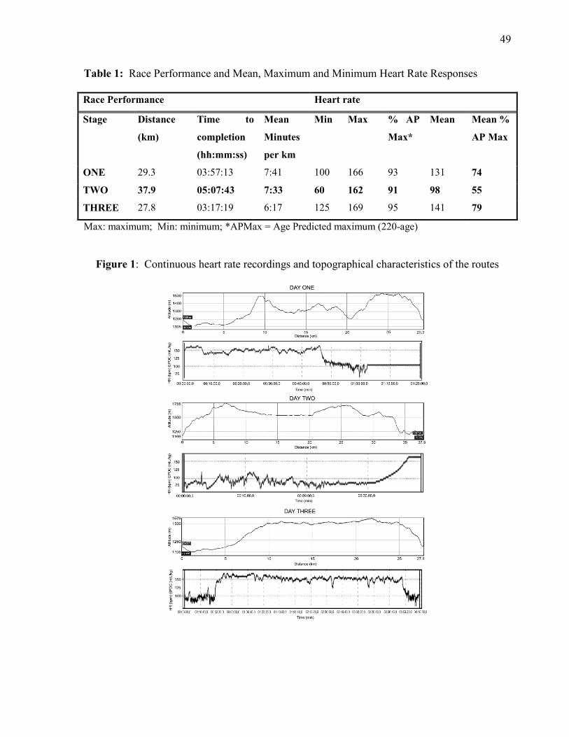
49
Table 1: Race Performance and Mean, Maximum and Minimum Heart Rate Responses
Max: maximum; Min: minimum; *APMax = Age Predicted maximum (220-age)
Figure 1: Continuous heart rate recordings and topographical characteristics of the routes
Race Performance Heart rate
Stage Distance
(km)
Time to
completion
(hh:mm:ss)
Mean
Minutes
per km
Min Max % AP
Max*
Mean Mean %
AP Max
ONE 29.3 03:57:13
7:41 100 166 93 131 74
TWO 37.9 05:07:43 7:33 60 162 91 98 55
THREE 27.8 03:17:19 6:17 125 169 95 141 79

50
Table 2: Haematological analyses and markers of muscle damage, inflammation and hydration status
RBC: Red blood cell; Hb: Haemoglobin; Hct: Haematocrit; Serum CPK: Serum Creatine
phosphokinase; Serum hsCRP: Serum High sensitivity C-Reactive Protein; TC: Thigh circumference;
DOMS: Delayed onset muscle soreness; USG: Urine specific gravity; *ND: not detected
Her red blood cell and platelet indices reflected a healthy profile. Race-induced elevations in
leukocyte and neutrophil concentrations were modest, remaining within reference range (Table
2).
Variable PreS1 PostS1 PreS2 PostS2 PreS3 PostS3 24h 72h Reference
range
RBC( 10^12/l) 4.37 4.01 4.03 4.17 3.79 4.01 3.86 3.88 3.8 – 4.8
Hb (g/dl) 14.1 12.9 13.3 13.3 11.9 12.8 12.3 12.5 12 – 15
Hct (%) 42.8 39.1 39.2 40.9 37.5 39.9 38.4 40.4 36 – 46
Platelet (10^9/L) 236 238 225 245 207 246 187 208 150 -450
Leukocyte (10^9/L) 4.35 6.79 4.75 7.5 4.86 5.05 4.21 6.75 3.92– 9.88
Neutrophil (10^9/L) 1.83 5.20 2.02 5.79 2.25 5.05 1.85 4.64 2.00 – 7.50
Serum CPK (U/L) 134 215 269 478 470 665 391 140 39 – 308
Urinary myoglobin
(mcg/ml)
ND* ND* ND* ND* ND* ND* ND* ND* <1
Serum hsCRP(mg/l) 0.2 - - - - 1.9 1.4 0.6 <1.0
TC (cm) 49.5 47 49.5 49 49 49 49 49
DOMS Score 0 4 4 7 5 5 2 0
Serum Cortisol
(nmol/l)
845 617 846 943 755 670 - - 190 – 690
Serum Osmolality
(mOsm/kg)
290 292 291 305 290 303 - - 275 – 295
Urine Osmolality
(mOsm/kg)
754 482 564 705 1005 802 - - 300 – 800
USG 1.020 1.015 1.020 1.025 1.03 1.03 - - Dehydration
> 1.025
% body mass loss 3.2 4.5 2.7

51
As shown in Table 2, pre-race serum CPK concentrations (134U/L) were within the reference
range (39-308 U/L) and rose to a maximum of 665U/L following S3. No uMb was detected at
any reading. Serum CRP concentration rose from 0.2 mg/l to 1.9 mg/l post race, although she
reported DOMS ratings of 4/10, 7/10 and 5/10 respectively during the 3 days of racing and
2/10 for 24h and 48h PR. Early morning pre-race serum cortisol concentration (845 nmol/l)
rose to maximum of 943 nmol/l following S2. Post-stage TC did not increase.
sOSm concentrations and USG were indicative of substantial dehydration after S2, during
which she experienced nausea and an inability to ingest carbohydrates and fluids. The
dehydration persisted until completion of the race and is confirmed by the body mass loss of
4.5% during S2 (Table 2).
Discussion
Statins have been found to be the most effective medications for reducing elevated blood
concentrations of low density lipoprotein cholesterol and are currently being prescribed to
more patients in greater dosages (11). Although statins have been shown to reduce chronic
inflammation in patients with coronary artery diseases and cardiovascular related mortality (8),
dose related ( ≥ 1.0mg/kg) statin myotoxicity (7), has been found to have adverse effects on
muscle which may affect performance in sport (2,3,7,10,11).
Seachrist et al. (10) reported that 78 % of professional athletes with familial
hypercholesterolaemia could not tolerate statin therapy due to muscular problems and
predisposing risk factors include being female, advancing age and the use of concurrent
medications (2). It was therefore of interest to observe how this female athlete, aged 42 years,
ingesting a dose of 20mg/52 kg Aspavor combined with 10 mg Ezetrol per day, would respond
to participation in a three-day 95 km trail run.
Atorvastatin and simvastatin are lipophilic statins which promote ease of diffusion through
passive transport across the bilipid cell membrane, causing augmented toxic effects on muscle
at a dosage of >80mg/day (2,8). At 0.5mg/kg, Seachrist et al. (10) demonstrated changes to the
morphology of mitochondria, which might sensitize muscle to injury, but without concurrent

52
changes to the myofibre. At doses of ≥1mg/kg/day significant alterations in the myofibre and
increases in serum CPK, alanine aminotransferase (ALT), aspartate aminotransferase (AST)
and plasma lactate were reported which were greater in Type II glycolytic fibres than in Type I
muscle fibres. They concluded that ≥1mg/kg/day might promote statin-induced muscle damage
and subsequent muscle mass loss.
As athletes regularly present with CPK values in the “thousands” following ultradistance road
running events (4,6), this subject’s accumulative CPK concentrations following the three
stages of the event indicate very little exercise-induced muscle damage. Her training status, a
repeated bout effect and reduced muscle damage resulting from the inclusion of hill training in
her program, might, together with genetic factors and the dosage of her medication (0.4
mg/kg), have contributed to her lower serum CPK concentrations (5).
According to her HR data (Figure 1, Table 1), the greatest effort was during S3 although
during all three stages her HR sporadically reached a maximum of over 90% APmax. This
indicates a moderate intensity of prolonged effort during S1 and S3. The reduced intensity
during S2 (Figure 1, Table 1), was probably due to her reported nausea and vomiting and
evident dehydration (Table 2), which could have reduced muscle damage, resulting in an
attenuated serum CPK concentration and the absence of uMb throughout the race. This is also
confirmed by her post-race concentration of serum hsCRP, 1.9 mg/l, which is indicative of
only mild inflammation due to muscle damage, when compared to the mean concentration
hsCRP in previous ultradistance events (6). The anti-inflammatory effect of Atorvastatin (8) is
likely to be a factor responsible for this attenuation.
Her decreased thigh circumference after S1 (49.5cm – 47cm) may not be muscle mass loss due
to statin therapy, but rather confirm previous findings (4) at the 2006 Isarrun, a 5-stage 338 km
ultra- endurance run, where athletes experienced significant losses in skeletal muscle mass
(particularly after S1), but not body mass during the race.
Although her pre stage serum cortisol concentrations were above the reference range (Table 2),
it increased further only following S2, which confirms greater physiological stress, possibly
53
linked to her reported inability to ingest carbohydrates during this stage. However, it did not
affect her muscle damage during that stage (Table 2). Post S1 and S3 serum cortisol
concentrations indicated that she was running comfortably within her physiological range.
Pre race sOsm indicates that she started the race adequately hydrated. In view of the reported
effect of dehydration on exercise-induced muscle damage (4) it is interesting to note that
despite dehydration occurring during S2 and S3, as indicated by the substantial increases in
sOsm,UOsm and USG and a body mass loss of 4.5% during S2, there was no excessive
increase in the markers of muscle damage and inflammation (Table2).
Although her pre-race RBC, Hb and HCT values reflect the absence of anaemia, the slight drop
in RBC indices over the 3-day period may be due to transient osmotic stress, membrane lipid
peroxidation caused by free radicals released by leucocytes, or even intramuscular destruction
of RBC (9), which returned to normal within 72 hours post race. The anti-inflammatory effect
of Atorvastatin (8) probably contributed to the attenuation of the typical increase in leukocyte
count during intense prolonged exercise, indicating an increase in metabolic activity and
activation of neutrophil release (4, 5, 6).
In conclusion we report that this subject, despite presenting with evidence of dehydration after
S2 and S3, experienced no symptoms of statin- induced myalgia, fatigue or cramps during the
3-day trail run. The adverse effects of Atorvastatin, combined with simultaneous intake of
Ezetrol, were not evident at 0.4mg/kg. Although the nausea experienced may have been an
adverse effect, the fact that it only occurred during the more challenging strenuous S2, points
to greater likelihood of exercise-induced gastric irritation.

54
Acknowledgements:
Ms Heidi Mocke and the Wildlands Conservation Trust are thanked for permitting the
collection of these data at the 2011 Three Cranes Challenge multiday trail run and for
providing the research team with accommodation in the “race village” located in the
Bushwillow Campsite.
The SA Weather Office is thanked for provision of precise data regarding the environmental
conditions during the different stages of the race.
Mr. Navin Singh, Dr Anton de Waard, Prof. Andrew McKune and Mr Ronnie Naicker are
thanked for their support with the field side collection of data reported in this paper.
Ampath Laboratory is thanked for their assistance with phlebotomny and chemical pathology.
Mr Asokaran Rajh is thanked for graphic artwork.

55
References:
1. Balady GJ, Williams MA, Ades PA, et al. Core Components of Cardiac
Rehabilitation/Secondary Prevention Programs. Update. Circulation. 2007;115:2675–2682.
2. Di Stasi SL, MacLeod TD, Winters JD, Binder-Macleod SA. Effects of Statins on Skeletal
Muscle: A Perspective for Physical Therapists. Phys Ther. 2010;90(10):1530-1542.
3. Evans M and Rees A. Effects of HMG-CoA Reductase Inhibitors on Skeletal Muscle: Are
all Statins the Same? Drug Safety. 2002;25(9):649-663.
4. Knechtle B and Kohler G. Running 338 kilometres within five days has no effect on body
mass and body fat but reduces skeletal muscle mass – the Isarrun 2006. J Sports Sci and Med.
2007;6:401-407.
5. McHugh MP. Recent advances in the understanding of the repeated bout effect: the
protective effect against muscle damage from a single bout of eccentric exercise. Scand J Med
Sci Spor. 2003;13:88–97.
6. Millet GY, Tomazin K, Verges S, Boisson RC, Gergele L, Feasson L, Martin V.
Neuromuscular Consequences of an Extreme Mountain Ultra-Marathon. PLoS One. 2011;
6:e17059
7. Pasternak RC, Smith SC, Bairey-Merz CN, Grundy, SM, Cleeman JI, Lenfant C.
ACC/AHA/NHLBI Clinical Advisory on the Use and Safety of Statins. J Am Coll Card.
2002;40(3):567–572.
8. Riesen WF, Engler H, Risch M, Korte W, Noseda G. Short-term effects of Atorvastatin on
C-reactive protein. Eur Heart J. 2002;23:794–799.
9. Robinson Y, Christancho E, Boning D. Intravascular Hemolysis and Mean Red Blood Cell
Age in Athletes. Med Sci Sport Exer. 2006;38(3):480-483.
56
10. Seachrist JL, Loi CM, Evans MG, Criswell KA, Rothwell CE. Roles of Exercise and
Pharmakokinetics in Cerivastatin-Induced Skeletal Muscle Toxicity. Toxicol Sci.
2005;88(2):551–561.
11. Thompson PD, Parker BA, Clarkson PM et al. A Randomized Clinical Trial to Assess the
Effect of Statins on Skeletal Muscle Function and Performance: Rationale and Study Design.
Prev Cardiol. 2010; Summer:104–111.
57
CHAPTER FIVE
CONCLUSION AND RECOMMENDATIONS
5.1 CONCLUSION
This study set out to determine the effect of multiday trail running on systemic and urinary
markers of muscle damage and inflammation and the relationship with factors affecting muscle
damage and inflammation during this endurance event. The findings of this thesis reflect that
i. the eccentric component of skeletal muscle contraction, when compared to road
running events of similar duration, was not augmented during the three days of trail
running
ii. low markers of muscle damage and inflammation were recorded despite the
maintenance of a mean intensity of effort of 77-84% APmax and the evidence of
hyperthermia and dehydration, which have previously been found to augment exercise
induced muscle damage and inflammation
iii. markers of rhabdomyolysis and cardiac muscle damage were absent
iv. the attenuated skeletal muscle damage and inflammation was confirmed by the small
changes in DOMS and TC, which returned to baseline values by 72PR.
It is concluded that the reduced muscle damage and inflammation are attributed to the softer
surface underfoot and the technical difficulty of the terrain, which forced athletes to employ
varied muscle recruitment patterns and to reduce their pace while maintaining a moderately
high level of intensity. It is therefore possible that trail running, although it involves a greater
element of danger and hence greater predisposition to acute injuries, is less damaging than road
running in terms of intrinsic exercise-induced skeletal muscle damage.
The systemic and urinary evidence of muscle damage and inflammation in the case study was
also attenuated and, in contrast to our hypothesis, she did not experience either muscle cramps
or excessive fatigue, despite the use of Atorvastatin combined with Ezetimibe, which have both

58
been associated with muscle cramping, myalgia, fatigue and in rare cases, rhabdomyoloysis
and death.
It is thus suggested that the lower dosage of medication benefits this hypercholesterolaemic
athlete, allowing her to participate in sport without experiencing the adverse events associated
with higher dosages of the prescribed medication.
In conclusion it is however important to highlight the limitations of this study. These include
i. the use of a limited convenience sample from whom full compliance was obtained,
which may not have been representative of the population,
ii. financial constraints limiting the number of tests performed and
iii. lack of more comprehensive monitoring of the activity levels of the subjects during the
post-stage recovery period.
5.2 RECOMMENDATIONS FOR FUTURE RESEARCH
Directions for further research include
i. investigating the difference in skeletal muscle damage and inflammation between
multi-day road and trail running, using the same athletes whilst controlling variables
such as their intensity of effort, nutrition, hydration and the activities of the athletes
during the events and replicating the elevations of the routes, to match road and trail
running routes. This would include exploring the difference in the extent and duration
of fatigue experienced after multiday trail running events compared to road running;
ii. investigating the hormonal control of fluid balance during multiday trail running
events;
iii. investigating the changes in cardiac structure and function during multiday events and
the permanency of these changes, followed over a 5-10 year period.

59
Further research investigating the effects of the administration of different dosages of
Atorvastatin with or without the use of Ezetimibe in double blind randomly selected placebo-
controlled studies is also suggested, enforcing strict control of above mentioned variables.

60
REFERENCES
1. Ballantyne CM, Houri J, Notarbartolo A, Melani L, Lipka LJ, Suresh R, Sun S, LeBeaut
AP, Sager PT, Veltri EP, for the Ezetimibe Study Group. Effect of Ezetimibe
Coadministered With Atorvastatin in 628 Patients With Primary Hypercholesterolemia.
A Prospective, Randomized, Double-Blind Trial. Circulation 2003: 107: 2409-2415.
2. Bennett M, Best TM, Babul S, Taunton J, Lepawsky M. Hyperbaric oxygen therapy for
delayed onset muscle soreness and closed soft tissue injury. Cochrane Database Syst
Rev 2005: Oct 19(4):CD004713.
3. Blake DJ, Weir A, Newey SE, Davies KE. Function and Genetics of Dystrophin and
Dystrophin-Related Proteins in Muscle. Physiol Rev 2002: 82: 291-329.
4. Borer K. Exercise Endocrinology. Champaign IL Human Kinetics Publishers 2003: 65-
76.
5. Boriek AM, Capetanaki Y, Hwang W, Officer T, Badshah M, Rodarte J, Tidball JG.
Desmin integrates the three dimensional mechanical properties of muscles. Am J
Physiol-Cell PH 2001: 280: C46 – C52.
6. Bruunsgaard H and Pedersen BK. Effects of exercise on the immune system in the
elderly population. Imm Cell Biol 2000: 78: 523-531.
7. Casa DJ., Stearns RL, Lopes RM, Ganio MS, McDermott BP, Walker Yeargin S;
Yamamoto LM, Mazerolle SM, Roti MW, Armstrong LE, Maresh CM. Influence of
Hydration on Physiological Function and Performance during Trail Running in the Heat.
J Athl Trainingaining 2010: 45: 147-156.
8. Cheuvront SN and Haymes EM. Thermoregulation and Marathon Running. Biological
and Environmental Influences. Sports Med 2001: 31(10): 743-762.
9. Clarkson PM, Kearns AK, Rouzier P, Rubin R, Thompson PD. Serum Creatine Kinase
Levels and Renal Function Measures in Exertional Muscle Damage. Med Sci Sport
Exer 2006: 38(4): 623-627.
10. Clarkson PM, Hubal MJ. Exercise-Induced Muscle Damage in Humans. Am J Physiol
Med Rehab 2002: 81(Suppl): S52-S69.

61
11. Cleary MA, Sweeney LA, Kendrick ZV, Sitler MR. Dehydration and Symptoms of
Delayed-Onset Muscle Soreness in Hyperthermic Males. J Athl Trainingaining 2005:
40(4): 288-297.
12. Coffey VG, Hawley JA. The molecular bases of training adaptation. Sports Med 2007:
37(9): 737-63.
13. Connolly DA, McHugh MP, Padilla-Zakour OI, Carlson L, Sayers SP. Efficacy of a
tart cherry juice blend in preventing the symptoms of muscle damage. Brit J Sports Med
2007: Jun 41(6): 392-7. Epub 2007 Jan 29.
14. Devaney JM, Hoffman EP, Gordish-Dressman H, Kearns A, Zambraski E, Clarkson
PM. IGF-II gene region polymorphisms related to exertional muscle damage. J Appl
Physiol 2007: 102: 1815-1823.
15. Di Stasi SL, MacLeod TD, Winters JD, Binder-Macleod SA. Effects of Statins on
Skeletal Muscle: A Perspective for Physical Therapists. Phys Ther 2010: 90(10): 1530
– 1542.
16. Donovan RM. Characteristics of Myosin Heavy Chain and Titin in Strength and
Endurance Athletes, UW-L J of Undergraduate Research VII 2004.
17. Durnin JVGA, Womersley J. Body fat assessed from total body density and its
estimation from skinfold thickness: measurements on 481 men and women aged from 16
to 72 years. Brit J Nutrition 1974: 32:77-97.
18. Eston RG, Mickleborough J, Baltzopoulos V. Eccentric activation and muscle damage:
biomechanical and physiological considerations during downhill running. Brit J Sports
Med 1995: 29(2): 89-94.
19. Evans M and Rees A. Effects of HMG-CoA Reductase Inhibitors on Skeletal Muscle:
Are all Statins the Same? Drug Safety 2002: 25 (9): 649 – 663.
20. Feasson L, Stockholm D, Freyssenet D, Richard I, Duguez S, Beckmann JS, Denis C.
Molecular adaptations of neuromuscular disease associated proteins in response to
eccentric exercise in human skeletal muscle. J Physiol 2002: 543.1: 297-306.
21. Frassl W, Kowoll R, Speth M, Stangl A, Brechtel L, Joscht B, Boldt LH, Meier-
Buttermilch R, Schlemmer M, Roecker L, Gunga HC. Cardiac markers (BNP, NT-pro-
BNP), Troponnin I, Troponin T) in female amateur runners before and up until three
days after a marathon. Clin Lab 2008: 54(3-4): 81-7.
62
22. Friden J and Lieber R. Eccentric exercise-induced injuries to contractile and
cytoskeletal muscle fibre components. Acta Physiol Scand 2001: 171: 321-326.
23. Goldspink G. Gene expression in muscle in response to exercise. J Muscle Res Cell M
2003: 24(2-3): 121-6.
24. Grobler LA, Collins M, Lambert MI, Sinclair-Smith C, Derman W, St Clair Gibson A,
Noakes T. Skeletal Muscle Pathology in endurance athletes with acquired training
intolerance. Brit J Sports Med 2004: 38: 697-703.
25. Hamada K, Vannier E, Sacheck JM, Witsell AL, Roubenoff R. Senescence of human
skeletal muscle impairs the local inflammatory cytokine response to acute eccentric
execise. FASEB J 2004: doi: 10.1096/fj.03-1286fje.
26. Hawke TJ, Garry DJ. Myogenic satellite cells: physiology to molecular biology. J
Appl Physiol 2001: 91: 534-551.
27. Herbert RD, de Noronha M. Stretching to prevent or reduce muscle soreness after
exercise. Cochrane Database Syst Rev 2007: Oct 17: (4): CD004577.
28. Huxley AF. Muscle structure and theroies of contraction. Prog Biophys Biophys Chem
1957: (7): 255-318.
29. Jackson MJ, Khassaf M, Esanu C, Vasilaki A, Brodie DA, McArdle A. Vitamin C
Supplements suppress the stress response in human muscle. Free Radic Bio Med 1999:
27: S36.
30. Knechtle B, Duff B, Schulze I, Kohler G. A multi-stage ultra-endurance run over 1,200
km leads to a continuous accumulation of total body water. J Sports Sci and Med 2008:
7: 357-364.
31. Knechtle B and Kohler G. Running 338 kilometres within five days has no effect on
body mass and body fat but reduces skeletal muscle mass – the Isarrun 2006. J Sports
Sci and Med 2007: 6: 401-407.
32. Kohut ML, Senchina DS. Reversing age-associated immunosenescence via exercise.
Exerc Immunol Rev 2004: 10: 6-41.
33. Krause, M. Exercise, sarcopenia and Immunosenescence. 2003: [email protected]
34. La Gerche A, Connelly KA, Mooney DJ, Macisaac AI, Prior DL. Biochemical and
functional abnormalities of left and right ventricular function after ultra-endurace
exercise. Heart 2008: 94: 860-866.

63
35. Lambert MI and Dennis SC. Delayed-Onset Muscle-Soreness. SA J Sports Med 1994:
9: 18-20.
36. Leers MP, Schepers R, Baumgarten R. Effects of a long-distance run on cardiac
markers in healthy athletes. Clin Chem Lab Med 2006: 44(8): 999-1003.
37. Lieber RL, Friden J. Mechanisms of muscle injury gleaned from animal models. Am J
Physiol Med Rehab 2002: 81(Suppl): S70-S79.
38. Mahoney Dj, Safdar A, Parise G, Melov S, Fu M, MacNeil L, Kazcor j, Payne ET,
Tarnopolsky MA. Gene expression profiling in human skeletal muscle during recovery
from eccentric exercise. Am J Physiol Regul Integr Comp Physiol 2008: Jun 294(6):
R1901-10. Epub 2008 Mar 5.
39. Malm C, Sjodin The Late B, Sjoberg B, Lenkei R, Renstrom P, Lundberg IE, Ekblom B.
Leukocytes, cytokines, growth factors and hormones in human skeletal muscle and
blood after uphill or downhill running. J Physiol 2004: 556.3: 983-1000.
40. Maresh CM, Gabaree-Boulant CL, Armstrong LE, Judelson DA, Hoffman JR,
Castellani JW, Kenefick RW, Bergeron MF, Casa DJ. Effect of hydration status on
thirst, drinking, and related hormonal responses during low-intensity exercise in the
heat. J Appl Physiol 2004: 97: 39-44.
41. Martinez-Amat A, Boulaiz H, Prados J, Marchal JA, Padial Puche P, Caba O,
Rodriguez-Serrano F, Aranega A. Release of alpha-actin into serum after skeletal
muscle damage. Brit J Sports Med 2005: 39: 830-834.
42. Maughan R J, Donnelly A E, Gleeson M, Whiting P H, Walker K A, Clough P J.
Delayed-onset muscle damage and lipid peroxidation in man after a downhill run.
Muscle Nerve 1989: 12: 332–336. doi: 10.1002/mus.880120412.
43. McArdle A, Vasilaki A, Jackson M. Exercise and skeletal muscle ageing: cellular and
molecular mechanisms. Ageing Research Reviews 2002: 1: 79-93.
44. McHugh MP. Recent advances in the understanding of the repeated bout effect: the
protective effect against muscle damage from a single bout of eccentric exercise. Scand
J Med Sci Spor 2003: 13: 88-97.
45. McHugh MP, Connolly DAJ, Eston RG, Gleim GW. Exercise-induced muscle
damage and potential mechanisms for the repeated bout effect. Sports Med 1999: Mar
27(3): 157-170.

64
46. Middleton N, George K, Whyte G, Gaze D, Collinson P, Shave R. Cardiac Troponin T
Release is Stimulated by Endurance Exercise in Healthy Humans. J Am Coll Cardiol
2008: 52(22): 1813-6.
47. Millet GY, Tomazin K, Verges S, Vincent C, Bonnefoy R, Boisson RC, Gergele L,
Feasson L, Martin V. Neuromuscular Consequences of an Extreme Mountain Ultra-
Marathon. PLoS One 2011: 6(2): e17059.
48. Morgan DL. New insights into the behavior of muscle during active lengthening.
Biophys J 1990: 57: 209-221.
49. Nieman DC, Dumke CL, Henson DA, McAnulty SR, Gross SJ, Lind RH. Muscle
Damage is linked to cytokine changes following a 160-km race. Brain, Behav and
Immun 2005: 19: 398-403.
50. Noakes TD, Myburgh KH, Du Plessis J, Lang L, Lambert M, Van Der Riet C, Schall R.
Metabolic rate, not percent dehydration, predicts rectal temperature in marathon runners.
Med Sci Sport Exerc 1991: 23: 443-9.
51. Pasternak RC, Smith SC, Bairey-Merz CN, Grundy SM, Cleeman JI, Lenfant C.
ACC/AHA/NHLBI Clinical Advisory on the Use and Safety of Statins. J Am Coll Card
2002: 40(3): 567 – 572.
52. Paulsen G, Egner IM, Drange M, Langberg H, Benestad HB, Fjeld JG, Hallen J, Raastad
T. A COX-2 inhibitor reduces muscle soreness, but does not influence recovery and
adaptation after eccentric exercise. Scand J Med Sci Sport 2009: Jun 10.
53. Peake J, Nosaka K, Suzuki K. Inflammation and eccentric exercise. Exercise Immunol
Rev 2005: 11: 64-85.
54. Pedersen BK, Steensberg A, Fischer C, Keller C, Ostrowski K, Schjerlin P. Exercise
and cytokines with particular focus on muscle-derived IL-6. Exercise Immunol Rev
2001: 7: 18-31.
55. Pelsers MM, Hermens WT, Glatz JF. Fatty acid-binding proteins as plasma markers of
tissue injury. Chim Acta 2005: Feb 352 (1-2): 15-35.
56. Peters EM. Interleukin-6 – exercise factor and modulator of metabolic function? J SA
Sports Med 2004: 16(3): 8-13.
57. Peters EM, Anderson R, Nieman DC, Fickl H, Jogessar V. Vitamin C supplementation
attenuates the increases in circulating cortisol, adrenaline and anti-inflammatory
65
polypeptides following ultramarathon running. Int J Sports Med 2001: Oct 22(7): 537-
43.
58. Peters EM, Anderson R, Theron AJ. Attenuation of Increase in Circulating Cortisol and
Enhancement of the Acute Phase Protein Response in Vitamin C-Supplemented
Ultramarathoners. Int J Sports Med 2001: Feb 22(2): 120-126.
59. Peters T, Weiss JM, Sindrilaru A, Wang H, Oreshkova T, Waschek M, Malty P,
Reimann J, Scharffetter-Kochanek K. Reactive oxygen intermediate-induced
pathomechanisms contribute to immunosenescence, chronic inflammation and
autoimmunity. Mech Ageing Dev 2009: Sept 130(9): 564-87.
60. Petersen AMW, Pedersen BK. The anti-inflammatory effect of exercise. J Appl Physiol
2005: 98: 1154-62.
61. Pietzsch FJG. The Role of vibratory massage on treating delayed onset muscle
soreness. University of Brighton, Chelsea School, Eastborne, East Sussex 2004(no
journal/publication details available).
62. Pournot H, Bieuzen F, Louis J, Fillard JR, Barbiche E, Hausswirth C. Time-Course of
Changes in Inflammatory Response after Whole-Body Cryotherapy Multi Exposures
following Severe Exercise PloS One 2011: 6(7): e22748. doi:
10.1371/journal.pone.0022748.
63. Proske U, Morgan DL. Muscle damage from eccentric exercise mechanism, mechanical
signs, adaptation and clinical application. J Physiol 2001: 537.2: 333-345.
64. Puterman E, Lin J, Blackburn E, O’Donovan A, Adler N, Epel E. The power of
exercise: buffering the effect of chronic stress on telomere length. PLoS One 2010:
5(5): e10837. doi: 10.1371/journal.pone.0010837.
65. Radak Z, Chung HY, Goto S. Exercise and hormesis: oxidative stress-related
adaptation for successful aging. Biogerontology 2005: 6: 71-75.
66. Raastad T, Risoy, BA, Benestad HB, Fjeld JG, Hallen, J. Temporal relation between
leukocyte accumulation in muscles and halted recovery 10-20h after strength exercise. J
Appl Physiol 2003: 95(6): 2503-9.
67. Radom-Aizik S, Zaldivar F jr, Leu Sy, Cooper DM. A brief bout of exercise alters gene
expression and distinct gene pathways in peripheral blood mononuclear cells of early-
and late-pubertal females. J Appl Physiol 2009: Jul 107(1): 168-75.

66
68. Riesen WF, Engler H, Risch M, Korte W, Noseda G. Short-term effects of
Atorvastatin on C-reactive protein. Eur Heart J 2002: 23: 794 – 799.
69. Robson-Ansley P, Barwood M, Canavan J, Hack S, Eglin C, Davey S, Hewitt J, Hull J,
Ansley L. The effect of repeated endurance exercise on IL-6 and sIL-6R and their
relationship with sensations of fatigue at rest. Cytokine 2009: 45: 111-116.
70. Ronkainen PHA, Kovanen V, Alen M, Pollanen E, Palonen AM, Ankarberg-Lindgren
C, Hamalainen E, Turpeinen U, Kujala UM, Puolakka J, Kaprio J, Sipila S.
Postmenopausal hormone replacement therapy modifies skeletal muscle composition
and function: a study with monozygotic twin pairs. J Appl Physiol 2009: 107: 25-33.
71. Roth SM, Martel GF, Ivey FM, Lemmer JT, Metter EJ, Hurley BF, Rogers M. High-
volume, heavy resistance strength training and muscle damage in young and older
women. J Appl Physiol 2000: 88: 1112-1118.
72. Sawka MN, Young AJ, Francesconi RP, Muza SR, Pandolf KB. Thermoregulatory and
blood responses during exercise at graded hypohydration levels. J Appl Physiol 1985:
59(5): 1394-1401.
73. Seachrist JL, Loi CM, Evans MG, Criswell KA, Rothwell CE. Roles of Exercise and
Pharmakokinetics in Cerivastatin-Induced Skeletal Muscle Toxicity. Toxicol Sci 2005:
88(2): 551 – 561.
74. Seene T, Kaasik P, Umnova M. Structural reaarangements in contractile apparatus and
resulting skeletal muscle remodeling: effect of exercise training. J Sports Med Fitness
2009: Dec 49(40): 410-23.
75. Sellwood KL, Brukner P, Williams D, Nicol A, Hinman R. Ice-water immersion and
delayed-onset muscle soreness: a randomized controlled trial. Brit J Sports Med 2007:
Jun 41(6): 392-7.
76. Simpson RJ, Guy K. Coupling Aging immunity with a sedentary lifestyle: has the
damage already been done? - A mini-review. Gerontology 2009: doi:
10.1159/000270905.
77. Simpson RJ, Florida-James GD, Cosgrave C, Whyte GP, Macrae S, Pircher H, Guy K.
High-intensity exercise elicits the mobilization of senescent T lymphocytes into the
peripheral blood compartment of human subjects. J Appl Physiol 2007: 103: 396-401.

67
78. Skenderi KP, Kavouras SA, Anastasiou CA, Yiannakouris N, Matalas AL. Exertional
rhabdomyolysis during a 246-km continuous running race. Med & Sc in Sports and
Exerc 2006: doi: 10.1249/01.mss.0000222831.35897.5f .
79. Sorichter S, Mair J, Koller A, Pelsers NMAL, Puschendorf B, Glatz JFC. Early
assessment of exercise induced skeletal muscle injury using fatty acid binding protein.
Brit J Sports Med 1998: 32: 121-124.
80. Stearns RL, Casa DJ, Lopes RM. Influence of Hydration status on pacing during Trail
Running in the Heat. J Strength Cond Res 2009: 23: 2533-2541.
81. Stupka N, Tarnopolsky MA, Yardley NJ, Phillips SM. Cellular adaptation to repeated
eccentric exercise-induced muscle damage. J Appl Physiol 2001: 91: 1669-1678.
82. Tartibian B, Maleki BH, Abbasi A. The effects of ingestion of Omega-3 Fatty Acids on
perceived pain and external symptoms of delayed onset muscle soreness in untrained
men. Clin J Sport Med 2009: 19:115-119.
83. Thompson PD, Parker BA, Clarkson PM, Pescatello LS, White CM, Grimaldi AS,
Levine BD, Haller RG, Hoffman EP. A Randomized Clinical Trial to Assess the Effect
of Statins on Skeletal Muscle Function and Performance: Rationale and Study Design.
Prev Cardiol 2010: Summer: 104 – 111.
84. Urso ML, Clarkson PM, Hittel D, Hoffman EP, Thompson PD. Changes in ubiquitin
proteasome pathway gene expression in skeletal muscle with exercise and statins.
Arterioscl Throm Vas 2005: 25:2560-2566.
85. Virtanen C, Takahashi M. Muscling in on microarrays. Appl Physiol Nutr Metab 2008:
Feb 33(1): 124-9.
86. Yanagida T. Muscle Contraction Mechanism Based on Actin Filament Rotation. Adv
Exp Med Biol 2007: Vol592 VI: 359-367.
87. Zainuddin Z, Newton M, Sacco P, Nosaka K. Massage Therapy effectively alleviates
muscle soreness caused by exercise. J Athl Training 2005: Sept 40 (3): 174-180.

68
APPENDICES

69

70
APPENDIX B
Discipline of Physiology School of Medical Sciences Faculty of Health Sciences 2011-07-14
THREE-CRANES TRAIL RUN 2011 Study Title: Physiological assessment of participants in a three day trail run event: markers of muscle damage, hydration and immune status. Dear PW, Thank you very much for agreeing to take part in the above trial. Although the final research papers are not yet published, we are happy to provide you with your results. Your Physical Characteristics Your value Predicted norm for active
non-athlete of your age, gender and BMI
Height (m) 1.78 Mass (kg) 80.1
BMI 25.28 19 – 26 Triceps skin fold (mm) 17.0 Biceps skin fold (mm) 8.0 Sub scapular skin fold (mm) 9.67 Suprailliac skin fold (mm) 12.13 Body Fat % (determined from triceps, biceps, suprailliac and sub scapular skin folds)
20.8
11-17% (athletes 5 – 12%)
Blood Pressure (mmHg)– Baseline 127/70 120/80 Heart Rate (bpm)– Baseline 53 65 Your BMI and resting blood pressure and heart rate fall within normal for a male athlete of your caliber, though your body fat % is higher than the norm. Enclosed are charts which you may find interesting for a comparison of body fat percentages of athletes in various disciplines.

71
Body Fat Percentage for The Average Population
Age Up to 30 30-50 50+
Females 14 - 21% 15-23% 16-25%
Males 9-15% 11-17% 12-19%
Average Body Fat Percentage of Athletes
Sport Male Female Sport Male Female
Baseball 12-
15% 12-18% Rowing 6-14% 12-18%
Basketball 6-12% 20-27% Shot Putters 16-
20% 20-28%
Body building 5-8% 10-15% Skiing (X
country) 7-12% 16-22%
Cycling 5-15% 15-20% Sprinters 8-10% 12-20%
Football (Backs) 9-12% No data Swimming 9-12% 14-24%
Football (Linemen) 15-
19% No data Tennis
12-
16% 16-24%
Gymnastics 5-12% 10-16% Triathlon 5-12% 10-15%
High/long Jumpers 7-12% 10-18% Volleyball 11-
14% 16-25%
Ice/field Hockey 8-15% 12-18% Weightlifters 9-16% No data
Racquetball 8-13% 15-22% Wrestlers 5-16% No data
Adapted From: Sports Fitness Advisor-Sports Training Tips for Athletic Peak Performance (www.sport-fitness-advisor.com) Ambient weather conditions during the three stages Stage
Ambient temperature range (º C)
Windspeed Range (m/s)
Relative Humidity %
Barometric Pressure (hPa)
1 11.5 - 21.7 0 - 2.3 63- 95 895.2 - 896.1 2 12.4 - 22.8 0 - 2.8 54- 97 896.5 - 897.7 3 12.1 - 21.2 0 - 2.3 64- 96 896.8 - 897.9 Elevation gains and losses Running speed varied depending on topography of the routes. elevation gains reached 1020, 1226 and 680m, while elevation losses were recorded at 1021, 1231 and 687m during Stages One, Two and Three respectively.
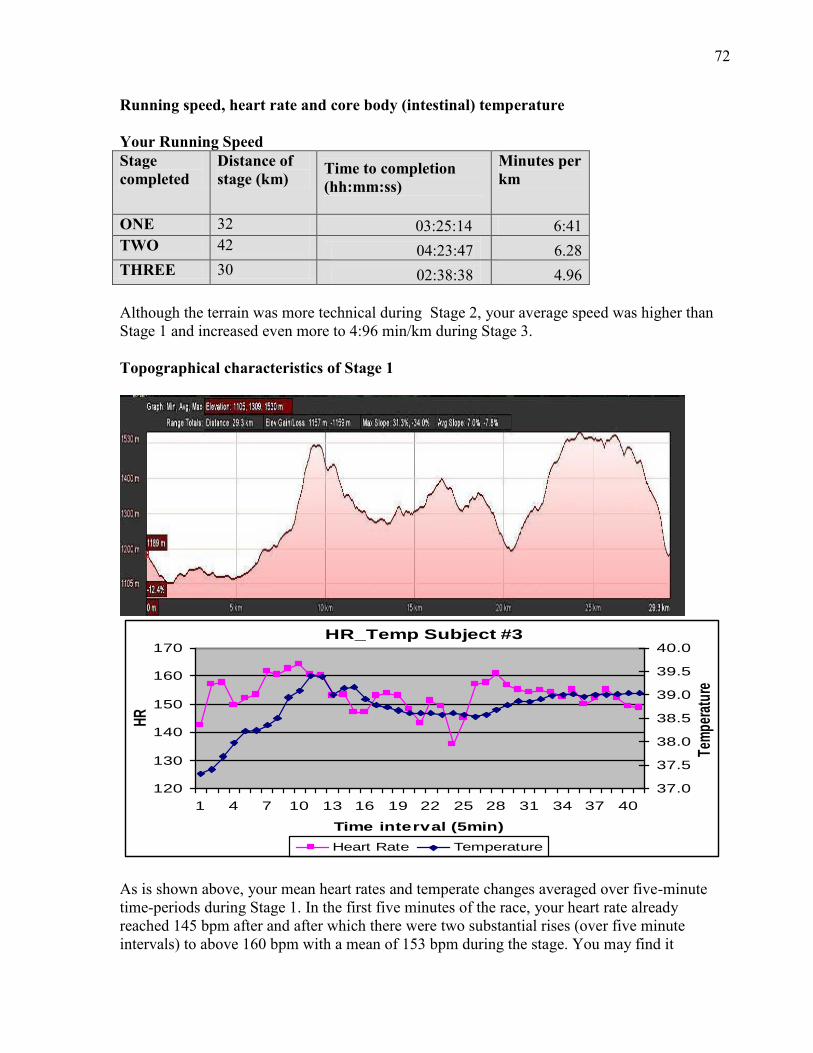
72
Running speed, heart rate and core body (intestinal) temperature Your Running Speed Stage completed
Distance of stage (km)
Time to completion (hh:mm:ss)
Minutes per km
ONE 32 03:25:14 6:41 TWO 42 04:23:47
6.28 THREE 30 02:38:38
4.96 Although the terrain was more technical during Stage 2, your average speed was higher than Stage 1 and increased even more to 4:96 min/km during Stage 3. Topographical characteristics of Stage 1
HR_Temp Subject #3
120
130
140
150
160
170
1 4 7 10 13 16 19 22 25 28 31 34 37 40
Time interval (5min)
HR
37.0
37.5
38.0
38.5
39.0
39.5
40.0
Tem
pera
ture
Heart Rate Temperature As is shown above, your mean heart rates and temperate changes averaged over five-minute time-periods during Stage 1. In the first five minutes of the race, your heart rate already reached 145 bpm after and after which there were two substantial rises (over five minute intervals) to above 160 bpm with a mean of 153 bpm during the stage. You may find it
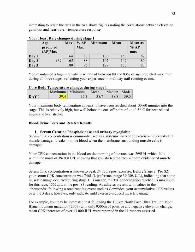
73
interesting to relate the data in the two above figures noting the correlations between elevation gain/loss and heart rate – temperature response. Your Heart Rate changes during stage 1 Age
predicted (AP)Max
Max % AP Max
Minimum Mean Mean as % AP max
Day 1 187
164 88 136 153 82 Day 2 165 88 107 149 80 Day 3 180 96 127 155 83 You maintained a high intensity heart rate of between 80 and 83% of age predicted maximum during all three stages, reflecting your experience in multiday trail running events. Core Body Temperature changes during stage 1 Maximum Minimum Mean Median Mode DAY 1 39.4 37.3 38.7 38.8 39.0 Your maximum body temperature appears to have been reached about 55-60 minutes into the stage. This is relatively high, but well below the cut- off point of > 40.5 º C for heat related injury and heat stroke. Blood/Urine Tests and Related Results
1. Serum Creatine Phosphokinase and urinary myoglobin Serum CPK concentration is commonly used as a systemic marker of exercise-induced skeletal muscle damage. It leaks into the blood when the membrane surrounding muscle cells is damaged. Your CPK concentration in the blood on the morning of the race was 269U/L which falls within the norm of 39-308 U/L showing that you started the race without evidence of muscle damage. Serum CPK concentration is known to peak 24 hours post exercise. Before Stage 2 (Pre S2) your serum CPK concentration was 746U/L (reference range 39-308 U/L), indicating that some muscle damage occurred during stage 1. Your serum CPK concentration reached its maximum for this race, 1562U/L at the post S3 reading. As athletes present with values in the “thousands” following a road running event such as Comrades, your accumulative CPK values over the 3 days, however, only indicate mild exercise-induced muscle damage. For example, you may be interested that following the 166km North Face Ultra Trail du Mont Blanc mountain marathon (2009) with only 9500m of positive and negative elevation change, mean CPK increases of over 13 000 IU/L were reported in the 11 runners assessed.
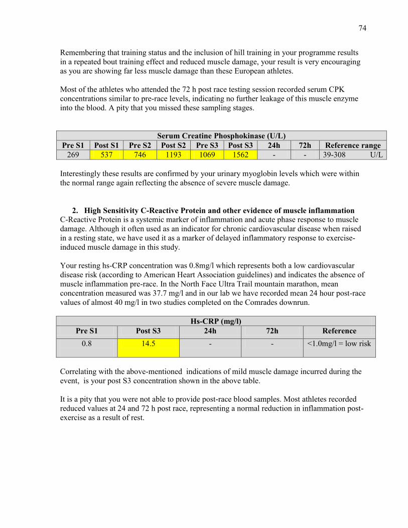
74
Remembering that training status and the inclusion of hill training in your programme results in a repeated bout training effect and reduced muscle damage, your result is very encouraging as you are showing far less muscle damage than these European athletes. Most of the athletes who attended the 72 h post race testing session recorded serum CPK concentrations similar to pre-race levels, indicating no further leakage of this muscle enzyme into the blood. A pity that you missed these sampling stages.
Serum Creatine Phosphokinase (U/L) Pre S1 Post S1 Pre S2 Post S2 Pre S3 Post S3 24h 72h Reference range
269 537 746 1193 1069 1562 - - 39-308 U/L Interestingly these results are confirmed by your urinary myoglobin levels which were within the normal range again reflecting the absence of severe muscle damage.
2. High Sensitivity C-Reactive Protein and other evidence of muscle inflammation C-Reactive Protein is a systemic marker of inflammation and acute phase response to muscle damage. Although it often used as an indicator for chronic cardiovascular disease when raised in a resting state, we have used it as a marker of delayed inflammatory response to exercise-induced muscle damage in this study. Your resting hs-CRP concentration was 0.8mg/l which represents both a low cardiovascular disease risk (according to American Heart Association guidelines) and indicates the absence of muscle inflammation pre-race. In the North Face Ultra Trail mountain marathon, mean concentration measured was 37.7 mg/l and in our lab we have recorded mean 24 hour post-race values of almost 40 mg/l in two studies completed on the Comrades downrun.
Hs-CRP (mg/l) Pre S1 Post S3 24h 72h Reference
0.8
14.5 - - <1.0mg/l = low risk
Correlating with the above-mentioned indications of mild muscle damage incurred during the event, is your post S3 concentration shown in the above table. It is a pity that you were not able to provide post-race blood samples. Most athletes recorded reduced values at 24 and 72 h post race, representing a normal reduction in inflammation post-exercise as a result of rest.

75
Thigh Circumference (cm): Pre S1
Post S1
+4hours Pre S2
Post S2
+ 4hours
Pre S3
Post S3
59 54.5 59 59 57 58 59 57.6
A study by Knechtle and Kohler (2007) conducted on the 2006 Isarrun, a 5-stage 338 km ultra- endurance run in Bavaria, Germany, revealed that athletes experienced significant losses in skeletal body mass ( particularly after Stage 1 of the race), but not body mass during the race. Your decreased thigh circumference after Stage 1 therefore may well confirm these findings. The return to pre-race circumference within 4 hours post race confirms an inflammatory response which follows mild degrees of swelling and accumulation of fluid within the interstitial spaces between the active muscles.
3. Serum Cortisol concentration Cortisol is a hormone secreted by the adrenal glands and is often elevated in times of physical or mental stress. Cortisol increases the glucose available to the muscles by stimulating the breakdown of glycogen and also acts as a powerful anti-inflammatory hormone. Cortisol secretion in the body normally follows a diurnal pattern, being highest between 5 – 10am and lowest between 8pm – 4am. In the case of athletes running a race, blood cortisol concentrations may also be elevated in the morning due to anticipation or hyperglycaemia, but do tend to increase further during prolonged exercise. Your cortisol result for the morning of the first day was 711nmol/l which is higher than the norm, probably due to anticipatory anxiety. Usually during a race the cortisol values increase due to physiological stress. As shown in the table below, the metabolic demands/ anti-inflammatory response appear to have been increased during Stage 2 and greatest during Stage 3, confirming your increased average heart rate and speed maintained during Stage 3.
Serum Cortisol concentration (nmol/l) Pre S1 Post S1 Pre S2 Post S2 Pre S3 Post S3 Reference
711 740 562 938 604 983 190 – 690nmol/l
4. Phadiatop and Immunoglobulin E These blood tests are used to diagnose and monitor allergy reactions. The Phadiatop test shows whether you are allergic to a range of common inhalant allergens, including animal dander, pollen and mould. IgE concentrations represent the severity of the allergic reaction.
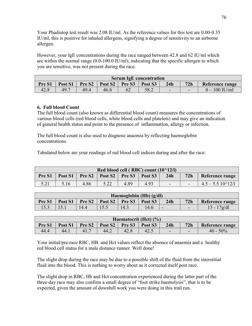
76
Your Phadiatop test result was 2.08 IU/ml. As the reference values for this test are 0.00-0.35 IU/ml, this is positive for inhaled allergens, signifying a degree of sensitivity to an airborne allergen. However, your IgE concentrations during the race ranged between 42.8 and 62 IU/ml which are within the normal range (0.0-100.0 IU/ml), indicating that the specific allergen to which you are sensitive, was not present during the race.
Serum IgE concentration Pre S1 Post S1 Pre S2 Post S2 Pre S3 Post S3 24h 72h Reference range
42.8 49.7 49.4 46.8 62 58.2 - - 0 – 100 IU/ml 6. Full blood Count The full blood count (also known as differential blood count) measures the concentrations of various blood cells (red blood cells, white blood cells and platelets) and may give an indication of general health status and point to the presence of inflammation, allergy or infection. The full blood count is also used to diagnose anaemia by reflecting haemoglobin concentrations. Tabulated below are your readings of red blood cell indices during and after the race:
Red blood cell ( RBC) count (10^12/l) Pre S1 Post S1 Pre S2 Post S2 Pre S3 Post S3 24h 72h Reference range
5.21 5.16 4.86 5.22 4.89 4.93 - - 4.5 – 5.5 10^12/l
Haemoglobin (Hb) (g/dl) Pre S1 Post S1 Pre S2 Post S2 Pre S3 Post S3 24h 72h Reference range
15.3 15.1 14.4 15.5 14.3 14.6 - - 13 - 17g/dl
Haematocrit (Hct) (%) Pre S1 Post S1 Pre S2 Post S2 Pre S3 Post S3 24h 72h Reference range
44.4 44.1 41.7 44.2 42.8 42.5 - - 40 - 50% Your initial/pre-race RBC, HB and Hct values reflect the absence of anaemia and a healthy red blood cell status for a male distance runner. Well done! The slight drop during the race may be due to a possible shift of the fluid from the interstitial fluid into the blood. This is nothing to worry about as it corrected itself post race. The slight drop in RBC, Hb and Hct concentration experienced during the latter part of the three-day race may also confirm a small degree of “foot strike haemolysis”, that is to be expected, given the amount of downhill work you were doing in this trail run.
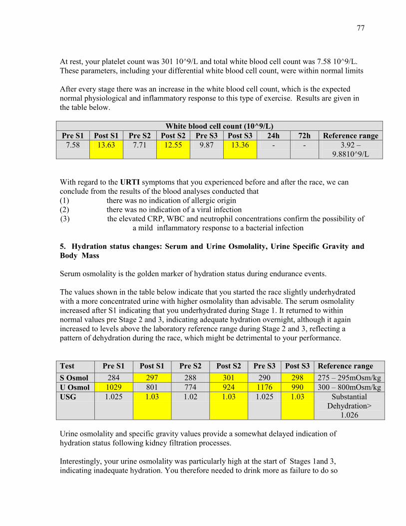
77
At rest, your platelet count was 301 10^9/L and total white blood cell count was 7.58 10^9/L. These parameters, including your differential white blood cell count, were within normal limits After every stage there was an increase in the white blood cell count, which is the expected normal physiological and inflammatory response to this type of exercise. Results are given in the table below.
White blood cell count (10^9/L) Pre S1 Post S1 Pre S2 Post S2 Pre S3 Post S3 24h 72h Reference range
7.58 13.63 7.71 12.55 9.87 13.36 - - 3.92 – 9.8810^9/L
With regard to the URTI symptoms that you experienced before and after the race, we can conclude from the results of the blood analyses conducted that (1) there was no indication of allergic origin (2) there was no indication of a viral infection (3) the elevated CRP, WBC and neutrophil concentrations confirm the possibility of
a mild inflammatory response to a bacterial infection
5. Hydration status changes: Serum and Urine Osmolality, Urine Specific Gravity and Body Mass Serum osmolality is the golden marker of hydration status during endurance events. The values shown in the table below indicate that you started the race slightly underhydrated with a more concentrated urine with higher osmolality than advisable. The serum osmolality increased after S1 indicating that you underhydrated during Stage 1. It returned to within normal values pre Stage 2 and 3, indicating adequate hydration overnight, although it again increased to levels above the laboratory reference range during Stage 2 and 3, reflecting a pattern of dehydration during the race, which might be detrimental to your performance. Test Pre S1 Post S1 Pre S2 Post S2 Pre S3 Post S3 Reference range S Osmol 284 297 288 301 290 298 275 – 295mOsm/kg U Osmol 1029 801 774 924 1176 990 300 – 800mOsm/kg USG 1.025 1.03 1.02 1.03 1.025 1.03 Substantial
Dehydration> 1.026
Urine osmolality and specific gravity values provide a somewhat delayed indication of hydration status following kidney filtration processes. Interestingly, your urine osmolality was particularly high at the start of Stages 1and 3, indicating inadequate hydration. You therefore needed to drink more as failure to do so

78
resulted in the high values throughout the race and might have affected your running performance. The above is confirmed by the changes in urine specific gravity (USG). A urine specific gravity of 1.010 is indicative of a well hydrated state. At no stage was your USG at that level. The changes in body mass also reflect some degree of fluid loss, although recent research (Nolte, 2010) has, however, confirmed that changes in body mass cannot be used as accurate surrogate for changes in total body water during prolonged exercise.
Body mass (kg) Pre S1 Post S1 % loss Pre S2 Post S2 % loss Pre S3 Post S3 %loss 79.68 77.5 2.7 79.86 76.95 3.6 79.74 78.04 2.1
We do hope that these findings are of interest to you and would once again like to thank you for being such a reliable research subject during the 3 days of racing. With very best wishes, . Prof E Peters-Futre Research Director Dr. Rentia Denissen Dr. Anton de Waard Mr Navin Singh Masters in Medical Science: Sports Medicine students

79
APPENDIX C
DEPARTMENT OF HUMAN PHYSIOLOGY FACULTY OF HEALTH SCIENCES
SUBJECT INFORMATION FORM
Study title: Physiological assessment of runners participating in three-day trail run event, with particular emphasis on markers of muscle damage, hydration and immune status. Dear Runner Ethical Approval: This study has received ethics approval from the University of KwaZulu-Natal, Nelson R Mandela
School of Medicine, Research Ethics Committee (Reference No: BF210/010)
Study Aims: The primary questions to be examined in this study are to include the following: Whether amateur athletes competing in a multistage mountain trail run
(1) incur muscle damage (2) are adequately, over or under hydrated and (3) experience an increased incidence of Upper Respiratory Tract Infections due to
airborne allergic responses
Who will qualify to participate in this study? 1. Healthy athletes (not in possession of CVS, renal, hepatic or endocrine dysfunction) between the ages of 25 and 50 years taking part in the 2010 3-Cranes Mountain Trail Run on 25-27 February 2010. 2. Residents in the greater eThekweni and Umzumduzi area. 3. Those willing to take part in the study and consent for all aspects of test e.g. regular blood tests, provision of saliva and urine samples, skin prick tests, weighing, filling in questionnaires, measuring urine output. What will be expected from volunteers in this study? You will be asked to: 1. Maintain your normal fluid use habits throughout the study.

80
2. Maintain your normal diet throughout the study. Outline of tests: Pre race Briefing – Day before race – REGISTRATION: 24 February 2011 1. Complete a medical questionnaire and undergo a full medical exam if indicated. 2. Complete a pre-race questionnaire concerning your training experience as well as details
about the fluids you consume when exercising 3. Assessment of height, mass and percentage body fat (to be estimated from skinfold
thicknesses). During race (before and after each stage)– Research station at Karkloof 1. Body mass measurement. 2. Three vials (approx 9-12 ml) of blood to be taken before and after the event. 3. Provide a urine sample 4. Complete a brief questionnaire. 5. Record all fluid consumed and all urine output during stage and overnight. 6. Wear a heart rate monitor throughout the race 24, 48 and 72 Hours after race - Exercise Laboratory, Physiology Department, E2 Building 1. Body mass measurement. 2. Three vials of blood (approx 9-12 ml) to be taken. 3 Provide a urine and saliva sample. 4. Complete a brief questionnaire . How can you benefit from participation in this study? Following the study, you will be given the results of your full blood counts and other biochemical/ laboratory tests which were run on your blood samples. As soon as the data has been collated, you will receive the study results. A furthering of knowledge regarding your fluid balance during endurance exercise as well as hydration status will be gained from this work. This will allow you to optimize your fluid use in the future and improve your performance. You will be the first to be notified of the results of this study. Will you be exposed to adverse effects of the study? There will be minimal risks to you by being involved in the study. Some people find venipuncture (taking blood) uncomfortable. There is a very slight risk of complications from this procedure, mainly infection at the site of puncture or inflammation (swelling) of the vein used. In the unlikely event of a complication occurring, it is not likely to prevent you from completing the event. A n experienced qualified nurse will be taking the samples. She will do my best to prevent complications by ensuring procedures are performed according to the same standards the subjects would experience in a hospital environment. Both of the above mentioned complications are easily treated. Can you withdraw from the study?

81
As participation is entirely voluntary, you may withdraw from the study at any time without penalty. Will your individual results remain confidential? Yes. The records identifying the participant will be kept confidential and, to the extent permitted by the applicable laws and/or regulations, will not be made publicly available. The study is for degree purposes however the results of the study may be published. In all cases the participant’s identity will remain confidential. Financial compensation. Any out-of-pocket expenses which you may incur as a result of your participation in this study (e.g. traveling expenses) will also be reimbursed by the research team. Further queries? Should you have any queries or wish to obtain further details regarding this study, please do not hesitate to contact us. Prof Edith Peters-Futre – (031) 260-4237(w); 261-3869(h). Study Director Contact details of REC administrator and chair – for reporting of complaints / problems. BIOMEDICAL RESEARCH ETHICS ADMINISTRATION University of KwaZulu-Natal Research Office, Westville Campus Govan Mbeki Building Private Bag X 54001, Durban, 4000 KwaZulu-Natal, SOUTH AFRICA Tel: 27 31 2604769 - Fax: 27 31 2604609 Email: [email protected] .

82
APPENDIX D
DEPARTMENT OF PHYSIOLOGY
COLLEGE OF HEALTH SCIENCES Physiological assessment of participants in a three-day trail run event: markers of muscle damage, hydration and immune status.
SUBJECT CONSENT FORM I, ………………………………………….. hereby agree to participate in a research study to be performed by Dr. Rentia Denissen, Dr Anton de Waard, Mr. Navin Runjit Singh and Professor Edith Peters-Futre in the Department of Human Physiology in the College of Health Sciences of the University of KwaZulu-Natal. I have been informed about the study by the principal investigator, Professor Peters-Futre. I understand that the basic procedures to be carried out are to include: 1. Completion of URTI questionnaire for 14 days prior to the race 2. Assessment of height, mass and percentage body fat, heart rate, blood pressure and peak flow prior to registration on 24 February 3. Completing two questionnaires as well as providing a small vial of blood (2ml) to perform an allergy test at the pre-race briefing 4. Completion of a brief questionnaire, giving a urine and saliva sample and the taking of three vials of blood (a total of no more than 14ml of blood), twice daily (before and after each stage of the three-day event) 5. Completion of a post-stage questionnaire, giving a urine sample and three vials of blood 24, 48 and 72 hrs after the race finishes 6. Accurately measuring all fluid consumed and urine voided throughout the duration of the race, and recording the incidence of upper respiratory tract infection symptoms during the 2-week post-race period 7. Completion of a URTI questionnaire for 14 days following the race The details of these procedures have been explained to me in full. I am aware that a certain level of discomfort may occur when the blood is taken and that this procedure may be accompanied by certain medical risks including infection and inflammation of the vein. I understand that the study forms part of the Masters degrees of Drs Rentia Denissen and Anton de Waard and that the results may be published. I understand that participation is entirely voluntary and that I may withdraw from the study at any time.
83
I may contact the principal investigator of the project, Professor Peters Futre at 073- 7597974 at any time if I have questions about the research or if I are injured as a result of the research. _________________ ______________ Signature of Participant Date ____________________ __________________ Signature of Witness Date (Where applicable)

84
APPENDIX E
DEPARTMENT OF PHYSIOLOGY FACULTY OF HEALTH SCIENCE THREE-CRANES RESEARCH PROJECT, 2011
PRE-RACE QUESTIONNAIRE
Physiological assessment of participants in a three-day trail run event: markers of muscle damage.
Name of Subject:………………...............................Number………………… SECTION A: PLEASE ANSWER ALL QUESTIONS. 1. Date of birth: ______________________ Age:___________ 2. Address: ______________________________________________________________________ 3 Telephone numbers:
Home: _______/_______________
Work: ______/________________
Cell: _______________________ 4. Running club: ________________________ 5. Occupation: ___________________________ Please tick one of the boxes in each of the following questions: 6. How many hours do you generally run per week?
□ < 5 □ 6 - 10 □ 11 - 15 □ 16 – 20
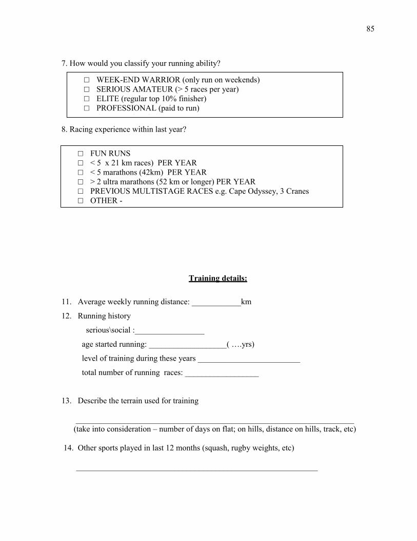
85
7. How would you classify your running ability? 8. Racing experience within last year?
Training details:
11. Average weekly running distance: ____________km
12. Running history
serious\social :_________________
age started running: ___________________( ….yrs)
level of training during these years _________________________
total number of running races: __________________
13. Describe the terrain used for training ____________________________________________________________________ (take into consideration – number of days on flat; on hills, distance on hills, track, etc) 14. Other sports played in last 12 months (squash, rugby weights, etc)
___________________________________________________________
□ WEEK-END WARRIOR (only run on weekends) □ SERIOUS AMATEUR (> 5 races per year) □ ELITE (regular top 10% finisher) □ PROFESSIONAL (paid to run) □ OTHER – SPECIFY………………………….
□ FUN RUNS □ < 5 x 21 km races) PER YEAR □ < 5 marathons (42km) PER YEAR □ > 2 ultra marathons (52 km or longer) PER YEAR □ PREVIOUS MULTISTAGE RACES e.g. Cape Odyssey, 3 Cranes □ OTHER -
SPECIFY……………………………………………………………………...
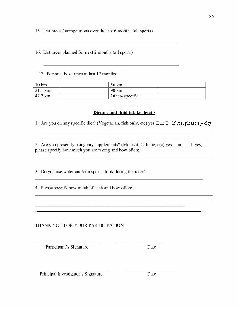
86
15. List races / competitions over the last 6 months (all sports) __________________________________________________________ 16. List races planned for next 2 months (all sports)
__________________________________________________________ 17. Personal best times in last 12 months: 10 km 56 km 21.1 km 90 km 42.2 km Other- specify
Dietary and fluid intake details 1. Are you on any specific diet? (Vegetarian, fish only, etc) yes ________________________________________________________________________________________________________________________________________________ 2. Are you presently using any supplements? (Multivit, Calmag, etc) yes no . If yes, please specify how much you are taking and how often: ________________________________________________________________________________________________________________________________________________ 3. Do you use water and/or a sports drink during the race? ________________________________________________________________________ 4. Please specify how much of each and how often: ________________________________________________________________________________________________________________________________________________________________________________________________________________________ ________________________________________________________________________ THANK YOU FOR YOUR PARTICIPATION ____________________________ ___________________ Participant’s Signature Date _________________________________ ____________________ Principal Investigator’s Signature Date

87
SECTION B: FOR LABORATORY USE ONLY
Pre-event/Post-event: Height (cm): _________________ Mass (kg): __________________
Thigh circumference (cm) ____________________________________ Skin – fold Measurements:
Biceps: ___________ ___________ ____________ __________
Triceps: ___________ ___________ ___________ __________
Suprailliac: __________ __________ __________ __________
Subscapular: _________ __________ ___________ __________
Body fat percentage: ___________________________________
Resting heart rate: _________ /min ______________/min
Resting blood pressure: __________________________________
STUDY NUMBER:…………………
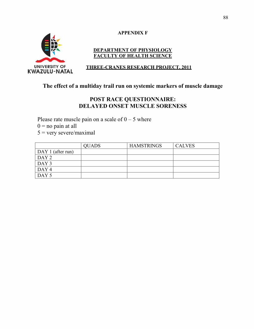
88
APPENDIX F
DEPARTMENT OF PHYSIOLOGY FACULTY OF HEALTH SCIENCE
THREE-CRANES RESEARCH PROJECT, 2011
The effect of a multiday trail run on systemic markers of muscle damage
POST RACE QUESTIONNAIRE: DELAYED ONSET MUSCLE SORENESS
Please rate muscle pain on a scale of 0 – 5 where 0 = no pain at all 5 = very severe/maximal
QUADS HAMSTRINGS CALVES DAY 1 (after run) DAY 2 DAY 3 DAY 4 DAY 5

5th February, 2012, Dear Emmerentia Denissen,
I have the pleasure to inform you that your paper "Low markers of muscle damage and inflammation following a three–day trail run" has been accepted for publication. The publishers will
contact you within 3 weeks and ask you to check the proofs of
your paper which will be published in issue 1, 2012. The PDF
version of the paper will be available for downloading once the
paper has been published.
Thank you for supporting the South African Journal of Sports
Medicine.
regards
Professor Mike Lambert
South African Journal of Sport Medicine
http://www.sajsm.org.za/index.php/sajsm
SOUTH AFRICAN SPORTS MEDICINE ASSOCIATION
Editor-in- Chief SASMA journal Professor Mike Lambert P.O. Box 115, Newlands, 7725 Tel: 021-650-4558 Fax: 021-686-7530 E-mail: [email protected]