Murine Cytomegalovirus Interference with Antigen ... · Murine Cytomegalovirus Interference with...
11
of July 14, 2017. This information is current as CD8 T Cell Response Size or the Effector Memory Phenotype of the Antigen Presentation Has Little Effect on the Murine Cytomegalovirus Interference with Ann B. Hill Mark K. Slifka, Ulrich H. Koszinowski, David H. Raulet and Christopher W. McMahon, Ann Kelly, Daniel G. Kavanagh, Marielle C. Gold, Michael W. Munks, Markus Wagner, http://www.jimmunol.org/content/172/11/6944 doi: 10.4049/jimmunol.172.11.6944 2004; 172:6944-6953; ; J Immunol References http://www.jimmunol.org/content/172/11/6944.full#ref-list-1 , 37 of which you can access for free at: cites 63 articles This article Subscription http://jimmunol.org/subscription is online at: The Journal of Immunology Information about subscribing to Permissions http://www.aai.org/About/Publications/JI/copyright.html Submit copyright permission requests at: Email Alerts http://jimmunol.org/alerts Receive free email-alerts when new articles cite this article. Sign up at: Print ISSN: 0022-1767 Online ISSN: 1550-6606. Immunologists All rights reserved. Copyright © 2004 by The American Association of 1451 Rockville Pike, Suite 650, Rockville, MD 20852 The American Association of Immunologists, Inc., is published twice each month by The Journal of Immunology by guest on July 14, 2017 http://www.jimmunol.org/ Downloaded from by guest on July 14, 2017 http://www.jimmunol.org/ Downloaded from
Transcript of Murine Cytomegalovirus Interference with Antigen ... · Murine Cytomegalovirus Interference with...
of July 14, 2017.This information is current as
CD8 T Cell ResponseSize or the Effector Memory Phenotype of theAntigen Presentation Has Little Effect on the Murine Cytomegalovirus Interference with
Ann B. HillMark K. Slifka, Ulrich H. Koszinowski, David H. Raulet andChristopher W. McMahon, Ann Kelly, Daniel G. Kavanagh, Marielle C. Gold, Michael W. Munks, Markus Wagner,
http://www.jimmunol.org/content/172/11/6944doi: 10.4049/jimmunol.172.11.6944
2004; 172:6944-6953; ;J Immunol
Referenceshttp://www.jimmunol.org/content/172/11/6944.full#ref-list-1
, 37 of which you can access for free at: cites 63 articlesThis article
Subscriptionhttp://jimmunol.org/subscription
is online at: The Journal of ImmunologyInformation about subscribing to
Permissionshttp://www.aai.org/About/Publications/JI/copyright.htmlSubmit copyright permission requests at:
Email Alertshttp://jimmunol.org/alertsReceive free email-alerts when new articles cite this article. Sign up at:
Print ISSN: 0022-1767 Online ISSN: 1550-6606. Immunologists All rights reserved.Copyright © 2004 by The American Association of1451 Rockville Pike, Suite 650, Rockville, MD 20852The American Association of Immunologists, Inc.,
is published twice each month byThe Journal of Immunology
by guest on July 14, 2017http://w
ww
.jimm
unol.org/D
ownloaded from
by guest on July 14, 2017
http://ww
w.jim
munol.org/
Dow
nloaded from
Murine Cytomegalovirus Interference with AntigenPresentation Has Little Effect on the Size or the EffectorMemory Phenotype of the CD8 T Cell Response1
Marielle C. Gold,* Michael W. Munks,* Markus Wagner, § Christopher W. McMahon,‡
Ann Kelly,* Daniel G. Kavanagh,* Mark K. Slifka, † Ulrich H. Koszinowski,§ David H. Raulet,‡
and Ann B. Hill 2*
As with most herpesviruses, CMVs encode viral genes that inhibit Ag presentation to CD8 T cells (VIPRs). VIPR function has beenassumed to be essential for CMV to establish its characteristic lifetime infection of its host. We compared infection of C57BL/6mice with wild-type murine CMV (MCMV) and a virus lacking each of MCMV’s three known VIPRs: m4, m6, and m152. Duringacute infection, there was very little difference between the two viruses with respect to the kinetics of viral replication andclearance, or in the size and kinetics of the virus-specific CD8 T cell response. During chronic infection, a large, effector memory,virus-specific CD8 T cell population (CD8lowCD62L�CD11c�NKG2A�) was maintained in both infections; the size and phenotypeof the CD8 T cell response to both viruses was remarkably similar. The characteristic effector memory phenotype of the CD8 Tcells suggested that both wild-type and�m4�m6�m152 virus continued to present Ag to CD8 T cells during the chronic phaseof infection. During the chronic phase of infection, MCMV cannot be isolated from immunocompetent mice. However, uponimmunosuppression, both�m4�m6�m152 and wild-type virus could be reactivated from mice infected for 6 wk. Thus, restoringthe ability of CD8 T cells to detect MCMV had little apparent effect on the course of MCMV infection and on the CD8 T cellresponse to it. These results challenge the notion that VIPR function is necessary for CMV persistence in the host.The Journalof Immunology, 2004, 172: 6944–6953.
V iral immune evasion has been an area of intensive in-vestigation for the past decade. Viruses have been foundto interfere with the functions of cytokines, chemokines,
APCs, apoptotic responses, Abs, complement, NK cells, and Agpresentation to both CD4 and CD8 T cells (1–3). The MHC classI pathway of Ag presentation is a frequent target of virus-encodedimmune evasion genes. Genes mediating this type of immune eva-sion have been called immunoevasins (4) or viral genes that inhibitAg presentation (VIPRs)3 (5). Herpesviruses in particular all seemto encode VIPRs. Most reports have used isolated VIPR genes tocharacterize them, few have looked at VIPR function in the context
of virus-infected cells, and even fewer have attempted to assess theimpact of VIPR function on the immunobiology of virus infection.We use the natural mouse pathogen murine CMV (MCMV) tostudy VIPR function, and report here a comparison of wild-typeMCMV with a virus that lacks MCMV’s three known VIPRs:m4,m6, andm152 (6). Because the other immune evasion functions ofMCMV are intact in both viruses, our study isolates the impact ofthese VIPRs on MCMV infection and the immune response to it.
MCMV is an excellent model in which to dissect the real evo-lutionary role of immune evasion. It is fully sequenced (7), and theexpression of its genome as a bacterial artificial chromosome(BAC) has revolutionized functional genetic analysis (8). Since itsisolation in 1954 (9), its immunobiology has been extensivelystudied, revealing complex and redundant immunological controlmechanisms (4, 10, 11). Any immunological investigation ofMCMV needs to take into account the marked strain difference insusceptibility to MCMV, which is mediated by NK cells. The mostimportant resistance locus (Cmvr-1) has been recently mapped tothe Ly49H gene (12–14), which C57BL/6 mice express andBALB/c mice do not. Ly49H is an activating NK cell receptor thatrecognizes the product of the MCMVm157 gene (15, 16). In con-sequence, MCMV replicates to much higher titers in BALB/c thanB6 mice, especially in the spleen. Most of the previous work onMCMV T cell immunology has been conducted in the BALB/cstrain, and much less is known about the immunobiology of thisvirus in B6 mice.
MCMV encodes three known VIPRS, all of which are glycop-roteins expressed in the early (E) phase of viral gene expression.m152/gp40 causes class I MHC to be retained in the endoplasmicreticulum/Golgi intermediate compartment (ERGIC) (17).m6/gp48 binds to class I and directs it to the lysosome for destruction
*Department of Molecular Microbiology and Immunology, Oregon Health and Sci-ence University, Portland, OR 97239;†Vaccine and Gene Therapy Institute, OregonHealth and Science University, Beaverton, OR 97006;‡Department of Molecular andCell Biology, University of California, Berkeley, CA 94720; and§Max von Petten-kofer Institute, Department for Virology, Ludwig-Maximilians-Universitat, Munich,Germany
Received for publication February 11, 2003. Accepted for publication March26, 2004.
The costs of publication of this article were defrayed in part by the payment of pagecharges. This article must therefore be hereby markedadvertisement in accordancewith 18 U.S.C. Section 1734 solely to indicate this fact.1 This work was funded by National Institutes of Health Grants AI51346 (to M.K.S.),MOI RR00334 (to M.K.S.), and A107474 (to A.B.H.); American Heart AssociationGrant 9650521N (to A.B.H.); and the Pew Charitable Trusts Grant P0176SC (toA.B.H.).2 Address correspondence and reprint requests to Dr. Ann B. Hill, Department ofMolecular Microbiology and Immunology L220, Oregon Health and Science Univer-sity, 3181 Southwest Sam Jackson Park Road, Portland, OR 97239. E-mail address:[email protected] Abbreviations used in this paper: VIPR, viral gene that inhibits Ag presentation;MCMV, murine CMV; BAC, bacterial artificial chromosome; LCMV, lymphocyticchoriomeningitis virus; IE, immediate early; MEF, mouse embryo fibroblast; vv-ova,recombinant vaccinia virus expressing chicken OVA; ICS, intracellular cytokinestaining; BrdU, 5-bromo-2�-deoxyuridine.
The Journal of Immunology
Copyright © 2004 by The American Association of Immunologists, Inc. 0022-1767/04/$02.00
by guest on July 14, 2017http://w
ww
.jimm
unol.org/D
ownloaded from
(18). m4/gp34 is primarily endoplasmic reticulum resident and as-sociates with class I there; a small amount of m4/gp34 complexedwith class I travels to the cell surface where they remain stablyassociated (19). Each of these three VIPRs has been shown toinhibit the ability of MCMV-specific CD8 CTLs to lyse infectedcells expressing H-2b MHC (20, 21); gp40 has also been shown toinhibit H-2d-restricted Ag presentation (22). The relative potencyof the individual VIPRs varies for different allelic variants of MHCclass I (6, 20). Mutagenesis of an MCMV BAC to delete thesegenes alone and in combination indicated that there are no otherMCMV genes that have a significant impact on cell surface MHCclass I (6).
The impact of VIPR function on MCMV infection remainslargely unexplored, and is not readily predicted from the literatureon the role of CD8 T cells in controlling MCMV infection. Forinstance, even when VIPRs are functional, CD8 T cells can impairwild-type MCMV replication in vivo. Adoptively transferred CD8T cells reduce virus titers in the lungs, spleen, liver, and adrenalglands of acutely infected, irradiated BALB/c mice (23, 24) Also,prior immunization with CD8 T cell epitopes protects BALB/cmice against a lethal dose of virus (25, 26). In contrast, animals canbe completely depleted of CD8 T cells in acute (27) or chronic (28)infection without impacting virus control, because of redundantmechanisms contributed by CD4 T cells and NK cells. We pre-dicted that removing VIPRs would enable CD8 T cells to play amuch more dominant and effective role in controlling virus.
There have been three reports of the effect of m152 on acuteinfection in vivo with MCMV. First, Jonjic and colleagues (22)showed that m152 affects the ability of CD8 T cells to control acuteinfection, and that this led to �1 log lower virus titers from days8 to 10 postinfection. This effect was seen in both BALB/c and B6mice. To increase the dependence of host defense on CD8 T cell-mediated control, all of the experiments reported in that paper usedimmunocompromised mice that were either neonatal, B cell defi-cient, or irradiated. Even so, the effect of m152 removal was mod-est, with at best a 1.5 log reduction in titer. In view of the recentdiscovery that m152 can also affect NK function, it should be notedthat this paper clearly showed that m152 affects the function ofCD8 T cells: removing m152 from the virus reduced virus titers atday 10 postinfection in normal B6 mice but had no effect on virustiters on day 10 postinfection in mice lacking �2-microglobulin orCD8 (22).
Second, it has recently been discovered that, in addition to in-hibiting MHC class I-restricted Ag presentation, m152 also inhibitsthe expression of the RAE-1 family of ligands for the NK cell-activating receptor NKG2D (29, 30). Jonjic and colleagues (29)found that, in BALB/c mice, this inhibition of NK function re-sulted in higher virus titers on day 3 postinfection. Thus, in addi-tion to being a VIPR, m152 can mediate anti-NK cell immuneevasion.
Third, we have studied the effect of m152 on the acute CD8 Tcell response to MCMV in B6 mice, looking at its impact on theAg specificity of the immune response. We found that m152 didnot affect the immunodominance of the Db-restricted M45 HGIR-NASFI epitope in acute infection (31). M45 is an early gene, ex-pressed at the same time in the infectious cycle as the immunemodulators. Because m152 profoundly inhibits the ability of M45-specific CD8 T cells to detect infected cells, including macro-phages and DCs, we interpreted this result to suggest that CD8 Tcells in acute MCMV infection are primed by cross-priming ratherthan by infected APCs.
Each of these three studies on the effect of m152 in vivo waslimited to acute infection. Because all herpesviruses appear to en-code VIPRs, and the hallmark of the herpesvirus lifestyle is life-
long survival in the infected host, it is generally assumed thatVIPRs will be necessary for this lifelong survival. In other words,the main phenotype of a VIPR-deficient virus would be predictedto be in the chronic phase of infection. Indeed, VIPR-deficientmutants of the gamma-2-herpesvirus murine gammaherpesvi-rus-68 showed little defect in the acute phase of infection in thelungs, but were markedly impaired in their ability to establishsplenic latency (32). After acute infection, MCMV establishes atrue latent infection that is characterized by periodic reactivation,sometimes aborted at the immediate-early (IE) phase of gene ex-pression, and at other times presumably progressing to replication(33, 34). However, unless mice are immunosuppressed, replicatingvirus is not detectable, and even the latent virus DNA load is nearor below the threshold of detection, especially in B6 mice. Virusactivity is believed to be kept to this very low level by effectiveimmune surveillance.
The concept of active immune surveillance in the chronic phaseof MCMV infection is supported by studies of the T cell responsein BALB/c mice. Memory CD8 T cells numbers remain high, theirantigenic specificity narrows with the duration of infection, andthey express an effector memory phenotype, remaining CD62L�
(26, 35, 36). Interestingly, a remarkably similar picture is emerg-ing in studies of human CMV infection. In healthy CMV-seropos-itive adults, �2% of CD4 T cells (37) and 6% of CD8 T cells (11%of memory CD8 T cells) (L. Picker, unpublished observations) arespecific for CMV for the life of the infected individual. In somehealthy individuals, �30% of CD8 T cells are CMV specific (38,39). Many human CMV-specific CD8 T cells have an effector mem-ory phenotype (CD57�CD45RAbrightCD28�CD27�) (40–42).Thus, in the chronic phase of CMV infection in immunocompetenthumans and mice, the size and phenotype of the CD8 T cell re-sponse may be the best indicator of virus activity.
In this study, we report a series of experiments designed toassess the effect of VIPRs on the course of MCMV infection in B6mice, and on the CD8 T cell response to it. We compared wild-type infection with a virus lacking all three known VIPRs: m4, m6,and m152. Our expectation was that a virus lacking VIPRs wouldestablish acute infection, and would probably be able to establishsome latent pool during the 5-day window before effective CD8 Tcell control develops. However, after that, we expected that theVIPR-deficient virus would be rapidly controlled, and as latentvirus reactivated, it would also be recognized and eradicated. Overtime, we expected that this would lead to a reduced or eliminatedlatent virus pool, and in consequence, we predicted that theMCMV-specific CD8s would come to display the familiar pheno-type seen in cleared virus infections such as lymphocytic chorio-meningitis virus (LCMV). We were surprised to find that removingVIPRs had little detectable effect on the course of virus infectionor on the antiviral CD8 T cell response.
Materials and MethodsMice
Female C57BL/6 (B6) mice were purchased from Simonson (Gilroy, CA)or The Jackson Laboratory (Bar Harbor, ME); mice from a single vendorwere used for each experiment. B cell-deficient (�mt) mice were purchasedfrom The Jackson Laboratory. OT-1 transgenic mice were kindly providedby D. Hinrichs (Veterans Affairs Hospital, Portland, OR). All mice werehoused in an isolation suite and used at age 6 wk or greater. Sentinel micewere routinely tested for mouse pathogens, and none were found in thecourse of this study.
Cells
Mouse embryo fibroblasts (MEFs) were grown from trypsin-digested day12–14 mouse embryos, and used between passages 2 and 6. NIH 3T3 cells(CRL-1658), BALB 3T3 cells (CCL-163), and JAWSII cells (CRL-11904)
6945The Journal of Immunology
by guest on July 14, 2017http://w
ww
.jimm
unol.org/D
ownloaded from
were obtained from American Type Culture Collection (Manassas, VA).Cells were maintained in DMEM/10% FBS. JAWSII cells were maintainedin �-MEM supplemented with 20% FBS, sodium pyruvate, nonessentialamino acids, and 5 ng/ml GM-CSF.
Viruses
MCMV Smith was purchased from American Type Culture Collection (no.1399-VR). �MS94.5 (lacking ORFs m151–165) (43), wild-type BACMCMV MW97.01 (44), and �04�m06�m152 (6) have been described.Viruses were grown on B6 MEFs. Stocks were prepared by sonication ofinfected cells. Virus was titered without centrifugal enhancement onBALB-3T3 cells to determine PFU for all stocks. Aliquots of the samevirus stock were used for each experiment. Mice were infected i.p. witheither 5 � 104 or 5 � 106 PFU of MCMV, as described in the figures. Thelower dose was used in earlier experiments in which we assessed CTLresponses only (see Figs. 1, 5, and 6). The higher dose was necessary todetect robust virus replication in B6 mice, and hence was used in the laterexperiments when virus load was assessed in addition to monitoring CD8T cell responses (see Figs. 2–4). A recombinant vaccinia virus expressingchicken OVA (vv-ova) (a gift from J. Yewdell, National Institutes ofHealth, Bethesda, MD) was grown on L929 cells and titered on Vero cells.
Detection of virus in tissues
Organs were dissected and immediately flash frozen in liquid nitrogen (seeFig. 2b), or placed on ice and homogenized by passing through a cellstrainer and frozen. DNA was extracted using DNA Blood mini-kit (no.51106; Qiagen, Valencia, CA) or High Pure Viral Nucleic Acid kit (no. 1858 874; Roche, Basel, Switzerland). Real-time PCR was performed todetect the MCMV IE1 gene exon 1 using the following primers: primer 1,TCG CCC ATC GTT TCG AGA; primer 2, TCT CGT AGG TCC ACTGAC GGA. PCR product was detected with the probe ACT CGA GTCGGA CGC TGC ATC AGA AT labeled with 6-FAM and black holequencher-1, manufactured by Biosearch Technologies (Novato, CA). Copynumbers were determined by reference to a standard curve obtained usingplasmid DNA expressing IE1, a gift from J. Nelson (Oregon Health andScience University).
Immunosuppression to reactivate latent MCMV
For the experiment in Fig. 5, mice were infected with 5 � 106 PFU ofMCMV. Thirty-seven (for �mt mice) or 42 (for B6 mice) days later, micewere injected with cyclophosphamide (150 mg/kg) and 0.3 ml of anti-lymphocyte serum (M4529; Sigma-Aldrich, St. Louis, MO). They werethen injected with 0.3 ml of anti-lymphocyte serum and hydrocortisonesuccinate (125 mg/kg) every 2 days until sacrifice 14 days later. To detectviable virus, salivary gland homogenates were plated onto MEFs until mostof the monolayer showed cytopathic effect. Cells were then scraped andpelleted, and DNA was extracted using a Qiagen DNA Blood mini-kit.Viral genes were detected by PCR using the following primers: IE1, primer1, CAC CAT GGA GCC CGC CGC A, and primer 2, CTT CTT GCT CTTCTT CTT GGG C; m4, primer 1, CAC CAT GTC TCT CGT ATG TCGGCT GGT GTT GGT G, and primer 2, GTT ACT CTT AAG CGG TTTGAA GTT C; m6, primer 1, CAC CAT GCC CAG TTG GAG CGA T, andprimer 2, TTT GGT AAG CAA GGG GGA AGT; and m152, primer 1,CAC CAT GCT GGG CGC TAT CA, and primer 2, CCA CAC GCG GCAGTT GAT GTA
CTL clones and assay
MCMV-specific CTL clones have been previously described (20). The51Cr release assay was conducted as previously described (14), using IFN-�-pretreated MEF targets infected for 16 h with virus at a multiplicity ofinfection of 50 in the presence of phosphonoacetic acid to prevent late geneexpression.
OT-1 T cell transfer
A total of 2�107 whole splenocytes from naive OT-1 TCR transgenic micewas transferred i.v. into unmanipulated naive B6 mice that were either leftuninfected or immediately injected i.p. with 5 � 104 PFU of �MS94.5 or2 � 105 PFU of vv-ova. Splenocytes were analyzed by FACS 7 days later.
Intracellular cytokine staining (ICS) and tetramer staining
For ICS, JAWSII cells were infected with MCMV (�MS94.5 at a multi-plicity of infection of 30–50) for 16 h in the presence of phosphonoaceticacid. Effector splenocytes were isolated from MCMV-infected mice andincubated at a ratio of 1:1 with infected or uninfected APCs for 6 h in thepresence of brefeldin A (GolgiPlug; BD PharMingen, San Diego, CA).
Cells were washed, incubated with FcBlock (BD Biosciences, MountainView, CA), and surface stained with Abs to the CD8� chain conjugated toPE-Cy5 (no. 15-0081-83; eBioscience, San Diego, CA) and to NKG2A/C/E using the mAb 20d5 (no. 550520; BD Biosciences), or to CD43 (no.558761; BD Biosciences), CD11c (no. 557400; BD Biosciences) orCD62L (no. 553150; BD Biosciences), all FITC conjugated. Cells werethen fixed and permeabilized using BD PharMingen’s Cytofix/Cytopermkit before staining with an Ab to IFN-� (no. 12-7311-82; BD Biosciences).For BrdU staining, the BD PharMingen BrdU Flow kit (2345KK) was usedaccording to the manufacturer’s protocol. CD8 T cells were analyzed byflow cytometry using CellQuest software (BD Biosciences). All furtheranalyses were performed using FlowJo software (Treestar, San Carlos,CA). Kb-SIINFEKL tetramers were purchased from the Trudeau InstituteMolecular Biology Core Facility (Saranac Lake, NY). Db-HGIRNASFItetramers were used to stain T cells specific for the immunodominant M45epitope (31). Tetramers were generated using HGIRNASFI peptide, fol-lowing the previously described protocols (45, 46), and coupled tostreptavidin-PE.
BrdU labeling
Mice infected for 1 day or 30 wk were injected i.p. with 1.8 mg of 5-bro-mo-2�-deoxyuridine (BrdU; Sigma-Aldrich) on day 1 and subsequently fed0.8 mg/ml BrdU in their drinking water for 7 days before sacrifice.
ResultsAssays used to monitor the CD8 T cell response to MCMVinfection
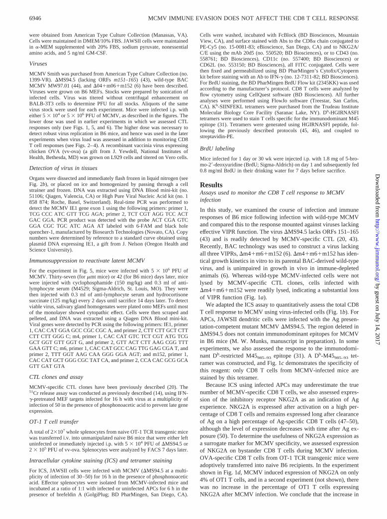
In this study, we examined the course of infection and immuneresponses of B6 mice following infection with wild-type MCMVand compared this to the response mounted against viruses lackingeffective VIPR function. The virus �MS94.5 lacks ORFs 151–165(43) and is readily detected by MCMV-specific CTL (20, 43).Recently, BAC technology was used to construct a virus lackingall three VIPRs, �m4�m6�m152 (6). �m4�m6�m152 has iden-tical growth kinetics in vitro to its parental BAC-derived wild-typevirus, and is unimpaired in growth in vivo in immune-depletedanimals (6). Whereas wild-type MCMV-infected cells were notlysed by MCMV-specific CTL clones, cells infected with�m4�m6�m152 were readily lysed, indicating a substantial lossof VIPR function (Fig. 1a).
We adapted the ICS assay to quantitatively assess the total CD8T cell response to MCMV using virus-infected cells (Fig. 1b). ForAPCs, JAWSII dendritic cells were infected with the Ag presen-tation-competent mutant MCMV �MS94.5. The region deleted in�MS94.5 does not contain immunodominant epitopes for MCMVin B6 mice (M. W. Munks, manuscript in preparation). In someexperiments, we also assessed the response to the immunodomi-nant Db-restricted M45985–93 epitope (31). A Db-M45985–93 tet-ramer was constructed, and Fig. 1c demonstrates the specificity ofthis reagent: only CD8 T cells from MCMV-infected mice arestained by this tetramer.
Because ICS using infected APCs may underestimate the truenumber of MCMV-specific CD8 T cells, we also assessed expres-sion of the inhibitory receptor NKG2A as an indication of Agexperience. NKG2A is expressed after activation on a high per-centage of CD8 T cells and remains expressed long after clearanceof Ag on a high percentage of Ag-specific CD8 T cells (47–50),although the level of expression decreases with time after Ag ex-posure (50). To determine the usefulness of NKG2A expression asa surrogate marker for MCMV specificity, we assessed expressionof NKG2A on bystander CD8 T cells during MCMV infection.OVA-specific CD8 T cells from OT-1 TCR transgenic mice wereadoptively transferred into naive B6 recipients. In the experimentshown in Fig. 1d, MCMV induced expression of NKG2A on only4% of OT1 T cells, and in a second experiment (not shown), therewas no increase in the percentage of OT1 T cells expressingNKG2A after MCMV infection. We conclude that the increase in
6946 MCMV IMMUNE EVASION DOES NOT AFFECT THE CD8 T CELL RESPONSE
by guest on July 14, 2017http://w
ww
.jimm
unol.org/D
ownloaded from
percentage of CD8 cells expressing NKG2A after MCMV infec-tion is a useful surrogate marker for MCMV-specific CD8 T cells.
Comparison of VIPR-deficient and wild-type virus during acuteinfection
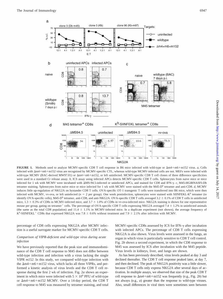
We have previously reported that the peak size and immunodomi-nance of the CD8 T cell response to M45 does not differ betweenwild-type infection and infection with a virus lacking the singleVIPR m152. In this study, we compared wild-type infection withthe �m4�m6�m152 virus, which lacks all three VIPRs, and per-formed a kinetic analysis of virus levels and the CD8 T cell re-sponse during the first 2 wk of infection. Fig. 2a shows an exper-iment in which mice were infected with 5 � 106 PFU of wild-typeor �m4�m6�m152 MCMV. Over a 14-day period, the CD8 Tcell response to M45 was measured by tetramer staining, and total
MCMV-specific CD8s assessed by ICS for IFN-� after incubationwith infected APCs. The percentage of CD8 T cells expressingNKG2A is also shown. Virus levels were assessed in the lungs, anorgan in which virus is particularly sensitive to CD8 T cell control.Fig. 2b shows a second experiment, in which the CD8 response toM45 was assessed by ICS after incubation with the M45 peptide.Virus levels in kidneys, liver, and lungs are shown.
As has been previously described, virus levels peaked at day 3 anddeclined thereafter. The CD8 T cell response peaked later, at day 7,and then declined. The peak of NKG2A positivity was a little slower,because CD8 T cells only express NKG2A after several days of ac-tivation. In multiple assays, we observed that size of the peak CD8 Tcell response to �m4�m6�m152 was frequently (e.g., Fig. 2b) butnot always (e.g., a) greater than the response to wild-type viruses.Also, small differences in viral titers were sometimes seen between
FIGURE 1. Methods used to analyze MCMV-specific CD8 T cell response in B6 mice infected with wild-type or �m4�m6�m152 virus. a, Cellsinfected with �m4�m6�m152 virus are recognized by MCMV-specific CTL, whereas wild-type MCMV-infected cells are not. MEFs were infected withwild-type MCMV (BAC-derived MW97.01) or �m4�m6�m152, or left uninfected. MCMV-specific CD8 T cell clones of three difference specificitieswere used in a standard Cr release assay. b, ICS assay using infected APCs detects MCMV-specific CD8 T cells. Splenocytes from naive mice or miceinfected for 1 wk with MCMV were incubated with �MS-94.5-infected or uninfected APCs, and stained for CD8 and IFN-�. c, M45-HGIRNASFI-Dbtetramer staining. Splenocytes from naive mice or mice infected for 1 wk with MCMV were stained with the M45-Db tetramer and anti-CD8. d, MCMVinduces little up-regulation of NKG2A on bystander CD8 T cells. OVA-specific OT-1 transgenic T cells were transferred into B6 mice, which were theninfected with MCMV, vv-ova, or left uninfected (n � 2 per group). One week postinfection, splenocytes were stained with SIINFEKL-Kb tetramer (toidentify OVA-specific cells), M45-Db tetramer, anti-CD8, and anti-NKG2A. OVA-specific CD8 T cells averaged 3.2 0.1% of CD8 T cells in uninfectedmice, 1.3 0.3% of CD8s in MCMV-infected mice, and 3.7 1.0% of CD8s in vv-ova-infected mice. NKG2A staining is shown for one representativemouse per group, gating on tetramer� cells. The percentage of OVA-specific CD8 T cells expressing NKG2A averaged 7.4 1.2% in uninfected animals(the same as the total CD8 population) and 11.4 1.1% in MCMV-infected mice. In a duplicate experiment (not shown), the average frequency ofKb-SIINFEKL� CD8s that expressed NKG2A was 7.8 0.6% without treatment and 7.9 2.1% after infection with MCMV.
6947The Journal of Immunology
by guest on July 14, 2017http://w
ww
.jimm
unol.org/D
ownloaded from
wild-type and �m4�m6�m152 at some time points. However, over-all, the remarkable feature of these experiments was the similaritybetween wild-type and �m4�m6�m152 infections.
Comparison of VIPR-deficient and wild-type viruses during thechronic/latent phase of infection
We were surprised that VIPR-mediated impairment of infectedcells had little effect on the course of acute MCMV infection andon the antiviral CD8 T cell response. However, as with many othervirus infections, the initial control of MCMV infection may beprimarily a function of NK cells: an effective CD8 response doesnot develop until day 5 postinfection, a time at which virus titersare already falling (Fig. 2). Furthermore, we have previously sug-gested that, in MCMV infection, naive CD8 T cells may be ini-tially primed in vivo primarily through cross-presented Ag (31),which would explain why VIPR function had little effect on thesize of the initial CD8 T cell response. However, because cellsinfected with �m4�m6�m152 are readily detected and lysed byCTL (Fig. 1a), we expected that the virus would be eventuallycleared and that the numbers of MCMV-specific CD8 T cellswould then decline and show the characteristic features of a CD8T cell response to a cleared virus infection. Specifically, we ex-pected that MCMV-specific CD8 T cell numbers in the memoryphase would be 5–10% of those seen at the peak, and the pheno-type would revert in time to a resting, central memory phenotype
(CD62L�). In contrast, we expected that mice infected with wild-type virus in which the VIPRs are functional would maintain ahigher level of virus activity, resulting in higher numbers of mem-ory CD8 T cells, and that these cells would display the character-istic effector memory phenotype (CD62L�) that has been de-scribed in chronic CMV infection in mice and humans.
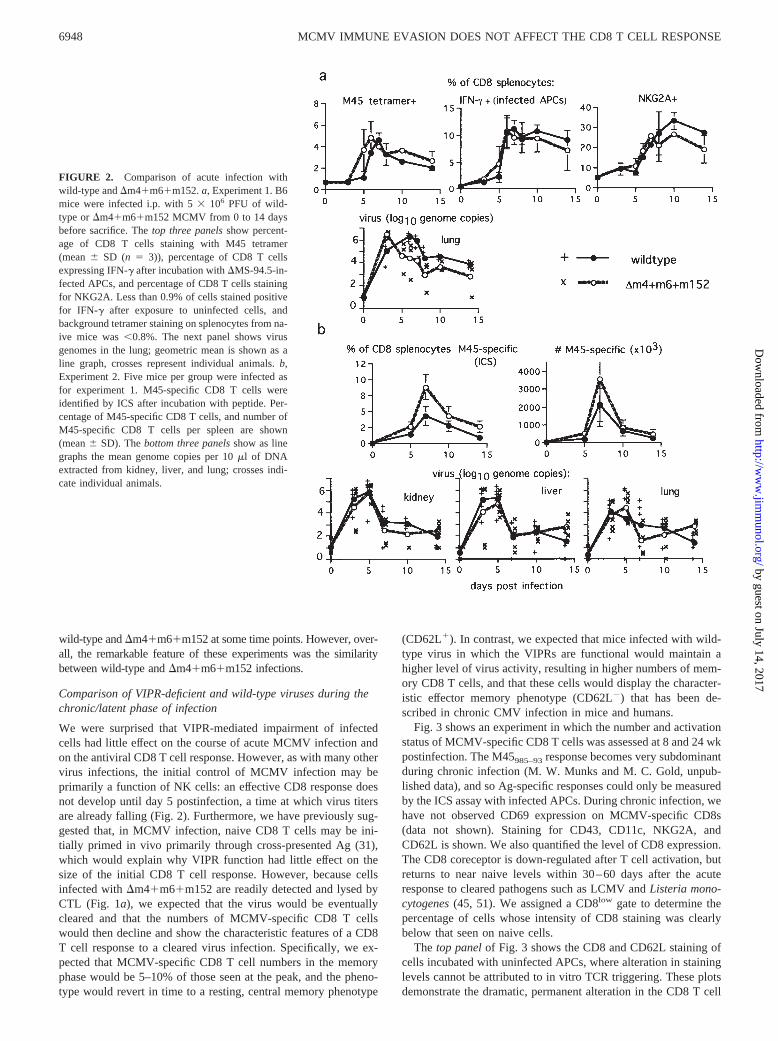
Fig. 3 shows an experiment in which the number and activationstatus of MCMV-specific CD8 T cells was assessed at 8 and 24 wkpostinfection. The M45985–93 response becomes very subdominantduring chronic infection (M. W. Munks and M. C. Gold, unpub-lished data), and so Ag-specific responses could only be measuredby the ICS assay with infected APCs. During chronic infection, wehave not observed CD69 expression on MCMV-specific CD8s(data not shown). Staining for CD43, CD11c, NKG2A, andCD62L is shown. We also quantified the level of CD8 expression.The CD8 coreceptor is down-regulated after T cell activation, butreturns to near naive levels within 30–60 days after the acuteresponse to cleared pathogens such as LCMV and Listeria mono-cytogenes (45, 51). We assigned a CD8low gate to determine thepercentage of cells whose intensity of CD8 staining was clearlybelow that seen on naive cells.
The top panel of Fig. 3 shows the CD8 and CD62L staining ofcells incubated with uninfected APCs, where alteration in staininglevels cannot be attributed to in vitro TCR triggering. These plotsdemonstrate the dramatic, permanent alteration in the CD8 T cell
FIGURE 2. Comparison of acute infection withwild-type and �m4�m6�m152. a, Experiment 1. B6mice were infected i.p. with 5 � 106 PFU of wild-type or �m4�m6�m152 MCMV from 0 to 14 daysbefore sacrifice. The top three panels show percent-age of CD8 T cells staining with M45 tetramer(mean SD (n � 3)), percentage of CD8 T cellsexpressing IFN-� after incubation with �MS-94.5-in-fected APCs, and percentage of CD8 T cells stainingfor NKG2A. Less than 0.9% of cells stained positivefor IFN-� after exposure to uninfected cells, andbackground tetramer staining on splenocytes from na-ive mice was 0.8%. The next panel shows virusgenomes in the lung; geometric mean is shown as aline graph, crosses represent individual animals. b,Experiment 2. Five mice per group were infected asfor experiment 1. M45-specific CD8 T cells wereidentified by ICS after incubation with peptide. Per-centage of M45-specific CD8 T cells, and number ofM45-specific CD8 T cells per spleen are shown(mean SD). The bottom three panels show as linegraphs the mean genome copies per 10 �l of DNAextracted from kidney, liver, and lung; crosses indi-cate individual animals.
6948 MCMV IMMUNE EVASION DOES NOT AFFECT THE CD8 T CELL RESPONSE
by guest on July 14, 2017http://w
ww
.jimm
unol.org/D
ownloaded from
compartment engendered by MCMV infection: mice infected withMCMV had a much expanded CD62L�CD8low effector memoryCD8 T cell population at both 8 and 22 wk. Strikingly, the patternwas very similar for wild-type and �m4�m6�m152 viruses. Therest of the panels show staining of cells exposed to infected APCs.Around 13% of CD8 T cells made IFN-� in response to infectedAPCs at both 8 and 22 wk. Most of these cells were NKG2A�,indicating Ag experience. About one-half expressed CD11c, whichis up-regulated on effector CD8 T cells (52–54). About one-halfwere CD8low, suggesting that they had encountered Ag within �60days. Only a small percentage (6–17%) displayed the acute effec-tor marker CD43. Interestingly, consistent with previous reports,the IFN-�� CD8 T cells were almost exclusively CD62L negative,which is considered the hallmark of the effector memory pheno-type. Although some CD62L loss may have occurred during the6-h incubation with brefeldin A, most of this loss is likely to haveoccurred in vivo, as a high proportion of CD8 T cells wereCD62L� without exposure to Ag (Fig. 3, top panel). Thus, a largeproportion of the MCMV-specific CD8 T cells in chronic infectionwere CD62L�, NKG2Ahigh, and CD8low, characteristic of an ef-fector memory phenotype and suggestive of repeated exposure toviral Ag. Remarkably, the number and phenotype of the CD8 Tcells in �m4�m6�m152-infected mice were strikingly similar towild-type infection.
Reactivation of wild-type and �m4�m6�m152 viruses frommice infected for 6 wk
During chronic infection, virus cannot be cultured from mice (55).We have occasionally detected viral DNA from homogenized or-gans by real-time PCR in both wild-type and VIPR-deficient in-fections, but frequency of positive samples for both infections istoo low in our hands for statistical comparison.
To assess whether the virus persisted but was undetectable dueto the ongoing immune response, we immunosuppressed chroni-cally infected mice to enable reactivating virus to replicate. Inaddition to B6 mice, we used B cell-deficient (�mt) mice, in whichreactivating virus is easier to detect because it cannot be eliminatedby neutralizing Ab (28). Mice were infected with either wild-typeor �m4�m6�m152 MCMV for 6 wk. They were then immuno-suppressed for 2 wk with a combination of cyclophosphamide,hydrocortisone, and anti-lymphocyte globulin. After sacrifice, viralload in liver, lungs, kidneys, and salivary glands was measured byreal-time PCR. For �mt mice, virus was detected in four of fourwild-type and four of four �m4�m6�m152-infected mice. For B6mice, virus was detected in three of three wild-type and three offour �m4�m6�m152-infected mice. Salivary gland extracts fromB6 mice were plated onto monolayers until viral CPE was de-tected; DNA was extracted and analyzed for presence of viral
FIGURE 3. Comparison of virus-specificCD8 T cell response to chronic infection with�m4�m6�m152 or wild type. Mice were in-fected with wild-type or �m4�m6�m152 virusfor 8 or 24 wk before sacrifice. Splenocyteswere incubated with uninfected or �MS 94.5-infected APCs, and stained for the cell surfacemarkers shown and intracellular IFN-�. FACSplots are shown for one representative mouseper group. The bar graphs show the mean SDof each group of three mice, first as a percentageof the total CD8� gate, and then as a percentageof the IFN-��CD8� gate. The top panel showsstaining of splenocytes exposed to uninfectedAPCs, to demonstrate the direct ex vivo pheno-type. The numbers indicate the percentage oftotal CD8� cells that were CD62L�. The sec-ond panel shows IFN-� staining after exposureto infected APCs. Background IFN-� positivityafter exposure to uninfected APCs was 0.5%,except for one mouse in which it was 1.08%.The next four panels are gated on CD8� cellsand show staining for NKG2A, CD43, CD11c,and CD62L. The numbers indicate the percent-age of IFN-�� cells displaying the indicatedmarker. The bottom panel shows the CD8 his-togram of IFN-�� CD8� cells (solid line), over-laying the CD8 histogram from a representativenaive mouse (dashed line). The number indi-cates the percentage of IFN-�� CD8� cellsscored as CD8low using the gate shown. Threesimilar experiments with two or more mice pergroup showed similar results.
6949The Journal of Immunology
by guest on July 14, 2017http://w
ww
.jimm
unol.org/D
ownloaded from
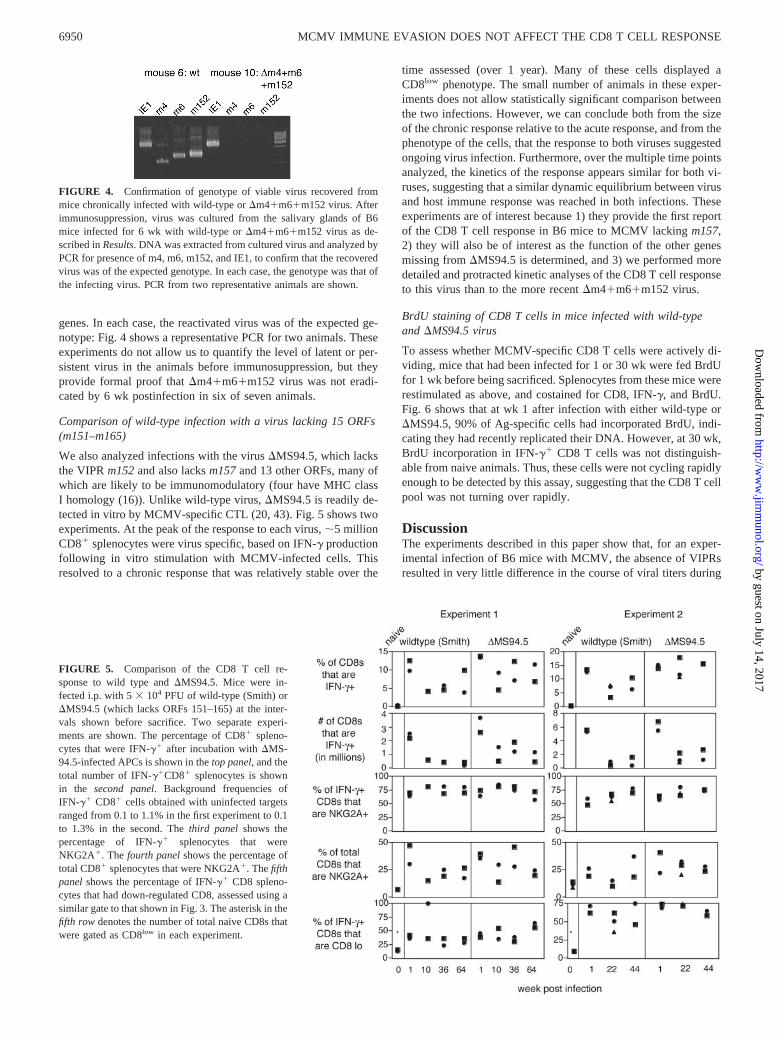
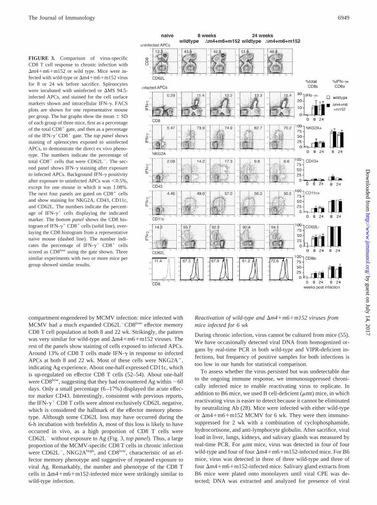
genes. In each case, the reactivated virus was of the expected ge-notype: Fig. 4 shows a representative PCR for two animals. Theseexperiments do not allow us to quantify the level of latent or per-sistent virus in the animals before immunosuppression, but theyprovide formal proof that �m4�m6�m152 virus was not eradi-cated by 6 wk postinfection in six of seven animals.
Comparison of wild-type infection with a virus lacking 15 ORFs(m151–m165)
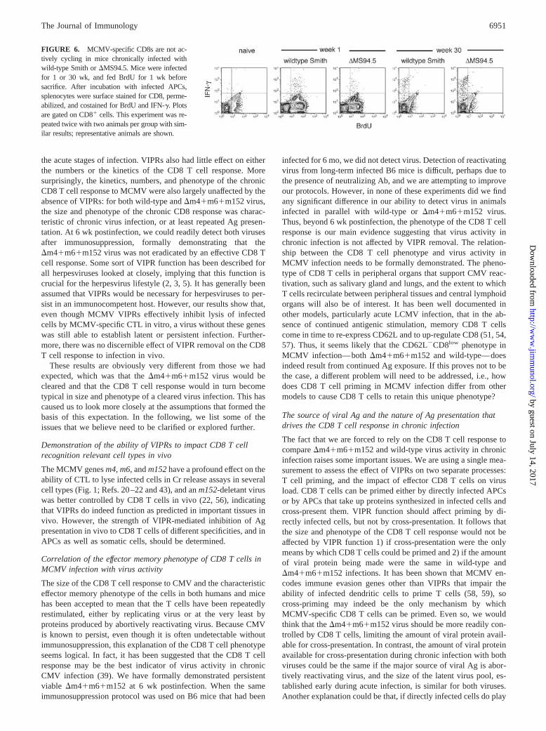
We also analyzed infections with the virus �MS94.5, which lacksthe VIPR m152 and also lacks m157 and 13 other ORFs, many ofwhich are likely to be immunomodulatory (four have MHC classI homology (16)). Unlike wild-type virus, �MS94.5 is readily de-tected in vitro by MCMV-specific CTL (20, 43). Fig. 5 shows twoexperiments. At the peak of the response to each virus, �5 millionCD8� splenocytes were virus specific, based on IFN-� productionfollowing in vitro stimulation with MCMV-infected cells. Thisresolved to a chronic response that was relatively stable over the
time assessed (over 1 year). Many of these cells displayed aCD8low phenotype. The small number of animals in these exper-iments does not allow statistically significant comparison betweenthe two infections. However, we can conclude both from the sizeof the chronic response relative to the acute response, and from thephenotype of the cells, that the response to both viruses suggestedongoing virus infection. Furthermore, over the multiple time pointsanalyzed, the kinetics of the response appears similar for both vi-ruses, suggesting that a similar dynamic equilibrium between virusand host immune response was reached in both infections. Theseexperiments are of interest because 1) they provide the first reportof the CD8 T cell response in B6 mice to MCMV lacking m157,2) they will also be of interest as the function of the other genesmissing from �MS94.5 is determined, and 3) we performed moredetailed and protracted kinetic analyses of the CD8 T cell responseto this virus than to the more recent �m4�m6�m152 virus.
BrdU staining of CD8 T cells in mice infected with wild-typeand �MS94.5 virus
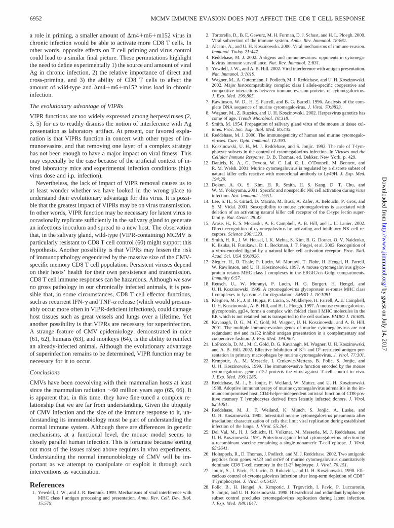
To assess whether MCMV-specific CD8 T cells were actively di-viding, mice that had been infected for 1 or 30 wk were fed BrdUfor 1 wk before being sacrificed. Splenocytes from these mice wererestimulated as above, and costained for CD8, IFN-�, and BrdU.Fig. 6 shows that at wk 1 after infection with either wild-type or�MS94.5, 90% of Ag-specific cells had incorporated BrdU, indi-cating they had recently replicated their DNA. However, at 30 wk,BrdU incorporation in IFN-�� CD8 T cells was not distinguish-able from naive animals. Thus, these cells were not cycling rapidlyenough to be detected by this assay, suggesting that the CD8 T cellpool was not turning over rapidly.
DiscussionThe experiments described in this paper show that, for an exper-imental infection of B6 mice with MCMV, the absence of VIPRsresulted in very little difference in the course of viral titers during
FIGURE 4. Confirmation of genotype of viable virus recovered frommice chronically infected with wild-type or �m4�m6�m152 virus. Afterimmunosuppression, virus was cultured from the salivary glands of B6mice infected for 6 wk with wild-type or �m4�m6�m152 virus as de-scribed in Results. DNA was extracted from cultured virus and analyzed byPCR for presence of m4, m6, m152, and IE1, to confirm that the recoveredvirus was of the expected genotype. In each case, the genotype was that ofthe infecting virus. PCR from two representative animals are shown.
FIGURE 5. Comparison of the CD8 T cell re-sponse to wild type and �MS94.5. Mice were in-fected i.p. with 5 � 104 PFU of wild-type (Smith) or�MS94.5 (which lacks ORFs 151–165) at the inter-vals shown before sacrifice. Two separate experi-ments are shown. The percentage of CD8� spleno-cytes that were IFN-�� after incubation with �MS-94.5-infected APCs is shown in the top panel, and thetotal number of IFN-��CD8� splenocytes is shownin the second panel. Background frequencies ofIFN-�� CD8� cells obtained with uninfected targetsranged from 0.1 to 1.1% in the first experiment to 0.1to 1.3% in the second. The third panel shows thepercentage of IFN-�� splenocytes that wereNKG2A�. The fourth panel shows the percentage oftotal CD8� splenocytes that were NKG2A�. The fifthpanel shows the percentage of IFN-�� CD8 spleno-cytes that had down-regulated CD8, assessed using asimilar gate to that shown in Fig. 3. The asterisk in thefifth row denotes the number of total naive CD8s thatwere gated as CD8low in each experiment.
6950 MCMV IMMUNE EVASION DOES NOT AFFECT THE CD8 T CELL RESPONSE
by guest on July 14, 2017http://w
ww
.jimm
unol.org/D
ownloaded from
the acute stages of infection. VIPRs also had little effect on eitherthe numbers or the kinetics of the CD8 T cell response. Moresurprisingly, the kinetics, numbers, and phenotype of the chronicCD8 T cell response to MCMV were also largely unaffected by theabsence of VIPRs: for both wild-type and �m4�m6�m152 virus,the size and phenotype of the chronic CD8 response was charac-teristic of chronic virus infection, or at least repeated Ag presen-tation. At 6 wk postinfection, we could readily detect both virusesafter immunosuppression, formally demonstrating that the�m4�m6�m152 virus was not eradicated by an effective CD8 Tcell response. Some sort of VIPR function has been described forall herpesviruses looked at closely, implying that this function iscrucial for the herpesvirus lifestyle (2, 3, 5). It has generally beenassumed that VIPRs would be necessary for herpesviruses to per-sist in an immunocompetent host. However, our results show that,even though MCMV VIPRs effectively inhibit lysis of infectedcells by MCMV-specific CTL in vitro, a virus without these geneswas still able to establish latent or persistent infection. Further-more, there was no discernible effect of VIPR removal on the CD8T cell response to infection in vivo.
These results are obviously very different from those we hadexpected, which was that the �m4�m6�m152 virus would becleared and that the CD8 T cell response would in turn becometypical in size and phenotype of a cleared virus infection. This hascaused us to look more closely at the assumptions that formed thebasis of this expectation. In the following, we list some of theissues that we believe need to be clarified or explored further.
Demonstration of the ability of VIPRs to impact CD8 T cellrecognition relevant cell types in vivo
The MCMV genes m4, m6, and m152 have a profound effect on theability of CTL to lyse infected cells in Cr release assays in severalcell types (Fig. 1; Refs. 20–22 and 43), and an m152-deletant viruswas better controlled by CD8 T cells in vivo (22, 56), indicatingthat VIPRs do indeed function as predicted in important tissues invivo. However, the strength of VIPR-mediated inhibition of Agpresentation in vivo to CD8 T cells of different specificities, and inAPCs as well as somatic cells, should be determined.
Correlation of the effector memory phenotype of CD8 T cells inMCMV infection with virus activity
The size of the CD8 T cell response to CMV and the characteristiceffector memory phenotype of the cells in both humans and micehas been accepted to mean that the T cells have been repeatedlyrestimulated, either by replicating virus or at the very least byproteins produced by abortively reactivating virus. Because CMVis known to persist, even though it is often undetectable withoutimmunosuppression, this explanation of the CD8 T cell phenotypeseems logical. In fact, it has been suggested that the CD8 T cellresponse may be the best indicator of virus activity in chronicCMV infection (39). We have formally demonstrated persistentviable �m4�m6�m152 at 6 wk postinfection. When the sameimmunosuppression protocol was used on B6 mice that had been
infected for 6 mo, we did not detect virus. Detection of reactivatingvirus from long-term infected B6 mice is difficult, perhaps due tothe presence of neutralizing Ab, and we are attempting to improveour protocols. However, in none of these experiments did we findany significant difference in our ability to detect virus in animalsinfected in parallel with wild-type or �m4�m6�m152 virus.Thus, beyond 6 wk postinfection, the phenotype of the CD8 T cellresponse is our main evidence suggesting that virus activity inchronic infection is not affected by VIPR removal. The relation-ship between the CD8 T cell phenotype and virus activity inMCMV infection needs to be formally demonstrated. The pheno-type of CD8 T cells in peripheral organs that support CMV reac-tivation, such as salivary gland and lungs, and the extent to whichT cells recirculate between peripheral tissues and central lymphoidorgans will also be of interest. It has been well documented inother models, particularly acute LCMV infection, that in the ab-sence of continued antigenic stimulation, memory CD8 T cellscome in time to re-express CD62L and to up-regulate CD8 (51, 54,57). Thus, it seems likely that the CD62L�CD8low phenotype inMCMV infection—both �m4�m6�m152 and wild-type—doesindeed result from continued Ag exposure. If this proves not to bethe case, a different problem will need to be addressed, i.e., howdoes CD8 T cell priming in MCMV infection differ from othermodels to cause CD8 T cells to retain this unique phenotype?
The source of viral Ag and the nature of Ag presentation thatdrives the CD8 T cell response in chronic infection
The fact that we are forced to rely on the CD8 T cell response tocompare �m4�m6�m152 and wild-type virus activity in chronicinfection raises some important issues. We are using a single mea-surement to assess the effect of VIPRs on two separate processes:T cell priming, and the impact of effector CD8 T cells on virusload. CD8 T cells can be primed either by directly infected APCsor by APCs that take up proteins synthesized in infected cells andcross-present them. VIPR function should affect priming by di-rectly infected cells, but not by cross-presentation. It follows thatthe size and phenotype of the CD8 T cell response would not beaffected by VIPR function 1) if cross-presentation were the onlymeans by which CD8 T cells could be primed and 2) if the amountof viral protein being made were the same in wild-type and�m4�m6�m152 infections. It has been shown that MCMV en-codes immune evasion genes other than VIPRs that impair theability of infected dendritic cells to prime T cells (58, 59), socross-priming may indeed be the only mechanism by whichMCMV-specific CD8 T cells can be primed. Even so, we wouldthink that the �m4�m6�m152 virus should be more readily con-trolled by CD8 T cells, limiting the amount of viral protein avail-able for cross-presentation. In contrast, the amount of viral proteinavailable for cross-presentation during chronic infection with bothviruses could be the same if the major source of viral Ag is abor-tively reactivating virus, and the size of the latent virus pool, es-tablished early during acute infection, is similar for both viruses.Another explanation could be that, if directly infected cells do play
FIGURE 6. MCMV-specific CD8s are not ac-tively cycling in mice chronically infected withwild-type Smith or �MS94.5. Mice were infectedfor 1 or 30 wk, and fed BrdU for 1 wk beforesacrifice. After incubation with infected APCs,splenocytes were surface stained for CD8, perme-abilized, and costained for BrdU and IFN-�. Plotsare gated on CD8� cells. This experiment was re-peated twice with two animals per group with sim-ilar results; representative animals are shown.
6951The Journal of Immunology
by guest on July 14, 2017http://w
ww
.jimm
unol.org/D
ownloaded from
a role in priming, a smaller amount of �m4�m6�m152 virus inchronic infection would be able to activate more CD8 T cells. Inother words, opposite effects on T cell priming and virus controlcould lead to a similar final picture. These permutations highlightthe need to define experimentally 1) the source and amount of viralAg in chronic infection, 2) the relative importance of direct andcross-priming, and 3) the ability of CD8 T cells to affect theamount of wild-type and �m4�m6�m152 virus load in chronicinfection.
The evolutionary advantage of VIPRs
VIPR functions are too widely expressed among herpesviruses (2,3, 5) for us to readily dismiss the notion of interference with Agpresentation as laboratory artifact. At present, our favored expla-nation is that VIPRs function in concert with other types of im-munoevasins, and that removing one layer of a complex strategyhas not been enough to have a major impact on viral fitness. Thismay especially be the case because of the artificial context of in-bred laboratory mice and experimental infection conditions (highvirus dose and i.p. infection).
Nevertheless, the lack of impact of VIPR removal causes us toat least wonder whether we have looked in the wrong place tounderstand their evolutionary advantage for this virus. It is possi-ble that the greatest impact of VIPRs may be on virus transmission.In other words, VIPR function may be necessary for latent virus tooccasionally replicate sufficiently in the salivary gland to generatean infectious inoculum and spread to a new host. The observationthat, in the salivary gland, wild-type (VIPR-containing) MCMV isparticularly resistant to CD8 T cell control (60) might support thishypothesis. Another possibility is that VIPRs may lessen the riskof immunopathology engendered by the massive size of the CMV-specific memory CD8 T cell population. Persistent viruses dependon their hosts’ health for their own persistence and transmission.CD8 T cell immune responses can be hazardous. Although we sawno gross pathology in our chronically infected animals, it is pos-sible that, in some circumstances, CD8 T cell effector functions,such as recurrent IFN-� and TNF-� release (which would presum-ably occur more often in VIPR-deficient infections), could damagehost tissues such as great vessels and lungs over a lifetime. Yetanother possibility is that VIPRs are necessary for superinfection.A strange feature of CMV epidemiology, demonstrated in mice(61, 62), humans (63), and monkeys (64), is the ability to reinfectan already-infected animal. Although the evolutionary advantageof superinfection remains to be determined, VIPR function may benecessary for it to occur.
Conclusions
CMVs have been coevolving with their mammalian hosts at leastsince the mammalian radiation �60 million years ago (65, 66). Itis apparent that, in this time, they have fine-tuned a complex re-lationship that we are far from understanding. Given the ubiquityof CMV infection and the size of the immune response to it, un-derstanding its immunobiology must be part of understanding thenormal immune system. Although there are differences in geneticmechanisms, at a functional level, the mouse model seems toclosely parallel human infection. This is fortunate because sortingout most of the issues raised above requires in vivo experiments.Understanding the normal immunobiology of CMV will be im-portant as we attempt to manipulate or exploit it through suchinterventions as vaccination.
References1. Yewdell, J. W., and J. R. Bennink. 1999. Mechanisms of viral interference with
MHC class I antigen processing and presentation. Annu. Rev. Cell. Dev. Biol.15:579.
2. Tortorella, D., B. E. Gewurz, M. H. Furman, D. J. Schust, and H. L. Ploegh. 2000.Viral subversion of the immune system. Annu. Rev. Immunol. 18:861.
3. Alcami, A., and U. H. Koszinowski. 2000. Viral mechanisms of immune evasion.Immunol. Today 21:447.
4. Reddehase, M. J. 2002. Antigens and immunoevasins: opponents in cytomega-lovirus immune surveillance. Nat. Rev. Immunol. 2:831.
5. Yewdell, J. W., and A. B. Hill. 2002. Viral interference with antigen presentation.Nat. Immunol. 3:1019.
6. Wagner, M., A. Gutermann, J. Podlech, M. J. Reddehase, and U. H. Koszinowski.2002. Major histocompatibility complex class I allele-specific cooperative andcompetitive interactions between immune evasion proteins of cytomegalovirus.J. Exp. Med. 196:805.
7. Rawlinson, W. D., H. E. Farrell, and B. G. Barrell. 1996. Analysis of the com-plete DNA sequence of murine cytomegalovirus. J. Virol. 70:8833.
8. Wagner, M., Z. Ruzsics, and U. H. Koszinowski. 2002. Herpesvirus genetics hascome of age. Trends Microbiol. 10:318.
9. Smith, M. 1954. Propagation of salivary gland virus of the mouse in tissue cul-tures. Proc. Soc. Exp. Biol. Med. 86:435.
10. Reddehase, M. J. 2000. The immunogenicity of human and murine cytomegalo-viruses. Curr. Opin. Immunol. 12:390.
11. Koszinowski, U. H., M. J. Reddehase, and S. Jonjic. 1993. The role of T-lym-phocyte subsets in the control of cytomegalovirus infection. In Viruses and theCellular Immune Response. D. B. Thomas, ed. Dekker, New York, p. 429.
12. Daniels, K. A., G. Devora, W. C. Lai, C. L. O’Donnell, M. Bennett, andR. M. Welsh. 2001. Murine cytomegalovirus is regulated by a discrete subset ofnatural killer cells reactive with monoclonal antibody to Ly49H. J. Exp. Med.194:29.
13. Dokun, A. O., S. Kim, H. R. Smith, H. S. Kang, D. T. Chu, andW. M. Yokoyama. 2001. Specific and nonspecific NK cell activation during virusinfection. Nat. Immunol. 2:951.
14. Lee, S. H., S. Girard, D. Macina, M. Busa, A. Zafer, A. Belouchi, P. Gros, andS. M. Vidal. 2001. Susceptibility to mouse cytomegalovirus is associated withdeletion of an activating natural killer cell receptor of the C-type lectin super-family. Nat. Genet. 28:42.
15. Arase, H., E. S. Mocarski, A. E. Campbell, A. B. Hill, and L. L. Lanier. 2002.Direct recognition of cytomegalovirus by activating and inhibitory NK cell re-ceptors. Science 296:1323.
16. Smith, H. R., J. W. Heusel, I. K. Mehta, S. Kim, B. G. Dorner, O. V. Naidenko,K. Iizuka, H. Furukawa, D. L. Beckman, J. T. Pingel, et al. 2002. Recognition ofa virus-encoded ligand by a natural killer cell activation receptor. Proc. Natl.Acad. Sci. USA 99:8826.
17. Ziegler, H., R. Thale, P. Lucin, W. Muranyi, T. Flohr, H. Hengel, H. Farrell,W. Rawlinson, and U. H. Koszinowski. 1997. A mouse cytomegalovirus glyco-protein retains MHC class I complexes in the ERGIC/cis-Golgi compartments.Immunity 6:57.
18. Reusch, U., W. Muranyi, P. Lucin, H. G. Burgert, H. Hengel, andU. H. Koszinowski. 1999. A cytomegalovirus glycoprotein re-routes MHC classI complexes to lysosomes for degradation. EMBO J. 18:1081.
19. Kleijnen, M. F., J. B. Huppa, P. Lucin, S. Mukherjee, H. Farrell, A. E. Campbell,U. H. Koszinowski, A. B. Hill, and H. L. Ploegh. 1997. A mouse cytomegalovirusglycoprotein, gp34, forms a complex with folded class I MHC molecules in theER which is not retained but is transported to the cell surface. EMBO J. 16:685.
20. Kavanagh, D. G., M. C. Gold, M. Wagner, U. H. Koszinowski, and A. B. Hill.2001. The multiple immune-evasion genes of murine cytomegalovirus are notredundant: m4 and m152 inhibit antigen presentation in a complementary andcooperative fashion. J. Exp. Med. 194:967.
21. LoPiccolo, D. M., M. C. Gold, D. G. Kavanagh, M. Wagner, U. H. Koszinowski,and A. B. Hill. 2002. Effective Inhibition of Kb- and Db-restricted antigen pre-sentation in primary macrophages by murine cytomegalovirus. J. Virol. 77:301.
22. Krmpotic, A., M. Messerle, I. Crnkovic-Mertens, B. Polic, S. Jonjic, andU. H. Koszinowski. 1999. The immunoevasive function encoded by the mousecytomegalovirus gene m152 protects the virus against T cell control in vivo.J. Exp. Med. 190:1285.
23. Reddehase, M. J., S. Jonjic, F. Weiland, W. Mutter, and U. H. Koszinowski.1988. Adoptive immunotherapy of murine cytomegalovirus adrenalitis in the im-munocompromised host: CD4-helper-independent antiviral function of CD8-pos-itive memory T lymphocytes derived from latently infected donors. J. Virol.62:1061.
24. Reddehase, M. J., F. Weiland, K. Munch, S. Jonjic, A. Luske, andU. H. Koszinowski. 1985. Interstitial murine cytomegalovirus pneumonia afterirradiation: characterization of cells that limit viral replication during establishedinfection of the lungs. J. Virol. 55:264.
25. Del Val, M., H. J. Schlicht, H. Volkmer, M. Messerle, M. J. Reddehase, andU. H. Koszinowski. 1991. Protection against lethal cytomegalovirus infection bya recombinant vaccine containing a single nonameric T-cell epitope. J. Virol.65:3641.
26. Holtappels, R., D. Thomas, J. Podlech, and M. J. Reddehase. 2002. Two antigenicpeptides from genes m123 and m164 of murine cytomegalovirus quantitativelydominate CD8 T-cell memory in the H-2d haplotype. J. Virol. 76:151.
27. Jonjic, S., I. Pavic, P. Lucin, D. Rukavina, and U. H. Koszinowski. 1990. Effi-cacious control of cytomegalovirus infection after long-term depletion of CD8�
T lymphocytes. J. Virol. 64:5457.28. Polic, B., H. Hengel, A. Krmpotic, J. Trgovcich, I. Pavic, P. Luccaronin,
S. Jonjic, and U. H. Koszinowski. 1998. Hierarchical and redundant lymphocytesubset control precludes cytomegalovirus replication during latent infection.J. Exp. Med. 188:1047.
6952 MCMV IMMUNE EVASION DOES NOT AFFECT THE CD8 T CELL RESPONSE
by guest on July 14, 2017http://w
ww
.jimm
unol.org/D
ownloaded from
29. Krmpotic, A., D. H. Busch, I. Bubic, F. Gebhardt, H. Hengel, M. Hasan,A. A. Scalzo, U. H. Koszinowski, and S. Jonjic. 2002. MCMV glycoprotein gp40confers virus resistance to CD8� T cells and NK cells in vivo. Nat. Immunol.3:529.
30. Lodoen, M., K. Ogasawara, J. A. Hamerman, H. Arase, J. P. Houchins,E. S. Mocarski, and L. L. Lanier. 2003. NKG2D-mediated natural killer cellprotection against cytomegalovirus is impaired by viral gp40 modulation of reti-noic acid early inducible 1 gene molecules. J. Exp. Med. 197:1245.
31. Gold, M. C., M. W. Munks, M. Wagner, U. H. Koszinowski, A. B. Hill, andS. P. Fling. 2002. The murine cytomegalovirus immunodomulatory gene m152prevents recognition of infected cells by M45-specific CTL, but does not alter theimmunodominance of the M45-specific CD8 T cell response in vivo. J. Immunol.169:359.
32. Stevenson, P. G., J. S. May, X. G. Smith, S. Marques, H. Adler, U. H. Koszinowski,J. P. Simas, and S. Efstathiou. 2002. K3-mediated evasion of CD8� T cells aidsamplification of a latent �-herpesvirus. Nat. Immunol. 3:733.
33. Kurz, S., H. P. Steffens, A. Mayer, J. R. Harris, and M. J. Reddehase. 1997.Latency versus persistence or intermittent recurrences: evidence for a latent stateof murine cytomegalovirus in the lungs. J. Virol. 71:2980.
34. Kurz, S. K., and M. J. Reddehase. 1999. Patchwork pattern of transcriptionalreactivation in the lungs indicates sequential checkpoints in the transition frommurine cytomegalovirus latency to recurrence. J. Virol. 73:8612.
35. Holtappels, R., M. F. Pahl-Seibert, D. Thomas, and M. J. Reddehase. 2000. En-richment of immediate-early 1 (m123/pp89) peptide-specific CD8 T cells in apulmonary CD62Llo memory-effector cell pool during latent murine cytomega-lovirus infection of the lungs. J. Virol. 74:11495.
36. Karrer, U., S. Sierro, M. Wagner, A. Oxenius, H. Hengel, U. H. Koszinowski,R. E. Phillips, and P. Klenerman. 2003. Memory inflation: continuous accumu-lation of antiviral CD8� T cells over time. J. Immunol. 170:2022.
37. Sester, M., U. Sester, B. Gartner, B. Kubuschok, M. Girndt, A. Meyerhans, andH. Kohler. 2002. Sustained high frequencies of specific CD4 T cells restricted toa single persistent virus. J. Virol. 76:3748.
38. Lang, K. S., A. Moris, C. Gouttefangeas, S. Walter, V. Teichgraber, M. Miller,D. Wernet, K. Hamprecht, H. G. Rammensee, and S. Stevanovic. 2002. Highfrequency of human cytomegalovirus (HCMV)-specific CD8� T cells detected ina healthy CMV-seropositive donor. Cell. Mol. Life Sci. 59:1076.
39. Khan, N., N. Shariff, M. Cobbold, R. Bruton, J. A. Ainsworth, A. J. Sinclair,L. Nayak, and P. A. Moss. 2002. Cytomegalovirus seropositivity drives the CD8T cell repertoire toward greater clonality in healthy elderly individuals. J. Im-munol. 169:1984.
40. Weekes, M. P., A. J. Carmichael, M. R. Wills, K. Mynard, and J. G. Sissons.1999. Human CD28�CD8� T cells contain greatly expanded functional virus-specific memory CTL clones. J. Immunol. 162:7569.
41. Kern, F., E. Khatamzas, I. Surel, C. Frommel, P. Reinke, S. L. Waldrop,L. J. Picker, and H. D. Volk. 1999. Distribution of human CMV-specific memoryT cells among the CD8pos. subsets defined by CD57, CD27, and CD45 isoforms.Eur. J. Immunol. 29:2908.
42. Gillespie, G. M., M. R. Wills, V. Appay, C. O’Callaghan, M. Murphy, N. Smith,P. Sissons, S. Rowland-Jones, J. I. Bell, and P. A. Moss. 2000. Functional het-erogeneity and high frequencies of cytomegalovirus-specific CD8� T lympho-cytes in healthy seropositive donors. J. Virol. 74:8140.
43. Thale, R., U. Szepan, H. Hengel, G. Geginat, P. Lucin, and U. H. Koszinowski.1995. Identification of the mouse cytomegalovirus genomic region affecting ma-jor histocompatibility complex class I molecule transport. J. Virol. 69:6098.
44. Wagner, M., S. Jonjic, U. H. Koszinowski, and M. Messerle. 1999. Systematicexcision of vector sequences from the BAC-cloned herpesvirus genome duringvirus reconstitution. J. Virol. 73:7056.
45. Busch, D. H., I. M. Pilip, S. Vijh, and E. G. Pamer. 1998. Coordinate regulationof complex T cell populations responding to bacterial infection. Immunity 8:353.
46. Murali-Krishna, K., J. D. Altman, M. Suresh, D. J. Sourdive, A. J. Zajac,J. D. Miller, J. Slansky, and R. Ahmed. 1998. Counting antigen-specific CD8 Tcells: a reevaluation of bystander activation during viral infection. Immunity8:177.
47. McMahon, C. W., and D. H. Raulet. 2001. Expression and function of NK cellreceptors in CD8� T cells. Curr. Opin. Immunol. 13:465.
48. Moser, J. M., J. Gibbs, P. E. Jensen, and A. E. Lukacher. 2002. CD94-NKG2Areceptors regulate antiviral CD8� T cell responses. Nat. Immunol. 3:189.
49. Miller, J. D., M. Peters, A. E. Oran, G. W. Beresford, L. Harrington, J. M. Boss,and J. D. Altman. 2002. CD94/NKG2 expression does not inhibit cytotoxic func-tion of lymphocytic choriomeningitis virus-specific CD8� T cells. J. Immunol.169:693.
50. McMahon, C. W., A. J. Zajac, A. M. Jamieson, L. Corral, G. E. Hammer,R. Ahmed, and D. H. Raulet. 2002. Viral and bacterial infections induce expres-sion of multiple NK cell receptors in responding CD8� T cells. J. Immunol.169:1444.
51. Slifka, M. K., and J. L. Whitton. 2000. Activated and memory CD8� T cells canbe distinguished by their cytokine profiles and phenotypic markers. J. Immunol.164:208.
52. Huleatt, J. W., and L. Lefrancois. 1995. Antigen-driven induction of CD11c onintestinal intraepithelial lymphocytes and CD8� T cells in vivo. J. Immunol.154:5684.
53. Kim, S. K., K. S. Schluns, and L. Lefrancois. 1999. Induction and visualizationof mucosal memory CD8 T cells following systemic virus infection. J. Immunol.163:4125.
54. Kaech, S. M., S. Hemby, E. Kersh, and R. Ahmed. 2002. Molecular and func-tional profiling of memory CD8 T cell differentiation. Cell 111:837.
55. Reddehase, M. J., J. Podlech, and N. K. Grzimek. 2002. Mouse models of cy-tomegalovirus latency: overview. J. Clin. Virol. 25(Suppl. 2):S23.
56. Holtappels, R., J. Podlech, M. F. Pahl-Seibert, M. Julch, D. Thomas,C. O. Simon, M. Wagner, and M. J. Reddehase. Cytomegalovirus misleads itshost by priming of CD8 T cells specific for an epitope not presented in infectedcells. J. Exp. Med. In press.
57. Wherry, E. J., V. Teichgraber, T. C. Becker, D. Masopust, S. M. Kaech, R. Antia,U. H. von Andrian, and R. Ahmed. 2003. Lineage relationship and protectiveimmunity of memory CD8 T cell subsets. Nat. Immunol. 4:225.
58. Andrews, D. M., C. E. Andoniou, F. Granucci, P. Ricciardi-Castagnoli, andM. A. Degli-Esposti. 2001. Infection of dendritic cells by murine cytomegalovi-rus induces functional paralysis. Nat. Immunol. 2:1077.
59. Mathys, S., T. Schroeder, J. Ellwart, U. H. Koszinowski, M. Messerle, andU. Just. 2003. Dendritic cells under influence of mouse cytomegalovirus have aphysiologic dual role: to initiate and to restrict T cell activation. J. Infect. Dis.187:988.
60. Jonjic, S., W. Mutter, K. Munch, H. Buhring, and U. Koszinowski. 1989. Site-restricted persistent cytomegalovirus infection after selective long-term depletionof CD4 lymphocytes. J. Exp. Med. 169:1199.
61. Booth, T. W., A. A. Scalzo, C. Carrello, P. A. Lyons, H. E. Farrell,G. R. Singleton, and G. R. Shellam. 1993. Molecular and biological character-ization of new strains of murine cytomegalovirus isolated from wild mice. Arch.Virol. 132:209.
62. Rizvanov, A. A., A. G. van Geelen, S. Morzunov, E. W. Otteson, C. Bohlman,G. S. Pari, and S. C. St. Jeor. 2003. Generation of a recombinant cytomegalovirusfor expression of a hantavirus glycoprotein. J. Virol. 77:12203.
63. Boppana, S. B., L. B. Rivera, K. B. Fowler, M. Mach, and W. J. Britt. 2001.Intrauterine transmission of cytomegalovirus to infants of women with precon-ceptional immunity. N. Engl. J. Med. 344:1366.
64. Strelow, L., S. Hansen, K. Buxton, J. Leid, J. Edgar, J. Walker, A. Legasse,M. Axthelm, P. Barry, L. J. Picker, et al. 2003. Evidence of CMV re-infection ina sero-positive host. In 28th International Herpesvirus Workshop, Madison, WI,p. 1.10.
65. McGeoch, D. J., A. Dolan, and A. C. Ralph. 2000. Toward a comprehensivephylogeny for mammalian and avian herpesviruses. J. Virol. 74:10401.
66. McGeoch, D. J., S. Cook, A. Dolan, F. E. Jamieson, and E. A. Telford. 1995.Molecular phylogeny and evolutionary timescale for the family of mammalianherpesviruses. J. Mol. Biol. 247:443.
6953The Journal of Immunology
by guest on July 14, 2017http://w
ww
.jimm
unol.org/D
ownloaded from


![Home | Cancer Research - Transduction and …...[CANCER RESEARCH 51. 3657-3662. July 15. 1991] Transduction and Expression of the Human Carcinoembryonic Antigen Gene in a Murine Colon](https://static.fdocuments.us/doc/165x107/5f56cc44d1215262b86320e8/home-cancer-research-transduction-and-cancer-research-51-3657-3662-july.jpg)