Multiple uses of fibrin sealant for nervous system ... · Multiple uses of fibrin sealant for...
11
REVIEW Open Access Multiple uses of fibrin sealant for nervous system treatment following injury and disease Natalia Perussi Biscola 1,2,3 , Luciana Politti Cartarozzi 3 , Suzana Ulian-Benitez 3,4 , Roberta Barbizan 3,5 , Mateus Vidigal Castro 3 , Aline Barroso Spejo 3 , Rui Seabra Ferreira Jr. 1,2 , Benedito Barraviera 1,2 and Alexandre Leite Rodrigues Oliveira 3* Abstract Lesions to the nervous system often produce hemorrhage and tissue loss that are difficult, if not impossible, to repair. Therefore, scar formation, inflammation and cavitation take place, expanding the lesion epicenter. This significantly worsens the patient conditions and impairment, increasing neuronal loss and glial reaction, which in turn further decreases the chances of a positive outcome. The possibility of using hemostatic substances that also function as a scaffold, such as the fibrin sealant, reduces surgical time and improve postoperative recovery. To date, several studies have demonstrated that human blood derived fibrin sealant produces positive effects in different interventions, becoming an efficient alternative to suturing. To provide an alternative to homologous fibrin sealants, the Center for the Study of Venoms and Venomous Animals (CEVAP, Brazil) has proposed a new bioproduct composed of certified animal components, including a thrombin-like enzyme obtained from snake venom and bubaline fibrinogen. Thus, the present review brings up to date literature assessment on the use of fibrin sealant for nervous system repair and positions the new heterologous bioproduct from CEVAP as an alternative to the commercial counterparts. In this way, clinical and pre-clinical data are discussed in different topics, ranging from central nervous system to peripheral nervous system applications, specifying positive results as well as future enhancements that are necessary for improving the use of fibrin sealant therapy. Keywords: Central nervous system, Peripheral nervous system, Commercial fibrin sealant, New heterologous fibrin sealant, Nervous system injury, Fibrin tissue adhesive Background The nervous system is immensely complex and respon- sible for most of the biological responses and maintenance of homeostasis. It is, however, subject to injuries and pathologies that usually require surgical intervention. Due to its cellular organization, high vascularization and the presence of the blood–brain barrier, to interfere in the nervous tissue parenchyma constitutes a major challenge. The possibility of using a biological scaffold to provide hemostasis, reestablishment of subarachnoid space tightness as well as a vehicle for drug and stem cell delivery opened a new and promising field of research. The use of homologous commercial fibrin sealants (FS) in a number of surgical procedures is now consolidated as an efficient method to avoid suturing, enhancing success ratio and reducing patient recovery time. To provide an alternative to human blood derived fibrin sealants, the Center for the Study of Venoms and Venomous Animals (CEVAP – UNESP) has proposed a new heterologous bioproduct composed of certified animal components, including a thrombin-like enzyme obtained from snake venom and a buffalo-cryoprecipitate rich in fibrinogen [1–4]. After more than 20 years of efforts, this study is now under clinical trials [5–10]. * Correspondence: [email protected] 3 Department of Structural and Functional Biology, Institute of Biology, University of Campinas (UNICAMP), Laboratory of Nerve Regeneration, CEP 13083-970 Campinas, SP, Brazil Full list of author information is available at the end of the article © The Author(s). 2017 Open Access This article is distributed under the terms of the Creative Commons Attribution 4.0 International License (http://creativecommons.org/licenses/by/4.0/), which permits unrestricted use, distribution, and reproduction in any medium, provided you give appropriate credit to the original author(s) and the source, provide a link to the Creative Commons license, and indicate if changes were made. The Creative Commons Public Domain Dedication waiver (http://creativecommons.org/publicdomain/zero/1.0/) applies to the data made available in this article, unless otherwise stated. Biscola et al. Journal of Venomous Animals and Toxins including Tropical Diseases (2017) 23:13 DOI 10.1186/s40409-017-0103-1
Transcript of Multiple uses of fibrin sealant for nervous system ... · Multiple uses of fibrin sealant for...

REVIEW Open Access
Multiple uses of fibrin sealant for nervoussystem treatment following injury anddiseaseNatalia Perussi Biscola1,2,3, Luciana Politti Cartarozzi3, Suzana Ulian-Benitez3,4, Roberta Barbizan3,5,Mateus Vidigal Castro3, Aline Barroso Spejo3, Rui Seabra Ferreira Jr.1,2, Benedito Barraviera1,2
and Alexandre Leite Rodrigues Oliveira3*
Abstract
Lesions to the nervous system often produce hemorrhage and tissue loss that are difficult, if not impossible, torepair. Therefore, scar formation, inflammation and cavitation take place, expanding the lesion epicenter. Thissignificantly worsens the patient conditions and impairment, increasing neuronal loss and glial reaction, which inturn further decreases the chances of a positive outcome. The possibility of using hemostatic substances that alsofunction as a scaffold, such as the fibrin sealant, reduces surgical time and improve postoperative recovery. To date,several studies have demonstrated that human blood derived fibrin sealant produces positive effects in differentinterventions, becoming an efficient alternative to suturing. To provide an alternative to homologous fibrin sealants,the Center for the Study of Venoms and Venomous Animals (CEVAP, Brazil) has proposed a new bioproductcomposed of certified animal components, including a thrombin-like enzyme obtained from snake venom andbubaline fibrinogen. Thus, the present review brings up to date literature assessment on the use of fibrin sealant fornervous system repair and positions the new heterologous bioproduct from CEVAP as an alternative to thecommercial counterparts. In this way, clinical and pre-clinical data are discussed in different topics, ranging fromcentral nervous system to peripheral nervous system applications, specifying positive results as well as futureenhancements that are necessary for improving the use of fibrin sealant therapy.
Keywords: Central nervous system, Peripheral nervous system, Commercial fibrin sealant, New heterologous fibrinsealant, Nervous system injury, Fibrin tissue adhesive
BackgroundThe nervous system is immensely complex and respon-sible for most of the biological responses and maintenanceof homeostasis. It is, however, subject to injuries andpathologies that usually require surgical intervention. Dueto its cellular organization, high vascularization and thepresence of the blood–brain barrier, to interfere in thenervous tissue parenchyma constitutes a major challenge.The possibility of using a biological scaffold to providehemostasis, reestablishment of subarachnoid space
tightness as well as a vehicle for drug and stem celldelivery opened a new and promising field ofresearch.The use of homologous commercial fibrin sealants (FS)
in a number of surgical procedures is now consolidated asan efficient method to avoid suturing, enhancing successratio and reducing patient recovery time. To provide analternative to human blood derived fibrin sealants, theCenter for the Study of Venoms and Venomous Animals(CEVAP – UNESP) has proposed a new heterologousbioproduct composed of certified animal components,including a thrombin-like enzyme obtained from snakevenom and a buffalo-cryoprecipitate rich in fibrinogen[1–4]. After more than 20 years of efforts, this studyis now under clinical trials [5–10].
* Correspondence: [email protected] of Structural and Functional Biology, Institute of Biology,University of Campinas (UNICAMP), Laboratory of Nerve Regeneration, CEP13083-970 Campinas, SP, BrazilFull list of author information is available at the end of the article
© The Author(s). 2017 Open Access This article is distributed under the terms of the Creative Commons Attribution 4.0International License (http://creativecommons.org/licenses/by/4.0/), which permits unrestricted use, distribution, andreproduction in any medium, provided you give appropriate credit to the original author(s) and the source, provide a link tothe Creative Commons license, and indicate if changes were made. The Creative Commons Public Domain Dedication waiver(http://creativecommons.org/publicdomain/zero/1.0/) applies to the data made available in this article, unless otherwise stated.
Biscola et al. Journal of Venomous Animals and Toxins includingTropical Diseases (2017) 23:13 DOI 10.1186/s40409-017-0103-1
The present review positions CEVAP heterologousfibrin sealant (HFS) in the context of nervous systemrepair following trauma and diseases, indicating a pos-sible advantageous use in different instances. Recentliterature is provided and discussed in different topics,ranging from central nervous system (CNS) to periph-eral nervous system (PNS) applications, specifyingpositive results as well as future enhancements that arenecessary for improving the use of fibrin sealant therapy.
BrainThe use of FS in brain lesions is not restricted to itscommon use in the treatment and prevention ofcerebrospinal fluid (CSF) leaks [11]. It has expanded to awide range of surgical interventions including as ahemostatic agent following the total or partial extractionof brain tumors and for coaptation of nerves and brainvessels, replacing conventional sutures [11–13]. Thereare also promising results from combining this valuableadjunct with various drugs and other agents to enhanceregenerative and therapeutic effects after a widespectrum of brain traumas whether accidental, surgicalor even congenital [14–17]. Since the early 20th century,fibrin has been used for stopping cerebral hemorrhageand it is currently being employed through the sealant invarious reparative procedures [2, 18].A cerebrospinal fluid fistula is a condition in which
there is a leak of CSF to the nasal cavity, due to fractureof the skull, resulting from traumatic causes (accidentalor surgical) and non-traumatic causes, also known asspontaneous fistulas [11, 19]. In both cases, the persistentleakage of CSF might cause complications that are respon-sible for significant mortality and morbidity [20]. Mostleaks provoked by head trauma will seal without interven-tion; however, spontaneous or surgically-induced leaksoften require operative repair [20].Some authors described treatment by FS of acute
(intraoperative) cerebrospinal fluid leaks [21, 22]. Greenet al. [21] evaluated FS as an adjunct to sutured duralrepair to obtain intraoperative watertight closure inpatients undergoing elective cranial surgery. The studydemonstrated the superiority of FS over sutures in estab-lishing intraoperative tight closure of a dural incision.Furthermore, Hobbs et al. [22] demonstrated the effect-iveness of FS in 120 patients undergoing pituitarysurgery procedures with intraoperative CSF leaks. Allintraoperative leaks were managed using the FS withdifferent materials, resulting in a low incidence of post-operative CSF leakage.Other authors described FS as preventing postoperative
cerebrospinal fluid leaks [23, 24]. Its use was predomin-antly in cranial procedures with low incidences of postop-erative CSF leaks [11]. Many cases involving patientsundergoing transsphenoidal surgery in which postoperative
CSF leaks significantly decreased were reported [11]. Forexample, Yoshimoto et al. [23] evaluated a FS for preven-tion of postoperative extra dural fluid collection throughthe dural sutures in patients undergoing craniotomy for anunruptured aneurysm. Once again, the study demonstratedthe superiority of the fibrin sealant over sutures. Further-more, a retrospective (historical) study by Kassam et al.[24] evaluated the efficacy and cost-effectiveness of fibrinin patients with intracranial pathological lesions. Theincidence of CSF leaking in matched groups treated withFS or without it were compared. There were no cases ofCSF leak in the group of patients receiving FS. Thus, theauthors conclude that the FS reduces the incidence ofpostoperative CSF leaks.Recent studies in animal models are corroborating the
hypothesis that FS prevents CSF leakage. Hutchinson etal. [25] compared two available FS with a synthetic poly-ethylene glycol (PEG) hydrogel sealant in a canine durot-omy repair model. This well-characterized modelemployed 27 mongrel dogs to assess the ability ofsealants to achieve intraoperative tight seals of the duramater, as well as long-term safety and efficacy. Theapplication of these sealants was 100% effective in pre-venting CSF leakage.Finally, a few authors described FS as a treatment for
persistent CSF leaks. Cappabianca et al. [26] locallyinjected FS in patients following different neurosurgicalprocedures. The injection of FS has proven to be effectivein filling or sealing postoperative recesses and treatingminor or initial CSF leaks, adding another possibility forthreatening postoperative leaks.Besides CSF leaks, postoperative subdural fluid collec-
tion (SFC) is another complication of craniotomy, beingmost frequently employed after aneurysm surgery [27].Most SFC cases eventually disappear or are clinicallyasymptomatic. However, some SFCs enlarge, leading tohygromas or subdural hematomas, which requiresurgical treatment [27]. In this sense, arachnoid plastyhas been demonstrated to be effective for preventingSFC. Several arachnoid plasty methods have beenreported including its sealing with FS or covering withappropriate materials and FS. Thus, Abe et al. [27] ex-amined the efficacy of arachnoid plasty with collagensheet and FS after the clipping of unruptured aneurysms.The procedure achieved favorable outcomes with zeroincidence of SFC or complications such as surgicalinfection.Lee et al. [12] described a series of 26 patients who
underwent microneurosurgical operations in which FSwas used. The patients had various neurological disorders:11 had cerebral aneurysms, 11 had brain tumors, two hadlipomyelomeningoceles, one had cerebral arteriovenousmalformation and one had torn dura resulting from amastoidectomy. The FS was tested and effective in the
Biscola et al. Journal of Venomous Animals and Toxins including Tropical Diseases (2017) 23:13 Page 2 of 11

following procedures: reinforcement of aneurysmal clip-ping; local hemostasis; protection of cerebral veins andsealing of CSF leakage.Fujimura et al. [13] studied the incidence of chronic
hydrocephalus by analyzing a series of 39 patients withsubarachnoid hemorrhage, who underwent perivascularcoating with FS of cerebral arteries after clipping of ananeurysm. The authors concluded that there were nocomplications caused by FS and that it protected thecerebral arteries during the acute phase.Furthermore, there are also promising results associating
fibrin sealants with other components, even in cell therapy.An example of this association is the combination ofcollagen foil or fleece with FS. It is known that the collagenhas been successfully employed as a dural graft for years,but when used in combination with fibrin sealant, it en-hances sealing and tissue regeneration properties, positivelyreflecting on hemostasis and stimulation of tissue repair.Besides, such combination prevents fibrin sealant to bewashed away in cases of CSF leakage. Thus, a combinationof collagen and FS is effective, safe and biocompatible. Nofurther adverse events, complications or toxicity werereported [14–16, 28, 29].Another example is the FS association with stem cells.
Chen et al. [17] investigated the therapeutic effects ofsubdural transplantation of inducible pluripotent stemcells (iPS) mixed with fibrin sealant (iPS-FS) on rats withcerebral ischemia induced by middle cerebral arteryocclusion (MCAO). They demonstrated that subduraliPS-FS enhances recovery from induced stroke and isable to avoid iatrogenic injury to brain parenchyma, thuscomprising a safer alternative approach. In this respect,due to the feasibility of obtaining formulations withvarying characteristics (customization), the use of thederived snake venom sealant enables an association withpotentially different compounds beneficial for the regen-eration process of the nervous system.Thus, it is evident that the FS is a valuable adjuvant to
various microneurosurgical procedures, and potentiallyuseful by contributing to the improvement of surgicaltechniques related to different disorders and adversitiesin the brain and surrounding environment.
Spinal cordSpinal cord injury (SCI) by compression or spondylo-listhesis usually results in cavitation and glial scar forma-tion. Biomatrices with immunomodulatory propertiesare of interest since they may be used to bridge thelesion, reducing the formation of scar tissue, as well asfacilitating axonal growth. In this context, FS could actas a carrier for therapeutic agents, such as neurotrophicfactors and stem cells [30–32].Guest et al. [33] combined fibroblast growth factor
(FGF) and FS to human Schwann-cell grafts which were
engrafted to transected rat spinal cords. Such therapyreduced retrograde axonal degeneration stimulating fiberregeneration throughout the implant. In human patients,a therapeutic combination of FGF and FS was applied tothe injured spinal segment and used to prevent postop-erative CSF leakage. The treatment resulted in signifi-cant motor and sensory improvements [34].FS can be complexed with FGF and nerve grafts as
well. Kuo et al. [35] used autologous peripheral intercos-tal nerve segments combined with FGF in an FS scaffold,implanted to bridge the 5 mm gap in transected ratspinal cords. FGF treatment induced IL-4 expressionwhile nerve grafts induced nerve grow factor (NGF) andbrain-derived neurotrophic factor (BDNF) expression.This combined treatment has also been applied to ani-mals with chronic complete SCI by the removal of scartissue to expose fresh tissue at the surface of the spinalcord stumps [36]. Such approach restored a degree ofhind-limb function [36, 37]. Tsai et al. [38] also treatedspinal cord transection with peripheral nerve grafts andspinal cord anastomosis, both including FGF1 in an FSscaffold. Rats recovered both motor-evoked potentials,recorded at the lumbar level and locomotor functiondue to long tract regeneration.Proteins can be complexed with fibrin matrix. Lord-
Fontaine et al. [39] used rat contusion model and topicalapplication of the protein BA-210 onto the spinal cordusing an FS formulation. BA-210 inactivates Rho, whichactivation is a conserved response in various types ofcentral injuries, thus significantly reducing tissue loss inthe perilesional area and rostrocaudal spreading of lesioncavity. Significant walking abilities were regained morerapidly and more consistently in rats treated withBA-210 [39]. Although a previous work has shown apotential scaffold role for FS, which enhanced FGFand BA-210 treatments, FS action itself has not beenfully evaluated [39].FS is already applied by neurosurgeons as a hemostatic
agent and for the control of cerebrospinal fluid (CSF)leaks [15, 32, 40–42]. In this sense, postoperative CSFleakage is a known complication of spinal surgery. Theideal material to be used in the dural closure is still amatter of debate [43]. Prompt surgery is recommendedto prevent the complications such as meningitis, CSFfistulas, and pseudocyst formation with potential nervecompression [44]. In this regard, FS has been consideredeffective for prevention of CSF leakage in the field ofneurosurgery and spinal surgery [45]. Frequently, durot-omy margin is uneven, and watertight dural closurecannot be achieved only by single sutures. In such cases,the use of a sealant is helpful [45–49].Many authors recommend FS to reinforce the site of
durotomy and have reported that the FS-treated patientspresented a significantly higher rate of tight closure than
Biscola et al. Journal of Venomous Animals and Toxins including Tropical Diseases (2017) 23:13 Page 3 of 11

controls as well as decreased postoperative drainageoutput and time spent at the hospital [45, 48–51]. Percu-taneous therapy of FS in humans with postoperative CSFleaks generated a 50% success rate, similar to the 56%success rate in rats with direct application of FS alone,after experimental induction of CSF rhinorrhea [44, 52].Patel et al. [44] recommend autologous cryoprecipitateuse whenever possible to avoid the risk of blood-bornepathogens, including hepatitis C. The preparation ofcryoprecipitate from autologous blood requires threedays, and 500 mL of whole blood generates 20 to 25 mLof cryoprecipitate [44].An autologous FS has also been used by Nakamura
et al. [45] – in comparison to commercial FS – in patientsundergoing spinal surgery. No complications such as in-fection or continuous CSF leak were observed in any case.The volume of drainage fluid was significantly reduced inthe group subjected to either autologous or commercialFS, when compared to the group without FS. As to safety,the autologous adhesive was equal to the commercialcounterpart. The preventive effects of both adhesives wereequivalent, but the autologous adhesive is much cheaperand provides the advantage of being risk-free of transfu-sion infection.As a treatment for sacral meningeal cysts, Paulsen
et al. [53] determined whether placement of FS afteraspiration could offer a more definitive therapy. The useof FS resulted in marked improvement in all patients, withno evidence of pathology recurrence [54].Although FS use has produced positive results, there are
reports of inconsistent outcomes. Thus, in a retrospectiveanalysis done by Balasubramaniam et al. [43], evaluatingchildren submitted to surgery for various spinal patholo-gies, FS had no effect, though the numbers were statisti-cally too small. Jankowitz et al. [55] reached a similarconclusion that the use of FS did not significantly decreasethe incidence of subsequent CSF leakage while studyingthe potential efficacy of FS TISSEEL® (Baxter) for enhan-cing dural repair after lumbar spine surgery. Consideringthe risk of healing inhibition, the findings did not supportthe prophylactic use of FS when a primary repair isdeemed adequate. Augmentation with muscle, fat, FS, orgraft should be considered when the dural closure issuboptimal.When used to fill the lesion gap after SCI, FS provided
neuroprotective effects. Tissucol® (Baxter) FS was usedby Petter-Puchner et al. [32] after thoracic spinal cordhemisection in rats. Three and seven days after lesion,histology showed a more pronounced inflammatoryresponse triggered by macrophages in the FS-treatedgroup. This difference did not impair behavioral orreflex tests performed at the same time points. At day28, recruitment of macrophages and microglia hadsubstantially decreased and no intergroup difference was
detectable. Substantial benefits were found in relation tomotor function and proprioceptive recovery in the FS-treated group [32]. A similar result was achieved afterintramedullary axotomy and a new heterologous fibrinsealant (HFS) treatment. The HFS-treated group dis-played improved motoneuronal survival after lesion andshowed upregulation of iNOS2 and arginase1 genes,proinflammatory (TNFα and IL1β) and antiinflammatorycytokines (IL10, IL4, and IL13). Thus, HFS enhancedearly macrophage recruitment and proinflammatory cyto-kine expression, which contributed to an acceleration ofinflammation resolution, shown by the increased expres-sion of M2 macrophage markers and antiinflammatorycytokines. The greater inflammation was coupled withbetter motor performance in the walking track test [56].
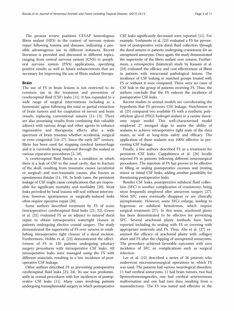
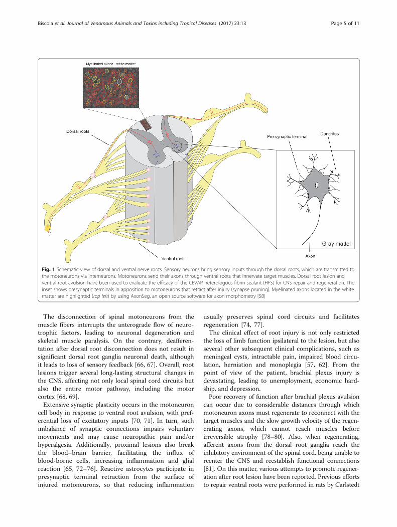
Spinal cord ventral and dorsal rootsSpinal motoneurons are located in the spinal cordventral horn and send their axon towards the peripheryto innervate skeletal muscles. These efferent fibers,among other functions, control the voluntary movementsin response to central brain stimulation and/or sensoryfeedback. Afferent fibers bring sensorial information(touch, temperature, pressure, pain and proprioception)from the periphery to the CNS through the dorsal roots.Sensorial feedback and motor control are crucial in oureveryday life, given their roles in the controlling andadjusting of movements and in adaption to environmentalchanges [57]. Unfortunately, nerve roots can be damaged,thereby disrupting complex and highly specialized neuralnetworks, impairing neural signal transmission.A schematic view of dorsal and ventral nerve roots, as
well as structures of gray and white matter, are repre-sented in Fig. 1. It also illustrates the ventral root avulsionand dorsal root section lesions. Axons in the white matterare highlighted with the program AxonSeg, availableonline [58].Proximal root injury, differently from peripheral nerve
lesion, results in extensive degeneration of adult moto-neurons and loss of sensory feedback since axons cannotregenerate into the spinal cord [59, 60]. When this injuryhappens in an abrupt traction, it is called avulsion[61, 62]. Such lesion or damage frequently occurs insevere brachial plexus injuries due to the high impact ofthe trauma [61]. In cats and dogs, avulsion is normally as-sociated with being hit by vehicles [63, 64]. Nevertheless,in humans, it often happens in vehicle or sport accidentswith limb traction or shoulder depression. In suchaccidents, the brachial plexus can be damaged when thehead is pushed away from the shoulder [57, 65]. Compres-sion or crushing, industrial trauma, and iatrogenic injuryare mechanisms that can also produce root avulsion[61, 62]; and a similar lesion can also happen in thenewborn during childbirth [57].
Biscola et al. Journal of Venomous Animals and Toxins including Tropical Diseases (2017) 23:13 Page 4 of 11
The disconnection of spinal motoneurons from themuscle fibers interrupts the anterograde flow of neuro-trophic factors, leading to neuronal degeneration andskeletal muscle paralysis. On the contrary, deafferen-tation after dorsal root disconnection does not result insignificant dorsal root ganglia neuronal death, althoughit leads to loss of sensory feedback [66, 67]. Overall, rootlesions trigger several long-lasting structural changes inthe CNS, affecting not only local spinal cord circuits butalso the entire motor pathway, including the motorcortex [68, 69].Extensive synaptic plasticity occurs in the motoneuron
cell body in response to ventral root avulsion, with pref-erential loss of excitatory inputs [70, 71]. In turn, suchimbalance of synaptic connections impairs voluntarymovements and may cause neuropathic pain and/orhyperalgesia. Additionally, proximal lesions also breakthe blood–brain barrier, facilitating the influx ofblood-borne cells, increasing inflammation and glialreaction [65, 72–76]. Reactive astrocytes participate inpresynaptic terminal retraction from the surface ofinjured motoneurons, so that reducing inflammation
usually preserves spinal cord circuits and facilitatesregeneration [74, 77].The clinical effect of root injury is not only restricted
the loss of limb function ipsilateral to the lesion, but alsoseveral other subsequent clinical complications, such asmeningeal cysts, intractable pain, impaired blood circu-lation, herniation and monoplegia [57, 62]. From thepoint of view of the patient, brachial plexus injury isdevastating, leading to unemployment, economic hard-ship, and depression.Poor recovery of function after brachial plexus avulsion
can occur due to considerable distances through whichmotoneuron axons must regenerate to reconnect with thetarget muscles and the slow growth velocity of the regen-erating axons, which cannot reach muscles beforeirreversible atrophy [78–80]. Also, when regenerating,afferent axons from the dorsal root ganglia reach theinhibitory environment of the spinal cord, being unable toreenter the CNS and reestablish functional connections[81]. On this matter, various attempts to promote regener-ation after root lesion have been reported. Previous effortsto repair ventral roots were performed in rats by Carlstedt
Fig. 1 Schematic view of dorsal and ventral nerve roots. Sensory neurons bring sensory inputs through the dorsal roots, which are transmitted tothe motoneurons via interneurons. Motoneurons send their axons through ventral roots that innervate target muscles. Dorsal root lesion andventral root avulsion have been used to evaluate the efficacy of the CEVAP heterologous fibrin sealant (HFS) for CNS repair and regeneration. Theinset shows presynaptic terminals in apposition to motoneurons that retract after injury (synapse pruning). Myelinated axons located in the whitematter are highlighted (top left) by using AxonSeg, an open source software for axon morphometry [58]
Biscola et al. Journal of Venomous Animals and Toxins including Tropical Diseases (2017) 23:13 Page 5 of 11

et al. [82] followed by Cullheim et al. in cats [83]. In thesestudies, the avulsed ventral roots were reimplanted on thesurface of spinal cord lateral funiculus. A similartechnique was applied to humans; however, with limitedsuccess [57, 84]. Further experimental ventral rootimplant approaches were carried out using 9/0 non-absorbable sutures (EthilonH®), lithium chloride, tissueglue (TisseelH®), fibrin sealant (TissueCol®; BaxterBVUtrecht, the Netherlands), nerve grafting, biodegrad-able scaffolds and nerve transfer [79, 85–91]. For dorsalroot repair, some promising results towards regenerationwere obtained by using inhibitors of chondroitin sulfateproteoglycans, myelin associated proteins, and by knock-ing down neurotrophin receptors [92–103].The heterologous fibrin sealant derived from snake
venom (HFS), alone or in association with cell therapy,has already shown promising results in the treatment ofdorsal and ventral root injuries [67, 104]. Figure 1 showsthe dorsal root rhizotomy. The HFS usage to reconnectventral and dorsal roots also resulted in the statisticallysignificant preservation of injured motoneurons, improvedsynaptic circuitry recovery, upregulation of trophic factors,and substantial recovery of sensory and motor function[67, 104–107]. Such studies provide a novel approach fortreating spinal cord root lesions, aiming at restoring CNS/PNS interface integrity.
Vidigal de Castro et al. [107] showed a significantrestoration of weight-bearing capacity following ventralroot avulsion (VRA) and reimplantation with theheterologous (HFS) and commercial fibrin sealant (FS),showed by the overview of CatWalk System (Fig. 2) andAdditional file 1 (VRA only), Additional file 2 (VRA +HFS) and Additional file 3 (VRA + FS).
Peripheral nervous systemPeripheral nerve injuries lead to the disconnection of thenervous system with target organs, resulting in paralysisand numbness. Incomplete injuries usually cause pharma-cologically resistant neuropathic pain [108]. Thus, theprimary concern after nerve lesion is to secure theanatomical continuity, allowing regeneration of the axonstowards the periphery.End-to-end coaptation, with or without grafting is the
gold-standard technique used to repair a sectionedperipheral nerve [109, 110]. Thus, the surgical approachdepends on the degree of the lesion. Direct nerve repairwith epineural suturing is possible when a tension-free ad-justment and adequate vascularization can be achieved.When there is a gap between the stumps, generatingsignificant tension for direct epineural repair, the interpos-ition of autologous nerve grafts is required. In acute andclean nerve transection, the primary repair should be
Fig. 2 Paw prints and walking profile after ventral root avulsion and treatment with two different fibrin sealants, obtained with the CatWalk System(Noldus®). a-c Preoperative; d-f ventral root avulsion (VRA) only; g-i VRA followed by reimplantation with new heterologous fibrin sealant derived fromsnake venom (VRA + HFS); j-l VRA followed by reimplantation with commercial fibrin sealant (VRA + FS). It is possible to observe that (h and k) rootreimplantation results in paw print partial recovery, whereas (e) avulsion alone leads to permanent paralysis
Biscola et al. Journal of Venomous Animals and Toxins including Tropical Diseases (2017) 23:13 Page 6 of 11
performed as soon as possible to improve neuronalsurvival and decrease fibrosis of the distal stump.Minimizing the number of sutures can also decrease iat-
rogenic nerve tissue trauma. In this sense, alternative repairtechniques have been studied in order to improve the sta-bility of end-to-end coaptation. Tissue adhesives, such asthe fibrin sealant, can either supplement or replace suturesand present advantages including reduction of surgery time[111, 112]. Besides, the FS may reduce suture-associatedinflammation and enhance axonal regeneration [113].A study comparing the use of FS alone, suturing and
the combination of both techniques after sciatic nerveinjury showed that fibrin sealant presented better resultsthan suturing considering recovery of evoked motor ac-tion potential [114]. Other studies comparing the use ofFS associated with peroneal nerve tubulization demon-strate that FS allows nerve regeneration and functionalrecovery without formation of neuroma [115, 116].End-to-side coaptation has been proposed to repair
peripheral nerves in the absence of the proximal stump.When associated with FS in rats, a greater number ofregenerating fibers and improved motor recovery wereobserved [117].Additional to FS coaptation, the use of neuroprotective
and pro-regenerative substances, such as atorvastatin,was analyzed after sciatic nerve lesion demonstratingbeneficial effects on muscle strength [118–121].Wood et al. [122] evaluated the effects of glial cell-
derived neurotrophic factor (GDNF) microspheres associ-ated with FS, showing improvement in axonal regenerationand size of regrown axons. Tubular conduits preparedfrom FS can also improve short- and long-term regener-ation following peripheral nerve injury, with regard toaxonal sprouting and muscle weight recovery [123, 124].Also, the evaluation of FS with bone marrow mononuclearstem cells on sciatic nerve injury demonstrated betterresults compared with FS alone as to morphometricparameters [125].Importantly, the better understanding of nerve regen-
eration approaches requires careful evaluation of motorand sensory behavior. Such functional recovery is crucialfor validation of morphological and molecular (e.g. geneexpression) data [126]. In this sense, our group hasdedicated much effort to combine reparative approacheswith histological and molecular analyses and behavioraltests in order to improve the completeness of the resultsand findings [127, 128].The fibrin sealant derived from snake venom (HFS)
has been used for rat neonatal sciatic nerve coaptationfacilitating the regenerative process. Furthermore, thecomparison between HFS with another commerciallyavailable sealant (FS) revealed that both present similarperformance in peripheral nerve repair [127]. Additionalfiles 4, 5 and 6 illustrate normal gait pattern, evaluation
after neonatal sciatic nerve axotomy and following coap-tation, respectively.An early study comparing HFS with other commercially
available sealants showed that the new sealant promotedadequate sciatic nerve adherence and repair, highlightingthat the nerve without repair showed extensive fibrosisand absence of nerve fibers [129]. More recently, anotherstudy using HFS, performed to evaluate functional recov-ery following sciatic nerve coaptation, showed improvedrecovery of neurophysiological parameters relative toaction potential and muscle reinnervation [130]. The useof low-level laser therapy (LLLT) was also tested with HFSto observe the collateral repair of axons originating fromthe vagus nerve to the interior of a sural nerve graft,demonstrating that the HFS supports axonal regeneration[131]. Cartarozzi et al. [128] also observed sciatic nerveregeneration after combining mesenchymal stem cells andHFS in a polycaprolactone-based tubular prosthesis afternerve transection. CEVAP heterologous fibrin sealantscaffold combined with cell therapy improved Schwanncell reactivity, myelination and gait recovery.
ConclusionsBiological sealants have long been used in research toprovide a scaffold for substances and regrowth of axonsand have been employed in neurosurgery for over 20 yearswithout inducing damage to the nervous system [86, 101,102, 132]. Sealant efficacy is similar or even better whencompared to sutures in most of the cases [133–137].However, commercial sealants have the disadvantage ofusing human blood that can lead to eventual transmissionof infectious diseases, necrosis, and seroma formation [2].As to the repair of nerves, the ideal sealant must
possess specific biological, mechanical and structuralproperties, while presenting minimal risk of diseasetransmission, antigenicity, and toxicity. Furthermore, thesealant should not induce fibrosis, that can lead to nervecompression, and should not act as a barrier to axon re-generation, thereby preserving normal axon architecture.Adherence produced by the sealant should provide ad-equate mechanical strength to avoid nerve rupture, pro-viding a stable scaffold for axonal growth. Additionally,it should be easy to handle, reducing operative time.Taking all the above into account, the new heterologous
fibrin sealant from snake venom (HFS) represents a con-sistent alternative, since it is produced without humanblood to avoid transmission of infectious diseases. Its for-mulation can be customized to surgical needs; the clottingtime can be adjusted and degradation time can be con-trolled. Moreover, HFS prevents fluid loss, promotes tissueadhesion, reduces surgery time and decreases hemorrhage[2, 127]. In addition, it is cheaper than commercial heter-ologous sealants, since the technology and productionprocesses have been optimized [2, 4].
Biscola et al. Journal of Venomous Animals and Toxins including Tropical Diseases (2017) 23:13 Page 7 of 11

Additional files
Additional file 1: Video showing loss of weight-bearing capacity followingventral root avulsion (VRA), 12 weeks post-surgery, in the the CatWalk System.(AVI 10692 kb)
Additional file 2: Video showing restoration of weight-bearing capacityfollowing ventral root avulsion (VRA) and reimplantation with theheterologous fibrin sealant (HFS), 12 weeks post-surgery, CatWalk System.(AVI 12966 kb)
Additional file 3: Video showing restoration of weight-bearing capacityfollowing ventral root avulsion (VRA) and reimplantation with commercialfibrin sealant (FS), 12 weeks post-surgery, CatWalk System. (AVI 15923 kb)
Additional file 4: Video showing normal gait pattern evaluation in acontrol animal. (AVI 17628 kb)
Additional file 5: Video showing gait pattern evaluation after neonatal(P2) sciatic nerve axotomy followed by coaptation: sciatic nerve repairwith the heterologous (HFS) sealant. (AVI 14347 kb)
Additional file 6: Video showing gait pattern evaluation after neonatal(P2) sciatic nerve axotomy followed by coaptation: recovery followingnerve repair using commercial fibrin sealant (FS), 12 weeks post-surgery.(AVI 10675 kb)
AbbreviationsBDNF: Brain-derived neurotrophic factor; CEVAP: Center for the Study of Venomsand Venomous Animals (Brazil); CNS: Central nervous system; CSF: Cerebrospinalfluid; FGF: Fibroblast growth factor; FS: Fibrin sealant; GDNF: Glial cell-derivedneurotrophic factor; HFS: CEVAP heterologous fibrin sealant; IL10: Interleukin 10;IL13: Interleukin 13; IL-1β: Interleukin 1-beta; IL4: Interleukin 4; iPS: Induciblepluripotent stem cells; iPS-FS: Inducible pluripotent stem cells mixed with fibrinsealant; LLLT: Low-level laser therapy; MCAO: Middle cerebral artery occlusion;NGF: Nerve growth factor; PEG: Polyethylene glycol; PNS: Peripheral nervoussystem; SCI: Spinal cord injury; SFC: Subdural fluid collection; TNF: Tumornecrosis factor; VRA: Ventral root avulsion
AcknowledgmentsThe authors would like to thank Giullio Piettro Gomes da Silva for drawingFig. 1. Thanks are also due to the Center for the Study of Venoms andVenomous Animals (CEVAP) of UNESP for enabling the publication of thispaper (Edital Toxinologia CAPES no. 063/2010, Process no. 230.38.006285/2011-21, AUXPE Toxinologia 1219/2011).
FundingThe authors are thankful to São Paulo Research Foundation (FAPESP) forproviding financial support (2009/53846-9, 2010/00729-2, 2011/23377-7,2011/02889-0, 2012/22750-9, 2012/02689-3, 2012/08101-8, 2012/13514-0,2012/20456-6, 2013/23030-2, 2013/04409-0, 2014/06892-3, 2014/11405-4) andto the National Council for Scientific and Technological Development (CNPq)(Proc. No. 300553/2013-9, 563582/2010-3). This work was also supported bythe Coordination for the Improvement of Higher Education Personnel(CAPES) through Edital Toxinologia CAPES no. 063/2010, Process no.230.38.006285/2011-21, AUXPE Toxinologia 1219/2011. RSF Jr. is a CNPq DTIfellow researcher (310395/2014-3).
Authors’ contributionsNPB, LPC, SUB, RBP, MVC and ABS, search the databases (PubMed, Scopus,Scielo and Web of Science) and wrote the review. SUB and ALRO preparedFig. 1. RSFJr, BB and ALRO corrected the manuscript. All authors read andapproved the final manuscript.
Competing interestsOne of the authors of this article, Dr. Benedito Barraviera, is the Editor-in-Chiefof Journal of Venomous Animals and Toxins including Tropical Diseases. He didnot get involved in the peer review process of this manuscript.
Consent for publicationNot applicable.
Ethics approval and consent to participateNot applicable.
Author details1Graduate Program in Tropical Diseases, Botucatu Medical School, UnivEstadual Paulista (UNESP), Botucatu, SP, Brazil. 2Center for the Study ofVenoms and Venomous Animals (CEVAP), Univ Estadual Paulista (UNESP),Botucatu, SP, Brazil. 3Department of Structural and Functional Biology,Institute of Biology, University of Campinas (UNICAMP), Laboratory of NerveRegeneration, CEP 13083-970 Campinas, SP, Brazil. 4Neuro Development Lab,School of Biosciences, University of Birmingham, Birmingham, England, UK.5The School of Medicine at Mucuri (FAMMUC), Federal University ofJequitinhonha and Mucuri Valleys (UFVJM), 39803-371 Teófilo Otoni, MG,Brazil.
Received: 17 November 2016 Accepted: 23 February 2017
References1. Ferreira Junior RS, Barros LC, Abbade LF, Barraviera SS, Silvares MC, Pontes LG,
et al. Heterologous fibrin sealant derived from snake venom: from bench tothe bedside - an overview. J Venom Anim Toxins incl Trop Dis. 2017.
2. Barros LC, Ferreira Junior RS, Barraviera SR, Stolf HO, Thomazinni-Santos IA,Mendes-Giannini MJ, et al. A new fibrin sealant from Crotalus durissusterrificus venom: applications in medicine. J Toxicol Environ Health B CritRev. 2009;12(8):553–71. doi:10.1080/10937400903442514.
3. Barros LC AMS, Costa FL, Rodrigues VM, Fuly AL, Giglio JR, et al. Biochemicaland biological evaluation of gyroxin isolated from Crotalus durissus terrificusvenom. J Venom Anim Toxins incl Trop Dis. 2011;17(1):10.
4. Ferreira Junior RS. Autologous or heterologous fibrin sealant scaffold: whichis the better choice? J Venom Anim Toxins incl Trop Dis. 2014;20:31.doi:10.1186/1678-9199-20-31.
5. Stolf HO. The use of fibrin adhesive derived from snake venom and theevaluation of skin grafting using skin from the patient’s nasolabial fold.J Venom Anim Toxins. 1999;5(2):227.
6. Barbosa MD, Gregh SL, Passanezi E. Fibrin adhesive derived from snakevenom in periodontal surgery. J Periodontol. 2007;78(10):2026–31.doi:10.1902/jop.2007.070005.
7. Barbosa MD, Stipp AC, Passanezi E, Greghi SL. Fibrin adhesive derived fromsnake venom in periodontal surgery: histological analysis. J Appl Oral Sci.2008;16(5):310–5.
8. Chiquito GCM. Comparison between suture and fibrin adhesive derived fromsnake venom for fixation of connective tissue graft in correction of marginaltissue recession. J Venom Anim Toxins incl Trop Dis. 2007;13(2):559.
9. Gatti MAN, Vieira LM, Barraviera B, Barraviera SRCS. Treatment of venousulcers with fibrin sealant derived from snake venom. J Venom Anim Toxinsincl Trop Dis. 2011;17(2):4.
10. Abbade LPF, Barraviera SRCS, Silvares MRC, Carneiro MTR, Medolago NB,Ferreira Jr RS, et al. A new fibrin sealant derived from snake venomcandidate to treat chronic venous ulcers. J Am Acad Dermatol.2015;72(5 Supplement 1):AB271.
11. Esposito F, Angileri FF, Kruse P, Cavallo LM, Solari D, Esposito V, et al. Fibrinsealants in dura sealing: a systematic literature review. PLoS One.2016;11(4), e0151533. doi:10.1371/journal.pone.0151533.
12. Lee KC, Park SK, Lee KS. Neurosurgical application of fibrin adhesive. YonseiMed J. 1991;32(1):53–7.
13. Fujimura M, Sugawara T, Seki H, Oku T, Niimura K, Otawara Y, et al.Perivascular coating with fibrin glue of cerebral arteries in patients withaneurysmal subarachnoid hemorrhage; incidence of chronic hydrocephalus.Tohoku J Exp Med. 1996;179(4):267–72.
14. Cappabianca P, Cavallo LM, Valente V, Romano I, D’Enza AI, Esposito F, et al.Sellar repair with fibrin sealant and collagen fleece after endoscopicendonasal transsphenoidal surgery. Surg Neurol. 2004;62(3):227–33.doi:10.1016/j.surneu.2004.01.016. discussion 233.
15. Cappabianca P, Esposito F, Cavallo LM, Messina A, Solari D, di Somma LG,et al. Use of equine collagen foil as dura mater substitute in endoscopicendonasal transsphenoidal surgery. Surg Neurol. 2006;65(2):144–8.doi:10.1016/j.surneu.2005.08.023. discussion 149.
16. Parlato C, di Nuzzo G, Luongo M, Parlato RS, Accardo M, Cuccurullo L, et al.Use of a collagen biomatrix (TissuDura) for dura repair: a long-term
Biscola et al. Journal of Venomous Animals and Toxins including Tropical Diseases (2017) 23:13 Page 8 of 11
neuroradiological and neuropathological evaluation. Acta Neurochir (Wien).2011;153(1):142–7. doi:10.1007/s00701-010-0718-2.
17. Chen SJ, Chang CM, Tsai SK, Chang YL, Chou SJ, Huang SS, et al. Functionalimprovement of focal cerebral ischemia injury by subdural transplantationof induced pluripotent stem cells with fibrin glue. Stem Cells Dev.2010;19(11):1757–67. doi:10.1089/scd.2009.0452.
18. Grey EG. Fibrin as a hemostatic in cerebral surgery. Surg Gynecol Obstet.1915;21:452–4.
19. Al-Sebeih K, Karagiozov K, Elbeltagi A, Al-Qattan F. Non-traumaticcerebrospinal fluid rhinorrhea: diagnosis and management. Ann Saudi Med.2004;24(6):453–8.
20. Rock JP, Sierra DH, Castro-Moure F, Jiang F. Skull base cerebrospinal fluidleakage control with a fibrin-based composite tissue adhesive. Skull BaseSurgery. 1996;6(3):137–40.
21. Green AL, Arnaud A, Batiller J, Eljamel S, Gauld J, Jones P, et al. Amulticentre, prospective, randomized, controlled study to evaluate the useof a fibrin sealant as an adjunct to sutured dural repair. Br J Neurosurg.2014:1–7. doi:10.3109/02688697.2014.948808.
22. Hobbs CGL, Darr A, Carlin WV. Management of intra-operative cerebrospinalfluid leak following endoscopic trans-sphenoidal pituitary surgery.J Laryngol Otol. 2011;125(3):311–3. doi:10.1017/S0022215110002483.
23. Yoshimoto T, Sawamura Y, Houkin K, Abe H. Effectiveness of fibrin glue forpreventing postoperative extradural fluid leakage. Neurol Med Chir (Tokyo).1997;37(12):886–9. discussion 889–90.
24. Kassam A, Horowitz M, Carrau R, Snyderman C, Welch W, Hirsch B, et al. Useof Tisseel fibrin sealant in neurosurgical procedures: incidence ofcerebrospinal fluid leaks and cost-benefit analysis in a retrospective study.Neurosurgery. 2003;52(5):1102–5. discussion 1105.
25. Hutchinson RW, Mendenhall V, Abutin RM, Muench T, Hart J. Evaluation offibrin sealants for central nervous system sealing in the mongrel dogdurotomy model. Neurosurgery. 2011;69(4):921–8. doi:10.1227/NEU.0b013e318222ad63. discussion 929.
26. Cappabianca P, Esposito F, Magro F, Cavallo LM, Solari D, Stella L, et al.Natura abhorret a vacuo–use of fibrin glue as a filler and sealant inneurosurgical “dead spaces”. Technical note. Acta Neurochir (Wien).2010;152(5):897–904. doi:10.1007/s00701-009-0580-2.
27. Abe J, Ichinose T, Terakawa Y, Tsuyuguchi N, Tsuruno T, Ohata K. Efficacy ofarachnoid plasty with collagen sheets and fibrin glue: An in vitroexperiment and a case review. Surg Neurol Int. 2015;6:90.doi:10.4103/2152-7806.157792.
28. Cho JM, Ahn JY, Chang JH, Kim SH. Prevention of cerebrospinal fluidrhinorrhea after transsphenoidal surgery by collagen fleece coated withfibrin sealant without autologous tissue graft or postoperative lumbardrainage. Neurosurgery. 2011;68(1 Suppl Operative):130–6; discussion 136–7.doi:10.1227/NEU.0b013e318207b4ea.
29. Reddy M, Schöggl A, Reddy B, Holzer A, Saringer W, Steiger C, et al.Watertightness and effectiveness of a fibrinogen-based collagen fleece(TachoComb®) in neurosurgery. Eur Surg. 2003;35(5):278–81.doi:10.1007/s10353-003-0027-6.
30. Iwakawa M, Mizoi K, Tessler A, Itoh Y. Intraspinal implants of fibrin gluecontaining glial cell line-derived neurotrophic factor promote dorsal rootregeneration into spinal cord. Neurorehabil Neural Repair. 2001;15(3):173–82.
31. Cheng H, Huang SS, Lin SM, Lin MJ, Chu YC, Chih CL, et al. Theneuroprotective effect of glial cell line-derived neurotrophic factor in fibringlue against chronic focal cerebral ischemia in conscious rats. Brain Res.2005;1033(1):28–33. doi:10.1016/j.brainres.2004.10.067.
32. Petter-Puchner AH, Froetscher W, Krametter-Froetscher R, Lorinson D, RedlH, van Griensven M. The long-term neurocompatibility of human fibrinsealant and equine collagen as biomatrices in experimental spinal cordinjury. Exp Toxicol Pathol. 2007;58(4):237–45. doi:10.1016/j.etp.2006.07.004.
33. Guest JD, Hesse D, Schnell L, Schwab ME, Bunge MB, Bunge RP. Influence ofIN-1 antibody and acidic FGF-fibrin glue on the response of injuredcorticospinal tract axons to human Schwann cell grafts. J Neurosci Res.1997;50(5):888–905. doi:10.1002/(SICI)1097-4547(19971201)50:5<888::AID-JNR24>3.0.CO;2-W.
34. Wu JC, Huang WC, Chen YC, Tu TH, Tsai YA, Huang SF, et al. Acidicfibroblast growth factor for repair of human spinal cord injury: a clinical trial.J Neurosurg Spine. 2011;15(3):216–27. doi:10.3171/2011.4.SPINE10404.
35. Kuo HS, Tsai MJ, Huang MC, Chiu CW, Tsai CY, Lee MJ, et al. Acid fibroblastgrowth factor and peripheral nerve grafts regulate Th2 cytokine expression,macrophage activation, polyamine synthesis, and neurotrophin expression
in transected rat spinal cords. J Neurosci. 2011;31(11):4137–47.doi:10.1523/JNEUROSCI.2592-10.2011.
36. Olson L. Combinatory treatments needed for spinal cord injury. Exp Neurol.2013;248:309–15. doi:10.1016/j.expneurol.2013.06.024.
37. Fraidakis MJ. Lugaro’s forgotten legacy: the hypothesis of negativeneurotropism. J Hist Neurosci. 2010;19(3):239–52. doi:10.1080/09647040903148621.
38. Tsai EC, Krassioukov AV, Tator CH. Corticospinal regeneration intolumbar grey matter correlates with locomotor recovery after completespinal cord transection and repair with peripheral nerve grafts,fibroblast growth factor 1, fibrin glue, and spinal fusion. J NeuropatholExp Neurol. 2005;64(3):230–44.
39. Lord-Fontaine S, Yang F, Diep Q, Dergham P, Munzer S, Tremblay P, et al.Local inhibition of Rho signaling by cell-permeable recombinant proteinBA-210 prevents secondary damage and promotes functional recoveryfollowing acute spinal cord injury. J Neurotrauma. 2008;25(11):1309–22.doi:10.1089/neu.2008.0613.
40. Novikova LN, Novikov LN, Kellerth JO. Biopolymers and biodegradable smartimplants for tissue regeneration after spinal cord injury. Curr Opin Neurol.2003;16(6):711–5. doi:10.1097/01.wco.0000102620.38669.3e.
41. Kataoka K, Suzuki Y, Kitada M, Hashimoto T, Chou H, Bai H, et al. Alginateenhances elongation of early regenerating axons in spinal cord of youngrats. Tissue Eng. 2004;10(3–4):493–504. doi:10.1089/107632704323061852.
42. Woerly S, Doan VD, Evans-Martin F, Paramore CG, Peduzzi JD. Spinal cordreconstruction using NeuroGel implants and functional recovery afterchronic injury. J Neurosci Res. 2001;66(6):1187–97. doi:10.1002/jnr.1255.
43. Balasubramaniam C, Rao SM, Subramaniam K. Management of CSF leakfollowing spinal surgery. Childs Nerv Syst. 2014;30(9):1543–7.doi:10.1007/s00381-014-2496-2.
44. Patel MR, Louie W, Rachlin J. Postoperative cerebrospinal fluid leaks of thelumbosacral spine: management with percutaneous fibrin glue. AJNR Am JNeuroradiol. 1996;17(3):495–500.
45. Nakamura H, Matsuyama Y, Yoshihara H, Sakai Y, Katayama Y, Nakashima S,et al. The effect of autologous fibrin tissue adhesive on postoperativecerebrospinal fluid leak in spinal cord surgery: a randomized controlled trial.Spine (Phila Pa 1976). 2005;30(13):E347–51.
46. Kim KD, Wright NM. Polyethylene glycol hydrogel spinal sealant (DuraSealSpinal Sealant) as an adjunct to sutured dural repair in the spine: results ofa prospective, multicenter, randomized controlled study. Spine (Phila Pa1976). 2011;36(23):1906–12. doi:10.1097/BRS.0b013e3181fdb4db.
47. Tan LA, Takagi I, Straus D, O’Toole JE. Management of intended durotomyin minimally invasive intradural spine surgery: clinical article. J NeurosurgSpine. 2014;21(2):279–85. doi:10.3171/2014.3.SPINE13719.
48. Yeom JS, Buchowski JM, Shen HX, Liu G, Bunmaprasert T, Riew KD. Effect offibrin sealant on drain output and duration of hospitalization aftermultilevel anterior cervical fusion: a retrospective matched pair analysis.Spine (Phila Pa 1976). 2008;33(16):E543–7. doi:10.1097/BRS.0b013e31817c6c9b.
49. Won YI, Kim CH, Chung CK, Jahng TA, Park SB. The use fibrin sealant afterspinal intradural tumor surgery: is it necessary? Korean J Spine.2016;13(1):24–9. doi:10.14245/kjs.2016.13.1.24.
50. Dafford EE, Anderson PA. Comparison of dural repair techniques. Spine J.2015;15(5):1099–105. doi:10.1016/j.spinee.2013.06.044.
51. Wright NM, Park J, Tew JM, Kim KD, Shaffrey ME, Cheng J, et al. Spinalsealant system provides better intraoperative watertight closure thanstandard of care during spinal surgery: a prospective, multicenter,randomized controlled study. Spine (Phila Pa 1976). 2015;40(8):505–13.doi:10.1097/BRS.0000000000000810.
52. Nishihira S, McCaffrey TV. The use of fibrin glue for the repair ofexperimental CSF rhinorrhea. Laryngoscope. 1988;98(6 Pt 1):625–7.doi:10.1288/00005537-198806000-00009.
53. Paulsen RD, Call GA, Murtagh FR. Prevalence and percutaneous drainage ofcysts of the sacral nerve root sheath (Tarlov cysts). AJNR Am J Neuroradiol.1994;15(2):293–7. discussion 298–9.
54. Patel MR, Louie W, Rachlin J. Percutaneous fibrin glue therapy of meningealcysts of the sacral spine. AJR Am J Roentgenol. 1997;168(2):367–70.doi:10.2214/ajr.168.2.9016209.
55. Jankowitz BT, Atteberry DS, Gerszten PC, Karausky P, Cheng BC, Faught R, etal. Effect of fibrin glue on the prevention of persistent cerebral spinal fluidleakage after incidental durotomy during lumbar spinal surgery.Eur Spine J. 2009;18(8):1169–74. doi:10.1007/s00586-009-0928-6.
Biscola et al. Journal of Venomous Animals and Toxins including Tropical Diseases (2017) 23:13 Page 9 of 11

56. Spejo A, Chiarotto G, Ferreira Junior RS, Barraviera B, Oliveira ALR.Neuroprotection and functional recovery after spinal cord injury followedby mesenchymal stem cell and fibrin sealant treatment. Annals of 2ndFALAN (Federation of Latin American and Caribbean Neuroscience Socities)Congress 2016. p. 127.
57. Carlstedt T. Nerve root replantation. Neurosurg Clin N Am. 2009;20(1):39–50.doi:10.1016/j.nec.2008.07.020.
58. Zaimi A, Duval T, Gasecka A, Côté D, Stikov N, Cohen-Adad J. AxonSeg:open source software for axon and myelin segmentation andmorphometric analysis. Front Neuroinform. 2016;10:37.doi:10.3389/fninf.2016.00037.
59. Oliveira AL, Langone F. GM-1 ganglioside treatment reduces motoneurondeath after ventral root avulsion in adult rats. Neurosci Lett. 2000;293(2):131–4.
60. Carlstedt T. Nerve fibre regeneration across the peripheral-centraltransitional zone. J Anat. 1997;190(Pt 1):51–6.
61. Teixeira MJ, da Paz MGS, Bina MT, Santos SN, Raicher I, Galhardoni R, et al.Neuropathic pain after brachial plexus avulsion - central and peripheralmechanisms. BMC Neurol. 2015;15:73. doi:10.1186/s12883-015-0329-x.
62. Thatte MR, Babhulkar S, Hiremath A. Brachial plexus injury in adults:diagnosis and surgical treatment strategies. Ann Indian Acad Neurol.2013;16(1):26–33. doi:10.4103/0972-2327.107686.
63. Arias MVB, Stopiglia AJ. Avulsão do plexo braquial em cães. Aspectosclínicos e neurológicos. Brachial plexus avulsion in dogs. Clinical andneurological aspects. Cienc Rural. 1997;27(1):5.
64. Steinberg HS. Brachial plexus injuries and dysfunctions. Vet Clin North AmSmall Anim Pract. 1988;18(3):565–80.
65. Fraher JP. The transitional zone and CNS regeneration. J Anat.1999;194(Pt 2):161–82.
66. Oliveira AL, Hydling F, Olsson E, Shi T, Edwards RH, Fujiyama F, et al. Cellularlocalization of three vesicular glutamate transporter mRNAs and proteins inrat spinal cord and dorsal root ganglia. Synapse. 2003;50(2):117–29.doi:10.1002/syn.10249.
67. Benitez SU, Barbizan R, Spejo AB, Ferreira Jr RS, Barraviera B, Goes AM, et al.Synaptic plasticity and sensory-motor improvement following fibrin sealantdorsal root reimplantation and mononuclear cell therapy. Front Neuroanat.2014;8:96. doi:10.3389/fnana.2014.00096.
68. Darian-Smith C. Monkey models of recovery of voluntary hand movementafter spinal cord and dorsal root injury. ILAR J. 2007;48(4):396–410.
69. Chew DJ, Leinster VH, Sakthithasan M, Robson LG, Carlstedt T, Shortland PJ.Cell death after dorsal root injury. Neurosci Lett. 2008;433(3):231–4.doi:10.1016/j.neulet.2008.01.012.
70. Brännstrom T, Kellerth JO. Changes in synaptology of adult cat spinal alpha-motoneurons after axotomy. Exp Brain Res. 1998;118(1):1–13.
71. Linda H, Shupliakov O, Ornung G, Ottersen OP, Storm-Mathisen J, Risling M,et al. Ultrastructural evidence for a preferential elimination of glutamate-immunoreactive synaptic terminals from spinal motoneurons afterintramedullary axotomy. J Comp Neurol. 2000;425(1):10–23.
72. Kozlova EN. Differentiation and migration of astrocytes in the spinal cordfollowing dorsal root injury in the adult rat. Eur J Neurosci. 2003;17(4):782–90.
73. Scorisa JM, Zanon RG, Freria CM, de Oliveira AL. Glatiramer acetatepositively influences spinal motoneuron survival and synaptic plasticity afterventral root avulsion. Neurosci Lett. 2009;451(1):34–9. doi:10.1016/j.neulet.2008.12.017.
74. Barbizan R, Oliveira ALR. Impact of acute inflammation on spinalmotoneuron synaptic plasticity following ventral root avulsion.J Neuroinflammation. 2010;7:29. doi:10.1186/1742-2094-7-29.
75. Piehl F, Lundberg C, Khademi M, Bucht A, Dahlman I, Lorentzen JC, et al.Non-MHC gene regulation of nerve root injury induced spinal cordinflammation and neuron death. J Neuroimmunol. 1999;101(1):87–97.
76. Aldskogius H, Liu L, Svensson M. Glial responses to synaptic damage andplasticity. J Neurosci Res. 1999;58(1):33–41.
77. Aldskogius H, Kozlova EN. Central neuron-glial and glial-glial interactionsfollowing axon injury. Prog Neurobiol. 1998;55(1):1–26.
78. Höke A. Mechanisms of disease: what factors limit the success of peripheralnerve regeneration in humans? Nat Clin Pract Neurol. 2006;2(8):448–54.doi:10.1038/ncpneuro0262.
79. Fu R, Tang Y, Ling ZM, Li YQ, Cheng X, Song FH, et al. Lithium enhancessurvival and regrowth of spinal motoneurons after ventral root avulsion.BMC Neurosci. 2014;15:84. doi:10.1186/1471-2202-15-84.
80. Carlstedt T. Root repair review: basic science background and clinicaloutcome. Restor Neurol Neurosci. 2008;26(2–3):225–41.
81. Silver J, Miller JH. Regeneration beyond the glial scar. Nat Rev Neurosci.2004;5(2):146–56. doi:10.1038/nrn1326.
82. Carlstedt T, Linda H, Cullheim S, Risling M. Reinnervation of hind limbmuscles after ventral root avulsion and implantation in the lumbar spinalcord of the adult rat. Acta Physiol Scand. 1986;128(4):645–6.doi:10.1111/j.1748-1716.1986.tb08024.x.
83. Cullheim S, Carlstedt T, Linda H, Risling M, Ulfhake B. Motoneuronsreinnervate skeletal muscle after ventral root implantation into the spinalcord of the cat. Neuroscience. 1989;29(3):725–33.
84. Fournier HD, Mercier P, Menei P. Repair of avulsed ventral nerve roots bydirect ventral intraspinal implantation after brachial plexus injury. Hand Clin.2005;21(1):109–18. doi:10.1016/j.hcl.2004.09.001.
85. Brunelli G, Brunelli F. Use of anterior nerves of cervical plexus to partiallyneurotize the avulsed brachial plexus. In: Brunelli G, editor. Textbook ofmicrosurgery. Milan: Masson; 1988. p. 803–7.
86. Gu YD, Wu MM, Zhen YL, Zhao JA, Zhang GM, Chen DS, et al. Phrenicnerve transfer for brachial plexus motor neurotization. Microsurgery.1989;10(4):287–9.
87. Seddon HJ. The use of autogenous grafts for the repair of large gaps inperipheral nerves. Br J Surg. 1947;35(138):151–67.
88. Gu YD, Zhang GM, Chen DS, Yan JG, Cheng XM, Chen L. Seventh cervicalnerve root transfer from the contralateral healthy side for treatment ofbrachial plexus root avulsion. J Hand Surg Br. 1992;17(5):518–21.
89. Grahn PJ, Vaishya S, Knight A, Chen BK, Schmeichel A, Currier B, et al.Implantation of cauda equina nerve roots through a biodegradablescaffold at the conus medullaris in rat. Spine J. 2014;14(9):2172–7.doi:10.1016/j.spinee.2014.01.059.
90. Flores LP, Socolovsky M. Phrenic nerve transfer for reconstruction of elbowextension in severe brachial plexus injuries. J Reconstr Microsurg.2016;32(7):546–50. doi:10.1055/s-0036-1583302.
91. Sinha S, Khani M, Mansoori N, Midha R. Adult brachial plexus injuries:surgical strategies and approaches. Neurol India. 2016;64(2):289–96.doi:10.4103/0028-3886.177597.
92. Steinmetz MP, Horn KP, Tom VJ, Miller JH, Busch SA, Nair D, et al. Chronicenhancement of the intrinsic growth capacity of sensory neuronscombined with the degradation of inhibitory proteoglycans allowsfunctional regeneration of sensory axons through the dorsal root entryzone in the mammalian spinal cord. J Neurosci. 2005;25(35):8066–76.doi:10.1523/JNEUROSCI.2111-05.2005.
93. Peng X, Zhou Z, Hu J, Fink DJ, Mata M. Soluble Nogo receptor down-regulates expression of neuronal Nogo-A to enhance axonal regeneration.J Biol Chem. 2010;285(4):2783–95. doi:10.1074/jbc.M109.046425.
94. Scott AL, Ramer MS. Schwann cell p75NTR prevents spontaneous sensoryreinnervation of the adult spinal cord. Brain. 2010;133(Pt 2):421–32.doi:10.1093/brain/awp316.
95. Carlstedt T, Aldskogius H, Rosario C. Extension of dorsal horn neurons intothe severed and implanted dorsal root. Restor Neurol Neurosci.1991;3(4):205–9. doi:10.3233/RNN-1991-3405.
96. Tan CL, Andrews MR, Kwok JC, Heintz TG, Gumy LF, Fässler R, et al. Kindlin-1enhances axon growth on inhibitory chondroitin sulfate proteoglycans andpromotes sensory axon regeneration. J Neurosci. 2012;32(21):7325–35.doi:10.1523/JNEUROSCI.5472-11.2012.
97. Ramer MS, McMahon SB, Priestley JV. Axon regeneration across the dorsalroot entry zone. Prog Brain Res. 2001;132:621–39. doi:10.1016/S0079-6123(01)32107-6.
98. Ramer MS, Duraisingam I, Priestley JV, McMahon SB. Two-tiered inhibition ofaxon regeneration at the dorsal root entry zone. J Neurosci. 2001;21(8):2651–60.
99. Kelamangalath L, Tang X, Bezik K, Sterling N, Son YJ, Smith GM.Neurotrophin selectivity in organizing topographic regeneration ofnociceptive afferents. Exp Neurol. 2015;271:262–78.doi:10.1016/j.expneurol.2015.06.007.
100. Tang XQ, Heron P, Mashburn C, Smith GM. Targeting sensory axonregeneration in adult spinal cord. J Neurosci. 2007;27(22):6068–78.doi:10.1523/JNEUROSCI.1442-07.2007.
101. Tang XQ, Cai J, Nelson KD, Peng XJ, Smith GM. Functional repair after dorsalroot rhizotomy using nerve conduits and neurotrophic molecules.Eur J Neurosci. 2004;20(5):1211–8. doi:10.1111/j.1460-9568.2004.03595.x.
102. Zhang Y, Dijkhuizen PA, Anderson PN, Lieberman AR, Verhaagen J. NT-3delivered by an adenoviral vector induces injured dorsal root axons toregenerate into the spinal cord of adult rats. J Neurosci Res. 1998;54(4):554–62.doi:10.1002/(SICI)1097-4547(19981115)54:4<554::AID-JNR12>3.0.CO;2-M.
Biscola et al. Journal of Venomous Animals and Toxins including Tropical Diseases (2017) 23:13 Page 10 of 11

103. Fagoe ND, Attwell CL, Kouwenhoven D, Verhaagen J, Mason MR.Overexpression of ATF3 or the combination of ATF3, c-Jun, STAT3 andSmad1 promotes regeneration of the central axon branch of sensoryneurons but without synergistic effects. Hum Mol Genet. 2015;24(23):6788–800.doi:10.1093/hmg/ddv383.
104. Barbizan R, Castro MV, Barraviera B, Ferreira Jr RS, Oliveira ALR. Influence ofdelivery method on neuroprotection by bone marrow mononuclear celltherapy following ventral root reimplantation with fibrin sealant. PLoS One.2014;9(8), e105712. doi:10.1371/journal.pone.0105712.
105. Barbizan R, Castro MV, Rodrigues AC, Barraviera B, Ferreira RS, Oliveira AL.Motor recovery and synaptic preservation after ventral root avulsion andrepair with a fibrin sealant derived from snake venom. PLoS One.2013;8(5), e63260. doi:10.1371/journal.pone.0063260.
106. Spejo AB, Carvalho JL, Goes AM, Oliveira AL. Neuroprotective effects ofmesenchymal stem cells on spinal motoneurons following ventral rootaxotomy: synapse stability and axonal regeneration. Neuroscience.2013;250:715–32. doi:10.1016/j.neuroscience.2013.07.043.
107. Vidigal de Castro M, Barbizan R, Seabra Ferreira Jr R, Barraviera B, LeiteRodrigues de Oliveira A. Direct Spinal ventral root repair following avulsion:effectiveness of a new heterologous fibrin sealant on motoneuron survivaland regeneration. Neural Plast. 2016;2016:2932784. doi:10.1155/2016/2932784.
108. Campbell WW. Evaluation and management of peripheral nerve injury.Clin Neurophysiol. 2008;119(9):1951–65. doi:10.1016/j.clinph.2008.03.018.
109. IJkema-Paassen J, Jansen K, Gramsbergen A, Meek MF. Transection ofperipheral nerves, bridging strategies and effect evaluation. Biomaterials.2004;25(9):1583–92.
110. Wu R, Wang L, Chen F, Huang Y, Shi J, Zhu X, et al. Evaluation of artificialnerve conduit and autografts in peripheral nerve repair in the rat model ofsciatic nerve injury. Neurol Res. 2016;38(5):461–6. doi:10.1080/01616412.2016.1181346.
111. Isaacs J. Treatment of acute peripheral nerve injuries: current concepts.J Hand Surg [Am]. 2010;35(3):491–7. doi:10.1016/j.jhsa.2009.12.009. quiz 498.
112. Félix SP, Pereira Lopes FR, Marques SA, Martinez AM. Comparison betweensuture and fibrin glue on repair by direct coaptation or tubulization ofinjured mouse sciatic nerve. Microsurgery. 2013;33(6):468–77.doi:10.1002/micr.22109.
113. Koulaxouzidis G, Reim G, Witzel C. Fibrin glue repair leads to enhancedaxonal elongation during early peripheral nerve regeneration in an in vivomouse model. Neural Regen Res. 2015;10(7):1166–71.
114. Martins RS, Siqueira MG, da Silva CF, de Godoy BO, Plese JPP.Electrophysiologic assessment of regeneration in rat sciatic nerve repairusing suture, fibrin glue or a combination of both techniques.Arq Neuro-Psiquiatr. 2005;63(3a):601–4.
115. Rafijah G, Bowen AJ, Dolores C, Vitali R, Mozaffar T, Gupta R. The effects ofadjuvant fibrin sealant on the surgical repair of segmental nerve defects inan animal model. J Hand Surg [Am]. 2013;38(5):847–55. doi:10.1016/j.jhsa.2013.01.044.
116. Pertici V, Laurin J, Marqueste T, Decherchi P. Comparison of a collagenmembrane versus a fibrin sealant after a peroneal nerve section and repair: afunctional and histological study. Acta Neurochir (Wien). 2014;156(8):1577–90.
117. Nunes e Silva D, Coelho J, Frazílio FO, Odashiro AN, Carvalho PTC, PontesERJC, et al. End-to-side nerve repair using fibrin glue in rats. Acta Cir Bras.2010;25(2):4.
118. Chen J, Zhang ZG, Li Y, Wang Y, Wang L, Jiang H, et al. Statins induceangiogenesis, neurogenesis, and synaptogenesis after stroke. Ann Neurol.2003;53(6):743–51. doi:10.1002/ana.10555.
119. Paintlia AS, Paintlia MK, Singh I, Skoff RB, Singh AK. Combination therapy oflovastatin and rolipram provides neuroprotection and promotes neurorepairin inflammatory demyelination model of multiple sclerosis. Glia.2009;57(2):182–93. doi:10.1002/glia.20745.
120. Déry MA, Rousseau G, Benderdour M, Beaumont E. Atorvastatin preventsearly apoptosis after thoracic spinal cord contusion injury and promoteslocomotion recovery. Neurosci Lett. 2009;453(1):73–6. doi:10.1016/j.neulet.2009.01.062.
121. Cloutier FC, Rouleau DM, Hébert-Davies J, Beaumont PH, Beaumont E.Atorvastatin is beneficial for muscle reinnervation after complete sciaticnerve section in rats. J Plast Surg Hand Surg. 2013;47(6):446–50.
122. Wood MD, Kim H, Bilbily A, Kemp SW, Lafontaine C, Gordon T, et al. GDNFreleased from microspheres enhances nerve regeneration after delayedrepair. Muscle Nerve. 2012;46(1):122–4. doi:10.1002/mus.23295.
123. Kalbermatten DF, Pettersson J, Kingham PJ, Pierer G, Wiberg M, Terenghi G.New fibrin conduit for peripheral nerve repair. J Reconstr Microsurg.2009;25(1):27–33. doi:10.1055/s-0028-1090619.
124. Pettersson J, Kalbermatten D, McGrath A, Novikova LN. Biodegradable fibrinconduit promotes long-term regeneration after peripheral nerve injury inadult rats. J Plast Reconstr Aesthet Surg. 2010;63(11):1893–9.doi:10.1016/j.bjps.2009.11.024.
125. Kurwale NS, Suri V, Srivastava A, Suri A, Mohanti S, Yadav P, et al. Role ofbone marrow derived pluripotent stem cells in peripheral nerve repair inadult rats: a morphometric evaluation. J Neurosci Rural Pract.2015;6(2):152–9. doi:10.4103/0976-3147.153218.
126. Navarro X. Functional evaluation of peripheral nerve regeneration andtarget reinnervation in animal models: a critical overview. Eur J Neurosci.2016;43(3):271–86. doi:10.1111/ejn.13033.
127. Biscola NP, Cartarozzi LP, Ferreira Junior RS, Barraviera B, Oliveira ALR. Long-standing motor and sensory recovery following acute fibrin sealant basedneonatal sciatic nerve repair. Neural Plast. 2016;2016:9028126.doi:10.1155/2016/9028126.
128. Cartarozzi LP, Spejo AB, Ferreira Jr RS, Barraviera B, Duek E, Carvalho JL, et al.Mesenchymal stem cells engrafted in a fibrin scaffold stimulate Schwanncell reactivity and axonal regeneration following sciatic nerve tubulization.Brain Res Bull. 2015;112:14–24. doi:10.1016/j.brainresbull.2015.01.005.
129. Juan FC, Thomazini IA, Gianini MJM, Viterbo F, Toscano E, Moraes RA, et al.Reparation of peripheral nerves with fibrin glue prepared from snakevenom. Preliminary results. Sao Paulo Med J. 1995;113(5):1000–2.
130. Vicente EJD, Rodrigues AC, Gallacci M, Vicente PC, Santos SMR, da CostaDMN. Recuperação funcional do nervo ciático reparado pela cola de fibrina.HU Revista, Juiz de Fora. 2008;34(1):53–6. https://hurevista.ufjf.emnuvens.com.br/hurevista/article/viewFile/86/65.
131. Buchaim RL, Andreo JC, Barraviera B, Ferreira Junior RS, Buchaim DV, RosaJunior GM, et al. Effect of low-level laser therapy (LLLT) on peripheral nerveregeneration using fibrin glue derived from snake venom. Injury.2015;46(4):655–60. doi:10.1016/j.injury.2015.01.031.
132. de Vries J, Menovsky T, van Gulik S, Wesseling P. Histological effects of fibringlue on nervous tissue: a safety study in rats. Surg Neurol. 2002;57(6):415–22.discussion 422.
133. Tse R, Ko JH. Nerve glue for upper extremity reconstruction. Hand Clin.2012;28(4):529–40. doi:10.1016/j.hcl.2012.08.006.
134. Becker CM, Gueuning CO, Graff GL. Sutures or fibrin glue for divided ratnerves: Schwann cell and muscle metabolism. Microsurgery. 1985;6(1):1–10.
135. Povlsen B. A new fibrin seal in primary repair of peripheral nerves.J Hand Surg Br. 1994;19(1):43–7.
136. Faldini A, Puntoni P, Magherini PC, Lisanti M, Carlucci F, Risaliti R.Comparative neurophysiological assessments of nerve sutures performed bymicrosurgical methods and with fibrin glue: experimental study.Ital J Orthop Traumatol. 1984;10(4):527–32.
137. Inalöz SS, Ak HE, Vayla V, Akin M, Aslan A, Sari I, et al. Comparison ofmicrosuturing to the use of tissue adhesives in anastomosing sciatic nervecuts in rats. Neurosurg Rev. 1997;20(4):250–8.
• We accept pre-submission inquiries
• Our selector tool helps you to find the most relevant journal
• We provide round the clock customer support
• Convenient online submission
• Thorough peer review
• Inclusion in PubMed and all major indexing services
• Maximum visibility for your research
Submit your manuscript atwww.biomedcentral.com/submit
Submit your next manuscript to BioMed Central and we will help you at every step:
Biscola et al. Journal of Venomous Animals and Toxins including Tropical Diseases (2017) 23:13 Page 11 of 11