
Multiple site electromyograph amplitude estimation ...ted/full_text/00341833.pdfManuscript received...
9
IEEE TRANSACrIONS ON BIOMEDICAL ENGINEERING, VOL. 42, NO. 2, FEBRUARY 1995 203 Multiple Site Electromyograph Amplitude Estimation Edward A. Clancy and Neville Hogan Abstract- Temporal whitening of individual surface electromyograph (EMG) waveforms and spatial combination of multiple recording sites have separately been demonstrated to improve the performance of EMG amplitude estimation. This investigation combined these two techniques by first whitening, then combining the data from multiple EMG recording sites to form an EMG amplitude estimate. A phenomenological mathematical model of multiple sites of the surface EMG waveform, with analytic solution for an optimal amplitude estimate, is presented. Experimental surface EMG waveforms were then sampled from multiple sites during nonfatiguing, constant-force, isometric contractions of the biceps or triceps muscles, over the range of 1&75% maximum voluntary contraction. A signal-to-noise ratio (SNR) was computed from each amplitude estimate (deviations about the mean value of the estimate were considered as noise). Results showed that SNR performance: 1) increased with the number of EMG sites, 2) was a function of the sampling frequency, 3) was predominantly invariant to various methods of determining spatial uncorrelation filters, 4) was not sensitive to the intersite correlations of the electrode configuration investigated, and 5) was best at lower levels of contraction. A moving average root mean square estimator (245-ms window) provided an average f standard deviation (AfSD) SNR of 10.7 f 3.3 for single site unwhitened recordings. Temporal whitening and four combined sites improved the AfSD SNR to 24.6 f 10.4. On one subject, eight whitened combined sites were achieved, providing an A f S D SNR of 35.0 f 13.4. I. INTRODUCTION HE amplitude of the surface EMG waveform has been T observed to increase with the level of muscular con- traction. Estimates of the EMG amplitude are utilized as the control input to proportional control myoelectric prostheses and have also been investigated as an indicator of muscle force. Typical EMG amplitude estimators have poor SNR performance. Previous studies have experimentally demon- strated and/or analytically predicted two separate techniques for improving EMG amplitude estimation: 1) temporal whiten- ing of the EMG waveform prior to detection [1]-[7], and 2) combining the (unwhitened) information from multiple EMG waveform recording sites [2]-[4], 181. In this research, these two methods were combined and, for nonfatiguing, constant- force, isometric contractions about the elbow, the sensitivities, Manuscript received January 12, 1993; revised September 22, 1994. This work was supported by National Institutes of Health under grant AR40029 and the U.S. Department of Education under NIDRR grant H133E80024. E. A. Clancy was with the Department of Electrical Engineering and Computer Science, Massachusetts Institute of Technology, Cambridge, MA 02139 USA. He is now with the Liberty Mutual Research Center for Safety and Health, Hopkinton, MA 01748 USA. N. Hogan is with the Departments of Mechanical Engineering, and Brain and Cognitive Sciences, Massachusetts Institute of Technology, Cambridge, MA 02139 USA. IEEE Log Number 9407588. design considerations, and performance of various multiple site EMG amplitude estimators were studied. Results for temporal whitening of individual EMG’s have been reported previously VI. The first reported use of multiple sites for EMG ampli- tude estimation appears to be that of Hogan and Mann [21, [3]. They suggested that dispersing multiple electrodes about a single muscle would provide a broader, more complete measure of the underlying electrophysiologic activity, since a single differential electrode obtains most of its signal energy from a small portion of muscle adjacent to the electrode. They derived an optimal amplitude estimator assuming that separate EMG sites were spatially correlated but temporally uncorrelated. Using four electrodes, they achieved an SNR performance improvement of approximately 9 1 %, compared to the single site rectify and low-pass filter estimator of Inman et al. 191. The combination of multiple sites and whitening via electrode geometry yielded an SNR perfor- mance improvement of approximately 176%, compared to the estimator of Inman et al. The SNR performance of their algorithm was relatively insensitive to force levels over the range of 5-25% MVC. Hogan and Mann implemented their algorithm off-line on a digital computer and on-line with analog circuitry. Murray and Rolph [lo] implemented this algorithm in real time on a digital microprocessor. Harba and Lynn [4] used four electrode pairs to improve the quality of an EMG processor which tried to differentiate between four discrete contraction levels. They were able to improve the probability of correctly differentiating between contraction levels by 40-70% (compared to using one electrode). Recently, Thusneyapan and Zahalak [8] reported a nine site EMG amplitude estimator. Although the above studies demonstrated the advantages of multiple site combination, no systematic study on the influence of various multiple site combination techniques upon amplitude estimate performance has been reported. Further, no study has yet utilized both temporal whitening of individual surface EMG waveforms and multiple site combination. This paper presents such a study. Experimental surface EMG wave- forms were acquired, temporally whitened, and then numerous site combination filters compared. All site combiners utilized a spatial uncorrelation filter. The amount of data used to estimate the coefficients of the spatial uncorrelation filter, the number of sites participating in the estimate, the effect of contraction level, the effect of the sampling rate, and the effect of correlation between sites upon the performance of the amplitude estimate were investigated. In all cases, performance was defined as the SNR. Preliminary results of this study have previously been presented in [ 111. 0018-9294/95$04.00 0 1995 IEEE
Transcript of Multiple site electromyograph amplitude estimation ...ted/full_text/00341833.pdfManuscript received...
IEEE TRANSACrIONS ON BIOMEDICAL ENGINEERING, VOL. 42, NO. 2, FEBRUARY 1995 203
Multiple Site Electromyograph Amplitude Estimation Edward A. Clancy and Neville Hogan
Abstract- Temporal whitening of individual surface electromyograph (EMG) waveforms and spatial combination of multiple recording sites have separately been demonstrated to improve the performance of EMG amplitude estimation. This investigation combined these two techniques by first whitening, then combining the data from multiple EMG recording sites to form an EMG amplitude estimate. A phenomenological mathematical model of multiple sites of the surface EMG waveform, with analytic solution for an optimal amplitude estimate, is presented. Experimental surface EMG waveforms were then sampled from multiple sites during nonfatiguing, constant-force, isometric contractions of the biceps or triceps muscles, over the range of 1&75% maximum voluntary contraction. A signal-to-noise ratio (SNR) was computed from each amplitude estimate (deviations about the mean value of the estimate were considered as noise). Results showed that SNR performance: 1) increased with the number of EMG sites, 2) was a function of the sampling frequency, 3) was predominantly invariant to various methods of determining spatial uncorrelation filters, 4) was not sensitive to the intersite correlations of the electrode configuration investigated, and 5) was best at lower levels of contraction. A moving average root mean square estimator (245-ms window) provided an average f standard deviation (AfSD) SNR of 10.7 f 3.3 for single site unwhitened recordings. Temporal whitening and four combined sites improved the AfSD SNR to 24.6 f 10.4. On one subject, eight whitened combined sites were achieved, providing an AfSD SNR of 35.0 f 13.4.
I. INTRODUCTION HE amplitude of the surface EMG waveform has been T observed to increase with the level of muscular con-
traction. Estimates of the EMG amplitude are utilized as the control input to proportional control myoelectric prostheses and have also been investigated as an indicator of muscle force. Typical EMG amplitude estimators have poor SNR performance. Previous studies have experimentally demon- strated and/or analytically predicted two separate techniques for improving EMG amplitude estimation: 1) temporal whiten- ing of the EMG waveform prior to detection [1]-[7], and 2) combining the (unwhitened) information from multiple EMG waveform recording sites [2]-[4], 181. In this research, these two methods were combined and, for nonfatiguing, constant- force, isometric contractions about the elbow, the sensitivities,
Manuscript received January 12, 1993; revised September 22, 1994. This work was supported by National Institutes of Health under grant AR40029 and the U.S. Department of Education under NIDRR grant H133E80024.
E. A. Clancy was with the Department of Electrical Engineering and Computer Science, Massachusetts Institute of Technology, Cambridge, MA 02139 USA. He is now with the Liberty Mutual Research Center for Safety and Health, Hopkinton, MA 01748 USA. N. Hogan is with the Departments of Mechanical Engineering, and Brain
and Cognitive Sciences, Massachusetts Institute of Technology, Cambridge, MA 02139 USA.
IEEE Log Number 9407588.
design considerations, and performance of various multiple site EMG amplitude estimators were studied. Results for temporal whitening of individual EMG’s have been reported previously VI.
The first reported use of multiple sites for EMG ampli- tude estimation appears to be that of Hogan and Mann [21, [3]. They suggested that dispersing multiple electrodes about a single muscle would provide a broader, more complete measure of the underlying electrophysiologic activity, since a single differential electrode obtains most of its signal energy from a small portion of muscle adjacent to the electrode. They derived an optimal amplitude estimator assuming that separate EMG sites were spatially correlated but temporally uncorrelated. Using four electrodes, they achieved an SNR performance improvement of approximately 9 1 %, compared to the single site rectify and low-pass filter estimator of Inman et al. 191. The combination of multiple sites and whitening via electrode geometry yielded an SNR perfor- mance improvement of approximately 176%, compared to the estimator of Inman et al. The SNR performance of their algorithm was relatively insensitive to force levels over the range of 5-25% MVC. Hogan and Mann implemented their algorithm off-line on a digital computer and on-line with analog circuitry. Murray and Rolph [lo] implemented this algorithm in real time on a digital microprocessor. Harba and Lynn [4] used four electrode pairs to improve the quality of an EMG processor which tried to differentiate between four discrete contraction levels. They were able to improve the probability of correctly differentiating between contraction levels by 40-70% (compared to using one electrode). Recently, Thusneyapan and Zahalak [8] reported a nine site EMG amplitude estimator.
Although the above studies demonstrated the advantages of multiple site combination, no systematic study on the influence of various multiple site combination techniques upon amplitude estimate performance has been reported. Further, no study has yet utilized both temporal whitening of individual surface EMG waveforms and multiple site combination. This paper presents such a study. Experimental surface EMG wave- forms were acquired, temporally whitened, and then numerous site combination filters compared. All site combiners utilized a spatial uncorrelation filter. The amount of data used to estimate the coefficients of the spatial uncorrelation filter, the number of sites participating in the estimate, the effect of contraction level, the effect of the sampling rate, and the effect of correlation between sites upon the performance of the amplitude estimate were investigated. In all cases, performance was defined as the SNR. Preliminary results of this study have previously been presented in [ 111.
0018-9294/95$04.00 0 1995 IEEE
204 IEEE TRANSACTIONS ON BIOMEDICAL ENGINEERING, VOL. 42, NO. 2, FEBRUARY 1995
I I
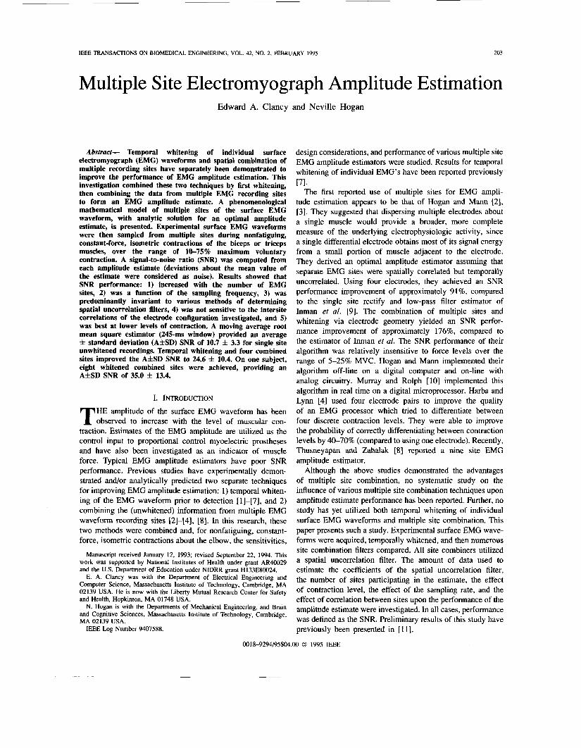
Independent Zero Filtering Mean, JWSS, CE, Effects of Jointly Gaussian, Muscle Tissue, White P ~ o c e ~ s e ~ of Bone, Skin and Unit Intensity Electrodes
surface EMG Waveforms
sites, including differences in signal strength. Such a restriction requires that outputs of the multidimensional filter can only be based on knowledge of the present inputs. Any use of past inputs would imply a contribution to the temporal correla- tion in the EMG. Hence, the multidimensional filter has no dynamics and can be represented as a linear transformation. The L outputs from the multidimensional shaping filter are passed through a bank of LTI shaping filters to form L dependent, zero-mean, JWSS, nonwhite, jointly CE, jointly Gaussian processes. The bank of shaping filters accounts for all of the time dependence in the EMG. These shaping filters are stable, causal, and have an inverse which is stable and causal. This model describes the electrical phenomenon, but does not account for details of the underlying physiology. The multiple site problem is, therefore, formulated as estimating the EMG amplitude (common standard deviation) from L zero-mean, JWSS, jointly CE, jointly Gaussian processes.
B. Maximum Likelihood Estimator
If the power spectral density (PSD) of each surface EMG Fig. 1 . Model of multiple sites of the EMG waveform. L independent, zero-mean, jointly wide sense stationary (JWSS), jointly correlation-ergodic (CE), jointly Gaussian, white processes of unit intensity are passed through the multidimensional filter Hspurc which accounts only for the spatial deuendence between sites. These filter oumuts are each uassed through a
waveform was white (uncorrelated temporally), and the mul- tiple recordings were both mutually uncomelated spatially and Of Over a particular time Deriod from each of L sites could be grouued into one data Set.
intensity* then W .
shaping filter H l l m e . l ( e J d ) and multiplied by the EMG amplitude 5 to Form the L sites of surface EMG waveforms. The EMG amplitude is assumed constant and the muscle contraction is assumed nonfatieuing.
kor such a set, sequential causal optimal maximum likelihood (ML) estimates of the EMG amplitude, i;, have been shown
1 -
to be found as [2], [13],
11. MATHEMATICAL MODEL
A. Model Description Viewed as a random signal, the surface EMG waveform
recorded from a single site resembles a band-limited Gaussian random process. For constant-force, isometric contractions, there is evidence that the random process is wide-sense sta- tionary and correlation-ergodic (CE) [ 121. When multiple electrodes are placed over a muscle, several correlated EMG’s are recorded. In order to form a useful model from this description, three fundamental assumptions were made: 1) the EMG amplitude could be identified from the surface EMG waveform, 2) for the case of nonfatiguing, constant-force, iso- metric muscle contraction, the EMG amplitude had a constant value, and 3) the mutually uncorrelated information from each recording site should be weighted equally in the determination of the EMG amplitude estimate. The EMG amplitude can thus be more formally defined as the common standard deviation of the equally weighted mutually uncorrelated information from each recording site.
Fig. 1 shows a phenomenological model of multiple sur- face EMG waveforms which embodies the above descrip- tion and assumptions. Spatial and temporal correlation are both accounted for in this model. L independent, zero-mean, jointly wide-sense stationary (JWSS), white, jointly CE, jointly Gaussian processes of unit intensity are passed through an L-input, L-output, linear time-invariant (LTI) shaping filter, Hspace, which is stable, causal, and whose inverse exists and is stable and causal. This multidimensional shaping filter is restricted to account only for the spatial dependence between
where i is the time index, 1 is the site index, and ml,; are the surface EMG waveform samples. The SNR for this estimator is PI, [31, WI
-112
=m where Nd is the number of degrees of freedom in the data, r() is the gamma function, and i is the EMG amplitude estimate. The approximation is useful for Nd large. For the present case,
When the surface EMG waveforms are spatially and tempo- rally correlated, consider the following two filter operations. First, filter each respective site by the temporal whitening filter HzAe, l (e ju) . These filters can be derived from the PSD’s of each site [2], [4], [7], [13]. The construction of the EMG model guarantees that these inverse filters exist and are stable, causal, and LTI. Thus, the filter outputs must be white Gaussian processes. Further, since HGie, l (e ju) is invertible, no information is lost due to the filter. Optimal estimation of s from the filtered processes is equivalent to optimal estimation of s from the original EMG samples. Second, filter the whitened sequences via the L-input, L-output
Nd = N . L .

CLANCY AND HOGAN: MULTIPLE SITE ELECTROMYOGRAPH AMPLITUDE ESTIMATION
-1,z - U2,Z
V 3 , i c*,i = .
205
,::.*#" DLi -#-
A. Experimental Apparatus and Methods
The experimental apparatus and methods have been de- scribed in detail elsewhere [7], [13]. Briefly, an Instrumented
Torque Chair was designed to measure the torque generated about the elbow. A subject was seated and secured into a straight-back chair via five quick release belts. The subject's right arm was oriented so that the upper arm and forearm were in a plane parallel to the floor (shoulder abducted 90" from the anatomic position), the upper arm was directed laterally outward from the shoulder (normal to the sagittal plane), and the angle between the upper arm and the forearm was 90". The subject's right wrist was mounted, via a wrist cuff, to an instrumented beam which was rigidly attached to the chair. Deflection of the beam (torque about the elbow) was measured by four active strain gauges arranged as a Wheatstone bridge. Up to eight commercial electrode-amplifiers (Liberty Mutual MY01 11 [14]) were placed side by side latitudinally across the flexor (biceps brachii) or extensor (triceps) muscles of the elbow, the two electrode contacts of each electrode-amplifier being oriented along the direction of action potential con- duction. The electrode-amplifiers were located approximately midway between the elbow and the midpoint of the upper arm, clustered about the muscle midline. Each electrode- amplifier consisted of a pair of 4-mm diameter, stainless steel, hemispherical electrode contacts separated by a distance of 15 mm (center to center). The distance between adjacent electrode-amplifiers was approximately 1.75 cm.
Five subjects (four male and one female, ranging in age from 23 to 37 years), with no known neuromuscular deficits of the right shoulder, arm, or hand, each participated in one experi- ment. Informed consent was received from each subject. Two experiments studied flexion, three studied extension. During an experimental trial, the output voltage of the strain gauge circuit and a target torque level were presented as the two displays of a dual trace oscilloscope. The subject was instructed to begin at rest, then gradually increase flexion/extension torque until the target torque level was achieved (typically over a period of 0.5-1 s). The subject maintained the target torque level until a five-second segment of data was recorded.
Two initial three-second maximum voluntary contraction (MVC) trials were averaged to provide a rough estimate of the strain gauge circuit output voltage corresponding to MVC. A sequence of five sets of constant-force, isometric contractions was conducted. Each set consisted of four trials, one trial each at 10, 25, 50, and 75% MVC. Trials within a set were randomized. A rest period of two minutes between trials (three minutes after 100% MVC trials) was provided to prevent fatigue [15]. EMG and strain gauge circuit output data were sampled at 2048 Hz using a 12-bit A D converter and processed off-line. A total of 100 multiple site recordings, comprised of 660 single site recordings, were acquired.
B . Methods of Analysis
The basic computational steps involved in optimal multi- ple site EMG amplitude estimation were temporal whitening of individual surface EMG waveforms, followed by spatial uncorrelation of the whitened data, followed by M A W S calculation. Temporal whitening results from these data have been discussed in detail previously [7]. For this multiple site analysis, a single fourth-order moving average whitening filter
206 IEEE TRANSACTIONS ON BIOMEDICAL ENGINEERING, VOL. 42. NO. 2, FEBRUARY 1995
I SNR = 23.0
f
SNR = 95.1 !I 1 .5 1 .1 1.5 1.1 1.5 3.1 3.5 4 . 1 4.5 5.0
n.. 1.~~~4.
a,,. 3, I .gc . l l" Whlt.n.d
1 .I 1 . 1 1.5 1.1 1.5 1.1 3.5 4 . 1 4.5 5.. TI.. I" a.I.,,A.
0 .5 1 .1 f .5 1.1 1.5 3.1 3.5 4 . 1 4 . 5 5.1 r m ~ ~ . ~ i .
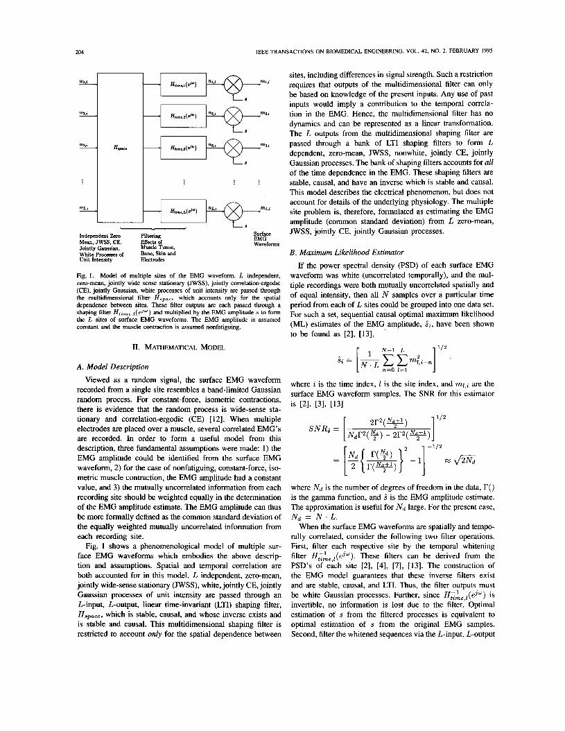
Fig. 3. EMG amplitude estimates. The top left plot is the measured torque for nonfatiguing, isometric contraction of elbow flexors at 25% MVC. The top right plot is the MARMS estimate (245" window) of the amplitude of EMG site 3. The bottom left plot is the temporally whitened estimate of the amplitude of EMG site 3. The bottom right plot is the temporally whitened, spatially uncorrelated (eight sites) estimate of the EMG amplitude. The beginning portion of each amplitude estimate depicts the rise time of the estimator. All data are from the same trial. (Reprinted with permission from [Il l , @ 1990 IEEE.)
was designed for each electrode-amplifier from each subject from one five-second recording (at the 50% MVC level), and applied to all trials recorded by that electrode-amplifier. All EMG's were temporally whitened before any sites were
where zl,i were the temporally whitened, spatially uncorre- lated data, the Z t h column of DT was comprised of the Z t h orthonormal eigenvector, and
In addition to the above computations, an estimate of the correlation coefficient matrix Rv, ,tg* ,1 of the random vector g+ was desired. Again, since the EMG's were J W S S , jointly CE, and zero-mean, each element of R g * , t , , z was the same for all time i and was estimated from the covariance matrix estimate as
- kv, ,* V I , I
' v ~ , z V k , % - JW'
kv, , 1 VUJ , 1 kV, . Z V k ,1
Performance differences between pairs of amplitude estimators were evaluated for statistical significance by paired t-tests [16]. SNR's were computed as the square root of the ratio of the squared amplitude estimate sample mean divided by the amplitude estimate sample variance.
IV. EXPERIMENTAL RESULTS All data analysis was performed in double-precision floating
point. Fig. 3 shows EMG amplitude estimates for single site unwhitened, single site whitened, and multiple site temporally whitened spatially uncorrelated data, all from the same trial. For the figure, temporal whitening filters and the spatial uncorrelation filter were designed from the data of a separate trial from the same subject. For comparison, the A f S D SNR of the 660 unwhitened single site estimates was 10.7 f 3.3.
A . Number of Sites spatially combined.
In order to form a spatial uncorrelation filter for L recording sites, the ensemble random vector of the whitened data x * , ~ was considered. The eigenvalues and eigenvectors of the covariance matrix Kv, ,,v* ,o (evaluated at a reference value for the EMG amplitude s) define the spatial uncorrelation filter. Thus, first an estimate of Kv*,tv,,z was formed. Since all EMG's were assumed to be JWSS processes, Kg,.,v,,, was the same for all time i . The j , k element of the covariance matrix for these CE zero-mean processes was estimated as
Initially, the number of EMG sites participating in the EMG amplitude estimate was investigated. The spatial uncorrelation filter for a particular trial was designed ("calibrated") from a separate trial from the same subject from the corresponding sites during an identical level of contraction. Two, four, and six site estimators were evaluated. (Four sites were available for all five subjects, six sites for four subjects, and eight sites for two subjects.) Since there was freedom in deciding which sites out of the total would participate in a particular estimate, nonadjacent electrode-amplifiers were selected whenever pos-
where P is the total number of time samples used to form the estimate.
Next, the eigenvalues and eigenvectors of the estimated covariance matrix were computed with an algorithm of Press er al. [16]. Because a covariance matrix is real and sym- metric, a complete orthonormal set of eigenvectors exists. Once the eigenvectors and corresponding eigenvalues (XI, Xa, X3. . . XL) were known, spatial uncorrelation of the random vector was performed as [21, [31, [131
sible. Results are presented in Table I, item 1. (Each multiple site result in Tables I and I1 is compared to the single site unwhitened results only from those sites which comprised the multiple site estimator.) Each increment in the number of EMG sites provided a statistically significant increase in SNR performance (p < 0.002 for all paired comparisons). Additional results from the two subjects with more than six EMG sites are presented later.
B. Calibration Length The investigations were repeated using 20 s of data for
calibration. The spatial uncorrelation filter for a particular trial was calibrated from the concatenation of the data from all four separate trials from the same subject from the corresponding
CLANCY AND HOGAN: MULTIPLE SITE ELECTROMYOGRAPH AMPLITUDE ESTIMATION 207
TABLE I TABULATED RESULTS OF MULTIPLE SITE AMPLITUDE
ESTIMATION-UNCORRELATION FILTER MATCHED TO LEVEL OF CONTRACTION. * DENTOES DATA FROM FOUR OF FIVE SUBJECTS
Unwhitcncd Single Site
Method of Determining 1 EMG Uncomlation Filter SNR fa
1) 5 seconds of data from same contraction level, same subject, for;
1 site, 10.7 f 3.3 2 sites, 4 sites. 6 sites’.
2) 20 seconds of data from same contraction level, same subject, for;
1 site, 2 sites, 4 sites, 6 sites’.
10.7 f 3.3
10.7 f 3.0
Whitened Multiple
Site EMG
SNR f a
17.4 f 6.1 22.2 f 7.9
26.8 f 10.0 2 9 . 0 f 11.9
17.4 f 6.1 22.2 f 7.9 26.8 f 9.9 29.3 f 11.8
3) Four EMG sites: Portion of one record from same contraction same rubject. with;
32 samples (15.625ma), 64 samples (31.25ms),
128 samples (62.5ms), 256 samples (125ms), 512 samples (250ms),
1024 samples (500ma), 2048 samples (Is), 4096 sampler (Zs), 6144 samples (3s). 8192 samples (4s).
11.0 f 3.6 1 1 . 0 f 3 . 6 11.0 f 3.6 11.0 f 3.6 11.0 f 3.6 11.0 f 3.6 11.0 f 3.6 l l . O f 3 . 6 11.0 f 3.6 11.0 f 3.6
4) 5 seconds of data from same contraction level, I
four EMG sites, for; 10% MVC, 12.4 f 4.7 25% MVC, 11.4 f 3.4
75% MVC. 9.8 f 2.3 50% MVC, 11 1 0 . 2 f 2 . 9
25.8 f 10.0 26.3 f 10.1 26.6 f 10.3 26.9 f 10.4 26.9 f 10.3 26.9 f 10.1 26.9 f 10.1 26.8 f 10.0 26.8 f 10.0 26.8 f 10.0
me subject,
35.8 f 8.6 30.5 f 8.8 22.8 f 6.2 18.0 f 4.9
5 ) Decimate data by 2: 20 seconds of data from same contractior level. same subiect. for:
1 site, 2 sites, 4 sites, 6 sites’.
10.7 f 3.3 10.9 f 4.2 11.0 f 3.6 10.7 f 3.0
13.5 f 4.5 14.2 f 3.2 18.0 f 4.6 20.2 f 6.2
11 I I 6) Decimate data by 2: 5 seconds of data from same contraction
1 site, 10.7 f 3.3 13.5 f 4.6 2 sites, 10.9 f 4.2 14.2 f 3.2 4 sites, 1 1 . 0 f 3 . 6 17.9f4.6 6 sites’.
level, same subject. for;
11 1 0 . 7 f 3.0 I 2O.Of 6.2 1
Percent Increase
in Average SNR
6 3 104 144 171
6 3 104 144 174
rel,
135 139 144 145 145 145 145 144 144 144
189 168 124 84
26 30 64 89
26 30 6 3 8 7
sites during an identical level of contraction. No statistical difference in SNR performance between using 5 and 20 s calibration lengths was found for any number of sites (p > 0.36 for all paired comparisons) (see Table I, item 2).
The above data suggested that, at most, five seconds of data were needed to calibrate the spatial uncorrelation filters. For real-time applications, it was of interest to determine if yet shorter time durations would perform as well. Spatial uncorrelation filters were designed from portions of five- second trials. Table I, item 3, lists the results for time durations between 15.625 ms and 4 s. Compared to the five second calibration length, none of the shorter calibration lengths significantly altered the SNR performance (0.09 < p < 0.85 for the various paired comparisons).
C . Effect of Contraction Level
In order to investigate if the SNR performance of the above tests was influenced by the contraction level, results
SNR
40
20
Four Site Results With Spatial Uncorrelation
I- 8 -I Single Site Results Without Whitening
T I
I I 1- I 1
10 25 50 75 Percent MVC
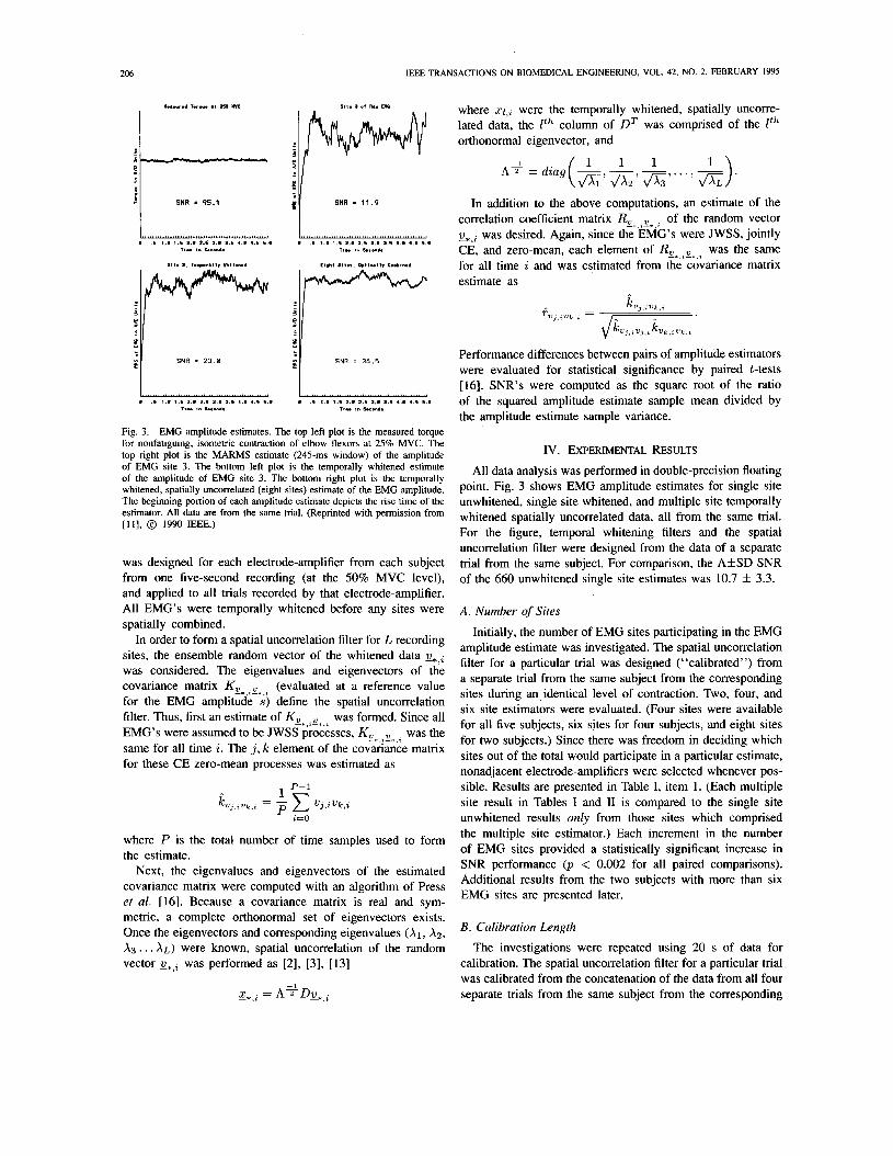
Fig. 4. Multiple site estimation at each contraction level. The mean and standard deviation SNR’s (245-ms smoothing window), averaged across 100 contraction trials, are presented. Mean values are graphed as small circles. Standard deviations are graphed as error bars about the mean. Solid-line error bars denote results from four site spatial uncorrelation filters calibrated from 5 s of EMG data from the same subject from the corresponding sites during an identical level of contraction. Dashed-line error bars denote single site results when no whitening filter was applied. Results are separated by the four contraction levels of lo%, 25%. 50%. and 75% MVC.
from the four site “Number of Sites” study were segregated by contraction level and plotted in Fig. 4 (also listed in Table I, item 4). The figure shows that there was a marked difference in performance as a function of contraction level. Performance at 10% MVC was almost twice that at 75% MVC. The performance differences between each pair of contraction levels were statistically significant (p < 0.007 for all paired comparisons). Note that high frequency (FZ 14 Hz) periodic oscillations were found superimposed on the torque recordings during the 50 and 75% MVC’s. These oscillations seemed to be due to normal tremor activity associated with the high torque output of the joint (see the discussion in [7]). With tremor activity during high contractions, muscle activation deviated, in part, from an isotonic contraction, and likely hindered the achieved SNR.
D. Effect of Sampling Rate
The effect of sampling rate was studied next. All data were decimated by a factor of two, providing an effective sampling frequency of 1024 Hz. Although sampling at 1024 Hz might have aliased the data, only a standard deviation estimate-not a complete reconstruction of the EMG-was desired. Aliased frequencies would still be represented in the data and, although not properly whitened, would still contribute to the EMG amplitude estimate. Amplitude estimates were formed using 5 and 20 s calibration lengths for two, four, and six sites. (In order to maintain a smoothing window of 245 ms, one-
‘Note that aliasing could have been avoided by passing the data through a digital low pass filter (cutoff frequency 5 512 Hz) prior to decimating. Assuming an ideal low pass filter, all information at frequencies above the cutoff frequency would be lost entirely, but all information at frequencies below the cutoff frequency could then be properly whitened. This altemative technique was not investigated in the present study.

208 IEEE TRANSAmIONS ON BIOMEDICAL ENGINEERING, VOL. 42, NO. 2, FEBRUARY 1995
10% MVC, 25% MVC, 50% MVC, 75% MVC. Equalized variances.
2) Four EMG sites: One 10% MVC, 25% MVC, 50% MVC, 76% MVC, Equaliced variances.
half of the number of discrete-time samples was used in these MARMS filters.) The results are presented in Table I, items 5 and 6. Again, there was no statistically significant difference between using 5 or 20 s calibration lengths (p > 0.11 for 2, 4, and 6 site comparisons). Each increment in the number of EMG sites provided a statistically significant increase in the SNR performance (p < 0.002 for all paired comparisons). The overall results, however, were well below those when the data were sampled at 2048 Hz. These performance decrements were similar to those found previously [7] when unwhitened single site estimation was compared to whitened single site estimation at these two sampling rates for these same data. Thus, the loss in performance seems to be associated with tem- poral whitening and not site combination. For comparison, one temporally whitened site sampled at 2048 Hz performed about as well as four temporally whitened, spatially uncorrelated sites sampled at 1024 Hz.
10.9 f 4.2 10.9 f 4.2 10.9 f 4.2 10.9 f 4.2 10.9 f 4.2
uncorrelation lilter 11.0f3.6 11.0f 3.6 11.0f 3.6 11.0f 3.6 11.0 f 3.6
E. One Spatial Uncorrelation Filter Per Subject
The next set of comparisons studied the feasibility of calibrating a single spatial uncorrelation filter from one trial from a subject, and applying that spatial uncorrelation filter to all trials for that subject. This investigation was performed four times with a two site estimator, once each by forming a spatial uncorrelation filter from a trial corresponding to 10,25,50, and 75% MVC. The entire process was then repeated with a four site estimator, a six site estimator (four out of five subjects), and an eight site estimator (one out of five subjects). In addition, two, four, and eight site estimators were also formed in which no spatial uncorrelation was performed. Rather, the data from each site were normalized (based on calibration from the data which determine the temporal whitening filters) and then smoothed. This filter was called the equal variance combiner. Table 11, items 1-4, list the results.
In general, all seven multiple site combination filters pro- vided considerable performance improvement. Compared to spatial uncorrelation filters calibrated from data at an identical contraction level (either 5 or 20 s of data), these single trial calibration filters performed marginally poorer, however, the statistical significance of this conclusion was weak. With four, six, or eight sites, the equal variance combiner performed progressively poorer, on average, than the other filters (p < 0.002 for all paired comparisons). All of the above differences, however, were small in strength. Thus, for most applications, the simpler equal variance combiner, which uses no spatial uncorrelation filter, is justified.
24.3 f 10.4 26.1 f 10.6 26.1 f 9.3 25.6f 8.4 25.1f9.0
,m trial at; 26.6 f 11.8 27.9 f 12.9 28.2 f 10.3 27.3f 9.2 25.5f 8.8
F. Seven and Eight Site Estimators Seven sites from subject FA were combined and all eight
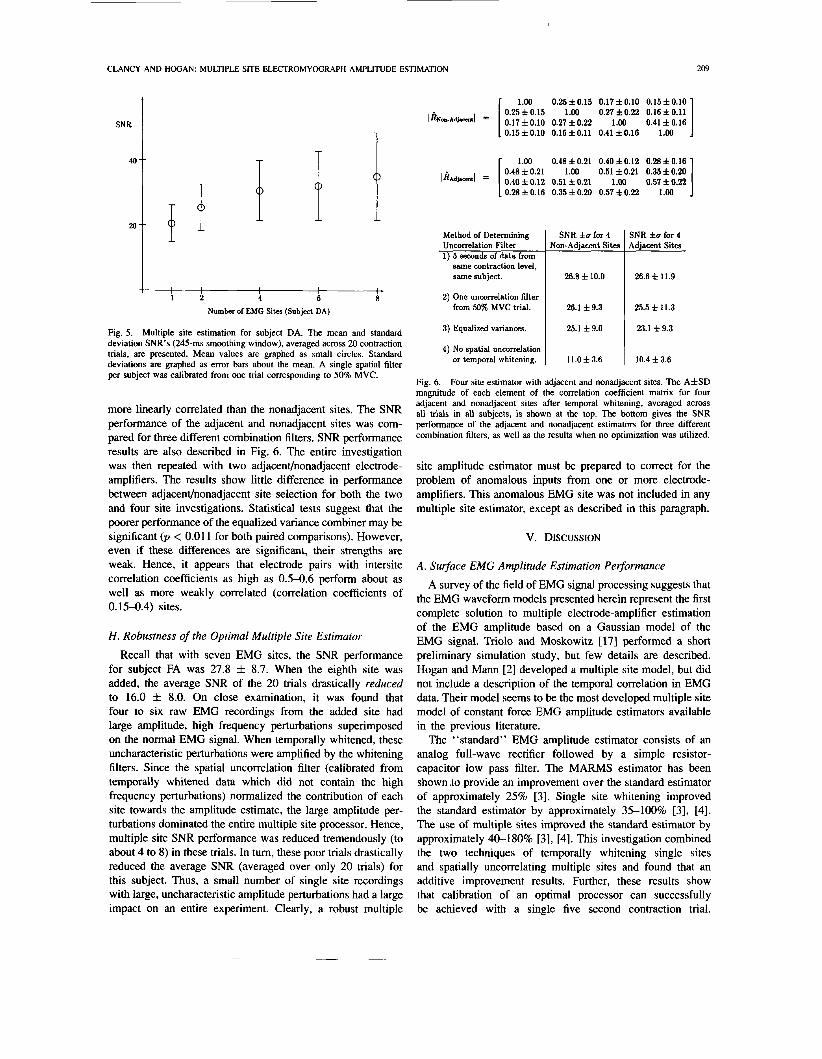
sites from subject DA were combined. A single spatial filter per subject was calibrated from a single trial corresponding to 50% MVC. Amplitude estimation was performed for two, four, and six sites as well. The results are listed in Table 11, items 5 and 6, and displayed in Fig. 5 (for subject DA). Considering these data pooled only from subjects DA and FA, each increment in the number of EMG sites provided a statistically significant increase in SNR performance (p <
121 137 137 133 128
149 161 164 155 138
TABLE I1 TABULATED RESULTS OF MULTIPLE SITE AMPLITUDE
E S T I M A T I O N ~ N E UNCORRELATION FILTER PER TRIAL. * DENTOES DATA FROM FOUR OF FIVE SUBJECTS
Unwhitened Whitened Percent Single Multiple I~CKMC
Site Site Method of Determining 1 EMG I EMC I
Uncorrelation Filter S N R f u S N R f u
1 site, 2 sites, 4 sites, 6 sites, 8 sites.
11.8f 2.6 20.5f 5.6 11.9f 2.8 25.4 f 7.1 11.8 f 2.7 30.6 f 9.2 11.8f 2.4 32.1 f 10.6 11.6f 2.5 35.0 f 13.4
,m trial at; 20.7 f 7.6 21.4 f 7.9 21.7 f 6.9 21.4 f 6.7 21.7 f 7.0
II I I 4) Eight EMG sites: Subject DA: One uncorrdation filter from
trial at; 10% MVC, 25% MVC, 50% MVC, 75% MVC, Equalized variances.
11.8f 2.5 11.8f 2.5 11.8f 2.5 11.8f 2.5 11.8f 2.5
90 96 99 96 99
35.9 f 17.4 37.5 f 19.1 35.0 f 13.4 33.5 f 10.6 31.8 f 10.9
204 218 197 184 169
74 113 169 172 197
74 121 169 192 216
0.002 for all paired comparisons). Increasing the number of electrode-amplifiers beyond eight may continue to provide a statistically significant improvement in SNR performance, however, the strength of this improvement should be small.
G . Effect of Intersite Correlation For all of the above investigations, when possible, the
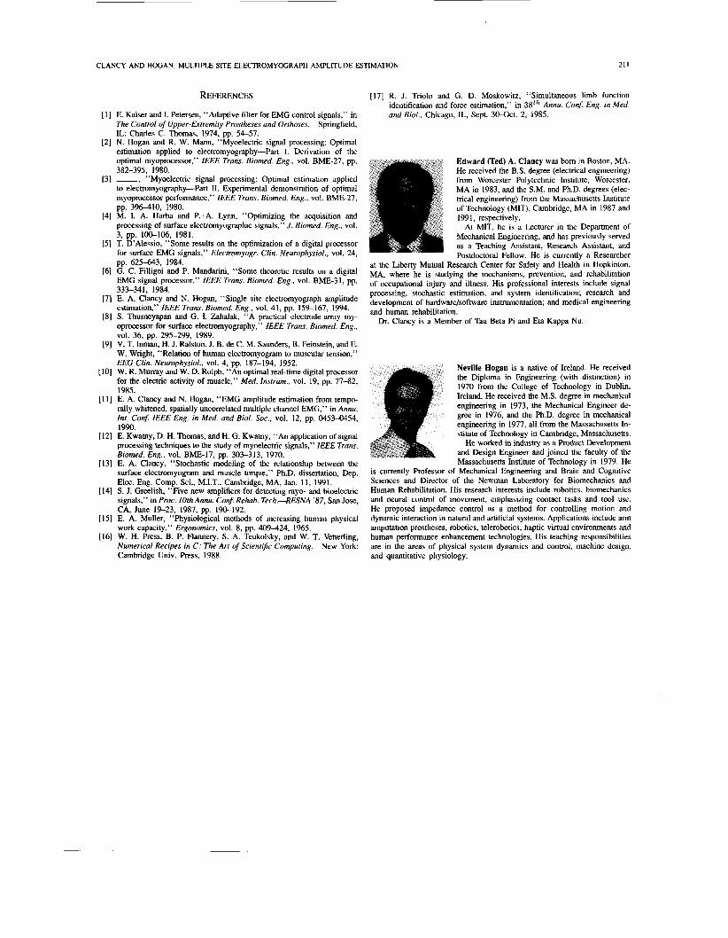
electrode-amplifiers for a particular multiple site estimator where chosen from nonadjacent locations on the muscle group. The next examination contrasted nonadjacent selection with adjacent selection. Presumably, adjacent sites were inherently more correlated than nonadjacent sites. Two sets of four site estimators were evaluated. The first set was comprised of non- adjacent sites, the second set of adjacent sites. The correlation coefficient matrix was estimated for each set of electrode- amplifiers. The AfSD (averaged across all 20 trials in all five subjects) of each element in these matrices was computed and is shown in Fig. 6. As expected, the adjacent sites were
CLANCY AND HOGAN: MULTIPLE SITE ELECTROMYOGRAPH AMPLITUDE ESTIMATION
t SNR I
40
20 I , 1 2 6 8
Number of EMG Sites (Subject DA)
Fig. 5. Multiple site estimation for subject DA. The mean and standard deviation SNR’s (245-111s smoothing window), averaged across 20 contraction trials, are presented. Mean values are graphed as small circles. Standard deviations are graphed as error bars about the mean. A single spatial filter per subject was calibrated from one trial corresponding to 50% MVC.
more linearly correlated than the nonadjacent sites. The SNR performance of the adjacent and nonadjacent sites was com- pared for three different combination filters. SNR performance results are also described in Fig. 6. The entire investigation was then repeated with two adjacenvnonadjacent electrode- amplifiers. The results show little difference in performance between adjacenvnonadjacent site selection for both the two and four site investigations. Statistical tests suggest that the poorer performance of the equalized variance combiner may be significant (p < 0.01 1 for both paired comparisons). However, even if these differences are significant, their strengths are weak. Hence, it appears that electrode pairs with intersite correlation coefficients as high as 0.5-0.6 perform about as well as more weakly correlated (correlation coefficients of 0.15-0.4) sites.
H . Robustness of the Optimal Multiple Site Estimator
Recall that with seven EMG sites, the SNR performance for subject FA was 27.8 f 8.7. When the eighth site was added, the average SNR of the 20 trials drastically reduced to 16.0 f 8.0. On close examination, it was found that four to six raw EMG recordings from the added site had large amplitude, high frequency perturbations superimposed on the normal EMG signal. When temporally whitened, these uncharacteristic perturbations were amplified by the whitening filters. Since the spatial uncorrelation filter (calibrated from temporally whitened data which did not contain the high frequency perturbations) normalized the contribution of each site towards the amplitude estimate, the large amplitude per- turbations dominated the entire multiple site processor. Hence, multiple site SNR performance was reduced tremendously (to about 4 to 8) in these trials. In turn, these poor trials drastically reduced the average SNR (averaged over only 20 trials) for this subject. Thus, a small number of single site recordings with large, uncharacteristic amplitude perturbations had a large impact on an entire experiment. Clearly, a robust multiple
209
1 1.00 0.25f0.15 0.17f0.10 0.15f0.10 0.25f0.15 1.00 0.27f0.22 0.16f0.11
0.15f0.10 0.16f0.11 0.41f0.16 1.00 0.17 f 0.10 0.27 f 0.22 1.00 0.41 f 0.16
1.00 0.48f0.21 0.40f0.12 0.28f0.16 0.48 f0.21 1.00 0.51 60.21 0.35f0.20 0.40 f 0.12 0.51 f 0.21 1.00 0.57 f 0.22 0.28f0.16 0.35f0.20 0.57f0.22 1.00
Method of Determining Uncorrelation Filter 1) 5 seconds of data from
same contraction level, same subject.
2) One uncorrelation filter from 50% MVC trial.
3) Equalized variances.
4) No spatial uncorrelation or temporal whitening.
SNR f u for 4 Non-Adjacent Sites
26.8 f 10.0
26.1 f 9.3
25.1 f 9.0
11.0 f 3.6
SNR f u for 4 Adjacent Sites
26.6 f 11.9
25.5 f 11.3
23.1 f 9.3
10.4 f 3.6
Fig. 6. Four site estimator with adjacent and nonadjacent sites. The A f S D magnitude of each element of the correlation coefficient matrix for four adjacent and nonadjacent sites after temporal whitening, averaged across all trials in all subjects, is shown at the top. The bottom gives the SNR performance of the adjacent and nonadjacent estimators for three different combination filters, as well as the results when no optimization was utilized.
site amplitude estimator must be prepared to correct for the problem of anomalous inputs from one or more electrode- amplifiers. This anomalous EMG site was not included in any multiple site estimator, except as described in this paragraph.
V. DISCUSSION
A . Surface EMG Amplitude Estimation Performance
A survey of the field of EMG signal processing suggests that the EMG waveform models presented herein represent the first complete solution to multiple electrode-amplifier estimation of the EMG amplitude based on a Gaussian model of the EMG signal. Triolo and Moskowitz [17] performed a short preliminary simulation study, but few details are described. Hogan and Mann [2] developed a multiple site model, but did not include a description of the temporal correlation in EMG data. Their model seems to be the most developed multiple site model of constant force EMG amplitude estimators available in the previous literature.
The “standard” EMG amplitude estimator consists of an analog full-wave rectifier followed by a simple resistor- capacitor low pass filter. The MARMS estimator has been shown to provide an improvement over the standard estimator of approximately 25% [3]. Single site whitening improved the standard estimator by approximately 35-100% [3], [4]. The use of multiple sites improved the standard estimator by approximately 40-1 80% [3], [4]. This investigation combined the two techniques of temporally whitening single sites and spatially uncorrelating multiple sites and found that an additive improvement results. Further, these results show that calibration of an optimal processor can successfully be achieved with a single five second contraction trial.

210 IEEE TRANSACTIONS ON BIOMEDICAL ENGINEERING, VOL. 42, NO. 2, FEBRUARY 1995
With an eight site estimator, the average SNR improved by approximately 197% over the single site MARMS estimator (or an estimated 274% improvement over the standard estimator).
B. Surface EMG Waveform Model The complexity of solving this multiple site optimal estima-
tion problem was greatly reduced by two modeling assump- tions. First, it was assumed that the spatio-temporal correlation in the EMG data could be represented by a spatial filter cascaded with a bank of temporal filters, as shown in Fig. 1. In a fully general stochastic model, spatio-temporal correlation would be represented by one composite filter. However, only a second-order characterization is needed for our purpose. The cascade model we assumed places no restrictions on the autocorrelation (and, equivalently, the autospectrum) of each individual EMG signal; that is, each channel can have a distinct autocorrelation. Further, the cross-correlation at zero time lag is not restricted-it is specified by the spatial filter. Second, it was assumed that each shaping filter (both spatial and temporal) was stable, causal, and had an inverse which was stable and causal. The invertible requirement prohibits the autospectrum from being null-valued at any frequency. (Note that the null value at zero Hz was considered as a special case.) The causality assumptions, by definition, limit the phase relationships in the model. These two assumptions are both plausible and practical. In particular, they allowed spatial uncorrelation and temporal whitening to be introduced into the solutions. Because the operations of spatial uncorrelation and temporal whitening are invertible, invertibly stable, and invertibly causal, any probabilistic inputs to these filters yield outputs in which no information is lost. Further, the outputs are spatially and/or temporally uncorrelated. Thus, in the event that the EMG is not distributed as a Gaussian random process (for example, [13] discusses a Laplacian model for the EMG probability distribution), inverse filtering remains beneficial. Uncorrelation/whitening can be considered separately from the formal ML development.
C . Multiple Site EMG Amplitude Estimation These results clearly confirm the benefit of combining the
information from multiple EMG sites on one muscle in order to estimate the EMG amplitude. These results also suggest that, for most applications, the simple equal variance combiner may be the most appropriate combination technique. With four or greater EMG sites, the equal variance combiner did perform statistically poorer than estimators which used an uncorrelation filter. However, this performance difference was small in strength and for most applications, may not be worth the added complexity.
If channel uncorrelation is selected, then these results sug- gest that SNR performance results are indifferent to the amount of data used to determine the uncorrelation filters. Calibration of a spatial uncorrelation filter from as little as 15.625 ms of data provides SNR performance equivalent to a filter calibrated from 5 s of data. Note that this result does not imply that the uncorrelation filters themselves were indifferent to the amount
of calibration data. In fact, the elements of the uncorrelation filter might be quite sensitive to the amount of calibration data, with little resultant change in SNR performance. Direct evaluation of the sensitivity of the uncorrelation filter elements to the amount of calibration data may provide further insight into these filters.
Finally, the results of this investigation must be cautiously interpreted when other electrode configurations are considered. The electrode configuration of this project used tightly spaced bipolar electrode contacts and tight interelectrode-amplifier spacing. Correlation coefficients did not exceed 0.5-0.6 in ad- jacent electrode-amplifiers. The tight electrode contact spacing would cause muscle fibers near each electrode-amplifier to dominate that electrode-amplifier’s signal. Altematively con- figured electrode-amplifiers which covered the same muscle area would be expected to acquire a different signal. Caution should be exercised in extrapolating the results of the current study if the spatial statistics of altemative configurations are grossly different. In particular, the limited success of spatial uncorrelation filters (as compared to the simple equal variance filter) may reflect the low correlation of signals from this project’s electrode-amplifier configuration. Altemative config- urations might increase the correlation between sites, resulting in uncorrelation filters becoming more efficacious.
VI. SUMMARY
Tables I and I1 summarize the results of the various mul- tiple site amplitude estimators. The results show that, as the number of EMG sites increased, so did the SNR, for all of the spatial uncorrelation filters. Spatial uncorrelation filters derived from between 15.625 ms and 5 s of data showed no SNR performance differences. As in the single site temporal whitening case [7], performance was severely degraded when the sampling frequency was reduced from 2048 Hz to 1024 Hz. All of the spatial uncorrelation filters provided an equitable performance improvement in the SNR, except for the equal variance filter which performed slightly poorer for four or more sites. Thus, for most applications, the simple equal variance combiner is justified. Detailed study of site correlations showed little difference between amplitude estimators based on adjacent or nonadjacent sites. For all spatial uncorrelation filters, SNR performance was best at lower levels of contraction, the 10% MVC level performing almost twice as well as the 75% MVC level. The multiple site estimators were heavily influenced by the robustness of single site temporal whitening.
ACKNOWLEDGMENT
The authors are grateful for the critical comments and suggestions provided to this work by Drs. W. Durfee, W. Peake, and G. Verghese at M.I.T. and Dr. A. Wiegner of the VA Medical Center, West Roxbury, MA. This work was performed at the Eric P. and Evelyn E. Newman Laboratory for Biomechanics and Human Rehabilitation at the Massachusetts Institute of Technology.
CLANCY AND HOGAN: MULTIPLE SITE ELECTROMYOGRAPH AMPLITUDE ESTIMATION 21 I
REFERENCES
[I] E. Kaiser and I. Petersen, “Adaptive filter for EMG control signals,” in Springfield,
[2] N. Hogan and R. W. Mann, “Myoelectric signal processing: Optimal estimation applied to electromyography-Part 1. Derivation of the optimal myoprocessor,” IEEE Trans. Biomed. Eng., vol. BME-27, pp.
[3] -, “Myoelectric signal processing: Optimal estimation applied to electromyography-Part 11. Experimental demonstration of optimal myoprocessor performance,” IEEE Trans. Biomed. Eng., vol. BME-27, pp. 396-410, 1980.
[4] M. I. A. Harba and P. A. LYM, “Optimizing the acquisition and processing of surface electromyographic signals,” J. Biomed. Eng.. vol. 3, pp. 100-106, 1981.
[5] T. D’Alessio, “Some results on the optimization of a digital processor for surface EMG signals,” Electromyogr. Clin. Neurophysiol., vol. 24, pp. 625-643, 1984.
[6] G. C. Filligoi and P. Mandarini, “Some theoretic results on a digital EMG signal processor,” IEEE Trans. Biomed. Eng., vol. BME-31, pp. 333-341, 1984.
[7] E. A. Clancy and N. Hogan, “Single site electromyograph amplitude estimation,” IEEE Trans. Biomed. Eng., vol. 41, pp. 159-167, 1994.
[8] S. Thusneyapan and G. I. Zahalak, “A practical electrode-array my- oprocessor for surface electromyography,” IEEE Trans. Biomed. Eng., vol. 36, pp. 295-299, 1989.
[9] V. T. Inman, H. J. Ralston, J. B. de C. M. Saunders, B. Feinstein, and E. W. Wright, “Relation of human electromyogram to muscular tension,” EEG Clin. Neurophysiol., vol. 4, pp. 187-194, 1952.
[IO] W. R. Murray and W. D. Rolph, “An optimal real-time digital processor for the electric activity of muscle,” Med. Instrum., vol. 19, pp. 77-82, 1985.
[ 111 E. A. Clancy and N. Hogan, “EMG amplitude estimation from tempo- rally whitened, spatially uncorrelated multiple channel EMG,” in Annu. Int. Conf. IEEE Eng. in Med. and Biol. Soc., vol. 12, pp. 0453-0454, 1990.
[12] E. Kwatny, D. H. Thomas, and H. G. Kwatny, “An application of signal processing techniques to the study of myoelectric signals,” JEEE Trans. Biomed. Eng., vol. BME-17, pp. 303-313, 1970.
[13] E. A. Clancy, “Stochastic modeling of the relationship between the surface electromyogram and muscle torque,” Ph.D. dissertation, Dep. Elec. Eng. Comp. Sci., M.I.T., Cambridge, MA, Jan. 1 1 , 1991.
[14] S. J. Greelish. “Five new amplifiers for detecting myo- and bioelectric signals,” in Proc. 1OthAnnu. Conf. Rehab. T e c h . 4 E S N A ‘87, San Jose, CA, June 19-23, 1987, pp. 190-192.
[I51 E. A. Muller, “Physiological methods of increasing human physical work capacity,” Ergonomics, vol. 8, pp. 409424, 1965.
[I61 W. H. Press, B. P. Flannery, S. A. Teukolsky, and W. T. Vetterling, Numerical Recipes in C: The Art of Scientific Computing. New York: Cambridge Univ. Press, 1988.
The Control of Upper-Extremity Prostheses and Orthoses. Charles C. Thomas, 1974, pp. 54-57.
382-395, 1980.
[I71 R. J. Trio10 and G. D. Moskowitz, “Simultaneous limb function identification and force estimation,” in 38”’ Annu. Conf. Eng. in Med. and Biol., Chicago, IL, Sept. 30-0ct. 2, 1985.
Edward (Ted) A. Clancy was bom in Boston, MA. He received the B.S. degree (electrical engineering) from Worcester Polytechnic Institute, Worcester, MA in 1983, and the S.M. and Ph.D. degrees (elec- trical engineering) from the Massachusetts Institute of Technology (MIT), Cambridge, MA in 1987 and 1991, respectively.
At MIT, he is a Lecturer in the Department of Mechanical Engineering, and has previously served as a Teaching Assistant, Research Assistant, and Postdoctoral Fellow. He is currently a Researcher
at the Liberty Mutual Research Center for Safety and Health in Hopkinton, MA, where he is studying the mechanisms, prevention, and rehabilitation of occupational injury and illness. His professional interests include signal processing, stochastic estimation, and system identification; research and development of hardwarehoftware instrumentation; and medical engineering and human rehabilitation.
Dr. Clancy is a Member of Tau Beta Pi and Eta Kappa Nu.
Neville Hogan is a native of Ireland. He received the Diploma in Engineering (with distinction) in 1970 from the College of Technology in Dublin, Ireland. He received the M.S. degree in mechanical engineering in 1973, the Mechanical Engineer de- gree in 1976, and the Ph.D. degree in mechanical engineering in 1977, all from the Massachusetts In- stitute of Technology in Cambridge, Massachusetts.
He worked in industry as a Product Development and Design Engineer and joined the faculty of the Massachusetts Institute of Technology in 1979. He
is currently Professor of Mechanical Engineering and Brain and Cognitive Sciences and Director of the Newman Laboratory for Biomechanics and Human Rehabilitation. His research interests include robotics, biomechanics and neural control of movement, emphasizing contact tasks and tool use. He proposed impedance control as a method for controlling motion and dynamic interaction in natural and artificial systems. Applications include arm amputation prostheses, robotics, telerobotics, haptic virtual environments and human performance enhancement technologies. His teaching responsibilities are in the areas of physical system dynamics and control, machine design, and quantitative physiology.


![[TM1960- David Duncan-Screenplay-2nd draft-Undated- Full ... · [TM1960- David_Duncan-Screenplay-2nd_draft-Undated- Full_Text#] [page_i] H. G. WELLS' THE TIME MACHINE An invention](https://static.fdocuments.us/doc/165x107/5f773e8cae173d10c9434ea3/tm1960-david-duncan-screenplay-2nd-draft-undated-full-tm1960-davidduncan-screenplay-2nddraft-undated-.jpg)