MULTIPLE PREGNANCY -...
88
MULTIPLE PREGNANCY Presented by, Mrs.C.Margret, Associate Professor, Obstetrics and Gynaecology Department, Annammal College Of Nursing, Kuzhithurai
-
Upload
nguyennhan -
Category
Documents
-
view
221 -
download
0
Transcript of MULTIPLE PREGNANCY -...
MULTIPLE
PREGNANCY
Presented by,Mrs.C.Margret,
Associate Professor, Obstetrics and Gynaecology
Department,
Annammal College Of Nursing, Kuzhithurai
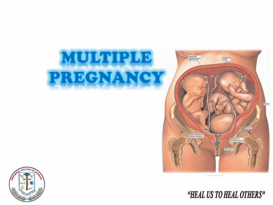
When two or more embryos develop in the uterus
at the same time the condition is known as multiple
pregnancy. These are considered as complicated
pregnancies because there is an appreciable
increase in morbidity and mortality.
High order multiples :Three or more offspring in one birth
Zygote: Fertilized ovum for the first three weeks following
conception.
Zygosity: It refers to the similarity of genes for a trait
Vanishing twin: Occasional death of one fetus and continuation of
pregnancy with surviving one. The dead fetus simply vanishes by
resorption
Chorionicity : Number of chorionic membranes surrounding babies
in a multiple pregnancy
Fetus papyraceous or compress: Is a state which occurs if one
of the fetus dies early .The dead fetus is flattened and
compressed between the membrane of the living fetus and
uterine wall
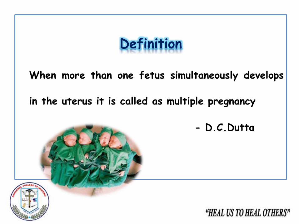
Definition
When more than one fetus simultaneously develops
in the uterus it is called as multiple pregnancy
- D.C.Dutta
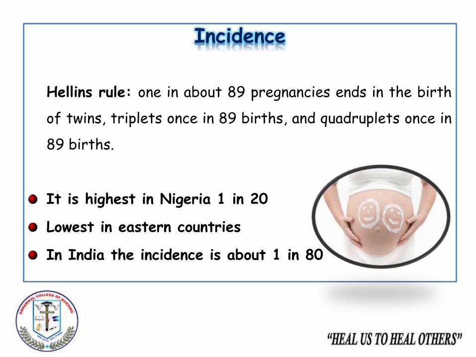
Incidence
Hellins rule: one in about 89 pregnancies ends in the birth
of twins, triplets once in 89 births, and quadruplets once in
89 births.
It is highest in Nigeria 1 in 20
Lowest in eastern countries
In India the incidence is about 1 in 80
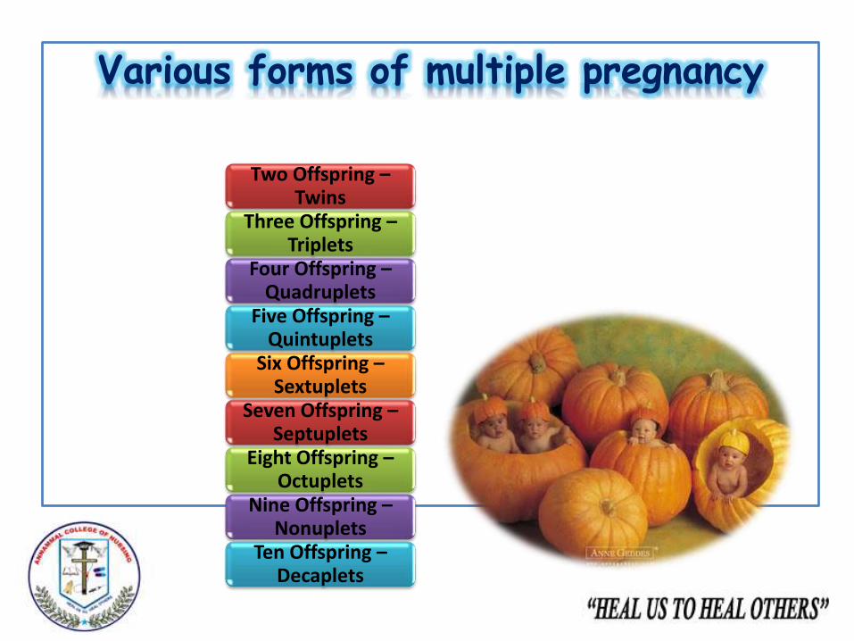
Various forms of multiple pregnancy
Two Offspring –Twins
Three Offspring –Triplets
Four Offspring –Quadruplets
Five Offspring –Quintuplets
Six Offspring –Sextuplets
Seven Offspring –Septuplets
Eight Offspring –Octuplets
Nine Offspring –Nonuplets
Ten Offspring –Decaplets
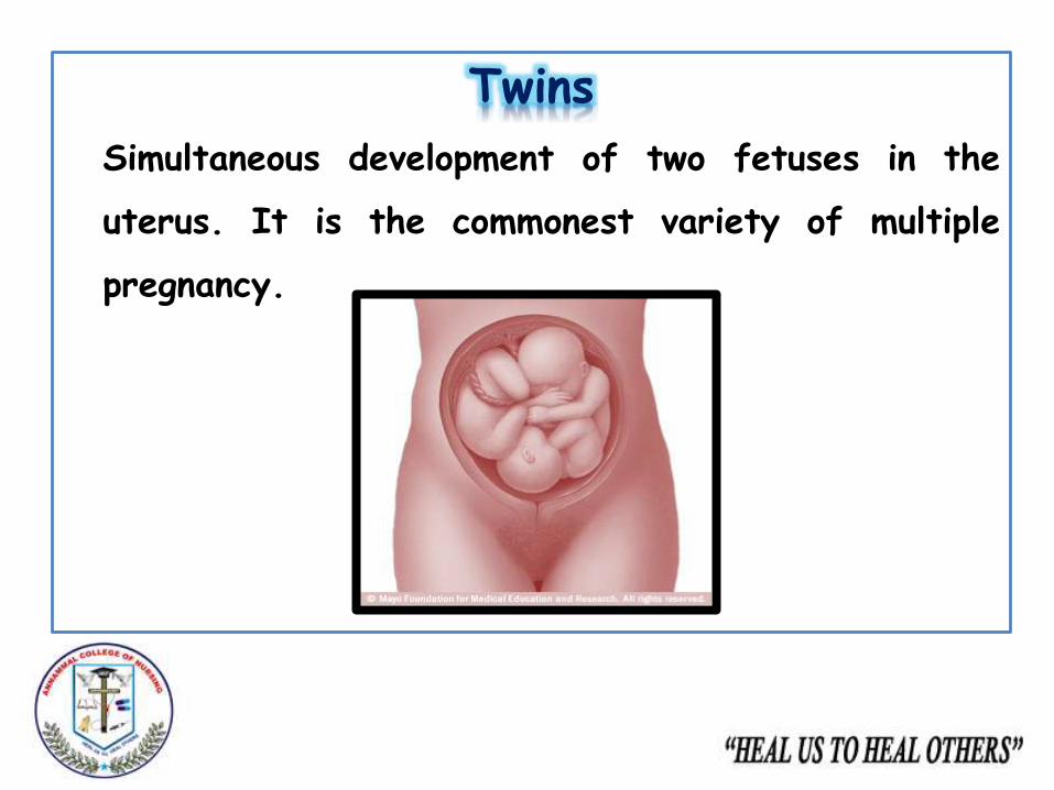
Twins
Simultaneous development of two fetuses in the
uterus. It is the commonest variety of multiple
pregnancy.
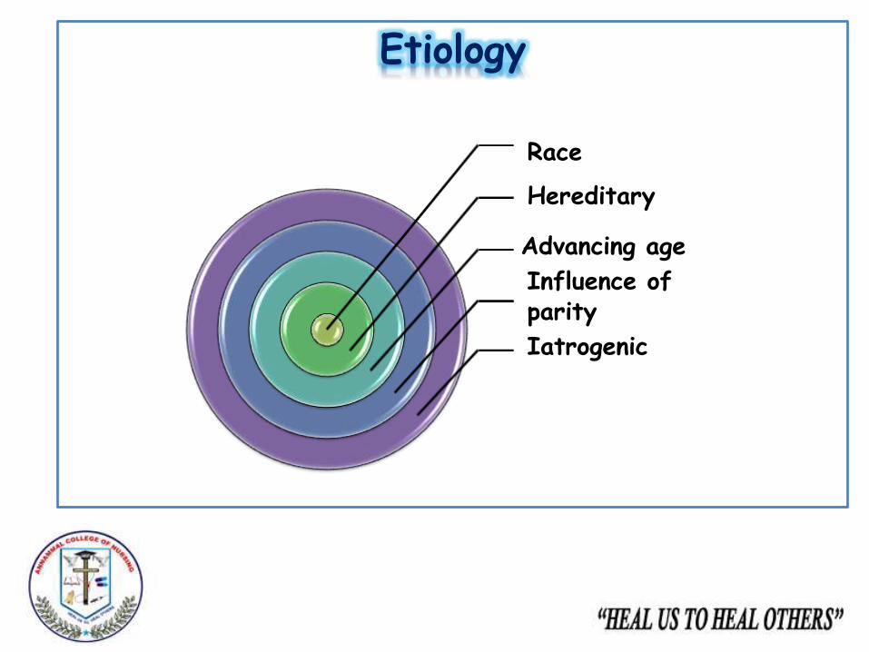
Etiology
Race
Hereditary
Advancing age
Influence of parity
Iatrogenic
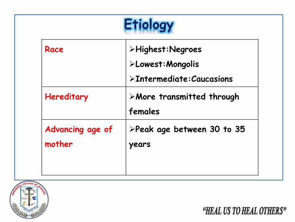
Etiology
Race Highest:Negroes
Lowest:Mongolis
Intermediate:Caucasions
Hereditary More transmitted through
females
Advancing age of
mother
Peak age between 30 to 35
years
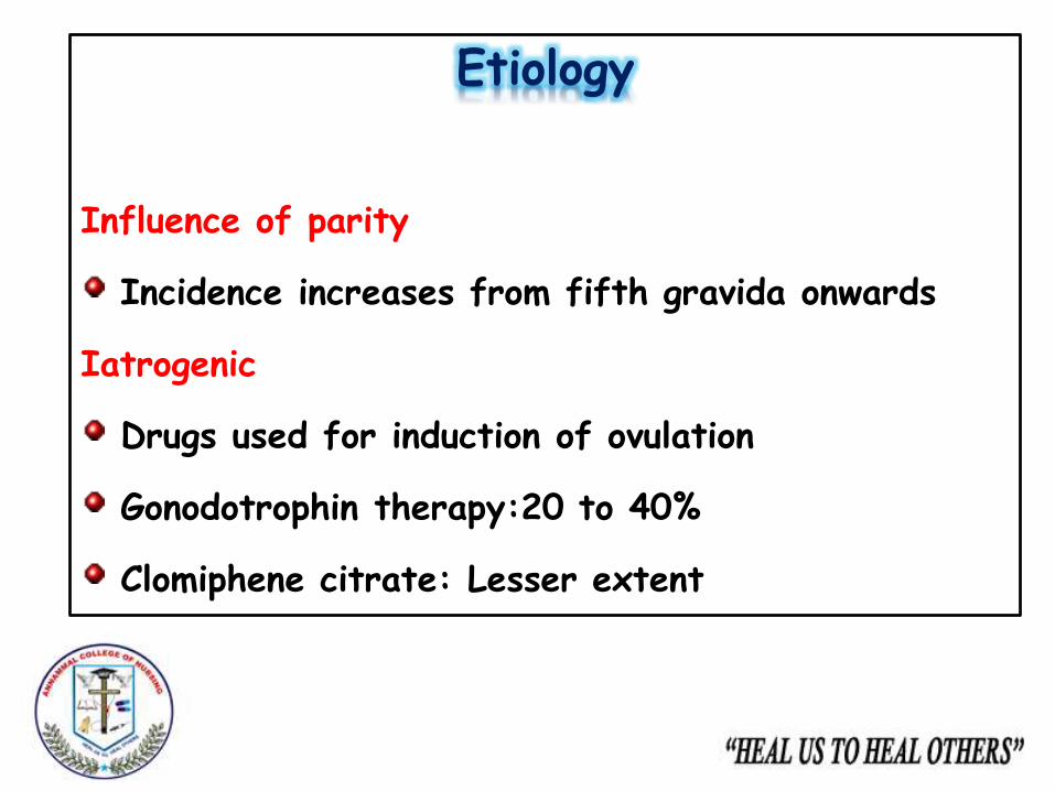
Etiology
Influence of parity
Incidence increases from fifth gravida onwards
Iatrogenic
Drugs used for induction of ovulation
Gonodotrophin therapy:20 to 40%
Clomiphene citrate: Lesser extent
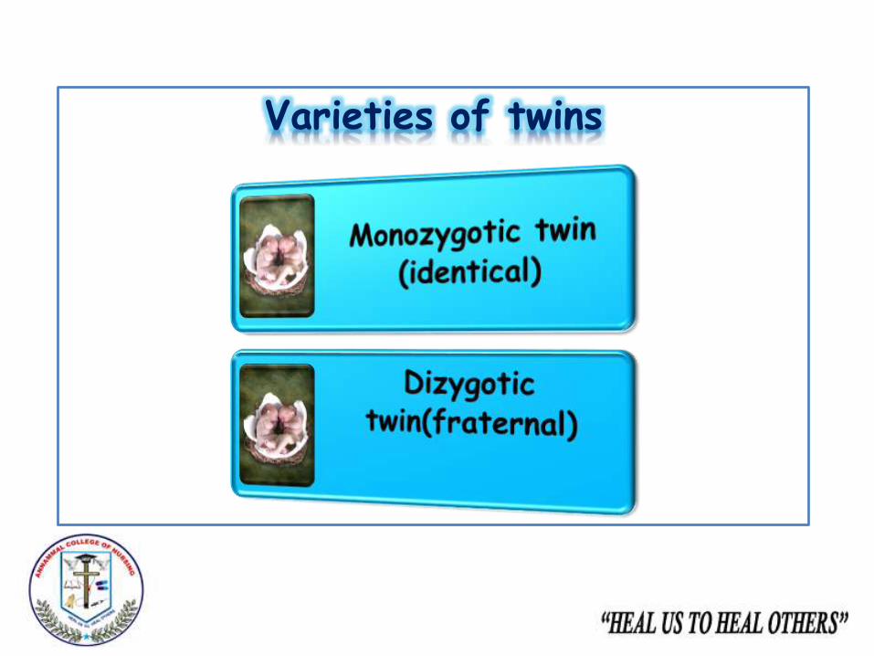
Varieties of twins

Genesis of twins
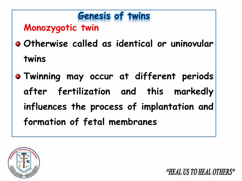
Genesis of twinsMonozygotic twin
Otherwise called as identical or uninovular
twins
Twinning may occur at different periods
after fertilization and this markedly
influences the process of implantation and
formation of fetal membranes
Genesis of twins
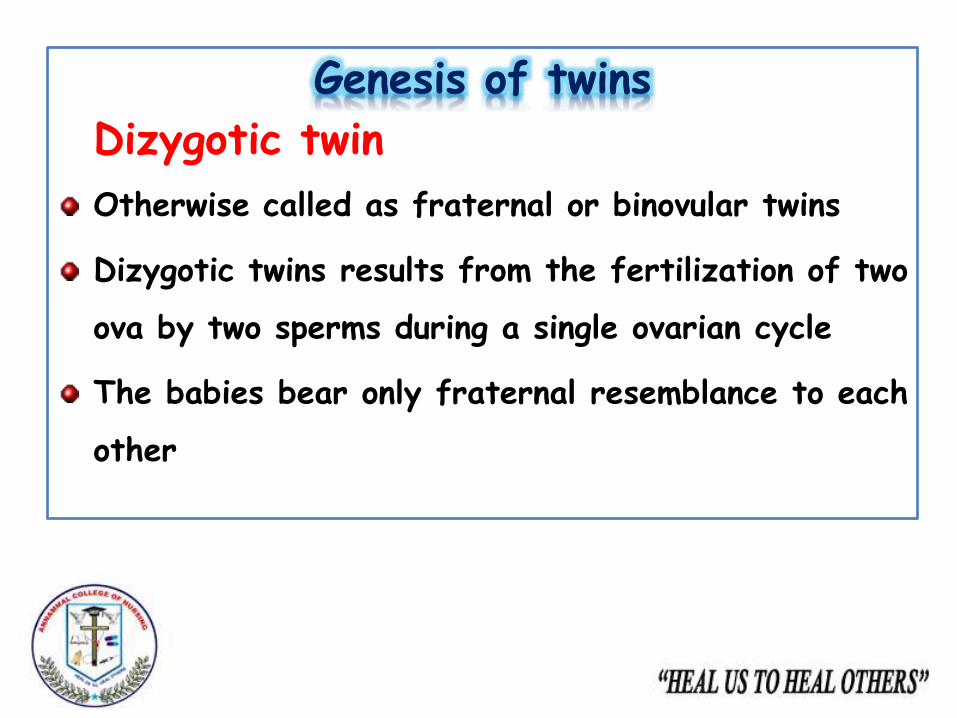
Dizygotic twin
Otherwise called as fraternal or binovular twins
Dizygotic twins results from the fertilization of two
ova by two sperms during a single ovarian cycle
The babies bear only fraternal resemblance to each
other
Genesis of twins
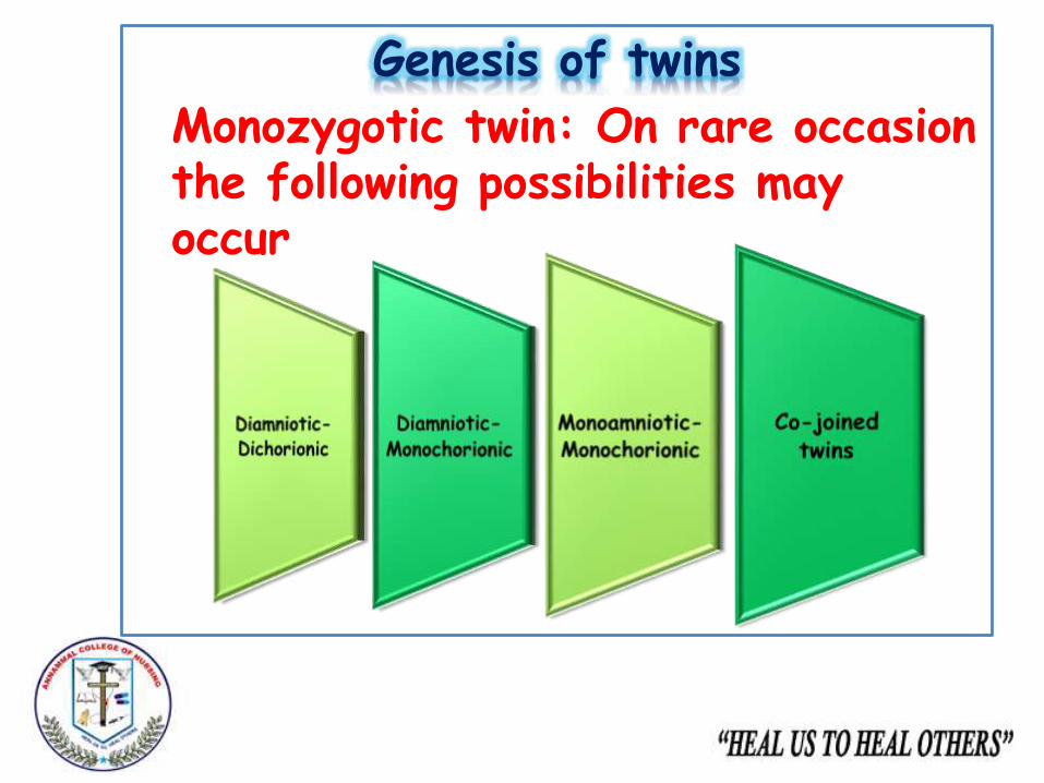
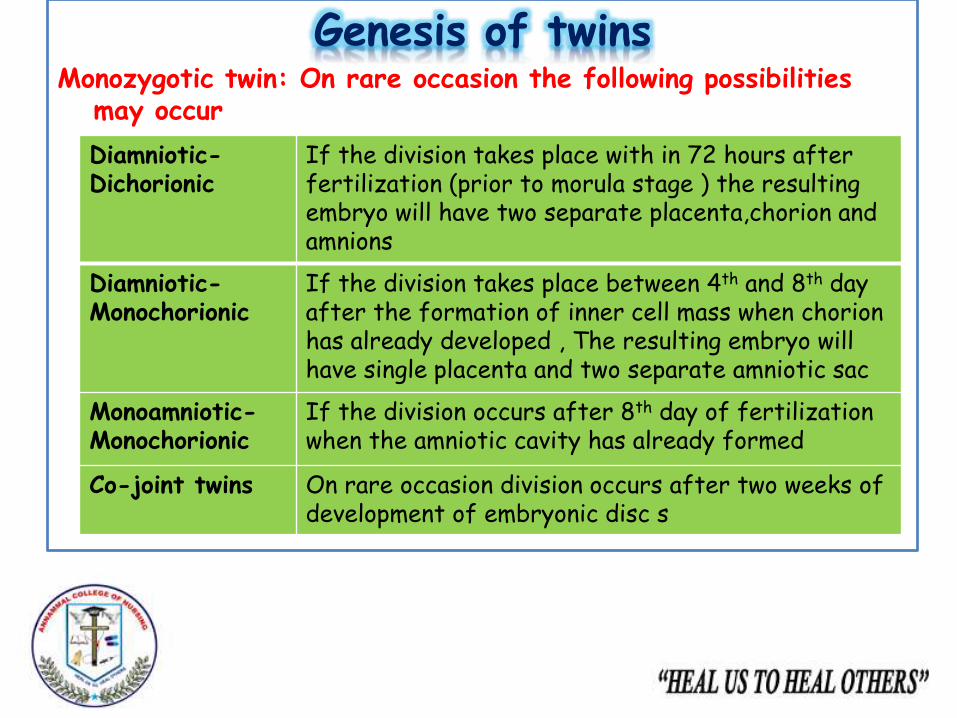
Monozygotic twin: On rare occasion the following possibilities may occur
Monozygotic twin: On rare occasion the followingpossibilities may occur
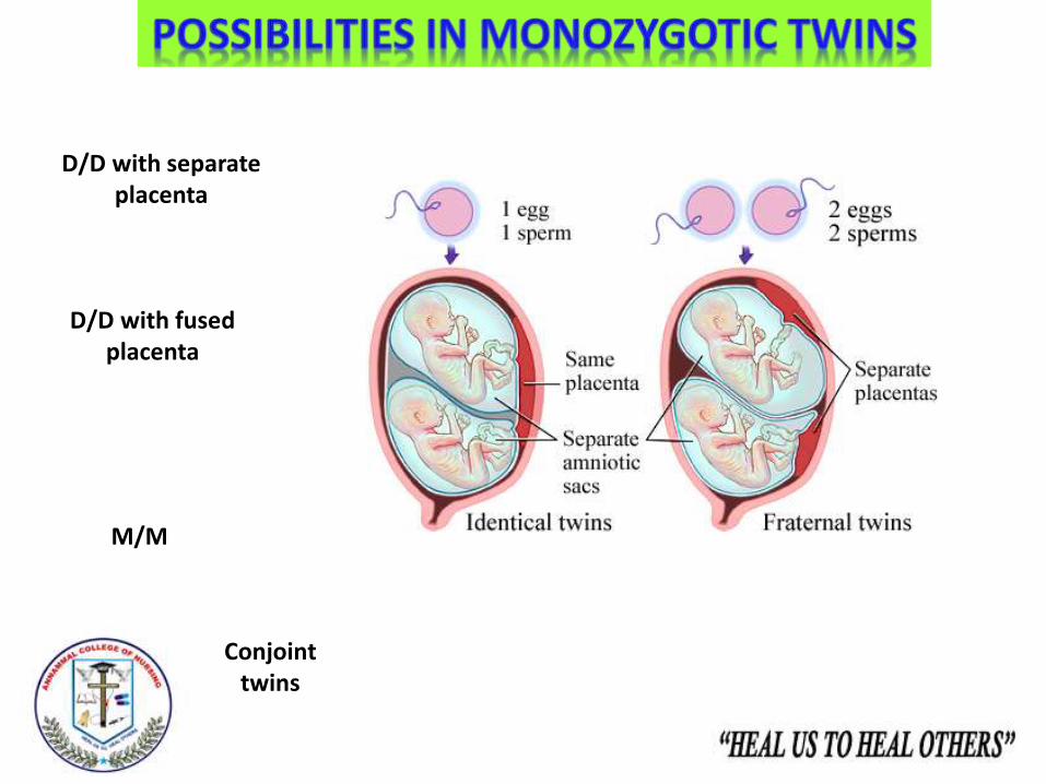
D/D with separate placenta
D/D with fused placenta
M/M
Conjoint twins
Genesis of twinsMonozygotic twin: On rare occasion the following possibilities
may occur
Diamniotic-Dichorionic
If the division takes place with in 72 hours after fertilization (prior to morula stage ) the resulting embryo will have two separate placenta,chorion and amnions
Diamniotic-Monochorionic
If the division takes place between 4th and 8th day after the formation of inner cell mass when chorionhas already developed , The resulting embryo will have single placenta and two separate amniotic sac
Monoamniotic-Monochorionic
If the division occurs after 8th day of fertilization when the amniotic cavity has already formed
Co-joint twins On rare occasion division occurs after two weeks of development of embryonic disc s
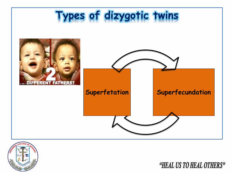
Types of dizygotic twins
SuperfecundationSuperfetation
Rare forms of multiple pregnancy
Superfecundation:Is the fertilization of two
different ova released in the same cycle by
separate acts of coitus within a short period of
time
Superfetation:Is the fertilization of two ova
released in different menstrual cycle
.
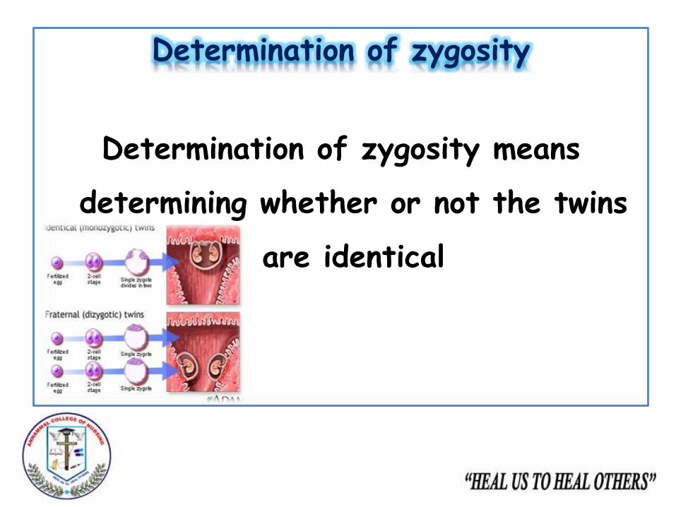
Determination of zygosity
Determination of zygosity means
determining whether or not the twins
are identical
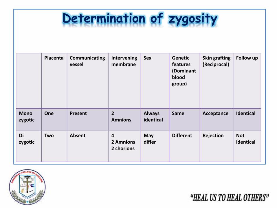
Determination of zygosity
Placenta Communicating vessel
Interveningmembrane
Sex Genetic features(Dominant blood group)
Skin grafting(Reciprocal)
Follow up
Monozygotic
One Present 2Amnions
Always identical
Same Acceptance Identical
Dizygotic
Two Absent 42 Amnions2 chorions
May differ
Different Rejection Not identical
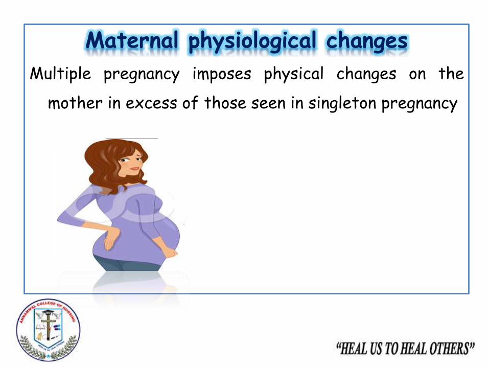
Maternal physiological changes
Multiple pregnancy imposes physical changes on the
mother in excess of those seen in singleton pregnancy
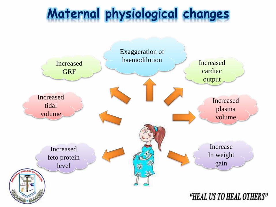
Maternal physiological changes
Increased
GRF
Increased
tidal
volume
Increase
In weight
gain
Increased
cardiac
output
Increased
plasma
volume
Increased
feto protein
level
Exaggeration of
haemodilution
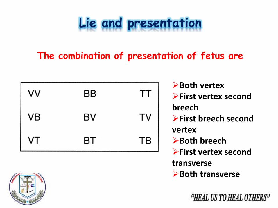
Lie and presentation
The combination of presentation of fetus are
Both vertexFirst vertex second breechFirst breech second vertexBoth breechFirst vertex second transverseBoth transverse
L
I
E
A
N
D
P
R
E
S
E
N
T
A
T
I
O
N
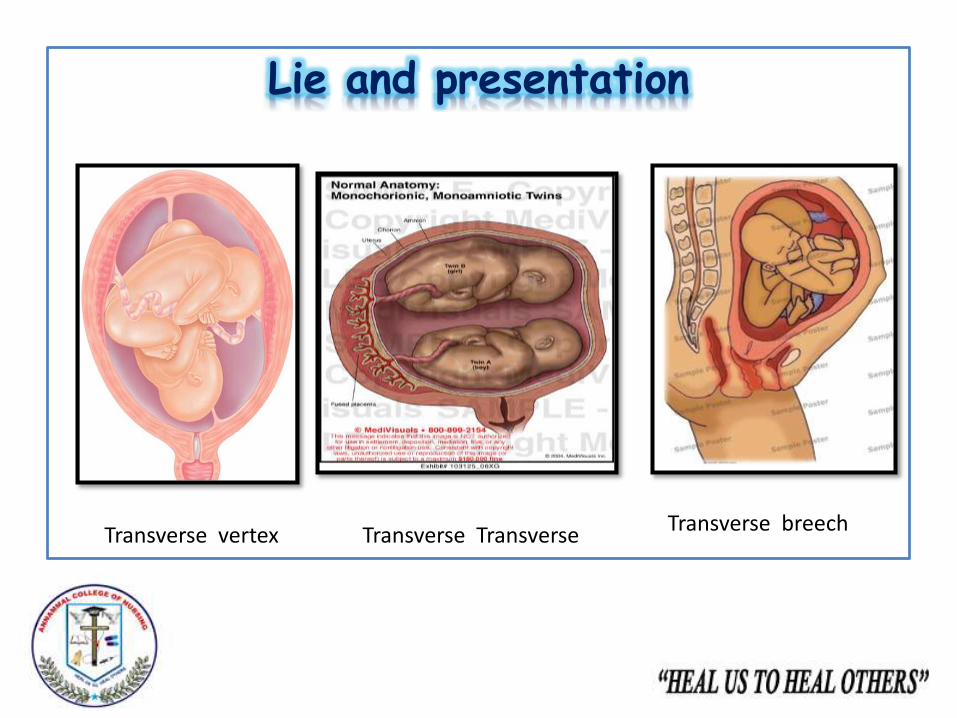
Lie and presentation
Transverse TransverseTransverse vertex Transverse breech
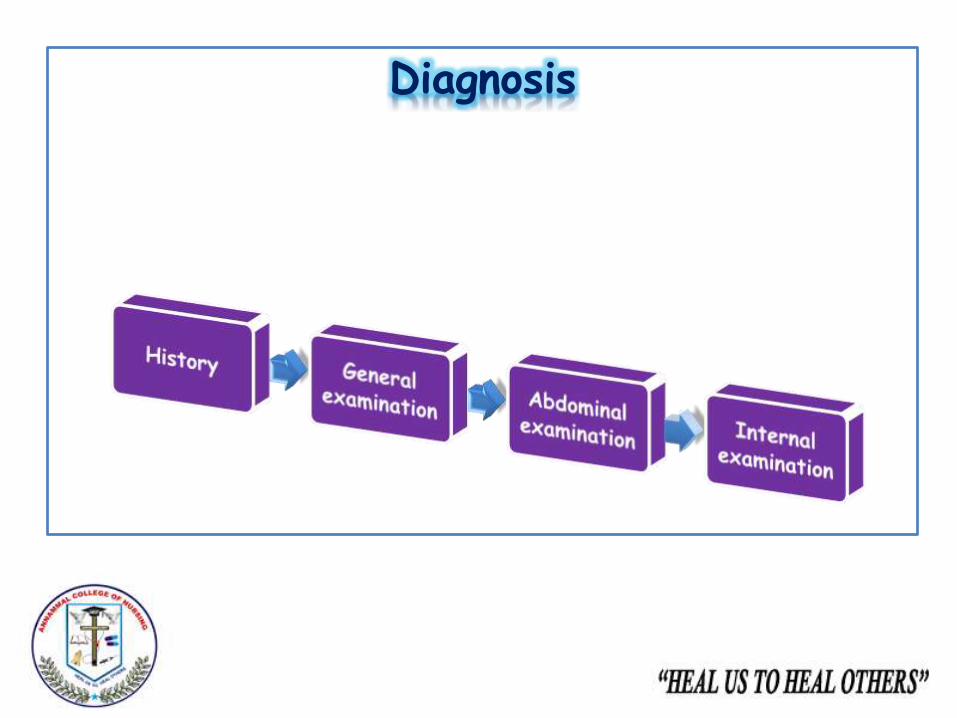
Diagnosis

Diagnosis
History collection:
History of ovulation inducing drugs specifically
gonadotrophins for infertility or use of ART
Family history of twinning
DiagnosisSymptoms:
Minor ailments of normal pregnancy are often exaggerated, Increased nausea and vomitingCardio respiratory embarrassment (palpitation, shortness of breath)Tendency of swelling of legsVaricose veinHaemorrhoidsUnusual rate of abdominal enlargementExcessive fetal movements
Diagnosis
General examination:
Prevalence of anaemia
Unusual weight gain
Evidence of pre eclampsia
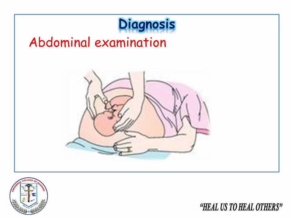
Diagnosis
Abdominal examination
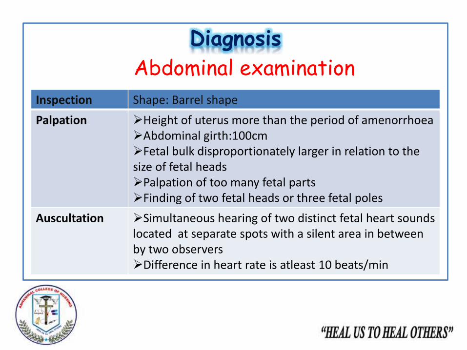
Diagnosis
Abdominal examination
Inspection Shape: Barrel shape
Palpation Height of uterus more than the period of amenorrhoeaAbdominal girth:100cmFetal bulk disproportionately larger in relation to the size of fetal headsPalpation of too many fetal partsFinding of two fetal heads or three fetal poles
Auscultation Simultaneous hearing of two distinct fetal heart sounds located at separate spots with a silent area in between by two observers Difference in heart rate is atleast 10 beats/min
Diagnosis
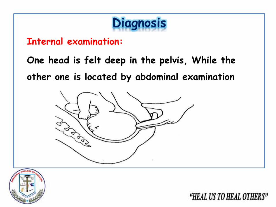
Internal examination:
One head is felt deep in the pelvis, While the
other one is located by abdominal examination
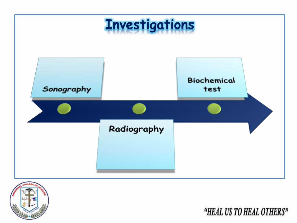
Investigations
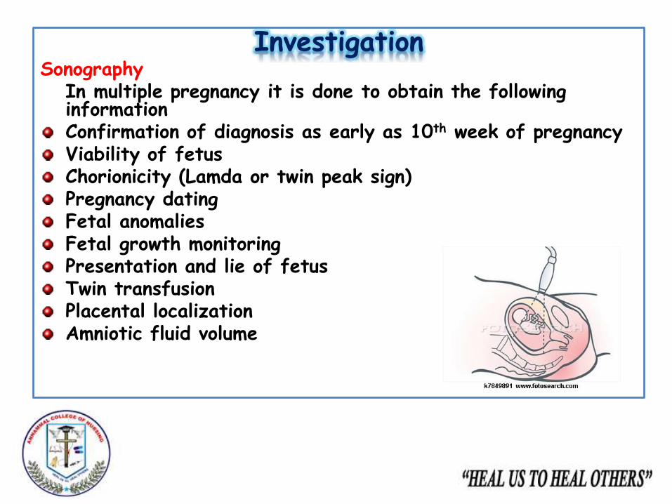
InvestigationSonography
In multiple pregnancy it is done to obtain the following informationConfirmation of diagnosis as early as 10th week of pregnancyViability of fetusChorionicity (Lamda or twin peak sign)Pregnancy datingFetal anomaliesFetal growth monitoringPresentation and lie of fetusTwin transfusionPlacental localizationAmniotic fluid volume
Investigations
Lamda or twin peak sign:
Chorionicity of the placenta is best diagnosed by
USG at 6 to 9 weeks of gestation
In dichorionic twins there is a thick septum
between the chorionic sacs .
It is best identified at the base of the membrane
where a triangular projection is seen this is known
as twin peak sign
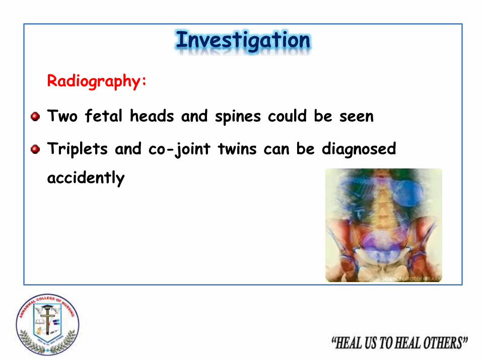
Investigation
Radiography:
Two fetal heads and spines could be seen
Triplets and co-joint twins can be diagnosed
accidently
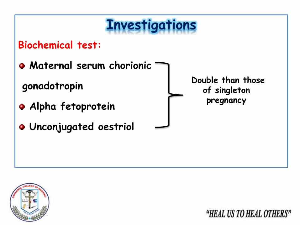
Investigations
Biochemical test:
Maternal serum chorionic
gonadotropin
Alpha fetoprotein
Unconjugated oestriol
Double than those of singleton pregnancy
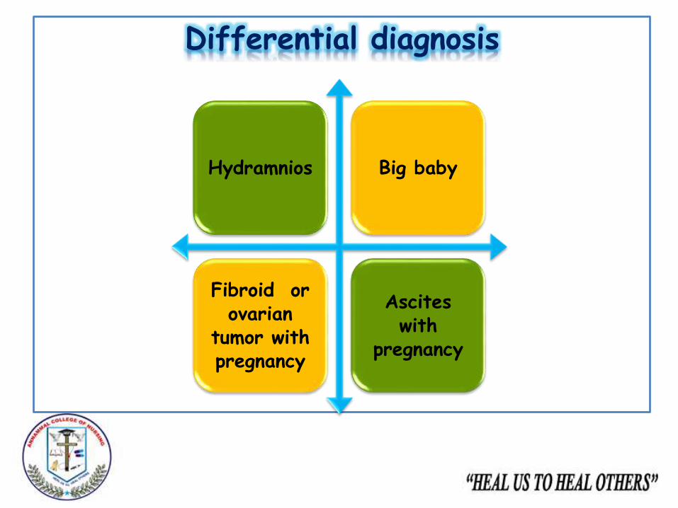
Differential diagnosis
Hydramnios Big baby
Fibroid or ovarian
tumor with pregnancy
Ascites with
pregnancy
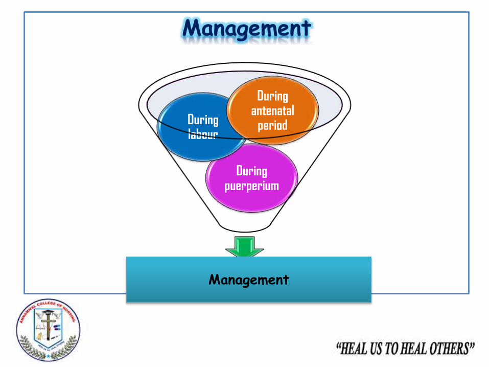
Management
Management
During puerperium
During labour
During antenatal
period
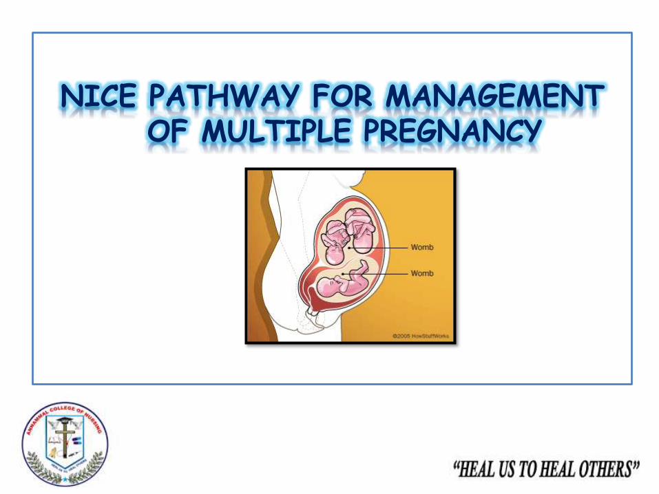
NICE PATHWAY FOR MANAGEMENT OF MULTIPLE PREGNANCY
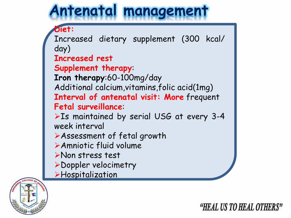
Antenatal managementDiet:Increased dietary supplement (300 kcal/day)Increased restSupplement therapy:Iron therapy:60-100mg/dayAdditional calcium,vitamins,folic acid(1mg)Interval of antenatal visit: More frequentFetal surveillance:Is maintained by serial USG at every 3-4week intervalAssessment of fetal growthAmniotic fluid volumeNon stress testDoppler velocimetryHospitalization
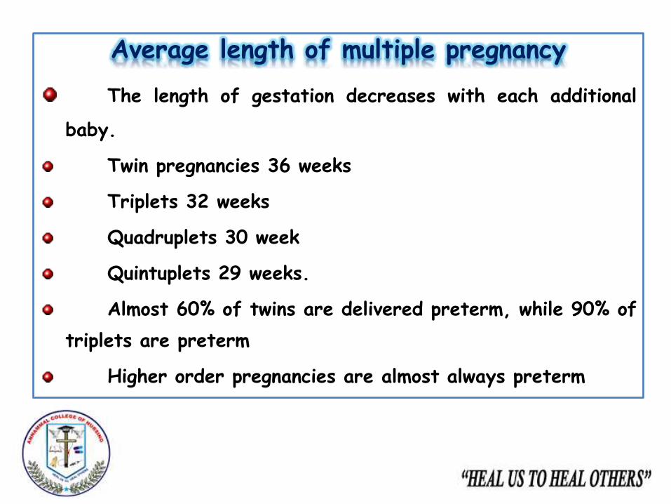
Average length of multiple pregnancy
The length of gestation decreases with each additional
baby.
Twin pregnancies 36 weeks
Triplets 32 weeks
Quadruplets 30 week
Quintuplets 29 weeks.
Almost 60% of twins are delivered preterm, while 90% of
triplets are preterm
Higher order pregnancies are almost always preterm
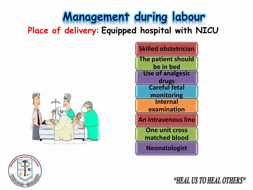
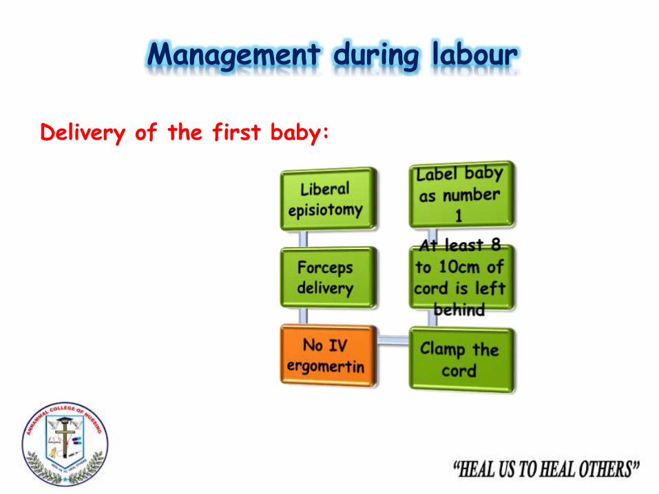
Management during labourPlace of delivery: Equipped hospital with NICU
Skilled obstetrician
The patient should be in bed
Use of analgesic drugs
Careful fetal monitoring
Internal examination
An intravenous line
One unit cross matched blood
Neonatologist
Management during labour
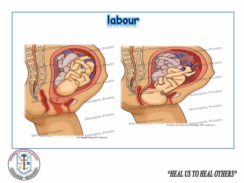
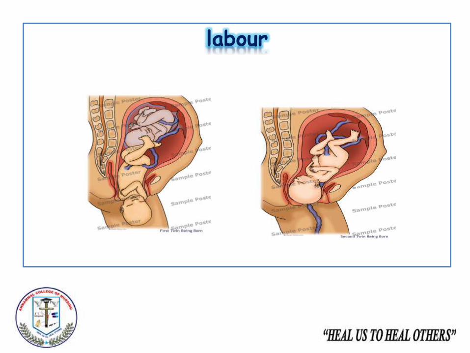
Delivery of the first baby:
Management during labour
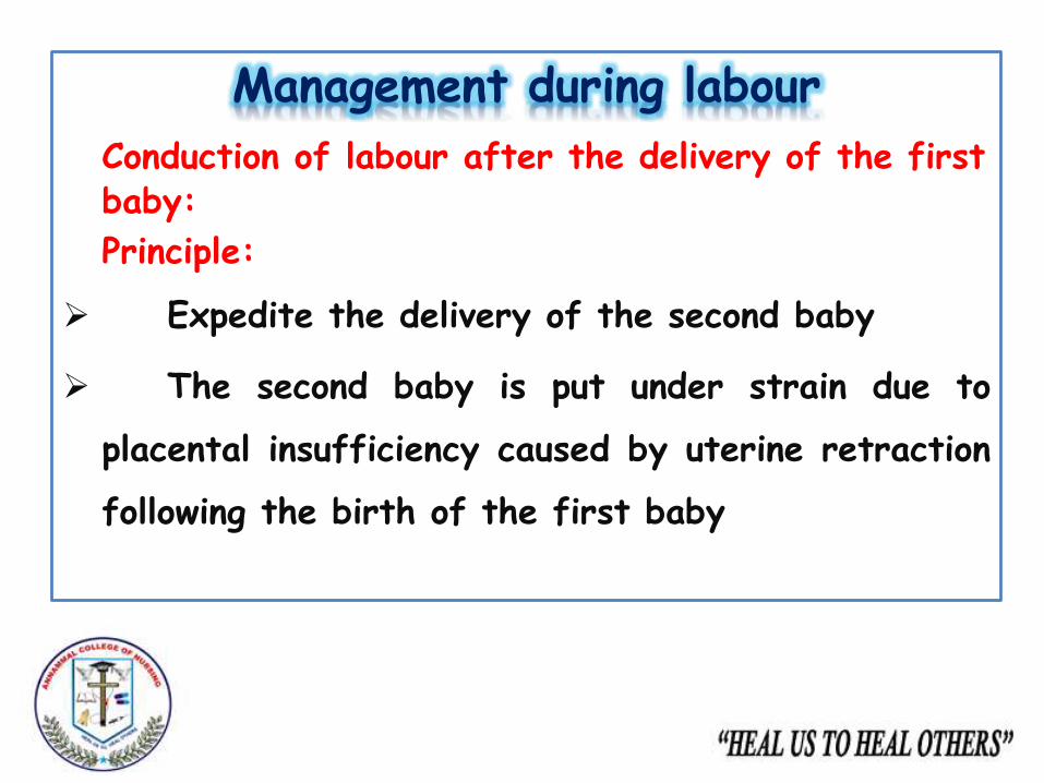
Conduction of labour after the delivery of the first baby:
Principle:
Expedite the delivery of the second baby
The second baby is put under strain due to
placental insufficiency caused by uterine retraction
following the birth of the first baby
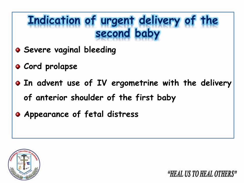
Indication of urgent delivery of the second baby
Severe vaginal bleeding
Cord prolapse
In advent use of IV ergometrine with the delivery
of anterior shoulder of the first baby
Appearance of fetal distress
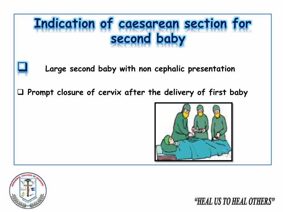
Indication of caesarean section for second baby
Large second baby with non cephalic presentation
Prompt closure of cervix after the delivery of first baby
labour
labour
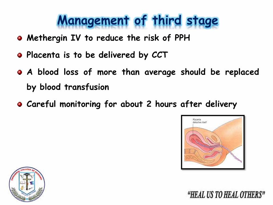
Management of third stage
Methergin IV to reduce the risk of PPH
Placenta is to be delivered by CCT
A blood loss of more than average should be replaced
by blood transfusion
Careful monitoring for about 2 hours after delivery
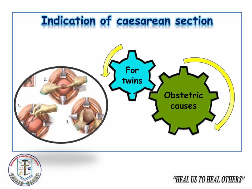
Indication of caesarean section
Obstetric causes
For twins
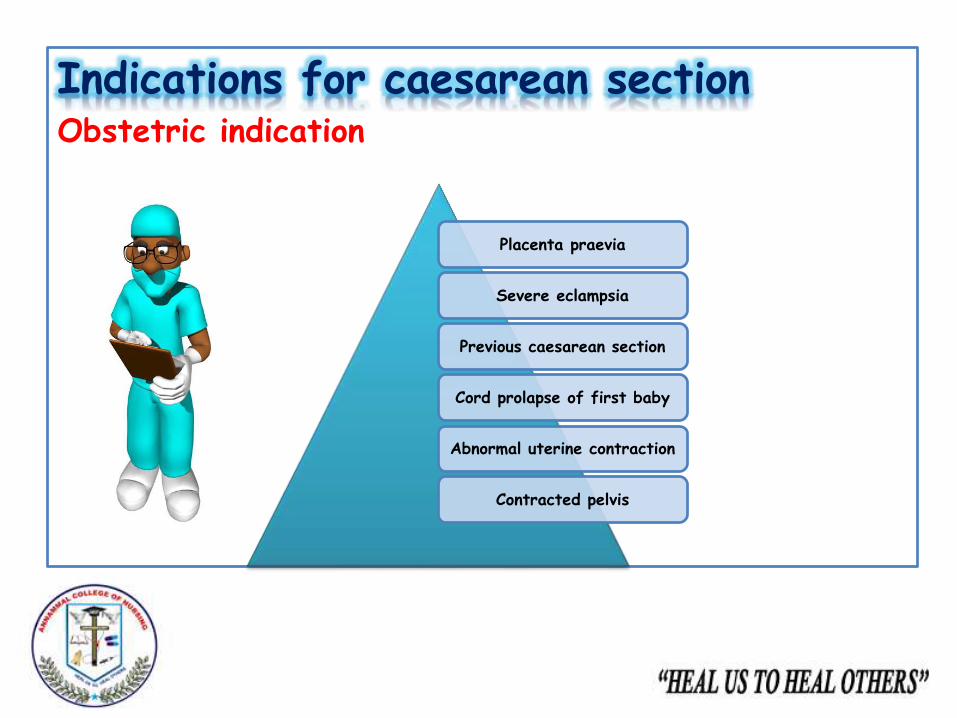
Indications for caesarean sectionObstetric indication
Placenta praevia
Severe eclampsia
Previous caesarean section
Cord prolapse of first baby
Abnormal uterine contraction
Contracted pelvis
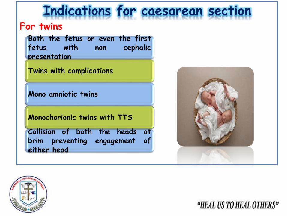
Indications for caesarean sectionFor twins
Both the fetus or even the firstfetus with non cephalicpresentation
Twins with complications
Mono amniotic twins
Monochorionic twins with TTS
Collision of both the heads atbrim preventing engagement ofeither head
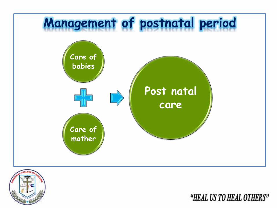
Management of postnatal period
Care of babies
Care of mother
Post natal care
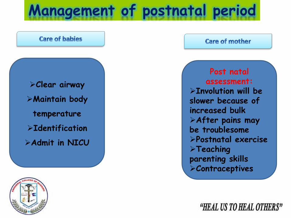
Clear airway
Maintain body
temperature
Identification
Admit in NICU
Post natal assessment:
Involution will be slower because of increased bulkAfter pains may be troublesomePostnatal exerciseTeaching parenting skillsContraceptives
Management of postnatal period
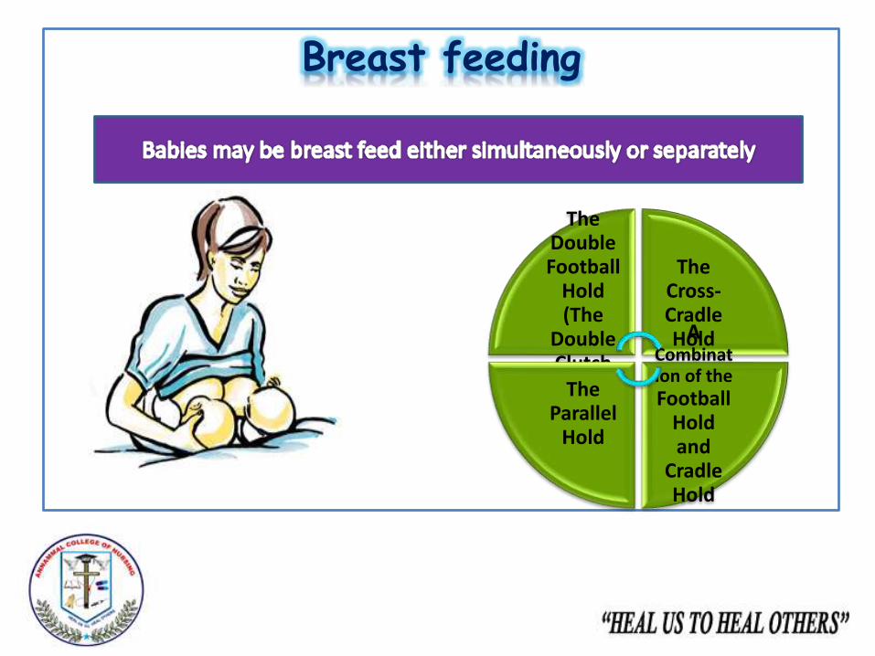
Breast feeding
The Double Football
Hold (The
Double Clutch Hold
The Cross-Cradle HoldA
Combination of the Football
Hold and
Cradle Hold
The Parallel
Hold
Breast feeding
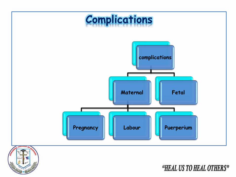
Complications
complications
Maternal
Pregnancy Labour Puerperium
Fetal
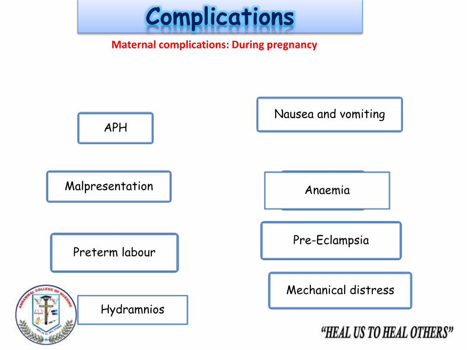
Nausea and vomiting
Anaemia
Hydramnios
Pre-Eclampsia
APH
Malpresentation
Mechanical distress
Preterm labour
ComplicationsMaternal complications: During pregnancy
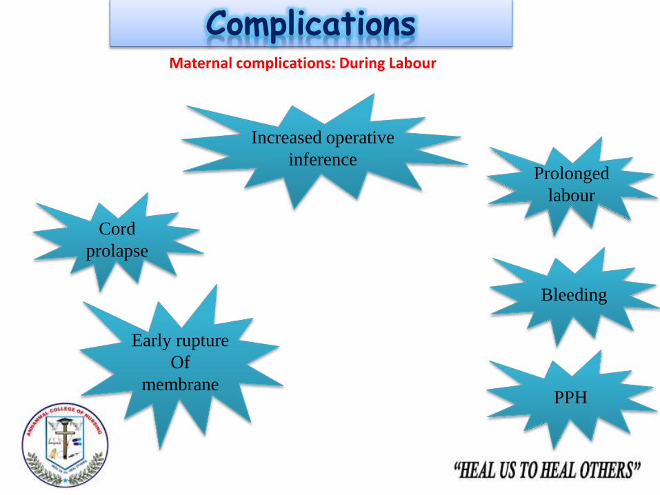
Cord
prolapse
Increased operative
inference
Early rupture
Of
membrane
Prolonged
labour
PPH
Bleeding
ComplicationsMaternal complications: During Labour
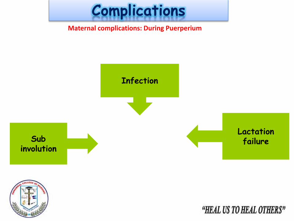
Sub involution
Infection
Lactation failure
ComplicationsMaternal complications: During Puerperium
Complications
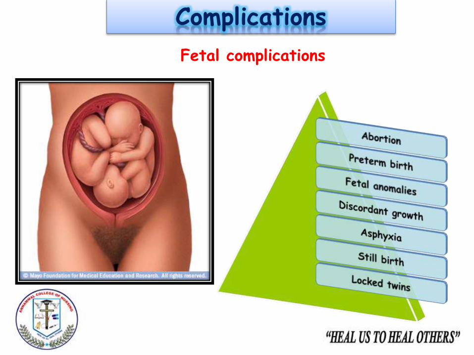
Fetal complications
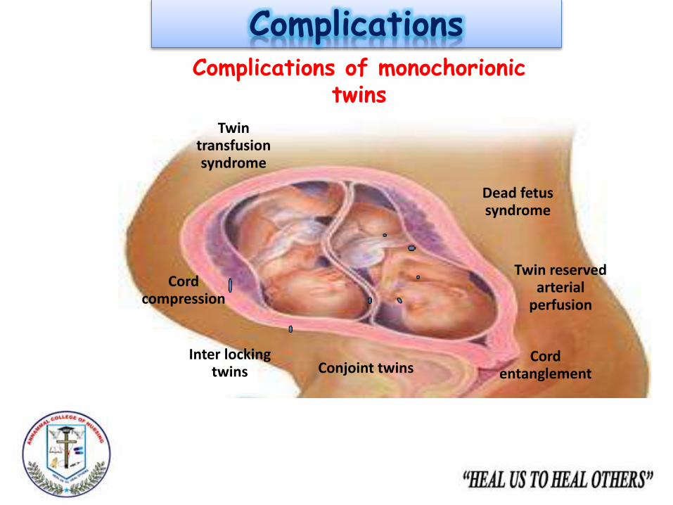
ComplicationsComplications of monochorionic
twins
Twin transfusion syndrome
Dead fetus syndrome
Twin reserved arterial
perfusion
Cord entanglementConjoint twins
Inter locking twins
Cord compression
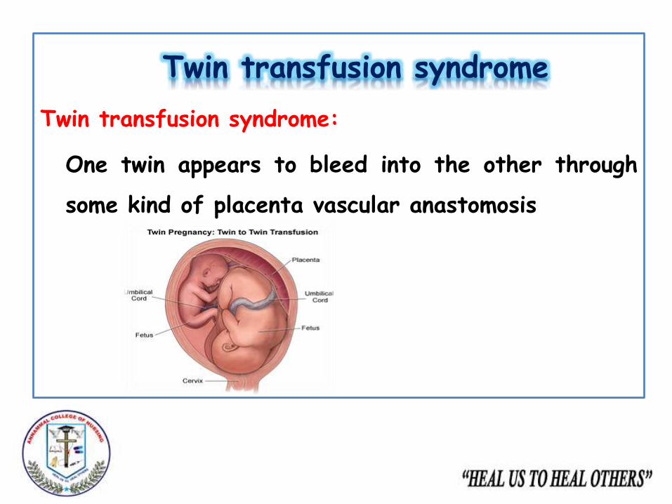
Twin transfusion syndrome
Twin transfusion syndrome:
One twin appears to bleed into the other through
some kind of placenta vascular anastomosis
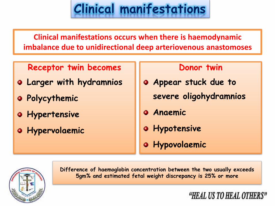
Donor twin
Appear stuck due to
severe oligohydramnios
Anaemic
Hypotensive
Hypovolaemic
Receptor twin becomes
Larger with hydramnios
Polycythemic
Hypertensive
Hypervolaemic
Clinical manifestations
Clinical manifestations occurs when there is haemodynamicimbalance due to unidirectional deep arteriovenous anastomoses
Difference of haemoglobin concentration between the two usually exceeds 5gm% and estimated fetal weight discrepancy is 25% or more
Twin transfusion syndrome
Management
Antenatal diagnosis is made by Ultrasound with
Doppler blood flow study in the placental vascular
bed
Amniocentesis
Laser photocoagulation
Selective reduction
Smaller twin have got
better outcome
The plethoric twin runs
the risk of CCF and
hydrops
Mortality:70%
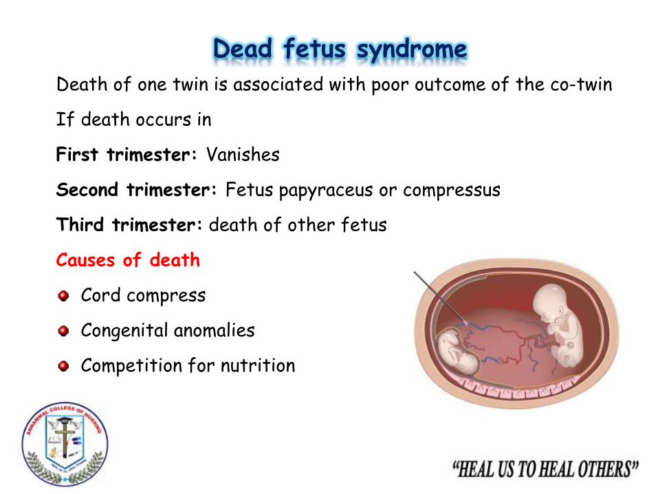
Dead fetus syndrome
Death of one twin is associated with poor outcome of the co-twin
If death occurs in
First trimester: Vanishes
Second trimester: Fetus papyraceus or compressus
Third trimester: death of other fetus
Causes of death
Cord compress
Congenital anomalies
Competition for nutrition
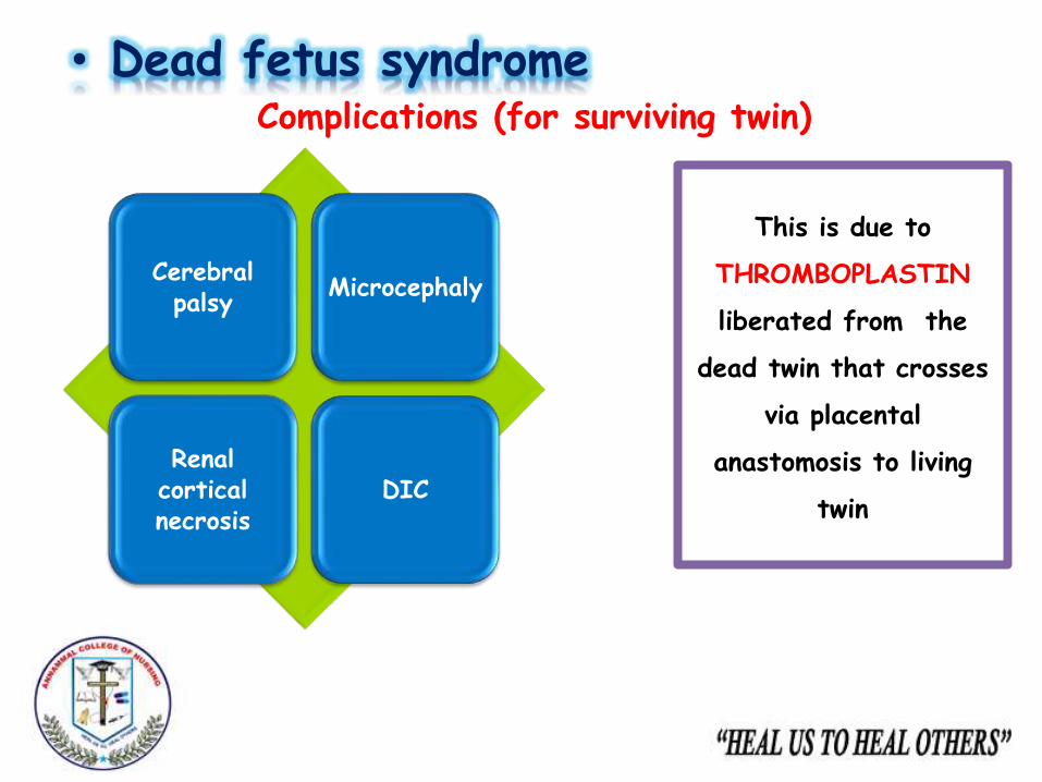
• Dead fetus syndromeComplications (for surviving twin)
Cerebral palsy
Microcephaly
Renal cortical necrosis
DIC
This is due to
THROMBOPLASTIN
liberated from the
dead twin that crosses
via placental
anastomosis to living
twin
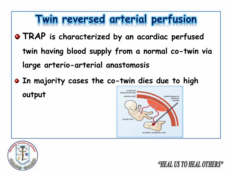
Twin reversed arterial perfusion
TRAP is characterized by an acardiac perfused
twin having blood supply from a normal co-twin via
large arterio-arterial anastomosis
In majority cases the co-twin dies due to high
output
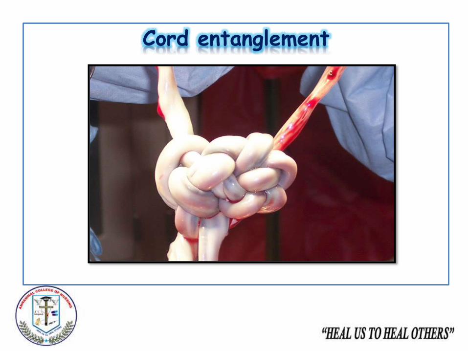
Cord entanglement
The close proximity and absence of amniotic membrane
separating the two umbilical cords makes it particularly
easy for the twins to become entangled in each other’s
cords, hindering fetal movement and development.
Additionally, entanglement may cause one twin to become
stuck in the birth canal during labour and expulsion..
Management:
Sulindac a prostaglandin synthase inhibitor used to reduce
the fetal urine output
Cord entanglement
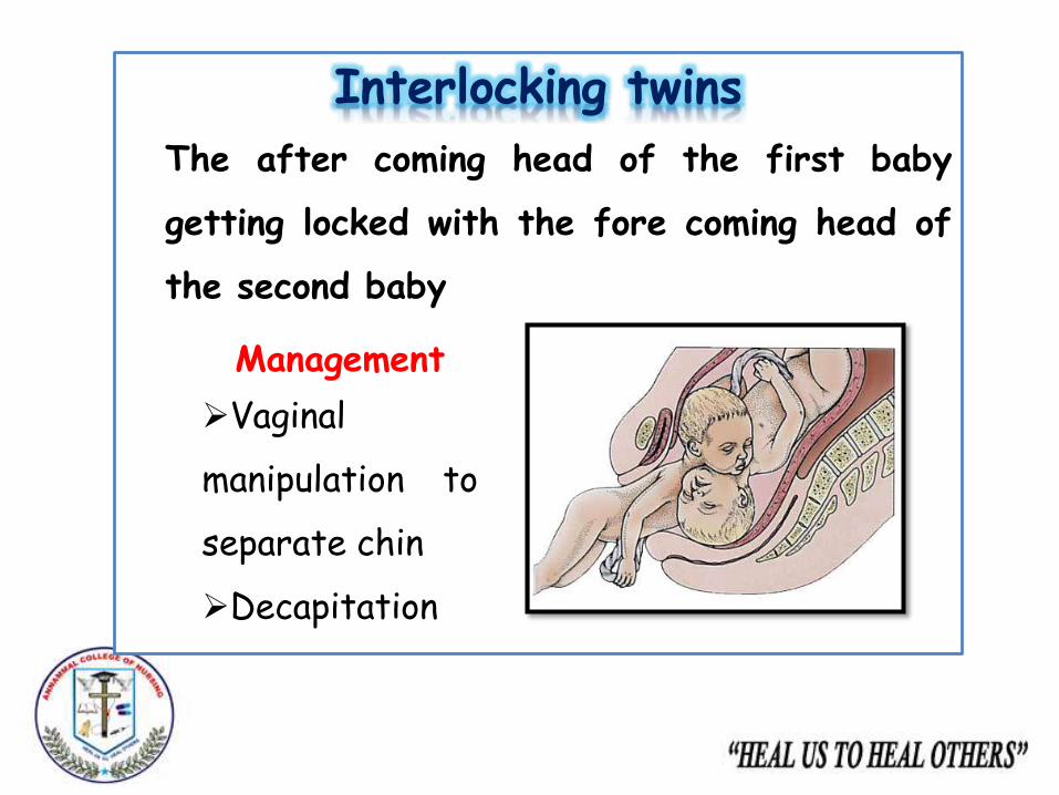
Interlocking twins
The after coming head of the first baby
getting locked with the fore coming head of
the second baby
Management
Vaginal
manipulation to
separate chin
Decapitation
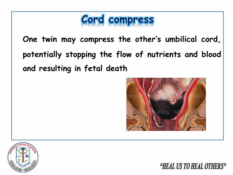
Cord compress
One twin may compress the other’s umbilical cord,
potentially stopping the flow of nutrients and blood
and resulting in fetal death
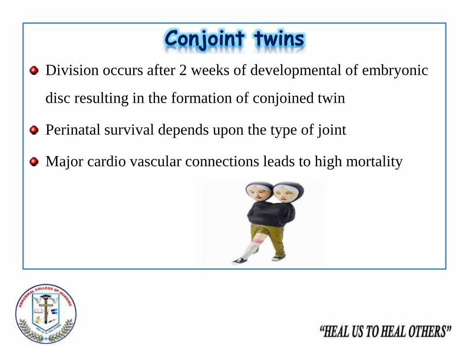
Conjoint twins
Division occurs after 2 weeks of developmental of embryonic
disc resulting in the formation of conjoined twin
Perinatal survival depends upon the type of joint
Major cardio vascular connections leads to high mortality
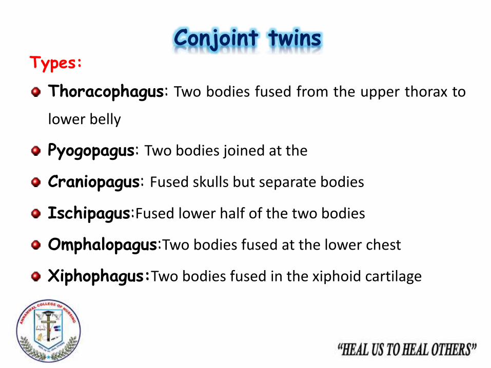
Conjoint twinsTypes:
Thoracophagus: Two bodies fused from the upper thorax to
lower belly
Pyogopagus: Two bodies joined at the
Craniopagus: Fused skulls but separate bodies
Ischipagus:Fused lower half of the two bodies
Omphalopagus:Two bodies fused at the lower chest
Xiphophagus:Two bodies fused in the xiphoid cartilage
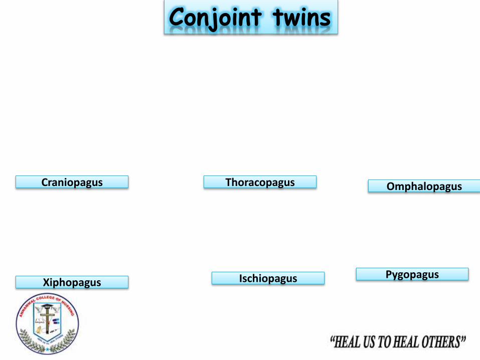
Conjoint twins
Craniopagus Omphalopagus
Xiphopagus
Thoracopagus
Ischiopagus Pygopagus
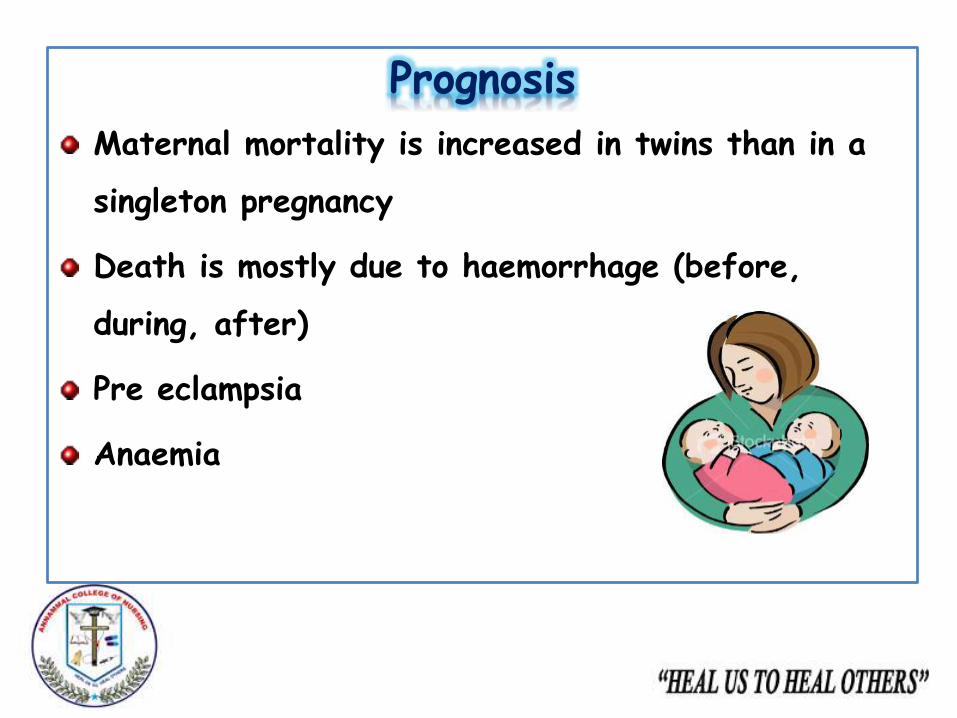
Prognosis
Maternal mortality is increased in twins than in a
singleton pregnancy
Death is mostly due to haemorrhage (before,
during, after)
Pre eclampsia
Anaemia
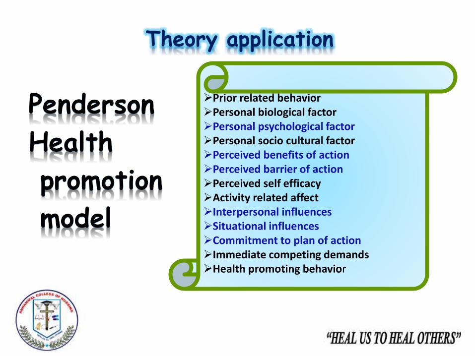
Theory application
Penderson
Health
promotion
model
Prior related behaviorPersonal biological factorPersonal psychological factorPersonal socio cultural factorPerceived benefits of actionPerceived barrier of actionPerceived self efficacyActivity related affectInterpersonal influencesSituational influencesCommitment to plan of actionImmediate competing demandsHealth promoting behavior
Journal presentation
BJOG an international journal ofobstetrics and Gynaecology
Nursing diagnosis Anxiety related to outcome of pregnancy as
manifested by increased frequency in asking doubts
Fatigue related to increased body functioningsecondary to multiple pregnancy
Body image disturbance related to increasedphysiological demand secondary to multiple pregnancy
Sleep pattern disturbance related to increased fetalmovements
Imbalanced nutritional status less than the bodyrequirement related to increased demand secondaryto multiple pregnancy

Conclusion
BibliographyAnnama jacob text book of midwifery and gynaecology 3rd edition
Jaypee publications pg no:336
D.C Dutta text book of obstetrics and gynaecology (2006) sixth
edition new central book publication pg no:210 to 212
Lowdermilk textbook of maternity and women health nursing 8th
edition Mosby publications pg 336