Multiple Endocrine Neoplasia Type 2A - ijsr.net · of using the term multiple endocrine...
7
International Journal of Science and Research (IJSR) ISSN (Online): 2319-7064 Index Copernicus Value (2015): 78.96 | Impact Factor (2015): 6.391 Volume 6 Issue 5, May 2017 www.ijsr.net Licensed Under Creative Commons Attribution CC BY Multiple Endocrine Neoplasia Type 2A I Made Pande Dwipayana 1 , Adrian Tri Sutjahjo 2 Division of Endocrinology Metabolism, Department of Internal Medicine Udayana University School of Medicine/Sanglah General Hospital Denpasar Abstract: An adult woman 43 years old, with Multiple Endocrine Neoplasia type 2A (MEN2A). Clinical diagnosis of MEN2A was made based on the chief complaints of abdominal pain and a lump in the front left side area of neck accompanied with weight loss, from laboratory examination revealed an increased level of plasma levels of calcitonin levels with the result of 6359 pg/ml and 24 hours metanephrine urine levels was 534 µg/24 hours. On the results of thyroid ultrasound examination showed nodules with malignant characteristics, multiple parathyroid gland hyperplasia and the discovery of a solid mass in the left adrenal gland area by an abdominal CT Scan with and without contrast. MEN2A case is considered as one of the few endocrine malignancies with a rare incidence especially in Indonesia. Keyword: Multiple endocrine neoplasia, thyroid, pheochromocytoma, endocrine. 1. Introduction Multiple Endocrine Neoplasia (MEN) is a hereditary syndrome of a benign and malignant endocrine neoplasia that are found in two or more different hormonal tissues. In this modern era, there are a few of those who meritorious of finding this syndrome. Werner is the first one who proposed of using the term multiple endocrine adenomatosis that defines of a syndrome of tumor that involves hypophysis gland, pancreatic islet cell, and parathyroid gland. Sipple explained about a phenomena of a thyroid malignancies and pheochromocytoma syndrome. Schimke reported of a subgroup from Sipple’s syndrome that is manifested as neurofibromatosis and other genetic abnormalities. Zollinger-Ellison syndrome (gastrin hormone-secreting tumors or so-called gastrinoma), which was initially thought something separate, are now considered a variant of MEN (1). MEN syndrome including rare diseases. The prevalence is estimated to be only about 0.2 to 2.0 per 100,000 population for MEN type 1 (MEN1) and 2.0 to 10 per 100,000 population for MEN type 2 (MEN2). While the incidence is estimated at about 2 to 20 per 100,000 population for MEN1 and 1 to 10 per 100,000 population for MEN2. Initially, the case is often found in residents of northern European descent, with the passage of time then these cases were also reported from Southern and Eastern Europe, Asia, and more rarely from Africa and the Americas (2). In Indonesia alone, the incidence of MEN is quite rare, therefore it was, the background of the authors to report the case. MEN2 is a combination of medullary thyroid carcinoma (MTC), pheochromocytoma and parathyroid tumors. There are three variants of MEN2, namely MEN type 2A (MEN2A), MEN type 2B (MEN2B), and familial MTC (FMTC). MEN2A patients have a normal phenotype, where MEN2B patients have different phenotypes (vide infra) with oral ganglioneuroma, marfanoid habitus, prominent corneal nerves, and generally no parathyroid disease, and FMTC which is a familial disorder in which patients have only MTC alone. Therefore we reported a case of an adult woman with MEN2A. This case was reported in case of a rarity incidence especially in Bali, as well as to be able to provide information on how to diagnosis and conduct an appropriate treatment. 2. Case A Woman aged 43 years old, was referred to our hospital with a chief complaint of an abdominal pain. Abdominal pain is felt in navel area since three years ago, intermittent, felt like it was stabbed, and did not improve with pain- killers. Sometimes abdominal pain feels moved to the chest and head with varying intensity. Patients also complained of a lump in the front of the neck to the left who felt since patients aged approximately 20 years. Lump is said to be enlarged and not accompanied by pain. Patients said weight loss of about 13 kilograms in the last one year. Other complaints such as palpitations, trembling, sweating was denied by the patient. Patient was fully allert during physical examination with blood pressure 170/100 mmHg, pulse 110 beats per minute, respiratory rate 20 breaths per minutes, axillary temperature of 36 0C, pain scale during the examination was 1 of 10. On examination of the neck appears the mass in front of the neck area to the left, two lateral finger palpation of the cricoid cartilage to find any solid mass of ± 3 cm in diameter, hard, helped move when the patient is asked to swallow and not found any pulsation. There enlarged lymph nodes preaurikular left. On examination of the heart and lungs are found within the normal range, while the physical examination did not reveal any abdominal masses, tenderness, and bowel sounds are within normal limits. Paper ID: ART20173176 DOI: 10.21275/ART20173176 411
-
Upload
trinhnguyet -
Category
Documents
-
view
215 -
download
0
Transcript of Multiple Endocrine Neoplasia Type 2A - ijsr.net · of using the term multiple endocrine...

International Journal of Science and Research (IJSR) ISSN (Online): 2319-7064
Index Copernicus Value (2015): 78.96 | Impact Factor (2015): 6.391
Volume 6 Issue 5, May 2017 www.ijsr.net
Licensed Under Creative Commons Attribution CC BY
Multiple Endocrine Neoplasia Type 2A
I Made Pande Dwipayana1, Adrian Tri Sutjahjo2
Division of Endocrinology Metabolism, Department of Internal Medicine Udayana University School of Medicine/Sanglah General Hospital Denpasar
Abstract: An adult woman 43 years old, with Multiple Endocrine Neoplasia type 2A (MEN2A). Clinical diagnosis of MEN2A was made based on the chief complaints of abdominal pain and a lump in the front left side area of neck accompanied with weight loss, from laboratory examination revealed an increased level of plasma levels of calcitonin levels with the result of 6359 pg/ml and 24 hours metanephrine urine levels was 534 µg/24 hours. On the results of thyroid ultrasound examination showed nodules with malignant characteristics, multiple parathyroid gland hyperplasia and the discovery of a solid mass in the left adrenal gland area by an abdominal CT Scan with and without contrast. MEN2A case is considered as one of the few endocrine malignancies with a rare incidence especially in Indonesia. Keyword: Multiple endocrine neoplasia, thyroid, pheochromocytoma, endocrine. 1. Introduction Multiple Endocrine Neoplasia (MEN) is a hereditary syndrome of a benign and malignant endocrine neoplasia that are found in two or more different hormonal tissues. In this modern era, there are a few of those who meritorious of finding this syndrome. Werner is the first one who proposed of using the term multiple endocrine adenomatosis that defines of a syndrome of tumor that involves hypophysis gland, pancreatic islet cell, and parathyroid gland. Sipple explained about a phenomena of a thyroid malignancies and pheochromocytoma syndrome. Schimke reported of a subgroup from Sipple’s syndrome that is manifested as neurofibromatosis and other genetic abnormalities. Zollinger-Ellison syndrome (gastrin hormone-secreting tumors or so-called gastrinoma), which was initially thought something separate, are now considered a variant of MEN (1). MEN syndrome including rare diseases. The prevalence is estimated to be only about 0.2 to 2.0 per 100,000 population for MEN type 1 (MEN1) and 2.0 to 10 per 100,000 population for MEN type 2 (MEN2). While the incidence is estimated at about 2 to 20 per 100,000 population for MEN1 and 1 to 10 per 100,000 population for MEN2. Initially, the case is often found in residents of northern European descent, with the passage of time then these cases were also reported from Southern and Eastern Europe, Asia, and more rarely from Africa and the Americas (2). In Indonesia alone, the incidence of MEN is quite rare, therefore it was, the background of the authors to report the case. MEN2 is a combination of medullary thyroid carcinoma (MTC), pheochromocytoma and parathyroid tumors. There are three variants of MEN2, namely MEN type 2A (MEN2A), MEN type 2B (MEN2B), and familial MTC (FMTC). MEN2A patients have a normal phenotype, where MEN2B patients have different phenotypes (vide infra) with oral ganglioneuroma, marfanoid habitus, prominent corneal nerves, and generally no parathyroid disease, and FMTC
which is a familial disorder in which patients have only MTC alone. Therefore we reported a case of an adult woman with MEN2A. This case was reported in case of a rarity incidence especially in Bali, as well as to be able to provide information on how to diagnosis and conduct an appropriate treatment. 2. Case A Woman aged 43 years old, was referred to our hospital with a chief complaint of an abdominal pain. Abdominal pain is felt in navel area since three years ago, intermittent, felt like it was stabbed, and did not improve with pain-killers. Sometimes abdominal pain feels moved to the chest and head with varying intensity. Patients also complained of a lump in the front of the neck to the left who felt since patients aged approximately 20 years. Lump is said to be enlarged and not accompanied by pain. Patients said weight loss of about 13 kilograms in the last one year. Other complaints such as palpitations, trembling, sweating was denied by the patient. Patient was fully allert during physical examination with blood pressure 170/100 mmHg, pulse 110 beats per minute, respiratory rate 20 breaths per minutes, axillary temperature of 36 0C, pain scale during the examination was 1 of 10. On examination of the neck appears the mass in front of the neck area to the left, two lateral finger palpation of the cricoid cartilage to find any solid mass of ± 3 cm in diameter, hard, helped move when the patient is asked to swallow and not found any pulsation. There enlarged lymph nodes preaurikular left. On examination of the heart and lungs are found within the normal range, while the physical examination did not reveal any abdominal masses, tenderness, and bowel sounds are within normal limits.
Paper ID: ART20173176 DOI: 10.21275/ART20173176 411
International Journal of Science and Research (IJSR) ISSN (Online): 2319-7064
Index Copernicus Value (2015): 78.96 | Impact Factor (2015): 6.391
Volume 6 Issue 5, May 2017 www.ijsr.net
Licensed Under Creative Commons Attribution CC BY
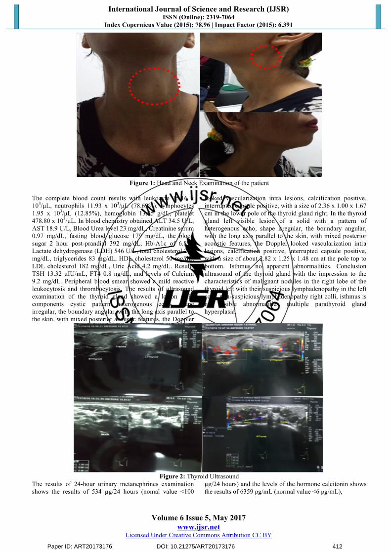
Figure 1: Head and Neck Examination of the patient
The complete blood count results with leukocyte 15.16 x 10³/µL, neutrophils 11.93 x 10³/µL (78.69%), lymphocytes 1.95 x 10³/µL (12.85%), hemoglobin 13.95 g/dL, platelet 478.80 x 10³/µL. In blood chemistry obtained ALT 34.5 U/L, AST 18.9 U/L, Blood Urea level 23 mg/dL, Creatinine serum 0.97 mg/dL, fasting blood glucose 179 mg/dL, the blood sugar 2 hour post-prandial 392 mg/dL, Hb-A1c of 6.8%, Lactate dehydrogenase (LDH) 546 U/L, total cholesterol 241 mg/dL, triglycerides 83 mg/dL, HDL cholesterol 50 mg/dL, LDL cholesterol 182 mg/dL, Uric Acid 4.2 mg/dL. Results TSH 13.32 µIU/mL, FT4 0.8 ng/dL, and levels of Calcium 9.2 mg/dL. Peripheral blood smear showed a mild reactive leukocytosis and thrombocytosis. The results of ultrasound examination of the thyroid gland showed a lesion solid components cystic pattern heterogenous echo, shape irregular, the boundary angular, with the long axis parallel to the skin, with mixed posterior acoustic features, the Doppler
looked vascularization intra lesions, calcification positive, interrupted capsule positive, with a size of 2.36 x 1.00 x 1.67 cm in the lower pole of the thyroid gland right. In the thyroid gland left visible lesion of a solid with a pattern of heterogenous echo, shape irregular, the boundary angular, with the long axis parallel to the skin, with mixed posterior acoustic features, the Doppler looked vascularization intra lesions, calcification positive, interrupted capsule positive, with a size of about 2.82 x 1.25 x 1.48 cm at the pole top to bottom. Isthmus no apparent abnormalities. Conclusion ultrasound of the thyroid gland with the impression to the characteristics of malignant nodules in the right lobe of the thyroid left with their suspicious lymphadenopathy in the left colli, non-suspicious lymphadenopathy right colli, isthmus is not visible abnormalities, multiple parathyroid gland hyperplasia.
Figure 2: Thyroid Ultrasound
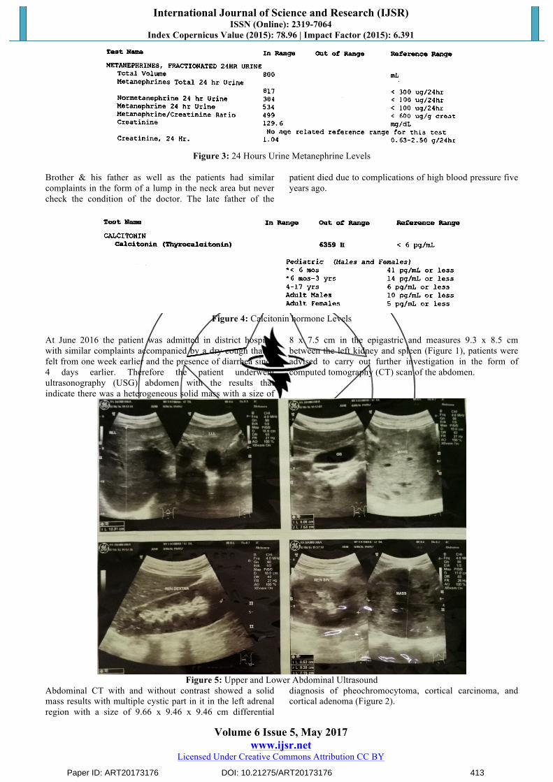
The results of 24-hour urinary metanephrines examination shows the results of 534 µg/24 hours (nomal value <100
µg/24 hours) and the levels of the hormone calcitonin shows the results of 6359 pg/mL (normal value <6 pg/mL),
Paper ID: ART20173176 DOI: 10.21275/ART20173176 412
International Journal of Science and Research (IJSR) ISSN (Online): 2319-7064
Index Copernicus Value (2015): 78.96 | Impact Factor (2015): 6.391
Volume 6 Issue 5, May 2017 www.ijsr.net
Licensed Under Creative Commons Attribution CC BY
Figure 3: 24 Hours Urine Metanephrine Levels
Brother & his father as well as the patients had similar complaints in the form of a lump in the neck area but never check the condition of the doctor. The late father of the
patient died due to complications of high blood pressure five years ago.
Figure 4: Calcitonin hormone Levels
At June 2016 the patient was admitted in district hospital with similar complaints accompanied by a dry cough that is felt from one week earlier and the presence of diarrhea since 4 days earlier. Therefore the patient underwent ultrasonography (USG) abdomen with the results that indicate there was a heterogeneous solid mass with a size of
8 x 7.5 cm in the epigastric and measures 9.3 x 8.5 cm between the left kidney and spleen (Figure 1), patients were advised to carry out further investigation in the form of computed tomography (CT) scan of the abdomen.
Figure 5: Upper and Lower Abdominal Ultrasound
Abdominal CT with and without contrast showed a solid mass results with multiple cystic part in it in the left adrenal region with a size of 9.66 x 9.46 x 9.46 cm differential
diagnosis of pheochromocytoma, cortical carcinoma, and cortical adenoma (Figure 2).
Paper ID: ART20173176 DOI: 10.21275/ART20173176 413

International Journal of Science and Research (IJSR) ISSN (Online): 2319-7064
Index Copernicus Value (2015): 78.96 | Impact Factor (2015): 6.391
Volume 6 Issue 5, May 2017 www.ijsr.net
Licensed Under Creative Commons Attribution CC BY
Figure 6: Abdominal CT with and wihout contrast
Patient was diagnosed with type 2 diabetes mellitus and MEN2A syndrome based on the finding of suspected MTC, unilateral pheochromocytoma and parathyroid gland hyperplasia. Initial management is given in the form of non-surgical therapy. Medical treatment for the provision of insulin aspart 6 units subcutaneously every 8 hours, insulin glargine 14 units subcutaneously every 24 hours, prazosin 1 mg every 8 hours orally, propranolol 10 mg every 8 hours orally, as well as paracetamol 750 mg every 8 hours if the headache reappears, Patients consulted to peer surgical oncology with a suspected diagnosis of multiple neoplasia syndrome type 2A, and was planned to be done exploratory laparotomy and biopsy of the adrenal glands. Until now, patients still awaiting for surgery to be done. The last condition of patients with complaints of abdominal pain that has rarely emerged and the results of physical examination found blood pressure 130/70 mmHg, pulse 80 beats/minute, respiratory rate 20 breaths/min, axillary temperature of 36.6oC, pain scale during examination was decreased to 0/10. 3. Discussion MEN syndrome is a rare malignant disease with a prevalence of 2.5 per 100,000 people in the general population (3). There were no sex differences in the prevalence of MEN in general or subtypes (2.4). Because of its prevalence is rare until now there has been no literature that outlines the differences of race as a factor predisposing to the emergence of MEN syndrome (1,6). There are two main types MEN syndrome, namely MEN1 and MEN2. MEN1 consists of two or more tumors derived from the pituitary, enteropankreatic, and parathyroid. MEN1 is the most heterogeneous tumor syndrome, causes tumors among the 25 major endocrine tissue (Table 1) (4). MEN2 is a combination of medullary thyroid carcinoma (MTC), pheochromocytoma and parathyroid tumors. There are three variants of MEN2, namely MEN2A where the patient has a phenotype that is normal, MEN2B where patients have different phenotypes (vide infra) with ganglioneuroma oral, marfanoid habitus, nerve cornea prominent, and generally no parathyroid disease, and Familial MTC (FMTC) namely familial disorder in which patients have only MTC (5). In the case of a woman
found 43 years of age, with MEN2A syndrome is confirmed by the discovery of medullary thyroid carcinoma (MTC), unilateral pheochromocytoma and parathyroid gland hyperplasia.
Table 1: The organs involved and the estimated percentage
in the MEN (4) Organs involved Estimated
percentage of tumors (%)
MEN1 • Parathyroid 90 • Enteropancreatic 30-75 Functioning Gastrin Insulin Glucagon Nonfunctioning Pancreatic Polypeptide Glucagon Vasoactive intestinal polypeptide Somatostatin Others: Calcitonin, serotonin, cromatogranin, neurotensin, growth hormones.
40-50 10-29
1
17-20 2-8 2 2
• Nonfunctioning foregut carcinoid Thymic Bronchial
16 2
2-8 • Gastric enterochromorffin-like 7-10 • Anterior Pituitary Prolactin Growth hormones and prolactin Growth hormones Adrenocorticotropic Thyrothropin Nonfunctional
18-47 20-30
5 5 2 1
5-10 • Thyroid 12 • Adrenal cortex 16-40 • Other non-endocrine tumors MEN2A • Medullary Thyroid Carcinoma 100 • Pheochromocytoma 19-50 • Parathyroid 15-30 • Cutaneus Lichen Amiloidosis Rare • Hirschsprung disease Rare MEN2B
Paper ID: ART20173176 DOI: 10.21275/ART20173176 414

International Journal of Science and Research (IJSR) ISSN (Online): 2319-7064
Index Copernicus Value (2015): 78.96 | Impact Factor (2015): 6.391
Volume 6 Issue 5, May 2017 www.ijsr.net
Licensed Under Creative Commons Attribution CC BY
• Medullary Thyroid Carcinoma 100 • Pheochromocytoma 25 • Parathyroid Rare • Ganglioneuroma Phenotye 100 KTMF • Medullary Thyroid Carcinoma 7
Both MEN syndrome is an inherited disease is autosomal dominant (1). MEN1 derived from a gene mutation on chromosome 11q13. This gene will transform into 613 amino acid protein intranuclear called "menin” whis is a tumor suppressor (8). Approximately 80% of patients had one or more MEN1 320 codon germline mutation that causes unnecessary downtime or loss of tumor suppression. In the event of the loss of the second allele suppressor, the tumor began to grow consistently. In MEN2 will find missense mutation codon 12 of the proto-oncogene germline rearranged during transfection (RET) on chromosome 10q12. 95% of cases will have a type of mutation MEN2 RET protooncogene 10q12 (7). Activation of the gene to a receptor tyrosine kinases will lead to the growth and differentiation of cells, and the onset of tumors. First, the occurrence of germinal mutations will increase the vulnerability towards malignant transformation. Second, somatic mutations that alter mutant cells into tumor cells. In MEN2 found a strong relationship between genotype and phenotype (7). MEN2A refers to the existence of MTC associated with pheochromocytoma in 50% of cases, and parathyroid gland abnormalities in 20-30% of cases (6). MTC himself appeared in 95% of cases MEN2A (2). MTC is generally a multifocal lesions and has spread to the nearby lymph nodes in 40% of cervical MEN2A newly diagnosed cases (8). Clinical findings are often found in a patient MTC is in the form of a lump in the neck that is palpable or the symptoms resulting calcitonin hormone levels in the body of excess such as diarrhea and flushing of the face that are temporary (flushing). Diarrhea usually only be found if very high levels of the hormone calcitonin (at least 10 times above the upper limit of normal levels of the hormone calcitonin). Quite possibly MTC has spread to regional lymph nodes when symptoms of diarrhea have emerged may have been a plus magnitude of distant metastases (9). In case, MTC diagnosis is made by the clinical findings in the form of a lump in the neck area with their palpability lymph nodes in the area preaurikularis left, symptoms obtained in the form of a history of intermittent diarrhea. Test results above reinforced by evidence of elevated levels of the hormone calcitonin in the blood to levels of 6359 pg / mL (normal range <6 pg / mL). Pheochromocytoma is a rare tumor of the adrenal gland (2). The words pheochromocytoma have a meaning of a brown colour in appearance where most of these tumors grow in the adrenal gland and only 10% outside the adrenal glands (paraganglioma) (2.8). Pheochromocytoma appears in 50% of cases MEN2A, although the number of events was lower in populations with a low risk of mutations (9). Pheochromocytoma is rarely be the initial manifestation in MEN2A in which is usually preceded by MTC finding characterized by symptoms caused by an increase in the hormone calcitonin (8.9). These tumors may secrete a variety
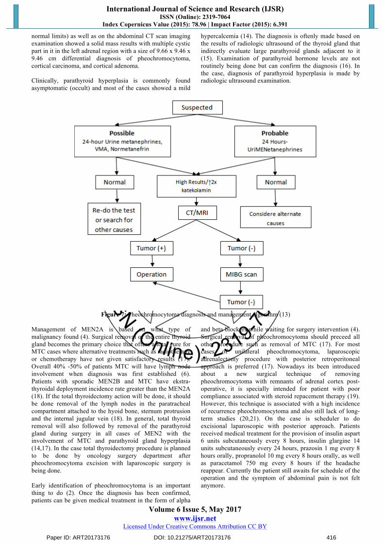
of hormones, especially norephinephrine, ephinephrine, and dopamine, with certain patterns are different in each individual (8). Pheochromocytoma types found in the case of MEN2A is usually benign, located in the adrenal glands, multicentric, and located adjacent with concurrent adrenal gland hyperplasia (9). Hormonal profile on MEN2A and MEN2B are very similar in which there are domination on the secretion of epinephrine when compared with other hormones, so that will appear on the examination of urine and plasma epinephrine and metanephrines (10). The pattern of hormone secretion is related to the regulation of several enzymes intratumor by RET (9.10). Clinical manifestations associated with overproduction of catecholamines such as headache, sweating, and palpitations (9). Other symptoms of excess catecholamines may be pale, orthostatic hypotension, blurred vision, papilledema eyes, weight loss, polyuria, polidipsi, an elevated ESR, hyperglycemia, psychiatric disorders, dilated cardiomyopathy (11). Because of lack of specific signs and symptoms and laboratory results is difficult, so pheochromocytoma are often found incidentally by CT scan or MRI (12). The diagnosis of pheochromocytoma is based on complaints and clinical symptoms and it requires laboratory confirmation by measuring the levels of catecholamines (metanephrines or normetanephrin) blood, urine or metabolites result (Figure 7) (13). Typical laboratory findings is an increase in catecholamine levels 5-10 times normal. If the levels are not too high, then the diagnosis of pheochromocytoma will not be confirmed and further investigation such as clonidine test in which will suppress norephinephrine levels (to normal) need to be done (13). Clonidine test procedure can be fairly complicated because prior to the procedure the patient should be in a condition of not consuming an alpha and/or beta inhibitor drugs within the last 48 hours in order to avoid false negative results. This procedure is performed in a dark room to avoid the presence of sympathetic stimulation, then the patient is asked to rest for a while (30 minutes) and blood samples were taken to measure levels of catecholamine hormones (metanephrines and normetanefrin). Patients were then asked to consume 0.3 mg oral clonidine and blood samples were taken back 180 minutes later. Positive results were obtained when there was a drop of at least 40% of basal levels of catecholamines (13). In addition to clonidine suppression test, there are other provocation tests such as regitin tests (fentolamin) by administering 5 mg fentolamin intravenously (positive result if there is a decrease in systolic blood pressure> 35 mm Hg and diastolic blood pressure of <25 mmHg) and a glucagon stimulation test by giving 1 mg of glucagon intravenously in patients who had fasted overnight before the procedure is performed, and blood samples were taken three minutes after (a positive result if there is an increase in blood catecholamine levels of> 3x from baseline). However, due to risk of rebound hypertensive crisis, both of these tests are no longer used (12,13). The diagnosis of pheochromocytoma in the case is based on the symptoms and signs of increased catecholamine levels include headache, weight loss, as well as an increase in blood pressure. In addition to the investigation of 24-hour urinary metanephrines levels show the results of 534 mg (above
Paper ID: ART20173176 DOI: 10.21275/ART20173176 415
International Journal of Science and Research (IJSR) ISSN (Online): 2319-7064
Index Copernicus Value (2015): 78.96 | Impact Factor (2015): 6.391
Volume 6 Issue 5, May 2017 www.ijsr.net
Licensed Under Creative Commons Attribution CC BY
normal limits) as well as on the abdominal CT scan imaging examination showed a solid mass results with multiple cystic part in it in the left adrenal region with a size of 9.66 x 9.46 x 9.46 cm differential diagnosis of pheochromocytoma, cortical carcinoma, and cortical adenoma. Clinically, parathyroid hyperplasia is commonly found asymptomatic (occult) and most of the cases showed a mild
hypercalcemia (14). The diagnosis is oftenly made based on the results of radiologic ultrasound of the thyroid gland that indirectly evaluate large parathyroid glands adjacent to it (15). Examination of parathyroid hormone levels are not routinely being done but can confirm the diagnosis (16). In the case, diagnosis of parathyroid hyperplasia is made by radiologic ultrasound examination.
Figure 7: Pheochromocytoma diagnosis and management algorithm (13)
Management of MEN2A is based on what type of malignancy found (4). Surgical removal of the entire thyroid gland becomes the primary choice that offers a total cure for MTC cases where alternative treatments such as radiotherapy or chemotherapy have not given satisfactory results (17). Overall 40% -50% of patients MTC will have lymph node involvement when diagnosis was first established (6). Patients with sporadic MEN2B and MTC have ekstra-thyroidal deployment incidence rate greater than the MEN2A (18). If the total thyroidectomy action will be done, it should be done removal of the lymph nodes in the paratracheal compartment attached to the hyoid bone, sternum protrusion and the internal jugular vein (18). In general, total thyroid removal will also followed by removal of the parathyroid gland during surgery in all cases of MEN2 with the involvement of MTC and parathyroid gland hyperplasia (14,17). In the case total thyroidectomy procedure is planned to be done by oncology surgery department after pheochromocytoma excision with laparoscopic surgery is being done. Early identification of pheochromocytoma is an important thing to do (2). Once the diagnosis has been confirmed, patients can be given medical treatment in the form of alpha
and beta blockers while waiting for surgery intervention (4). Surgical removal of pheochromocytoma should preceed all other procedure such as removal of MTC (17). For most cases of unilateral pheochromocytoma, laparoscopic adrenalectomy procedure with posterior retroperitoneal approach is preferred (17). Nowadays its been introduced about a new surgical technique of removing pheochromocytoma with remnants of adrenal cortex post-operative, it is specially intended for patient with poor compliance associated with steroid repacement therapy (19). However, this technique is associated with a high incidence of recurrence pheochromocytoma and also still lack of long-term studies (20,21). On the case is scheduler to do excisional laparoscopic with posterior approach. Patients received medical treatment for the provision of insulin aspart 6 units subcutaneously every 8 hours, insulin glargine 14 units subcutaneously every 24 hours, prazosin 1 mg every 8 hours orally, propranolol 10 mg every 8 hours orally, as well as paracetamol 750 mg every 8 hours if the headache reappear. Currently the patient still awaits for schedule of the operation and the symptom of abdominal pain is not felt anymore.
Paper ID: ART20173176 DOI: 10.21275/ART20173176 416

International Journal of Science and Research (IJSR) ISSN (Online): 2319-7064
Index Copernicus Value (2015): 78.96 | Impact Factor (2015): 6.391
Volume 6 Issue 5, May 2017 www.ijsr.net
Licensed Under Creative Commons Attribution CC BY
4. Summary It has been reported a case of an adult woman 43 years old, with Multiple Endocrine Neoplasia type 2A (MEN2A). Clinical diagnosis of MEN2A was made based on the chief complaints of abdominal pain and a lump in the front left side area of the neck accompanied with weight loss, from laboratory examination revealed an increased level of plasma levels of calcitonin levels with the result of 6359 pg/ml and 24 hours metanephrine urine levels was 534 µg/24 hours. On the results of thyroid ultrasound examination showed nodules with malignant characteristics, multiple parathyroid gland hyperplasia and the discovery of a solid mass in the left adrenal gland area by an abdominal CT Scan with and without contrast. MEN2A case is considered as one of the few endocrine malignancies with a rare incidence especially in Indonesia. If there is a delay in diagnosis and initial treatment, high morbidity and mortality can be the consequences. A careful history taking, physical examinations, supported with additional laboratory and imaging data can help a clinician in making of a proper diagnosis of MEN2A. References
[1] Gagel RF, Marx SJ. Multiple endocrine neoplasia. In:
Larsen PR et al. editors. Williams textbook of endocrinology. Tenth Edition. Philadelphia: Saunders, 2003; 1717-1748.
[2] Sizemore GW. Multiple endocrine neoplasia. In: Camacho PM et al. editors. Evidence-based endocrinology. Second Edition. Philadelphia: Lippincot Williams & Wilkins, 2007; 225-241.
[3] Frank-Raue K, Raue F. Multiple endocrine neoplasia type 2 (MEN 2). Eur J Cancer 2009; 45(suppl. 1): 267-73.
[4] Dashe AM. Multiple endocrine neoplasia (MEN) syndromes. In: Lavin N editors. Manual of endocrinology and metabolism. Fourth Edition. Philadelphia: Lippincott Williams & Wilkins, 2009; 749-753.
[5] Agarwal SK, Ozawa A, Mateo CM, Marx SJ. The MEN1 gene and pituitary tumours. Horm Res 2009; 71 (Suppl 2):131-138.
[6] El-Kholy LR. Multiple endocrine neoplasia syndromes. In: Henderson KE et al. editors. The Washington Manual Endocrinology Subspecialty Consult. Washington: Lippincott Williams & Wilkins, 2005; p213-218.
[7] Burgess J. How should the patients with multiple endocrine neoplasia type 1 (MEN1) be followed? Clin Endocrinol 2010; 72: 13-16.
[8] Marx SJ. Molecular genetics of multiple endocrine neoplasia type 2: 2007 Update. Horm res 2007; 68(suppl 5): 101-104.
[9] Thosani S, Ayala-Ramirez M, Palmer L. The characterization of pheochromocytoma and its impact on overall survival in multiple endocrine neoplasia type 2A. J Clin Endocinol Metab 2013; 98: E1813-1819.
[10] Eisenhofer G, Lenders JW, Timmers H. Measurements of plasma methoxytyramine, normetanephrine, and metanephrineas discriminators of different hereditary
forms of pheochromocytoma. Clin Chem 2011; 57: 411-420.
[11] Neumann HP, Berger DP, Sigmund G. Pheochromocytomas, multiple endocrine neoplasia type 2, and von Hippel-Lindau disease. N Eng J Med 2003; 329: 1531.
[12] Scholten A, Valk GD, Ulfman D. Unilateral subtotal adrenalectomy for pheochromocytoma in multiple endocrine neoplasia type 2 patients: a feasible surgical strategy. Ann Surg 2011; 254: 1022-1027.
[13] Efendi I. Feokromositoma. Buku Ajar Ilmu Penyakit Dalam. Edisi Keenam. Jakarta. Interna Publishing 2014; 2: 2206-2209.
[14] Raue F, Kraimps JL, Dralle L. Primary hyperparathyroidism in multiple endocrine neoplasia type 2A. J Intern Med 2012; 238: 369-273.
[15] Scholten A, SchreiMENakers JM, Pieterman CR. Evolution of surgical treatment of primary hyperparathyroidism in patients with multiple endocrine neoplasia type 2A. Endocr Pract 2011; 17: 7-15.
[16] Yoshida S, Imai T, Kikumori T. Long term parathyroid function following total parathyroidectomy with autotransplantation in adult patients with MEN type 2A. Endocr J 2009; 56: 545-551.
[17] Kloos RT, Eng C, Evans DB. Medullary thyroid cancer: management guidelines of the American Thyroid Association. Thyroid 2009; 19: 565-612.
[18] Russel CF, Van Heerden JA, Sizemore GW. The surgical management of medullary thyroid carcinoma. Ann Surg 2003; 197: 42-48.
[19] Mazzaglia PJ, Vezeridis MP. Laparoscopic adrenalectomy: balancing the operative indications with technical advances. J Surg Oncol 2010; 101: 739-744.
[20] Scholten A, Valk GD, Ulfman D. Unilateral subtotal adrenalectomy for pheochromocytoma in multiple endocrine neoplasia type 2 patients: a feasible surgical strategy. Ann Surg 2011; 254: 1022-1027.
[21] Asari R, Scheuba C, Kaczirek K, Niederle B. Estimated risk of pheochromocytoma recurrence after adrenal-sparing surgery in patients with multiple endocrine neoplasia type 2A. Arch Surgc 2006; 141: 1199-1205.
Paper ID: ART20173176 DOI: 10.21275/ART20173176 417