Multiple Diverse Circoviruses Infect Farm Animals and …jvi.asm.org/content/84/4/1674.full.pdfEast...
9
JOURNAL OF VIROLOGY, Feb. 2010, p. 1674–1682 Vol. 84, No. 4 0022-538X/10/$12.00 doi:10.1128/JVI.02109-09 Copyright © 2010, American Society for Microbiology. All Rights Reserved. Multiple Diverse Circoviruses Infect Farm Animals and Are Commonly Found in Human and Chimpanzee Feces † Linlin Li, 1,2 Amit Kapoor, 1,2 Beth Slikas, 1,2 Oderinde Soji Bamidele, 3 Chunlin Wang, 4,5 Shahzad Shaukat, 6 Muhammad Alam Masroor, 6 Michael L. Wilson, 7,8 Jean-Bosco N. Ndjango, 9 Martine Peeters, 10 Nicole D. Gross-Camp, 11 Martin N. Muller, 12 Beatrice H. Hahn, 13 Nathan D. Wolfe, 14,15 Hinda Triki, 16 Joanne Bartkus, 17 Sohail Zahoor Zaidi, 6 and Eric Delwart 1,2 * Blood Systems Research Institute, San Francisco, California 94118 1 ; Department of Laboratory Medicine, University of California, San Francisco, San Francisco, California 94118 2 ; Medical Microbiology Department, University of Maiduguri Teaching Hospital, 1414 Maiduguri, Borno State, Nigeria 3 ; Division of Infectious Diseases, Stanford University Medical Center, Stanford, California 94305 4 ; Stanford Genome Technology Center, Stanford, California 94304 5 ; Department of Virology, National Institute of Health, Islamabad 45500, Pakistan 6 ; Department of Anthropology, University of Minnesota, Minneapolis, Minnesota 55455 7 ; Jane Goodall Institute’s Center for Primate Studies, Department of Ecology, Evolution and Behavior, University of Minnesota, St. Paul, Minnesota 55108 8 ; University of Kisangani, Kisangani, Democratic Republic of the Congo 9 ; Institut de Recherche pour le De ´veloppement and University of Montpellier 1, Montpellier, France 10 ; University of East Anglia, Norwich NR4 7TJ, United Kingdom 11 ; Department of Anthropology, University of New Mexico, Albuquerque, New Mexico 87131 12 ; Department of Medicine and Microbiology, University of Alabama at Birmingham, Birmingham, Alabama 35294 13 ; Program in Human Biology, Stanford University, Stanford, California 94305 14 ; Global Viral Forecasting Initiative, San Francisco, California 94104 15 ; Laboratory of Clinical Virology, Institut Pasteur de Tunis, 1002 Tunis-Belve ´de `re, Tunisia 16 ; and Public Health Laboratory, Minnesota Department of Health, St. Paul, Minnesota 55164 17 Received 5 October 2009/Accepted 27 November 2009 Circoviruses are known to infect birds and pigs and can cause a wide range of severe symptoms with significant economic impact. Using viral metagenomics, we identified circovirus-like DNA sequences and characterized 15 circular viral DNA genomes in stool samples from humans in Pakistan, Nigeria, Tunisia, and the United States and from wild chimpanzees. Distinct genomic features and phylogenetic analysis indicate that some viral genomes were part of a previously unrecognized genus in the Circoviridae family we tentatively named “Cyclovirus” whose genetic diversity is comparable to that of all the known species in the Circovirus genus. Circoviridae detection in the stools of U.S. adults was limited to porcine circoviruses which were also found in most U.S. pork products. To determine whether the divergent cycloviruses found in non-U.S. human stools were of dietary origin, we genetically compared them to the cycloviruses in muscle tissue samples of commonly eaten farm animals in Pakistan and Nigeria. Limited genetic overlap between cycloviruses in human stool samples and local cow, goat, sheep, camel, and chicken meat samples indicated that the majority of the 25 Cyclovirus species identified might be human viruses. We show that the genetic diversity of small circular DNA viral genomes in various mammals, including humans, is significantly larger than previously recognized, and frequent exposure through meat consumption and contact with animal or human feces provides ample opportunities for cyclovirus transmission. Determining the role of cycloviruses, found in 7 to 17% of non-U.S. human stools and 3 to 55% of non-U.S. meat samples tested, in both human and animal diseases is now facilitated by knowledge of their genomes. Animal viruses with small, circular, single-stranded DNA (ssDNA) genomes comprise the Circoviridae family and the Anellovirus genus, while viruses in the Geminiviridae and Nano- viridae families infect plants (3, 25, 34, 37, 40). The genomes of these small viruses without a lipid envelope replicate through a rolling-circle mechanism, possibly sharing a common origin with bacterial plasmids (6), and show high recombination and nucleotide substitution rates (7, 19). The Circoviridae family consists of the Circovirus genus whose member species are currently known to infect only birds and pigs, and the Gyrovirus genus, including a single species, Chicken anemia virus (CAV). Circoviruses infect several avian groups, including parrots, pigeons, gulls, anserids (ducks, geese, and swans), and numerous passerines (ravens, canaries, finches, and starlings) (12, 15, 16, 22, 26, 31, 35, 38, 39). Avian circoviruses have been associated with a variety of symptoms, including developmental abnormalities, lymphoid depletion, and immunosuppression (22, 26, 28, 35, 39). Mammalian circo- viruses include only two closely related species, Porcine circo- virus 1 and 2 (PCV1 and PCV2, respectively), infecting pigs (21). PCV2 has been associated with porcine circovirus-asso- * Corresponding author. Mailing address: Blood Systems Research Institute, 270 Masonic Ave., San Francisco, CA 94118. Phone: (415) 923-5763. Fax: (415) 567-5899. E-mail: [email protected]. † Supplemental material for this article may be found at http://jvi .asm.org/. Published ahead of print on 9 December 2009. 1674 on May 19, 2018 by guest http://jvi.asm.org/ Downloaded from
Transcript of Multiple Diverse Circoviruses Infect Farm Animals and …jvi.asm.org/content/84/4/1674.full.pdfEast...

JOURNAL OF VIROLOGY, Feb. 2010, p. 1674–1682 Vol. 84, No. 40022-538X/10/$12.00 doi:10.1128/JVI.02109-09Copyright © 2010, American Society for Microbiology. All Rights Reserved.
Multiple Diverse Circoviruses Infect Farm Animals and AreCommonly Found in Human and Chimpanzee Feces�†
Linlin Li,1,2 Amit Kapoor,1,2 Beth Slikas,1,2 Oderinde Soji Bamidele,3 Chunlin Wang,4,5 Shahzad Shaukat,6
Muhammad Alam Masroor,6 Michael L. Wilson,7,8 Jean-Bosco N. Ndjango,9Martine Peeters,10 Nicole D. Gross-Camp,11 Martin N. Muller,12
Beatrice H. Hahn,13 Nathan D. Wolfe,14,15 Hinda Triki,16
Joanne Bartkus,17 Sohail Zahoor Zaidi,6 and Eric Delwart1,2*Blood Systems Research Institute, San Francisco, California 941181; Department of Laboratory Medicine, University of California,San Francisco, San Francisco, California 941182; Medical Microbiology Department, University of Maiduguri Teaching Hospital,1414 Maiduguri, Borno State, Nigeria3; Division of Infectious Diseases, Stanford University Medical Center, Stanford, California
943054; Stanford Genome Technology Center, Stanford, California 943045; Department of Virology, National Institute ofHealth, Islamabad 45500, Pakistan6; Department of Anthropology, University of Minnesota, Minneapolis, Minnesota 554557;
Jane Goodall Institute’s Center for Primate Studies, Department of Ecology, Evolution and Behavior, University ofMinnesota, St. Paul, Minnesota 551088; University of Kisangani, Kisangani, Democratic Republic of the Congo9;Institut de Recherche pour le Developpement and University of Montpellier 1, Montpellier, France10; University ofEast Anglia, Norwich NR4 7TJ, United Kingdom11; Department of Anthropology, University of New Mexico,
Albuquerque, New Mexico 8713112; Department of Medicine and Microbiology, University ofAlabama at Birmingham, Birmingham, Alabama 3529413; Program in Human Biology,
Stanford University, Stanford, California 9430514; Global Viral Forecasting Initiative,San Francisco, California 9410415; Laboratory of Clinical Virology, Institut Pasteur de
Tunis, 1002 Tunis-Belvedere, Tunisia16; and Public Health Laboratory,Minnesota Department of Health, St. Paul, Minnesota 5516417
Received 5 October 2009/Accepted 27 November 2009
Circoviruses are known to infect birds and pigs and can cause a wide range of severe symptoms with significanteconomic impact. Using viral metagenomics, we identified circovirus-like DNA sequences and characterized 15circular viral DNA genomes in stool samples from humans in Pakistan, Nigeria, Tunisia, and the United States andfrom wild chimpanzees. Distinct genomic features and phylogenetic analysis indicate that some viral genomes werepart of a previously unrecognized genus in the Circoviridae family we tentatively named “Cyclovirus” whose geneticdiversity is comparable to that of all the known species in the Circovirus genus. Circoviridae detection in the stoolsof U.S. adults was limited to porcine circoviruses which were also found in most U.S. pork products. To determinewhether the divergent cycloviruses found in non-U.S. human stools were of dietary origin, we genetically comparedthem to the cycloviruses in muscle tissue samples of commonly eaten farm animals in Pakistan and Nigeria. Limitedgenetic overlap between cycloviruses in human stool samples and local cow, goat, sheep, camel, and chicken meatsamples indicated that the majority of the 25 Cyclovirus species identified might be human viruses. We show thatthe genetic diversity of small circular DNA viral genomes in various mammals, including humans, is significantlylarger than previously recognized, and frequent exposure through meat consumption and contact with animal orhuman feces provides ample opportunities for cyclovirus transmission. Determining the role of cycloviruses, foundin 7 to 17% of non-U.S. human stools and 3 to 55% of non-U.S. meat samples tested, in both human and animaldiseases is now facilitated by knowledge of their genomes.
Animal viruses with small, circular, single-stranded DNA(ssDNA) genomes comprise the Circoviridae family and theAnellovirus genus, while viruses in the Geminiviridae and Nano-viridae families infect plants (3, 25, 34, 37, 40). The genomes ofthese small viruses without a lipid envelope replicate through arolling-circle mechanism, possibly sharing a common origin
with bacterial plasmids (6), and show high recombination andnucleotide substitution rates (7, 19).
The Circoviridae family consists of the Circovirus genuswhose member species are currently known to infect only birdsand pigs, and the Gyrovirus genus, including a single species,Chicken anemia virus (CAV). Circoviruses infect several aviangroups, including parrots, pigeons, gulls, anserids (ducks,geese, and swans), and numerous passerines (ravens, canaries,finches, and starlings) (12, 15, 16, 22, 26, 31, 35, 38, 39). Aviancircoviruses have been associated with a variety of symptoms,including developmental abnormalities, lymphoid depletion,and immunosuppression (22, 26, 28, 35, 39). Mammalian circo-viruses include only two closely related species, Porcine circo-virus 1 and 2 (PCV1 and PCV2, respectively), infecting pigs(21). PCV2 has been associated with porcine circovirus-asso-
* Corresponding author. Mailing address: Blood Systems ResearchInstitute, 270 Masonic Ave., San Francisco, CA 94118. Phone: (415)923-5763. Fax: (415) 567-5899. E-mail: [email protected].
† Supplemental material for this article may be found at http://jvi.asm.org/.
� Published ahead of print on 9 December 2009.
1674
on May 19, 2018 by guest
http://jvi.asm.org/
Dow
nloaded from

ciated diseases, which can manifest as a systemic disease,respiratory disease complex, enteric disease, porcine dermatitisand nephropathy syndrome or as reproductive problems, caus-ing great losses in the pork industry (1, 29, 32). Circovirusinfections are thought to occur mainly through fecal-oral trans-mission (37).
We describe here highly diverse, circovirus-like, circularDNA viral genomes discovered in human and chimpanzeestool samples, and we propose their inclusion in a new genus ofthe Circoviridae family that we tentatively name “Cyclovirus”pending review by the International Committee on Taxonomyof Viruses (ICTV). Cycloviruses were also found to be preva-lent in the muscle tissue of farm animals, such as chickens,cows, sheep, goats, and camels. The cyclovirus species found inhuman stool samples and in animal meat samples showedlimited genetic overlap, suggesting that most of the cyclovi-ruses found in human stool samples are not from consumedanimal meat. Rather, these cycloviruses in human stools mightcause human enteric infections. The presence of cycloviruses inhuman stool samples and in farm animal tissue also suggeststhe potential for frequent cross-species exposure and zoonotictransmissions.
MATERIALS AND METHODS
Detection of circoviruses using degenerate primers. Nucleic acids were ex-tracted from stool supernatants and plasma samples using the QIAamp viralRNA kit which extracts both RNA and DNA (Qiagen). DNA was extracted fromanimal tissue specimens using a QIAamp DNA minikit (Qiagen). Degenerateprimers for nested PCR were as follows: CV-F1 (5�-GGIAYICCICAYYTICARGG), CV-R1 (5�-AWCCAICCRTARAARTCRTC), CV-F2 (5�-GGIAYICCICAYYTICARGGITT), and CV-R2 (5�-TGYTGYTCRTAICCRTCCCACCA).The degenerate primers were designed on the basis of the consensus sequencefrom an alignment of replicase (Rep) proteins from CyCV1-PK5006 and 12representative Circovirus species. Multiple-sequence alignment of the Rep aminoacid sequences was performed using ClustalW2, with default settings. PCRs withthe degenerate Rep primers were performed with the following cycling profile: 5min at 95°C; 40 cycles, with 1 cycle consisting of 1 min at 95°C, 1 min at 52°C(56°C for the 2nd PCR round), and 1 min at 72°C; and a final incubation for 10min at 72°C. Products with a size of approximately 400 bp were purified andsequenced using primer CV-R2. Most of the products were sequenced directly.Amplicons with low concentrations or multiple bands were cloned to obtainhigh-quality sequence data.
Phylogenetic analysis. Phylogenetic analyses based on aligned amino acidsequences from full-length or partial Rep proteins were generated by the neigh-bor-joining (NJ) method in MEGA 4.1 (18), using amino acid p-distances, with1,000 bootstrap replicates. Other tree-building methods, maximum parsimony(MEGA) and maximum likelihood (PhyML [11]), were carried out to confirmthe NJ tree. The GenBank accession numbers of the Rep sequences fromplasmids, viruses, and protists used in the phylogenetic analyses are as follows(shown in brackets): Beak and feather disease virus (BFDV) [AF071878],Canary circovirus (CaCV) [AJ301633], Columbid circovirus (CoCV)[AF252610], Duck circovirus (DuCV) [DQ100076], Goose circovirus (GoCV)[AJ304456], Gull circovirus (GuCV) [DQ845074], Finch circovirus (FiCV)[DQ845075], Raven circovirus (RaCV) [DQ146997], Starling circovirus (StCV)[DQ1729062], Cygnus olor circovirus (SwCV) [EU056310], Porcine circovirus1 (PCV1) [AY660574], Porcine circovirus 2 (PCV2) [AY424401], Chickenanemia virus (CAV) [M55918], Milk vetch dwarf virus [AB009047], Peppergolden mosaic virus [U57457], Canarypox virus [NC_005309], Giardia intesti-nalis [AF059664], Bifidobacterium pseudocatenulatum plasmid p4M[NC_003527], and Entamoeba histolytica [XM_643662].
Genome analyses. Putative open reading frames (ORFs) with a coding capac-ity greater than 100 amino acids were predicted by Vector NTI Advance 10.3(Invitrogen). The stem-loop structure was predicted using Mfold (version 3.2)(43).
Sample collections. (i) Stool samples from South Asian children. A total of107 fecal specimens were collected by the WHO Regional Reference Labo-ratory for polio eradication at the National Institute of Health in Islamabad,
Pakistan, between December 2005 and May 2008: 57 samples from non-polio-infected children with acute flaccid paralysis (AFP), 9 from closely related buthealthy contacts, and 41 from clinically healthy children living in the samegeographic region. The median age of the children was 3 years (range, 1month to 15 years).
(ii) Stool samples from Nigerian children. Ninety-six stool samples fromnon-polio-infected children with AFP were collected by the WHO National PolioLaboratory at the University of Maiduguri Teaching Hospital in Maiduguri,Nigeria, during February to April 2007. The median age of these children was 2.5years (range, 6 months to 12 years).
(iii) Stool samples from Tunisian children and adults. A total of 192 stoolsamples were collected by the WHO Regional Reference Laboratory forPoliomyelitis and Measles, Institut Pasteur de Tunis, Tunis-Belvedere, Tuni-sia, from 2005 to 2008, including 94 stool samples from non-polio-infectedchildren with AFP and 82 samples from closely related healthy contact chil-dren. Two stool samples from AFP cases and 14 stool samples from healthycontacts were from adults (�15 years). The median age of the cohort was 5years (range, 6 months to 54 years).
(iv) Stool samples from Minnesota patients with gastroenteritis and healthycontrols. A total of 247 stool samples from the Minnesota Department of Healthwere collected from 2004 to 2006, including 107 specimens from clinically healthydonors and 140 specimens from patients with acute gastroenteritis.
(v) Stool samples from African chimpanzees. Forty-four stool samples fromindividual wild chimpanzees were collected from Central Africa (Tanzania, Cam-eroon, Rwanda, Uganda, Central African Republic, Republic of the Congo, andDemocratic Republic of the Congo). All of the samples were collected from thecommon chimpanzee (Pan troglodytes) between 2002 and 2007. The geographicsites where the chimpanzee stool samples were collected are shown in Table S1in the supplemental material.
(vi) Plasma specimens from U.S. blood donors. Ninety-six plasma specimenswere collected from unremunerated blood donors in the United States.
(vii) Plasma specimens from African bush hunters. A total of 113 plasmaspecimens were collected from nonsymptomatic bush-hunting African adults (95specimens) or adults with a nonmalarial fever (18 specimens).
(viii) U.S. pork. Thirteen specimens of pork products were purchased frommarkets in stores in the United States (San Francisco) in September 2008.
(ix) Meat products from Pakistan. A total of 57 meat samples were collectedfrom Islamabad, Rawalpindi, and Lahore in Pakistan between August 2008 andMarch 2009.
(x) Meat products from Nigeria. A total of 147 meat product samples frommarkets in Maiduguri, Nigeria, were collected during March to April 2009.
All studies were reviewed and approved by the University of California in SanFrancisco Committee on Human Research.
Nucleotide sequence accession numbers. The sequences of 15 genomes havebeen deposited in GenBank under accession numbers GQ404844 to GQ404858.Partial Rep gene sequences were deposited in GenBank under accession num-bers GQ404858 to GQ404986.
RESULTS
A highly divergent circovirus in human stool samples. Viralparticles in human stool samples from Pakistani children wereenriched by filtration, and contaminating host DNA and RNAwere digested by nuclease treatment. Nucleic acids protectedwithin viral capsids were then extracted, amplified using ran-dom PCR, and pyrosequenced (41). The resulting DNA se-quences were assembled into contigs, translated, and analyzedby protein similarity search (BLASTx). A contig (1,164 bp)composed of eight sequence reads from the stool sample of ahealthy South Asian child (PK5006) was found to have signif-icant similarity to the replicase (Rep) protein of circoviruses(E-value � 1e�10) (41). Since species in the genus Circovirushave a circular genome, the full viral genome was then ampli-fied by inverse nested PCR, and the amplicon was sequencedby primer walking. The virus of the assembled genome wastentatively named “Cyclovirus species 1 strain PK5006”(CyCV1-PK5006) (cyclo means circular in Greek). Sequencealignment of the putative Rep protein of CyCV1-PK5006 with
VOL. 84, 2010 DIVERSE CYCLOVIRUSES FOUND IN STOOL AND ANIMAL TISSUE 1675
on May 19, 2018 by guest
http://jvi.asm.org/
Dow
nloaded from
that of known species in the genus Circovirus identified severalhighly conserved amino acids motifs (see Fig. S1 in the sup-plemental material) (15, 31, 38).
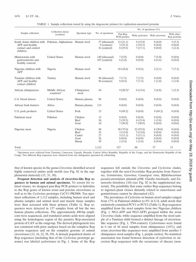
Frequent detection and analysis of circovirus-like Rep se-quences in human and animal specimens. To screen for re-lated viruses, we designed pan-Rep PCR primers to hybridizeto the Rep genes of known avian and porcine circoviruses aswell as to the Cyclovirus prototype CyCV1-PK5006. Ten spec-imen collections of 1,112 samples, including human stool andplasma samples and animal stool and muscle tissue sampleswere then screened with these primers (Table 1). Rep se-quences were detected in 137 samples from all but the twohuman plasma collections. The approximately 400-bp ampli-cons were sequenced, and translated amino acids were alignedusing the homologous region of the putative Rep-associatedprotein of CAV as the outgroup. The derived phylogenetic treewas consistent with prior analyses based on the complete Repprotein sequences and on the complete genome of animalcircoviruses (12, 16, 22, 38, 39). A densely populated cluster ofRep sequences (including that of the Cyclovirus prototype ge-nome) was labeled cycloviruses in Fig. 1. Some of the Rep
sequences fell outside the Circovirus and Cyclovirus clades,together with the non-Circoviridae Rep proteins from Nanovi-rus, Geminivirus, Gyrovirus, Canarypox virus, Bifidobacteriumpseudocatenulatum plasmid p4M, Giardia intestinalis, and En-tamoeba histolytica (10) (see Fig. S2 in the supplemental ma-terial). The possibility that some outlier Rep sequences belongto ingested plant viruses distantly related to nanoviruses andgeminiviruses cannot be discounted (42).
The prevalence of Cyclovirus in human stool samples rangedfrom 17% in Pakistani children to 0% in U.S. adult stools thatexclusively contained PCV1 or PCV2 (Table 1). Rep sequencesamplified from the stool samples from two Nigerian children(Fig. 1, NG1-AFP and NG3-AFP) grouped within the avianCircovirus clade, while sequence amplified from the stool sam-ple of a Tunisian child formed a distinct lineage of circovirus-like sequence (Fig. 1, TN4-contact). Cycloviruses were foundin 6 out of 44 stool samples from chimpanzees (14%), andavian circovirus-like sequences were amplified from another 3chimpanzee stool samples (Fig. 1, purple circles). No statisticalassociation was found between detection of cyclovirus or cir-covirus Rep sequences with the occurrence of disease (non-
TABLE 1. Sample collections tested by using the degenerate primers for replication-associated proteins
Sample collection Collection site(s)(country) Specimen type No. of specimens
No. of specimens (%)
Pan-RepPCR-positive With cyclovirus With circovirus With other
Rep proteins
South Asian children with Pakistan, Afghanistan Human stool 57 (diseased) 12 (21.1) 9 (15.8) 0 (0.0) 3 (5.3)AFP and healthy 9 (contact) 3 (33.3) 3 (33.3) 0 (0.0) 0 (0.0)contact and controlchildren
41 (control) 8 (19.5) 7 (17.1) 0 (0.0) 1 (2.4)
Minnesotans with United States Human stool 140 (diseased) 7 (5.0) 0 (0.0) 7 (5.0) 0 (0.0)gastroenteritis andhealthy controls
107 (control) 6 (5.6) 0 (0.0) 6 (5.6) 0 (0.0)
Nigerian children withAFP
Nigeria Human stool 96 18 (18.8) 9 (9.4) 2 (2.1) 7 (7.3)
Tunisian children with Tunisia Human stool 96 (diseased) 7 (7.3) 7 (7.3) 0 (0.0) 0 (0.0)AFP and healthycontact children
96 (contact) 9 (9.4) 7 (7.3) 1 (1.0) 1 (1.0)
African chimpanzees Middle Africancountriesa
Chimpanzeestool
44 9 (20.5)a 6 (13.6) 3 (6.8) 1 (2.3)
U.S. blood donors United States Human plasma 96 0 (0.0) 0 (0.0) 0 (0.0) 0 (0.0)
African bush hunters Africa Human plasma 113 0 (0.0) 0 (0.0) 0 (0.0) 0 (0.0)
U.S. pork products United States Pork 13 9 (69.2) 0 (0.0) 9 (69.2) 0 (0.0)
Pakistani meat Pakistan Chicken 13 0 (0.0) 0 (0.0) 0 (0.0) 0 (0.0)Beef 26 5 (19.2) 4 (15.4) 1 (3.8) 0 (0.0)Goat 18 7 (38.9) 7 (38.9) 0 (0.0) 0 (0.0)
Nigerian meat Nigeria Chicken 40 30 (75.0) 22 (55.0) 8 (20.0) 0 (0.0)Beef 25 3 (12.0) 3 (12.0) 0 (0.0) 0 (0.0)Camel 27 3 (11.1) 3 (11.1) 0 (0.0) 0 (0.0)Goat 26 0 (0.0) 0 (0.0) 0 (0.0) 0 (0.0)Sheep 29 1 (3.4) 1 (3.4) 0 (0.0) 0 (0.0)
Total 1,112 137 88 37 13
a Specimens were collected from Tanzania, Cameroon, Uganda, Rwanda, Central Africa Republic, Republic of the Congo, and the Democratic Republic of theCongo. Two different Rep sequences were obtained from one chimpanzee specimen by subcloning.
1676 LI ET AL. J. VIROL.
on May 19, 2018 by guest
http://jvi.asm.org/
Dow
nloaded from

polio AFP in Pakistan or Tunisia or unexplained gastroenter-itis in Minnesota).
Genome characteristics and phylogeny of cycloviruses. Toconfirm the presence of diverse cycloviruses and to character-ize the genome of this novel group, inverse PCR was used toamplify and sequence complete viral genomes from human andchimpanzee stool samples. Each of the 15 sequenced circulargenomes has two main open reading frames arranged in op-posite directions, encoding the putative Rep and capsid (Cap)proteins, an arrangement typical of circoviruses (Fig. 2). Thecomplete Rep proteins were used for phylogenetic analysis.The resulting tree confirms the presence of a new Cyclovirusclade within the Circoviridae, including now 12 genomes (Fig.3). The ORFs of the Cyclovirus genomes were similar to thoseof circoviruses but with some distinctive features (Fig. 2). Onaverage, cycloviruses have smaller genomes (average, 1,772 bp;range, 1,699 to 18,67 bp) than circoviruses do (average, 1,902bp; range, 1,759 to 2,063 bp), encoding relatively smaller Repand Cap proteins (Table 2). NG13 had the smallest genomesize of any reported virus (1,699 bp).
The 3� intergenic regions between the stop codons of the twomajor ORFs were either absent or only a few base pairs long incycloviruses, while those of circoviruses were significantlylarger. The 5� intergenic regions between the start codons ofthe two major ORFs of cycloviruses were larger than those ofcircoviruses (Table 2). The Rep ORFs of the two closely re-lated genomes, TN18 and TN25 (97% nucleotide similarity)were both interrupted by an apparent 171-bp intron with atypical splice donor site (GT) and splice acceptor site (AG)(Fig. 2).
The stem-loop structure with a conserved nonanucleotidemotif located at the 5� intergenic region of circovirus genomesis thought to initiate rolling-cycle replication (37). A highlyconserved stem-loop structure is also found in the 5� intergenicregions of cycloviruses (Fig. 2 and Fig. 4A). The consensussequence for the loop nonamer of the circoviruses is 5�-TAGTATTAC-3�, with slight variation among the sequenced ge-nomes (21, 26, 31, 35, 38, 39) (Fig. 4B). A different and con-served loop nonamer sequence (5�-TAATACTAT-3�) wasobserved for all the cycloviruses except CyCV-NG13, which isa cyclovirus group outlier but carries a typical circovirusnonamer (Fig. 4B). The highly conserved nonamer atop thestem-loop structure is one of the distinct characteristics of thenew Cyclovirus genus.
The two genomes derived from human stool samples in theUnited States (MN614 and MN500) shared 99% overall ge-nome nucleotide similarity with PCV2. The Chimp17 genome,from a chimpanzee stool sample, grouped with the raven cir-covirus RaCV, sharing 79% amino acid similarity to its Rep
FIG. 1. Phylogenetic analysis of the translated Rep sequences am-plified by pan-Rep PCR. Sequences derived from human stool samples(red), chimpanzee stool samples (purple), and farm animal meat sam-ples (green) are indicated. Cycloviruses in the same species are definedas having �85% identity in Rep region and are labeled by vertical bars1 to 25. The scale bar labeled 0.05 at the bottom of the figure repre-sents 5% estimated phylogenetic divergence. The countries of originare Pakistan (PK), Nigeria (NG), Tunisia (TN), and the United States(US).
VOL. 84, 2010 DIVERSE CYCLOVIRUSES FOUND IN STOOL AND ANIMAL TISSUE 1677
on May 19, 2018 by guest
http://jvi.asm.org/
Dow
nloaded from

protein. We have named this virus “Chimpanzee Stoolavian-like circovirus-chimp17” (CsaCV-chimp17). No suitablylocated ATG was identified for either ORF of CsaCV-chimp17. Considering the common usage of alternative startcodons in avian circoviruses, such as TCT (26, 31, 35), GTG(38), and ATA (39), CTG was considered the most likelycandidate for a start codon in the genome, producing ORFs ofexpected lengths.
The average amino acid similarity among cyclovirus Repproteins is 59% (range, 42 to 80%), and the value for circovirusRep proteins is 56% (range, 40 to 87%), reflecting a compa-rable range of viral diversity within both genera (see Table S2in the supplemental material). For the capsid protein, theaverage amino acid similarity is 29% (range, 11 to 56%) forcycloviruses and 34% (range, 18 to 76%) for circoviruses (seeTable S2 in the supplemental material). An amino acid align-ment shows that cycloviruses also possess some of the highlyconserved Rep amino acid motifs typical of circoviruses, in-cluding WWDGY, DDFYGW, and DRYP. Motifs associatedwith rolling-circle replication (FTLNN, TPHLQG, and CSK)
and deoxynucleoside triphosphate (dNTP) binding (G—GSK)were also identified, with some alterations (see Fig. S1 in thesupplemental material) (15, 31, 38). The N-terminal region ofthe cyclovirus Cap proteins was highly basic and arginine-rich,as is typical for circoviruses (17, 35).
PCVs frequently detected in U.S. human stool samples andpork products. All Rep sequences derived from human stoolsamples from the United States clustered closely with PCVs(Fig. 1, red diamonds). In order to test the possible dietaryorigin of these PCV sequences, pork specimens purchasedfrom different U.S. stores were tested. Out of 13 U.S. porkproducts, 9 (70%) were Rep positive, 7 of which clustered withPCV2, 1 with PCV1, and 1 highly divergent sequence (USporkNW2) (Fig. 1, green diamonds). Pork sample USporkNW2 may represent a yet uncharacterized porcine circo-virus species. Out of 23 Rep sequences from the U.S. samples,22, including all 14 from human stool samples and 8 out of 9from pork specimens, therefore belonged to PCV1 or PCV2.
Circovirus-like Rep sequences in consumed meats. The fre-quent detection of PCVs in U.S. stool samples and U.S. pork
FIG. 2. Genomic organizations of circoviruses (A) and cycloviruses (B). The 2 major ORFs, encoding the putative replication-associatedprotein (Rep) and the putative capsid protein (Cap), and other ORFs with a coding capacity greater than 100 amino acids are shown. The locationsof the stem-loop structures are marked.
1678 LI ET AL. J. VIROL.
on May 19, 2018 by guest
http://jvi.asm.org/
Dow
nloaded from

products suggested that the cycloviruses found in non-U.S.human stool samples and wild chimpanzee stool samplesmight similarly originate from the consumption of meatcontaminated with cycloviruses. To test this hypothesis,commonly eaten meat products were acquired from marketsin Pakistan and Nigeria and analyzed by pan-Rep PCR(Table 1). Of 204 meat samples tested, 24% were positive,and all amplicons were sequenced. The Rep sequence de-tection rates differed substantially between countries for thesame type of meat. None of 13 chicken samples from Paki-stan was positive, while 30 out of 40 (75%) chicken samplesfrom Nigeria were positive. Of the 30 Rep sequences fromNigerian chicken samples, 22 sequences clustered tightlywithin the Cyclovirus genus, and 8 sequences clustered to-gether in a cluster with pigeon Circovirus (as did the Repsequence NG1-AFP from the stool sample of a Nigerianchild). Of the 26 goat samples from Nigeria, none was pos-itive, while 7 out of 18 (38%) goat specimens from Pakistanwere positive for cycloviruses. Of the total 19 Rep sequencesobtained from farm mammals (cows, goats, sheep, and cam-els), only 1 (PK beef21) grouped deeply with the circovirusclade, while 18 fell within the cyclovirus clade. The majority(40 out of 49) of Rep sequences obtained from animal tissuetherefore belonged to the cyclovirus clade. Some cyclovirusRep sequences from different animal species (e.g., cows andgoats, even-toed ungulates in the Bovidae family) were veryclosely related (Fig. 1, species 22).
The ICTV defines different circovirus species based on se-quence similarity; genomic sequences having �75% nucleotideidentity and �70% identity in the capsid protein qualify asdifferent species (37). We adopted a criterion of �85% aminoacid identity in the highly conserved Rep protein region am-plified by pan-Rep PCR as the criterion for Cyclovirus speciesdesignation by comparing the amino acid identity of the sameRep region among known circovirus species. Using this crite-rion, 25 species of Cyclovirus were found in human and chim-panzee stool samples and meat samples from farm animals. Ofthese 25 species, only a single Cyclovirus species (Fig. 1, species2), represented by 16 out of a total of 88 Rep sequences(18.2%), was found in both human stool and farm animaltissue samples. Four species were specific to chimpanzee stoolsamples, and another four species were specific to farm ani-mals. Sixteen cyclovirus species were specific to non-U.S. hu-man stool samples. The consumption of meat from infectedanimals is therefore unlikely to account for the majority ofcycloviruses detected in non-U.S. human stool samples.
DISCUSSION
We report on the frequent detection of viral, circular DNAgenomes related to porcine and avian circoviruses in humanand chimpanzee stool samples and genetically characterize apreviously unrecognized genus in the family Circoviridae.These viruses were both widely dispersed (Tunisia, Pakistan,
FIG. 3. Phylogenetic analysis of 15 Circoviridae replicase proteins from 12 human stool samples and 3 chimpanzee stool samples. Outlier taxaare non-Circoviridae Rep proteins. Sample designation is the same as in Fig. 1. CyCV, cyclovirus.
VOL. 84, 2010 DIVERSE CYCLOVIRUSES FOUND IN STOOL AND ANIMAL TISSUE 1679
on May 19, 2018 by guest
http://jvi.asm.org/
Dow
nloaded from

and Nigeria) and highly prevalent (7 to 17% of children’s stoolsamples).
Cycloviruses are not closely related phylogenetically to the re-cently described circular DNA viruses chimpanzee stool-associ-
ated circular viruses (ChiSCV) found in chimpanzee stool sam-ples (5) or the circular ssDNA viruses in aquatic environments(20, 33), nor is their genome organization related to human oranimal anelloviruses (e.g., torque teno virus [TTV]) (4, 33).
FIG. 4. (A) Stem-loop of Cyclovirus prototype CyCV1-PK5006 and (B) nonamer sequences and stem lengths of the stem-loop structures forcircoviruses and cycloviruses.
TABLE 2. Genome organization of newly discovered cycloviruses and representative circoviruses
Virus type and circularDNA virus species
No. of nt inthe genome
No. of aa in protein Length (nt) of region (start–end)a
Rep Cap 5� Intergenic region 3� Intergenic region
CyclovirusesPK5006 1,723 278 219 230 (1516–22)PK5222 1,740 279 218 247 (1516–22)PK5510 1,759 280 219 271 (1679–190)PK5034 1,780 277 218 293 (1691–203)PK6197 1,741 279 218 248 (1516–22)Chimp11 1,750 280 220 258 (1515–22)Chimp12 1,747 280 220 255 (1515–22)NG12 1,794 281 218 284 (1691–180) 7 (1027–1033)NG14 1,795 286 230 245 (1707–156)TN18 1,867 286 222 160 (1762–54) 6 (1087–1092)TN25 1,867 286 222 160 (1762–54) 6 (1087–1092)NG13 1,699 307 221 105 (1622–27) 4 (952–955)
CircovirusesChimp17 1,935 291 232 198 (1772–34) 162 (911–1072)MN614 1,767 314 233 83 (1735–50) 37 (996–1032)MN500 1,768 314 233 83 (1736–50) 38 (996–1033)PCV1 1,759 312 233 82 (1724–46) 36 (986–1021)PCV2 1,768 314 233 83 (1736–50) 38 (996–1033)DuCV 1,991 292 257 110 (1929–47) 228 (927–1154)GoCV 1,821 293 250 132 (1762–72) 54 (955–1008)CoCV 2,037 317 273 90 (1988–40) 171 (995–1165)RaCV 1,898 291 243 86 (1848–35) 204 (912–1115)SwCV 1,785 293 251 107 (1726–47) 40 (930–969)BFDV 1,993 299 244 126 (1975–107) 232 (1008–1239)GuCV 2,035 305 245 207 (1928–99) 172 (1018–1189)FiCV 1,962 291 249 29 (1962–28) 307 (905–1211)StCV 2,063 289 276 79 (2021–36) 283 (907–1189)CaCV 1,952 290 250 77 (1907–31) 249 (905–1153)
a Fifteen genome sequences obtained in this study are shown in boldface type. Nucleotide position 1 was set at the residue A at position 8 of the nonamer sequence.nt, nucleotides; aa, amino acids.
1680 LI ET AL. J. VIROL.
on May 19, 2018 by guest
http://jvi.asm.org/
Dow
nloaded from

PCVs were frequently detected in stool samples from adultsin the United States (5%), and store-bought pork products alsofrequently contain PCV sequences (70%). These results indi-cated that detection of PCV DNA in stool may reflect dietaryconsumption of PCV-infected pork.
Evidence for circovirus infection in mammals other thanpigs is equivocal, and studies have been restricted to PCVs.PCV2 DNA in cows with respiratory symptoms and in abortedbovine fetuses has been reported only once (24). PCV2 wasalso reported in a colon biopsy specimen from a patient withulcerative colitis, although contamination with PCV2 fromstool is difficult to exclude in this case (2). No PCV DNA wasfound by PCR in screening more than 1,000 samples fromvarious tissues of both healthy and immunosuppressed humansand plasma samples from 18 xenotransplantation recipients ofpig islet cells (9, 13). In this study, the results of screeningplasma samples from 96 U.S. blood donors and 113 CentralAfrican bush hunters via pan-Rep PCR were also negative(Table 1). A study showed that viral protein expression, cyto-pathic effect, and DNA persistence occurs in human cell linesinfected with PCV2, but the virus could not be passed to newcultures (14). One study reported the presence of PCV-reac-tive antibodies, although with somewhat distinctive propertiesin sera of humans, cows, and mice (36), while another reportedthe lack of PCV antibodies in cows and horses (8). WhetherPCVs simply pass through or are capable of replication in thehuman gut remains unknown.
Avian circovirus-like DNA was found in 3/44 wild chimpan-zee stool samples and in 2/96 stool samples from Nigerianchildren (Table 1). This observation may reflect consumptionof infected birds or conceivably contamination of food withbird droppings.
Cycloviruses were found in the muscle tissue of all the spe-cies of farm animals tested (goats, sheep, cows, camels, andchickens), suggesting that viral infection occurs in these spe-cies. In previous studies, different tissues have been shown toretain small DNA viruses (e.g., parvoviruses) long after pri-mary infection viremia (23, 27). The detection of cyclovirusesand circoviruses in muscle tissue could therefore reflect priorand/or ongoing infection. The detection of closely related cy-clovirus Rep sequences in both cows and goats from Pakistan(Fig. 1, species 22) might reflect cross-species transmission.
A wide diversity of cycloviruses was identified in humanstool samples collected from children in developing countries.In contrast, in the United States, all Rep sequences obtainedfrom stool samples belonged to the PCV clade. An importantdistinction between U.S. and non-U.S. human stool sampleswas the younger age of the non-U.S. donors, which may haveimpacted host susceptibility to infections or the duration ofviral shedding. Exposure to cyclovirus may therefore also occurin the United States but was not detected because of the olderage of the subjects.
In total, 17 Cyclovirus species were identified in 395 humanstool samples, and 5 Cyclovirus species were found in muscletissue samples from 204 farm animals, with only a single spe-cies found in common in both groups of samples. The meatsamples analyzed were acquired from three major cities inPakistan and one major city in Nigeria, while the children fromthese countries shedding cycloviruses were geographicallymore dispersed. It is therefore conceivable that despite the
large number of cyclovirus replicase sequences generated,more geographically dispersed sampling of farm animals wouldhave shown greater overlap with human stool-derived cyclovi-rus sequences. Using the current sampling, the limited overlapbetween Cyclovirus species found in human stool samples andin meat from farm animals from the same countries does sug-gest that most of the cycloviruses found in the stool samples ofchildren in Nigeria and Pakistan were not from consumedmeat. Possibly, the 16 cycloviruses species found only in humanstool samples were transmitted via a fecal-oral route fromother infected children, a common pathway for many entericviral infections. The detection of cycloviruses in 14% of stoolsamples from chimpanzees (who consume very limitedamounts of meat) also argues in favor of transmission withinthis primate species rather than simply reflecting consumptionof infected meats. It is not known whether the viral speciesfound in both human stool samples and tissue samples fromfarm animals, such as PCVs in the United States and cyclovirusspecies 2 (Fig. 1, species 2) in Pakistan, Nigeria, and Tunisia,can replicate in their human host. Since transmission of PCV2from one pig to another through consumption of meat wasrecently shown (30), the potential for zoonotic transfer alsoexists for other circoviruses and cycloviruses.
Given the high prevalence of cyclovirus infections in non-U.S. farm animals, the possibility of cross-species transmission(cyclovirus species 22 in different members of the familyBovidae), the high diversity of cycloviruses in human stoolsamples, the documented pathogenicity of closely related Circo-virus species, and the high rate of mutation and recombinationof some ssDNA viruses, the pathogenic potential of cyclo-viruses in both humans and farm animals merits further study.
ACKNOWLEDGMENTS
This work was supported by Blood Systems Research Institute andNIH grant R01 HL083254 to E.D. The chimpanzee sample collectionwas supported by grants from National Institutes of Health to B.H.H.(R01 AI50529 and R01 AI58715), the Bristol Myers Freedom to Dis-cover Program, and the Jane Goodall Institute. Additional support wasprovided by the Global Viral Forecasting Initiative, Google.org, andthe Skoll Foundation.
We thank M. P. Busch for helpful discussions; Marycelin ManduBaba and David Nadeba Bukbuk from the WHO National Polio Lab-oratory, University of Maiduguri Teaching Hospital, Maiduguri, BornoState, Nigeria, for assistance collecting stool samples from non-polio-infected children with AFP; and John McGee, Jason Reilly, and MatsRynge from the Renaissance Computing Institute (RENCI) for assis-tance with computing analysis of the pyrosequencing data. We alsothank Farbod Babrzadeh and Baback Gharizadeh at Stanford Univer-sity for assistance with pyrosequencing. We thank the staff of ProjectPRESICA for chimpanzee sample collection in southern Cameroonand the Central African Republic; the staff of the Gombe StreamResearch Centre for sample collection in Gombe National Park inTanzania; Michael A. Huffman for sample collection in Mahale Moun-tain National Park in Tanzania; and Crickette Sanz and David Morganfor sample collection in the Goualougo Triangle in the DemocraticRepublic of Congo. We thank the Cameroonian Ministries of Health,Environment and Forestry, and Research for permission to collectsamples in Cameroon; the Democratic Republic of Congo Ministry ofScience and Technology and Ministry of Forest Economy for permis-sion to collect samples in the Goualougo Triangle in the DemocraticRepublic of Congo; the Tanzania National Parks, the Tanzania Com-mission for Science and Technology, and the Tanzania Wildlife Re-search Institute for permission to conduct research in Gombe Streamand Mahale Mountain National Parks in Tanzania; the Uganda Wild-life Authority, the Uganda National Council for Science and Technol-
VOL. 84, 2010 DIVERSE CYCLOVIRUSES FOUND IN STOOL AND ANIMAL TISSUE 1681
on May 19, 2018 by guest
http://jvi.asm.org/
Dow
nloaded from

ogy, and the Makerere University Biological Field Station for permis-sion to conduct research in Kibale National Park and Kyambura Gorgein Uganda; the Rwandan Office of Tourism and National Parks forpermission to collect samples in Nyungwe National Park in Rwanda;and the Department of Ecology and Management of Plant and AnimalResources (University of Kisangani) for authorization to collect sam-ples in the Democratic Republic of Congo.
REFERENCES
1. Allan, G. M., and J. A. Ellis. 2000. Porcine circoviruses: a review. J. Vet.Diagn. Investig. 12:3–14.
2. Bernstein, C. N., G. Nayar, A. Hamel, and J. F. Blanchard. 2003. Study ofanimal-borne infections in the mucosas of patients with inflammatory boweldisease and population-based controls. J. Clin. Microbiol. 41:4986–4990.
3. Biagini, P. 2005. Anellovirus, p. 335–341. In C. M. Fauquet, M. A. Mayo, J.Maniloff, U. Desselberger, and L. A. Ball (ed.), Virus taxonomy. Eighthreport of the International Committee on Taxonomy of Viruses. ElsevierAcademic Press, San Diego, CA.
4. Biagini, P. 2009. Classification of TTV and related viruses (anelloviruses).Curr. Top. Microbiol. Immunol. 331:21–33.
5. Blinkova, O., J. Victoria, Y. Li, B. Keele, C. Sanz, J. B. Ndjango, M. Peeters,D. Travis, E. Lonsdorf, M. Wilson, A. Pusey, B. Hahn, and E. Delwart. 2010.Novel circular DNA viruses in stool samples of wild-living chimpanzees.J. Gen. Virol. 91:74–96.
6. Cheung, A. K. 2006. Rolling-circle replication of an animal circovirus ge-nome in a theta-replicating bacterial plasmid in Escherichia coli. J. Virol.80:8686–8694.
7. Duffy, S., L. A. Shackelton, and E. C. Holmes. 2008. Rates of evolutionarychange in viruses: patterns and determinants. Nat. Rev. Genet. 9:267–276.
8. Ellis, J. A., C. Konoby, K. H. West, G. M. Allan, S. Krakowka, F. McNeilly,B. Meehan, and I. Walker. 2001. Lack of antibodies to porcine circovirustype 2 virus in beef and dairy cattle and horses in western Canada. Can. Vet.J. 42:461–464.
9. Garkavenko, O., M. C. Croxson, M. Irgang, A. Karlas, J. Denner, and R. B.Elliott. 2004. Monitoring for presence of potentially xenotic viruses in recip-ients of pig islet xenotransplantation. J. Clin. Microbiol. 42:5353–5356.
10. Gibbs, M. J., V. V. Smeianov, J. L. Steele, P. Upcroft, and B. A. Efimov. 2006.Two families of rep-like genes that probably originated by interspecies re-combination are represented in viral, plasmid, bacterial, and parasitic pro-tozoan genomes. Mol. Biol. Evol. 23:1097–1100.
11. Guindon, S., and O. Gascuel. 2003. A simple, fast, and accurate algorithm toestimate large phylogenies by maximum likelihood. Syst. Biol. 52:696–704.
12. Halami, M. Y., H. Nieper, H. Muller, and R. Johne. 2008. Detection of anovel circovirus in mute swans (Cygnus olor) by using nested broad-spectrumPCR. Virus Res. 132:208–212.
13. Hattermann, K., A. Maerz, H. Slanina, C. Schmitt, and A. Mankertz. 2004.Assessing the risk potential of porcine circoviruses for xenotransplantation:consensus primer-PCR-based search for a human circovirus. Xenotransplan-tation 11:547–550.
14. Hattermann, K., C. Roedner, C. Schmitt, T. Finsterbusch, T. Steinfeldt, andA. Mankertz. 2004. Infection studies on human cell lines with porcine cir-covirus type 1 and porcine circovirus type 2. Xenotransplantation 11:284–294.
15. Hattermann, K., C. Schmitt, D. Soike, and A. Mankertz. 2003. Cloning andsequencing of Duck circovirus (DuCV). Arch. Virol. 148:2471–2480.
16. Johne, R., D. Fernandez-de-Luco, U. Hofle, and H. Muller. 2006. Genome ofa novel circovirus of starlings, amplified by multiply primed rolling-circleamplification. J. Gen. Virol. 87:1189–1195.
17. Johne, R., R. Raue, C. Grund, E. F. Kaleta, and H. Muller. 2004. Recom-binant expression of a truncated capsid protein of beak and feather diseasevirus and its application in serological tests. Avian Pathol. 33:328–336.
18. Kumar, S., M. Nei, J. Dudley, and K. Tamura. 2008. MEGA: a biologist-centric software for evolutionary analysis of DNA and protein sequences.Brief. Bioinform. 9:299–306.
19. Lefeuvre, P., J. M. Lett, A. Varsani, and D. P. Martin. 2009. Widely con-served recombination patterns among single-stranded DNA viruses. J. Virol.83:2697–2707.
20. Lopez-Bueno, A., J. Tamames, D. Velazquez, A. Moya, A. Quesada, and A.Alcami. 2009. High diversity of the viral community from an Antarctic lake.Science 326:858–861.
21. Mankertz, A., R. Caliskan, K. Hattermann, B. Hillenbrand, P. Kurzendoer-fer, B. Mueller, C. Schmitt, T. Steinfeldt, and T. Finsterbusch. 2004. Mo-lecular biology of Porcine circovirus: analyses of gene expression and viralreplication. Vet. Microbiol. 98:81–88.
22. Mankertz, A., K. Hattermann, B. Ehlers, and D. Soike. 2000. Cloning and
sequencing of columbid circovirus (coCV), a new circovirus from pigeons.Arch. Virol. 145:2469–2479.
23. Manning, A., S. J. Willey, J. E. Bell, and P. Simmonds. 2007. Comparison oftissue distribution, persistence, and molecular epidemiology of parvovirusB19 and novel human parvoviruses PARV4 and human bocavirus. J. Infect.Dis. 195:1345–1352.
24. Nayar, G. P., A. L. Hamel, L. Lin, C. Sachvie, E. Grudeski, and G. Spear-man. 1999. Evidence for circovirus in cattle with respiratory disease and fromaborted bovine fetuses. Can. Vet. J. 40:277–278.
25. Ng, T. F., C. Manire, K. Borrowman, T. Langer, L. Ehrhart, and M. Breit-bart. 2009. Discovery of a novel single-stranded DNA virus from a sea turtlefibropapilloma by using viral metagenomics. J. Virol. 83:2500–2509.
26. Niagro, F. D., A. N. Forsthoefel, R. P. Lawther, L. Kamalanathan, B. W.Ritchie, K. S. Latimer, and P. D. Lukert. 1998. Beak and feather diseasevirus and porcine circovirus genomes: intermediates between the geminivi-ruses and plant circoviruses. Arch. Virol. 143:1723–1744.
27. Norja, P., K. Hokynar, L. M. Aaltonen, R. Chen, A. Ranki, E. K. Partio, O.Kiviluoto, I. Davidkin, T. Leivo, A. M. Eis-Hubinger, B. Schneider, H. P.Fischer, R. Tolba, O. Vapalahti, A. Vaheri, M. Soderlund-Venermo, and K.Hedman. 2006. Bioportfolio: lifelong persistence of variant and prototypicerythrovirus DNA genomes in human tissue. Proc. Natl. Acad. Sci. U. S. A.103:7450–7453.
28. Noteborn, M. H., G. F. de Boer, D. J. van Roozelaar, C. Karreman, O.Kranenburg, J. G. Vos, S. H. Jeurissen, R. C. Hoeben, A. Zantema, G. Koch,et al. 1991. Characterization of cloned chicken anemia virus DNA thatcontains all elements for the infectious replication cycle. J. Virol. 65:3131–3139.
29. Opriessnig, T., X. J. Meng, and P. G. Halbur. 2007. Porcine circovirus type2 associated disease: update on current terminology, clinical manifestations,pathogenesis, diagnosis, and intervention strategies. J. Vet. Diagn. Investig.19:591–615.
30. Opriessnig, T., A. R. Patterson, X. J. Meng, and P. G. Halbur. 2009. Porcinecircovirus type 2 in muscle and bone marrow is infectious and transmissibleto naive pigs by oral consumption. Vet. Microbiol. 133:54–64.
31. Phenix, K. V., J. H. Weston, I. Ypelaar, A. Lavazza, J. A. Smyth, D. Todd,G. E. Wilcox, and S. R. Raidal. 2001. Nucleotide sequence analysis of a novelcircovirus of canaries and its relationship to other members of the genusCircovirus of the family Circoviridae. J. Gen. Virol. 82:2805–2809.
32. Ramamoorthy, S., and X. J. Meng. 2009. Porcine circoviruses: a minusculeyet mammoth paradox. Anim. Health Res. Rev. 10:1–20.
33. Rosario, K., S. Duffy, and M. Breitbart. 2009. Diverse circovirus-like genomearchitectures revealed by environmental metagenomics. J. Gen. Virol. 90:2418–2424.
34. Stanley, J. 2005. Geminiviridae, p. 301–326. In C. M. Fauquet, M. A. Mayo,J. Maniloff, U. Desselberger, and L. A. Ball (ed.), Virus taxonomy. Eighthreport of the International Committee on Taxonomy of Viruses. ElsevierAcademic Press, San Diego, CA.
35. Stewart, M. E., R. Perry, and S. R. Raidal. 2006. Identification of a novelcircovirus in Australian ravens (Corvus coronoides) with feather disease.Avian Pathol. 35:86–92.
36. Tischer, I., L. Bode, J. Apodaca, H. Timm, D. Peters, R. Rasch, S. Pociuli,and E. Gerike. 1995. Presence of antibodies reacting with porcine circovirusin sera of humans, mice, and cattle. Arch. Virol. 140:1427–1439.
37. Todd, D. 2005. Circoviridae, p. 326–334. In C. M. Fauquet, M. A. Mayo, J.Maniloff, U. Desselberger, and L. A. Ball (ed.), Virus taxonomy. Eighthreport of the International Committee on Taxonomy of Viruses. ElsevierAcademic Press, San Diego, CA.
38. Todd, D., A. N. Scott, E. Fringuelli, H. L. Shivraprasad, D. Gavier-Widen,and J. A. Smyth. 2007. Molecular characterization of novel circoviruses fromfinch and gull. Avian Pathol. 36:75–81.
39. Todd, D., J. H. Weston, D. Soike, and J. A. Smyth. 2001. Genome sequencedeterminations and analyses of novel circoviruses from goose and pigeon.Virology 286:354–362.
40. Vetten, H. J. 2005. Nanoviridae, p. 343–352. In C. M. Fauquet, M. A. Mayo,J. Maniloff, U. Desselberger, and L. A. Ball (ed.), Virus taxonomy. Eighthreport of the International Committee on Taxonomy of Viruses. ElsevierAcademic Press, San Diego, CA.
41. Victoria, J. G., A. Kapoor, L. Li, O. Blinkova, B. Slikas, C. Wang, A. Naeem,S. Zaidi, and E. Delwart. 2009. Metagenomic analyses of viruses in stoolsamples from children with acute flaccid paralysis. J. Virol. 83:4642–4651.
42. Zhang, T., M. Breitbart, W. H. Lee, J. Q. Run, C. L. Wei, S. W. Soh, M. L.Hibberd, E. T. Liu, F. Rohwer, and Y. Ruan. 2006. RNA viral community inhuman feces: prevalence of plant pathogenic viruses. PLoS Biol. 4:e3.
43. Zuker, M. 2003. Mfold web server for nucleic acid folding and hybridizationprediction. Nucleic Acids Res. 31:3406–3415.
1682 LI ET AL. J. VIROL.
on May 19, 2018 by guest
http://jvi.asm.org/
Dow
nloaded from