Multifunctional Eu31- and Er31/ Yb31-doped GdVO4 nanoparticles ...
9
Multifunctional Eu 31 - and Er 31 / Yb 31 -doped GdVO 4 nanoparticles synthesized by reverse micelle method Tamara V. Gavrilovic ´, Dragana J. Jovanovic ´, Vesna Lojpur & Miroslav D. Dramic ´anin Vincˇa Institute of Nuclear Sciences, University of Belgrade, P.O. Box 522, 11001 Belgrade, Serbia. Synthesis of Eu 31 - and Er 31 /Yb 31 -doped GdVO 4 nanoparticles in reverse micelles and their multifunctional luminescence properties are presented. Using cyclohexane, Triton X-100, and n-pentanol as the oil, surfactant, and co-surfactant, respectively, crystalline nanoparticles with ,4 nm diameter are prepared at low temperatures. The particle size assessed using transmission electron microscopy is similar to the crystallite size obtained from X-ray diffraction measurements, suggesting that each particle comprises a single crystallite. Eu 31 -doped GdVO 4 nanoparticles emit red light through downconversion upon UV excitation. Er 31 /Yb 31 -doped GdVO 4 nanoparticles exhibit several functions; apart from the downconversion of UV radiation into visible green light, they act as upconvertors, transforming near-infrared excitation (980 nm) into visible green light. The ratio of green emissions from 2 H 11/2 R 2 I 15/2 and 4 S 3/2 R 4 I 15/2 transitions is temperature dependent and can be used for nanoscale temperature sensing with near-infrared excitation. The relative sensor sensitivity is 1.11%K 21 , which is among the highest sensitivities recorded for upconversion-luminescence-based thermometers. T he properties of nanoparticles (NPs) and nanostructured materials often drastically differ from those of bulk materials with the same chemical composition and are advantageous for numerous physical, biological, biomedical, and pharmaceutical applications 1 . Therefore, the preparation of nanomaterials with well- controlled shape, size, phase purity, chemical composition, and with targeted, often multifunctional, properties is one of the most challenging problems in materials science. Because multifunctional NPs provide several functionalities in a single assembly, they are more applicable than conventional monofunctional NPs. Consequently, the design and preparation of multifunctional NPs have garnered considerable interest in recent years. In this respect, inorganic NPs doped with small amounts of activator ions such as transition-metal and/or rare-earth (RE) ions have attracted much attention because of their fascinating optical, magnetic, and thermal properties. Their potential as multifunctional materials is exceptionally broad. For example, they can be useful in improving photovoltaic cell efficiency through solar spectral conversion by shifting short-wavelength sunlight (ultraviolet and blue) to longer wavelengths (downshifting, quantum cutting), or by shifting long-wavelength near-infrared (NIR) radiation to visible light (upconversion), which provides more radiation in the spectral region wherein the solar cell shows the largest quantum efficiency 2 . The luminescence decays of the majority of lanthanide and transition-metal ions (of the order of ms and larger) are much longer than those of biological materials (e.g., proteins and cells); therefore, NPs activated with these ions have high potential for biolabeling applications. Long-lasting emissions can be easily discriminated from the autofluorescence of biological materials using time-resolved measurements. Lanthanide- and transition-metal-ion-activated NPs do not show photo- bleaching and blinking, because the emission arises from many ions incorporated within the NPs. Also, these materials can be used in nanomedicine as anticancer drug carriers and magnetic resonance imaging (MRI) contrast agents, as well as in optical displays, cathode ray tubes, fluorescent lamps, light-emitting diodes, infrared detectors, scintillators, and fluorescent paints 3,4 . Gadolinium orthovanadate (GdVO 4 )-based materials have interesting luminescent and magnetic properties. Gd compounds can be easily doped with luminescent lanthanide ions because of the equal valence and similar ionic radii. They can be efficiently excited with UV radiation because of the strong absorption of the VO 4 32 groups and efficient energy transfer from GdVO 4 to lanthanide ions. Therefore, GdVO 4 is a used as a phosphor (doped with Eu 31 , Dy 31 , Sm 31 ), an upconvertor (doped with Er 31 /Yb 31 , Ho 31 /Yb 31 , or Tm 31 /Yb 31 ), and a laser (doped with Nd 31 ) 5,6 . In addition, GdVO 4 NPs can act as T1-positive contrast agents for MRI, because Gd 31 ions possess unpaired electrons that efficiently alter the relaxation time of the surrounding water protons 3,4,7 . OPEN SUBJECT AREAS: NANOPARTICLES OPTICAL MATERIALS SENSORS AND BIOSENSORS NANOPHOTONICS AND PLASMONICS Received 31 December 2013 Accepted 11 February 2014 Published 27 February 2014 Correspondence and requests for materials should be addressed to M.D.D. (dramican@ vinca.rs) SCIENTIFIC REPORTS | 4 : 4209 | DOI: 10.1038/srep04209 1
Transcript of Multifunctional Eu31- and Er31/ Yb31-doped GdVO4 nanoparticles ...

Multifunctional Eu31- and Er31/Yb31-doped GdVO4 nanoparticlessynthesized by reverse micelle methodTamara V. Gavrilovic, Dragana J. Jovanovic, Vesna Lojpur & Miroslav D. Dramicanin
Vinca Institute of Nuclear Sciences, University of Belgrade, P.O. Box 522, 11001 Belgrade, Serbia.
Synthesis of Eu31- and Er31/Yb31-doped GdVO4 nanoparticles in reverse micelles and their multifunctionalluminescence properties are presented. Using cyclohexane, Triton X-100, and n-pentanol as the oil,surfactant, and co-surfactant, respectively, crystalline nanoparticles with ,4 nm diameter are prepared atlow temperatures. The particle size assessed using transmission electron microscopy is similar to thecrystallite size obtained from X-ray diffraction measurements, suggesting that each particle comprises asingle crystallite. Eu31-doped GdVO4 nanoparticles emit red light through downconversion upon UVexcitation. Er31/Yb31-doped GdVO4 nanoparticles exhibit several functions; apart from thedownconversion of UV radiation into visible green light, they act as upconvertors, transformingnear-infrared excitation (980 nm) into visible green light. The ratio of green emissions from 2H11/2 R 2I15/2and 4S3/2 R 4I15/2 transitions is temperature dependent and can be used for nanoscale temperature sensingwith near-infrared excitation. The relative sensor sensitivity is 1.11%K21, which is among the highestsensitivities recorded for upconversion-luminescence-based thermometers.
The properties of nanoparticles (NPs) and nanostructured materials often drastically differ from those of bulkmaterials with the same chemical composition and are advantageous for numerous physical, biological,biomedical, and pharmaceutical applications1. Therefore, the preparation of nanomaterials with well-
controlled shape, size, phase purity, chemical composition, and with targeted, often multifunctional, propertiesis one of the most challenging problems in materials science. Because multifunctional NPs provide severalfunctionalities in a single assembly, they are more applicable than conventional monofunctional NPs.Consequently, the design and preparation of multifunctional NPs have garnered considerable interest in recentyears. In this respect, inorganic NPs doped with small amounts of activator ions such as transition-metal and/orrare-earth (RE) ions have attracted much attention because of their fascinating optical, magnetic, and thermalproperties. Their potential as multifunctional materials is exceptionally broad. For example, they can be useful inimproving photovoltaic cell efficiency through solar spectral conversion by shifting short-wavelength sunlight(ultraviolet and blue) to longer wavelengths (downshifting, quantum cutting), or by shifting long-wavelengthnear-infrared (NIR) radiation to visible light (upconversion), which provides more radiation in the spectralregion wherein the solar cell shows the largest quantum efficiency2. The luminescence decays of the majorityof lanthanide and transition-metal ions (of the order of ms and larger) are much longer than those of biologicalmaterials (e.g., proteins and cells); therefore, NPs activated with these ions have high potential for biolabelingapplications. Long-lasting emissions can be easily discriminated from the autofluorescence of biological materialsusing time-resolved measurements. Lanthanide- and transition-metal-ion-activated NPs do not show photo-bleaching and blinking, because the emission arises from many ions incorporated within the NPs. Also, thesematerials can be used in nanomedicine as anticancer drug carriers and magnetic resonance imaging (MRI)contrast agents, as well as in optical displays, cathode ray tubes, fluorescent lamps, light-emitting diodes, infrareddetectors, scintillators, and fluorescent paints3,4.
Gadolinium orthovanadate (GdVO4)-based materials have interesting luminescent and magnetic properties.Gd compounds can be easily doped with luminescent lanthanide ions because of the equal valence and similarionic radii. They can be efficiently excited with UV radiation because of the strong absorption of the VO4
32 groupsand efficient energy transfer from GdVO4 to lanthanide ions. Therefore, GdVO4 is a used as a phosphor (dopedwith Eu31, Dy31, Sm31), an upconvertor (doped with Er31/Yb31, Ho31/Yb31, or Tm31/Yb31), and a laser (dopedwith Nd31)5,6. In addition, GdVO4 NPs can act as T1-positive contrast agents for MRI, because Gd31 ions possessunpaired electrons that efficiently alter the relaxation time of the surrounding water protons3,4,7.
OPEN
SUBJECT AREAS:NANOPARTICLES
OPTICAL MATERIALS
SENSORS AND BIOSENSORS
NANOPHOTONICS ANDPLASMONICS
Received31 December 2013
Accepted11 February 2014
Published27 February 2014
Correspondence andrequests for materials
should be addressed toM.D.D. (dramican@
vinca.rs)
SCIENTIFIC REPORTS | 4 : 4209 | DOI: 10.1038/srep04209 1

So far, several methods have been used to prepare undoped andRE31-doped GdVO4 NPs with various sizes and shapes. GdVO4:Eu31
NPs have been successfully synthesized via a facile solvothermalroute4,8, a urea hydrolysis method9, a co-precipitation synthesis10,as well as facile hydrothermal methods11,12. Using these methods,tetragonal particles (d , 70 nm), spherical particles (d , 40 nm),ellipsoid particles (d , 20 nm, l , 25 nm), nanorods (d , 5 nm, l ,20 nm), and nanowires (d 5 15 nm, l 5 a few microns) were fabri-cated8–12. Submicronic polyhedrons with formula GdVO4:RE31
(RE31 5 Sm31, Dy31, Er31) (d , 300 nm) were obtained by facilehydrothermal methods13. Spherical GdVO4:Er31/Yb31 NPs with dia-meters ranging from 30 to 50 nm were produced by a simple hydro-thermal process assisted by polyvinylpyrrolidone (PVP)14.
One of the significant methods for the synthesis of nanoparticleswith desired diameter and shape is a reverse micelle route. Thispreparation method is widely applicable and is based on a simpleconcept. After mixing two reverse micelles, i.e., water-in-oil (oilbeing the nonpolar organic solvent) droplets containing reactionprecursors, an intermicellar exchange readily takes place, and achemical reaction between the precursors occurs, followed by nuc-leation and growth of nanocrystals. The growth of the NPs occurs inwater droplets dispersed in oil and stabilized by surfactant molecules.In other words, the droplets are coated by monolayer shells of aggre-gated surfactant molecules, with polar functional fragments (heads)oriented toward the aqueous phase and hydrocarbon chains (tails)immersed in oil. Clearly, the droplet size directly affects the final sizeof the NPs and depends on the surfactant type and water/surfactantmolar ratio. Several other parameters such as the working temper-ature, concentration of precursor ions, and presence of co-surfactants are important and should be taken into account. Thereverse micelle method has already been successfully employed inthe preparation of various types of materials, including metals, silicaand other oxides, polymers, semiconductors, superconductors, andbimetallic NPs with a core-shell or alloy structure15–18.
Herein, the aim was to prepare Eu31- and Er31/Yb31-dopedGdVO4 NPs by a reverse micelle route and to assess their multifunc-tional optical properties. The underlying motivation behind thisnovel synthesis approach to RE vanadates is the expectation that thismethod can provide small, highly crystalline NPs that incorporateRE dopant ions, and that this approach may be of significance in thepreparation of other RE31-based nanomaterials.
Results and discussionWe synthesized RE31-doped GdVO4 NPs in a reverse micelle med-ium using cyclohexane as the oil phase, Triton X-100 as a nonio-nic surfactant, and n-pentanol as a co-surfactant. In particular,
intermediate-chain-length alcohols as co-surfactants (n-pentanol,n-butanol, or n-hexanol) with a hydrophobic chain and a terminalhydroxyl group interact with surfactant monolayers at the interfaceand get distributed between the aqueous phase and phases in themicellar medium and may affect the final size distribution of theNPs19,20. A nonionic surfactant, Triton X-100, was used because ofits high degree of flexibility and because it has a much lower criticalmicelle concentration compared to ionic surfactants owing to theabsence of electronic repulsion between ionizable groups21. A sim-plified schematic of our synthesis is shown in Figure 1.
Structural and microstructural properties of Eu31- and Er31/Yb31-doped GdVO4 nanoparticles. Typical X-ray diffraction patterns ofEu31- and Er31/Yb31-doped GdVO4 bulk particles and NPs, togetherwith an example of structural refinement and a schematic repre-sentation of a zircon-type structure, are shown in Figure 2. Allpatterns clearly show the presence of a single tetragonal zircon-type phase of GdVO4 (space group I41/amd, JCPDS card no. 17-0260)22. The absence of impurity phases and very small shift ofreflections compared to the reflection positions of pure GdVO4
indicate that the dopant ions are successfully and uniformlyincorporated into the GdVO4 host lattice. In addition, therelatively intense reflection peaks suggest that the as-synthesizedNPs are highly crystalline, and no additional thermal treatment isnecessary. Such a high crystallinity may be attributed to homo-geneous nucleation in reverse micelles23. The average crystallitesizes of the GdVO4:Eu31 and GdVO4:Er31/Yb31 NPs (3.3 and3.9 nm, respectively) are estimated from the diffraction peaks bythe Halder–Wagner method, while the value of 4.2 nm is foundfrom structural refinement.
GdVO4 crystallizes with a zircon-type structure (Figure 2c), wherethe V51 ions in the [VO4]32 groups are tetrahedrally coordinatedwith O22 ions, and the Gd31 ions (with D2d point symmetry) arelocated within a distorted dodecahedron of eight O22 ions. The over-all structure is composed of alternating edge-sharing GdO8 dodeca-hedra and VO4 tetrahedra, forming chains parallel to the c-axis24.The local symmetry (the first coordination sphere) around the lan-thanide ions shows the highest effect on their luminescence prop-erties. Consequently, substitution of the Gd31 ions with Eu31 andEr31 will result in emissions that are characteristic of D2d pointsymmetry.
Structural details of the synthesized Eu31- and Er31/Yb31-dopedGdVO4 bulk particles and NPs were calculated by Rietveld refine-ment using Topas Academic software (Table 1). Rietveld data fit onX-ray diffraction data for GdVO4:Eu31 bulk particles and differencepattern between the simulated and experimental data are shown inFigure 2b. Previous reports on the structure of GdVO4 NPs (see for
Figure 1 | Simplified schematic of Eu31- and Er31/Yb31-GdVO4 nanoparticle synthesis by reverse micelle method based on Triton X-100/n-pentanol/cyclohexane/water system.
www.nature.com/scientificreports
SCIENTIFIC REPORTS | 4 : 4209 | DOI: 10.1038/srep04209 2
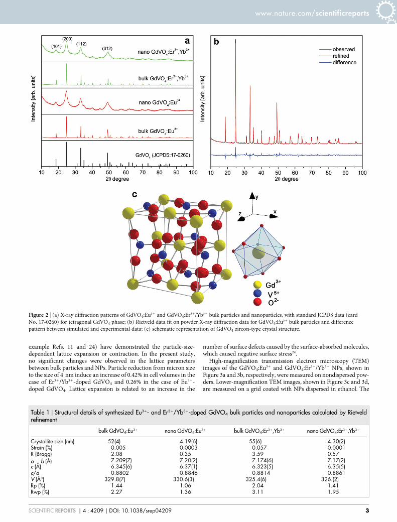
example Refs. 11 and 24) have demonstrated the particle-size-dependent lattice expansion or contraction. In the present study,no significant changes were observed in the lattice parametersbetween bulk particles and NPs. Particle reduction from micron sizeto the size of 4 nm induce an increase of 0.42% in cell volumes in thecase of Er31/Yb31-doped GdVO4 and 0.26% in the case of Eu31-doped GdVO4. Lattice expansion is related to an increase in the
number of surface defects caused by the surface-absorbed molecules,which caused negative surface stress24.
High-magnification transmission electron microscopy (TEM)images of the GdVO4:Eu31 and GdVO4:Er31/Yb31 NPs, shown inFigure 3a and 3b, respectively, were measured on nondispersed pow-ders. Lower-magnification TEM images, shown in Figure 3c and 3d,are measured on a grid coated with NPs dispersed in ethanol. The
Figure 2 | (a) X-ray diffraction patterns of GdVO4:Eu31 and GdVO4:Er31/Yb31 bulk particles and nanoparticles, with standard JCPDS data (card
No. 17-0260) for tetragonal GdVO4 phase; (b) Rietveld data fit on powder X-ray diffraction data for GdVO4:Eu31 bulk particles and difference
pattern between simulated and experimental data; (c) schematic representation of GdVO4 zircon-type crystal structure.
Table 1 | Structural details of synthesized Eu31- and Er31/Yb31-doped GdVO4 bulk particles and nanoparticles calculated by Rietveldrefinement
bulk GdVO4:Eu31 nano GdVO4:Eu31 bulk GdVO4:Er31,Yb31 nano GdVO4:Er31,Yb31
Crystallite size (nm) 52(4) 4.19(6) 55(6) 4.30(2)Strain (%) 0.005 0.0003 0.057 0.0001R (Bragg) 2.08 0.35 3.59 0.57a 5 b (Å) 7.209(7) 7.20(2) 7.174(6) 7.17(2)c (Å) 6.345(6) 6.37(1) 6.323(5) 6.35(5)c/a 0.8802 0.8846 0.8814 0.8861V (Å3) 329.8(7) 330.6(3) 325.4(6) 326.(2)Rp (%) 1.44 1.06 2.04 1.41Rwp (%) 2.27 1.36 3.11 1.95
www.nature.com/scientificreports
SCIENTIFIC REPORTS | 4 : 4209 | DOI: 10.1038/srep04209 3

TEM images show NPs of ,3–4 nm in diameter. This finding isconsistent with the crystallite size evaluated from the powder X-ray diffraction (XRD) measurements. In particular, similar valuesof the crystalline domain size and microscopically estimated averageparticle size of the NPs imply that each particle consists of a singlecrystallite. The selected area electron diffraction (SAED) pattern(inset of Figure 3b) shows features typical to polycrystalline powderswith extremely small particles.
Luminescent properties of GdVO4:Eu31-downshifting photonconversion. Downshifting, or downconversion (DC), is the processof converting high-energy photons into low-energy ones. In RE31-doped materials, DC usually consists of the absorption of high-energy photons by the host material, energy transfer from the hostto the excited states of the RE31 ions, and emission of low-energyphotons after radiative relaxation of the excited states of the RE31
ions to the ground state. GdVO4 is an excellent host matrix fordownshifting photon conversion. This material shows strongabsorption in the UV spectral region and efficient energy transferto RE31 dopants. The excitation spectra (monitored at 537 nm) ofthe GdVO4:Eu31 NPs and the bulk material recorded at roomtemperature are shown in Figure 4a. The spectra share similarfeatures; one can observe broad charge transfer bands (centered at280 and 320 nm)25 and several sharp bands at longer wavelengths(350–500 nm) from the absorption by Eu31.
It is well known that the 320-nm excitation band may be attributedto the VO4
32 groups, i.e., to a V51–O22 charge transfer (CT) from theexcited oxygen ligands (O22) to the central vanadium atom (V51) inthe VO4
32 ions. According to the molecular orbital theory, this corre-sponds to transitions from the 1A2(1T1) ground state to the 1A1(1E)and 1E(1T2) excited states of the VO4
32 ions, i.e., in crystallineGdVO4, the original Td symmetry of VO4
32 (free ion) is reduced toD2d by the crystal field; this causes a splitting of the degenerate levelsof VO4
32. The 280-nm excitation may be assigned to the combinedeffects of the V51–O22 CT and CT transition between Eu31 and O22.It seems that the V51–O22 CT occurs much more easily than the CTof Eu31–O22 because of the large differences in the charge and ionicradii between V51 (15, r 5 0.0355 nm) and Eu31 (13, r 5
0.107 nm)22,26–28. The excitation spectra above 350 nm exhibit sharplines at 363, 377, 381, 395, 418, and 466 nm, which exclusively corre-spond to the following f–f transitions within the 4f6 electronic shell of
Eu31 ions: 7F0 R 5D4, 7F0 R 5G4, 7F0 R 5G2, 7F0 R 5L6, 7F1 R 5L6, and7F0 R 5D2, respectively. The absorption of VO4
32 is stronger thanthat of Eu31, because the f–f electronic transitions of lanthanides arespin-forbidden. However, Figure 4a shows that this difference ismuch more pronounced in the NPs than in the bulk particles.
The room-temperature DC emission spectra of GdVO4:Eu31 NPsand the bulk material, recorded under excitation at 330 nm, areshown in Figure 4b. For the sake of convenience, the emission intens-ity values of the bulk material (because they are much larger) arescaled down by a factor of 100. As is known, the emission intensitiesof RE31-doped materials are generally lower for nanocrystals; thisphenomenon can be explained by surface effects. Nano-sized part-icles possess a higher surface-area-to-volume ratio and many ligatingspecies on their surface; therefore, a larger portion of RE31 emissionsis quenched.
The emission spectra are dominated (in the range above 500 nm)by the red 5D0 R 7F2 electronic transition of Eu31 at 618 nm, whilethree weaker lines at 537, 551, and 593 nm can be attributed to the5D1 R 7F1, 5D1 R 7F2, and 5D0 R 7F1 transitions, respectively.Further, the spectrum of the NPs shows an intense, broad band witha maximum at ,400 nm attributed to the vanadate group emission.However, the emissions from the vanadate groups are much smallerthan those from Eu31 in the bulk material, which further confirms themore pronounced nonradiative relaxation (quenching) of Eu31 in theNPs than that in the bulk material. Nevertheless, emission from theGdVO4:Eu31 NPs is sufficiently strong to be easily recorded. Aschematic representation of the transitions responsible for the emis-sion spectrum is shown in Figure 4c.
It is well known that the emission of Eu31 ions can serve as asensitive probe of local site symmetry29,30. It is possible to deduce thelocal symmetry around the Eu31 ion from the number of multiplet–multiplet transition bands. Moreover, the relative intensities of thetransitions also provide some information about the local symmetry.When Eu31 occupies a site with an inversion center, the magnetic-dipole transition of 5D0 R 7F1 (orange) is dominant. On thecontrary, the electric-dipole transition of 5D0 R 7F2 (red) will bedominant when Eu31 occupies a site without an inversion center.Here, the shape of the emission spectra (Figure 4b) confirms thatthe Eu31 ions occupy sites with D2d point symmetry (sites withoutan inversion center), i.e., they replace the Gd31 ions. This findingfurther confirms and complements those from the XRD results31,32.
Figure 3 | Transmission electron microscopy images of (a, c) GdVO4:Er31/Yb31 and (b, d) GdVO4:Eu31 nanoparticles. Images (a) and (b) are
obtained on nondispersed powder, and images (c) and (d) are obtained on a grids coated with nanoparticles dispersed in ethanol. Inset shows selected
area electron diffraction image of typical GdVO4:Eu31 particle.
www.nature.com/scientificreports
SCIENTIFIC REPORTS | 4 : 4209 | DOI: 10.1038/srep04209 4

Apart from the smaller emission intensities in the NPs compared tothe bulk material, no other differences between the emission spectracould be observed (no changes in the band shapes and no bandshifts). By taking into account the high sensitivity of Eu31 lumin-escence to changes in the symmetry of the local environment, it wasfound that negligible lattice aberrance in nanoparticles is confirmedby the absence of modulation of emission. Therefore, it is reasonableto conclude that reduction in the particle size does not produce anyconsiderable changes in the crystal structure of GdVO4 particles.
In solid materials, electrons in the 4fn orbitals of lanthanides arehighly localized; therefore, such electrons do not exhibit quantumconfinement even in nanocrystals33. Nanophenomena due to sizeconfinement in lanthanide nanoparticles may occur through changesof ion–phonon coupling, which influence the dynamics of 4f–4ftransitions, whereas the static energy levels of the 4f states experiencenegligible impact. In fact, majority of reported size-reduction effectson lanthanide luminescence from 4f-4f transitions are not results ofnano-confinement on electronic states. They are the consequences ofinduced structure distortion and surface defects that affect the localenvironments (site symmetry) surrounding the lanthanide ions. Incontrast to lanthanide 4fn states, the electronic states of VO4
32 ionsare sensitive to the particle size. This is also the case with the
lanthanide 4fn215d states, which, however, are not of relevance forthe current study. In these cases, electronic transitions between statesare spin-allowed, whereas the transitions between 4fn states are spin-forbidden. Therefore, the size effect appears in VO4
32 transitions andis responsible for the difference in the excitation spectra shown inFigure 4a.
Luminescent properties of GdVO4:Er31/Yb31: upconversion anddownconversion. In upconversion (UC), the sequential absorptionof two or more photons (via intermediate long-lived energy states)leads to the emission of light at a shorter wavelength than theexcitation wavelength. The most important class of UC materialsare based on transparent inorganic insulators doped with RE31
ions, because these ions have many long-lived energy states. UCNPs are of particular interest because of the following reasons:they have huge potential in biomedical applications (excitation inthe NIR spectral region where biological materials have minimalabsorption); they can be used to improve the solar cell efficiency(because NP layers scatter considerably less light than bulkparticles); and it is possible to fine-tune the various properties ofNPs for a specific purpose by controlling their shape and size atthe nanometer scale34.
Figure 4 | (a) Room-temperature excitation spectra and (b) downconversion (DC) emission spectra of bulk and nano-sized GdVO4:6mol%Eu31.
(c) Energy levels and transitions in Eu31.
www.nature.com/scientificreports
SCIENTIFIC REPORTS | 4 : 4209 | DOI: 10.1038/srep04209 5

Figure 5a and 5b presents the room-temperature UC emissionspectra of the GdVO4:Er31/Yb31 NPs and the bulk material and aschematic illustration of the energy levels and transitions for theYb31/Er31 system, respectively. Their emission spectra were mea-sured upon excitation with 980-nm radiation under identical condi-tions for comparing the emission intensities.
Er31/Yb31 doping of materials, where Yb31 is used as a sensitizingion and Er31 is used for providing the emission, is by far the mostfrequently used method for preparing RE-based UC materials. Thisis because Yb31 shows good absorption in the NIR spectral region(,980 nm) and a simple energy-level structure with only two levelswhose energy difference closely matches that of Er31; also, energytransfer from Yb31 to Er31 is efficient. The observed UC emissionpeaks (Figure 5a) are characteristic of Er31 emission and are presentin both the green (2H11/2 R 4I15/2 and 4S3/2 R 4I15/2 transitions) andred (4F9/2 R 4I15/2 transition) spectral regions.
A schematic representation of the processes responsible for UCemission is shown in Figure 5b. The energy of the 4I11/2 level of Er31 isvery similar to that of the 2F5/2 level of Yb31; thus, both Er31 and Yb31
absorb 980-nm photons. However, the absorption cross section ofthe 2F7/2 R 2F5/2 transition of Yb31 is very large than that of the 4I15/2
R 4I11/2 transition of Er31; therefore, the Yb31 ions absorb most of theexcitation energy. The excited Yb31 ions either relax to the groundstate (2F7/2) or participate in energy transfer to the neighboring Er31
ions. Therefore, the energy levels of Er31 are populated by ground-and excited-state absorption and by the charge transfer from theYb31 ions. Because of the long-lasting nature of the Er31 excitedstates, its higher energy levels can be populated after the absorptionof two or more photons by a combination of the above-mentionedprocesses. The radiative relaxation of these higher energy states pro-duces UC emission. In the cases of relaxation from 2H11/2 and 4S3/2
(green emission) and 4F9/2 (red emission) to the Er31 ground state(4I15/2), the overall UC process requires the absorption of twophotons in each case. UC emission from the NPs is less intense thanthe emission from the bulk material for the same reasons as for theDC emission with Eu31. Also, in this case, no changes in the bandshapes between the NP and bulk emission are observed, and the sameratio of green to red emission is present.
The room-temperature DC emission spectra of the GdVO4:Er31/Yb31 NPs and the bulk material recorded under excitation at 345 nmare shown in Figure 6. Two green bands are observed at ,525 and,555 nm, which correspond to the transitions from the excited-statelevels 2H11/2 and 4S3/2 to the Er31 ground state (4I15/2). Note that very
weak red emission (,625 nm) corresponding to the 4F9/2 R 4I15/2
transition is also observed, in contrast to the UC spectra upon 980-nm excitation (Figure 5b). This demonstrates that the 4F9/2 stateresponsible for the red emission is not populated via the 4F7/2 and4S3/2 levels or the upper levels, but by lower-lying levels viare-excitation35.
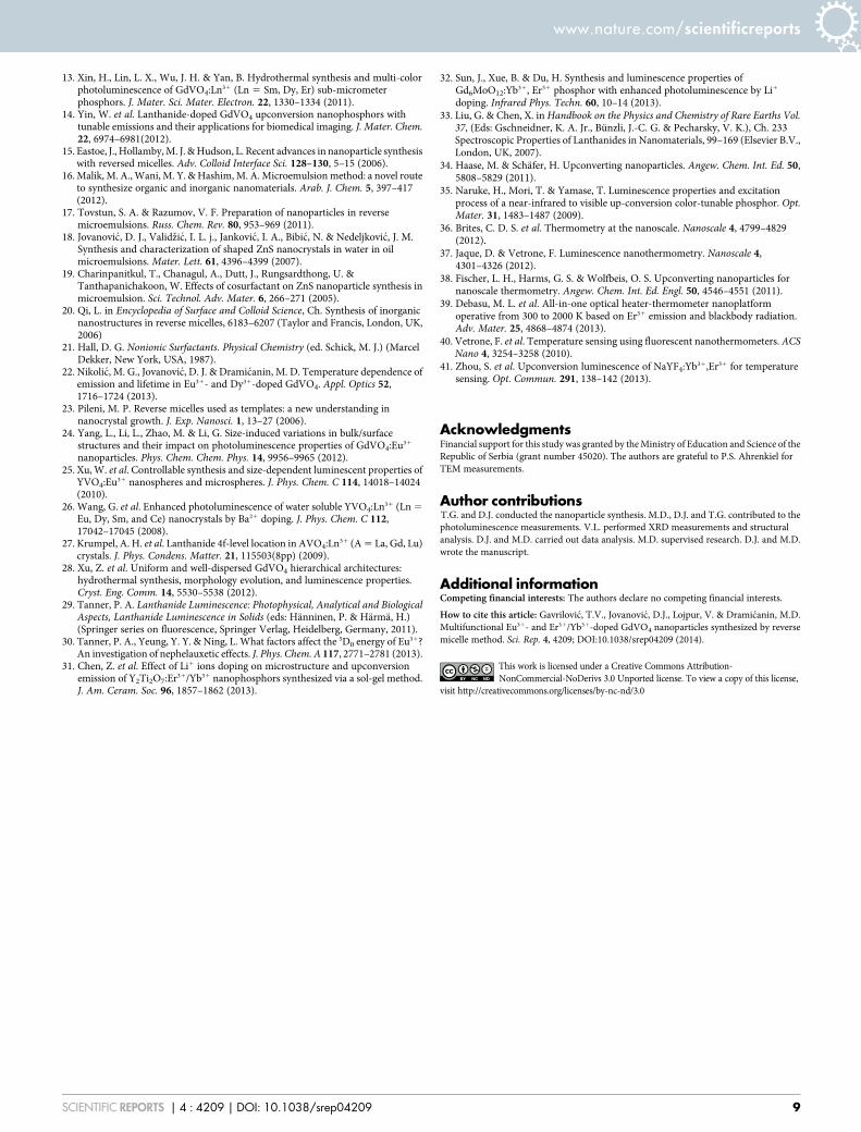
Temperature dependence of upconversion emission in Er31/Yb31-doped GdVO4 nanoparticles. Luminescence thermometry is apromising approach for temperature sensing in a nanoscaleenvironment36–38. Temperature evolution can be realized byremotely measuring the changes in the luminescent properties ofthe various types of NPs or organic dyes. This approach providestemperature measurements with spatial resolution of less than 1 mm,which cannot be achieved with traditional thermometers, and canbe highly beneficial in biomedicine, micro/nano-electronics, andintegrated photonics37. Temperature sensing with upconvertingNPs is of particular interest for biomedicine, because the excitationtypically occurs in the NIR spectral region, and therefore,autofluorescence (the natural emission of light by biologicalstructures such as mitochondria and lysosomes when they absorblight) from biological material does not affect the measurements.NIR excitation shows excellent tissue penetration, and a largenumber of rather inexpensive light sources operating in thisspectral region exist. Here, the potential of the Er31/Yb31-dopedGdVO4 NPs for temperature sensing is assessed. The UC emissionspectra of the GdVO4:Er31/Yb31 NPs recorded over the temperaturerange 307–473 K are shown in Figure 7.
Because two closely separated levels show Boltzmann-type relativepopulation, the integrated fluorescence intensity ratio (FIR) of tran-sitions from the 2H11/2 and 4S3/2 levels to the ground level could beapproximated using Boltzmann distribution as follows39:
FIR ~I 2H11=2?4I15=2
� �I 4S3=2? 4I15=2
� � ~gH AH hnH
gS AS hnSexp {
DEk T
� �
~ B exp {DEkT
� � ð1Þ
where gH and gS are the degeneracies of the 2H11/2 and 4S3/2 levels,respectively; AH, AS and nH, nS are the spontaneous emission rates andfrequencies of the 2H11/2 R 4I15/2 and 4S3/2 R 4I15/2 transitions, respect-ively; h is the Planck’s constant; k is the Boltzmann’s constant; and T isthe absolute temperature. Equation (1) can be expressed as follows:
Figure 5 | (a) Room-temperature upconversion (UC) emission spectra of GdVO4: 2mol%Er31, 10mol%Yb31 nanoparticles and bulk material.
(b) Partial energy-level diagrams of Er31 and Yb31 with proposed upconversion mechanism.
www.nature.com/scientificreports
SCIENTIFIC REPORTS | 4 : 4209 | DOI: 10.1038/srep04209 6

ln FIRð Þ~ ln Bð Þz {DEkT
� �~ ln Bð Þz {
CT
� �, ð2Þ
where B and C are the constants that need to be determined.Fitting of the experimental data with Equation (2), shown in
Figure 8a, shows good correlation between experiment and theory,and is also in agreement with previous reports on thermometryperformed using Er31 UC emission38,39. The obtained parameters,log(B) 5 2.81 and C 51047.52 cm21, provide the absolute sensorsensitivity, Sa [in K21]:
Sa ~LFIRLT
��������~FIR|
DEkT2
, ð3Þ
and the relative sensor sensitivity, Sr [in %K21]:
Sr ~100%|1
FIRLFIRLT
��������~100%|
DEkT2
: ð4Þ
The sensitivity values calculated from Equations (3) and (4) areshown in Figure 8b. The maximal value of the relative sensitivity,
1.11%K21, is found at 307 K; it is very similar to those found inNaYF4:Yb31,Er3140,41 and gold-decorated Gd2O3:Yb31,Er3139. Thisresult is among the highest relative sensitivities of thermometersbased on UC emission (for a comparison see Table S1 in theSupporting Information of Ref. 38). A temperature resolution of,1 K can be estimated from dFIR/Sa, where dFIR is the resolutionof the FIR calculated from the standard deviation of the residuals inthe polynomial interpolation of the experimental data points (tem-perature vs. FIR curve)36,39. Information on the temperature resolu-tion may also be deduced from the sensitivity of the detection systemand the calculated sensitivity.
In conclusion, preparation of GdVO4 powders using reversemicelles yields super thin NPs (3–4 nm in diameter) because ofhomogeneous nucleation. A zircon-type structure that incorporatesEu31 or Er31/Yb31 ions as dopants can be achieved without addi-tional thermal treatments; therefore, no further growth of the part-icles occurs. Moreover, in the absence of thermal treatment andbecause the particles were conserved in micelles, each particle con-sists of a single crystallite, as evidenced from the crystal coherencesize obtained from the XRD analysis and the particle size from theTEM images. Different optical functions are observed with Eu31 andEr31/Yb31 doping. In DC, the Eu31-doped GdVO4 NPs exhibit char-acteristic sharp emissions in the red region along with broad emis-sions from the vanadate groups centered at 400 nm. The intensity ofthe Eu31 emission is much smaller in the NPs than in the bulkpowders. This indicates that stronger nonradiative pathways existin the NPs because of the presence of ligands on their surface. Nochanges in the band positions or bandwidths are observed in theemission spectra of the NPs and bulk particles; this result suggeststhat were no significant alterations of the size and symmetry of thefirst coordination sphere around the dopant ions upon decreasingthe particle size to the nanoscale. The Er31/Yb31-doped NPs emitgreen light with both DC and UC processes. Similar to the Eu31
luminescence, NPs show smaller emissions compared to the bulkparticles, and no changes in the band shapes or positions areobserved. However, these emissions are of sufficient strength to beeasily detected and used; for example, in temperature sensing by UCemission. This might be a new and powerful function of Er31/Yb31-doped GdVO4 NPs; luminescence nanoprobes can be used to extractinformation about the local temperature of a given system with sub-micrometric spatial resolution, which cannot be achieved with
Figure 6 | Room-temperature downconversion (DC) emission spectra of GdVO4:2mol%Er31, 10 mol%Yb31 nanoparticles and bulk material.
Figure 7 | Upconversion emission spectra of GdVO4:Er31/Yb31
nanoparticles recorded over the temperature range 307–473 K.
www.nature.com/scientificreports
SCIENTIFIC REPORTS | 4 : 4209 | DOI: 10.1038/srep04209 7
traditional temperature sensors. Compared to other materialsrecently suggested for use in temperature sensing with UC, Er31/Yb31-doped GdVO4 NPs exhibit a high relative sensitivity (of1.11%K21) with an estimated resolution of 1 K.
Furthermore, temperature sensing with UC emission utilizesexcitation sources in the NIR spectral region, where biological tissuesshow low absorption; therefore, this type of sensor may have valuableapplications in the biomedicine and bioimaging fields. GdVO4 canact as a T1-positive contrast agent, because Gd31 ions possessunpaired electrons that could efficiently alter the relaxation time ofthe surrounding water protons. Phosphorescent emission of RE31-doped materials is useful for bioimaging, because it lasts longer thanthe autofluorescence of tissue, and thus, can be easily discriminatedusing time-resolved measurements. Also, unlike semiconductingquantum dots, RE31-doped NPs do not show photobleaching.Therefore, these NPs, because of their exceptionally small sizes,excellent crystallinity, and phosphorescent emission with the pos-sibility to sense temperature with a high spatial resolution, showsignificant potential for bioimaging and biolabeling.
MethodsSynthesis of Eu31- and Er31/Yb31-doped GdVO4 nanoparticles by reverse micellemethod. The following chemicals were used as received: gadolinium(III) nitratehexahydrate, Gd(NO3)3?6H2O (99.9%, Alfa Aesar), europium(III) nitratehexahydrate, Eu(NO3)3?6H2O (99.9%, Alfa Aesar), erbium(III) nitrate pentahydrate,Er(NO3)3?5H2O (99.9%, Alfa Aesar), ytterbium(III) nitrate pentahydrate,Yb(NO3)3?5H2O (99.9%, Alfa Aesar), ammonium vanadium oxide, NH4VO3 (min.99.0%, Alfa Aesar), and sodium hydroxide, NaOH (min. 99%, Moss Hemos).
A typical high-temperature solid-state reaction method was used for preparingRE31-doped GdVO4 bulk material; more details can be found in our previous work22.The GdVO4:Eu31 and GdVO4:Er31/Yb31 NPs were prepared by a reverse micellemethod. The doping levels were 6 mol% of Eu31 and 2 mol% of Er31/10 mol%of Yb31.
A typical synthesis using reverse micelles at room temperature was as follows. First,the following solutions were prepared: 1) an oil phase formed by mixing cyclohexane(100 mL), Triton X-100 (60 mL), and n-pentanol (20 mL) in a volume ratio of55351; 2 a) a solution (10 mL) formed by mixing aqueous solutions of 0.1 MGd(NO3)3?6H2O and 0.1 M Eu(NO3)3?6H2O in a stoichiometric ratio (6 mol% Eu31
with respect to Gd31); 2 b) a solution (10 mL) formed by mixing aqueous solutions of0.1 M Gd(NO3)3?6H2O (10 mL) and 0.1 M Er(NO3)3?6H2O and 0.1 MYb(NO3)3?6H2O in a stoichiometric ratio (2 mol% Er31 and 10 mol% Yb31 withrespect to Gd31); and 3) a 0.1 M solution of Na3VO4 (10 mL) formed by dissolvingNH4VO3 in 0.15 M NaOH. In the next step, the oil phase and solution containingVO4
32 were mixed; into the obtained mixture, the solution containing RE31 wasadded drop-wise under continuous magnetic stirring. In all syntheses, the water-to-oil volume ratio was maintained at 159.
At the end of each synthesis, a stable (single-phase) yellowish colloid solutioncontaining reverse micelles was obtained upon stirring for 60 min at room temper-ature. After aging for 24 h, methanol was added to destabilize the solution. Theobtained mixture was separated by centrifugation, and the resulting precipitate
(powder) was washed several times with methanol and water to remove excess sur-factant and then dried in an oven at 70uC for 20 h.
Characterization. XRD measurements were performed on a Rigaku SmartLabdiffractometer using Cu-Ka radiation (l 5 0.15405 nm). Diffraction data wererecorded with a step size of 0.02u and a counting time of 0.7u min21 over the angularrange 10u # 2h # 100u. Crystallite sizes were estimated using the Halder–Wagnermethod by analyzing all major diffraction peaks. TEM imaging was performed using aJEOL-JEM 2100 instrument (Akishima-shi, Japan) equipped with a LaB6 cathodeoperating at 200 kV.
Photoluminescence measurements were carried out on pellets prepared fromRE31-GdVO4 powders under a load of 2 ton cm22. All DC luminescence measure-ments were performed at room temperature using a Fluorolog-3 spectrofluorometer(model FL3-221, Horiba Jobin Yvon), which uses a 450-W xenon lamp as anexcitation source for emission measurements (lexc 5 330 nm for GdVO4:Eu31 andlexc 5 345 nm for GdVO4:Er31/Yb31) and a xenon–mercury pulsed lamp for decaytime measurements. The emission spectra were scanned in the wavelength ranges350–640 and 375–660 nm, respectively. The UC emission spectra were measuredupon excitation with 980-nm radiation (MDLH 980 3w) on an AvaSpec-2048 FiberOptic Spectrometer system.
1. Albanese, A., Tang, P. S. & Chan, W. C. The effect of nanoparticle size, shape, andsurface chemistry on biological systems. Annu. Rev. Biomed. Eng. 14, 1–16 (2012).
2. Huang, X., Han, S., Huang, W. & Liu, X. Enhancing solar cell effciency: the searchfor luminescent materials as spectral converters. Chem. Soc. Rev. 42, 173–201(2013).
3. Yen, W. M., Shionoya, S. & Yamamoto, H. Phosphor Handbook (CRC Press, BocaRaton, FL, USA, 2007).
4. Nunez, N. O. et al. Surface modified Eu:GdVO4 nanocrystals for optical and MRIimaging. Dalton Trans. 42, 10725–10734 (2013).
5. Singh, N. S. et al. Re-dispersion and film formation of GdVO4:Ln31 (Ln31 5 Dy31,Eu31, Sm31, Tm31) nanoparticles: particle size and luminescence studies. DaltonTrans. 41, 4404–4412 (2012).
6. Zhang, L. et al. Characteristics of Nd:GdVO4 laser with different Nd-dopingconcentrations. Chin. Phys. Lett. 23, 2088–2091 (2006).
7. Kang, X. et al. Poly(acrylic acid) modified lanthanide-doped GdVO4 hollowspheres for up-conversion cell imaging, MRI and pH-dependent drug release.Nanoscale 5, 253–261 (2013).
8. Kim, M.-J. & Huh, Y.-D. Preparation and photoluminescence of GdVO4:Eunanophosphors for flexible and transparent displays. Bull. Korean Chem. Soc. 32,4454–4457 (2011).
9. Singh, N. S. et al. Luminescence study of Eu31 doped GdVO4 nanoparticles:Concentration, particle size, and core/shell effects. J. Appl. Phys. 104, 104307(9pp) (2008).
10. Jovanovic, D. J. et al. Annealing effects on the microstructure andphotoluminescence of Eu31-doped GdVO4 powders. Opt. Mater. 35, 1797–1804(2013).
11. Yang, L., Li, L., Zhao, M., Fu, C. & Li, G. Is there lattice contraction inmulticomponent metal oxides? Case study for GdVO4:Eu31 nanoparticles.Nanotechnology 24, 305701(10pp) (2013).
12. Zheng, Y. et al. Facile hydrothermal synthesis and luminescent properties of large-scale GdVO4:Eu31 nanowires. Cryst. Growth Des. 9, 5101–5107 (2009).
Figure 8 | (a) Fluorescence intensity ratio (FIR) values of GdVO4:Er31/Yb31 nanopowder as a function of temperature (dots) and corresponding line
obtained by fitting Equation (2). (b) Absolute (blue line) and relative (red) sensitivity of FIR temperature sensor based on the upconversion of
GdVO4:Er31/Yb31 nanopowder.
www.nature.com/scientificreports
SCIENTIFIC REPORTS | 4 : 4209 | DOI: 10.1038/srep04209 8
13. Xin, H., Lin, L. X., Wu, J. H. & Yan, B. Hydrothermal synthesis and multi-colorphotoluminescence of GdVO4:Ln31 (Ln 5 Sm, Dy, Er) sub-micrometerphosphors. J. Mater. Sci. Mater. Electron. 22, 1330–1334 (2011).
14. Yin, W. et al. Lanthanide-doped GdVO4 upconversion nanophosphors withtunable emissions and their applications for biomedical imaging. J. Mater. Chem.22, 6974–6981(2012).
15. Eastoe, J., Hollamby, M. J. & Hudson, L. Recent advances in nanoparticle synthesiswith reversed micelles. Adv. Colloid Interface Sci. 128–130, 5–15 (2006).
16. Malik, M. A., Wani, M. Y. & Hashim, M. A. Microemulsion method: a novel routeto synthesize organic and inorganic nanomaterials. Arab. J. Chem. 5, 397–417(2012).
17. Tovstun, S. A. & Razumov, V. F. Preparation of nanoparticles in reversemicroemulsions. Russ. Chem. Rev. 80, 953–969 (2011).
18. Jovanovic, D. J., Validzic, I. L. j., Jankovic, I. A., Bibic, N. & Nedeljkovic, J. M.Synthesis and characterization of shaped ZnS nanocrystals in water in oilmicroemulsions. Mater. Lett. 61, 4396–4399 (2007).
19. Charinpanitkul, T., Chanagul, A., Dutt, J., Rungsardthong, U. &Tanthapanichakoon, W. Effects of cosurfactant on ZnS nanoparticle synthesis inmicroemulsion. Sci. Technol. Adv. Mater. 6, 266–271 (2005).
20. Qi, L. in Encyclopedia of Surface and Colloid Science, Ch. Synthesis of inorganicnanostructures in reverse micelles, 6183–6207 (Taylor and Francis, London, UK,2006)
21. Hall, D. G. Nonionic Surfactants. Physical Chemistry (ed. Schick, M. J.) (MarcelDekker, New York, USA, 1987).
22. Nikolic, M. G., Jovanovic, D. J. & Dramicanin, M. D. Temperature dependence ofemission and lifetime in Eu31- and Dy31-doped GdVO4. Appl. Optics 52,1716–1724 (2013).
23. Pileni, M. P. Reverse micelles used as templates: a new understanding innanocrystal growth. J. Exp. Nanosci. 1, 13–27 (2006).
24. Yang, L., Li, L., Zhao, M. & Li, G. Size-induced variations in bulk/surfacestructures and their impact on photoluminescence properties of GdVO4:Eu31
nanoparticles. Phys. Chem. Chem. Phys. 14, 9956–9965 (2012).25. Xu, W. et al. Controllable synthesis and size-dependent luminescent properties of
YVO4:Eu31 nanospheres and microspheres. J. Phys. Chem. C 114, 14018–14024(2010).
26. Wang, G. et al. Enhanced photoluminescence of water soluble YVO4:Ln31 (Ln 5
Eu, Dy, Sm, and Ce) nanocrystals by Ba21 doping. J. Phys. Chem. C 112,17042–17045 (2008).
27. Krumpel, A. H. et al. Lanthanide 4f-level location in AVO4:Ln31 (A 5 La, Gd, Lu)crystals. J. Phys. Condens. Matter. 21, 115503(8pp) (2009).
28. Xu, Z. et al. Uniform and well-dispersed GdVO4 hierarchical architectures:hydrothermal synthesis, morphology evolution, and luminescence properties.Cryst. Eng. Comm. 14, 5530–5538 (2012).
29. Tanner, P. A. Lanthanide Luminescence: Photophysical, Analytical and BiologicalAspects, Lanthanide Luminescence in Solids (eds: Hanninen, P. & Harma, H.)(Springer series on fluorescence, Springer Verlag, Heidelberg, Germany, 2011).
30. Tanner, P. A., Yeung, Y. Y. & Ning, L. What factors affect the 5D0 energy of Eu31?An investigation of nephelauxetic effects. J. Phys. Chem. A 117, 2771–2781 (2013).
31. Chen, Z. et al. Effect of Li1 ions doping on microstructure and upconversionemission of Y2Ti2O7:Er31/Yb31 nanophosphors synthesized via a sol-gel method.J. Am. Ceram. Soc. 96, 1857–1862 (2013).
32. Sun, J., Xue, B. & Du, H. Synthesis and luminescence properties ofGd6MoO12:Yb31, Er31 phosphor with enhanced photoluminescence by Li1
doping. Infrared Phys. Techn. 60, 10–14 (2013).33. Liu, G. & Chen, X. in Handbook on the Physics and Chemistry of Rare Earths Vol.
37, (Eds: Gschneidner, K. A. Jr., Bunzli, J.-C. G. & Pecharsky, V. K.), Ch. 233Spectroscopic Properties of Lanthanides in Nanomaterials, 99–169 (Elsevier B.V.,London, UK, 2007).
34. Haase, M. & Schafer, H. Upconverting nanoparticles. Angew. Chem. Int. Ed. 50,5808–5829 (2011).
35. Naruke, H., Mori, T. & Yamase, T. Luminescence properties and excitationprocess of a near-infrared to visible up-conversion color-tunable phosphor. Opt.Mater. 31, 1483–1487 (2009).
36. Brites, C. D. S. et al. Thermometry at the nanoscale. Nanoscale 4, 4799–4829(2012).
37. Jaque, D. & Vetrone, F. Luminescence nanothermometry. Nanoscale 4,4301–4326 (2012).
38. Fischer, L. H., Harms, G. S. & Wolfbeis, O. S. Upconverting nanoparticles fornanoscale thermometry. Angew. Chem. Int. Ed. Engl. 50, 4546–4551 (2011).
39. Debasu, M. L. et al. All-in-one optical heater-thermometer nanoplatformoperative from 300 to 2000 K based on Er31 emission and blackbody radiation.Adv. Mater. 25, 4868–4874 (2013).
40. Vetrone, F. et al. Temperature sensing using fluorescent nanothermometers. ACSNano 4, 3254–3258 (2010).
41. Zhou, S. et al. Upconversion luminescence of NaYF4:Yb31,Er31 for temperaturesensing. Opt. Commun. 291, 138–142 (2013).
AcknowledgmentsFinancial support for this study was granted by the Ministry of Education and Science of theRepublic of Serbia (grant number 45020). The authors are grateful to P.S. Ahrenkiel forTEM measurements.
Author contributionsT.G. and D.J. conducted the nanoparticle synthesis. M.D., D.J. and T.G. contributed to thephotoluminescence measurements. V.L. performed XRD measurements and structuralanalysis. D.J. and M.D. carried out data analysis. M.D. supervised research. D.J. and M.D.wrote the manuscript.
Additional informationCompeting financial interests: The authors declare no competing financial interests.
How to cite this article: Gavrilovic, T.V., Jovanovic, D.J., Lojpur, V. & Dramicanin, M.D.Multifunctional Eu31- and Er31/Yb31-doped GdVO4 nanoparticles synthesized by reversemicelle method. Sci. Rep. 4, 4209; DOI:10.1038/srep04209 (2014).
This work is licensed under a Creative Commons Attribution-NonCommercial-NoDerivs 3.0 Unported license. To view a copy of this license,
visit http://creativecommons.org/licenses/by-nc-nd/3.0
www.nature.com/scientificreports
SCIENTIFIC REPORTS | 4 : 4209 | DOI: 10.1038/srep04209 9