Multicomponent hydrogels from enantiomeric amino acid derivatives: helical nanofibers, handedness...
10
Multicomponent hydrogels from enantiomeric amino acid derivatives: helical nanofibers, handedness and self-sorting† Bimalendu Adhikari, Jayanta Nanda and Arindam Banerjee * Received 17th May 2011, Accepted 4th July 2011 DOI: 10.1039/c1sm05907f In this study, chiral helical nanofibers have been obtained from suitable, co-assembling, two oppositely charged amino acid based two component hydrogels. An equimolar mixture of an N-terminally protected amino acid Fmoc-(L/D)Glu (Fmoc: N-fluorenyl-9-methoxycarbonyl, Glu: glutamic acid) and (L/D)Lys (Lys: lysine) can co-assemble to form hydrogels. These hydrogels have been characterised using circular dichroism (CD), atomic force microscopy (AFM), transmission electron microscopy (TEM), X-ray powder diffraction, fluorescence spectroscopic and rheological studies. CD and AFM studies have been extensively used to examine the chiral/achiral nature of fibers obtained from different hydrogel systems. The equimolar mixture of two L-isomers, {Fmoc-(L)Glu + (L)Lys} in the assembled state, leads to the exclusive formation of left-handed helical nanofibers, whereas an equimolar mixture of two D-isomers, {Fmoc-(D)Glu + (D)Lys}, gives rise to right-handed helical nanofibers. The CD study of the gel obtained from the {Fmoc-(L)Glu + (L)Lys} system is exactly the mirror image of the CD signal obtained from the gel of the {Fmoc-(D)Glu + (D)Lys} system. These results suggest that the molecular chirality is being translated into the supramolecular helicity and the handedness of these fibers depends on the corresponding molecular chirality in the mixture of the two component system. Reversing the handedness of helical fibers is possible by using enantiomeric building blocks. Co- assembly of racemic and equimolar mixtures of all four components, i.e., [{Fmoc-(L)Glu + (L)Lys} + {Fmoc-(D)Glu + (D)Lys}] can also form hydrogels. Interestingly, in this racemic mixture self-sorting has been observed with the presence of almost equal amount of left- and right-handed helical nanofibers. The equimolar mixture of Fmoc-(L)Glu and L-ornithine/L-arginine also produces hydrogel with left-handed helical fibers. Moreover, the straight fiber has been observed from the two component hydrogel {Fmoc-(L)Glu + (L)Lys} system in the presence of Ca 2+ /Mg 2+ ions. This indicates the straight nanofibers are obtained under suitable conditions and acid–base interaction is responsible for making the helical fibers at the nanoscale. Introduction Helicity is omnipresent in nature ranging from nanoscopic helical structures in proteins, DNA double helices and collagen triple helix structures to microscopic viruses and macroscopic seashells. Chirality plays a pivotal role in chemistry, biology and material sciences through its various applications in chemical-/ bio-sensors, pharmaceutics, chiral catalysts, asymmetric synthesis, enantioselective separation, nonlinear optics, chiral devices, and also in other fields. 1 Being inspired by the impor- tance of chirality in biology and to explore interesting applica- tions of chirality in nanosciences, chemists have attempted to make helical structures using molecular assembly of one or multicomponent(s). 2 Chirality can be expressed at different levels, from chiral small molecules to helical conformation of macromolecules, and even to helical supramolecular nano- structures. Molecular assembly based on the rational control of noncovalent interactions including hydrogen bonding, aromatic Department of Biological Chemistry, Indian Association for the Cultivation of Science, Jadavpur, Kolkata, 700 032, India. E-mail: [email protected]; Fax: +91-33-2473-2805 † Electronic supplementary information (ESI) available: Fig. S1–S14; FT-IR spectrum of a dried hydrogel, CD spectra of two component systems in the solution state, CD spectra of two component hydrogels obtained from {Fmoc-(L)Glu + (L)ornithine} and {Fmoc-(L)Glu + (L) arginine} separately, CD spectrum of {Fmoc-(L)Glu + (L)Lys} in DMSO/water mixture, size distribution of width of fibers obtained from {Fmoc-(L)Glu + (L)Lys}, AFM images of two component hydrogels [Fmoc-(L)Glu + (L)arginine] and [Fmoc-(L)Glu + (L) ornithine], TEM image of hydrogel {Fmoc-(L)Glu + (L)Lys}, size distribution of widths of these fibers obtained from {Fmoc-(D)Glu + (D)Lys}, AFM image of two component hydrogel {Fmoc(L)Glu + (L) Lys} in the presence of 0.5 equimolar Ca 2+ , AFM image of two component hydrogel {Fmoc(L)Glu + (L)Lys} in the presence of monovalent Na + and K + , HRMS and 1 H NMR spectra of synthetic Fmoc-(L)Glu and Fmoc-(D)Glu, titration curve for pK a determination. See DOI: 10.1039/c1sm05907f This journal is ª The Royal Society of Chemistry 2011 Soft Matter , 2011, 7, 8913–8922 | 8913 Dynamic Article Links C < Soft Matter Cite this: Soft Matter , 2011, 7, 8913 www.rsc.org/softmatter PAPER Downloaded by Duke University on 29 September 2012 Published on 09 August 2011 on http://pubs.rsc.org | doi:10.1039/C1SM05907F View Online / Journal Homepage / Table of Contents for this issue
Transcript of Multicomponent hydrogels from enantiomeric amino acid derivatives: helical nanofibers, handedness...

Dynamic Article LinksC<Soft Matter
Cite this: Soft Matter, 2011, 7, 8913
www.rsc.org/softmatter PAPER
Dow
nloa
ded
by D
uke
Uni
vers
ity o
n 29
Sep
tem
ber
2012
Publ
ishe
d on
09
Aug
ust 2
011
on h
ttp://
pubs
.rsc
.org
| do
i:10.
1039
/C1S
M05
907F
View Online / Journal Homepage / Table of Contents for this issue
Multicomponent hydrogels from enantiomeric amino acid derivatives: helicalnanofibers, handedness and self-sorting†
Bimalendu Adhikari, Jayanta Nanda and Arindam Banerjee*
Received 17th May 2011, Accepted 4th July 2011
DOI: 10.1039/c1sm05907f
In this study, chiral helical nanofibers have been obtained from suitable, co-assembling, two oppositely
charged amino acid based two component hydrogels. An equimolar mixture of an N-terminally
protected amino acid Fmoc-(L/D)Glu (Fmoc: N-fluorenyl-9-methoxycarbonyl, Glu: glutamic acid) and
(L/D)Lys (Lys: lysine) can co-assemble to form hydrogels. These hydrogels have been characterised
using circular dichroism (CD), atomic force microscopy (AFM), transmission electron microscopy
(TEM), X-ray powder diffraction, fluorescence spectroscopic and rheological studies. CD and AFM
studies have been extensively used to examine the chiral/achiral nature of fibers obtained from different
hydrogel systems. The equimolar mixture of two L-isomers, {Fmoc-(L)Glu + (L)Lys} in the assembled
state, leads to the exclusive formation of left-handed helical nanofibers, whereas an equimolar mixture
of two D-isomers, {Fmoc-(D)Glu + (D)Lys}, gives rise to right-handed helical nanofibers. The CD study
of the gel obtained from the {Fmoc-(L)Glu + (L)Lys} system is exactly the mirror image of the CD
signal obtained from the gel of the {Fmoc-(D)Glu + (D)Lys} system. These results suggest that the
molecular chirality is being translated into the supramolecular helicity and the handedness of these
fibers depends on the corresponding molecular chirality in the mixture of the two component system.
Reversing the handedness of helical fibers is possible by using enantiomeric building blocks. Co-
assembly of racemic and equimolar mixtures of all four components, i.e., [{Fmoc-(L)Glu + (L)Lys} +
{Fmoc-(D)Glu + (D)Lys}] can also form hydrogels. Interestingly, in this racemic mixture self-sorting
has been observed with the presence of almost equal amount of left- and right-handed helical
nanofibers. The equimolar mixture of Fmoc-(L)Glu and L-ornithine/L-arginine also produces hydrogel
with left-handed helical fibers. Moreover, the straight fiber has been observed from the two component
hydrogel {Fmoc-(L)Glu + (L)Lys} system in the presence of Ca2+/Mg2+ ions. This indicates the straight
nanofibers are obtained under suitable conditions and acid–base interaction is responsible for making
the helical fibers at the nanoscale.
Department of Biological Chemistry, Indian Association for the Cultivationof Science, Jadavpur, Kolkata, 700 032, India. E-mail: [email protected];Fax: +91-33-2473-2805
† Electronic supplementary information (ESI) available: Fig. S1–S14;FT-IR spectrum of a dried hydrogel, CD spectra of two componentsystems in the solution state, CD spectra of two component hydrogelsobtained from {Fmoc-(L)Glu + (L)ornithine} and {Fmoc-(L)Glu + (L)arginine} separately, CD spectrum of {Fmoc-(L)Glu + (L)Lys} inDMSO/water mixture, size distribution of width of fibers obtainedfrom {Fmoc-(L)Glu + (L)Lys}, AFM images of two componenthydrogels [Fmoc-(L)Glu + (L)arginine] and [Fmoc-(L)Glu + (L)ornithine], TEM image of hydrogel {Fmoc-(L)Glu + (L)Lys}, sizedistribution of widths of these fibers obtained from {Fmoc-(D)Glu +(D)Lys}, AFM image of two component hydrogel {Fmoc(L)Glu + (L)Lys} in the presence of 0.5 equimolar Ca2+, AFM image of twocomponent hydrogel {Fmoc(L)Glu + (L)Lys} in the presence ofmonovalent Na+ and K+, HRMS and 1H NMR spectra of syntheticFmoc-(L)Glu and Fmoc-(D)Glu, titration curve for pKa determination.See DOI: 10.1039/c1sm05907f
This journal is ª The Royal Society of Chemistry 2011
Introduction
Helicity is omnipresent in nature ranging from nanoscopic
helical structures in proteins, DNA double helices and collagen
triple helix structures to microscopic viruses and macroscopic
seashells. Chirality plays a pivotal role in chemistry, biology and
material sciences through its various applications in chemical-/
bio-sensors, pharmaceutics, chiral catalysts, asymmetric
synthesis, enantioselective separation, nonlinear optics, chiral
devices, and also in other fields.1 Being inspired by the impor-
tance of chirality in biology and to explore interesting applica-
tions of chirality in nanosciences, chemists have attempted to
make helical structures using molecular assembly of one or
multicomponent(s).2 Chirality can be expressed at different
levels, from chiral small molecules to helical conformation of
macromolecules, and even to helical supramolecular nano-
structures. Molecular assembly based on the rational control of
noncovalent interactions including hydrogen bonding, aromatic
Soft Matter, 2011, 7, 8913–8922 | 8913

Dow
nloa
ded
by D
uke
Uni
vers
ity o
n 29
Sep
tem
ber
2012
Publ
ishe
d on
09
Aug
ust 2
011
on h
ttp://
pubs
.rsc
.org
| do
i:10.
1039
/C1S
M05
907F
View Online
stacking, electrostatic interactions, van der Waals interactions
and/or metal coordination interactions provides powerful tools
for the design and construction of hierarchical structures from
nano- to micrometre scale.2,3
There are several reports of constructing supramolecular
helical nanostructures including helical fibers,4 helical rib-
bons,1h,2g,5 or helical tubes6 by using different strategies from self-
assembling organic molecules. Meijer and his workers have
produced supramolecular helical polymers using the self-recog-
nition of hydrogen bonds.2h A long fiber with left handed helicity
has been obtained from a disk-shaped molecule with chiral tails
upon self-assembly in chloroform and also a nonhelical rod has
been formed in the presence of potassium ions.4a The formation
of left-handed helical nanoropes has been reported from the self-
assembly of a rigid p-conjugated oligo(p-phenylenevinylene)
with remote chiral handles.4b Yashima and his coworkers have
reported supramolecular cholesteric twist based on controlled
polymerization of an enantiomerically pure isocyanide.4d Stupp
and his coworkers have developed a class of tripeptide amphi-
philes that are self-assembled to form superhelical twisting
nanofibers.4f Liu and coworkers have made helical nanofibers
based on gels containing achiral porphyrin units.4g Huang and
coworkers have reported the double stranded helical nanofibers
based on a sugar–lipid amphiphilic hydrogel.4i Hamley and
coworkers have reported the formation of a helical nanoribbon
based on a short segment of amyloid b peptide.5d
Helical to nonhelical conversion and even inversion of helicity
in supramolecular assembly and also in certain macromolecules
can be achieved by using either enantiomeric molecular building
blocks,7 or by the slight structural change in the molecular
building blocks,8 or by the changing solvent or temperature,6d,9 or
upon binding with chiral/achiral counterions,10 or upon binding
with chiral/achiral neutral guest molecules,11 or by changing the
pH,12 or by irradiating with light.4e,13 The inversion of the
handedness of a helical fiber using the enantiomeric form of the
molecular building block is an interesting phenomenon.7 One of
the oldest reports in the construction of helical nanofibers was
based on a supramolecular liquid-crystalline polymer consisting
of the polyassociation of the complementary chiral components
TP2 and TU2, which were derived from the nucleobase pyrimi-
dine (P) anduracil (U) derivatives and from the D, L, ormeso forms
of tartaric acid (T).7a The handedness of helical fibers is governed
by the chirality of the tartaric acid. Other examples of the
formation of helical fibers include the self-assembly of dithienyl-
ethene functionalized chiral amides,7c dendron rodcoil triblock
molecules7d and artificial b-sheet-conforming peptides.7e The
handedness of the helical fibers can be reversed by using the
mirror-image form of the molecular building block. Previous
results of our research group have shown that the handedness of
the helical nanofibers can be reversed either by using mirror-
imaged tri-amidemolecules7forbyutilizing the enantiomeric form
of pseudopeptide-based building blocks.7h
The principle of self-sorting is efficiently used in nature in the
formation of functional architectures like DNA. In DNA four
nucleobases (adenine (A), thymine (T), guanine (G), and cytosine
(C)) self-sort to form specific self-complementary base pairs (AT
and GC) in DNA hybridisation. This self-sorting principle is also
found in the crystallization of racemates into conglomerates.
Lehn and his coworkers have shown that the racemic mixture of
8914 | Soft Matter, 2011, 7, 8913–8922
helical fiber forming components gives rise to spontaneous
resolution to form both left- and right-handed supramolecular
helical nanofibers in the liquid-crystalline system in chloro-
form.7a This type of self-sorting phenomenon has been observed
in other artificial systems.14 Examples include the occurrence of
self-sorting in a multi-component mixture of functionalized
different organic groups (crown ethers and ammonium ions),14a
and tetra-urea calix[4]arenes system.14b A self-sorting phenom-
enon is also observable in some organogel systems including
dendritic peptide,14d perylene bisimide derivative14e and long
chain containing carbamate based chiral pyrrolidine deriva-
tives.14g In dendritic peptide based assembly, Smith and co-
workers have investigated three parameters namely ‘‘size’’,
‘‘shape’’ and ‘‘chirality’’.14d They have observed that the mixtures
of dendritic peptides with different ‘‘size’’ and ‘‘chirality’’ can be
self-organized in their molecular scaffolds, whereas mixtures
of dendritic peptides with different ‘‘shapes’’ can break one
another’s self-association process.14d Chiral pyrrolidine deriva-
tive based organogels obtained from the two enantiomers of
gelators and their mixtures exhibited enantiomeric discrimina-
tion and this results in a self-sorting process.14g
However, none of these above mentioned examples address
multiple issues including reversing the handedness of helical
fibers, disrupting the helicity and the self-sorting of both left- and
right-handed helical fibers in two or multicomponent systems. In
this study, we present the construction of chiral helical nano-
fibers from the co-assembly of the complementary amino acid
based two component hydrogel system, reversing the chirality of
helical fibers by changing the intrinsic molecular chirality of
constituent amino acid building blocks, self-sorting of both left-
and right-handed helical nanofibers in a four component racemic
mixture and disrupting the helicity of nanofibers in the presence
of Ca2+/Mg2+ ions. The equimolar mixture of two L isomers,
{Fmoc-(L)Glu + (L)Lys}, forms left-handed helical nanofibers,
whereas an equimolar mixture of two D isomers, {Fmoc-(D)Glu
and (D)Lys}, results in right-handed helical nanofibers. More-
over, the racemic mixture of the above mentioned four compo-
nent system, i.e., [{Fmoc-(L)Glu + (L)Lys} + {Fmoc-(D)Glu + (D)
Lys}], yields the co-existence of helical nanofibers of opposite
handedness, indicating the occurrence of the self-sorting process.
Interestingly, an achiral straight fiber has been obtained from the
multicomponent hydrogel system involving {Fmoc-(L)Glu and
(L)Lys} in the presence of Ca2+/Mg2+ ions. This suggests that the
presence of acid–base interactions involving two oppositely
charged amino acids in a co-assembling system can play a role in
the formation of chiral (helical) fibers. To the best of our
knowledge, this is the unique example of the observation of
several phenomena including construction of helicity, reversing
of helicity, self-sorting of both left- and right-handed helical
fibers and abolishing the helicity of the fibers based on co-
assembly of the multicomponent hydrogel system under specific
conditions.
Results and discussion
Gel formation and characterization
Low molecular weight supramolecular hydrogels are an inter-
esting class of soft materials.15 Formation of two-component gels
This journal is ª The Royal Society of Chemistry 2011
Fig. 1 X-ray powder diffraction of dried hydrogel obtained from the
two component system [Fmoc-(L)Glu + (L)Lys]. A photograph of
hydrogel obtained from [Fmoc-(L)Glu + (L)Lys].
Dow
nloa
ded
by D
uke
Uni
vers
ity o
n 29
Sep
tem
ber
2012
Publ
ishe
d on
09
Aug
ust 2
011
on h
ttp://
pubs
.rsc
.org
| do
i:10.
1039
/C1S
M05
907F
View Online
through supramolecular complex formation is an emerging field
of research.16 Recently, Fmoc containing small molecule based
hydrogels have been reported.17 In this study, assembling prop-
erties of the two component system, Fmoc-Glu and Lys (see
Scheme 1), were first examined by gelation experiments in water
at pH 7.4 using phosphate buffer as well as distilled water (milli
Q). Several attempts using different approaches were made to
examine whether Fmoc-(L/D)Glu alone can form hydrogels or
not. Fmoc-(L/D)Glu exhibited very low solubility in distilled
water (milli Q) at pH 7.4 even at high temperature (�100 �C).However, by cooling down the temperature to room tempera-
ture, the soluble portion was precipitated out. The Fmoc-(L/D)
Glu showed moderate solubility at pH 7.4 using phosphate
buffer at high temperature and precipitation was observed upon
cooling down the temperature to room temperature. The Fmoc-
(L/D)Glu alone is also unable to form any kind of hydrogel using
the phosphate buffer under the condition studied (pH 1–11). The
Fmoc-(L/D)Glu did not show any hydrogel formation using
sodium carbonate or sodium hydrogen carbonate or sodium
hydroxide. No hydrogel from Fmoc-(L/D)Glu was found, by
dissolving it in basic pH and then decrease the pH of the medium
gradually using AcOH or using the hydrolysis of glucono-d-
lactone (GdL) as described elsewhere.17d
The equimolar mixture of these two components {Fmoc-(L)
Glu + (L)Lys} was dissolved in water by heating for a few
minutes. A translucent hydrogel was obtained, when this hot
solution was cooled down to room temperature (Fig. 1, inset).
The minimum gelation concentration is 0.05 (M) with respect to
each of these components using phosphate buffer solution at pH
7.4 and the hydrogel is stable within the pH range 2–9 using
phosphate buffer. The final pH values after mixing both
components in all cases were changed. After hydrogel formation
the pH was determined using the pH meter. After gel formation
using buffer solution at pH 7.4 and 9.0, the pH of the hydrogels
were decreased to pH 5.20 and 5.38 respectively. However, the
increase in pH from 2.0 to 4.16 was observed, when the two
component gel was formed using the buffer solution at pH 2.0. A
Scheme 1 Chemical structures of the components involved in two
component hydrogels: (A) acidic side chain containing amino acid and
(B) basic side chain containing amino acids.
This journal is ª The Royal Society of Chemistry 2011
viscous solution was observed from the two component system
above pH 9 and upto pH 10 using phosphate buffer. However,
a clear solution was obtained with a further increase in the pH
above 10 using the buffer. No gel formation was observed using
buffer solution below pH 2.0, instead a precipitation was
occurred. On the basis of pKa values obtained from the titration
experiment, the degree of ionizations of two COOH groups
present in Fmoc-Glu and one COOH group and twoNH2 groups
in lysine have been calculated at the final pH value of 5.2 within
the hydrogel. In this case the degree of ionizations (or deproto-
nation) of a-COOH and side chain-(g)-COOH, of Fmoc-Glu,
were found to be 0.9754 and 0.585 respectively. The degree of
ionization of a-COOH and the degree of protonation of a-NH2
and side chain-(3)-NH2 of lysine were determined to be 0.99991,
0.99968 and 0.999995 respectively. The degree of ionization of
COOH groups and the degree of protonation of NH2 groups of
the two component system in other pH values are mentioned in
the ESI†.
This gel is thermoreversible in nature and the Tgel is found to
be 41 �C. By mixing these two oppositely charged amino acids
(Fmoc-(L)Glu and (L)Lys) in water a supramolecular hydrogel is
formed via the electrostatic attraction (acid–base type) triggered
co-assembly (see Scheme 2). The FT-IR study indicates the
presence of intermolecular hydrogen bonding interaction
between the amino acid backbones and the X-ray powder
diffraction study suggests the existence of p–p stacking inter-
action between the aromatic fluorenyl rings, in the gel state
(Fig. S1† in the ESI and ‘X-Ray powder diffraction study’
section discussed later). So electrostatic interaction, hydrogen
bonding and p–p stacking interactions are the driving force for
hydrogelation. It is important to note that the lysine can be
replaced only either by ornithine or by arginine among all other
amino acids to produce hydrogels. This suggests that simple
acid–base interaction can be responsible for the co-assembly and
hydrogelation as at pH 7.4, two carboxylic groups of Glu are
deprotonated and two amino groups of lysine/ornithine and one
amino and one guanidino group of arginine are protonated.
Other proteinaceous amino acids (Glycine, Alanine, Valine,
Leucine, Isoleucine, Phenylalanine, Tyrosine, Tryptophan,
Soft Matter, 2011, 7, 8913–8922 | 8915

Scheme 2 Tentative model for the molecular packing of gelator mole-
cules in the assembled state. Blue dotted lines indicate acid–base type
interactions, while orange dashed lines indicate aromatic p–p stacking
interactions. Molecules A {Fmoc-(L)Glu} and B {(L)Lys} are involved in
acid–base interactions to form a co-assembled adduct first, and this is
further assembled to form a bilayer structure. This bilayer structure acts
as a repeating structural unit and each black block line indicates one of
these structural units.
Dow
nloa
ded
by D
uke
Uni
vers
ity o
n 29
Sep
tem
ber
2012
Publ
ishe
d on
09
Aug
ust 2
011
on h
ttp://
pubs
.rsc
.org
| do
i:10.
1039
/C1S
M05
907F
View Online
Histidine, Proline, Aspartic acid, Glutamic acid) with only one
primary amino group are unable to co-assemble with Fmoc-Glu
to form hydrogel. The mixture of Fmoc-Glu and any other
amino acids (other than lysine, arginine and ornithine) remains
insoluble in distilled water. This suggests that for co-assembly
and/or hydrogelation acid–base type interactions between two
carboxylic acid groups of Fmoc-Glu and two amino/guanidino
group(s) of lysine/ornithine/arginine are responsible. These co-
assembling adducts can be further assembled using p–p stacking
interactions utilizing aromatic moieties (of Fmoc-Glu residue)
and hydrogen bonding to form gel phase materials (see Scheme
2). Gelation tests were also performed using propyl amine (one
and two equivalent separately) and one equivalent of organic
diamines (1,3-diamine or 1,4-diamine or 1,6-diamine) with
Fmoc-(L/D)Glu. However, no gelation was observed in any of
these cases. This suggests that two amino groups containing
amino acids were required for this type of multicomponent based
hydrogelation.
Fig. 2 CD spectra of hydrogels at the near gel state with different
stereochemical compositions as indicated in the figure.
X-Ray powder diffraction study
X-Ray powder diffraction, a technique for ascertaining the
molecular packing of assemblies, was used to get the internal
assembling structure of the co-assembling two component
hydrogel. Fig. 1 shows the small angle X-ray powder
diffraction (XRPD) patterns of the xerogel obtained from the
{Fmoc-(L)Glu + (L)Lys} system. A strong reflection peak cor-
responding to a d-spacing of 15.29 �A (at 2q ¼ 5.77) was found
and this d-spacing value is nearly close to the calculated molec-
ular length (18.19 �A) of the co-assembled state. In the small angle
8916 | Soft Matter, 2011, 7, 8913–8922
region (at 2q ¼ 3.03) one sharp peak corresponding to a d-spac-
ing of 29.07 �Awas observed, which may correspond to the higher
order lamellar organization of the co-assembling molecules. In
the higher angle region, a peak around 2q ¼ 22.76 (d ¼ 3.90 �A) is
a characteristic of the p–p stacking distance of two aromatic
fluorenyl groups.18 Another peak at 2q ¼ 16.55 (d ¼ 5.35 �A)
corresponds to the distance between the two hydrogen bonded
molecules in a strand.18 Fmoc-(L)Glu and (L)Lys are involved in
acid–base interactions to form a coassembled adduct (a pair) and
these co-assembled adducts are further assembled to form
a bilayer structure by p–p stacking interactions in the assembled
gel state. The length of 2.907 nm (obtained from XRPD) is larger
than the extended coassembled adduct’s length (1.819 nm).
However, it is smaller than two times the co-assembled adduct’s
length. To satisfy the intermolecular hydrogen bonding interac-
tion, acid–base interaction and p–p stacking interaction, we
believe that assembled adducts form bilayer structures (having
a width of 2.81 nm) and this bilayer structure is the repeating
structural unit.13d The width of a bilayer structure (2.81 nm) is
calculated from the energy minimized tentative model structure
and it is comparable with the distance obtained from the small
angle XRPD diffraction peak (2.907 nm). Based on these above
observations a tentative model of molecular packing has been
proposed in Scheme 2.
Circular dichroism study
Circular dichroism (CD) is a useful tool for determining the
chiral molecular arrangement in assemblies. This is because
intermolecular interactions, especially between chromophoric
molecules, may produce striking chiroptical responses and
generate CD signals often much stronger in associated state
compared to their isolated molecular state. The CD study of two
component hydrogels at the gel state with different stereochem-
ical compositions is presented in Fig. 2. Circular dichroism of the
gel obtained from {Fmoc-(L)Glu + (L)Lys} has shown a strong
negative signal around 304 nm. The Cotton effect at 304 nm
(p–p* transition) indicates the superhelical arrangement formed
by the fluorenyl groups in the hydrogel state16d and the CD signal
at 304 nm is presumably induced by the formation of chiral
This journal is ª The Royal Society of Chemistry 2011
Dow
nloa
ded
by D
uke
Uni
vers
ity o
n 29
Sep
tem
ber
2012
Publ
ishe
d on
09
Aug
ust 2
011
on h
ttp://
pubs
.rsc
.org
| do
i:10.
1039
/C1S
M05
907F
View Online
(helical) structures. It can be noted that the amide region in the
CD spectra (200–230 nm) is noisy due to the high absorbance in
this region, with the high concentration of gelators.17e However,
in the solution state with low concentration of gelators, a Cotton
effect at 223 nm containing positive signal appeared (Fig. S2,
ESI†). This is due to n–p* transition.16d
Moreover, it can be mentioned that each of these hydrogels
obtained from {Fmoc-(L)Glu + (L)ornithine} and {Fmoc-(L)Glu
+ (L)arginine} also produces a strong negative signal around 306
nm (Fig. S3, ESI†). The CD spectrum of {Fmoc-(D)Glu + (D)
Lys} in the gel state has shown a strong positive signal around
304 nm and this CD signal is nearly the mirror image of the CD
signal obtained from the {Fmoc-(L)Glu + (L)Lys} gel system (see
Fig. 2). This suggests that the chirality of the gel obtained from
the co-assembly of {Fmoc-(L)Glu + (L)Lys} is opposite in nature
with respect to the gel obtained from the D-isomers, i.e., {Fmoc-
(D)Glu + (D)Lys}.
This type of chirality is generated due to the formation of
supramolecular chirality in the gel state.5c To establish this
temperature dependent, concentration dependent and solvent
dependent CD studies have been performed. In the case of the
temperature dependent CD study (see Fig. 3), the signal at
304 nm has been gradually decreased with an increase in the
temperature and surprisingly the signal has approached almost
the base line at the temperature higher than the Tgel value, where
gelator molecules remain in the solution state and not in the
assembled state. This clearly suggests the presence of supramo-
lecular chirality in the co-assembled state. The temperature
dependency of the CD signals clearly suggests that the CD
response is a consequence of the co-assembly of chiral compo-
nents to supramolecular chirality, rather than the inherent
molecular chirality of the individual amino acid components.5c,16j
Thus, each of these two component systems (e.g., {Fmoc-(L)Glu
+ (L)Lys}) has appeared as a supramolecular chiroptical switch
in the sol–gel process. However, the chirality disappeared when
the gel was heated to solution, and it reappeared again upon
cooling the system to get the gel phase material. In DMSO/water
or 1,4-dioxane/water solvent mixture, the mixture of these two
components remains in the solution state at the similar
Fig. 3 Temperature dependent CD spectra of {Fmoc-(D)Glu + (D)Lys}
hydrogel at the respective gel state starting from 25 �C (room tempera-
ture) to 50 �C (>Tgel).
This journal is ª The Royal Society of Chemistry 2011
concentration that is required for gelation in water. There is no
characteristic CD of signal of {Fmoc-(L)Glu + (L)Lys} in
DMSO/water or 1,4-dioxane/water solvent mixture (see Fig. S4,
ESI†). This further supports the fact that the CD signal of the
hydrogel is arising from the supramolecular chirality generated
in the co-assembled gel state and it does not arise from the
individual chirality of the corresponding amino acid
components.
Interestingly, the CD spectrum of the gel obtained from the
racemic mixture of both L and D isomers of these components,
i.e., [{Fmoc-(L)Glu + (L)Lys} + {Fmoc-(D)Glu + (D)Lys}] is
almost flat and it is close to the baseline (see Fig. 2). The observed
almost zero Cotton effect for [{Fmoc-(L)Glu + (L)Lys} + {Fmoc-
(D)Glu + (D)Lys}] suggests that the inter-chromophore orienta-
tion is either disordered, achiral, or is chiral but racemic (i.e.
equal amounts of left- and right-handed helical orientations are
present).2a Similarly, in the presence of divalent cations Ca2+ or
Mg2+ this two component system (e.g., {Fmoc-(L)Glu + (L)Lys})
does not show any characteristic CD signal (see Fig. 2) sug-
gesting the achiral nature of the supramolecular aggregates in the
presence of any of these divalent metal ions (Ca2+ or Mg2+). The
molar ratio of divalent cations (Ca2+ or Mg2+) to gelators is 1 : 1.
Morphological study
To investigate the morphology of the binary component hydro-
gels microscopic experiments were carried out using atomic force
microscopy (AFM). Fig. 4–6 show the tapping mode AFM
images of hydrogels with two different stereochemical composi-
tions {Fmoc-(L)Glu + (L)Lys} and {Fmoc-(D)Glu + (D)Lys} and
their racemic mixture.
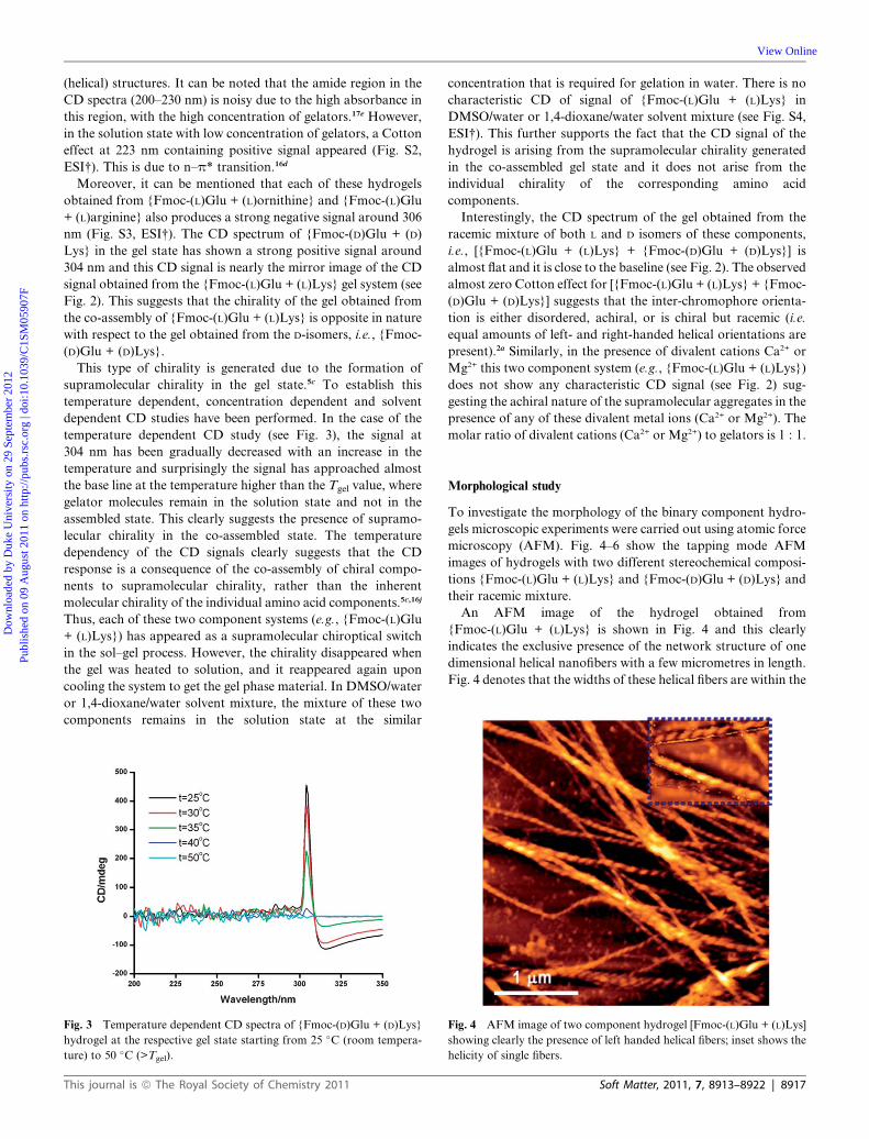
An AFM image of the hydrogel obtained from
{Fmoc-(L)Glu + (L)Lys} is shown in Fig. 4 and this clearly
indicates the exclusive presence of the network structure of one
dimensional helical nanofibers with a few micrometres in length.
Fig. 4 denotes that the widths of these helical fibers are within the
Fig. 4 AFM image of two component hydrogel [Fmoc-(L)Glu + (L)Lys]
showing clearly the presence of left handed helical fibers; inset shows the
helicity of single fibers.
Soft Matter, 2011, 7, 8913–8922 | 8917

Fig. 5 (a) AFM image of two component hydrogel [Fmoc-(D)Glu + (D)
Lys] showing right handed helical fibers; inset shows the helicity of single
fiber; (b) zoomed version of some part of the image (a).
Fig. 6 (a) AFM image of racemic mixture in multi-component (four
componets) hydrogel [Fmoc-(L + D)Glu + (L + D)Lys] showing the
presence of both left- and right-handed helical fibers; (b) zoomed version
of green color marked region showing the right-handed helical fiber
clearly; (c) zoomed version of violet color marked region showing the left-
handed helical fiber clearly.
Fig. 7 AFM image of aggregates obtained from {Fmoc-(L)Glu + (L)
Lys} in the presence of Ca2+ ions.
Dow
nloa
ded
by D
uke
Uni
vers
ity o
n 29
Sep
tem
ber
2012
Publ
ishe
d on
09
Aug
ust 2
011
on h
ttp://
pubs
.rsc
.org
| do
i:10.
1039
/C1S
M05
907F
View Online
range 52–127 nm and the majority of these fibers fall within the
range 60–100 nm (Fig. S5, ESI†) with an average diameter of
82.52 nm. The morphology of a single helix is twisted with
uniform left-handed bias along the fiber long axis (see Fig. 4).
The helical pitch of these fibers varies from 98 to 140 nm from
one fiber to the other fibers. However, the helical pitch of a single
helical fiber remains uniform along the fiber and this indicates
that the underlying forces causing helix formation are uniform
throughout the fiber structure. The observed left-handed helicity
is compatible with the observed negative Cotton effect for the
circular dichroism (CD) measurements.19 It can be mentioned
that hydrogels obtained from {Fmoc-(L)Glu + (L)ornithine} and
{Fmoc-(L)Glu + (L)arginine} also individually produce left-
handed helical nanofibers (see Fig. S6, ESI†). This suggests that
simple acid–base interactions are responsible for the co-assembly
in the formation of hydrogel and helical fibers.
A TEM experiment was also carried out using the two
component hydrogel {Fmoc-(L)Glu + (L)Lys} system to examine
the morphological inner detail of helical structure and to probe
whether it is a fiber or a tube (with inner hollowness). The TEM
image is shown in Fig. S7† in the ESI section and this image
clearly demonstrates the formation of helical nanofibers, instead
of a helical nanotubular structure.
8918 | Soft Matter, 2011, 7, 8913–8922
On the other hand, AFM image analysis of the hydrogel
formed by {Fmoc-(D)Glu + (D)Lys} reveals the presence of the
network structure of right handed helical nanofibers (see Fig. 5).
These helical fibers are a few micrometres in length and the width
of these fibers varies from 62 to 135 nm, with an average diameter
of 84.5 nm. The majority of these fibers fall within the range of
62–99 nm (Fig. S8, ESI†). The pitch length of the helical fibers
varies from 70.5 to 90 nm. However, the pitch of a single fiber is
uniform in nature. Therefore, it can be concluded that the helicity
of fibers could be easily controlled by the chirality of the
constituent amino acid components of the hydrogel system.
It is interesting to examine the morphology of the gel obtained
from the racemic mixtures {Fmoc-(L + D)Glu + (L + D)Lys} of
the four component system. There are three following possibili-
ties: (a) change in the morphology of helical to non-helical
(straight) fibers or any other shape, (b) co-existence of both left-
and right-handed helical fibers within the same system and (c) the
presence of both left- and right-handedness within the same
helical fiber with one half left handed and other portion right
handed.
However, the AFM images of hydrogels obtained from
racemic mixture {Fmoc-(L + D)Glu + (L + D)Lys} reveal the
presence of both left- and right-handed helical fibers in the same
system and not within the same fiber (see Fig. 6). In this study,
widths of both left- and right-handed fibers are within the range
of 70–90 nm. This spontaneous occurrence of racemate resolu-
tion by chiral selection in molecular recognition directed co-
assembly of the components in the supramolecular hydrogel
system is rare.7a In most of these cases a racemic mixture
produces either achiral morphology e.g. straight fiber or
a different morphology like vesicles.7e However, in this study,
self-sorting behavior of these components has been observed.
The chirality at the supramolecular level depends on the
configuration of the stereogenic centers of the amino acids in the
two component hydrogel system.
It is interesting to address the point whether in the presence of
divalent cations Ca2+ or Mg2+ the hydrogel based helical fibers
are formed or not. Straight fibers are obtained from this two
component hydrogel system in the presence of divalent cations
Ca2+/Mg2+ with 1 : 1 molecular ratio (see Fig. 7). Fig. 7 shows
that no helical (left- or right-handed) fibers have been formed
and only straight fibers have been observed with an average
width of 170 nm. Therefore, helical fiber to straight fiber
This journal is ª The Royal Society of Chemistry 2011

Dow
nloa
ded
by D
uke
Uni
vers
ity o
n 29
Sep
tem
ber
2012
Publ
ishe
d on
09
Aug
ust 2
011
on h
ttp://
pubs
.rsc
.org
| do
i:10.
1039
/C1S
M05
907F
View Online
transformation is possible in the presence of bivalent metal ions
such as Ca2+ or Mg2+. This is because Ca2+ ions can easily bind to
the two carboxylate moiety of Fmoc-glutamic acid. This inhibits
the co-assembly process between Fmoc-glutamic acid and lysine/
ornithine/arginine using acid–base type interaction, which is
responsible for helicity. In this study, the gel formation is not
affected by the presence of Ca2+/Mg2+ ions, however the
morphology of the hydrogel changes from helical to straight
fibers upon binding with Ca2+ or Mg2+ into the multicomponent
gel system.
AFM experiments have also been carried out using different
concentrations of divalent cations (Ca2+/Mg2+) to explore the
dependency of helical to non-helical transformation of gel
nanofibers on the concentration of divalent cations. At lower
concentration of Ca2+ ions, both helical and non-helical
(straight) gel fibers have been observed (Fig. S9, ESI†). However,
at the relatively higher concentration of Ca2+ ions, i.e., the
equimolar ratio of each of the gelators and Ca2+ ions, only
straight nanofibers have been obtained. In other words, it can be
stated that to disrupt the helicity completely the required molar
ratio of divalent metal ions to gelators is 1 : 1. It is interesting to
observe that in the presence of monovalent metal ions (e.g., Na+,
K+) two component hydrogel systems also produce helical
nanofibers (Fig. S10, ESI†). This suggests that helical to non-
helical transition is due to the binding of divalent metal ions with
two carboxylic acid of glutamic acid residue.
Fluorescence study
Fluorescence experiments have been utilized to follow the
assembling nature of these gelator molecules within hydrogels
because the fluorescence study may provide useful information
regarding the change in the microenvironment of the fluorophore
moiety during the gelation process. The concentration dependent
fluorescence starting from very dilute solution to the assembling
gel state has been studied and it has been shown in Fig. 8, where
(a) is a very dilute solution of gelators at a concentration of
0.0031 (M), (b) is a dilute solution of gelators at a concentration
of 0.0062 (M), (c) is a solution of gelators at a concentration of
Fig. 8 Concentration dependent fluorescence spectra of two-component
gelators {Fmoc-(L)Glu + (L)Lys} in water starting from very dilute
solution (concentration 0.0031 M) to gel state (concentration 0.05 M) as
indicated in the figure.
This journal is ª The Royal Society of Chemistry 2011
0.0125 (M), (d) is a concentrated, viscous solution of gelators at
a concentration of 0.025 (M) and (e) is the gel state of gelators at
a concentration of 0.0500 (M) (minimum gelation concentra-
tion). Following observations have been noted.
(1) In the case of (a), the strong emission maximum centered at
317 nm has been obtained for the excitation at 300 nm. This
emission is arising from monomeric Fmoc-Glu molecules.16d,i,20
Fluorescence intensity slowly increases by increasing the
concentration from (a) to (c). This is due to the enhancement of
concentrations of gelator molecules. (2) By increasing the
concentrations of both gelators further, i.e. the enhancement of
concentration (c) sol to (d) viscous solution, a huge red shifting of
fluorescence emission maximum has been observed from 320 nm
to 367 nm, without changing the emission intensity significantly.
This indicates the presence of aggregation at this state. (3)
Further enhancement of both gelator’s concentration from (d) to
gel state (e) has resulted in a red shifting from 367 nm to 380 nm.
The emission maximum at 380 nm suggests that fluorenyl groups
are dimerized in the gel state due to a strong p–p interaction16d,i
(Scheme 2). In last two cases, i.e. in the concentration region of
(d) and (e), a small shoulder emission peak at 468 nm has also
been observed. This indicates that some fluorene moieties
aggregate more efficiently involving more than two fluorene
groups in p–p stacking interactions in the hydrogel state.16d,i
Rheological study
Viscoelastic properties of two component hydrogels at different
stereo-chemical compositions were examined by measuring their
rheological properties using these gels at a fixed concentration
0.2 (M) with respect to each of these components. In a typical
frequency sweep experiment, the variation of storage modulus
(G0) and loss modulus (G0 0) was monitored as a function of
applied angular frequency under a constant strain 0.1%. It is
worth mentioning that G0 and G00 respectively symbolize the
ability of the deformed material to restore its original geometry
and tendency of a material to flow. For an ideal liquid, G0 ¼ 0,
and for an ideal solid, G0 0 ¼ 0. For viscoelastic materials like gels,
G0 is greater than G0 0, and it shows that elastic behavior of the
system is dominant. Fig. 9 shows the linear viscoelastic frequency
sweep responses of these hydrogels. All of these
Fig. 9 Frequency dependence of the dynamic storage moduli (G0) andthe loss moduli (G0 0) of hydrogels at different stereo-chemical composi-
tions as indicated in the figure.
Soft Matter, 2011, 7, 8913–8922 | 8919

Dow
nloa
ded
by D
uke
Uni
vers
ity o
n 29
Sep
tem
ber
2012
Publ
ishe
d on
09
Aug
ust 2
011
on h
ttp://
pubs
.rsc
.org
| do
i:10.
1039
/C1S
M05
907F
View Online
multi-component systems at different stereo-chemical composi-
tions exhibit a very weak frequency dependence within the
experimental frequency limit, with G0 greater than G0 0. This
suggests that they are effective physical hydrogels.
Conclusions
This study addresses multipurpose issues: formation of helical
(chiral) nanofibers, reversing the handedness of helicity and self-
sorting of both left- and right-handed helical nanofibers. In this
study, one-dimensional helical nanofibers are successfully con-
structed from suitable co-assembling complementary chiral
amino acid based two component hydrogel systems utilizing
acid–base type interaction (electrostatic interaction), hydrogen-
bonding and aromatic p–p stacking interactions. This study also
demonstrates the reversing of handedness of the helical (chiral)
nanofiber using the enantiomeric form of constituent amino
acids, the conversion of helical to straight fibers in the presence of
bivalent metal ions such as Ca2+/Mg2+ and self-sorting of both
left- and right-handed helical fibers in a racemic mixture of all
four L and D components of the amino acid based gel system.
These observations suggest that the molecular chirality is being
transferred into the supramolecular chirality and ultimately to
the nanoscale level (helical nanofiber) and the handedness of
these helical nanofibers can be reversed by using the enantio-
meric molecular building blocks. These findings also indicate
that the presence of acid–base interactions between two oppo-
sitely charged amino acids can have a definite role in the helical
fiber formation as the presence of Ca2+/Mg2+ ions disrupts the co-
assembly of Fmoc-(L/D)Glu and (L/D)Lys.
Experimental section
Materials
L-Glutamic acid, D-glutamic acid, L-lysine, D-lysine, L-arginine,
L-ornithine and Fmoc-chloride were purchased from Sigma-
Aldrich. The water used in all experiments was of Millipore
Milli-Q grade.
Synthesis of Fmoc-Glu
2 mmol of Glu was dissolved in a basic sodium carbonate solu-
tion (15 mL). It was cooled in an ice-water bath and a cooled
solution of 3 mmol Fmoc-Cl in dioxane (15 mL) was added to it.
The reaction mixture was allowed to come to room temperature
and stirred for 24 h. Then the solution was concentrated in
vacuum to about 15 mL, cooled in an ice water bath, covered
with a layer of ethyl acetate (about 30 mL), and acidified with
a dilute HCl to neutral pH. The aqueous phase was extracted
with ethyl acetate and this operation was done twice. The ethyl
acetate extract was pooled, dried over anhydrous Na2SO4 and
evaporated in vacuum. A white material was obtained and this
was characterized by mass spectrometry, 1H-NMR spectroscopy
(for spectra see Fig. S11–S13†).
Circular dichroic (CD) study
Circular dichroism spectroscopy was used for determining the
chiral molecular arrangement of assemblies within hydrogels. All
8920 | Soft Matter, 2011, 7, 8913–8922
CD spectra were recorded by using a quartz cuvette of 1 mm path
length in a Jasco J-815 spectropolarimeter. All the CD experi-
ments were performed for gel samples (homochiral and racemic)
using the same gelators concentration.
Atomic force microscopic (AFM) study
Morphologies of these reported hydrogels were investigated
using a tapping-mode atomic force microscope (AFM). AFM
studies were done by placing a small amount of wet hydrogel at
its minimum gelation concentration on a microscope cover glass.
The material was then allowed to dry in air by slow evaporation
first and then under vacuum at room temperature for two days.
Images were recorded by exploiting an Autoprobe CP Base Unit
di CP-II instrument (model no. AP-0100).
X-Ray powder diffraction (XRPD)
XRPD of dried hydrogel material was performed by using an
X-ray diffractometer (Bruker D8 Advance) equipped with
a conventional CuKa X-ray radiation (l ¼ 1.54 �A) source and
Bragg diffraction setup (Seifert 3000P).
Transmission electron microscopic (TEM) study
The morphology of the two component hydrogel obtained from
{Fmoc-(L)Glu + (L)Lys} was investigated using a transmission
electron microscope (TEM). The sample was prepared through
depositing a small amount of the near gel phase material on
a TEM grid (300 mesh Cu grid) coated with Formvar and carbon
film. The grid was then allowed to dry under vacuum for two
days. A TEM image was taken by a JEOL electron microscope
operated at an accelerating voltage of 200 kV.
Fluorescence spectroscopy
The emission spectra were recorded by using a Horiba Jobin
Yvon Fluoromax 3 instrument with a 1 cm path length quartz
cell in a concentration range from 0.0031%, w/v, to 0.050%, w/v
(MGC). The excitation and emission slit width were 5 and 5 nm,
respectively.
Rheological study
A rheological experiment was performed with an AR 2000
advanced rheometer.
Acknowledgements
B.A. and J.N. thank the CSIR, New Delhi, India, for financial
assistance. We also acknowledge the support by the DST, India,
project No.SR/S1/OC-73/2009.
Notes and references
1 (a) J. M. Lehn, Supramolecular Chemistry, Wiley VCH, Weinheim,1995, pp. 1–271; (b) E. Yashima, K. Maeda, H. Iida, Y. Furushoand K. Nagai, Chem. Rev., 2009, 109, 6102–6211; (c) R. Tashiroand H. Sugiyama, J. Am. Chem. Soc., 2005, 127, 2094–2097; (d)A. Brizard, R. Oda and I. Huc, Top. Curr. Chem., 2005, 256, 167–218; (e) H. Goto, Y. Furusho and E. Yashima, Chem. Commun.,2009, 1650–1652; (f) T. Verbiest, S. Van Elshocht, M. Kauranen,L. Hellemans, J. Snauwaert, C. Nuckolls, T. J. Katz and
This journal is ª The Royal Society of Chemistry 2011

Dow
nloa
ded
by D
uke
Uni
vers
ity o
n 29
Sep
tem
ber
2012
Publ
ishe
d on
09
Aug
ust 2
011
on h
ttp://
pubs
.rsc
.org
| do
i:10.
1039
/C1S
M05
907F
View Online
A. Persoons, Science, 1998, 282, 913–915; (g) J. H. Jung, Y. Ono,K. Hanabusa and S. Shinkai, J. Am. Chem. Soc., 2000, 122, 5008–5009; (h) E. D. Sone, E. R. Zubarev and S. I. Stupp, Angew. Chem.,Int. Ed., 2002, 41, 1705–1709.
2 (a) C. C. Lee, C. Grenier, E. W. Meijer and A. P. H. J. Schenning,Chem. Soc. Rev., 2009, 38, 671–683; (b) D. Pijper andB. L. Feringa, Soft Matter, 2008, 4, 1349–1372; (c)A. R. A. Palmans and E. W. Meijer, Angew. Chem., Int. Ed., 2007,46, 8948–8968; (d) D. K. Smith, Chem. Soc. Rev., 2009, 38, 684–694; (e) K. Murata, M. Aoki, T. Suzuki, T. Harada, H. Kawabata,T. Komori, F. Ohseto, K. Ueda and S. Shinkai, J. Am. Chem. Soc.,1994, 116, 6664–6676; (f) R. Oda, I. Huc and S. J. Candau, Angew.Chem., Int. Ed., 1998, 37, 2689–2691; (g) R. Oda, I. Huc,M. Schmutz, S. J. Candau and F. C. MacKintosh, Nature, 1999,399, 566–569; (h) J. H. K. K. Hirschberg, L. Brunsveld, A. Ramzi,J. A. J. M. Vekemans, R. P. Sijbesma and E. W. Meijer, Nature,2000, 407, 167–170; (i) W.-Y. Yang, E. Lee and M. Lee, J. Am.Chem. Soc., 2006, 128, 3484–3485; (j) A. Ajayaghosh, P. Chithraand R. Varghese, Angew. Chem., Int. Ed., 2007, 46, 230–233; (k)T.-F. Lin, R.-M. Ho, C.-H. Sung and C.-S. Hsu, Chem. Mater.,2008, 20, 1404–1409; (l) Y. Qiao, Y. Lin, Y. Wang, Z. Yang, J. Liu,J. Zhou, Y. Yan and J. Huang, Nano Lett., 2009, 9, 4500–4504; (m)Y. Lin, Y. Qiao, C. Gao, P. Tang, Y. Liu, Z. Li, Y. Yan andJ. Huang, Chem. Mater., 2010, 22, 6711–6717; (n) M. Kimura,T. Hatanaka, H. Nomoto, J. Takizawa, T. Fukawa, Y. Tatewakiand H. Shirai, Chem. Mater., 2010, 22, 5732–5738; (o) S. Qu,L. Wang, X. Liu and M. Li, Chem.–Eur. J., 2011, 17, 3512–3518.
3 (a) J.-M. Lehn, Proc. Natl. Acad. Sci. U. S. A., 2002, 99, 4763–4768;(b) M. M. L. Nieuwenhuizen, T. F. A. de Greef, R. L. J. van derBruggen, J. M. J. Paulusse, W. P. J. Appel, M. M. J. Smulders,R. P. Sijbesma and E. W. Meijer, Chem.–Eur. J., 2010, 16, 1601–1612; (c) S. Yagai, H. Aonuma, Y. Kikkawa, S. Kubota,T. Karatsu, A. Kitamura, S. Mahesh and A. Ajayaghosh, Chem.–Eur. J., 2010, 16, 8652–8661.
4 (a) H. Engelkamp, S. Middelbeek and R. J. M. Nolte, Science, 1999,284, 785–788; (b) S. J. George, A. Ajayaghosh, P. Jonkheijm,A. P. H. J. Schenning and E. W. Meijer, Angew. Chem., Int. Ed.,2004, 43, 3421–3425; (c) J. Bae, J.-H. Choi, Y.-S. Yoo, N.-K. Oh,B.-S. Kim and M. Lee, J. Am. Chem. Soc., 2005, 127, 9668–9669;(d) T. Kajitani, K. Okoshi, S.-I. Sakurai, J. Kumaki andE. Yashima, J. Am. Chem. Soc., 2006, 128, 708–709; (e) R. Iwauraand T. Shimizu, Angew. Chem., Int. Ed., 2006, 45, 4601–4604; (f)L.-s. Li, H. Jiang, B. W. Messmore, S. R. Bull and S. I. Stupp,Angew. Chem., Int. Ed., 2007, 46, 5873–5876; (g) Y. Li, T. Wangand M. Liu, Soft Matter, 2007, 3, 1312–1317; (h) R. P. Nagarkar,R. A. Hule, D. J. Pochan and J. P. Schneider, J. Am. Chem. Soc.,2008, 130, 4466–4474; (i) Y. Lin, A. Wang, Y. Qiao, C. Gao,M. Drechsler, J. Ye, Y. Yan and J. Huang, Soft Matter, 2010, 6,2031–2036.
5 (a) J. H. Jung, H. Kobayashi, M. Masuda, T. Shimizu and S. Shinkai,J. Am. Chem. Soc., 2001, 123, 8785–8789; (b) T. Sumiyoshi,K. Nishimura, M. Nakano, T. Handa, Y. Miwa and K. Tomioka,J. Am. Chem. Soc., 2003, 125, 12137–12142; (c) S. J. Lee, E. Kim,M. L. Seo, Y. Do, Y.-A. Lee, S. S. Lee, J. H. Jung, M. Kogiso andT. Shimizu, Tetrahedron, 2008, 64, 1301–1308; (d) V. Castelletto,I. W. Hamley, R. A. Hule and D. Pochan, Angew. Chem., Int. Ed.,2009, 48, 2317–2320; (e) E. T. Pashuck and S. I. Stupp, J. Am.Chem. Soc., 2010, 132, 8819–8821; (f) S. Yagai, Y. Nakano, S. Seki,A. Asano, T. Okubo, T. Isoshima, T. Karatsu, A. Kitamura andY. Kikkawa, Angew. Chem., Int. Ed., 2010, 49, 9990–9994; (g)X. Zhu, P. Duan, L. Zhang and M. Liu, Chem.–Eur. J., 2011, 17,3429–3437; (h) J. Adamcik, V. Castelletto, S. Bolisetty,I. W. Hamley and R. Mezzenga, Angew. Chem., Int. Ed., 2011, 50,5495–5498.
6 (a) T. Shimizu, M. Masuda and H. Minamikawa, Chem. Rev., 2005,105, 1401–1443; (b) J. P. Hill, W. Jin, A. Kosaka, T. Fukushima,H. Ichihara, T. Shimomura, K. Ito, T. Hashizume, N. Ishii andT. Aida, Science, 2004, 304, 1481–1483; (c) A. Ajayaghosh,R. Varghese, S. Mahesh and V. K. Praveen, Angew. Chem., Int.Ed., 2006, 45, 7729–7732; (d) B. Isare, M. Linares, L. Zargarian,S. Fermandjian, M. Miura, S. Motohashi, N. Vanthuyne,R. Lazzaroni and L. Bouteiller, Chem.–Eur. J., 2010, 16, 173–177;(e) X. Zhu, Y. Li, P. Duan and M. Liu, Chem.–Eur. J., 2010, 16,8034–8040.
This journal is ª The Royal Society of Chemistry 2011
7 (a) T. Gulik-Krzywicki, C. Fouquey and J.-M. Lehn, Proc. Natl.Acad. Sci. U. S. A., 1993, 90, 163–167; (b) K. Hanabusa,M. Yamada, M. Kimura and H. Shirai, Angew. Chem., Int. Ed.Engl., 1996, 35, 1949–1951; (c) J. J. D. de Jong, L. N. Lucas,R. M. Kellogg, J. H. van Esch and B. L. Feringa, Science, 2004,304, 278–281; (d) B. W. Messmore, P. A. Sukerkar and S. I. Stupp,J. Am. Chem. Soc., 2005, 127, 7992–7993; (e) T. Koga,M. Matsuoka and N. Higashi, J. Am. Chem. Soc., 2005, 127,17596–17597; (f) P. P. Bose, M. G. B. Drew, A. K. Das andA. Banerjee, Chem. Commun., 2006, 3196–3198; (g) Z. Yang,G. Liang, M. Ma, Y. Gao and B. Xu, J. Mater. Chem., 2007, 17,850–854; (h) S. Guha, M. G. B. Drew and A. Banerjee, Small, 2008,4, 1993–2005; (i) S. J. Lee, S. S. Lee, J. S. Kim, J. Y. Lee andJ. H. Jung, Chem. Mater., 2005, 17, 6517–6520; (j) F. Aparicio,F. Garc�ıa, G. Fern�andez, E. Matesanz and L. S�anchez, Chem.–Eur.J., 2011, 17, 2769–2776.
8 (a) O. Henze, W. J. Feast, F. Gardebien, P. Jonkheijm, R. Lazzaroni,P. Lecl�ere, E. W.Meijer and A. P. H. J. Schenning, J. Am. Chem. Soc.,2006, 128, 5923–5929; (b) Y. Yan, K. Deng, Z. Yu and Z.Wei,Angew.Chem., Int. Ed., 2009, 48, 2003–2006.
9 (a) R. S. Johnson, T. Yamazaki, A. Kovalenko andH. Fenniri, J. Am.Chem. Soc., 2007, 129, 5735–5743; (b) M. M. J. Smulders,I. A. W. Filot, J. M. A. Leenders, P. van der Schoot,A. R. A. Palmans, A. P. H. J. Schenning and E. W. Meijer, J. Am.Chem. Soc., 2010, 132, 611–619.
10 (a) K. Maeda, N. Yamamoto and Y. Okamoto, Macromolecules,1998, 31, 5924–5926; (b) H. Miyake, H. Kamon, I. Miyahara,H. Sugimoto and H. Tsukube, J. Am. Chem. Soc., 2008, 130, 792–793; (c) Y. Zhang, P. Chen, Y. Ma, S. He and M. Liu, ACS Appl.Mater. Interfaces, 2009, 1, 2036–2043.
11 (a) D. Franke, M. Vos, M. Antonietti, N. A. J. M. Sommerdijk andC. F. J. Faul, Chem. Mater., 2006, 18, 1839–1847; (b) Y. Yan,Z. Yu, Y. Huang, W. Yuan and Z. Wei, Adv. Mater., 2007, 19,3353–3357; (c) Y. Hase, K. Nagai, H. Iida, K. Maeda, N. Ochi,K. Sawabe, K. Sakajiri, K. Okoshi and E. Yashima, J. Am. Chem.Soc., 2009, 131, 10719–10732.
12 P. G. A. Janssen, A. Ruiz-Carretero, D. Gonz�alez-Rodr�ıguez,E. W. Meijer and A. P. H. J. Schenning, Angew. Chem., Int. Ed.,2009, 48, 8103–8106.
13 (a) J. Lie, G. B. Schuster, K.-S. Cheon, M. M. Green andJ. V. Selinger, J. Am. Chem. Soc., 2000, 122, 2603–2612; (b)T. Muraoka, H. Cui and S. I. Stupp, J. Am. Chem. Soc., 2008, 130,2946–2947; (c) J. del Barrio, R. M. Tejedor, L. S. Chinelatto,C. S�anchez, M. Pi~nol and L. Oriol, Chem. Mater., 2010, 22, 1714–1723; (d) P. Duan, Y. Li, L. Li, J. Deng and M. Liu, J. Phys. Chem.B, 2011, 115, 3322–3329.
14 (a) W. Jiang and C. A. Schalley, Proc. Natl. Acad. Sci. U. S. A., 2009,106, 10425–10429; (b) Y. Rudzevich, V. Rudzevich, F. Klautzsch,C. A. Schalley and V. B€ohmer, Angew. Chem., Int. Ed., 2009, 48,3867–3871; (c) M. K€olbel and F. M. Menger, Langmuir, 2001, 17,4490–4492; (d) A. R. Hirst, B. Huang, V. Castelletto, I. W. Hamleyand D. K. Smith, Chem.–Eur. J., 2007, 13, 2180–2188; (e) S. Ghosh,X.-Q. Li, V. Stepanenko and F. W€urthner, Chem.–Eur. J., 2008, 14,11343–11357; (f) J. R. Moffat and D. K. Smith, Chem. Commun.,2009, 316–318; (g) S. Cicchi, G. Ghini, L. Lascialfari, A. Brandi,F. Betti, D. Berti, P. Baglioni, L. D. Bari, G. Pescitelli, M. Manniniand A. Caneschi, Soft Matter, 2010, 6, 1655–1661; (h)D. G. Vel�azquez and R. Luque, Chem.–Eur. J., 2011, 17, 3847–3849.
15 (a) Y. Zhang, Y. Kuang, Y. Gao and B. Xu, Langmuir, 2011, 27, 529–537; (b) D. J. Adams and P. D. Topham, Soft Matter, 2010, 6, 3707–3721; (c) S. Banerjee, R. K. Das and U. Maitra, J. Mater. Chem.,2009, 19, 6649–6687; (d) S. Bhattacharya and S. K. Samanta,Langmuir, 2009, 25, 8378–8381; (e) Z. Yang, G. Liang and B. Xu,Acc. Chem. Res., 2008, 41, 315–326; (f) M. de Loos, B. L. Feringaand J. H. van Esch, Eur. J. Org. Chem., 2005, 3615–3631; (g)M.-O. M. Piepenbrock, G. O. Lloyd, N. Clarke and J. W. Steed,Chem. Rev., 2010, 110, 1960–2004; (h) M.-O. M. Piepenbrock,N. Clarke and J. W. Steed, Soft Matter, 2011, 7, 2412–2418; (i)R. K. Das, R. Kandanelli, J. Linnanto, K. Bose and U. Maitra,Langmuir, 2010, 26, 16141–16149; (j) H. Komatsu, S. Matsumoto,S.-i. Tamaru, K. Kaneko, M. Ikeda and I. Hamachi, J. Am. Chem.Soc., 2009, 131, 5580–5585; (k) L. E. Buerkle, Z. Li,A. M. Jamieson and S. J. Rowan, Langmuir, 2009, 25, 8833–8840;(l) F. Rodr�ıguez-Llansola, J. F. Miravet and B. Escuder, Chem.Commun., 2011, 47, 4706–4708; (m) B. Verdejo, F. Rodr�ıguez-
Soft Matter, 2011, 7, 8913–8922 | 8921

Dow
nloa
ded
by D
uke
Uni
vers
ity o
n 29
Sep
tem
ber
2012
Publ
ishe
d on
09
Aug
ust 2
011
on h
ttp://
pubs
.rsc
.org
| do
i:10.
1039
/C1S
M05
907F
View Online
Llansola, B. Escuder, J. F. Miravet and P. Ballester, Chem. Commun.,2011, 47, 2017–2019.
16 (a) K. Hanabusa, T. Miki, Y. Taguchi, T. Koyama and H. Shirai, J.Chem. Soc., Chem. Commun., 1993, 1382–1384; (b) U. Maitra,P. V. Kumar, N. Chandra, L. J. D’Souza, M. D. Prasanna andA. R. Raju, Chem. Commun., 1999, 595–596; (c) M. Ayabe,T. Kishida, N. Fujita, K. Sada and S. Shinkai, Org. Biomol. Chem.,2003, 1, 2744–2747; (d) Z. Yang, H. Gu, Y. Zhang, L. Wang andB. Xu, Chem. Commun., 2004, 208–209; (e) A. R. Hirst andD. K. Smith, Chem.–Eur. J., 2005, 11, 5496–5508; (f) A. Saha,S. Manna and A. K. Nandi, Chem. Commun., 2008, 3732–3734; (g)H. Basit, A. Pal, S. Sen and S. Bhattacharya, Chem.–Eur. J., 2008,14, 6534–6545; (h) M. Suzuki, H. Saito and K. Hanabusa,Langmuir, 2009, 25, 8579–8585; (i) Z. Yang, L. Wang, J. Wang,P. Gao and B. Xu, J. Mater. Chem., 2010, 20, 2128–2132; (j) A. Pal,H. Basit, S. Sen, V. K. Aswal and S. Bhattacharya, J. Mater.Chem., 2009, 19, 4325–4334; (k) Z. D�zoli�c, K. Wolsperger andM. �Zini�c, New J. Chem., 2006, 30, 1411–1419; (l) T. Shu, J. Wu,M. Lu, L. Chen, T. Yi, F. Li and C. Huang, J. Mater. Chem., 2008,
8922 | Soft Matter, 2011, 7, 8913–8922
18, 886–893; (m) S. Yagai, M. Higashi, T. Karatsu andA. Kitamura, Chem. Mater., 2004, 16, 3582–3585.
17 (a) D. M. Ryan, T. M. Doran and B. L. Nilsson, Chem. Commun.,2011, 47, 475–477; (b) V. Castelletto, G. Cheng, B. W. Greenlandand I. W. Hamley, Langmuir, 2011, 27, 2980–2988; (c) D. M. Ryan,S. B. Anderson and B. L. Nilsson, Soft Matter, 2010, 6, 3220–3231;(d) D. J. Adams, L. M. Mullen, M. Berta, L. Chen and W. J. Frith,Soft Matter, 2010, 6, 1971–1980; (e) A. R. Hirst, S. Roy, M. Arora,A. K. Das, N. Hodson, P. Murray, S. Marshall, N. Javid, J. Sefcik,J. Boekhoven, J. H. van Esch, S. Santabarbara, N. T. Hunt andR. V. Ulijn, Nat. Chem., 2010, 2, 1089–1094.
18 G. Palui, J. Nanda, S. Ray and A. Banerjee, Chem.–Eur. J., 2009, 15,6902–6909.
19 R. Iwaura, F. J. M. Hoeben, M. Masuda, A. P. H. J. Schenning,E. W. Meijer and T. Shimizu, J. Am. Chem. Soc., 2006, 128, 13298–13304.
20 V. Jayawarna, M. Ali, T. A. Jowitt, A. F. Miller, A. Saiani,J. E. Gough and R. V. Ulijn, Adv. Mater., 2006, 18, 611–614.
This journal is ª The Royal Society of Chemistry 2011