EICOSANOIDS (,, ) EICOSANOIDS (prostaglandins, thromboxanes, leukotrienes)
HAL Id: hal-02154450https://hal.archives-ouvertes.fr/hal-02154450
Submitted on 3 Dec 2019
HAL is a multi-disciplinary open accessarchive for the deposit and dissemination of sci-entific research documents, whether they are pub-lished or not. The documents may come fromteaching and research institutions in France orabroad, or from public or private research centers.
L’archive ouverte pluridisciplinaire HAL, estdestinée au dépôt et à la diffusion de documentsscientifiques de niveau recherche, publiés ou non,émanant des établissements d’enseignement et derecherche français ou étrangers, des laboratoirespublics ou privés.
MS-based targeted metabolomics of eicosanoids andother oxylipins: Analytical and inter-individual
variabilitiesCécile Gladine, A. I. Ostermann, J. W. Newman, N. H. Schebb
To cite this version:Cécile Gladine, A. I. Ostermann, J. W. Newman, N. H. Schebb. MS-based targeted metabolomicsof eicosanoids and other oxylipins: Analytical and inter-individual variabilities. Free Radical Biologyand Medicine, Elsevier, 2019, 144 (SI), �10.1016/j.freeradbiomed.2019.05.012�. �hal-02154450�
MANUSCRIP
T
ACCEPTED
ACCEPTED MANUSCRIPT
1
MS-based targeted metabolomics of eicosanoids and other oxylipins: analytical and inter-1
individual variabilities. 2
3
Cécile Gladine1*
, Annika I. Ostermann2, John W Newman
3,4, Nils Helge Schebb
2 4
1Université Clermont Auvergne, INRA, UNH, Unité de Nutrition Humaine, CRNH Auvergne, Clermont-5
Ferrand, France. 6
2 Chair of Food Chemistry, Faculty of Mathematics and Natural Sciences, Gaußstraße 20, University of 7
Wuppertal, 42119, Wuppertal Germany 8
3 United States Department of Agriculture, Agricultural Researh Service, Western Human Nutrition 9
Research Center, Davis CA, USA. 10
4 University of California Davis, Department of Nutrition, Davis CA, USA 11
*Correspondance : Dr Cécile Gladine, [email protected] , +33 4 73 62 42 30 12
113
1 Abbreviations : (U)HPLC-MS, (ultra) high performance liquid chromatography-mass spectrometry ; AA,
arachidonic acid ; PUFAs, polyunsaturated fatty acids ; COX, cycloxygenase ; PG, prostaglandin ; Tx,
thromboxanes ; LOX, lipoxygenase ; LT, leukotrienes ; CYP, cytochrome P450 ; EPA, eicosapentaenoic acid ;
GPCR, G protein-coupled receptor ; TRPs, transient receptor potential channels ; PPARs, peroxisome
proliferator-activated receptors ; NFκB, nuclear factor kappa B ; DHA, docosahexaenoic acid ; EpOME,
epoxyoctadecenoic acid ; HODE, hydroxyoctadecadienoic acid; KODE, ketooctadecadienoic acid ; CRP, C-
reactive protein ; HETE, hydroxyeicosatetraenoic acid ; HEPE, hydroxyeicosapentaenoic acid ; HDHA,
hydroxydocosahexaenoic acid ; EDTA, ethylenediaminetetraacetic acid ; TriHOME, trihydroxyoctadecenoic acid
; BHT, butylated hydroxytoluene ; SPE, solid phase extraction ; ESI-MS, electrospray ionization-mass
spectrometry ; GLC-MS, gas liquid chromatography mass spectrometry ; QqQ-MS, triple quadrupole mass
spectrometry ; TOF, time of flight ; EpETrE, epoxyeicosatrienoic acid ; SPM, specialized pro-resolving mediators
; LX, lipoxin ; MaR, maresin ; PD, protectin ; LLOQ, lower limit of quantification ; LOD, limit of detection ; HMG-
CoA, 3-Hydroxy-3-Methyl-Glutaryl-CoA reductase ; FLAP, 5-lipoxygenase-activating protein ; CVD,
cardiovascular diseases ; sEH, soluble epoxyhydroalse ; CAD, coronary artery diseases.

MANUSCRIP
T
ACCEPTED
ACCEPTED MANUSCRIPT
2
Abstract (150 words). 14
Oxylipins, including the well-known eicosanoids, are potent lipid mediators involved in numerous 15
physiological and pathological processes. Therefore, their quantitative profiling has gained a lot of 16
attention during the last years notably in the active field of health biomarker discovery. Oxylipins 17
include hundreds of structurally and stereochemically distinct lipid species which today are most 18
commonly analyzed by (ultra) high performance liquid chromatography-mass spectrometry based 19
((U)HPLC-MS) methods. To maximize the utility of oxylipin profiling in clinical research, it is crucial to 20
understand and assess the factors contributing to the analytical and biological variability of oxylipin 21
profiles in humans. In this review, these factors and their impacts are summarized and discussed, 22
providing a framework for recommendations expected to enhance the interlaboratory comparability 23
and biological interpretation of oxylipin profiling in clinical research. 24
25
26
27
28
MANUSCRIP
T
ACCEPTED
ACCEPTED MANUSCRIPT
3
1. Introduction. 29
Eicosanoids and other oxylipins represent a superfamily of signalling lipids generated from 30
arachidonic acid (AA) and related polyunsaturated fatty acids (PUFAs) through a complex network of 31
biochemical reactions involving over 50 unique and cell-specific enzymes (1). To regulate a wide array 32
of biological processes, PUFAs are converted to oxylipins via four major pathways (Figure 1): the 33
cyclooxygenase (COX) pathway producing prostanoids such as prostaglandins (PG) and 34
thromboxanes (Tx); the lipoxygenase (LOX) pathway producing hydroperoxy-PUFAs which are 35
rearranged into monohydroxy-PUFAs or further converted by LOX catalyzed reactions to leukotrienes 36
(LT) and numerous dihydroxy- and trihydroxy-PUFAs, including specialized pro-resolving mediators 37
(e.g. lipoxins, resolvins, protectins); the cytochrome P450 pathway (CYP), primarily producing ω-/ω-1 38
hydroxy- and epoxy-PUFAs, with the epoxides being further transformed to vicinal (i.e. adjacent or 39
1,2-)dihydroxy-PUFAs; and the nonenzymatic pathway producing various hydro(peroxy) PUFAs, 40
epoxy-PUFAs (2) as well as iso- and neuroprostanes (3, 4). In response to external stimuli 41
(e.g. bradykinin, thrombin, inflammatory insult), PUFAs (5) and oxylipins (6) are released from 42
membrane phospholipids by phospholipases including phospholipase A2 (PLA2). Of note, hundreds 43
of structurally and strereochemically distinct oxylipins can be produced from AA and other PUFAs. 44
For instance, depending on the stimuli and the cell type, AA and EPA can be converted into PGE2 and 45
PGE3 respectiveley through the COX pathway or structurally distinct oxylipins (e.g. LTB4 and LTB5) via 46
the 5-LOX pathway (Figure 1). 47
The structural specificity of oxylipins leads to specificity in their biological activities, many of 48
which are still being elucidated. Important to clinical research, they are notably involved in the 49
regulation of inflammation, thrombosis, endothelial function, vascular tone and insulin secretion, 50
each of these systems being either stimulated or inhibited by the different oxylipin types as 51
simplified in Figure 2. Many oxylipins exert their biological effects by binding to cognate receptors, 52
which are members of the G protein-coupled receptor (GPCR). However, for several oxylipins such as 53
MANUSCRIP
T
ACCEPTED
ACCEPTED MANUSCRIPT
4
epoxy-PUFAs, the receptors characterized to date cannot explain all of the biological effects elicited 54
by these compounds (7). Other known routes of oxylipin elicited effects include directly influencing 55
the open-state probability of membrane ion channels including the calcium sensitive potassium 56
channels (KCa) and transient receptor potential channels (TRPs) (8-10), activation of intracellular 57
transcription factors such as PPARs (11, 12), and interference with intracellular signalling pathways 58
such as NFκB (13-15). 59
Oxylipins derived from omega-3 fatty acids can be either more or less potent than or antagonistic 60
to their omega-6-derived analogs (16), e.g epoxy-omega-3-PUFA are more potent antihypertensive 61
compounds than their arachidonic acid-derived counterparts (17). Even two oxylipins derived from 62
the same PUFA can be antogonists. For instance, TxA2 and PGI2, both derived from the oxygenation 63
of AA through the COX-dependent metabolism, respectively activate or inhibit thrombosis. Such 64
regulatory cross talk among metabolic cascades is common (18-20). Finally, depending on the 65
receptor, the tissue or the dose, a single oxylipin can also have opposite effects. For example, PGE2 66
can exert either pro- or anti-aggregatory effects depending on its dose or the type of EP receptor it 67
binds to (21). Similarly, PGE2 mediates lung inflammation in human cells (22) whereas it inhibits 68
inflammatory signalling in murine peritoneal macrophages (23). Moreover, while PGD2 synthesis can 69
have both pro- and anti-inflammatory impacts, and is synthesized by two convergent gene products 70
(24), regulation of this system controls the onset and resolution of inflammation in some models 71
(25). 72
While oxylipins are found in all tissues, cells are highly selective as to the type of oxylipin they 73
synthesize. For instance, TxA2 is mainly produced by platelets, while the endothelium is a major 74
source of PGI2. Of note, TxA synthase is also expressed in lung and macrophages and significant levels 75
of PGI synthase is found in smooth muscle cells (26, 27). PGF2α is mainly produced by uterus, PGE2 is 76
the major oxylipin generated in kidney (5) and skin (28), and the hematopeietic form of PGD 77
synthases is highly expressed in immune and inflammatory cells, but also identified in brain and 78
MANUSCRIP
T
ACCEPTED
ACCEPTED MANUSCRIPT
5
ovary (24, 29). Some LOXs also have preferential cell distribution with LOX-5 being mainly expressed 79
in leukocytes, macrophages and dendritic cells 12/15-LOX (ALOX15) has a broad tissue distribution 80
(30) but is notably abundant in eosinophils and bronchial epithelium, 15-LOX2 (ALOX15B) is highly 81
expressed in the skin and prostate, while 12-LOX (ALOX12) is mainly found in platelets (31). 82
Interestingly the regiospecificity of products from a single enzyme can differ for unique PUFAs. For 83
example while 15-LOX yields the 15-hydroperoxy metabolite of AA, it produces similar amounts of 84
14- and 17-hydroperoxy metabolites of DHA (32). While CYPs are highly expressed in liver, there are 85
also high levels of expression in brain, lungs, kidneys, gastro-intestinal tract and heart (33). Humans 86
express ~50 CYP isoforms, ~20 of which are shown to biosynthesize oxylipins, notably members of 87
the CYP2C (e.g. CYP2C8, CYP2C9, CYP2C19) and CYP2J (e.g. CYP2J2) families that are the predominant 88
human epoxygenases, while CYP4A and CYP4F have predominantly ω/ω-1-hydrolase activity (34). In 89
plasma, the major part of hydroxy-PUFA and epoxy-PUFA are found esterified into lipids (35), e.g. the 90
glycerolipids of lipoproteins (> 95% in rats) (36). Other vectors of circulating oxylipins include albumin 91
that can both passively adsorb oxylipins (37) or form covalent adducts (38) and extracellular vesicles 92
that are both carriers and producers of oxylipins (39). Free oxylipins are also detected in plasma and 93
are dramatically influenced postprandially by the nature of the diet (40, 41). 94
Oxylipin profiling has the potential to provide a wealth of information regarding global changes in 95
the homeostasis of a vast array of biological processes. Current MS-based oxylipin targeted 96
metabolomics allows the assessment of changes in a vast array of oxylipins simultaneously within 97
acceptable run-times, resulting in increased interest in oxylipin profiling during recent years, notably 98
in the active field of biomarker discovery. This review will present applications of oxylipin profiling 99
over the past several years, while describing the main analytical and biological factors contributing to 100
the variability in oxylipin profiles. 101
2. Applications of the MS-Based profiling of oxylipins. 102
MANUSCRIP
T
ACCEPTED
ACCEPTED MANUSCRIPT
6
The synthesis of oxylipins and subsequent induction of cell signalling pathways is tightly 103
regulated under normal physiological conditions. Oxylipin synthesizing enzymes and/or receptor 104
dysregulation is associated with a variety of diseases including cardiovascular diseases (CVD) and 105
various immune-related diseases. Moreover, the manipulation of oxylipin synthesis, through 106
modulation of precursor PUFAs or enzyme inhibitors via nutritional or therapeutic approaches, has 107
great potential in the prevention and management of disease. Therefore, oxylipin profiling of 108
biological fluids is being used to identify potential disease biomarkers, to characterize inflammatory 109
and oxidative status, and to monitor the effects of nutrition or drugs on these regulatory systems. 110
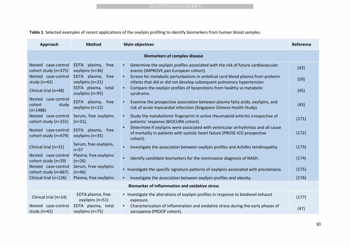
Recent studies addressing one of these goals are presented in Table 1. 111
Of note, the study design (i.e. power, participant selection and matching), the analytical choices 112
(free or total, plasma or serum), and the metabolic coverage of the MS-method applied (number and 113
variety of oxylipins) are very different between the studies and more importantly these parameters 114
may have significant consequences on the relevance and robustness of the biomarkers identified. 115
While the natural variance and reference value of oxylipin levels in healthy humans is not clinically 116
established, recent results show that the mean coeifficient of variation (CV) of oxylipins in the plasma 117
of clinically healthy human can reach 87% and the magnitude of variance depends on the type and 118
concentration of oxylipin considered (42) (Figure 3). This high variance highlights the need for large 119
clinical cohorts of well-selected and matched participants to properly power studies to identify 120
discriminant oxylipins. However, while the absolute concentration of individual oxylipins is likely 121
important, shifts in oxylipin relative abundance or pattern may hold great potential as complex 122
disease biomarkers. Therefore, as assays increase their metabolic coverage of representative 123
oxylipins, they will provide a higher resolution pattern to be considered for biomarker discovery. 124
In the field of disease biomarkers, most studies applying oxylipin profiling are related to 125
cardiometabolic disorders. For instance, in a recent nested case-control cohort study involving 175 126
cardiovascular cases and 172 controls (matched for recruitment center, age, sex, diabetes status, 127
MANUSCRIP
T
ACCEPTED
ACCEPTED MANUSCRIPT
7
insulin use, statin use, and smoking) and covering 36 free oxylipins, one factor containing 10 free 128
fatty acids and 19 oxylipins was significantly associated with cardiovascular events (42). Of note, this 129
association was only observed in non-diabetic patients highlighting the need for patient stratification 130
in clinical investigation. Moreover, linoleic-acid derived epoxides, alcohols and ketones (i.e. EpOMEs, 131
9HODE, 13-HODE and 13-KODE) were the metabolites most strongly associated with change in inter-132
adventitia common carotid artery diameter over time. In a Chinese population, a large nested case-133
control cohort study involving 744 incident acute myocardial infarction cases and 744 controls 134
matched for gender, dialect group, date of birth, date of recruitment and date of blood collection 135
failed to detect significant association between free oxylipin clusters and acute myocardial infarction 136
(besides TXB2 which may reflect sample collection, processing or storage) (43). Interestingly, the 137
authors suggested that examining oxylipins within the esterified pool of lipoprotein particules rather 138
than free oxylipins could provide better predictive biomarkers of coronary heart disease. This lays 139
emphasis on this critical point when focusing on oxylipins as potential disease biomarkers. Although 140
the origin and role of most free and almost all esterified oxylipins are not well understood, assessing 141
the esterified pool could be more relevant in a context of biomarker discovery for three main 142
reasons: (i) it represents the major portion of circulating oxylipins (especially epoxy- and hydroxy-143
PUFA); (ii) it is known to be biologically active (44); and (iii) it fluctuates as a function of lipoprotein 144
particle concentrations and composition (45), which is of importance since other factors associated 145
with such lipidomic fluctuations are known disease risk factors (e.g. hypertriglyceridemia, cholesterol 146
distributions). Moreover, one can expect (whereas this has not been assessed) that the esterified 147
oxylipins are more stable and therefore less affected by sample collection, processing or storage. 148
Another common application for the targeted metabolomics of oxylipins is to generate an 149
integrative assessment of the inflammatory and oxidative stress status of an individual. The utility of 150
such data is also enhanced by broad metabolic coverage to include complementary and opposing 151
oxylipins derived from all pathways and multiple substrates to allow relevant biological 152
interpretation. Inflammation and oxidative stress are the drivers for the onset of many diseases and 153
MANUSCRIP
T
ACCEPTED
ACCEPTED MANUSCRIPT
8
assessing them accurately is therefore crucial in clinical research. Inflammatory status is usually 154
quantified by markers like C-reactive protein (CRP), interleukin 1, TNF-α, and fibrinogen while the 155
non-enzymatically formed isoprostanes are considered to be the “gold standard” biomarkers of 156
endogenous lipid peroxidation and oxidative stress (46). However, we recently observed in patients 157
in the early phases of sarcopenia that the classic inflammatory biomarkers do not identify subtle 158
differences in inflammatory and oxidative stress status (47). Indeed, although CRP and F2-159
isoprostanes were similar in patients with different muscle status, several oxylipins (e.g. 15-HETE, 5-160
HEPE, 9-HETE, 9-HEPE and 14-HDHA) demonstrated subtle differences in inflammation and oxidative 161
stress providing a better characterization of the early phases of sarcopenia. 162
Targeted metabolomics of circulating oxylipins has also been widely used to monitor and 163
subsequently understand the effect of diet (mainly omega-3 fatty acids) or drugs (e.g. anti-164
inflammatory drugs) on health. The impacts of these two environmental factors on oxylipin patterns 165
will be detailed later (see section 4.1), but important observations arised from studies presented in 166
Table 1. The first is related to the variability of response which is systematicaly reported by the 167
authors, depending for example on the basal status of omega-3 fatty acids (48, 49), participant 168
health status (45), age and sex (50). The second observation refers to the necessity to assess all 169
pathways of oxylipin biosynthesis to identifiy unexpected effects of treatments (51). As with the 170
influence of basal omega-3 fatty acid status on oxylipin responses to fish oil consumption (52), 171
examination of response dynamics through the use of regression analysis can actually take advantage 172
of such variance to provide new information. Similarly, if unique response phenotypes are observed 173
within an experimental population, it may be important to develop new hypotheses surrounding the 174
associations of oxylipins and health risks to truely understand the utility of these potential 175
biomarkers in clinical research. 176
MS-based targeted metabolomics of oxylipins represents a very interesting tool for various 177
applications in clinical research, but the impact of analytical and biological parameters on the 178
MANUSCRIP
T
ACCEPTED
ACCEPTED MANUSCRIPT
9
variability of oxylipin patterns needs further understanding to avoid inappropriate study design and 179
subsequent biased biological interpretation. Moreover, expansive oxylipin coverage will increase our 180
ability to interpret the interactions and cross-talk among the various biosynthetic routes and 181
substratedependent metabolites, informing our understanding and possible treatment of 182
dysoxylipinemic diseases. 183
3. Analytical variability linked to the MS-based profiling of circulating oxylipins. 184
To enhance our understanding of the physiological roles of oxylipins that influence health and 185
disease, high quality data are required from a wide array of biological matrices. Therefore, robust 186
analytical methods with high sensitivity, accuracy and precision, are needed. Due to the complex and 187
interactive nature of lipid mediator signaling, it is now well accepted that the physiological effects of 188
these compounds result from a shift in the overall oxylipin pattern, e.g. from a pro- to an anti-189
inflammatory status (53), rather than from the absolute concentration of individual mediators. Thus, 190
state-of-the-art analytical methods aim to precisely detect as many members of the oxylipin cascades 191
(such as the arachidonic acid cascasde) as possible. This broad, and if possible comprehensive, 192
monitoring of such an endogenous pathway is typically referred to as targeted metabolomics (54). 193
The analysis of oxylipins as a superclass of endogenous compounds presents a number of challenges 194
including a diverse array of compound polarity, stability, and endogenous concentrations that can 195
vary ≥4-orders of magnitude (55, 56). Therefore, to quantify oxylipins with high sensitivity and 196
precision, critical considerations include sample collection and storage, instrument selection, 197
analytical breadth and the biological matrix of concern. Each of these factors will affect method 198
development aspects such as the modes of analyte extraction. 199
3.1 Sample collection procedures – A major source of variability in analysis is the 200
formation/degradation of oxylipins during or after sample collection. Oxylipins, particularly COX-1 201
derived Tx as well as 12-LOX generated hydro(peroxy)-PUFA are formed during blood coagulation 202
(57, 58). Thus, serum has to be regarded as an “ex-vivo” coagulation assay and it is likely that the 203

MANUSCRIP
T
ACCEPTED
ACCEPTED MANUSCRIPT
10
time and temperature for coagulation before spinning and freezing affects the formation of oxylipins 204
(59). The anticoagulant – e.g. ethylenediaminetetraacetic acid (EDTA) or heparin – used for the 205
preparation of plasma has also been shown to modulate the oxylipin pattern (57, 60, 61). Thus, levels 206
between differently generated plasmas may vary considerably and should not be directly compared. 207
EDTA is used in most studies investigating the oxylipin profile (Table 1). Best practice is to centrifuge 208
and store samples immediately after collection and define minimal/maximal duration and conditions 209
for transitory storage. Even short periods of storage (1-2 h) of plasma at room temperature or in the 210
refrigerator lead to ex vivo changes in the oxylipin profiles (57 , 60). For example, Figure 4 shows the 211
massive variability induced by differences in the handling of whole blood during the first two hours 212
post-blood collection at different temperatures prior to plasma isolation. Although the direct 213
addition of methanol (57 , 60) or additives (inhibitors or antioxidants) might have beneficial effects 214
on the stability of analytes. However, these procedures are seldom feasible in clinical settings, since 215
commercially available collection tubes containing such materials are not currently available. Similar 216
considerations are needed in the standardization of post-coagulation freezing delays when serum 217
analyses are considered experimentally relevant (59). Serial sampling from indwelling catheters 218
offers unique challenges, and the process of catheter maintenance must be carefully chosen. If 219
heparin is used for catheter maintenance, or in association with other medical interventions (e.g. 220
heart lung machine action) large increases in circulating non-esterified oxylipins are observed (61 , 221
62, 63). Although heparin stabilization of lipoprotein lipase can be reversed by the administration of 222
protamine sulfate, great care should be taken when analyzing and interpreting such samples due to 223
the effect heparin can have on the oxylipin profile in these settings. 224
These data illustrate the importance of standardized procedures and protocols in clinical 225
studies to reduce sample collection induced variability in oxylipin concentrations. Regardless, the 226
analysis of blood samples from cohort studies collected with non-documented and/or variable 227
sample conditions may be valuable to help define the physiological role of oxylipins. However, 228
researchers should consider that such data sets may have higher than expected variance and 229
MANUSCRIP
T
ACCEPTED
ACCEPTED MANUSCRIPT
11
carefully consider interpretation of data pertaining to metabolites with known instabilities. If future 230
studies can identify robust markers of poor sample handling, these may provide non-biased tools for 231
the exclusion of samples with evidence of significant ex-vivo changes in oxylipin profiles, increasing 232
the utility of such studies. In at least 3 studies to date we have found that greatly exaggerated 233
concentrations of 9-HETE and the linoleate-derived trihydroxy metabolites (TriHOMEs) may be such 234
potential markers of poor sample handling (unpublished data). Moreover, in order to correctly 235
interpret the results, quantitative data on the variability induced by transitory storage at each step of 236
plasma generation prior storage at -80°C is urgently needed. 237
3.2 Sample storage procedures – Variability of oxylipin levels is also induced by storage time 238
and conditions. In a recent article, Giera and coworkers show an impressive data set demonstrating 239
that only 4 weeks storage at -20°C leads to artificial oxylipin formation (57). This is consistent with 240
earlier reports showing that short term storage at 4°C as well as freeze/thaw cycling massively 241
influence oxylipin patterns (60). Interestingly, Giera and coworkers also show that the oxylipin 242
pattern in EDTA plasma is not stable when stored at -80°C (57). For most of the oxylipins, small 243
changes were observed over the time span of one year. However, two critical oxylipins, i.e. TxB2 and 244
12-HETE, were found to increase (2-20 fold and 1.5-15 fold, respectively) under certain conditions 245
(without methanol or butylated hydroxytoluene (BHT, radical scavenger) addition). As these 246
metabolites are halmark indicators of platelet degranulation (21, 64), for long term storage, the 247
preparation of platelet depleated plasma may be advantageous. Taking into account that clinical 248
samples are stored under these conditions, the data for TxB2 and 12-HETE should be evaluated with 249
caution in samples which have been stored over a long period, e.g. samples from prospective cohort 250
studies. In our hands, the oxylipin pattern in human plasma is stable during storage at -80°C for 2.5 251
years as shown for selected oxylipins in Figure 5. Though this data is only obtained over a period of 252
2.5 years from a pool of well-prepared EDTA plasma from healthy subjects, it implies that oxylipins 253
can be evaluated in biological samples that have been stored at -80°C for many years. Overall, these 254

MANUSCRIP
T
ACCEPTED
ACCEPTED MANUSCRIPT
12
data demonstrates the importance of carefully controlling all aspects of sample collection and 255
storage to minimize oxylipin variability. 256
3.3 Sample preparation approach - In general, oxylipins occur in plasma esterified into 257
complex lipids such as phospholipids, triglycerides and cholesterylesters (>90% of oxylipins (hydroxy-258
PUFA and epoxy-PUFA)), or as free oxylipins, i.e. in their non-esterified form. Esterified oxylipins are 259
commonly quantified in a sum parameter consisting of esterified and free oxylipins (total oxylipins) 260
following hydrolysis by saponification. It should be noted that alkaline conditions destroy the β-261
hydroxy-keto PG (e.g. PGEs, PGDs) and Tx, as well as cysteinyl leukotrienes and ketones but not the 262
β-hydroxy-alcohols (PGFs) (65). Reported conditions for saponification vary using e.g. potassium 263
hydroxide (0.2-0.5 M in sample (66, 67), sodium hydroxide (0.5-3.75 M in sample (35, 68-70)) or 264
sodium bicarbonate (0.1 M in sample (71)). Temperature and duration of hydrolysis range from 4-265
90°C for 20 min to 18 h (35, 66-71). Moreover, to promote efficient hydrolysis of complex lipids with 266
low solubility including triglycerides and cholesterylesters, transesterification of lipids in methanolic 267
sodium hydroxide in the presence of lipidclass surrogates (i.e. phospholipids, triglycerides, 268
cholesterylesters, and free fatty acids), followed by hydrolysis using water has been established (65). 269
In some protocols an initial liquid-liquid extraction (e.g. chloroform/methanol or 270
cyclohexane/isopropanol/ammonium acetate), or protein precipitation with organic solvents has 271
been used to extract the lipids from the biological sample before hydrolysis (65, 66, 68, 70, 71). 272
These different strategies may influence the amount of oxylipins isolated and liberated during 273
hydrolysis and thus directly affect apparent (total) oxylipin concentrations resulting in variations in 274
the reported concentrations. However, only a few reports data on method optimization are 275
presented – as in (67) – and thus so far, the effects of different saponification strategies has not 276
been systematically evaluated. 277
For analysis of free oxylipins in biological samples, solid phase extraction (SPE) is the most 278
commonly applied technique today, and SPE materials with different retention mechanisms as well 279
MANUSCRIP
T
ACCEPTED
ACCEPTED MANUSCRIPT
13
as different elution solvents are used (72-75) and have been summarized and compared in (76). SPE 280
allows relative enrichement of analytes from larger sample sizes to lower detection limits. Protein 281
precipitation (57 , 77) and liquid-liquid-extraction (78 , 79) procedures have also been reported. 282
While counterintuitive, the dilution of samples by protein precipitation procedures can effectively 283
lower detection limits by reducing ion suppression/enhancement influences of co-extracted 284
metabolites. In the SPE protocols, sample pre-treatment strategies have to be optimized for each 285
protocol and include protein precipitation followed by dilution of the samples with buffer – often 286
coupled to pH adjustment (e.g. using anion-exchange cartridge material) – or acidification of 287
samples. Following elution with appropriate organic solvents, samples are reduced and reconstituted 288
to achieve concentration of analytes of up to 10-fold (compared to the plasma sample). Differing SPE 289
protocols are most likely one of the most important factors contributing to the high variability in the 290
concentrations of oxylipins in biological samples, such as human plasma of healthy individuals. In a 291
direct comparison of the SPE protocols using the same instrumental setup and the same set of 292
samples (76) it has been shown that apparent recoveries of internal standards added to the samples 293
at the beginning of preparation (i.e. analytical surrogates) varied significantly between protocols. This 294
could be explained by differences in analyte extraction efficiency and/or differential removal of ion 295
suppressing matrix components, such as phospholipids (Figure 6). 296
Matrix induced ion suppression/enhancement is a key problem when using electrospray 297
ionization-mass spectrometry (ESI-MS) for quantitative analysis in biological matrices (80). Ion 298
suppression can be particularly problematic in targeted oxylipin metabolomics since only a few 299
isotopically labeled internal standards are used for a large number of oxylipins eluting at different 300
retention times. In this context, it has to be kept in mind that matrix differences between individual 301
samples such as different human plasma samples (i.e. healthy vs disease, fasting vs postprandial, 302
normolipidemic vs hyperlipidemic) may also increase variability in results, and assessing the 303
consistency of suppression across samples within any single study should be considered. Moreover, 304
there is an optimum balance between sample extraction and final concentration that can vary by 305
MANUSCRIP
T
ACCEPTED
ACCEPTED MANUSCRIPT
14
sample matrix (e.g. adipose vs liver vs plasma) and may be influenced by the nature of sample 306
extraction and the total lipid levels of the tissues. 307
Hence, a carefully optimized and well-characterized extraction protocol leading to 308
reproducible analyte concentrations in the analyzed matrix is of the utmost importance for the 309
production of biologically meaningful oxylipin data. If ion suppression is ignored – or not 310
characterized – apparent changes in oxylipin concentration may result from differences in the matrix 311
and not changes in the associated metabolic cascades. Therefore, ideal methods will limit the 312
observable matrix-dependent ionization effects across the entire chromatographic run. 313
3.4 Analytical hardware - While gas liquid chromatography-MS (GLC-MS) methods were 314
commonly used in the early days of oxylipin quantification, these have been almost completely 315
replaced by (ultra) high performance liquid chromatography-MS ((U)HPLC-MS) methods on reversed 316
phase (RP) columns, which can cover >170 analytes from up to six PUFAs precursors including 317
linoleate (C18:2n6), alpha-linolenate (C18:3n3), dihomo-gamma-linolenate (C20:3n6), arachidonate 318
(C20:4n6), eicosapentaenoate (C20:5n3) and docosahexaenoate (C22:6n3) (Table 2). Rare analyses 319
of oxylipins from adrenic acid (C22:4n6) (4, 81, 82) or n3- or n6-docosapentaenoic acid (DPA, 320
C22:5n3/n6) (4, 83), and very long chain elovanoids (84) have also been reported. Recent articles 321
describe the use of super-critcial-fluid chromatography (85). Because oxylipins in their non-esterified 322
form bear the acidic carboxy-moiety of the polyunsaturated fatty acid precursor, ionization is 323
dominantly carried out by ESI in negative ion mode. Only in a few applications different techniques 324
are used, e.g. atmospheric pressure chemical ionization following derivatization (86) (Table 2). 325
Today, oxylipins are most often quantified using triple quadrupole MS (QqQ-MS) (Table 2). 326
This type of analyzer is well suited for the detection of oxylipins because of its high ion transmission 327
and matrix independence (compared to Paul traps or linear ion traps). In addition, these systems are 328
generally coupled with either photomultipliers or electron multipliers, providing linear detector 329
responses over concentration ranges of 5-6 orders of magnitude. The latter is crucial for oxylipin 330
MANUSCRIP
T
ACCEPTED
ACCEPTED MANUSCRIPT
15
quantification because levels of analytes differ in the very same sample by well over three orders of 331
magnitude, e.g. in plasma of healthy humans from 0.050 to 11 nM (11,12-DiHETE and 15,16-DiHODE 332
(41)) or in healthy mouse plasma (following feeding with omega-3 fatty acids) from 0.064 to 495 nM 333
for PGE2 and 12-HEPE (56) (data obtained from the mean reported concentraton within one group). 334
Pathophysiological conditions induce strong changes where a linear range of at least four orders of 335
magnitude is needed (45, 55). A key feature of QqQ-MS is that oxylipins are detected following 336
fragmentation. Given that oxylipins are formed by oxygenation of unsaturated bounds within the 337
PUFA backbone, their (exact) molecular masses cannot distinguish regioisomeric structures. Thus, 338
only fragmentation makes a specific MS-detection of isobaric compounds possible. It is important to 339
remember, however, that oxylipin regioisomers can often generate non-specific fragments, and care 340
must be taken in both quantitative ion selection and chromatographic resolution of certain spieces 341
(87). However, QqQ-MS only allows the analysis of pre-selected species, i.e. “targeted 342
metabolomics”, and today analyses of 50-200 oxylipins are common (Table 2). Using time of flight 343
(TOF) or orbitrap mass analyzers equipped with a quadrupole collision cell (e.g. qTOF or Q Exactive™) 344
allow acquisition of full (fragment) spectra making simultaneous targeted and non-targeted oxylipin 345
metabolomics/lipidomics possible. To date, few applications with these instruments have been 346
published, most being qualitative (88 , 89). At present, using two methods for non-targeted 347
(orbitrap) and targeted (QqQ) metabolomics, as recently described by Wheelock and coworkers (42), 348
is the most promising approach for gaining both, lipidomics data and quantitative information on 349
oxylipins. 350
3.5. Isomeric and enantiomeric complexity - Due to the complexity of oxylipin isomer 351
profiles present in biological samples, careful selection of collision induced mass transitions along 352
with high chromatographic resolution and stable retention times are required to yield the selectivity 353
and sensitivity needed to correctly identify and quantify oxylipins. Thus, effort is required for 354
optimizing both the mass spectrometric parameters and the chromatographic method, to avoid peak 355
misalignment and/or mass spectrometric overlap of secondary ions which can lead to the generation 356
MANUSCRIP
T
ACCEPTED
ACCEPTED MANUSCRIPT
16
of inaccurate data leading to false conclusions. The importance of selective mass transitions is 357
illustrated by Figure 7. Two transitions of the hydroxylated-DHA 17-HDHA were used to quantify the 358
analyte in human plasma and rat liver. In both matrices, the selective transition yields clean 359
chromatograms with only one peak while using the unspecific transition results in several peaks. 360
These interfering peaks hamper peak integration and thus might lead to variations in apparent 361
concentrations. 362
In addition to selective MS detection, the large number of isomeric oxylipins require 363
excellent chromatographic separation. Currently, separation is typically carried out by reversed 364
phase chromatography often using columns filled with sub-2-µm or fused core particles (Table 2). 365
The fragment spectra of several oxylipins, particularly regio- and stereoisomers are often too similar 366
to be resolved spectrally. An example of such a critical separation pair, i.e. two oxylipins with the 367
same precursor mass giving rise to similar fragment spectra, are the hydroxylated-AA metabolites 9-368
HETE and 12-HETE (90). Similarly the most sensitive transition for 8(9)EpETrE also shows signals for 369
11(12)EpETrE (87) making these two epoxy-AAs another critical pair requiring chromatographic 370
baseline separation. Even using optimized MS-transitions and optimal (state of the art) reversed-371
phase liquid chromatography, isobaric interferences from the sample matrix could overlap with the 372
oxylipin peak. This is particularly a challenge in the detection of multiple-hydroxylated PUFA such as 373
specialized pro-resolving mediators (SPM), with a large number of possible regio- and stereo-374
isomers. Well characterized examples for this, which – if not recognized – lead to strong variations in 375
the apparent concentration (i.e. poor accuracy), are the interference between LTB4 and 5(S),12(S)-376
diHETE (91) and different isobaric compounds mimicking lipoxin A4 (LXA4) (79). Similarly, interfering 377
peaks for Maresin 1 (MaR1), and the protectins PD1 and PDX occur in improperly stored plasma (57). 378
Complicating matters, trans-double bond containing oxylipins also exist, produced by either the 379
metabolism of trans-fatty acids or the rearrangement of formed oxylipins (92-94). 380

MANUSCRIP
T
ACCEPTED
ACCEPTED MANUSCRIPT
17
Another significant consideration in oxylipin analyses is the chirality of the detected species. 381
In particular, independent detection and quantification of enantiomers can aid in interpretation of 382
results in a biologically relevant context. For instance, a variety of oxylipins can be produced by the 383
direct interaction of reactive oxygen species with unsaturated lipids, with the resulting products 384
having no enantiomeric enrichment, while enzymatic reactions generally have high degrees of 385
enantiomeric enrichment (95-102). Notably, the clotting process yields dramatic shifts in oxylipin 386
enantiomeric patterns (99). In addition, some enzymes yield the opposite enantiomers of the same 387
regioisomers, for example 12(R)-HETE production by CYPs and psoriatic lipoxygenases, and 12(S)-388
HETE production by the ALOX12 gene product (103, 104). Historically, these techniques have relied 389
upon normal phase chromatography of pentaflurobenzyl ester derivatives, however new reverse 390
phase chiral materials are opening the door to routine application of these methods in the future. 391
(100-102). Recently developed approaches couple a non-chiral reversed phase-column with a chiral 392
amylose-based column that can be used with reversed phase solvents (101, 105). These were used 393
e.g. to successfully unveil the stereospecific formation/degradation of epoxy-PUFA and hydroy-PUFA 394
(101) as well as the characterizing the configuration of SPMs (105). 395
3.6. Accessibility of analytical calibrants and internal standards - To accurately quantify 396
oxylipins, a calibration with authentic reference compounds is required, typically carried out by 397
external calibration with internal standards introduced to the samples, thus allowing correction for 398
methodological variance (e.g. sample loss, ion suppression). Currently, several hundred non-399
esterified oxylipins are commercially available through a small handful of companies. However, 400
oxylipins esterified into complex lipids including triacylglycerides, phospholipids, cholesterylesters 401
and appropriate isotopically labeled analogs are essentially unavailable through the commercial 402
market place and thus only available to those scientists with the skill and resources to synthesize and 403
purify them. With respect to the available standards, varying purities of these reference compounds 404
have been reported (106), which is likely one cause of the massive differences in reported 405
concentrations (52, 107). The limited availability of isotopically labeled structural analogs, and their 406
MANUSCRIP
T
ACCEPTED
ACCEPTED MANUSCRIPT
18
non-uniform application in these analyses likely exacerbates this problem. Recently, verified-407
concentration standards, i.e. quantitative grade standards, became available for selected 408
compounds, such as MaxSpecTM
(Cayman Chemicals, Ann Abor, MI, USA) and are supplied with 409
batch-specific certificates of analysis. However, up to now only a few standards are available and we 410
recently suggested a strategy to quantify non-verified material with these few verified-concentration 411
standards (106). 412
Despite optimizing and standardizing sample collection, preparation and analysis, inter- and 413
intra-day variation will remain. Recent studies show that an intra-day variation of <20% can be 414
achieved for most analytes in human plasma when present at concentrations above the lower limit 415
of quantification (LLOQ) (41). However, variability increases as analyte concentrations approache the 416
LLOQ. Therefore, methodological robustness should be considered when interpreting results, 417
particularly when the null hypothesis is not rejected. Such robustness factors should include method 418
variability along with other validation criteria such as internal standard recoveries, detected matrix 419
effects, sensitivity (i.e. LLOQs), and calibration stability and accuracy. To this end, we would 420
encourage the routine reporting of these robustness factors, which are rarely reported outside of 421
method development studies at this time. 422
When comparing oxylipin concentrations from different laboratories (Table 2), it is important to 423
consider the analytical choices within each method. Round-robin trials comparing methods 424
established and routinely implemented in different laboratories will be the key to identifying 425
strengths, weaknesses, and intercomparability of different approaches. Ultimately, such efforts will 426
allow the implementation of internationally agreed upon sample preparation procedures and/or a 427
routine performance standard that will allow the highest confidence in these important data sets, 428
and promote the direct comparison of studies performed at multiple locations. Ongoing effort in 429
different national projects e.g. German Research Foundation 41696725 430
(http://gepris.dfg.de/gepris/projekt/4169672511) or international projects such as the EU JPI-HDHL 431
MANUSCRIP
T
ACCEPTED
ACCEPTED MANUSCRIPT
19
OXYGENATE project (https://www6.inra.fr/jpi-hdhl-biomarkers-oxygenate/) aim to address these 432
questions. However, until these procedures have been established a comparison of the (absolute) 433
reported concentration of oxylipins should be performed carefully, and only the relative change 434
within the studies seems to be a measure which can be robustly compared. 435
Accurate analyte quantification requires signal to noise ratios to exceed a defined threshold. 436
This is comprehensively discussed in internationally accepted guidelines on method validation 437
(European Medicines Agency, US food and Drug Administration, US Environmental Protection 438
Agency, etc.). With a signal/noise ratio of at least 5, the typically reported LLOQ for oxylipins on 439
state-of-the-art QqQ instruments is ~0.5 to 50 fmol on column (Table 2). The LLOQ varies about 440
10-fold between different oxylipins based on their ionization and fragmentation behavior. Proper 441
definition of analyte specific LLOQs in the methods used is crucial, because it impacts the breadth 442
and number of oxylipins that can be reported in a biological sample. Reporting levels below the 443
LLOQ need to be identified as they are prone to high variation and inacceptable accuracy. It should 444
be stressed, however, that while measures below the limit of detection (LOD) are not an indication of 445
the presence or absence of a compound, and those below the LLOQ (above the LOD) are subject to 446
higher false positive/false negative rates (108), they may have statistical value in maintaining the 447
number of observations across all analytes, and may be better than removal and imputation or 448
arbitrary replacement by the LLOQ, procedures that should be implemented with great care (109-449
111). Taking an injection volume of 5 to 10 µL and an average molecular mass of 300 Da into 450
account, as a rule of the thumb the LLOQ for oxylipins in the injected solution is between 0.1 and 10 451
nmol/L (0.3 to30 ng/mL). The detection of lower concentration of oxylipins becomes possible by pre-452
concentration during sample preparation if a concurent increase in ion suppression is not observed. 453
4. Inter-individual variability of plasma oxylipin patterns. 454
As illustrated in Figure 3, plasma concentrations of oxylipins are highly variable in clinically healthy 455
humans and each oxylipin has its own variability. Both, environmental and intrinsic factors can 456
MANUSCRIP
T
ACCEPTED
ACCEPTED MANUSCRIPT
20
contribute to variance in measured oxylipin patterns, which are independent of analytical variability. 457
Such natural variance of systemic oxylipins can stem from changes in either substrate availability or 458
modulating the expression and/or activity of responsible biosynthetic enzymes. 459
4.1 Influence of environmental factors on oxylipin patterns - Oxylipin patterns reflect the 460
integration of many factors, most notably the availability of unsaturated fatty acid precursors, the 461
relative abundance and activity of specific enzymes, and the degree of oxidative stress present in the 462
individual at the time of sample collection. Each of these factors is influenced by the environment. 463
Diet constitutes the single largest environmental factor that modulates both the type and dose 464
of PUFAs available for oxylipin biosynthesis. Oxylipins can be produced either directly in phospholipid 465
membranes (e.g. non-enzymatically or via 15-LOX pathways) (112, 113), however for most enzymes, 466
the esterified PUFAs have to be released from membranes by the action of phospholipase A2 (PLA2) 467
(114). In most human tissues and circulating cells, the omega-6 PUFAs (i.e. arachidonic acid and 468
linoleic acid) are the most abundant PUFAs stored in membranes (115, 116) and therefore the main 469
substrates for oxylipin biosynthesis. However, changing dietary PUFAs (type and dose) can readily 470
modify the fatty acid composition of cellular lipids. This has been well demonstrated with increased 471
intake of long chain omega-3 PUFAs (i.e. EPA and DHA) from fish or fish oil based supplements, which 472
leads to dose- and time-dependent incorporation of both fatty acids in blood lipids, blood cell lipids, 473
and many tissue pools (49, 117, 118) (and see review (119) for more details). The incorportion of long 474
chain omega-3 PUFAs is usually accompanied by a decrease in omega-6 PUFAs including linoleic, 475
dihomo-γ-linolenic and arachidonic acids (119), but the rate and magnitude of these changes are 476
dependent on an individuals basal omega-3 status (49). Changing the relative proportion of omega-3 477
and omega-6 PUFAs in membranes influences oxylipin patterns because of both shifts in substrate 478
availabilityand variable substrate affinity with the different enzymes. For instance, cytosolic PLA2 has 479
a strong affinity for AA and EPA but very weak interaction with DHA, which is preferentially 480
hydrolyzed by the calcium-independent PLA2 (iPLA2, Type VIA) (120). A difference in substrate 481
MANUSCRIP
T
ACCEPTED
ACCEPTED MANUSCRIPT
21
affinity was also reported for COX-2, which preferentially oxidizes AA while having a much weaker 482
interaction with EPA, and almost no affinity for DHA (121). Concerning the CYP pathway, the affinitiy 483
of CYP isoforms with PUFAs depends on the carbon chain length as well as the type and number of 484
unsaturations. Interestingly, double bonds in the omega-3 position offer a preferential epoxidation 485
site for many CYPs including CYP1A, CYP2C, CYP2J and CYP2E, while CYP4A also predominantly 486
hydroxylates the terminal methyl-group of EPA and DHA (see (122) for more details). Consistently, 487
Fisher et al. demonstrated that CYP dependent-epoxide formation from EPA and DHA were 8.6-fold 488
and 2.2-fold more efficient than AA, respectively (123). Basically, in healthy subjects, long chain 489
omega-3 PUFA supplementation can elevate total EPA and DHA oxylipins at the expense of total AA 490
oxylipins (45, 49 , 52). Similar, but less consistant results have been reported for free oxylipins (52). 491
Relative changes in EPA are generally more pronounced. Crossvalidating the effects of omega-3 PUFA 492
supplementation on the different pathways of biosynthesis between different studies produces no 493
systematic trends (52). Several authors, however, reported predominant changes in the CYP 494
pathways (123) consistent with a higher affinity of CYP enzymes with omega-3 PUFAs. Recent data 495
demonstrate that the plasma levels of hydroxy-PUFA, epoxy-PUFA and dihydroxy-PUFA derived from 496
n3-PUFA increase in a linear fashion with the intake of n3-PUFA (124). 497
While PUFA instake can alter substrate availability, other factors can alter oxylipin patterns 498
through modification of the expression and/or activity of enzymes of the oxylipin pathways. The first 499
environmental factor to consider when interpreting oxylipin profiles should be the use of drugs that 500
specifically target oxylipin pathways. These notably include non-steroidal anti-inflammatory drugs 501
(NSAIDs), which act via the inhibition of COX isoenzymes and are one of the most widely used drugs 502
in global pharmacological management of acute and chronic pain (125). Other examples are 5-LOX 503
inhibitors (e.g. Zileuton) in the treatment of allergy and asthma, or antithrombotic agents (e.g. 504
Dazoxiben, Camonagrel, Picotamide) that inhibit Tx synthases. Of note, even for these widely used 505
and well characterized drugs, responses are complex and impacts on oxylipin patterns are often 506
broader than expected (51). For instance, the use of celecoxib, a specific COX-2 inhibitor, in patients 507
MANUSCRIP
T
ACCEPTED
ACCEPTED MANUSCRIPT
22
with colon polyp, was associated with increased levels of CYP- and LOX-derived oxylipins (126), which 508
could be a result of substrate shunting into these alternative pathways. Other drugs not specifically 509
designed to modulate enzymes of the oxylipin pathways can also influence oxylipin patterns. This is 510
notably the case of statins, inhibitors of HMG-CoA reductase, which are widely used in the treatment 511
of hypercholesterolemia. Statins have been shown to increase the production of COX-2-derived 512
oxylipins in various tissues via S-nitrosylation of COX-2 (127-130), while only modest changes in 5-513
LOX metabolites appeared in the non-esterified plasma pool with high-dose simvastatin (131). 514
Phytochemicals in medicinal herbs or plant-based foods (i.e. alkaloids, polyphenols, terpenoids and 515
plant-derived lipids) also influence the activity of oxylipin enzymes. Among them, polyphenols are 516
representative and nutritionally significant coumpounds investigated for the last 30 years. Numerous 517
in vitro cell-based or enzyme-based assays support the inhibitory effects of various dietary 518
polyphenols including flavonoids, curcumin, stilbenes and secoiridoids on PLA2, COX and 5-LOX and 519
CYP gene expression and/or activity (132-137). Of note, some of this in vitro evidence should be 520
interpreted with caution since they often arise from experiments conducted using native compounds 521
rather than circulating forms of polyphenols and at high doses not achievable through diet. However, 522
short term or long term controlled dietary interventions have reported significant modulation of 523
systemic COX- and LOX-derived oxylipin levels following intake of polyphenol-rich foods in healthy 524
humans (138, 139) even though interindividual variability sometimes hamper such demonstration 525
(140). Other general life style habits such as smoking (141, 142), alcohol consumption (143) or 526
physical activity (144, 145) can also influence systemic oxylipin patterns through an increased 527
inflammatory state caused by the combined effects of inhaled mediators and/or oxidative stress. 528
4.2 Influence of intrinsic factors on oxylipin patterns - Intrinsic factors that can modulate 529
oxylipin profiles include such variables as age, sex, and genetic polymorphisms, all of which have the 530
potential to modulate biosynthetic enzyme expression and/or activity. Various disease states such as 531
infections or cardiometabolic disturbances (e.g. obesity, type 2 diabetes) also influence oxylipin 532
biosynthesis but this will not be discussed here. 533
MANUSCRIP
T
ACCEPTED
ACCEPTED MANUSCRIPT
23
While not comprehensively investigated, several independent studies in both animal models 534
and humans have reported a significant influence of age and sex on oxylipin levels (see (146, 147) for 535
recent compilation of the literature). Aging is generally associated with increased levels of COX-536
derived oxylipins including PGE2, 6-keto-PGF1a, TxB2 and their stable metabolites 2,3-dinor TxB2 and 537
2,3-dinor-6-keto-PGF1α. Levels of pro-inflammatory leukotrienes (e.g. LTB4, LTC4) and hydroxy fatty 538
acids (e.g. 5-HETE) derived from the LOX-pathway are also higher in biological fluids or tissues from 539
aged individuals compared to younger ones. On the contrary, pro-resolving LOX-derived oxylipins 540
(e.g. LXA4, LXB4, Mar1, RvD1) are generally found in lower abundance in aged individuals with the 541
exception of PD1, which was reported to be increased in the brain of aged mice (146). Other oxylipins 542
produced by sequential LOX and CYP enzymes or autoxidation of linoleic acid (98 , 148), namely 543
9,10,13- and 9,12,13-TriHOME, were also higher in plasma of old healthy men and women in 544
comparison with younger controls (146). The effects of aging on CYP-derived oxylipins in humans are 545
more scarce and contradictory (149). Most studies have investigated the expression or activity of 546
CYPs at the hepatic level, but the difficulty in accessing human liver samples hampered the 547
demonstration of a significant effect of aging on human CYPs and its consequences on the production 548
of CYP-derived oxylipins. According to the free radical theory of aging (150) and knowing the role of 549
reactive oxygen species (151) and lipid peroxides (3) in the regulation of the expression/activity of 550
COX and LOX, oxidative stress is likely a contributing factor to the effect of aging on oxylipin patterns. 551
Modulation of oxylipin biosynthesis by sex was reported more than 50 years ago (152). 552
Sexual dimorphism of oxylipin pathways depends on the type of tissue investigated as well as the 553
hormonal status of individuals, which is itself age related. However, males generally have higher 554
levels of PGE2 and Tx derived from the COX pathway, while females have higher levels of leukotrienes 555
but lower levels of pro-resolving oxylipins, both produced by LOX enzymes (See detail in (147). This is 556
consistent with the higher preponderance of leukotriene-related diseases such as asthma and 557
rheumatoid arthritis in women. Sex is a known factor influencing CYP-dependent drug metabolism 558
(153), but conflicting results were reported in relation to oxylipin metabolism and it is difficult to 559

MANUSCRIP
T
ACCEPTED
ACCEPTED MANUSCRIPT
24
ascertain if gender has a significant influence on the expression/activity of oxylipin generating CYPs 560
(149). However, there are clear sex differences in the levels and distribution of the soluble epoxide 561
hydrolase (sEH), an important epoxy fatty acid converting enzyme (154). It should be noted that, 562
when looking at comprehensive oxylipin profiles, which has been rarely done so far, the overall effect 563
of sex and its interaction with other factors makes interpretation more complicated. Recent studies 564
in rats showed that ~40% of oxylipins (from profiles including 60-71 oxylipins) were influenced by sex 565
and almost all were higher in male rats (155, 156). These studies also showed that sex differences are 566
influenced by diet (e.g. male rats have higher levels of adipose oxylipins when fed a high DHA diet, 567
while females have higher oxylipins when fed a high EPA diet), by the type of precursor PUFA (e.g. rat 568
kidney DHA derived oxylipins are higher in females, while AA derived oxylipins are more often higher 569
in male) and by tissue (e.g. in kidney, DHA oxylipins are influenced by diet and sex whereas no 570
change is observed in liver). Mechanisms underlying the influence of sex on enzymes of the oxylipin 571
biosynthetic pathways logically include a regulation of expression/activity by sex hormones. Their 572
role as modulators of COX pathways remains unclear, with investigations mainly focused on primary 573
enzymes of the pathway (i.e. COX). On the other hand, differences in LOX-derived oxylipins have 574
been directly related to variant androgen levels in men and women (147). This can be the result of a 575
direct modulation of LOX enzymes by sex hormones but other regulatory aspects of the LOX-576
dependent oxylipin formation including interaction with other proteins (e.g. 5-lipoxygenase-577
activating protein or FLAP), subcellular localization, phosphorylation and other factors (e.g. ATP, 578
glyceride, redox tone, Ca2+
) (3) may also be affected by sex and sex hormones. 579
Genes of enzymes involved in the biosynthesis of oxylipins are highly polymorphic, which 580
may contribute to the variability of oxylipin patterns in healthy humans. However, human genetic 581
studies mostly focused on clinical outcomes rather than oxylipin metabolism. Several large studies 582
investigated the associations between the polymorphism of primary oxylipin enzymes (i.e. COX, LOX 583
and CYP) and the risk of diseases involving oxylipins, such as CVD and cancer. For instance, Ross et al. 584
conducted a remarkable study to prospectively explore the association of a COX-2 variant (i.e. 585
MANUSCRIP
T
ACCEPTED
ACCEPTED MANUSCRIPT
25
rs20417) in 49,232 participants. The results showed that rs20417 variant was associated with a 586
reduced risk of major cardiovascular events (157). Common polymorphisms in the 5-LOX pathway 587
including variants of 5-LOX, FLAP, LTA4H and LTC4S were associated with myocardial infarction in a 588
recent case-cohort study conducted in 3000 participants of the Danish Diet, Cancer and Health study 589
(158), and promoter region variants in the ALOX5 have been linked to the efficacy of fish oil 590
modulation on cardiovascular risk (159). Genetic variability of 5-, 12- and 15-LOX enzymes and FLAP 591
may also affect colorectal neoplasia as reported in three US population-based case-control studies of 592
colorectal cancer involving 5,625 subjects (160). CYP metabolites (i.e. EpETrE) having recognized 593
cardioprotective effects (161), associations between CYP and sEH polymorphisms and CVD have been 594
intensively investigated. Even though some studies detected significant associations between several 595
variants of CYP and CVD, contradictory results, probably due to ethnic variability, environmental 596
factors (e.g. smoking) and gender specific effects, have been reported and hamper conclusions of 597
significant associations (162, 163). Associations between sEH polymorphisms and CVD have been 598
investigated in case-cohort samples of the Atherosclerosis Risk in Communities (ARIC) study. In a first 599
sample of 1,336 participants, two common haplotypes with opposing effects showed significant 600
associations with the risk of ischemic stroke in African American subjects (164). In a second study 601
involving 2,065 participants of the ARIC cohort, Caucasians with the K55R polymorphic variant allele 602
of sEH were found to have a higher risk of CAD and an increase in the relative abundance of 603
linoleate-derived diols relative to their epoxide precursors (165). Similarly, the most common 604
epoxygenase gene polymorphism has been associated with a significant decrease of enzymatic 605
activity and consequently EpETrE biosynthesis (162). Other evidence of a polymorphism-dependent 606
effect on oxylipin levels were reported by Ross et al. (157) in which COX-2/rs20417 carriers had 607
significantly lower urinary levels of Tx and prostacyclin compared to non-carriers. Concerning the LOX 608
pathway, common variants generally have no functional consequences and loss-of-function variation 609
of the human LOX genes are rare (global allele frequency <0.1%) (166). Nevertheless, an intervention 610
study conducted in 116 healthy adults reported that 5-LOX gene variants were associated with 611
MANUSCRIP
T
ACCEPTED
ACCEPTED MANUSCRIPT
26
altered production of AA, EPA and DHA derived LOX-metabolites (i.e. hydroxy and oxo-fatty acids but 612
not leukotrienes) and different response to omega-3 (i.e. fish oil) supplementation (159). 613
While poorly studied and being more a concern of intra-individual variability, temporal 614
changes in both plasma and urinary oxylipins have been reported. Temporal changes in plasma 615
oxylipins may be linked to meal induced changes (41), however, circadian rythms in arachidonic acid 616
metabolism have also been reported (167) and circadian effects on other aspects of lipid metabolism 617
are well documented (168). Similarly, urinary CYP-dependent metabolites were found to oscilate 618
independent of meals in free feeding men and women (87). As oxylipins are also important 619
regulators of renal water retention and as urinary but not plasma levels are dramatically influenced 620
by sodium intake and depuration (169, 170), these are also important factors to consider when 621
urinary oxylipin profiling is considered. 622
5. Summary and outlook 623
Oxylipins represent an important regulatory cascade and continuing to unravel this system and 624
its intricate interactions has the potential to offer great value to society. To maximize the utility of 625
this information and accelerate our gain in knowledge, it will be critical (i) to promote our ability to 626
directly compare results generated by independent research teams and (ii) to be able to distinguish 627
abberant oxylipins pattern from natural variance. To do this, sources of variance, both analytical and 628
biological, must be identified, minimized where possible, and considered during data analysis and 629
interpretation. 630
The preanalytical sources of variance are large and best practices along with international 631
standardization in sample collection, storage and preparation should be established. Based on our 632
own understanding of the current literature, we would argue for the use of EDTA plasma and to 633
centrifuge and store samples at -80°C immediately after collection. A first important point to 634
consider for sample preparation is the choice of analyzing total (i.e. free and esterified) or only free 635
oxylipins, as this choice will have important consequences on the oxylipin profiles and subsequent 636
MANUSCRIP
T
ACCEPTED
ACCEPTED MANUSCRIPT
27
biological interpretation. Of note, total oxylipins represent the major portion of circulating oxylipins 637
and may be less affected by sample collection, processing or storage, but their quantitative anaysis 638
currently requires hydrolysis, which destroys several COX-derived oxylipins. Subaliquots of 100 µL of 639
plasma for total (free and esterified) oxylipin analysis or of 500 µL for free oxylipin analysis should be 640
stored at -80°C in methanol pre-cleaned polyethylene tubes until use. This procedure should reduce 641
artifacts associated with freeze/thaw cycling and platelet activation after prolonged storage. 642
Differences in sample preparation procedures (i.e. SPE, liquid-liquid extraction, protein 643
precipitation), the application of analytical internal standard corrections (i.e. which isotope labeled 644
internal standard is being used to correct for which specific analytes) and the purity of calibration 645
standards are likely sources of variance in comparing results from different laboratories. In 646
particular, matrix effects resulting in ion suppression of analytical internal standards that do not 647
coelute with their analytical target may lead to inaccurate, while reproducible results (systemic 648
error). Therefore, researchers should take care in assessing the stability of their internal standard 649
recoveries across all samples within their study. First, they should be sure that ion suppression does 650
not segregate by experimental group and they should report all internal standard recoveries as 651
metadata to the study so that others can assess to what degree corrections for loss/suppression are 652
being applied to the reported data, as this may influence the accuracy and thus global comparability 653
of the results. 654
In terms of analytical variability, in general, modern analytical hardware if well maintained is 655
exceptionally stable. Much more important are the standards used to calibrate these instruments, 656
and control over factors including ion suppression/enhancement. The greatest need in the scientific 657
community are the availability of analytical standards and an increased suite of isotopically labeled 658
analogs, including those which allow the direct quantitative analysis of oxylipins within complex 659
lipids. It would be valuable for the research community to establish priorities for the production of 660
new analytical resources for this field. Moreover, the concentration of all standards used should be 661
MANUSCRIP
T
ACCEPTED
ACCEPTED MANUSCRIPT
28
validated based on concentration-verified standard material. Until certified standards are available 662
for all oxylipins quality assessment strategies as described by Hartung et al. (106) have to be 663
implemented in each analytical lab. 664
Beyond these procedural and analytical factors, environmental and intrinsic factors 665
tremendously contribute to the natural variability of oxylipin profiles. Care should be taken during 666
the analysis of human studies to truely test assumptions of population normality prior to committing 667
to simple mean testing as a primary outcome. While the accumulating literature shows that dietary 668
and pharmacological components together with age, sex and genetics all have the potential to 669
modulate substrate PUFAs and/or enzyme activities to shift oxylipin profiles, the natural variance of 670
each circulating oxylipin has not been established so far. This will be crucial to appropriately power 671
experimental designs and to enhance the identification of reliable and relevant biomarkers of 672
disease. Increasing our knowledge regarding the natural variance of oxylipin biosynthesis is also of 673
great interest in personalized nutrition or medicine. These efforts will provide new understanding 674
regarding the variability of individual’ responses to dietary factors and therapeutics, thus offering 675
insights into diseases susceptibilitythat may allow for patient stratification and personnalization of 676
disease management. Similarly, if unique response phenotypes are observed within an experimental 677
population, it may be important to develop new hypotheses surrounding the associations of oxylipins 678
and health risks to truely understand the utility of these potential biomarkers in clinical research. 679
Therefore, moving forward, if we conciously control analytical variance while embracing 680
biological variance as a research community, we are sure to enhance the impact of our efforts and 681
accelerate our discovery of new knowledge regarding the biological regulation of these important 682
regulatory factors. 683
6. Acknowledment 684
This work was in part supported by the EU JPI-HDHL OXYGENATE project (https://www6.inra.fr/jpi-685
hdhl-biomarkers-oxygenate/). Additional support was provided by USDA Project 2032-51530-686
022-00D and 2032-51530-025-00D. The USDA is an equal opportunity provider and employeer. We 687

MANUSCRIP
T
ACCEPTED
ACCEPTED MANUSCRIPT
29
would like to thank Laura Kutzner, Laura Froehlich, Sarah Reuber and Christopher Millan Hidalgo for 688
their help summarizing the data on reported oxylipin methods (Table 2). 689
690
MANUSCRIP
T
ACCEPTED
ACCEPTED MANUSCRIPT
30
Table 1. Selected examples of recent applications of the oxylipin profiling to identify biomarkers from human blood samples.
Approach Method Main objectives Reference
Biomarkers of complex disease
Nested case-control
cohort study (n=375)
EDTA plasma, free
oxylipins (n=36)
• Determine the oxylipin profiles associated with the risk of future cardiovascular
events (IMPROVE pan European cohort). (42)
Nested case-control
study (n=42)
EDTA plasma, free
oxylipins (n=21)
• Screen for metabolic perturbations in umbilical cord blood plasma from preterm
infants that did or did not develop subsequent pulmonary hypertension. (59)
Clinical trial (n=48) EDTA plasma, total
oxylipins (n=95)
• Compare the oxylipin profiles of lipoproteins from healthy vs metabolic
syndrome. (45)
Nested case-control
cohort study
(n=1488)
EDTA plasma, free
oxylipins (n=12)
• Examine the prospective association between plasma fatty acids, oxylipins, and
risk of acute myocardial infarction (Singapore Chinese Health Study). (43)
Nested case-control
cohort study (n=231)
Serum, free oxylipins
(n=31),
• Study the metabolomic fingerprint in active rheumatoid arthritis irrespective of
patients' response (BiOCURA cohort). (171)
Nested case-control
cohort study (n=479)
EDTA plasma, free
oxylipins (n=35)
• Determine if oxylipins were associated with ventricular arrhythmias and all cause
of mortality in patients with systolic heart failure (PROSE-ICD prospective
cohort).
(172)
Clinical trial (n=31) Serum, free oxylipins,
n=37 • Investigate the association between oxylipin profiles and Achilles tendinopathy. (173)
Nested case-control
cohort study (n=29)
Plasma, free oxylipins
(n=26) • Identify candidate biomarkers for the noninvasive diagnosis of NASH. (174)
Nested case-control
cohort study (n=667)
Serum, Free oxylipins
(n=46) • Investigate the specific signature patterns of oxylipins associated with preclampsia. (175)
Clinical trial (n=126) Plasma, free oxylipins • Investigate the association between oxylipin profiles and obesity. (176)
Biomarker of inflammation and oxidative stress
Clinical trial (n=14) EDTA plasma, free
oxylipins (n=51)
• Investigate the alterations of oxylipin profiles in response to biodiesel exhaust
exposure. (177)
Nested case-control
study (n=42)
EDTA plasma, total
oxylipins (n=75)
• Characterization of inflammation and oxidative stress during the early phases of
sarcopenia (PROOF cohort). (47)

MANUSCRIP
T
ACCEPTED
ACCEPTED MANUSCRIPT
31
Clinical trial (n=42) Plasma, free oxylipins
(n=95)
• Evaluate levels of inflammatory markers and blood oxylipins in obese subjects
before and after weight reduction. (178)
Clinical trial (n=1) EDTA plasma, free
oxylipins (n=37) • Investigate postprandial inflammation through changes of oxylipins profiles. (179)
Clinical trial (n=10) EDAT plasma, total
oxylipins (n=27)
• Characterize oxylipin profile and vascular inflammation following a moderately
high-fat meal in Alzheimer's patients. (180)
Biomarkers of nutrition and drug effects
Dietary intervention
(n=35)
EDTA plasma, free
oxylipins(n=56)
• Determine the impact of long-term treatment with omega-3 fatty acids on plasma
oxylipin patterns in patients with severe hyperlipidemia and CVD on standard
lipid-lowering and cardioprotective medications.
(181)
Dietary intervention
(n=19)
EDTA plasma, free
oxylipins (n=59)
• Determine the effect of a high-ALA diet on EPA and DHA levels in red blood cells
and their oxylipins in the plasma of subjects with a low EPA and DHA status. (48)
Dietary intervention EDTA plasma, total
oxylipins (n=95)
• Compare the oxylipin profiles of lipoproteins from healthy vs metabolic syndrome
subjects with or without supplementation with omega-3 fatty acids. (45)
Dietary intervention
(n=12)
EDTA plasma, Free
oxylipins (n=46)
• Compare the short and long term effects of DHA supplementation on the oxylipin
plasma signature and oxylipin response of stimulated blood cells in healthy men. (182)
Nested case-control
study (n=231)
Serum, free oxylipins
(n=31), • Identify predictors for the outcome of TNFi therapy (BiOCURA cohort). (171)
Ex-vivo experiment
N=22, Heparin HWB,
free oxylipins
(n=122)
• Quantitatively map the effects of small-molecule drugs on the entire arachidonic
acid cascade. (51)
Dietary intervention
(n=40)
Serum , free oxylipins
(n=20)
• Investigate the effects of dietary omega-3 fatty acids on the metabolic signatures
of post-menopausal women. (183)
Dietary intervention
(n=38)
Plasma, free oxylipins
(n=19)
• Evaluate the acute and short-term effects of walnut intake on changes in
microvascular function and the relationship of these effects to plasma epoxides. (184)
Dietary intervention
(n=20)
Plasma, free oxylipins
(n=38)
• Determine if oxylipin concentrations change with the consumption of flaxseed
enriched foods or differ with subject age. (50)
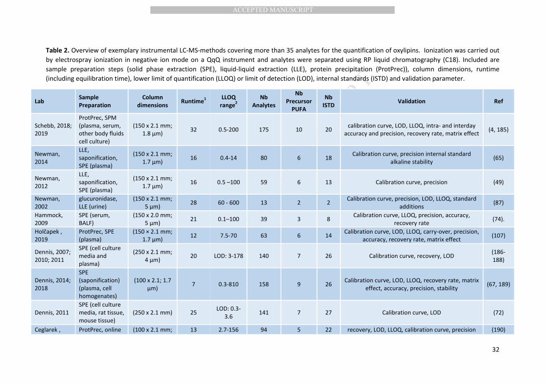
MANUSCRIP
T
ACCEPTED
ACCEPTED MANUSCRIPT
32
Table 2. Overview of exemplary instrumental LC-MS-methods covering more than 35 analytes for the quantification of oxylipins. Ionization was carried out
by electrospray ionization in negative ion mode on a QqQ instrument and analytes were separated using RP liquid chromatography (C18). Included are
sample preparation steps (solid phase extraction (SPE), liquid-liquid extraction (LLE), protein precipitation (ProtPrec)), column dimensions, runtime
(including equilibration time), lower limit of quantification (LLOQ) or limit of detection (LOD), internal standards (ISTD) and validation parameter.
Lab Sample
Preparation
Column
dimensions Runtime
1
LLOQ
range2
Nb
Analytes
Nb
Precursor
PUFA
Nb
ISTD Validation Ref
Schebb, 2018;
2019
ProtPrec, SPM
(plasma, serum,
other body fluids
cell culture)
(150 x 2.1 mm;
1.8 µm) 32 0.5-200 175 10 20
calibration curve, LOD, LLOQ, intra- and interday
accuracy and precision, recovery rate, matrix effect (4, 185)
Newman,
2014
LLE,
saponification,
SPE (plasma)
(150 x 2.1 mm;
1.7 µm) 16 0.4-14 80 6 18
Calibration curve, precision internal standard
alkaline stability (65)
Newman,
2012
LLE,
saponification,
SPE (plasma)
(150 x 2.1 mm;
1.7 µm) 16 0.5 –100 59 6 13 Calibration curve, precision (49)
Newman,
2002
glucuronidase,
LLE (urine)
(150 x 2.1 mm;
5 µm) 28 60 - 600 13 2 2
Calibration curve, precision, LOD, LLOQ, standard
additions (87)
Hammock,
2009
SPE (serum,
BALF)
(150 x 2.0 mm;
5 µm) 21 0.1–100 39 3 8
Calibration curve, LLOQ, precision, accuracy,
recovery rate (74).
Holčapek ,
2019
ProtPrec, SPE
(plasma)
(150 × 2.1 mm;
1.7 μm) 12 7.5-70 63 6 14
Calibration curve, LOD, LLOQ, carry-over, precision,
accuracy, recovery rate, matrix effect (107)
Dennis, 2007;
2010; 2011
SPE (cell culture
media and
plasma)
(250 x 2.1 mm;
4 µm) 20 LOD: 3-178 140 7 26 Calibration curve, recovery, LOD
(186-
188)
Dennis, 2014;
2018
SPE
(saponification)
(plasma, cell
homogenates)
(100 x 2.1; 1.7
µm) 7 0.3-810 158 9 26
Calibration curve, LOD, LLOQ, recovery rate, matrix
effect, accuracy, precision, stability (67, 189)
Dennis, 2011
SPE (cell culture
media, rat tissue,
mouse tissue)
(250 x 2.1 mm) 25 LOD: 0.3-
3.6 141 7 27 Calibration curve, LOD (72)
Ceglarek , ProtPrec, online (100 x 2.1 mm; 13 2.7-156 94 5 22 recovery, LOD, LLOQ, calibration curve, precision (190)
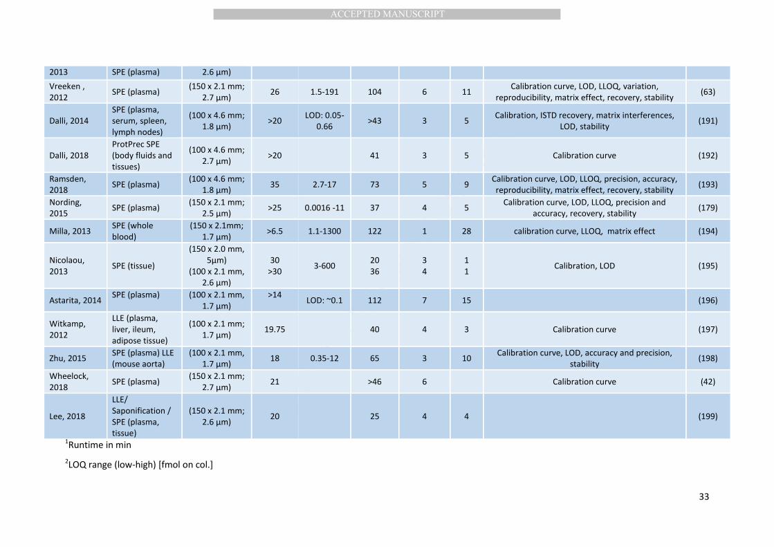
MANUSCRIP
T
ACCEPTED
ACCEPTED MANUSCRIPT
33
2013 SPE (plasma) 2.6 µm)
Vreeken ,
2012 SPE (plasma)
(150 x 2.1 mm;
2.7 µm) 26 1.5-191 104 6 11
Calibration curve, LOD, LLOQ, variation,
reproducibility, matrix effect, recovery, stability (63)
Dalli, 2014
SPE (plasma,
serum, spleen,
lymph nodes)
(100 x 4.6 mm;
1.8 µm) >20
LOD: 0.05-
0.66 >43 3 5
Calibration, ISTD recovery, matrix interferences,
LOD, stability (191)
Dalli, 2018
ProtPrec SPE
(body fluids and
tissues)
(100 x 4.6 mm;
2.7 µm) >20 41 3 5 Calibration curve (192)
Ramsden,
2018 SPE (plasma)
(100 x 4.6 mm;
1.8 µm) 35 2.7-17 73 5 9
Calibration curve, LOD, LLOQ, precision, accuracy,
reproducibility, matrix effect, recovery, stability (193)
Nording,
2015 SPE (plasma)
(150 x 2.1 mm;
2.5 µm) >25 0.0016 -11 37 4 5
Calibration curve, LOD, LLOQ, precision and
accuracy, recovery, stability (179)
Milla, 2013 SPE (whole
blood)
(150 x 2.1mm;
1.7 µm) >6.5 1.1-1300 122 1 28 calibration curve, LLOQ, matrix effect (194)
Nicolaou,
2013 SPE (tissue)
(150 x 2.0 mm,
5µm)
(100 x 2.1 mm,
2.6 µm)
30
>30 3-600
20
36
3
4
1
1 Calibration, LOD (195)
Astarita, 2014 SPE (plasma) (100 x 2.1 mm,
1.7 µm)
>14 LOD: ~0.1 112 7 15 (196)
Witkamp,
2012
LLE (plasma,
liver, ileum,
adipose tissue)
(100 x 2.1 mm;
1.7 µm) 19.75 40 4 3 Calibration curve (197)
Zhu, 2015 SPE (plasma) LLE
(mouse aorta)
(100 x 2.1 mm,
1.7 µm) 18 0.35-12 65 3 10
Calibration curve, LOD, accuracy and precision,
stability (198)
Wheelock,
2018 SPE (plasma)
(150 x 2.1 mm;
2.7 µm) 21 >46 6 Calibration curve (42)
Lee, 2018
LLE/
Saponification /
SPE (plasma,
tissue)
(150 x 2.1 mm;
2.6 µm) 20 25 4 4 (199)
1Runtime in min
2LOQ range (low-high) [fmol on col.]
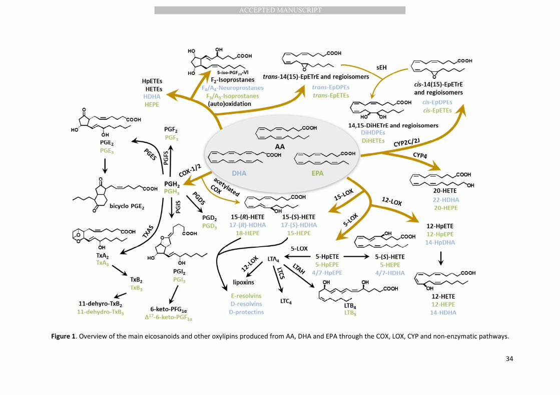
MANUSCRIP
T
ACCEPTED
ACCEPTED MANUSCRIPT
34
Figure 1. Overview of the main eicosanoids and other oxylipins produced from AA, DHA and EPA through the COX, LOX, CYP and non-enzymatic pathways.
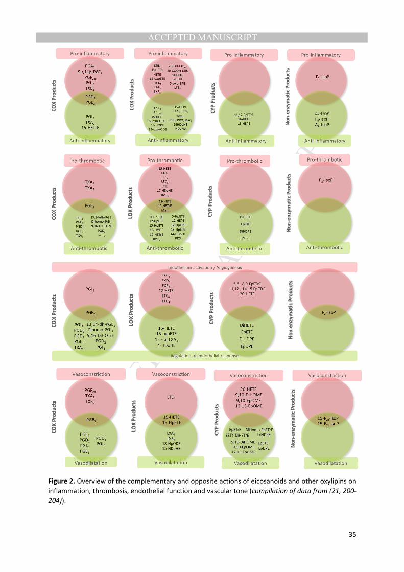
MANUSCRIP
T
ACCEPTED
ACCEPTED MANUSCRIPT
35
Figure 2. Overview of the complementary and opposite actions of eicosanoids and other oxylipins on
inflammation, thrombosis, endothelial function and vascular tone (compilation of data from (21, 200-
204)).

MANUSCRIP
T
ACCEPTED
ACCEPTED MANUSCRIPT
36
Figure 3. Box-plots from different oxylipins measured in plasma of 133 healthy individuals from the
IMPROVE cohort. Mean CV of oxylipin levels for this sample of the IMPROVE cohort was estimated at
87%. (Illustration created from supplementary dataset associated with (42)).
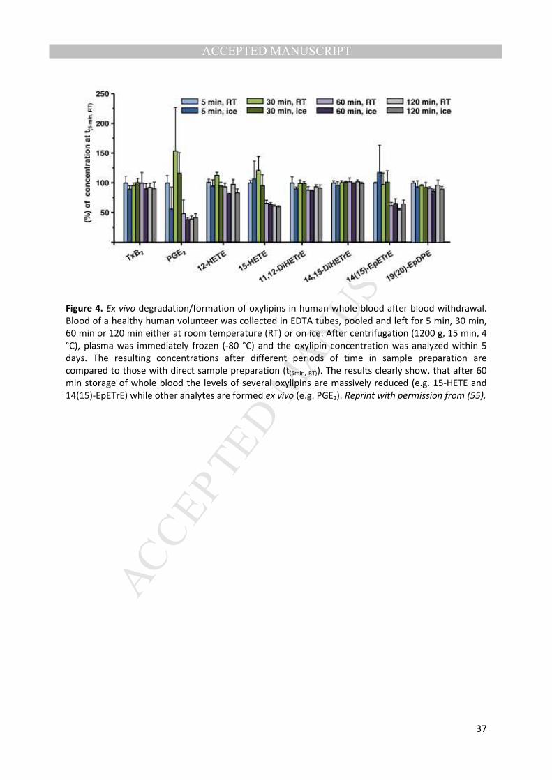
MANUSCRIP
T
ACCEPTED
ACCEPTED MANUSCRIPT
37
Figure 4. Ex vivo degradation/formation of oxylipins in human whole blood after blood withdrawal.
Blood of a healthy human volunteer was collected in EDTA tubes, pooled and left for 5 min, 30 min,
60 min or 120 min either at room temperature (RT) or on ice. After centrifugation (1200 g, 15 min, 4
°C), plasma was immediately frozen (-80 °C) and the oxylipin concentration was analyzed within 5
days. The resulting concentrations after different periods of time in sample preparation are
compared to those with direct sample preparation (t(5min, RT)). The results clearly show, that after 60
min storage of whole blood the levels of several oxylipins are massively reduced (e.g. 15-HETE and
14(15)-EpETrE) while other analytes are formed ex vivo (e.g. PGE2). Reprint with permission from (55).
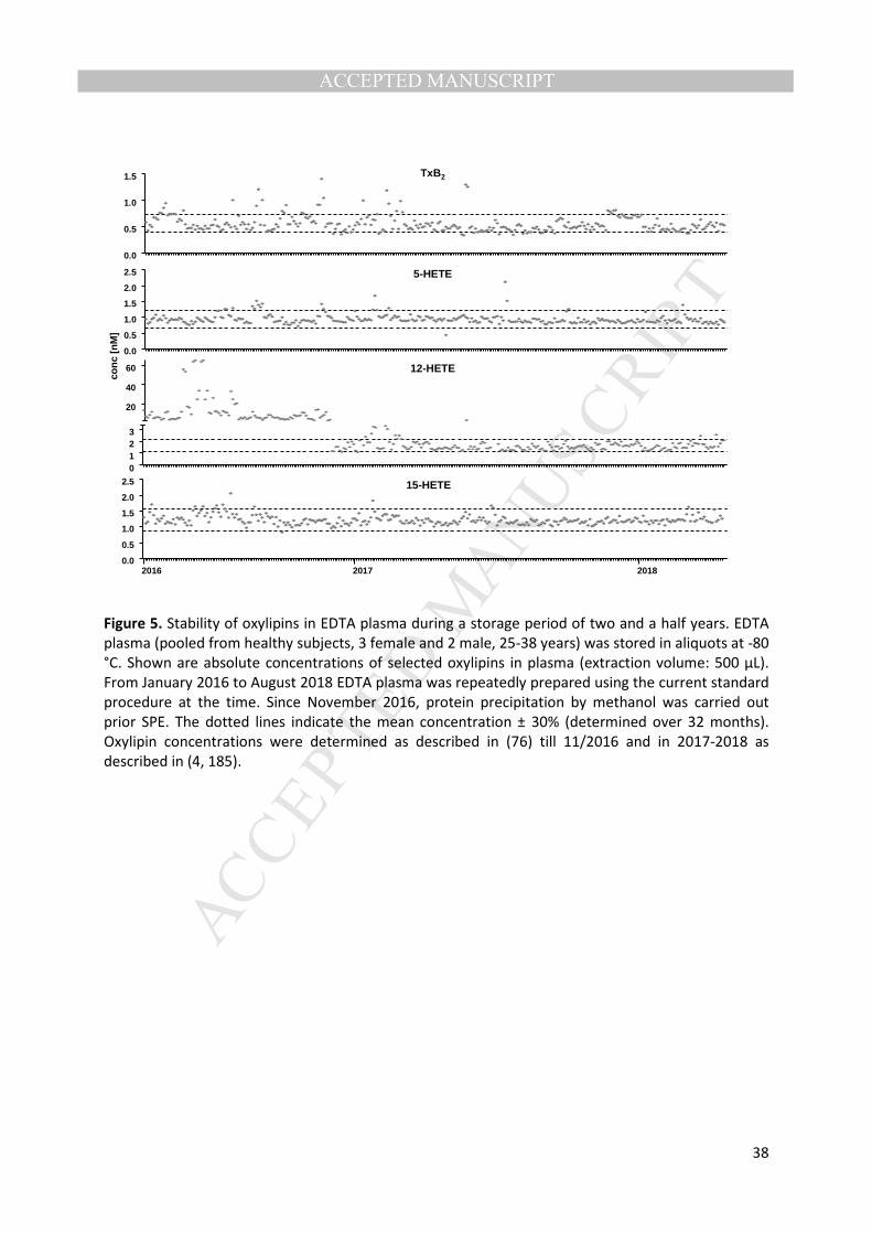
MANUSCRIP
T
ACCEPTED
ACCEPTED MANUSCRIPT
38
Figure 5. Stability of oxylipins in EDTA plasma during a storage period of two and a half years. EDTA
plasma (pooled from healthy subjects, 3 female and 2 male, 25-38 years) was stored in aliquots at -80
°C. Shown are absolute concentrations of selected oxylipins in plasma (extraction volume: 500 µL).
From January 2016 to August 2018 EDTA plasma was repeatedly prepared using the current standard
procedure at the time. Since November 2016, protein precipitation by methanol was carried out
prior SPE. The dotted lines indicate the mean concentration ± 30% (determined over 32 months).
Oxylipin concentrations were determined as described in (76) till 11/2016 and in 2017-2018 as
described in (4, 185).
TxB2
0.0
0.5
1.0
1.5
12-HETE
0123
20
40
60
con
c[n
M]
15-HETE
0.0
0.5
1.0
1.5
2.0
2.5
2016 2017 2018
5-HETE
0.0
0.5
1.0
1.5
2.0
2.5
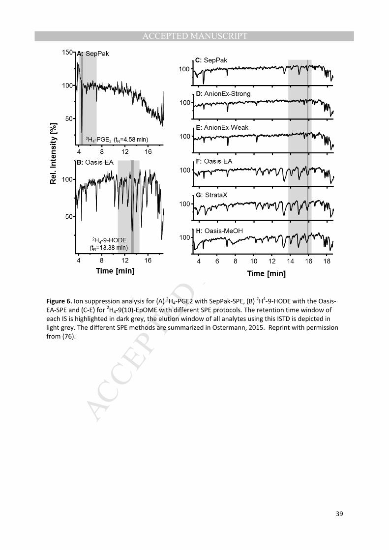
MANUSCRIP
T
ACCEPTED
ACCEPTED MANUSCRIPT
39
Figure 6. Ion suppression analysis for (A) 2H4-PGE2 with SepPak-SPE, (B)
2H
4-9-HODE with the Oasis-
EA-SPE and (C-E) for 2H4-9(10)-EpOME with different SPE protocols. The retention time window of
each IS is highlighted in dark grey, the elution window of all analytes using this ISTD is depicted in
light grey. The different SPE methods are summarized in Ostermann, 2015. Reprint with permission
from (76).
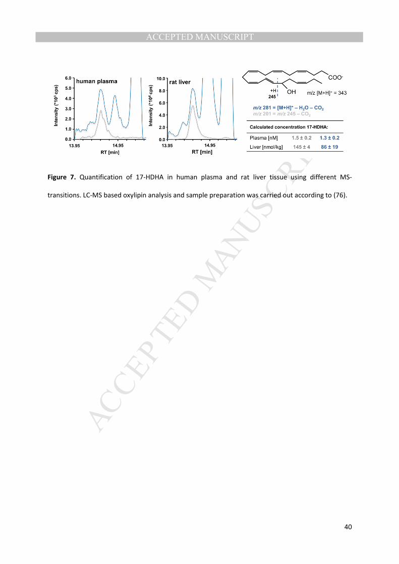
MANUSCRIP
T
ACCEPTED
ACCEPTED MANUSCRIPT
40
Figure 7. Quantification of 17-HDHA in human plasma and rat liver tissue using different MS-
transitions. LC-MS based oxylipin analysis and sample preparation was carried out according to (76).
MANUSCRIP
T
ACCEPTED
ACCEPTED MANUSCRIPT
41
7. References
1. Buczynski, M. W., Dumlao, D. S., and Dennis, E. A. (2009) Thematic Review Series:
Proteomics. An integrated omics analysis of eicosanoid biology. J Lipid Res 50, 1015-1038
2. Rund, K. M., Heylmann, D., Seiwert, N., Wecklein, S., Oger, C., Galano, J., Durand, T., Chen, R.,
Gueler, F., Fahrer, J., Bornhorst, J., and Schebb, N. H. (2019) Formation of trans-epoxy fatty
acids correlates with formation of isoprostanes and could serve as biomarker of oxidative
stress. Prostaglandins and Other Lipid Mediators
3. Smith WL, and Murphy RC. (2002) The eicosanoids: cyclooxygenase, lipoxygenase, and
epoxygenase pathways. In Biochemistry of Lipids, Lipoproteins and Membranes (4th eds
(Vance, D. E. V. a. J. E., ed), Elsevier Science B.V.
4. Rund, K. M., Ostermann, A. I., Kutzner, L., Galano, J. M., Oger, C., Vigor, C., Wecklein, S.,
Seiwert, N., Durand, T., and Schebb, N. H. (2018) Development of an LC-ESI(-)-MS/MS
method for the simultaneous quantification of 35 isoprostanes and isofurans derived from
the major n3- and n6-PUFAs. Anal Chim Acta 1037, 63-74
5. Smith, W. L., DeWitt, D. L., and Garavito, R. M. (2000) Cyclooxygenases: structural, cellular,
and molecular biology. Annu Rev Biochem 69, 145-182
6. O’Donnell, V. B., and Murphy, R. C. (2017) Directing eicosanoid esterification into
phospholipids. Journal of Lipid Research 58, 837-839
7. Park, S. K., Herrnreiter, A., Pfister, S. L., Gauthier, K. M., Falck, B. A., Falck, J. R., and Campbell,
W. B. (2018) GPR40 is a low-affinity epoxyeicosatrienoic acid receptor in vascular cells. J Biol
Chem 293, 10675-10691
8. Meves, H. (2008) Arachidonic acid and ion channels: an update. Br J Pharmacol 155, 4-16
9. Watanabe, H., Vriens, J., Prenen, J., Droogmans, G., Voets, T., and Nilius, B. (2003)
Anandamide and arachidonic acid use epoxyeicosatrienoic acids to activate TRPV4 channels.
Nature 424, 434-438
10. Wen, H., Ostman, J., Bubb, K. J., Panayiotou, C., Priestley, J. V., Baker, M. D., and Ahluwalia,
A. (2012) 20-Hydroxyeicosatetraenoic acid (20-HETE) is a novel activator of transient
receptor potential vanilloid 1 (TRPV1) channel. J Biol Chem 287, 13868-13876
11. Bos, C. L., Richel, D. J., Ritsema, T., Peppelenbosch, M. P., and Versteeg, H. H. (2004)
Prostanoids and prostanoid receptors in signal transduction. Int J Biochem Cell Biol 36, 1187-
1205
12. Hardwick, J. P., Eckman, K., Lee, Y. K., Abdelmegeed, M. A., Esterle, A., Chilian, W. M., Chiang,
J. Y., and Song, B. J. (2013) Eicosanoids in metabolic syndrome. Adv Pharmacol 66, 157-266
13. Bosviel, R., Joumard-Cubizolles, L., Chinetti-Gbaguidi, G., Bayle, D., Copin, C., Hennuyer, N.,
Duplan, I., Staels, B., Zanoni, G., Porta, A., Balas, L., Galano, J. M., Oger, C., Mazur, A., Durand,
T., and Gladine, C. (2017) DHA-derived oxylipins, neuroprostanes and protectins,
differentially and dose-dependently modulate the inflammatory response in human
macrophages: Putative mechanisms through PPAR activation. Free Radic Biol Med 103, 146-
154
14. Brooks, J. D., Musiek, E. S., Koestner, T. R., Stankowski, J. N., Howard, J. R., Brunoldi, E. M.,
Porta, A., Zanoni, G., Vidari, G., Morrow, J. D., Milne, G. L., and McLaughlin, B. (2011) The
fatty acid oxidation product 15-A3t-isoprostane is a potent inhibitor of NFkappaB
transcription and macrophage transformation. J Neurochem 119, 604-616
15. Musiek, E. S., Brooks, J. D., Joo, M., Brunoldi, E., Porta, A., Zanoni, G., Vidari, G., Blackwell, T.
S., Montine, T. J., Milne, G. L., McLaughlin, B., and Morrow, J. D. (2008) Electrophilic
cyclopentenone neuroprostanes are anti-inflammatory mediators formed from the
peroxidation of the omega-3 polyunsaturated fatty acid docosahexaenoic acid. J Biol Chem
283, 19927-19935
MANUSCRIP
T
ACCEPTED
ACCEPTED MANUSCRIPT
42
16. Tatsuno, I., Saito, H., Chang, K. J., Tamura, Y., and Yoshida, S. (1990) Comparison of the effect
between leukotriene B4 and leukotriene B5 on the induction of interleukin 1-like activity and
calcium mobilizing activity in human blood monocytes. Agents Actions 29, 324-327
17. Ulu, A., Stephen Lee, K. S., Miyabe, C., Yang, J., Hammock, B. G., Dong, H., and Hammock, B.
D. (2014) An omega-3 epoxide of docosahexaenoic acid lowers blood pressure in
angiotensin-II-dependent hypertension. J Cardiovasc Pharmacol 64, 87-99
18. Lotzer, K., Jahn, S., Kramer, C., Hildner, M., Nusing, R., Funk, C. D., and Habenicht, A. J. (2007)
5-Lipoxygenase/cyclooxygenase-2 cross-talk through cysteinyl leukotriene receptor 2 in
endothelial cells. Prostaglandins Other Lipid Mediat 84, 108-115
19. Oni-Orisan, A., Deng, Y., Schuck, R. N., Theken, K. N., Edin, M. L., Lih, F. B., Molnar, K.,
DeGraff, L., Tomer, K. B., Zeldin, D. C., and Lee, C. R. (2013) Dual modulation of
cyclooxygenase and CYP epoxygenase metabolism and acute vascular inflammation in mice.
Prostaglandins Other Lipid Mediat 104-105, 67-73
20. Rouzer, C. A., and Marnett, L. J. (2011) Endocannabinoid oxygenation by cyclooxygenases,
lipoxygenases, and cytochromes P450: cross-talk between the eicosanoid and
endocannabinoid signaling pathways. Chem Rev 111, 5899-5921
21. Yeung, J., Hawley, M., and Holinstat, M. (2017) The expansive role of oxylipins on platelet
biology. J Mol Med (Berl) 95, 575-588
22. Lee, I. T., Lin, C. C., Lin, W. N., Wu, W. L., Hsiao, L. D., and Yang, C. M. (2013) Lung
inflammation caused by adenosine-5'-triphosphate is mediated via Ca2+/PKCs-dependent
COX-2/PGE2 induction. Int J Biochem Cell Biol 45, 1657-1668
23. Shinomiyaa Shiho , Hiroaki, N., Akinori, U., Iku, U., Takayuki, M., Shuichi, O., Fumitaka, U.,
Koichi, Y., Shuh, N., Yukihiko, S., Atsushi, I., and Sachiko, O.-i. (2001) Regulation of TNFa and
interleukin-10 production by prostaglandins I2 and E2: studies with prostaglandin receptor-
deficient mice and prostaglandin E-receptor subtype-selective synthetic agonists.
Biochemical Pharmacology 61 1153-1160
24. Urade, Y., and Eguchi, N. (2002) Lipocalin-type and hematopoietic prostaglandin D synthases
as a novel example of functional convergence. Prostaglandins & Other Lipid Mediators 68-69,
375-382
25. Rajakariar, R., Hilliard, M., Lawrence, T., Trivedi, S., Colville-Nash, P., Bellingan, G., Fitzgerald,
D., Yaqoob, M. M., and Gilroy, D. W. (2007) Hematopoietic prostaglandin D2 synthase
controls the onset and resolution of acute inflammation through PGD2 and 15-deoxyDelta12
14 PGJ2. Proc Natl Acad Sci U S A 104, 20979-20984
26. Funk, C. D. (2001) Prostaglandins and leukotrienes: advances in eicosanoid biology. Science
294, 1871-1875
27. Ueno, N., Murakami, M., Tanioka, T., Fujimori, K., Tanabe, T., Urade, Y., and Kudo, I. (2001)
Coupling between cyclooxygenase, terminal prostanoid synthase, and phospholipase A2. J
Biol Chem 276, 34918-34927
28. Li, Y., Lei, D., Swindell, W. R., Xia, W., Weng, S., Fu, J., Worthen, C. A., Okubo, T., Johnston, A.,
Gudjonsson, J. E., Voorhees, J. J., and Fisher, G. J. (2015) Age-Associated Increase in Skin
Fibroblast-Derived Prostaglandin E2 Contributes to Reduced Collagen Levels in Elderly
Human Skin. J Invest Dermatol 135, 2181-2188
29. Farhat, A., Philibert, P., Sultan, C., Poulat, F., and Boizet-Bonhoure, B. (2011) Hematopoietic-
Prostaglandin D2 synthase through PGD2 production is involved in the adult ovarian
physiology. J Ovarian Res 4, 3
30. Shureiqi, I. (2000) 15-LOX-1: a Novel Molecular Target of Nonsteroidal Anti-inflammatory
Drug-Induced Apoptosis in Colorectal Cancer Cells. Journal of the National Cancer Institute
92, 1136-1142
31. Kuhn, H., Banthiya, S., and van Leyen, K. (2015) Mammalian lipoxygenases and their
biological relevance. Biochimica et Biophysica Acta 1851, 308-330
MANUSCRIP
T
ACCEPTED
ACCEPTED MANUSCRIPT
43
32. Kutzner, L., Goloshchapova, K., Heydeck, D., Stehling, S., Kuhn, H., and Schebb, N. H. (2017)
Mammalian ALOX15 orthologs exhibit pronounced dual positional specificity with
docosahexaenoic acid. Biochim Biophys Acta Mol Cell Biol Lipids 1862, 666-675
33. Jamieson, K. L., Endo, T., Darwesh, A. M., Samokhvalov, V., and Seubert, J. M. (2017)
Cytochrome P450-derived eicosanoids and heart function. Pharmacol Ther 179, 47-83
34. Westphal, C., Konkel, A., and Schunck, W. H. (2015) Cytochrome p450 enzymes in the
bioactivation of polyunsaturated Fatty acids and their role in cardiovascular disease. Adv Exp
Med Biol 851, 151-187
35. Schebb, N. H., Ostermann, A. I., Yang, J., Hammock, B. D., Hahn, A., and Schuchardt, J. P.
(2014) Comparison of the effects of long-chain omega-3 fatty acid supplementation on
plasma levels of free and esterified oxylipins. Prostaglandins Other Lipid Mediat 113-115, 21-
29
36. Shearer, G. C., and Newman, J. W. (2008) Lipoprotein lipase releases esterified oxylipins from
very low-density lipoproteins. Prostaglandins, Leukotrienes and Essential Fatty Acids 79, 215-
222
37. Ek-Von Mentzer, B. A., Zhang, F., and Hamilton, J. A. (2001) Binding of 13-HODE and 15-HETE
to phospholipid bilayers, albumin, and intracellular fatty acid binding proteins. implications
for transmembrane and intracellular transport and for protection from lipid peroxidation. J
Biol Chem 276, 15575-15580
38. Maclouf, J., Kindahl, H., Granstrom, E., and Samuelsson, B. (1980) Interactions of
prostaglandin H2 and thromboxane A2 with human serum albumin. Eur J Biochem 109, 561-
566
39. Boilard, E. (2018) Extracellular vesicles and their content in bioactive lipid mediators: more
than a sack of microRNA. J Lipid Res 59, 2037-2046
40. Wopereis, S., Wolvers, D., van Erk, M., Gribnau, M., Kremer, B., van Dorsten, F. A., Boelsma,
E., Garczarek, U., Cnubben, N., Frenken, L., van der Logt, P., Hendriks, H. F., Albers, R., van
Duynhoven, J., van Ommen, B., and Jacobs, D. M. (2013) Assessment of inflammatory
resilience in healthy subjects using dietary lipid and glucose challenges. BMC Med Genomics
6, 44
41. Ostermann, A. I., Greupner, T., Kutzner, L., Hartung, Nicole M., Hahn, A., Schuchardt, J. P.,
and Schebb, N. H. (2018) Intra-individual variance of the human plasma oxylipin pattern: low
inter-day variability in fasting blood samples versus high variability during the day. Analytical
Methods 10, 4935-4944
42. Kamleh, M. A., McLeod, O., Checa, A., Baldassarre, D., Veglia, F., Gertow, K., Humphries, S. E.,
Rauramaa, R., de Faire, U., Smit, A. J., Giral, P., Kurl, S., Mannarino, E., Tremoli, E., Silveira, A.,
Orvik, J., Hamsten, A., and Wheelock, C. E. (2018) Increased Levels of Circulating Fatty Acids
Are Associated with Protective Effects against Future Cardiovascular Events in Nondiabetics. J
Proteome Res 17, 870-878
43. Sun, Y., Koh, H. W., Choi, H., Koh, W. P., Yuan, J. M., Newman, J. W., Su, J., Fang, J., Ong, C. N.,
and van Dam, R. M. (2016) Plasma fatty acids, oxylipins, and risk of myocardial infarction: the
Singapore Chinese Health Study. J Lipid Res 57, 1300-1307
44. Hammond, V. J., and O'Donnell, V. B. (2012) Esterified eicosanoids: generation,
characterization and function. Biochim Biophys Acta 1818, 2403-2412
45. Shearer, G. C., Borkowski, K., Puumala, S. L., Harris, W. S., Pedersen, T. L., and Newman, J. W.
(2018) Abnormal lipoprotein oxylipins in metabolic syndrome and partial correction by
omega-3 fatty acids. Prostaglandins Leukot Essent Fatty Acids 128, 1-10
46. Milne, G. L., Yin, H., and Morrow, J. D. (2008) Human biochemistry of the isoprostane
pathway. J Biol Chem 283, 15533-15537
47. Dalle, C., Ostermann, A. I., Konrad, T., Coudy-Gandilhon, C., Decourt, A., Barthelemy, J. C.,
Roche, F., Feasson, L., Mazur, A., Bechet, D., Schebb, N. H., and Gladine, C. (2018) Muscle loss
associated changes of oxylipin signatures during biological aging: an exploratory study from
the PROOF cohort. J Gerontol A Biol Sci Med Sci
MANUSCRIP
T
ACCEPTED
ACCEPTED MANUSCRIPT
44
48. Greupner, T., Kutzner, L., Nolte, F., Strangmann, A., Kohrs, H., Hahn, A., Schebb, N. H., and
Schuchardt, J. P. (2018) Effects of a 12-week high-alpha-linolenic acid intervention on EPA
and DHA concentrations in red blood cells and plasma oxylipin pattern in subjects with a low
EPA and DHA status. Food Funct 9, 1587-1600
49. Keenan, A. H., Pedersen, T. L., Fillaus, K., Larson, M. K., Shearer, G. C., and Newman, J. W.
(2012) Basal omega-3 fatty acid status affects fatty acid and oxylipin responses to high-dose
n3-HUFA in healthy volunteers. J Lipid Res 53, 1662-1669
50. Caligiuri, S. P., Aukema, H. M., Ravandi, A., and Pierce, G. N. (2014) Elevated levels of pro-
inflammatory oxylipins in older subjects are normalized by flaxseed consumption. Exp
Gerontol 59, 51-57
51. Song, J., Liu, X., Rao, T. S., Chang, L., Meehan, M. J., Blevitt, J. M., Wu, J., Dorrestein, P. C.,
and Milla, M. E. (2015) Phenotyping drug polypharmacology via eicosanoid profiling of blood.
Journal of Lipid Research 56
52. Ostermann, A. I., and Schebb, N. H. (2017) Effects of omega-3 fatty acid supplementation on
the pattern of oxylipins: a short review about the modulation of hydroxy-, dihydroxy-, and
epoxy-fatty acids. Food Funct 8, 2355-2367
53. Arita, M. (2012) Mediator lipidomics in acute inflammation and resolution. J Biochem 152,
313-319
54. Roberts, L. D., Souza, A. L., Gerszten, R. E., and Clish, C. B. (2012) Targeted metabolomics.
Curr Protoc Mol Biol Chapter 30, Unit 30 32 31-24
55. Willenberg, I., Rund, K., Rong, S., Shushakova, N., Gueler, F., and Schebb, N. H. (2015)
Characterization of changes in plasma and tissue oxylipin levels in LPS and CLP induced
murine sepsis. Inflammation Research 65, 133-142
56. Ostermann, A. I., Waindok, P., Schmidt, M. J., Chiu, C. Y., Smyl, C., Rohwer, N., Weylandt, K.
H., and Schebb, N. H. (2017) Modulation of the endogenous omega-3 fatty acid and oxylipin
profile in vivo-A comparison of the fat-1 transgenic mouse with C57BL/6 wildtype mice on an
omega-3 fatty acid enriched diet. PLoS One 12, e0184470
57. Jonasdottir, H. S., Brouwers, H., Toes, R. E. M., Ioan-Facsinay, A., and Giera, M. (2018) Effects
of anticoagulants and storage conditions on clinical oxylipid levels in human plasma. Biochim
Biophys Acta Mol Cell Biol Lipids 1863, 1511-1522
58. Schuchardt, J. P., Schmidt, S., Kressel, G., Dong, H., Willenberg, I., Hammock, B. D., Hahn, A.,
and Schebb, N. H. (2013) Comparison of free serum oxylipin concentrations in hyper- vs.
normolipidemic men. Prostaglandins Leukot Essent Fatty Acids 89, 19-29
59. La Frano, M. R., Carmichael, S. L., Ma, C., Hardley, M., Shen, T., Wong, R., Rosales, L.,
Borkowski, K., Pedersen, T. L., Shaw, G. M., Stevenson, D. K., Fiehn, O., and Newman, J. W.
(2018) Impact of post-collection freezing delay on the reliability of serum metabolomics in
samples reflecting the California mid-term pregnancy biobank. Metabolomics 14
60. Dorow, J., Becker, S., Kortz, L., Thiery, J., Hauschildt, S., and Ceglarek, U. (2016) Preanalytical
Investigation of Polyunsaturated Fatty Acids and Eicosanoids in Human Plasma by Liquid
Chromatography–Tandem Mass Spectrometry. Biopreservation and Biobanking 14, 107-113
61. Goodfriend, T. L., Pedersen, T. L., Grekin, R. J., Hammock, B. D., Ball, D. L., and Vollmer, A.
(2007) Heparin, lipoproteins, and oxygenated fatty acids in blood: a cautionary note.
Prostaglandins Leukot Essent Fatty Acids 77, 363-366
62. Nakamura, H., Kim, D. K., Philbin, D. M., Peterson, M. B., Debros, F., Koski, G., and Bonventre,
J. V. (1995) Heparin-enhanced plasma phospholipase A2 activity and prostacyclin synthesis in
patients undergoing cardiac surgery. J Clin Invest 95, 1062-1070
63. Strassburg, K., Huijbrechts, A. M. L., Kortekaas, K. A., Lindeman, J. H., Pedersen, T. L., Dane,
A., Berger, R., Brenkman, A., Hankemeier, T., van Duynhoven, J., Kalkhoven, E., Newman, J.
W., and Vreeken, R. J. (2012) Quantitative profiling of oxylipins through comprehensive LC-
MS/MS analysis: application in cardiac surgery. Analytical and Bioanalytical Chemistry 404,
1413-1426
64. Goodall, A. H. (2014) Platelet 12-LOX scores a HIT. Blood 124, 2166-2168
MANUSCRIP
T
ACCEPTED
ACCEPTED MANUSCRIPT
45
65. Gladine, C., Newman, J. W., Durand, T., Pedersen, T. L., Galano, J. M., Demougeot, C.,
Berdeaux, O., Pujos-Guillot, E., Mazur, A., and Comte, B. (2014) Lipid profiling following
intake of the omega 3 fatty acid DHA identifies the peroxidized metabolites F4-
neuroprostanes as the best predictors of atherosclerosis prevention. PLoS One 9, e89393
66. Dupuy, A., Le Faouder, P., Vigor, C., Oger, C., Galano, J. M., Dray, C., Lee, J. C., Valet, P.,
Gladine, C., Durand, T., and Bertrand-Michel, J. (2016) Simultaneous quantitative profiling of
20 isoprostanoids from omega-3 and omega-6 polyunsaturated fatty acids by LC-MS/MS in
various biological samples. Anal Chim Acta 921, 46-58
67. Quehenberger, O., Dahlberg-Wright, S., Jiang, J., Armando, A. M., and Dennis, E. A. (2018)
Quantitative determination of esterified eicosanoids and related oxygenated metabolites
after base hydrolysis. J Lipid Res 59, 2436-2445
68. Newman, J. W., Kaysen, G. A., Hammock, B. D., and Shearer, G. C. (2007) Proteinuria
increases oxylipid concentrations in VLDL and HDL but not LDL particles in the rat. J Lipid Res
48, 1792-1800
69. Arnold, C., Markovic, M., Blossey, K., Wallukat, G., Fischer, R., Dechend, R., Konkel, A., von
Schacky, C., Luft, F. C., Muller, D. N., Rothe, M., and Schunck, W.-H. (2010) Arachidonic Acid-
metabolizing Cytochrome P450 Enzymes Are Targets of ω-3 Fatty Acids. Journal of Biological
Chemistry 285, 32720-32733
70. Duflot, T., Pereira, T., Roche, C., Iacob, M., Cardinael, P., Hamza, N. E., Thuillez, C.,
Compagnon, P., Joannides, R., Lamoureux, F., and Bellien, J. (2017) A sensitive LC-MS/MS
method for the quantification of regioisomers of epoxyeicosatrienoic and
dihydroxyeicosatrienoic acids in human plasma during endothelial stimulation. Anal Bioanal
Chem 409, 1845-1855
71. Morisseau, C., Inceoglu, B., Schmelzer, K., Tsai, H.-J., Jinks, S. L., Hegedus, C. M., and
Hammock, B. D. (2010) Naturally occurring monoepoxides of eicosapentaenoic acid and
docosahexaenoic acid are bioactive antihyperalgesic lipids. Journal of Lipid Research 51,
3481-3490
72. Dumlao, D. S., Buczynski, M. W., Norris, P. C., Harkewicz, R., and Dennis, E. A. (2011) High-
throughput lipidomic analysis of fatty acid derived eicosanoids and N-acylethanolamines.
Biochim Biophys Acta 1811, 724-736
73. Arnold, C., Markovic, M., Blossey, K., Wallukat, G., Fischer, R., Dechend, R., Konkel, A., von
Schacky, C., Luft, F. C., Muller, D. N., Rothe, M., and Schunck, W. H. (2010) Arachidonic acid-
metabolizing cytochrome P450 enzymes are targets of {omega}-3 fatty acids. J Biol Chem
285, 32720-32733
74. Yang, J., Schmelzer, K., Georgi, K., and Hammock, B. D. (2009) Quantitative profiling method
for oxylipin metabolome by liquid chromatography electrospray ionization tandem mass
spectrometry. Anal Chem 81, 8085-8093
75. Giera, M., Ioan-Facsinay, A., Toes, R., Gao, F., Dalli, J., Deelder, A. M., Serhan, C. N., and
Mayboroda, O. A. (2012) Lipid and lipid mediator profiling of human synovial fluid in
rheumatoid arthritis patients by means of LC-MS/MS. Biochim Biophys Acta 1821, 1415-1424
76. Ostermann, A. I., Willenberg, I., and Schebb, N. H. (2015) Comparison of sample preparation
methods for the quantitative analysis of eicosanoids and other oxylipins in plasma by means
of LC-MS/MS. Anal Bioanal Chem 407, 1403-1414
77. Pedersen, T. L., and Newman, J. W. (2018) Establishing and Performing Targeted Multi-
residue Analysis for Lipid Mediators and Fatty Acids in Small Clinical Plasma Samples.
Methods Mol Biol 1730, 175-212
78. Fromel, T., Jungblut, B., Hu, J., Trouvain, C., Barbosa-Sicard, E., Popp, R., Liebner, S.,
Dimmeler, S., Hammock, B. D., and Fleming, I. (2012) Soluble epoxide hydrolase regulates
hematopoietic progenitor cell function via generation of fatty acid diols. Proc Natl Acad Sci U
S A 109, 9995-10000
79. Ferreirós, N., Homann, J., Labocha, S., Grossmann, N., Hahn, J. S., Brüne, B., and Geisslinger,
G. (2014) Lipoxin A4: Problems with its determination using reversed phase
MANUSCRIP
T
ACCEPTED
ACCEPTED MANUSCRIPT
46
chromatography–tandem mass spectrometry and confirmation with chiral chromatography.
Talanta 127, 82-87
80. Stahnke, H., Reemtsma, T., and Alder, L. (2009) Compensation of matrix effects by
postcolumn infusion of a monitor substance in multiresidue analysis with LC-MS/MS. Anal
Chem 81, 2185-2192
81. Campbell, W. B., Falck, J. R., Okita, J. R., Johnson, A. R., and Callahan, K. S. (1985) Synthesis of
dihomoprostaglandins from adrenic acid (7,10,13,16-docosatetraenoic acid) by human
endothelial cells. Biochim Biophys Acta 837, 67-76
82. Kopf, P. G., Zhang, D. X., Gauthier, K. M., Nithipatikom, K., Yi, X. Y., Falck, J. R., and Campbell,
W. B. (2010) Adrenic acid metabolites as endogenous endothelium-derived and zona
glomerulosa-derived hyperpolarizing factors. Hypertension 55, 547-554
83. Markworth, J. F., Kaur, G., Miller, E. G., Larsen, A. E., Sinclair, A. J., Maddipati, K. R., and
Cameron-Smith, D. (2016) Divergent shifts in lipid mediator profile following
supplementation with n-3 docosapentaenoic acid and eicosapentaenoic acid. FASEB J 30,
3714-3725
84. Bhattacharjee, S., Jun, B., Belayev, L., Heap, J., Kautzmann, M. A., Obenaus, A., Menghani, H.,
Marcell, S. J., Khoutorova, L., Yang, R., Petasis, N. A., and Bazan, N. G. (2017) Elovanoids are a
novel class of homeostatic lipid mediators that protect neural cell integrity upon injury. Sci
Adv 3, e1700735
85. Berkecz, R., Lisa, M., and Holcapek, M. (2017) Analysis of oxylipins in human plasma:
Comparison of ultrahigh-performance liquid chromatography and ultrahigh-performance
supercritical fluid chromatography coupled to mass spectrometry. J Chromatogr A 1511, 107-
121
86. Meckelmann, S. W., Hellhake, S., Steuck, M., Krohn, M., and Schebb, N. H. (2017) Comparison
of derivatization/ionization techniques for liquid chromatography tandem mass
spectrometry analysis of oxylipins. Prostaglandins Other Lipid Mediat 130, 8-15
87. Newman, J. W., Watanabe, T., and Hammock, B. D. (2002) The simultaneous quantification of
cytochrome P450 dependent linoleate and arachidonate metabolites in urine by HPLC-
MS/MS. J Lipid Res 43, 1563-1578
88. Watrous, J. D., Niiranen, T. J., Lagerborg, K. A., Henglin, M., Xu, Y. J., Rong, J., Sharma, S.,
Vasan, R. S., Larson, M. G., Armando, A., Mora, S., Quehenberger, O., Dennis, E. A., Cheng, S.,
and Jain, M. (2019) Directed Non-targeted Mass Spectrometry and Chemical Networking for
Discovery of Eicosanoids and Related Oxylipins. Cell Chem Biol
89. Aoyagi, R., Ikeda, K., Isobe, Y., and Arita, M. (2017) Comprehensive analyses of oxidized
phospholipids using a measured MS/MS spectra library. J Lipid Res 58, 2229-2237
90. Willenberg, I., Ostermann, A. I., and Schebb, N. H. (2015) Targeted metabolomics of the
arachidonic acid cascade: current state and challenges of LC-MS analysis of oxylipins. Anal
Bioanal Chem 407, 2675-2683
91. Jonasdottir, H. S., Papan, C., Fabritz, S., Balas, L., Durand, T., Hardardottir, I., Freysdottir, J.,
and Giera, M. (2015) Differential mobility separation of leukotrienes and protectins. Anal
Chem 87, 5036-5040
92. Roy, U., Loreau, O., and Balazy, M. (2004) Cytochrome P450/NADPH-dependent formation of
trans epoxides from trans-arachidonic acids. Bioorg Med Chem Lett 14, 1019-1022
93. Balazy, M., and Chemtob, S. (2008) Trans-arachidonic acids: new mediators of nitro-oxidative
stress. Pharmacol Ther 119, 275-290
94. Adkins, Y., Belda, B. J., Pedersen, T. L., Fedor, D. M., Mackey, B. E., Newman, J. W., and
Kelley, D. S. (2017) Dietary Docosahexaenoic Acid and trans-10, cis-12-Conjugated Linoleic
Acid Differentially Alter Oxylipin Profiles in Mouse Periuterine Adipose Tissue. Lipids 52, 399-
413
95. Lee, S. H., Williams, M. V., and Blair, I. A. (2005) Targeted chiral lipidomics analysis.
Prostaglandins Other Lipid Mediat 77, 141-157
MANUSCRIP
T
ACCEPTED
ACCEPTED MANUSCRIPT
47
96. Mesaros, C., Lee, S. H., and Blair, I. A. (2009) Targeted quantitative analysis of eicosanoid
lipids in biological samples using liquid chromatography-tandem mass spectrometry. J
Chromatogr B Analyt Technol Biomed Life Sci 877, 2736-2745
97. Karara, A., Dishman, E., Blair, I., Falck, J. R., and Capdevila, J. H. (1989) Endogenous
epoxyeicosatrienoic acids: Cytochrome P-450 controlled stereoselectivity of the hepatic
arachidonic acid epoxygenase. J. Biol. Chem. 264, 19855-19827
98. Fuchs, D., Hamberg, M., Skold, C. M., Wheelock, A. M., and Wheelock, C. E. (2018) An LC-
MS/MS workflow to characterize 16 regio- and stereoisomeric trihydroxyoctadecenoic acids.
J Lipid Res 59, 2025-2033
99. Mazaleuskaya, L. L., Salamatipour, A., Sarantopoulou, D., Weng, L., FitzGerald, G. A., Blair, I.
A., and Mesaros, C. (2018) Analysis of HETEs in human whole blood by chiral UHPLC-
ECAPCI/HRMS. J Lipid Res 59, 564-575
100. Mesaros, C., and Blair, I. A. (2012) Targeted chiral analysis of bioactive arachidonic Acid
metabolites using liquid-chromatography-mass spectrometry. Metabolites 2, 337-365
101. Blum, M., Dogan, I., Karber, M., Rothe, M., and Schunck, W. H. (2019) Chiral lipidomics of
monoepoxy and monohydroxy metabolites derived from long-chain polyunsaturated fatty
acids. J Lipid Res 60, 135-148
102. Oh, S. F., Vickery, T. W., and Serhan, C. N. (2011) Chiral lipidomics of E-series resolvins:
aspirin and the biosynthesis of novel mediators. Biochim Biophys Acta 1811, 737-747
103. Falgueyret, J. P., Leblanc, Y., Rokach, J., and Riendeau, D. (1988) NAD(P)H-dependent
reduction of 12-ketoeicosatetraenoic acid to 12(R)- and 12(S)-hydroxyeicosatetraenoic acid
by rat liver microsomes. Biochem Biophys Res Commun 156, 1083-1089
104. Boeglin, W. E., Kim, R. B., and Brash, A. R. (1998) A 12R-lipoxygenase in human skin:
mechanistic evidence, molecular cloning, and expression. Proc Natl Acad Sci U S A 95, 6744-
6749
105. Homann, J., Lehmann, C., Kahnt, A. S., Steinhilber, D., Parnham, M. J., Geisslinger, G., and
Ferreiros, N. (2014) Chiral chromatography-tandem mass spectrometry applied to the
determination of pro-resolving lipid mediators. J Chromatogr A 1360, 150-163
106. Hartung, N. M., Mainka, M., Kampschulte, N., Ostermann, A. I., and Schebb, N. H. (2019) A
strategy for validating concentrations of oxylipin standards for external calibration.
Prostaglandins Other Lipid Mediat 141, 22-24
107. Chocholouskova, M., Jirasko, R., Vrana, D., Gatek, J., Melichar, B., and Holcapek, M. (2019)
Reversed phase UHPLC/ESI-MS determination of oxylipins in human plasma: a case study of
female breast cancer. Anal Bioanal Chem 411, 1239-1251
108. D, M., WB, C., and al., e. (1980) Guidelines for Data Acquisition and Data Quality Evaluation in
Environmental Chemistry. Analytical Chemistry 52, 2242-2249
109. Dong, Y., and Peng, C. Y. (2013) Principled missing data methods for researchers.
Springerplus 2, 222
110. Helsel, D. (2010) Much ado about next to nothing: incorporating nondetects in science. Ann
Occup Hyg 54, 257-262
111. Ogden, T. L. (2010) Handling results below the level of detection. Ann Occup Hyg 54, 255-256
112. Thomas, C. P., Morgan, L. T., Maskrey, B. H., Murphy, R. C., Kuhn, H., Hazen, S. L., Goodall, A.
H., Hamali, H. A., Collins, P. W., and O'Donnell, V. B. (2010) Phospholipid-esterified
eicosanoids are generated in agonist-activated human platelets and enhance tissue factor-
dependent thrombin generation. J Biol Chem 285, 6891-6903
113. O'Donnell, V. B., and Murphy, R. C. (2012) New families of bioactive oxidized phospholipids
generated by immune cells: identification and signaling actions. Blood 120, 1985-1992
114. Klett, E. L., Chen, S., Yechoor, A., Lih, F. B., and Coleman, R. A. (2017) Long-chain acyl-CoA
synthetase isoforms differ in preferences for eicosanoid species and long-chain fatty acids. J
Lipid Res 58, 884-894
MANUSCRIP
T
ACCEPTED
ACCEPTED MANUSCRIPT
48
115. Abbott, S. K., Else, P. L., Atkins, T. A., and Hulbert, A. J. (2012) Fatty acid composition of
membrane bilayers: importance of diet polyunsaturated fat balance. Biochim Biophys Acta
1818, 1309-1317
116. Arterburn, L. M., Hall, E. B., and Oken, H. (2006) Distribution, interconversion, and dose
response of n-3 fatty acids in humans. Am J Clin Nutr 83, 1467S-1476S
117. Browning, L. M., Walker, C. G., Mander, A. P., West, A. L., Madden, J., Gambell, J. M., Young,
S., Wang, L., Jebb, S. A., and Calder, P. C. (2012) Incorporation of eicosapentaenoic and
docosahexaenoic acids into lipid pools when given as supplements providing doses
equivalent to typical intakes of oily fish. Am J Clin Nutr 96, 748-758
118. Flock, M. R., Skulas-Ray, A. C., Harris, W. S., Etherton, T. D., Fleming, J. A., and Kris-Etherton,
P. M. (2013) Determinants of erythrocyte omega-3 fatty acid content in response to fish oil
supplementation: a dose-response randomized controlled trial. J Am Heart Assoc 2, e000513
119. Calder, P. C. (2015) Marine omega-3 fatty acids and inflammatory processes: Effects,
mechanisms and clinical relevance. Biochimica et Biophysica Acta (BBA) - Molecular and Cell
Biology of Lipids 1851, 469-484
120. Strokin, M., Sergeeva, M., and Reiser, G. (2003) Docosahexaenoic acid and arachidonic acid
release in rat brain astrocytes is mediated by two separate isoforms of phospholipase A2 and
is differently regulated by cyclic AMP and Ca2+. Br J Pharmacol 139, 1014-1022
121. Wada, M., DeLong, C. J., Hong, Y. H., Rieke, C. J., Song, I., Sidhu, R. S., Yuan, C., Warnock, M.,
Schmaier, A. H., Yokoyama, C., Smyth, E. M., Wilson, S. J., FitzGerald, G. A., Garavito, R. M.,
Sui de, X., Regan, J. W., and Smith, W. L. (2007) Enzymes and receptors of prostaglandin
pathways with arachidonic acid-derived versus eicosapentaenoic acid-derived substrates and
products. J Biol Chem 282, 22254-22266
122. Konkel, A., and Schunck, W. H. (2011) Role of cytochrome P450 enzymes in the bioactivation
of polyunsaturated fatty acids. Biochim Biophys Acta 1814, 210-222
123. Fischer, R., Konkel, A., Mehling, H., Blossey, K., Gapelyuk, A., Wessel, N., von Schacky, C.,
Dechend, R., Muller, D. N., Rothe, M., Luft, F. C., Weylandt, K., and Schunck, W. H. (2014)
Dietary omega-3 fatty acids modulate the eicosanoid profile in man primarily via the CYP-
epoxygenase pathway. J Lipid Res 55, 1150-1164
124. Ostermann AI, West AL, Schoenfeld K, Browning LM, Walker CG, Jebb SA, PC, C., and NH, S.
(2019) Plasma oxylipins respond in a linear dose-response manner with increased intake of
EPA and DHA: results from a randomized controlled trial in healthy humans. American
Journal of Clinical Nutrition 109
125. Brune, K., and Patrignani, P. (2015) New insights into the use of currently available non-
steroidal anti-inflammatory drugs. . J Pain Res 8, 105-118
126. Martinez, J. A., Yang, J., Wertheim, B. C., Roe, D. J., Schriewer, A., Lance, P., Alberts, D. S.,
Hammock, B. D., and Thompson, P. A. (2018) Celecoxib use and circulating oxylipins in a
colon polyp prevention trial. PLoS One 13, e0196398
127. Birnbaum, Y., Ye, Y., Lin, Y., Freeberg, S. Y., Nishi, S. P., Martinez, J. D., Huang, M.-H., Uretsky,
B. F., and Perez-Polo, J. R. (2006) Augmentation of Myocardial Production of 15-Epi-Lipoxin-
A4 by Pioglitazone and Atorvastatin in the Rat. Circulation 114, 929-935
128. Gottschall, H., Schmöcker, C., Hartmann, D., Rohwer , N., Rund, K., Kutzner, L., Nolte, F.,
Ostermann, A. I., Schebb, N. H., and Weylandt, K. H. (2018) Aspirin alone and combined with
a statin suppresses eicosanoid formation in human colon tissue. J. Lipid Res. 59, 864-871
129. Jesmond, D., Chiang, N., and Charles, S. (2015) Elucidation of novel 13-series resolvins that
increase with atorvastatin and clear infections. Nat Med. 21, 1071-1107
130. L, L. (2003) Statins stimulate arachidonic acid release and prostaglandin I2 production in rat
liver cells. Lipids in Health and Disease
131. Snowden, S. G., Grapov, D., Settergren, M., D'Alexandri, F. L., Haeggstrom, J. Z., Fiehn, O.,
Hyotylainen, T., Pedersen, T. L., Newman, J. W., Oresic, M., Pernow, J., and Wheelock, C. E.
(2014) High-dose simvastatin exhibits enhanced lipid-lowering effects relative to
simvastatin/ezetimibe combination therapy. Circ Cardiovasc Genet 7, 955-964
MANUSCRIP
T
ACCEPTED
ACCEPTED MANUSCRIPT
49
132. Apaya, M. K., Chang, M.-T., and Shyur, L.-F. (2016) Phytomedicine polypharmacology: Cancer
therapy through modulating the tumor microenvironment and oxylipin dynamics.
Pharmacology & Therapeutics 162 58-68
133. Mitjavila, M. T., and Moreno, J. J. (2012) The effects of polyphenols on oxidative stress and
the arachidonic acid cascade. Implications for the prevention/treatment of high prevalence
diseases. Biochemical Pharmacology 84 1113-1122
134. de la Puerta, R., Ruiz Gutierrez, V., and Hoult, J. R. (1999) Inhibition of leukocyte 5-
lipoxygenase by phenolics from virgin olive oil. Biochem Pharmacol 57, 445-449
135. Beauchamp, G. K., Keast, R. S., Morel, D., Lin, J., Pika, J., Han, Q., Lee, C. H., Smith, A. B., and
Breslin, P. A. (2005) Phytochemistry: ibuprofen-like activity in extra-virgin olive oil. Nature
437, 45-46
136. Steuck, M., Hellhake, S., and Schebb, N. H. (2016) Food Polyphenol Apigenin Inhibits the
Cytochrome P450 Monoxygenase Branch of the Arachidonic Acid Cascade. J Agric Food Chem
64, 8973-8976
137. Willenberg, I., Meschede, A. K., Gueler, F., Jang, M. S., Shushakova, N., and Schebb, N. H.
(2015) Food Polyphenols Fail to Cause a Biologically Relevant Reduction of COX-2 Activity.
PLoS One 10, e0139147
138. Schramm, D. D., Wang, J. F., Holt, R. R., Ensunsa, J. L., Gonsalves, J. L., Lazarus, S. A., Schmitz,
H. H., German, J. B., and Keen, C. L. (2001) Chocolate procyanidins decrease the leukotriene-
prostacyclin ratio in humans and human aortic endothelial cells. Am J Clin Nutr 73, 36-40
139. Visioli, F., Caruso, D., Grande, S., Bosisio, R., Villa, M., Galli, G., Sirtori, C., and Galli, C. (2005)
Virgin Olive Oil Study (VOLOS): vasoprotective potential of extra virgin olive oil in mildly
dyslipidemic patients. Eur J Nutr 44, 121-127
140. Agrawal, K., Melliou, E., Li, X., Pedersen, T. L., Wang, S. C., Magiatis, P., Newmana, J. W., and
Holt, R. R. (2017) Oleocanthal-rich extra virgin olive oil demonstrates acute anti-platelet
effects in healthy men in a randomized trial. Journal of Functional Foods 36, 84-93
141. Benowitz, N. L., Fitzerald, G. A., Wilson, M., and Zhang, Q. (1993) Nicotine Effects on
Eicosanoid Formation and Hemostatic Function : Comparison of Transdermal Nicotine and
Cigarette Smoking. J Am Coll Cardiol 22, 1159-1167
142. Saareks, V., P, Y., J, A., I, M., and A, R. (2001) Effects of smoking cessation and nicotine
substitution on systemic eicosanoid production in man. Arch Pharmacol. 363, 556-561
143. Puri, P., Xu, J., Vihervaara, T., Katainen, R., Ekroos, K., Daita, K., Min, H.-K., Joyce, A.,
Mirshahi, F., sukamoto, H. T., and Sanyal, A. J. (2016) Alcohol produces distinct hepatic
lipidome and eicosanoid signature in lean and obese. J. Lipid Res. 57, 1017-1028
144. Capó, X., Martorell, M., Sureda, A., Tur, J. A., and Pons, A. (2016 ) Effects of dietary
Docosahexaenoic, training and acute exercise on lipid mediators. Journal of the International
Society of Sports Nutrition 13
145. Williamson, S., Varma, D., Brown, M., and Jansen, S. (2011) Eicosanoid Production following
One Bout of Exercise in Middle-Aged African American Pre- and Stage 1 Hypertensives.
Journal of Aging Research 2011, 7
146. Caligiuri, S. P. B., Parikh, M., Stamenkovic, A., Pierce, G. N., and Aukema, H. M. (2017) Dietary
modulation of oxylipins in cardiovascular disease and aging. Am J Physiol Heart Circ Physiol
313, H903-H918
147. Pace, S., Sautebin, L., and Werz, O. (2017) Sex-biased eicosanoid biology: Impact for sex
differences in inflammation and consequences for pharmacotherapy. Biochemical
Pharmacology 145 1-11
148. Picklo, M. J., Sr., and Newman, J. W. (2015) Antioxidant supplementation and obesity have
independent effects on hepatic oxylipin profiles in insulin-resistant, obesity-prone rats. Free
Radic Biol Med 89, 182-191
149. Shahabi, P., Siest, G., Meyer, U. A., and Visvikis-Siest, S. (2014) Human cytochrome P450
epoxygenases: Variability in expression and role in inflammation-related disorders.
Pharmacology & Therapeutics 144 134-161
MANUSCRIP
T
ACCEPTED
ACCEPTED MANUSCRIPT
50
150. Cui, H., Kong, Y., and Zhang, H. (2012) Oxidative Stress,Mitochondrial Dysfunction, and Aging.
Journal of Signal Transduction, 13
151. Korbecki, J., Bara nowska-Bosiacka, I., Gutowska, I., and Chlubek, D. ( 2013) The effect of
reactive oxygen species on the synthesis of prostanoids from arachidonic acid. Journal of
Physiology and Pharmacology 64, 409-421
152. Monsen, E. R., Okey, R., and Lyman, R. L. (1962) Effect of diet and sex on the relative lipid
composition of plasma and red blood cells in the rat. Metabolism 11 1113-1124
153. Chang, T. K. H., and Waxman, D. J. (2012) Sex Differences in Drug Metabolism. In
Encyclopedia of Drug Metabolism and Interactions
154. Yang, Y. M., Sun, D., Kandhi, S., Froogh, G., Zhuge, J., Huang, W., Hammock, B. D., and Huang,
A. (2018) Estrogen-dependent epigenetic regulation of soluble epoxide hydrolase via DNA
methylation. Proc Natl Acad Sci U S A 115, 613-618
155. Leng, S., Winter, T., and Aukema, H. M. (2018) Dietary ALA, EPA and DHA have distinct effects
on oxylipin profiles in female and male rat kidney, liver and serum. J Nutr Biochem 57, 228-
237
156. Mendonca, A. M., Cayer, L. G. J., Pauls, S. D., Winter, T., Leng, S., Taylor, C. G., Zahradka, P.,
and Aukema, H. M. (2018) Distinct effects of dietary ALA, EPA and DHA on rat adipose
oxylipins vary by depot location and sex. Prostaglandins Leukot Essent Fatty Acids 129, 13-24
157. Ross, S., Eikelboom, J., Anand, S. S., Eriksson, N., Gerstein, H. C., Mehta, S., Connolly, S. J.,
Rose, L., Ridker, P. M., Wallentin, L., Chasman, D. I., Yusuf, S., and Pare, G. (2014) Association
of cyclooxygenase-2 genetic variant with cardiovascular disease. European Heart Journal 35,
2242-2248
158. Anders, G., S., N. M., Lundbye-Christensen, S., Tjønneland, A., Schmidt, E. B., and Overvad, K.
( 2016) Common Polymorphisms in the 5-Lipoxygenase Pathway and Risk of Incident
Myocardial Infarction: A Danish Case-Cohort Study. PLOS ONE 11
159. Stephensen , C. B., Armstrong , P., Newman , J. W., Pedersen , T. L., Legault , J., Schuster , G.
U., Kelley , D., Vikman , S., Hartiala , J., Nassir , R., Seldin , M. F., and Allayee, H. (2011 ) ALOX5
gene variants affect eicosanoid production and response to fi sh oil supplementation. J. Lipid
Res. 52, 991-1003
160. Kleinstein, S. E., Heath, L., Makar, K. W., Poole, E. M., Seufert, B. L., Slattery, M. L., Xiao, L.,
Duggan, D. J., Hsu, L., Curtin, K., Koepl, L., Muehling, J., Taverna, D., Caan, B. J., Carlson, C. S.,
Potter, J. D., and Ulrich, C. M. (2013) Genetic variation in the lipoxygenase pathway and risk
of colorectal neoplasia. Genes Chromosomes Cancer 52, 437-449
161. Batchu, S. N., Lee, S. B., Qadhi, R. S., Chaudhary, K. R., El-Sikhry, H., Kodela, R., Falck, J. R., and
Seubert, J. M. (2011) Cardioprotective effect of a dual acting epoxyeicosatrienoic acid
analogue towards ischaemia reperfusion injury. Br J Pharmacol 162, 897-907
162. Zordoky, B. N. M., and El-Kadi, A. O. S. (2010) Effect of cytochrome P450 polymorphism on
arachidonic acid metabolism and their impact on cardiovascular diseases. Pharmacology &
Therapeutics 125 446-463
163. Luo, X. H., Li, G. R., and Li, H. Y. (2015) Association of the CYP4F2 rs2108622 genetic
polymorphism with hypertension: a meta-analysis. Genet Mol Res 14, 15133-15139
164. Fornage, M., Lee, C. R., Doris, P. A., Bray, M. S., Gerardo, H., Zeldin, D. C., and Boerwinkle, E.
(2005) The soluble epoxide hydrolase gene harbors sequence variation associated with
susceptibility to and protection from incident ischemic stroke. Human Molecular Genetics 14,
2829-2837
165. Lee, C. R., North, K. E., Bray, M. S., Fornage, M., Seubert, J. M., Newman, J., Hammock, B. D.,
Couper, D. J., Heiss, G., and Zeldin, D. C. (2006) Genetic variation in soluble epoxide
hydrolase (EPHX2) and risk of coronary heart disease: The Atherosclerosis Risk in
Communities (ARIC) study. Hum Mol Genet 15, 1640-1649
166. Horn, T., Kakularamb, K. R., Anton , M., Richter , C., Reddanna, P., and Hartmut, K. (2013)
Functionalcharacterizationofgeneticenzymevariations in humanlipoxygenases. RedoxBiology
1, 566-577
MANUSCRIP
T
ACCEPTED
ACCEPTED MANUSCRIPT
51
167. Kunkel, G., Nigam, S., Herold, D., Jusuf, L., and Albright, D. L. (1988) Arachidonic acid
metabolites and their circadian rhythm in patients with allergic bronchial asthma. Chronobiol
Int 5, 387-394
168. Gooley, J. J., and Chua, E. C. (2014) Diurnal regulation of lipid metabolism and applications of
circadian lipidomics. J Genet Genomics 41, 231-250
169. Dreisbach, A. W., Rice, J. C., Japa, S., Newman, J. W., Sigel, A., Gill, R. S., Hess, A. E., Cemo, A.
C., Fonseca, J. P., Hammock, B. D., Lertora, J. J., and Hamm, L. L. (2008) Salt loading increases
urinary excretion of linoleic acid diols and triols in healthy human subjects. Hypertension 51,
755-761
170. Elijovich, F., Milne, G. L., Brown, N. J., Laniado-Schwartzman, M., and Laffer, C. L. (2018) Two
Pools of Epoxyeicosatrienoic Acids in Humans: Alterations in Salt-Sensitive Normotensive
Subjects. Hypertension 71, 346-355
171. Cuppen, B. V., Fu, J., van Wietmarschen, H. A., Harms, A. C., Koval, S., Marijnissen, A. C.,
Peeters, J. J., Bijlsma, J. W., Tekstra, J., van Laar, J. M., Hankemeier, T., Lafeber, F. P., van der
Greef, J., and all Society for Rheumatology Research Utrecht, i. (2016) Exploring the
Inflammatory Metabolomic Profile to Predict Response to TNF-alpha Inhibitors in
Rheumatoid Arthritis. PLoS One 11, e0163087
172. Zhang, Y., Guallar, E., Blasco-Colmenares, E., Harms, A. C., Vreeken, R. J., Hankemeier, T.,
Tomaselli, G. F., and Cheng, A. (2016) Serum-Based Oxylipins Are Associated with Outcomes
in Primary Prevention Implantable Cardioverter Defibrillator Patients. PLoS One 11, e0157035
173. Gouveia-Figueira, S., Nording, M. L., Gaida, J. E., Forsgren, S., Alfredson, H., and Fowler, C. J.
(2015) Serum levels of oxylipins in achilles tendinopathy: an exploratory study. PLoS One 10,
e0123114
174. Loomba, R., Quehenberger, O., Armando, A., and Dennis, E. A. (2015) Polyunsaturated fatty
acid metabolites as novel lipidomic biomarkers for noninvasive diagnosis of nonalcoholic
steatohepatitis. J Lipid Res 56, 185-192
175. Kuc, S., Koster, M. P., Pennings, J. L., Hankemeier, T., Berger, R., Harms, A. C., Dane, A. D.,
Schielen, P. C., Visser, G. H., and Vreeken, R. J. (2014) Metabolomics profiling for
identification of novel potential markers in early prediction of preeclampsia. PLoS One 9,
e98540
176. Pickens, C. A., Sordillo, L. M., Comstock, S. S., Harris, W. S., Hortos, K., Kovan, B., and Fenton,
J. I. (2015) Plasma phospholipids, non-esterified plasma polyunsaturated fatty acids and
oxylipids are associated with BMI. Prostaglandins Leukot Essent Fatty Acids 95, 31-40
177. Gouveia-Figueira, S., Karimpour, M., Bosson, J. A., Blomberg, A., Unosson, J., Sehlstedt, M.,
Pourazar, J., Sandstrom, T., Behndig, A. F., and Nording, M. L. (2018) Mass spectrometry
profiling reveals altered plasma levels of monohydroxy fatty acids and related lipids in
healthy humans after controlled exposure to biodiesel exhaust. Anal Chim Acta 1018, 62-69
178. Moller, K., Ostermann, A. I., Rund, K., Thoms, S., Blume, C., Stahl, F., Hahn, A., Schebb, N. H.,
and Schuchardt, J. P. (2016) Influence of weight reduction on blood levels of C-reactive
protein, tumor necrosis factor-alpha, interleukin-6, and oxylipins in obese subjects.
Prostaglandins Leukot Essent Fatty Acids 106, 39-49
179. Gouveia-Figueira, S., Spath, J., Zivkovic, A. M., and Nording, M. L. (2015) Profiling the Oxylipin
and Endocannabinoid Metabolome by UPLC-ESI-MS/MS in Human Plasma to Monitor
Postprandial Inflammation. PLoS One 10, e0132042
180. R, A., AH, K., JW, N., and JC, R. (2014) The Postprandial Effects of a Moderately High-Fat Meal
on Lipid Profiles and Vascular Inflammation in Alzheimer’s Disease Patients: A Pilot Study.
Journal of general practice 2
181. Schmocker, C., Zhang, I. W., Kiesler, S., Kassner, U., Ostermann, A. I., Steinhagen-Thiessen, E.,
Schebb, N. H., and Weylandt, K. H. (2018) Effect of Omega-3 Fatty Acid Supplementation on
Oxylipins in a Routine Clinical Setting. Int J Mol Sci 19
MANUSCRIP
T
ACCEPTED
ACCEPTED MANUSCRIPT
52
182. Schuchardt, J. P., Ostermann, A. I., Stork, L., Fritzsch, S., Kohrs, H., Greupner, T., Hahn, A., and
Schebb, N. H. (2017) Effect of DHA supplementation on oxylipin levels in plasma and immune
cell stimulated blood. Prostaglandins Leukot Essent Fatty Acids 121, 76-87
183. Watkins, B. A., Kim, J., Kenny, A., Pedersen, T. L., Pappan, K. L., and Newman, J. W. (2016)
Circulating levels of endocannabinoids and oxylipins altered by dietary lipids in older women
are likely associated with previously identified gene targets. Biochim Biophys Acta 1861,
1693-1704
184. Holt, R. R., Yim, S. J., Shearer, G. C., Hackman, R. M., Djurica, D., Newman, J. W., Shindel, A.
W., and Keen, C. L. (2015) Effects of short-term walnut consumption on human microvascular
function and its relationship to plasma epoxide content. J Nutr Biochem 26, 1458-1466
185. Kutzner, L., Rund, K. M., Ostermann, A. I., Hartung, N. M., Galano, J. M., Balas, L., Durand, T.,
Balzer, M. S., David, S., and Schebb, N. H. (2019) Development of an Optimized LC-MS
Method for the Detection of Specialized Pro-Resolving Mediators in Biological Samples. Front
Pharmacol 10, 169
186. Deems, R., Buczynski, M. W., Bowers-Gentry, R., Harkewicz, R., and Dennis, E. A. (2007)
Detection and quantitation of eicosanoids via high performance liquid chromatography-
electrospray ionization-mass spectrometry. Methods Enzymol 432, 59-82
187. Quehenberger, O., Armando, A. M., Brown, A. H., Milne, S. B., Myers, D. S., Merrill, A. H.,
Bandyopadhyay, S., Jones, K. N., Kelly, S., Shaner, R. L., Sullards, C. M., Wang, E., Murphy, R.
C., Barkley, R. M., Leiker, T. J., Raetz, C. R., Guan, Z., Laird, G. M., Six, D. A., Russell, D. W.,
McDonald, J. G., Subramaniam, S., Fahy, E., and Dennis, E. A. (2010) Lipidomics reveals a
remarkable diversity of lipids in human plasma. J Lipid Res 51, 3299-3305
188. Quehenberger, O., Yamashita, T., Armando, A. M., Dennis, E. A., and Palinski, W. (2011)
Effect of gestational hypercholesterolemia and maternal immunization on offspring plasma
eicosanoids. Am J Obstet Gynecol 205, 156 e115-125
189. Wang, Y., Armando, A. M., Quehenberger, O., Yan, C., and Dennis, E. A. (2014)
Comprehensive ultra-performance liquid chromatographic separation and mass
spectrometric analysis of eicosanoid metabolites in human samples. J Chromatogr A 1359,
60-69
190. Kortz, L., Dorow, J., Becker, S., Thiery, J., and Ceglarek, U. (2013) Fast liquid chromatography-
quadrupole linear ion trap-mass spectrometry analysis of polyunsaturated fatty acids and
eicosanoids in human plasma. J Chromatogr B Analyt Technol Biomed Life Sci 927, 209-213
191. Colas, R. A., Shinohara, M., Dalli, J., Chiang, N., and Serhan, C. N. (2014) Identification and
signature profiles for pro-resolving and inflammatory lipid mediators in human tissue. Am J
Physiol Cell Physiol 307, C39-54
192. Dalli, J., Colas, R. A., Walker, M. E., and Serhan, C. N. (2018) Lipid Mediator Metabolomics Via
LC-MS/MS Profiling and Analysis. Methods Mol Biol 1730, 59-72
193. Yuan, Z. X., Majchrzak-Hong, S., Keyes, G. S., Iadarola, M. J., Mannes, A. J., and Ramsden, C. E.
(2018) Lipidomic profiling of targeted oxylipins with ultra-performance liquid
chromatography-tandem mass spectrometry. Anal Bioanal Chem 410, 6009-6029
194. Song, J., Liu, X., Wu, J., Meehan, M. J., Blevitt, J. M., Dorrestein, P. C., and Milla, M. E. (2013)
A highly efficient, high-throughput lipidomics platform for the quantitative detection of
eicosanoids in human whole blood. Anal Biochem 433, 181-188
195. Massey, K. A., and Nicolaou, A. (2013) Lipidomics of oxidized polyunsaturated fatty acids.
Free Radic Biol Med 59, 45-55
196. Astarita, G., McKenzie, J. H., Wang, B., Strassburg, K., Doneanu, A., Johnson, J., Baker, A.,
Hankemeier, T., Murphy, J., Vreeken, R. J., Langridge, J., and Kang, J. X. (2014) A protective
lipidomic biosignature associated with a balanced omega-6/omega-3 ratio in fat-1 transgenic
mice. PLoS One 9, e96221
197. Balvers, M. G., Verhoeckx, K. C., Meijerink, J., Bijlsma, S., Rubingh, C. M., Wortelboer, H. M.,
and Witkamp, R. F. (2012) Time-dependent effect of in vivo inflammation on eicosanoid and
MANUSCRIP
T
ACCEPTED
ACCEPTED MANUSCRIPT
53
endocannabinoid levels in plasma, liver, ileum and adipose tissue in C57BL/6 mice fed a fish-
oil diet. Int Immunopharmacol 13, 204-214
198. Zhang, X., Yang, N., Ai, D., and Zhu, Y. (2015) Systematic metabolomic analysis of eicosanoids
after omega-3 polyunsaturated fatty acid supplementation by a highly specific liquid
chromatography-tandem mass spectrometry-based method. J Proteome Res 14, 1843-1853
199. Lee, Y. Y., and Lee, J. C. (2018) LC-MS/MS Analysis of Lipid Oxidation Products in Blood and
Tissue Samples. Methods Mol Biol 1730, 83-92
200. Bauer, J., Ripperger, A., Frantz, S., Ergun, S., Schwedhelm, E., and Benndorf, R. A. (2014)
Pathophysiology of isoprostanes in the cardiovascular system: implications of isoprostane-
mediated thromboxane A2 receptor activation. Br J Pharmacol 171, 3115-3131
201. Dennis, E. A., and Norris, P. C. (2015) Eicosanoid storm in infection and inflammation. Nat
Rev Immunol 15, 511-523
202. Gabbs, M., Leng, S., Devassy, J. G., Monirujjaman, M., and Aukema, H. M. (2015) Advances in
Our Understanding of Oxylipins Derived from Dietary PUFAs. Adv Nutr 6, 513-540
203. Milne, G. L., Dai, Q., and Roberts, L. J., 2nd. (2015) The isoprostanes--25 years later. Biochim
Biophys Acta 1851, 433-445
204. Waldman, M., Peterson, S. J., Arad, M., and Hochhauser, E. (2016) The role of 20-HETE in
cardiovascular diseases and its risk factors. Prostaglandins Other Lipid Mediat 125, 108-117

MANUSCRIP
T
ACCEPTED
ACCEPTED MANUSCRIPTHighlights
1. Oxylipin signatures can provide biomarkers of disease, diet and drug effects.
2. Expansive oxylipin coverage is essential for integrative biological interpretation.
3. Targeted metabolomics of oxylipins has to be standardized.
4. The natural variance of oxylipin biosynthesis has to be assessed.