MRI guided Therapies: seeing what you treat · anatomy and temperature mapping position and power...
54
MRI guided Therapies: seeing what you treat Chrit Moonen
-
Upload
phungkhuong -
Category
Documents
-
view
218 -
download
1
Transcript of MRI guided Therapies: seeing what you treat · anatomy and temperature mapping position and power...

MRI guided Therapies:
seeing what you treat
Chrit Moonen

Interventional radiology
• Vascular interventions
• Image guided biopsies
• Image guided drug infusion
• Tumor ablation
• Radiofrequency
• Microwave
• Laser
• High Intensity Focused Ultrasound (HIFU)
• US guided HIFU
• MRI guided HIFU
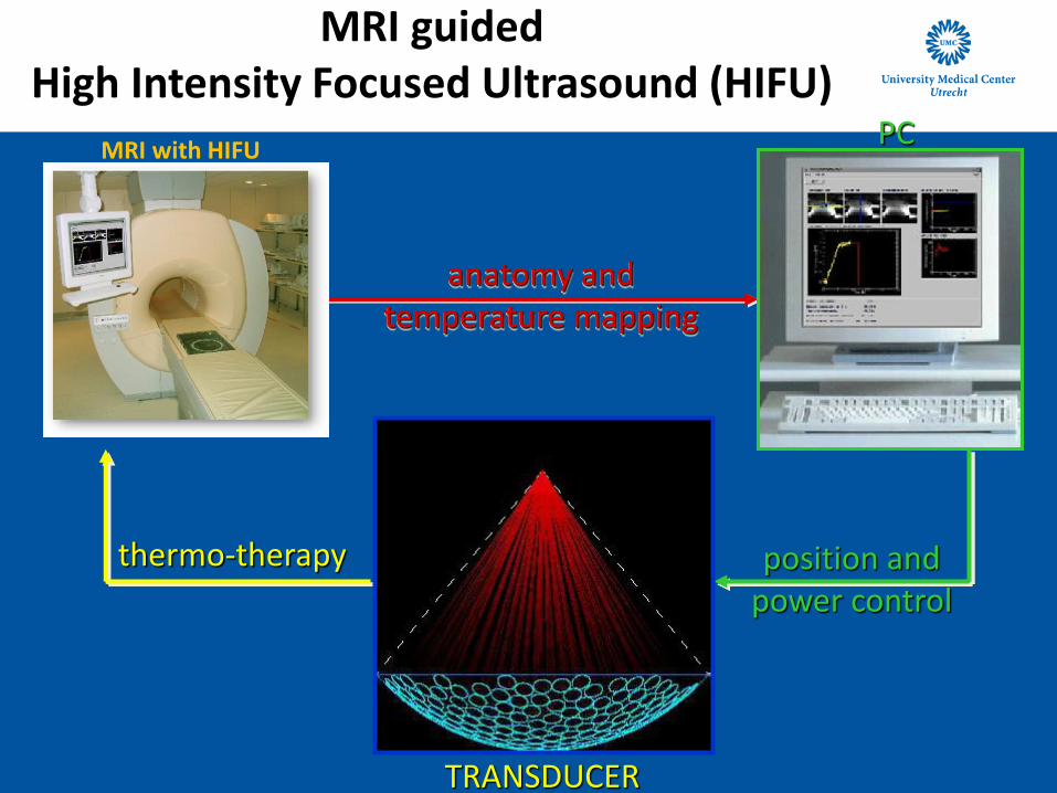
anatomy and temperature mapping
position and power control
TRANSDUCER
PC
thermo-therapy
MRI with HIFU
MRI guided High Intensity Focused Ultrasound (HIFU)

MRI guided Focused ultrasound:
clinical applications
After
Before
HIFU Ablation
Patient 1 (Uterine Fibroid) 20/11/2008, Philips/CHU Bordeaux (Pr Trillaud)
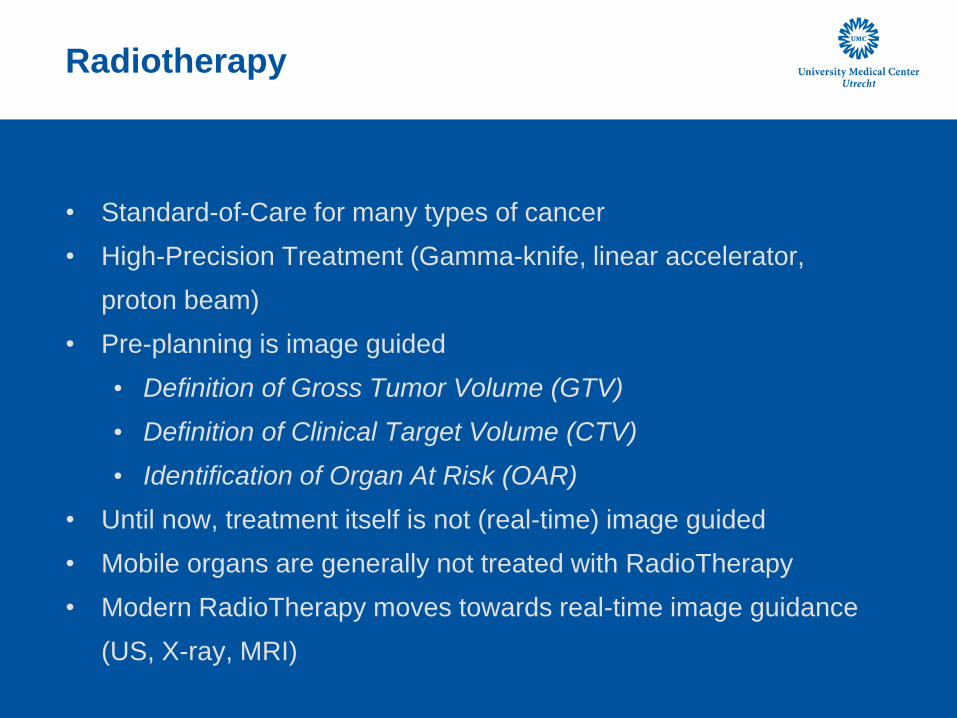
Radiotherapy
• Standard-of-Care for many types of cancer
• High-Precision Treatment (Gamma-knife, linear accelerator,
proton beam)
• Pre-planning is image guided
• Definition of Gross Tumor Volume (GTV)
• Definition of Clinical Target Volume (CTV)
• Identification of Organ At Risk (OAR)
• Until now, treatment itself is not (real-time) image guided
• Mobile organs are generally not treated with RadioTherapy
• Modern RadioTherapy moves towards real-time image guidance
(US, X-ray, MRI)

MRI offers superb soft tissue visualization
GTV primary tumor
rectum
GTV pathological lymph nodes (right)
bladder
GTV pathological lymph nodes (left)
CTVprimary (cervix, corpus uteri)
T2 weighted MRI sequence cervix

Real time breathing related motion
irregular breathing von Hippel Lindau kidney tumour

Breathing related motion
irregular breathing von Hippel Lindau kidney tumour

New 3D T2-FFE sequence with unique potential
lymph nodes breast cancer patients
• 3D T2-FFE with some intrinsic diffusion weighting, fat
suppression and black blood imaging
• Resolution 0.7x0.7x1 mm
• Geometrically correct, targeting 1.5 T MRL
T2-FFE MRI axillary lymph nodes
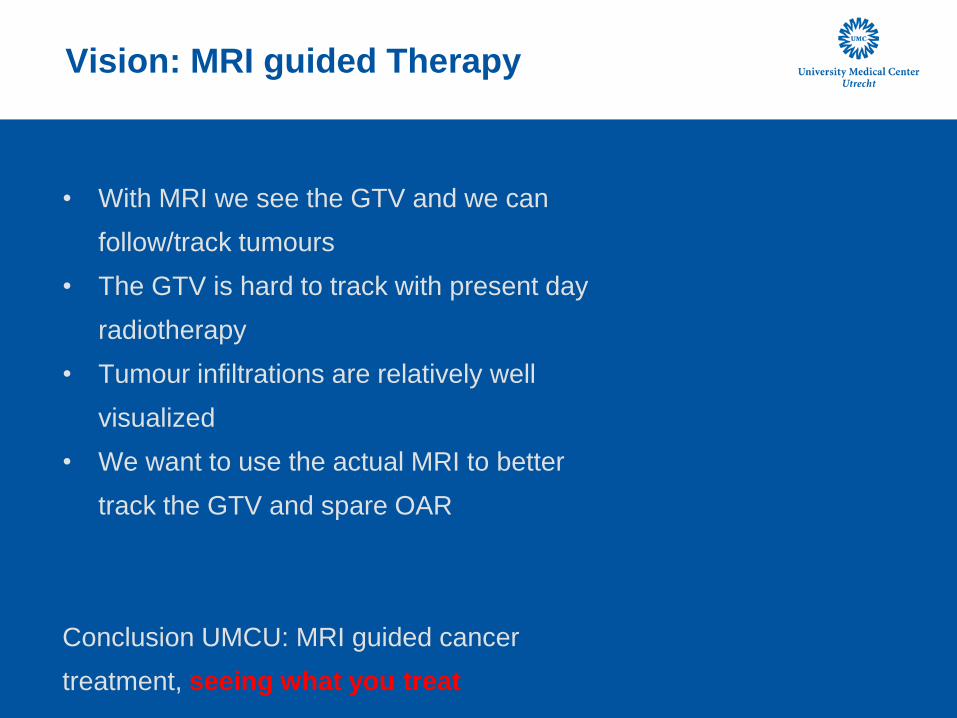
Vision: MRI guided Therapy
• With MRI we see the GTV and we can
follow/track tumours
• The GTV is hard to track with present day
radiotherapy
• Tumour infiltrations are relatively well
visualized
• We want to use the actual MRI to better
track the GTV and spare OAR
Conclusion UMCU: MRI guided cancer
treatment, seeing what you treat

Outline
• MRI guided RadioTherapy
• MRI guided Focused Ultrasound
• Image Guided Chemotherapy
• Center for Image Guided Oncological Interventions

Outline
• MRI guided RadioTherapy
• MRI guided Focused Ultrasound
• Image Guided Chemotherapy
• Center for Image Guided Oncological Interventions

Volumetric MR-HIFU ablation of breast
cancer using a dedicated breast platform
• Phase 1 study on patients with pathologically proven invasive
breast cancer (n=10)
• Feasibility study to assess safety and treatment accuracy in
patients with breast cancer
• Treat-and-resect protocol
• Surgery between 48 hours and one week after HIFU treatment
• Sentinel lymph node procedure
• Peritumoral injection of radioactive colloid just before surgery
• Detection during surgery

Dedicated breast MR-HIFU system
14
“Conventional” approach Dedicated system
with lateral sonication
transducer top view

Dedicated breast platform Sonalleve Breast MR-HIFU
Table top without covers
Water box with transducer
and motors
Close-up of breast cup, single-
element RF coil, and transducer

Results: 3 Tesla MRI

Results: MR-HIFU
3 4 5 6 7 8 9 10 1130
35
40
45
50
55
60
Time (s)
Temp (°C)
Results: after MR-HIFU ablation
• Minimal pain after MR-HIFU ablation
• Lumpectomy three days after MR-HIFU ablation
• Detection of sentinel lymph node
• No damage to pectoral muscle
• Pathology
• No macroscopic or microscopic changes visible in tumor tissue
(patient 1)
• Macroscopic changes visible in tumor tissue (patient 2) with
diameter corresponding with thermal dose threshold

Outline
• MRI guided RadioTherapy
• MRI guided Focused Ultrasound
• Image Guided Chemotherapy
• Center for Image Guided Oncological Interventions
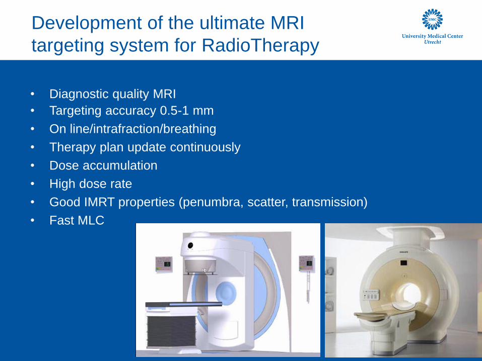
Development of the ultimate MRI
targeting system for RadioTherapy
• Diagnostic quality MRI
• Targeting accuracy 0.5-1 mm
• On line/intrafraction/breathing
• Therapy plan update continuously
• Dose accumulation
• High dose rate
• Good IMRT properties (penumbra, scatter, transmission)
• Fast MLC
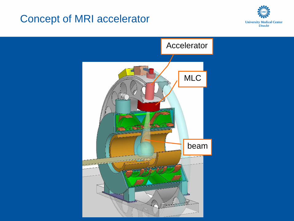
Concept of MRI accelerator
Accelerator
MLC
beam

Principle of active B field shielding
B0=Bpin-Bcin
B0out=Bpout-Bcout=0
0 T area
0 T area
Bpout Bcout
+ =
cross section through magnet

Radiation windows
accelerator
Gradient coils
RF coils
Present design:
field 24 cm long x 40 cm wide
window for beam

Specifications MRI accelerator
• 1.5 T diagnostic MRI
• 6 MV linac
• Simultaneous irradiation and MRI
• Continuously rotation
• Both directions
• 10 RPM
• 0.1 degree accuracy
• 1 mm spherical volume as target
• MLC Field size 24x56 cm2
• 7 mm leaves at isoc
Philips and Elekta go MRI
• Tumor characterization
• MRI simulation: delineation
• MRI guidance
• MRI treatment guidance external beam
• MRI treatment response assessment

Outline
• MRI guided RadioTherapy
• MRI guided Focused Ultrasound
• Liver- preclinical (large animal)
• Image Guided Chemotherapy
• Center for Image Guided Oncological Interventions

Magnetic Resonance guided HIFU of liver
and kidney
Challenges :
1. motion:
• Artifacts in MRI thermometry
• Target tracking/gated HIFU
2. Presence of ribs
• Block propagation of HIFU
• Burn risk in and around ribs
3. Highly perfused organs
• Cooling due to flow/perfusion
• High HIFU energy deposition
• Burn risk in near and far field

MRgHIFU for cancer therapy:
Challenges for HIFU in the liver
#1: Respiratory motion
• Motion Tracking: - MRI - Ultrasound
• HIFU guidance
- Beam steering - Gated sonication

MRgHIFU for cancer therapy:
Challenges for HIFU in the liver
#1: Respiratory motion
Abdomen of a free Breathing volunteer
Reference Position Current Position Difference after local registration
1) Global affine image registration 2) Optical flow based image registration image registration
Difference after global registration
Difference of both images
identify designated target position Look up the current target location
Update focal point position
For respiratory motion this is done at a rate of 10Hz with a latency below 100ms.
phased-array ultrasound transducer

Towards clinical MR-HIFU
treatment in the liver
Interventional
Pre-Planning
•Preparatory scanning
•Evalute acoustic access
•Determine diffractive/refractive effects
•Estimate duty-cycle/volumetric ablation rate (treatment duration)
Treatment
• Inter-costal firing
•Respiratory motion compensation
• Feed-back control
Evaluation
•Comparison of Contrast-Enhanced MR images to thermal dose maps
•Correlation of histopathological Findings and thermal dose maps
30

Treatment
• Inter-costal firing
• Respiratory motion compensation
• Temperature feed-back control
31
Liver ablations under clinical conditions

Continuous sonication with gated
thermometry
smearing of energy due to breathing motion

Reference: gated sonication
• adapted duration to duty-cycle
• isotropic shape
layered structures in beam-path strongly heated oedema! (all sonications)

34
Power calibration animal 4
shot pattern
What about the 8mm 349W sonication?
Gd-enhanced contrast
dose contours

Case study: Larger volume ablation
• ablation of region with 10mm diameter
• location: 35mm from skin, 12mm inside liver
• 7 cells @ 4mm
• cooling time between shots > 10 min
• check for oedema with T2 while waiting
feasible for unobstructed, shallow shot

Larger volume ablation: pathology puzzle
from DSC_8762 from DSC_8760 from DSC_8765
from DSC_8777 from DSC_8772
from DSC_8769
from DSC_8767

37
Treatment simulations for patient selection
MRI / CT based preplanning tools
Intercostal-firing: The problem
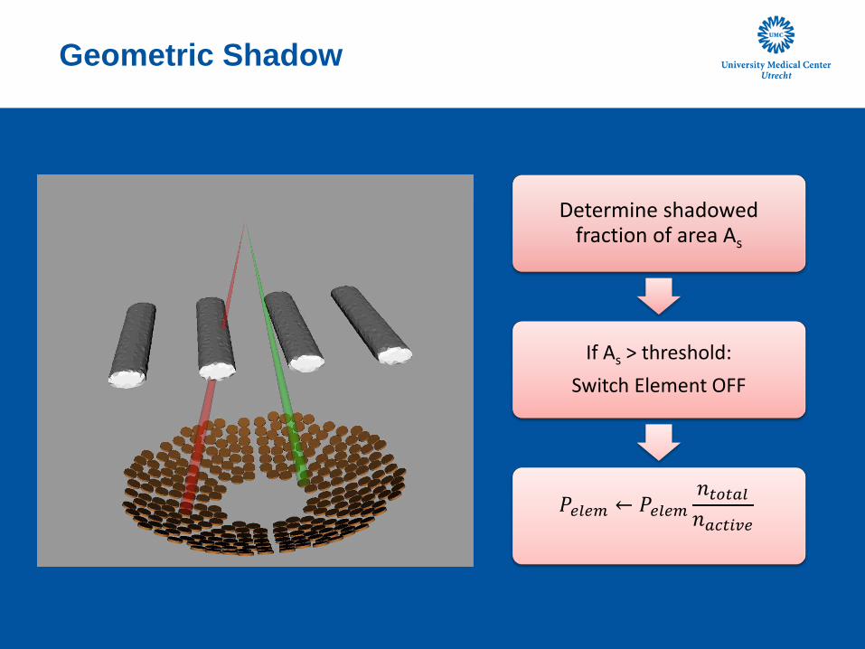
Geometric Shadow
Determine shadowed fraction of area As
If As > threshold:
Switch Element OFF
𝑃𝑒𝑙𝑒𝑚 ← 𝑃𝑒𝑙𝑒𝑚𝑛𝑡𝑜𝑡𝑎𝑙𝑛𝑎𝑐𝑡𝑖𝑣𝑒

Intensity-based thresholding
Element Directivity Pattern
10% of focus intensity @ |x| = 6mm
• Shadow casting ignores
transmission characteristics
• ‘Blocked’ elements can
contribute to focus
• ‘Unblocked’ elements
can expose the ribs
X
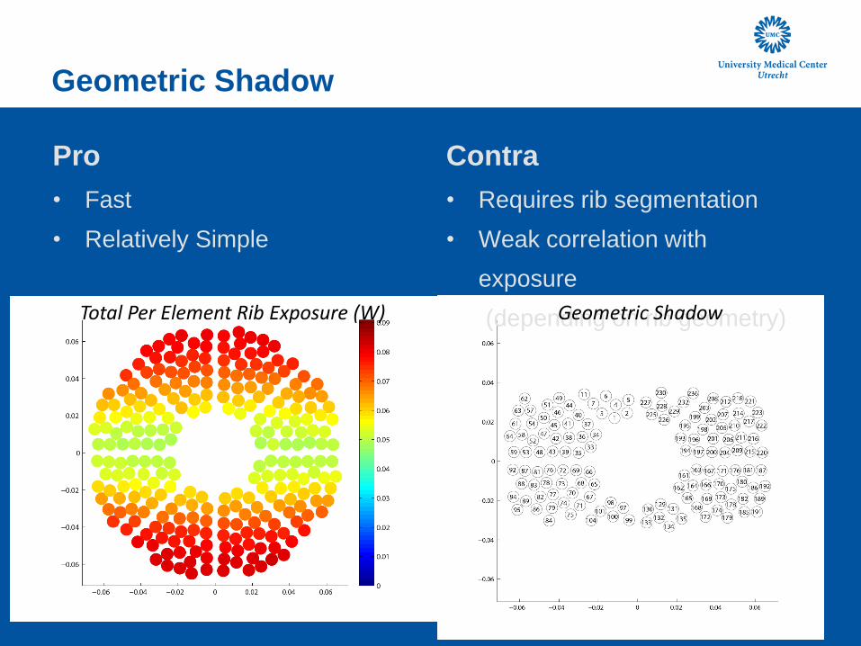
Geometric Shadow
Pro
• Fast
• Relatively Simple
Contra
• Requires rib segmentation
• Weak correlation with
exposure
(depending on rib geometry)
Total Per Element Rib Exposure (W) Geometric Shadow
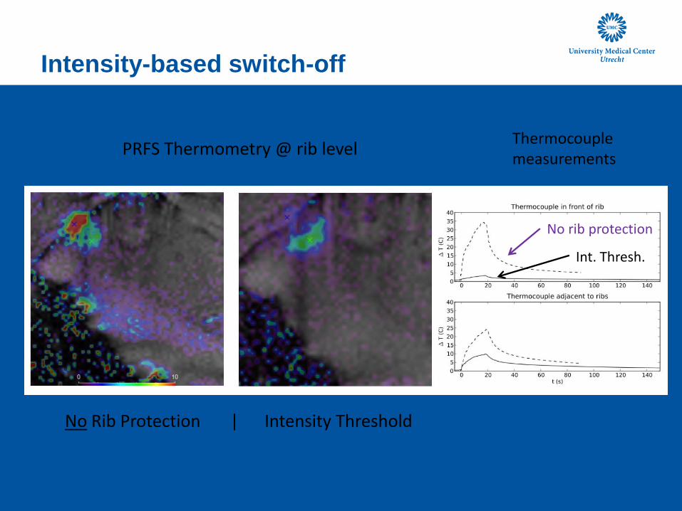
Intensity-based switch-off
No Rib Protection | Intensity Threshold
Thermocouple measurements
No rib protection
Int. Thresh.
PRFS Thermometry @ rib level

Outline
• MRI guided RadioTherapy
• MRI guided Focused Ultrasound
• Image Guided Chemotherapy
• Center for Image Guided Oncological Interventions

Center for Image Guided Oncological Interventions at Utrecht
Planning, real-time guidance, and monitoring of oncological interventions
Image guided Chemotherapy
MRI guided HIFU MRI guided Linear Accelerator
Applications of Focused
Ultrasound
• MR-HIFU for Image Guided, Local Drug Delivery
• Extravasation
• Membrane permeabilization
• Triggered drug release from nanocarriers

Blood Brain Barrier (BBB)
• The primary hurdle to the use of drugs in the
central nervous system for most small
molecule agents and all large molecule agents
• Methods developed to bypass the BBB are
invasive, non-targeted and/or require the
development of new drugs
Nathan McDannold, Harvard Univ

Benoit Larrat et al ISMRM 2011
Focused Ultrasound and Microbubbles: BBB opening

Applications of Focused
Ultrasound
• MR-HIFU for Image Guided, Local Drug Delivery and
Gene
• Extravasation
• Membrane permeabilization
• Triggered drug release from nanocarriers
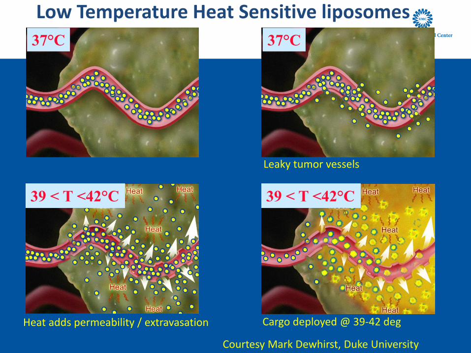
Low Temperature Heat Sensitive liposomes
37°C 37°C
Leaky tumor vessels
39 < T <42°C 39 < T <42°C
Heat adds permeability / extravasation
Cargo deployed @ 39-42 deg
Courtesy Mark Dewhirst, Duke University
49
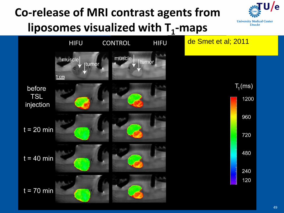
HIFU CONTROL HIFU
Co-release of MRI contrast agents from liposomes visualized with T1-maps
49
de Smet et al; 2011
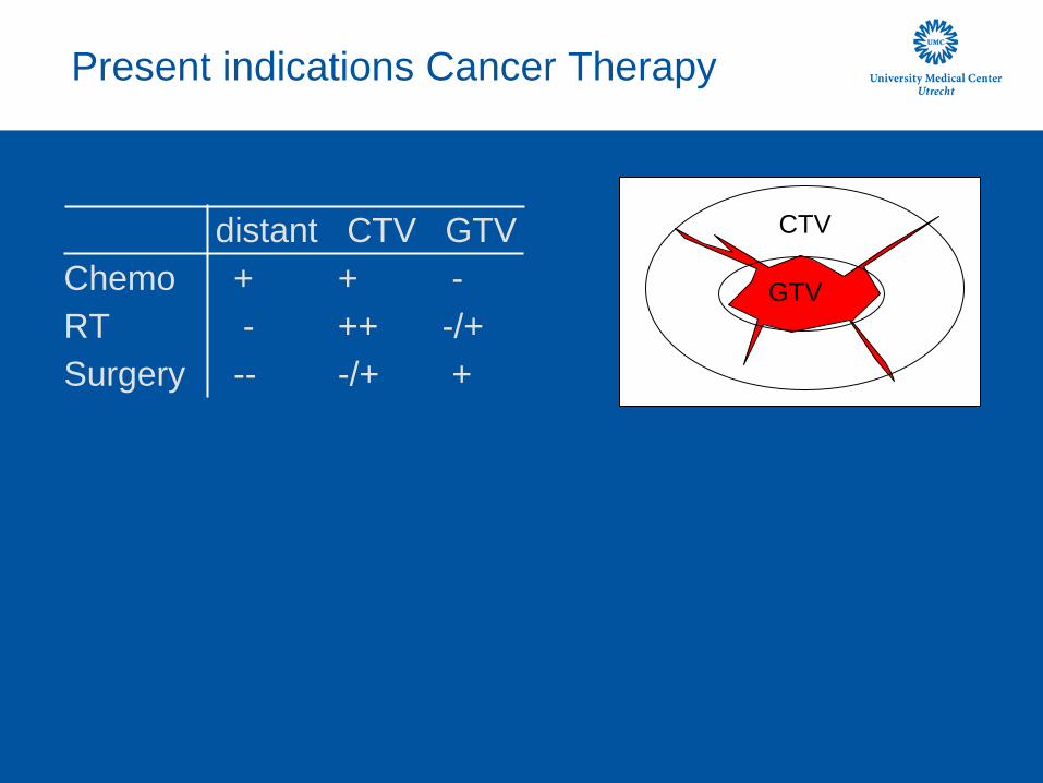
Present indications Cancer Therapy
distant CTV GTV
Chemo + + -
RT - ++ -/+
Surgery -- -/+ +
GTV
CTV

Development MR-HIFU and MR-LINAC
distant CTV GTV
Chemo + + -
RT - ++ ++
Surgery -- -/+ +
HIFU - + ++
MR-HIFU
MR-LINAC

HIFU
HDR robotic brachytherapy
MRI linac
Centre for Image Guided Oncological
Interventions (CIGOI)
MR-LINAC
MRI guided brachytherapy
MR-HIFU

Summary
• MRI guidance of RadioTherapy and MR guided HIFU will
set the next stage in high-precision tumor therapy • Synergy in development (motion descriptors, target
tracking)
• MR-LINAC will be the next standard-of-care in RadioTherapy
• MR-HIFU offers many complementary features and may be added to the Surgical, RT and Chemo therapies
• MR-HIFU may lead to MR guided Drug Delivery

Imaging Division, UMCU; Pharmaceutical Sciences UU Jan Lagendijk, Marco van Vulpen, Bas Raaijmakers, Baudouin Denis de Senneville, Mario Ries, Clemens Bos, Anna Yudina, Wilbert Bartels, Gert Storm, Maurice van den Bosch, Willem Mali et al
Philips Healthcare Charles Mougenot, Max Köhler, Sham Sokka and the Helsinki team Financial support European Union (Project SonoDrugs), CTMM project s VOLTA and HIFU-CHEM, ERC project Sound Pharma