MOVEMENT SYSTEM DIAGNOSES...
31
Property of Scheets, Crowner, Norton, Sahrmann, and Stith – DO NOT COPY WITHOUT PERMISSION April, 2014 MOVEMENT SYSTEM DIAGNOSES NEUROMUSCULAR CONDITIONS Examination Patricia L. Scheets, PT DPT NCS [email protected] Collaborators from Washington University Beth E. Crowner, PT DPT NCS MPPA Barbara J. Norton, PT PhD FAPTA Shirley A. Sahrmann, PT PhD FAPTA Jennifer S. Stith, PT PhD LCSW
-
Upload
trinhkhanh -
Category
Documents
-
view
217 -
download
0
Transcript of MOVEMENT SYSTEM DIAGNOSES...
Property of Scheets, Crowner, Norton, Sahrmann, and Stith – DO NOT COPY WITHOUT PERMISSION
April, 2014
MOVEMENT SYSTEM DIAGNOSES NEUROMUSCULAR CONDITIONS
Examination
Patricia L. Scheets, PT DPT NCS [email protected]
Collaborators from Washington University Beth E. Crowner, PT DPT NCS MPPA
Barbara J. Norton, PT PhD FAPTA Shirley A. Sahrmann, PT PhD FAPTA
Jennifer S. Stith, PT PhD LCSW
Property of Scheets, Crowner, Norton, Sahrmann, and Stith – DO NOT COPY WITHOUT PERMISSION
April, 2014
2
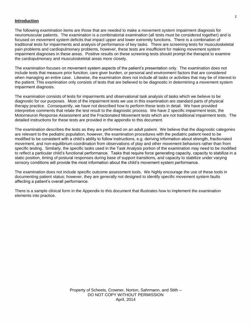
Introduction The following examination items are those that are needed to make a movement system impairment diagnosis for neuromuscular patients. The examination is a combinatorial examination (all tests must be considered together) and is focused on movement system deficits that impact upper and lower extremity functions. There is a combination of traditional tests for impairments and analysis of performance of key tasks. There are screening tests for musculoskeletal pain problems and cardiopulmonary problems, however, these tests are insufficient for making movement system impairment diagnoses in these areas. Positive results on these screening tests should prompt the therapist to examine the cardiopulmonary and musculoskeletal areas more closely. The examination focuses on movement system aspects of the patient’s presentation only. The examination does not include tests that measure prior function, care giver burden, or personal and environment factors that are considered when managing an entire case. Likewise, the examination does not include all tasks or activities that may be of interest to the patient. This examination only consists of tests that are believed to be diagnostic in determining a movement system impairment diagnosis. The examination consists of tests for impairments and observational task analysis of tasks which we believe to be diagnostic for our purposes. Most of the impairment tests we use in this examination are standard parts of physical therapy practice. Consequently, we have not described how to perform these tests in detail. We have provided interpretive comments that relate the test result to the diagnostic process. We have 2 specific impairment tests, the Motorneuron Response Assessment and the Fractionated Movement tests which are not traditional impairment tests. The detailed instructions for these tests are provided in the appendix to this document. The examination describes the tests as they are performed on an adult patient. We believe that the diagnostic categories are relevant to the pediatric population, however, the examination procedures with the pediatric patient need to be modified to be consistent with a child’s ability to follow instructions, e.g. deriving information about strength, fractionated movement, and non-equilibrium coordination from observations of play and other movement behaviors rather than from specific testing. Similarly, the specific tasks used in the Task Analysis portion of the examination may need to be modified to reflect a particular child’s functional performance. Tasks that require force generating capacity, capacity to stabilize in a static position, timing of postural responses during base of support transitions, and capacity to stabilize under varying sensory conditions will provide the most information about the child’s movement system performance. The examination does not include specific outcome assessment tools. We highly encourage the use of these tools in documenting patient status; however, they are generally not designed to identify specific movement system faults affecting a patient’s overall performance. There is a sample clinical form in the Appendix to this document that illustrates how to implement the examination elements into practice.

Property of Scheets, Crowner, Norton, Sahrmann, and Stith – DO NOT COPY WITHOUT PERMISSION
April, 2014
3
History/Systems Review From the patient and/or the medical record, obtain the following information:
medical diagnosis(es)
medical treatments which have an effect on muscle, nerve, or movement
prior level of function including use of assistive devices (what and how long)
complaint of motion sensitivity such as dizziness watching television, in crowds, or with driving Objective MENTAL STATUS
Perform traditional tests of mental status and basic cognitive functioning to determine:
Level of consciousness (LOC)
Attention
Ability to apply meaning to situational demands
Ability to follow instructions
Interpretation:
1Significant deficits may indicate the classification, Cognitive Deficit.
2 Moderate to significant deficits coupled with other movement related impairments indicate the use of a
modifier to the diagnosis, e.g. confusion, cognitive deficits.
3 Mild deficits are relevant to treatment but are unlikely to be relevant to diagnosis.
JOINT LIMITATION
Perform traditional tests of joint range of motion.
Interpretation:
1Use information about joint limitation to guide the use of a descriptor to the diagnosis, e.g. with
biomechanical deficit, hip. Significant limitation should guide the therapist to examine for a specific musculoskeletal diagnosis.
MUSCLE TONE / HYPEREXCITABILITY
Motorneuron Response Assessment
For the purposes of diagnosis, assessment of muscle tone / hyperexcitability is important in its relationship to a patient’s ability to fractionate movement. Patient’s with more signs of hyperexcitability such as spasticity, the inability to relax after effort, and associated reactions, are likely to have difficulty isolating movement of one limb segment from movement of other limb segments.
i At different points in time of a patient’s recovery from a neurological deficit, the signs of
hyperexcitability may be apparent before there is return of active movement. In these situations, the signs of hyperexcitability may give an earlier indication of the patient’s movement system diagnosis than the patient’s ability to fractionate movement. The Motorneuron Response Assessment (MRA) is a tool that is designed specifically to identify the multiple signs of hyperexcitability. The tool is reliable among multiple testers and valid when compared with the Ashworth scale.
ii The MRA is the preferred test for measuring hyperexcitability for the purposes of diagnosis. It is
described in the Appendix to this document.
Ashworth or Modified Ashworth Scales The Ashworth or Modified Ashworth Scales are commonly used measures of spasticity.
iii,iv,v,vi,vii,viii,ix We have not formally
studied the relationship between scores on the Ashworth or Modified Ashworth scale and the results of the Fractionated Movement test.
Property of Scheets, Crowner, Norton, Sahrmann, and Stith – DO NOT COPY WITHOUT PERMISSION
April, 2014
4
Rigidity
Rigidity is defined as persistent muscle activity at rest. Determine “yes” or “no” rigidity is present by slowly moving the extremities and observing them at rest. It may be useful to have the patient attend to a secondary task during testing.
x
yes = resistance is present in both directions and there is the appearance of persistent muscle activity at rest
Interpretation:xi,xii,xiii
1 MRA Classification of Moderate, Marked, or Severe or Modified Ashworth scores of 3 or 4 are indicative of
Fractionated Movement Deficit 2 MRA Classification of Flaccid is indicative of Force Production Deficit with poor prognosis
3 MRA Classification of Normal or Mild when there is a CNS lesion is indicative of Movement Pattern
Coordination Deficit or Force Production Deficit with good prognosis 4If the LE MRA category is different from the UE MRA category, consider making an UE and LE diagnosis.
5 Patients with rigidity related to brain injury often demonstrate varying levels of muscle tone based on
position. In this situation, attempt to measure the amount of excitability during functional tasks and consider that level when making a diagnosis.
MOVEMENT
Spontaneous Movement
Determine “yes” or “no” there is spontaneous movement by observing the patient and noting whether the patient can move against gravity. yes = movement against gravity in at least 2 extremities
Fractionated Movement (FM)
Determine “yes” or “no” there is fractionated movement by completing the following standardized examination detailed in the Appendix.
Interpretation:xiv,xv, xvi
1FM is an important test for patients with CNS lesions. Patients who have non-fractionated movement are
likely to be associated with higher degrees of hyperexcitability and are likely to have a diagnosis of Fractionated Movement Deficit.
Selective Control Assessment of the Lower Extremity (SCALE)
The SCALE is commonly used in the pediatric population to measure the ability to fractionated movement.xvii
We have not formally studied the relationship between scores on the SCALE and the MRA or other muscle tone scale. However, we expect that values ≤ 5 for either LE would indicate Fractionated Movement Deficit.
Strength
Follow principles of manual muscle testing as defined by Kendallxviii
in testing the following muscle groups: shoulder flexion, elbow flexion, elbow extension, wrist extension, hip extension, hip flexion, hip abduction, knee extension, dorsiflexion, weight bearing plantarflexion. According to these principles, only fractionated movement is tested. Use the following scale: 0 = no contraction felt in muscle 1 = a feeble contraction may be felt or the tendon may become prominent but there is not visible movement 2 = able to move the part through a small arc of motion with gravity lessened 3 = able to move the part into the test position and hold against gravity 4 = able to hold the test position against gravity and moderate pressure 5 = able to hold the test position against gravity and maximum pressure
Property of Scheets, Crowner, Norton, Sahrmann, and Stith – DO NOT COPY WITHOUT PERMISSION
April, 2014
5
Interpretation:
1Mucles grades less than 4/5 in the majority of muscles in one or more limbs or more focal weakness in a key
muscle group is indicative of Force Production Deficit with a good prognosis.
2A muscle grade less than 3+/5 in the majority of muscles in one or more limbs or more focal weakness in a
key muscle group is indicative of Force Production Deficit.
Fatigue
The purpose of this test is to determine if the patient demonstrates signs of skeletal muscle fatigue. Determine “yes” or “no,” the patient demonstrates skeletal muscle fatigue by performing one of the following tests. Two levels of testing are described; the lower level test is used only if the patient cannot participate in the higher level test. Low level test: the patient is supine
flex the shoulder to 90 and support the upper arm; ask the patient to extend the elbow 10 times
flex the hip to 90 and support the upper leg; ask the patient to extend the knee 10 times High level test: ask the patient to come to standing from an 18-20 inch surface without UE support 10 times. If
the patient cannot initiate standing without UE support, he may use UE support during the initiation phase only. For either test note a decrease in range of motion of the movement and change in speed. yes = decrement in range of motion observed
Interpretation: 1Fatigue noted during either test is indicative of Force Production Deficit.
2The purpose of these tests is to draw out skeletal muscle fatigue; if the patient fails for other reasons, such
as shortness of breath, increased heart rate, etc, see test for activity tolerance and consider a cardiopulmonary diagnosis.
Motor Planning
Determine “yes” or “no” the patient has deficits in motor planning as evidenced by difficulty organizing necessary movement patterns into purposeful actions yes = inconsistency between the degree of fractionated movement produced in isolated testing and in functional
activities
Interpretation: 1Patients with significant motor planning deficits are not likely to fit into one of the defined categories.
Non-equilibrium Coordination UE Accuracy: ask the patient to touch the examiner’s finger then touch his (the patient’s) nose. The examiner’s finger should be at a distance that requires the patient to extend his upper extremity fully and should be placed at 5 varying points before the patient.
xix The patient is told to try to hit the target, i.e. the tip of the finger and the tip of the nose. Count
the number of times the patient hits the target (nose or finger), and determine if the patient is: not impaired 0-1 inaccuracies mildly impaired 2-6 inaccuracies markedly impaired 7-10 inaccuracies LE Accuracy: ask the patient to place the heel of one foot on the knee of the other leg and slide the heel down and up
the tibia 5 times.19
Tell the patient to be as precise as possible. Count the number of times the heel does not maintain contact with the tibia. Determine if the patient is: not impaired 0-1 inaccuracies mildly impaired 2-6 inaccuracies markedly impaired 7-10 inaccuracies
Property of Scheets, Crowner, Norton, Sahrmann, and Stith – DO NOT COPY WITHOUT PERMISSION
April, 2014
6
UE Reciprocal Movement: ask the patient to rapidly supinate and pronate the forearm for 10-20 seconds.19
Determine if the patient is: normal slow LE Reciprocal Movement: ask the patient to sit with the heel on the ground and rapidly tap the toe for 10-20 seconds.
19
Determine if the patient is: normal slow
Interpretation: 1More than mild deficits are indicative Dysmetria.
SENSATION
Joint Position Sense
Have the patient sit or lie with the heel and leg supported. Grasp the lateral aspect of the great toe and move the toe into flexion and extension passively.
19 Encourage the patient to relax. Show the patient that a position into flexion is “down”
and a position into extension is “up.” Ask the patient to close his eyes. Move the toe into flexion or extension randomly 5 times. After each movement, ask the patient to tell you if the toe is up or down. Determine the accuracy of the patient’s responses using the following scale: not impaired = no inaccuracies mildly impaired = inaccurate 1-2 times moderately impaired = inaccurate 3-4 times severely impaired = inaccurate 5 or more times Repeat the test in the same fashion at the ankle and knee.
Interpretation:
1More than mild deficits at the toe and ankle or mild deficits at the ankle and knee are indicative of Sensory
Detection Deficit.
2If a loss of joint position sense is coupled with a marked motor impairment, the diagnosis is most likely to be
related to the motor impairment; in this circumstance consider using “with sensory loss” as a modifier to the diagnosis.
Contraversive Pushing or Backward Disequilibrium Behavior
This movement behavior may be observed in either the medial/lateral (contraversive pushing) or posterior (retropulsive pushing) directions. The hallmark of contraversive pushing behavior is: 1) abduction and extension of the limbs in sitting or standing either spontaneously or when changing position and 2) resistance to passive correction. Contraversive pushing behavior can be measured using the Scale for Contraversive Pushing.
xx, xxi
Retropulsive pushing behavior is characterized by: 1) trunk extension in sitting or posterior displacement of COM through ankle PF in standing and 2) resistance to passive correction. Retropulsive pushing behavior may be measured using the Backward Disequilibrium Scale.
xxii
yes = both hallmark signs of either contraversive or retropulsive pushing behavior present or scores > 0 on the “Use of the nonparetic extremities” and “Resistance to passive correction of tilted posture” subscales on the SCP.
xxiii
Interpretation:
1Contraversive or Retropulsive pushing behavior is indicative of Postural Vertical Deficit.
2If the deficit related to pushing behavior is significant and coupled with a motor deficit, the diagnosis is most
likely Postural Vertical Deficit.
Property of Scheets, Crowner, Norton, Sahrmann, and Stith – DO NOT COPY WITHOUT PERMISSION
April, 2014
7
Vertical Orientation (without pushing behavior)
Observe the patient during postural control tests and ambulation for displacement of the center of mass toward one side or in the posterior direction. Provide physical assistance and/or visual cues to see if the patient can correct his vertical orientation. yes = COM consistently shifted away from the COM alignment; able to correct with physical assistance or cues
Interpretation:
1Altered vertical orientation without pushing behavior is indicative of Sensory Selection and Weighting Deficit
Disregard
Observe the patient’s attention to his environment and note any inconsistencies in attention to one side more than the other. Examples may include lying on one side of the bed or positioning himself toward one side of a chair, lack of head turning to one side, inability to clear objects on one side during locomotion. yes = decreased attention to one side
Interpretation:
1Although disregard for a side of the body is often associated with altered perception of midline, it is not
always the case.
2If the test for disregard is positive but the test for midline perception is not, use “with disregard,” as a modifier
to the diagnosis; in this situation the diagnosis should be consistent with the motor deficit.
Sensitivity to Sensory Stimuli Ask the patient to visually track your finger to the side, up and down, and randomly while holding the head still. Ask the patient if he has any symptoms while performing this test such as dizziness, headache, nausea, etc. Note any signs of gaze aversion. If the patient is not impaired in this test but reports other specific motion related triggers of symptoms,
modify test in accordance with the patient’s specific complaints, e.g. 180 or 360 turns, repeated turns, repeated head turning, etc. Observe the patient’s response to auditory and visual motion cues in the environment. Look for increased postural sway, loss of balance, and/or stoppage of movement in the presence of these sensory cues. Observe the patient for self-stimulation behaviors such as repeated rocking, spinning, or hitting of the limb or head.
yes = dizziness or dizziness related symptoms and/or signs of movement deterioration related to sensory stimuli while performing any of the above tests
Interpretation:
1Symptoms with eye and/or head movements may indicate the diagnosis Sensory Selection and Weighting
Deficit.
2If there are abnormal motor findings with eye movements and no diagnosed neurological pathology, consult
a physician. 3Signs of movement deterioration with sensory stimuli or efforts to increase sensory stimuli may indicate the
diagnosis Sensory Selection and Weighting Deficit.
PAIN Ask the patient to rate the severity of pain on a scale of 0 to 10 where: 0 = no pain 10 = extreme pain Determine “yes” or “no” the pain is musculoskeletal in origin.

Property of Scheets, Crowner, Norton, Sahrmann, and Stith – DO NOT COPY WITHOUT PERMISSION
April, 2014
8
yes = pain decreases with change in alignment, positioning, or support
Interpretation:
1This test is used as a screening test for musculoskeletal pain; this system of diagnoses does not address
musculoskeletal pain syndromes.
ACTIVITY TOLERANCE Determine “yes” or “no” the patient is able to tolerate activity by assessing vital signs and signs of distress during exertion. No = decrease in heart rate, irregular rhythm, or inability to recover to resting rate after two minutes or decrease in systolic blood pressure, increase in diastolic blood pressure greater than 10 mm HG or labored breathing as evidenced by increased use of accessory muscles and increased rate or sustained decreased in o2 saturation below 90 or other medical guideline or increase in intracranial pressure above 15 or other medical guideline
Interpretation: 1This test is a screening test for cardiopulmonary impairments; this system of diagnoses does not address specific
cardiopulmonary syndromes.
TASK ANALYSIS
Analysis of mobility consists of systematic observation of the kinematic changes that occur during changes in position or alignment. These changes in angles and displacement of limbs and limb segments are the movement components of a task. While we appreciate that there are many movement patterns that a patient may use to be successful in completing a task, we have identified essential movement components for each task. We believe that the essential movement components describe the movement pattern for each task that is the most common, potentially the most efficient given the demands of the task, and the least likely to produce unnecessary stress on the musculoskeletal system. We have included in the examination, those tasks on which we believe patients of different types perform differently. Our purpose is not to examine every task that a person needs to perform in every day life, but rather, to examine those tasks we feel are necessary in order to diagnose the patient’s movement system problem. The tasks that are included in the examination are:
quiet sitting sit to stand quiet standing standing feet together step-up gait complex gait
And for patients with primary upper extremity involvement: reach grasp in-hand manipulation
Additional tasks may be needed to identify deficits, especially for higher level adult patients and in children. This additional testing may especially be necessary when making distinctions among patients with Movement Pattern Coordination Deficit, Force Production Deficit, or Sensory Selection and Weighting Deficit. Examples of these tasks in adults might include:
Property of Scheets, Crowner, Norton, Sahrmann, and Stith – DO NOT COPY WITHOUT PERMISSION
April, 2014
9
Standing on foam Standing on a tilt board Standing on a narrow beam Stair climbing Walking on heels and toes
Examples of additional or substitute tasks in children might include: Prone on elbows Rolling Pull to sit Creeping Floor to stand Running Jumping Hopping
Analysis of movement tasks in phases is useful for precise systematic observation. For all mobility tasks, the tester must observe all 3 of the following phases: Initiation those changes that occur in order to overcome inertia of the body at rest Execution intersegmental movements that allow for the movement of COM into a new position Termination those changes that occur to decelerate the movement of the COM as the body stabilizes into a
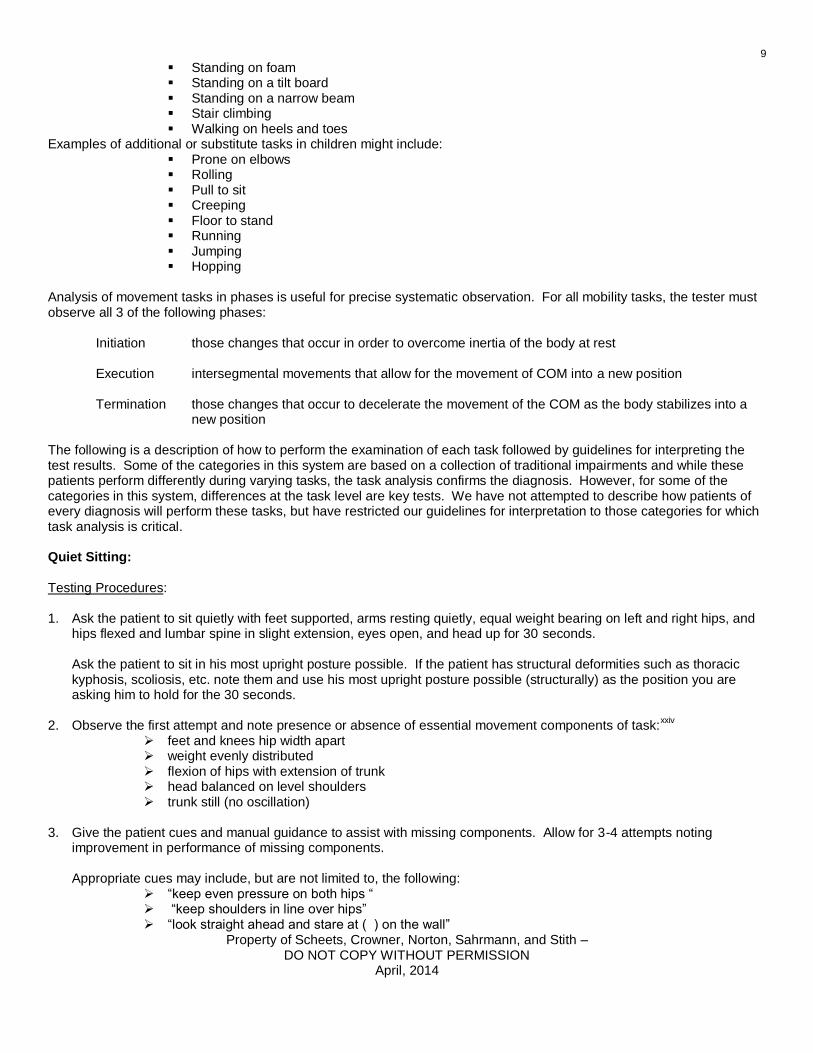
new position The following is a description of how to perform the examination of each task followed by guidelines for interpreting the test results. Some of the categories in this system are based on a collection of traditional impairments and while these patients perform differently during varying tasks, the task analysis confirms the diagnosis. However, for some of the categories in this system, differences at the task level are key tests. We have not attempted to describe how patients of every diagnosis will perform these tasks, but have restricted our guidelines for interpretation to those categories for which task analysis is critical. Quiet Sitting: Testing Procedures: 1. Ask the patient to sit quietly with feet supported, arms resting quietly, equal weight bearing on left and right hips, and
hips flexed and lumbar spine in slight extension, eyes open, and head up for 30 seconds.
Ask the patient to sit in his most upright posture possible. If the patient has structural deformities such as thoracic kyphosis, scoliosis, etc. note them and use his most upright posture possible (structurally) as the position you are asking him to hold for the 30 seconds.
2. Observe the first attempt and note presence or absence of essential movement components of task:xxiv
feet and knees hip width apart weight evenly distributed flexion of hips with extension of trunk head balanced on level shoulders trunk still (no oscillation)
3. Give the patient cues and manual guidance to assist with missing components. Allow for 3-4 attempts noting
improvement in performance of missing components.
Appropriate cues may include, but are not limited to, the following: “keep even pressure on both hips “ “keep shoulders in line over hips” “look straight ahead and stare at ( ) on the wall”
Property of Scheets, Crowner, Norton, Sahrmann, and Stith – DO NOT COPY WITHOUT PERMISSION
April, 2014
10
“relax your arms at your sides” Manual guidance should be proximal. Patients should be discouraged from using UE to maintain sitting.
Quiet Sitting
Observation Interpretation
gross abnormality; COM shifted significantly from midline
Biomechanical deficit (modifier)
unable to sit unsupported; appears weak; would fall without support
Force Production Deficit (FPD)
sits asymmetrically; may require assistance but only minimal
Fractionated Movement Deficit if associated with moderate, marked, or severe LE MRA
Sensory Selection and Weighting Deficit
patient resists correction of altered midline position or loss of balance
Perceptual Deficit
improvement in performance with repetition/practice
Movement Pattern Coordination Deficit (MPCD) Fractionated Movement Deficit if associated with
Moderate, Marked, or Severe MRA FPD Sensory Selection and Weighting Deficit
decrement in performance with repetition FPD (may demonstrate brief initial improvement followed by decrement in performance)
excessive sway at trunk; requires UE support; no improvement with practice
Dysmetria
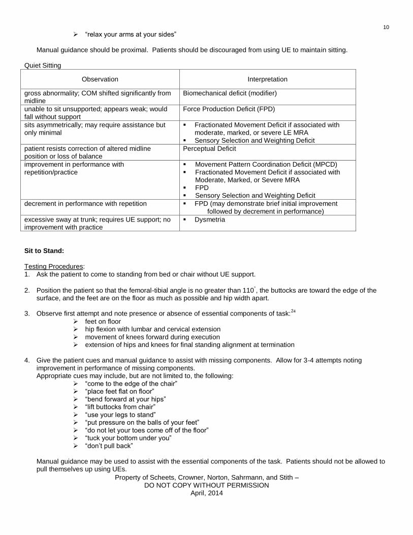
Sit to Stand: Testing Procedures: 1. Ask the patient to come to standing from bed or chair without UE support.
2. Position the patient so that the femoral-tibial angle is no greater than 110, the buttocks are toward the edge of the surface, and the feet are on the floor as much as possible and hip width apart.
3. Observe first attempt and note presence or absence of essential components of task:
24
feet on floor hip flexion with lumbar and cervical extension movement of knees forward during execution extension of hips and knees for final standing alignment at termination
4. Give the patient cues and manual guidance to assist with missing components. Allow for 3-4 attempts noting
improvement in performance of missing components. Appropriate cues may include, but are not limited to, the following:
“come to the edge of the chair” “place feet flat on floor” “bend forward at your hips” “lift buttocks from chair” “use your legs to stand” “put pressure on the balls of your feet” “do not let your toes come off of the floor” “tuck your bottom under you” “don’t pull back”
Manual guidance may be used to assist with the essential components of the task. Patients should not be allowed to pull themselves up using UEs.

Property of Scheets, Crowner, Norton, Sahrmann, and Stith – DO NOT COPY WITHOUT PERMISSION
April, 2014
11
5. If physical assistance is required, give only the assistance necessary at each phase. At each phase, relax or release
the assistance at least momentarily and observe patient’s movement response. Sit to Stand
Observation Interpretation
initiation requires assistance in order to initiate lift of buttocks from chair; if support removed, patient falls rapidly into chair
FPD
unable to passively position feet appropriately resulting in a more challenging starting position; may require assistance
Biomechanical deficit (modifier)
lack of preparatory movements or very slow preparatory movements; may require assistance; if support removed, patient falls slowly into chair
Hypokinesia
increased BOS FPD Dysmetria
repeated efforts; momentum strategy FPD Hypokinesia
excessive trunk sway Dysmetria
execution extends knees before hips in first half of movement sequence; may push on legs to extend trunk; may require assistance; if support removed segments rapidly fall in direction opposite of movement
FPD Sensory Detection
Deficit if associated with loss of JPS
requires assistance; associated with joint pain or stiffness
Biomechanical deficit (modifier)
arrest of ongoing movement; may require assistance Hypokinesia
altered sequencing of segmental movement ( most commonly insufficient DF of leg over foot); improves with guidance and practice
MPCD
shifts COM toward weaker side or back; resists correction; may fix foot (feet) and push away
Postural Vertical Deficit
shifts COM to one side; improves with practice and instruction
Sensory Selection and Weighting Deficit
excessive trunk sway Dysmetria
termination sway at ankle; may require a step MPCD
shifts COM toward weaker side or back; resists correction; may fix foot (feet) and push away
Postural Vertical Deficit
repeated stepping in order to find and maintain balance
Dysmetria FPD
excessive trunk sway at hips Dysmetria
Knee hyperextension against surface; improves with manual cues and instruction
MPCD
Knee hyperextension against surface; no change with manual cues and instruction
FPD Sensory Detection
Deficit
Property of Scheets, Crowner, Norton, Sahrmann, and Stith – DO NOT COPY WITHOUT PERMISSION
April, 2014
12
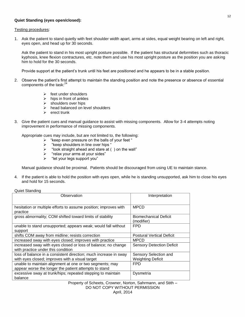
Quiet Standing (eyes open/closed): Testing procedures: 1. Ask the patient to stand quietly with feet shoulder width apart, arms at sides, equal weight bearing on left and right,
eyes open, and head up for 30 seconds.
Ask the patient to stand in his most upright posture possible. If the patient has structural deformities such as thoracic kyphosis, knee flexion contractures, etc. note them and use his most upright posture as the position you are asking him to hold for the 30 seconds. Provide support at the patient’s trunk until his feet are positioned and he appears to be in a stable position.
2. Observe the patient’s first attempt to maintain the standing position and note the presence or absence of essential components of the task:
24
feet under shoulders hips in front of ankles shoulders over hips head balanced on level shoulders erect trunk
3. Give the patient cues and manual guidance to assist with missing components. Allow for 3-4 attempts noting
improvement in performance of missing components.
Appropriate cues may include, but are not limited to, the following: “keep even pressure on the balls of your feet “ “keep shoulders in line over hips “ “look straight ahead and stare at ( ) on the wall” “relax your arms at your sides” “let your legs support you”
Manual guidance should be proximal. Patients should be discouraged from using UE to maintain stance.
4. If the patient is able to hold the position with eyes open, while he is standing unsupported, ask him to close his eyes and hold for 15 seconds.
Quiet Standing
Observation Interpretation
hesitation or multiple efforts to assume position; improves with practice
MPCD
gross abnormality; COM shifted toward limits of stability Biomechanical Deficit (modifier)
unable to stand unsupported; appears weak; would fall without support
FPD
shifts COM away from midline; resists correction Postural Vertical Deficit
increased sway with eyes closed; improves with practice MPCD
increased sway with eyes closed or loss of balance; no change with practice under this condition
Sensory Detection Deficit
loss of balance in a consistent direction; much increase in sway with eyes closed; improves with a visual target
Sensory Selection and Weighting Deficit
unable to maintain alignment at one or two segments; may appear worse the longer the patient attempts to stand
FPD
excessive sway at trunk/hips; repeated stepping to maintain balance
Dysmetria

Property of Scheets, Crowner, Norton, Sahrmann, and Stith – DO NOT COPY WITHOUT PERMISSION
April, 2014
13
Feet Together (eyes open/closed): Perform this test if the patient is successful with Quiet Standing with eyes open. Testing procedures: 1. Ask the patient to stand quietly with feet touching at toes and heels, arms at sides, equal weight bearing on left and
right, eyes open, and head up for 15 seconds.
Ask the patient to stand in his most upright posture possible. If the patient has structural deformities such as thoracic kyphosis, knee flexion contractures, etc. note them and use his most upright posture as the position you are asking him to hold for the 15 seconds. Provide support at the patient’s trunk until his feet are positioned and he appears to be in a stable position.
2. Observe the patient’s first attempt to maintain the feet together position and note the presence or absence of essential components of the task:
feet together hips in front of ankles shoulders over hips head balanced on level shoulders erect trunk
3. Give the patient cues and manual guidance to assist with missing components. Allow for 3-4 attempts noting
improvement in performance of missing components.
Appropriate cues may include, but are not limited to, the following: “keep even pressure on the balls of your feet“ “keep shoulders in line over hips“ “look straight ahead and stare at ( ) on the wall” “relax your arms at your sides” “let your legs support you.”
Manual guidance should be proximal. Patients should be discouraged from using UE to maintain stance.
4. If the patient is able to hold the position with eyes open, while he is standing unsupported, ask him to close his eyes and hold for 15 seconds.
Feet Together
Observation Interpretation
hesitation or multiple efforts to assume position; improves with practice
MPCD
unable to stand unsupported; appears weak; would fall without support
FPD
increased sway with eyes closed; improves with practice MPCD
increased sway with eyes closed or loss of balance; no change with practice under this condition
Sensory Detection Deficit
loss of balance in a consistent direction; much increase in sway with eyes closed; improves with a visual target
Sensory Selection and Weighting Deficit
unable to maintain alignment at one or two segments; may appear worse the longer the patient attempts to stand
FPD
excessive sway at trunk/hips; repeated stepping to maintain balance
Dysmetria
Property of Scheets, Crowner, Norton, Sahrmann, and Stith – DO NOT COPY WITHOUT PERMISSION
April, 2014
14
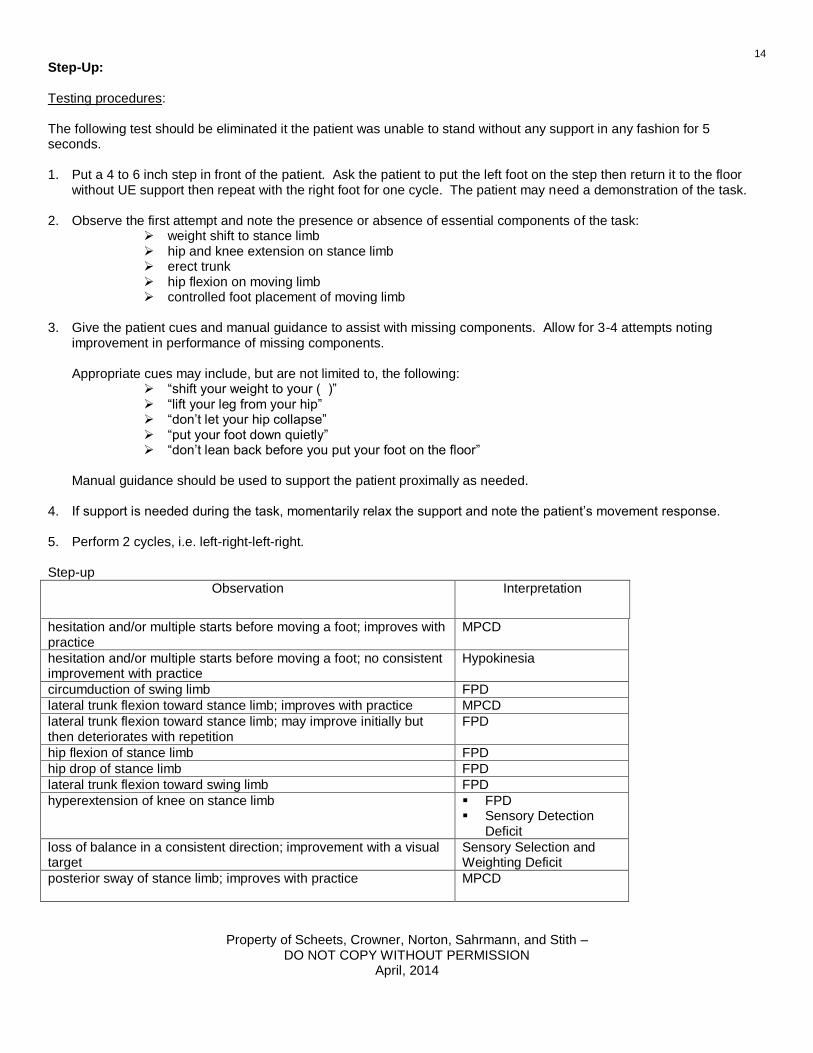
Step-Up: Testing procedures: The following test should be eliminated it the patient was unable to stand without any support in any fashion for 5 seconds. 1. Put a 4 to 6 inch step in front of the patient. Ask the patient to put the left foot on the step then return it to the floor
without UE support then repeat with the right foot for one cycle. The patient may need a demonstration of the task. 2. Observe the first attempt and note the presence or absence of essential components of the task:
weight shift to stance limb hip and knee extension on stance limb erect trunk hip flexion on moving limb controlled foot placement of moving limb
3. Give the patient cues and manual guidance to assist with missing components. Allow for 3-4 attempts noting
improvement in performance of missing components.
Appropriate cues may include, but are not limited to, the following: “shift your weight to your ( )” “lift your leg from your hip” “don’t let your hip collapse” “put your foot down quietly” “don’t lean back before you put your foot on the floor”
Manual guidance should be used to support the patient proximally as needed.
4. If support is needed during the task, momentarily relax the support and note the patient’s movement response. 5. Perform 2 cycles, i.e. left-right-left-right. Step-up
Observation Interpretation
hesitation and/or multiple starts before moving a foot; improves with practice
MPCD
hesitation and/or multiple starts before moving a foot; no consistent improvement with practice
Hypokinesia
circumduction of swing limb FPD
lateral trunk flexion toward stance limb; improves with practice MPCD
lateral trunk flexion toward stance limb; may improve initially but then deteriorates with repetition
FPD
hip flexion of stance limb FPD
hip drop of stance limb FPD
lateral trunk flexion toward swing limb FPD
hyperextension of knee on stance limb FPD Sensory Detection
Deficit
loss of balance in a consistent direction; improvement with a visual target
Sensory Selection and Weighting Deficit
posterior sway of stance limb; improves with practice MPCD
Property of Scheets, Crowner, Norton, Sahrmann, and Stith – DO NOT COPY WITHOUT PERMISSION
April, 2014
15
Observation Interpretation
excessive sway at trunk/hips; may overshoot foot placement on step; repeated stepping to maintain balance
Dysmetria
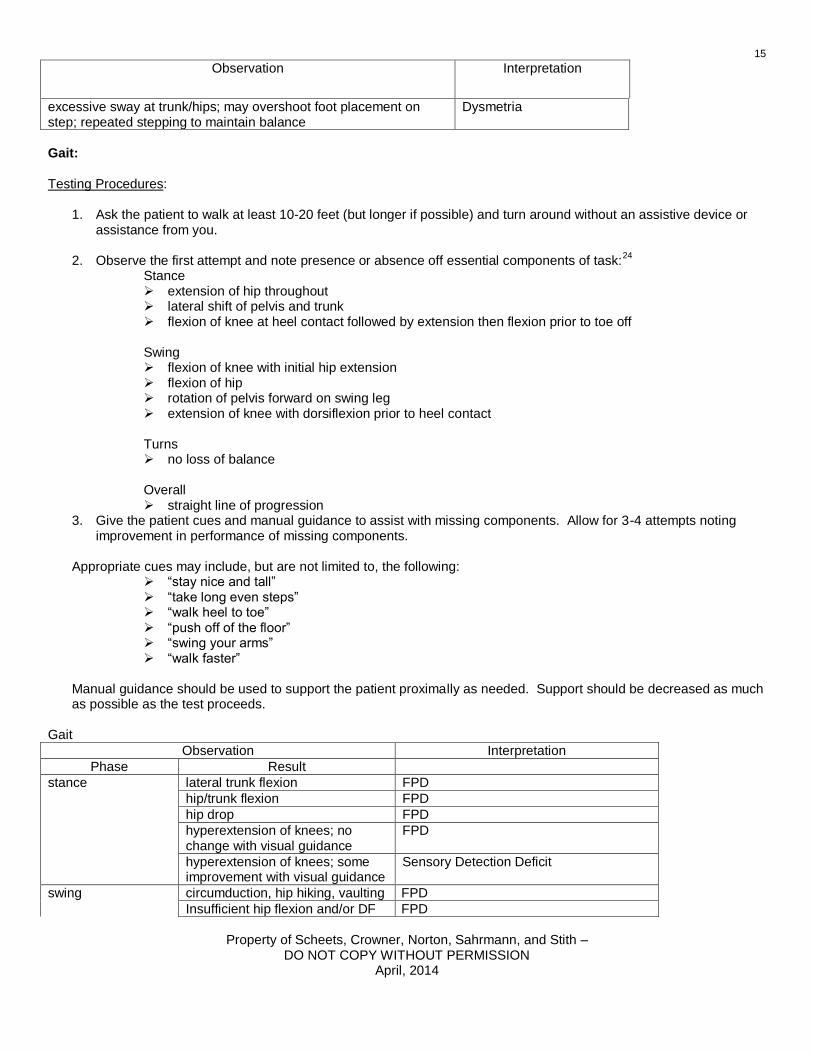
Gait:
Testing Procedures:
1. Ask the patient to walk at least 10-20 feet (but longer if possible) and turn around without an assistive device or assistance from you.
2. Observe the first attempt and note presence or absence off essential components of task:
24
Stance extension of hip throughout lateral shift of pelvis and trunk flexion of knee at heel contact followed by extension then flexion prior to toe off
Swing flexion of knee with initial hip extension flexion of hip rotation of pelvis forward on swing leg extension of knee with dorsiflexion prior to heel contact
Turns no loss of balance
Overall straight line of progression
3. Give the patient cues and manual guidance to assist with missing components. Allow for 3-4 attempts noting improvement in performance of missing components.
Appropriate cues may include, but are not limited to, the following:
“stay nice and tall” “take long even steps” “walk heel to toe” “push off of the floor” “swing your arms” “walk faster”
Manual guidance should be used to support the patient proximally as needed. Support should be decreased as much as possible as the test proceeds.
Gait
Observation Interpretation
Phase Result
stance lateral trunk flexion FPD
hip/trunk flexion FPD
hip drop FPD
hyperextension of knees; no change with visual guidance
FPD
hyperextension of knees; some improvement with visual guidance
Sensory Detection Deficit
swing circumduction, hip hiking, vaulting FPD
Insufficient hip flexion and/or DF FPD

Property of Scheets, Crowner, Norton, Sahrmann, and Stith – DO NOT COPY WITHOUT PERMISSION
April, 2014
16
Observation Interpretation
variable foot placement; improves with practice and cues
MPCD
multiple stops and starts; decreased step length
Hypokinesia
overall variable line of progression MPCD Sensory Selection and Weighting
Deficit Dysmetria
variable foot placement; no improvement with practice and cues
Dysmetria
Scissoring of steps Fractionated Movement Deficit
line of progression deviates toward one side
Disregard (modifier) Sensory Selection and Weighting
Deficit
throughout improvement in performance with practice and repetition
MPCD Sensory Selection and Weighting
Deficit
decrement in performance with repetition
FPD
Complex Gait:
Testing Procedures: While walking, ask the patient to perform the following:
1. turn head to left for 2-3 steps, to center for 2-3 steps, and to right for 2-3 steps. 2. step forward a few steps and step backward a few steps without stopping 3. step over a small obstacle
Give time to settle into a normal walking pattern between each of these tasks. Note change in performance with each task such as decreasing speed, deviation in line of progression, stopping, or stumbling.
Interpretation:
1Deviation in line of progression while walking with head turning may indicate Sensory Selection and
Weighting Deficit if coupled with other positive tests or Movement Pattern Coordination Deficit.
2Hesitation or extra steps in changing direction while walking but improvement with practice is indicative of
Movement Pattern Coordination Deficit.
3Hesitation and inability to step backward or extra steps may be indicative of Force Production Deficit.
Assess weight bearing strength of plantarflexors and consider improvement with practice when making diagnosis.
4Hesitation in stepping over obstacle or poor adjustment to step length when stepping over obstacle but
improvement in practice is indicative of Movement Pattern Coordination Deficit.
5Stopping movement with long hesitation before stepping and lack of support moment at hip of stance limb is
indicative of Force Production Deficit.
6Hesitation, stopping and starting, small steps, and poor control of momentum associated with Hypokinesia
7Difficulty transitioning from one sensory condition to another associated with Sensory Selection and
Weighting Deficit
8Consistent variability with extra steps for balance and wide base of support without change with practice
associated with Dysmetria

Property of Scheets, Crowner, Norton, Sahrmann, and Stith – DO NOT COPY WITHOUT PERMISSION
April, 2014
17
Dual Task Performance: Testing Procedures: While walking, ask the patient to perform the following:
1. walk while performing a cognitive task such as count backward by 3s, say the alphabet backward, or do multiplication tables
2. walk while performing a secondary manual tasks such as carrying a full cup of water, retrieving a coin from a pocket, or retrieve keys from a purse
Give time to settle into a normal walking pattern between each of these tasks. Note change in gait performance with each tasks such as decreasing speed, deviation in line of progression, stopping, or stumbling. Note change in the secondary task such has stopping, having to start the secondary task again, or errors. Note which task takes priority for the patient, ambulation and stability or the secondary task.
Interpretation:
9Deteriortation in performance of either ambulation or the secondary task indicates the need for a descriptor,
“with dual task difficulty” or similar language. Priority of the secondary task over ambulation stability may indicate a greater risk for falls.
Reach and Grasp: Testing Procedures: 1) Ask the patient to perform the following tests:
xxv
a) point to objects in front, at the ipsilateral and contralateral side, at shoulder height, and overhead b) reach for objects in the front, at the ipsilateral and contralateral side, at shoulder height, and overhead c) lift and release 3 objects of different size, weight, shape, and texture d) stack 3-4 small objects
2) Observe the reach and grasp components of each task and determine the following: a) is the motion fractionated b) is there sufficient active range of motion c) is the hand position being shaped during reach d) is the hand position awkward e) is the hand opening wide enough f) do the fingers contact the object before the web space g) is appropriate force applied to the object
Interpretation:
1Insufficient active range of motion but motion is fractionated is associated with Force Production Deficit.
2Inability to sustain grasp to lift object and motion is fractionated is associated with Force Production Deficit.
3Fractionated movement and dyscoordination of the hand movement during reach or of the hand about
objects is associated with Movement Pattern Coordination Deficit.
4Significant failure in accuracy of test indicates Dysmetria.
In-Hand Manipulation
Testing Procedures: 1) Place a pencil in the open palm of the patient’s hand and ask him to adjust the position of the pencil for use. (An
alternate test is to place a quarter in the palm and ask the patient to adjust it as if to put it in a vending machine.)25
2) Observe the task and determine the following:
a) is the motion fractionated b) is there sufficient active range of motion c) is the hand movement awkward d) is the hand movement slow e) is appropriate force applied to the object
Interpretation:
1Insufficient active range of motion but motion is fractionated is associated with Force Production Deficit

Property of Scheets, Crowner, Norton, Sahrmann, and Stith – DO NOT COPY WITHOUT PERMISSION
April, 2014
18
2Inability to sustain grasp and motion is fractionated is associated with Force Production Deficit.
3Fractionated movement and dyscoordination or slowness of the hand are associated with Movement Pattern
Coordination Deficit.
APPENDIX
Motorneuron Response Assessment Following the description and interpretation of the MRA are comments related to other standardized tools used to assess muscle tone and their use for the purposes of diagnosis. Motorneuron Response Assessment (MRA) The following is the description of the Motorneuron Response Assessment (MRA). The MRA was developed for patients with hemiplegia. The tool as written will be presented first. It will be followed by instructions for how to use the tests of the tool in different patient types. 1) Purpose
a) examine the overall level of excitability b) tests for spasticity, ability to relax after attempted active movement, and associated reactions
2) General guidelines
a) test all movements in supine b) perform each movement with the uninvolved extremity first in order to compare with the involved extremity c) prior to testing, check the passive range of motion (PROM) of each extremity involved d) explain to the patient that he should remain as relaxed as possible except for those tests in which he is to perform
a movement actively e) instruct the patient that with active movements, his effort should be just enough to accomplish the movement
requested f) determine that the patient understands the directions before rating his response; if he has difficulty with directions,
note this. g) assure the patient that the test will not harm him h) perform each test 3 times in order to measure the consistency of response i) occasionally, during testing of a specific item, a patient may not respond consistently; if one of the responses is
atypical, rate the item based on the response during the other movements and not the inconsistent response j) determine the overall classification according to the appropriate criteria k) record any additional information that would influence or help to interpret the results of the test
3) Upper Extremity (UE) test
a) position the UE to be tested in approximately 45 of abduction b) position the upper arm in neutral shoulder rotation and stabilize testing by holding onto the upper arm as needed c) position the forearm in neutral pronation/supination d) there are 5 components to the test; they are as follows:
i) Passive movement of the entire upper extremity (PROM): Instruct the patient to remain relaxed and not
assist with the movements. With the extremity in the described testing position, passively range each joint at varying speeds noting any resistance to stretch. Note all deviations from normal.
ii) Passive flexion of the elbow to 90 and drop into extension (PROM with Drop): Instruct the patient to remain relaxed, not to push the forearm down, and to let the arm drop. Support the upper arm and passively flex the
elbow to 90. Release the forearm, allowing it to fall into extension. Make sure the patient does not internally rotate the shoulder to extend the elbow. Note the speed of the fall into extension.
iii) Active flexion of the elbow to 90 and drop into extension (AROM with Drop): From the starting position, ask
the patient to bend the elbow to 90 then relax, allowing the forearm to drop into extension. Remind the patient not to “push” the forearm into extension. Stabilize the upper arm with one hand and use the other hand as a “target” to which the patient is to flex the elbow. Note the speed of the fall into extension.
Property of Scheets, Crowner, Norton, Sahrmann, and Stith – DO NOT COPY WITHOUT PERMISSION
April, 2014
19
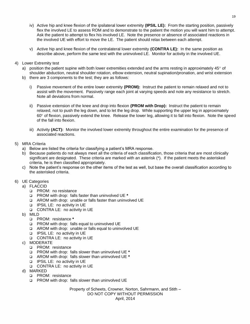
iv) Active hip and knee flexion of the ipsilateral lower extremity (IPSIL LE): From the starting position, passively
flex the involved LE to assess ROM and to demonstrate to the patient the motion you will want him to attempt. Ask the patient to attempt to flex his involved LE. Note the presence or absence of associated reactions in the involved UE with effort to move the LE. The patient should relax between each attempt.
v) Active hip and knee flexion of the contralateral lower extremity (CONTRA LE): In the same position as
describe above, perform the same test with the uninvolved LE. Monitor for activity in the involved UE. 4) Lower Extremity test
a) position the patient supine with both lower extremities extended and the arms resting in approximately 45 of shoulder abduction, neutral shoulder rotation, elbow extension, neutral supination/pronation, and wrist extension
b) there are 3 components to the test; they are as follows:
i) Passive movement of the entire lower extremity (PROM): Instruct the patient to remain relaxed and not to assist with the movement. Passively range each joint at varying speeds and note any resistance to stretch. Note all deviations from normal.
ii) Passive extension of the knee and drop into flexion (PROM with Drop): Instruct the patient to remain
relaxed, not to push the leg down, and to let the leg drop. While supporting the upper leg in approximately
60 of flexion, passively extend the knee. Release the lower leg, allowing it to fall into flexion. Note the speed of the fall into flexion.
iii) Activity (ACT): Monitor the involved lower extremity throughout the entire examination for the presence of
associated reactions. 5) MRA Criteria
a) Below are listed the criteria for classifying a patient’s MRA response. b) Because patients do not always meet all the criteria of each classification, those criteria that are most clinically
significant are designated. These criteria are marked with an asterisk (*). If the patient meets the asterisked criteria, he is then classified appropriately.
c) Note the patient’s response on the other items of the test as well, but base the overall classification according to the asterisked criteria.
6) UE Categories
a) FLACCID PROM: no resistance PROM with drop: falls faster than uninvolved UE * AROM with drop: unable or falls faster than uninvolved UE IPSIL LE: no activity in UE CONTRA LE: no activity in UE
b) MILD PROM: resistance *
PROM with drop: falls equal to uninvolved UE AROM with drop: unable or falls equal to uninvolved UE IPSIL LE: no activity in UE CONTRA LE: no activity in UE
c) MODERATE PROM: resistance PROM with drop: falls slower than uninvolved UE * AROM with drop: falls slower than uninvolved UE *
IPSIL LE: no activity in UE CONTRA LE: no activity in UE
d) MARKED PROM: resistance PROM with drop: falls slower than uninvolved UE

Property of Scheets, Crowner, Norton, Sahrmann, and Stith – DO NOT COPY WITHOUT PERMISSION
April, 2014
20
AROM with drop: falls slower than uninvolved UE IPSIL LE: activity in UE *
CONTRA LE: no activity in UE e) SEVERE
PROM: resistance PROM with drop: falls slower than uninvolved UE AROM with drop: falls slower than uninvolved UE IPSIL LE: activity in UE CONTRA LE: activity in UE *
f) NORMAL PROM: no resistance PROM with drop: falls equal to uninvolved UE AROM with drop: falls equal to uninvolved UE IPSIL LE: no activity in UE CONTRA LE: no activity in UE
7) LE Categories
a) FLACCID PROM: no resistance PROM with drop: falls faster than uninvolved LE *
ACT: no activity b) MILD
PROM: resistance * PROM with drop: falls equal to uninvolved LE ACT: no activity
c) MODERATE PROM: resistance PROM with drop: falls slower than uninvolved LE *
ACT: no activity d) SEVERE
PROM: resistance PROM with drop: falls slower than uninvolved LE ACT: activity *
e) NORMAL PROM: no resistance PROM with drop: falls equal to uninvolved LE ACT: no activity
8) Application to patients other than those with hemiplegia
While the MRA has been tested in our clinic on patients with hemiplegia, it is suggested that the test be considered as a series of tests that identify degrees of hyperexcitability. The test could be used on any patient with the Upper Motorneuron Syndrome, and patients with more positive responses on the tests would be considered more severe. The test position for the drop tests may also be changed in order to observe the speed of fall of a segment while different muscle groups are being stretched, e.g. prone with a lower leg drop test that passively stretches the hamstrings.
Fractionated Movement (FM)
Determine “yes” or “no” there is fractionated movement by completing the following standardized examination: 1) General Guidelines
a) test all movements in sitting with back supported unless medical status prohibits b) prior to testing, check the PROM for each extremity and joint involved c) beginning with the shoulder, ask the patient to perform isolated movements; instruction may be verbal and/or
visual

Property of Scheets, Crowner, Norton, Sahrmann, and Stith – DO NOT COPY WITHOUT PERMISSION
April, 2014
21
d) note the category with which the patient’s movement best corresponds e) record any additional information that would influence or help to interpret the results of the test f) perform all tests on the involved extremities
2) Upper Extremity test
a) there are 5 components of the test i) Ask the patient to flex his shoulder. Movement is fractionated if the patient moves the shoulder through at
least 50% of available range without substitution or other associated reactions. ii) Ask the patient to flex and extend the elbow while maintaining neutral supination/pronation. Movement is
fractionated if the patient moves the elbow through at least 50% of available range without substitution or other associated reactions.
iii) Ask the patient to flex and extend the wrist against gravity. Movement is fractionated if the patient moves the
wrist through 100% of available range without substitution or other associated reactions.
iv) Ask the patient to flex and extend the fingers against gravity. Movement is fractionated if the patient moves the fingers through 100% of available range without substitution or other associated reactions.
v) Ask the patient to flex and extend the index finger with the other fingers fully flexed. Movement is fractionated
if the patient moves the index finger through 100% of available range without substitution or other associated reactions.
3) Lower Extremity test
a) there are 3 components to the test i) Ask the patient to flex the hip in the sagittal plane. Movement is fractionated if the patient flexes the hip at
least 50% of available range of motion without substitution or other associated reactions.
ii) Ask the patient to extend the knee in the sagittal plane. Movement is fractionated if the patient extends the knee at least 50% of available range of motion without substitution or other associated reactions.
iii) Ask the patient to dorsiflex the ankle in the sagittal plane. Movement is fractionated if the patient dorsiflexes
the foot 100% of available range of motion without substitution or other associated reactions. 4) Fractionated Movement Category Criteria
a) Upper Extremity
Each joint is rated separately. If the patient is able to complete the task as defined, he is given a “yes” on the data sheet for that task. All tasks are rated for each patient. If the patient is unable to complete the task because he has no movement at a given segment, mark it on the data sheet.
b) Lower Extremity
Each joint is rated separately. If the patient is able to complete the task as defined, he is given a “yes” on the data sheet for that task. All tasks are rated for each patient. If the patient is unable to complete the task because he has no movement at a given segment, mark it on the data sheet.

Property of Scheets, Crowner, Norton, Sahrmann, and Stith – DO NOT COPY WITHOUT PERMISSION
April, 2014
22
SAMPLE CLINICAL FORM - Physical Therapy Initial Examination (Objective, Assessment, Plan) O: Observation: __________________________________________________________________________________
Vital signs: rest HR BP RR O2 sat _____
activity HR BP RR O2 sat _____ recovery HR BP RR O2 sat _____ Mental status: no deficits noted impaired in the following: ___________________________________
Special senses: (vision, hearing, etc.) ______________________________________________________________ SUPINE TESTS Edema: no deficits noted present in the following: ______________________________________
Skin: no deficits noted problems noted in the following: ____________________________________ ROM: _____ no deficits noted _____ deficits noted in the following: ______________________________________
Muscle tone: normal abnormal ______________________________________________________
Sensation: no deficits noted
impaired in the following:
lt touch: ___________________________________________________________________
pain/temperature: ___________________________________________________________
joint position sense: __________________________________________________________
protective sensation: _________________________________________________________
Strength: L R L R Shoulder flexion ______ ______ Hip flexion ______ ______ Elbow flexion ______ ______ Knee extension ______ ______ Elbow extension ______ ______ DF ______ ______ Wrist extension ______ ______ PF (NWB) ______ ______ Grip ______ ______ Skeletal muscle fatigue: fatigue with 10 reps elbow extension? left yes left no right yes right no fatigue with 10 reps knee extension? left yes left no right yes right no Rolling left: (without rails) assistance: with task: ___________________ analysis: (check all that apply)
essential movement components present
generates momentum with upper body on initiation
insufficient head/upper trunk rotation on initiation
insufficient shoulder flexion/horizontal add. on initiation
insufficient trunk rotation during execution
shifts left hip under right hip during execution
insufficient hip flexion/pelvic rotation on initiation
response to practice: improves fatigues no change
Property of Scheets, Crowner, Norton, Sahrmann, and Stith – DO NOT COPY WITHOUT PERMISSION
April, 2014
23
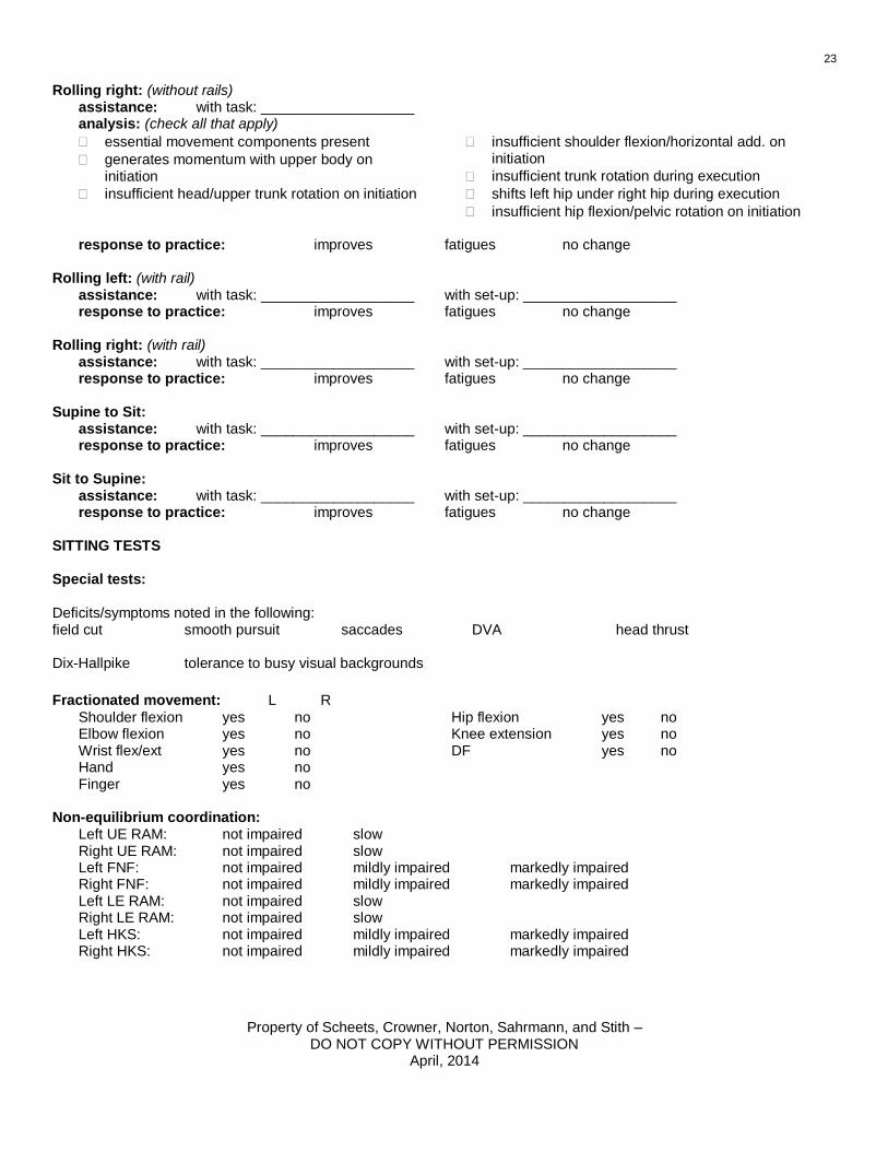
Rolling right: (without rails) assistance: with task: ___________________ analysis: (check all that apply)
essential movement components present
generates momentum with upper body on initiation
insufficient head/upper trunk rotation on initiation
insufficient shoulder flexion/horizontal add. on initiation
insufficient trunk rotation during execution
shifts left hip under right hip during execution
insufficient hip flexion/pelvic rotation on initiation
response to practice: improves fatigues no change
Rolling left: (with rail) assistance: with task: ___________________ with set-up: ___________________ response to practice: improves fatigues no change
Rolling right: (with rail) assistance: with task: ___________________ with set-up: ___________________ response to practice: improves fatigues no change
Supine to Sit: assistance: with task: ___________________ with set-up: ___________________ response to practice: improves fatigues no change
Sit to Supine: assistance: with task: ___________________ with set-up: ___________________ response to practice: improves fatigues no change
SITTING TESTS
Special tests:
Deficits/symptoms noted in the following: field cut
smooth pursuit saccades DVA head thrust
Dix-Hallpike
tolerance to busy visual backgrounds
Fractionated movement: L R
Shoulder flexion yes no Hip flexion yes no Elbow flexion yes no Knee extension yes no Wrist flex/ext yes no DF yes no Hand yes no Finger yes no
Non-equilibrium coordination:
Left UE RAM: not impaired slow Right UE RAM: not impaired slow Left FNF: not impaired mildly impaired markedly impaired Right FNF: not impaired mildly impaired markedly impaired Left LE RAM: not impaired slow Right LE RAM: not impaired slow Left HKS: not impaired mildly impaired markedly impaired Right HKS: not impaired mildly impaired markedly impaired
Property of Scheets, Crowner, Norton, Sahrmann, and Stith – DO NOT COPY WITHOUT PERMISSION
April, 2014
24
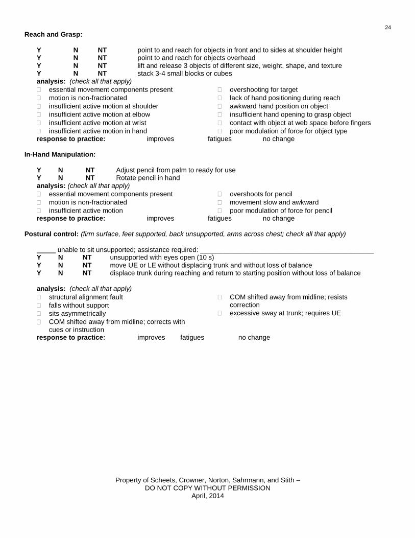
Reach and Grasp: Y N NT point to and reach for objects in front and to sides at shoulder height Y N NT point to and reach for objects overhead Y N NT lift and release 3 objects of different size, weight, shape, and texture Y N NT stack 3-4 small blocks or cubes analysis: (check all that apply)
essential movement components present
motion is non-fractionated
insufficient active motion at shoulder
insufficient active motion at elbow
insufficient active motion at wrist
insufficient active motion in hand
overshooting for target
lack of hand positioning during reach
awkward hand position on object
insufficient hand opening to grasp object
contact with object at web space before fingers
poor modulation of force for object type response to practice: improves fatigues no change In-Hand Manipulation: Y N NT Adjust pencil from palm to ready for use Y N NT Rotate pencil in hand analysis: (check all that apply)
essential movement components present
motion is non-fractionated
insufficient active motion
overshoots for pencil
movement slow and awkward
poor modulation of force for pencil response to practice: improves fatigues no change Postural control: (firm surface, feet supported, back unsupported, arms across chest; check all that apply) unable to sit unsupported; assistance required: ______________________________________________ Y N NT unsupported with eyes open (10 s) Y N NT move UE or LE without displacing trunk and without loss of balance Y N NT displace trunk during reaching and return to starting position without loss of balance
analysis: (check all that apply)
structural alignment fault
falls without support
sits asymmetrically
COM shifted away from midline; corrects with cues or instruction
COM shifted away from midline; resists correction
excessive sway at trunk; requires UE
response to practice: improves fatigues no change

Property of Scheets, Crowner, Norton, Sahrmann, and Stith – DO NOT COPY WITHOUT PERMISSION
April, 2014
25
Sit to Stand: (without UE support) assistance: ________________________________________________________________________________ analysis: (check all that apply)
essential movement components present
unable to assume normal starting position; stiffness
absent or delayed preparatory movements Initiation:
increased base of support
insufficient force production
uses a momentum strategy
excessive trunk sway Execution:
medial hip rotation
hip adduction
valgus of knee
varus of knee
extends knees before hips in first half
pushes on thighs to extend trunk
decreased weight bearing
insufficient translation of tibia over foot
shifts COM to one side
shifts COM to one side or back; corrects with cues/instruction
shifts COM to one side or back; resists correction
slow
arrests of ongoing movement Termination:
steps to find balance
repeated stepping to find and maintain balance
increased lumbar extension
inadequate hip extension
inadequate knee extension
posterior sway
shifts COM to one side
shifts COM to one side or back; corrects with cues/instruction
shifts COM to one side or back; resists correction
increased BOS with excessive sway at hips response to practice: improves fatigues no change Stand to Sit: (without UE support) assistance: ________________________________________________________________________________ analysis: (check all that apply)
essential movement components present
insufficient hip flexion during execution
insufficient knee flexion on initiation and during execution
inadequate control of descent into chair response to practice: improves fatigues no change Sit to Stand: (with UE support) assistance: _______________________________________________________________________________ analysis: (check all that apply)
essential movement components present
unable to assume normal starting position; stiffness
absent or delayed preparatory movements Initiation:
increased base of support
insufficient force production
uses a momentum strategy
excessive trunk sway Execution:
medial hip rotation
hip adduction
valgus of knee
varus of knee
extends knees before hips in first half
pushes on thighs to extend trunk
decreased weight bearing
insufficient translation of tibia over foot
shifts COM to one side
shifts COM to one side or back; corrects with cues/instruction
shifts COM to one side or back; resists correction
slow
arrests of ongoing movement Termination:
steps to find balance
repeated stepping to find and maintain balance
increased lumbar extension
inadequate hip extension
inadequate knee extension
posterior sway
shifts COM to one side
shifts COM to one side or back; corrects with cues/instruction
shifts COM to one side or back; resists correction
increased BOS with excessive sway at hips response to practice: improves fatigues no change
Property of Scheets, Crowner, Norton, Sahrmann, and Stith – DO NOT COPY WITHOUT PERMISSION
April, 2014
26
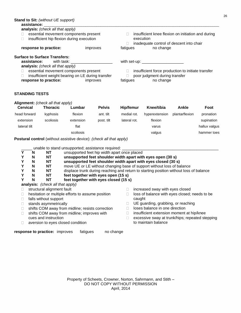
Stand to Sit: (without UE support) assistance: ________________________________________________________________________________ analysis: (check all that apply)
essential movement components present
insufficient hip flexion during execution
insufficient knee flexion on initiation and during execution
inadequate control of descent into chair response to practice: improves fatigues no change Surface to Surface Transfers: assistance: with task: ___________________ with set-up: ___________________ analysis: (check all that apply)
essential movement components present
insufficient weight bearing on LE during transfer
insufficient force production to initiate transfer
poor judgment during transfer response to practice: improves fatigues no change STANDING TESTS Alignment: (check all that apply)
Cervical Thoracic Lumbar Pelvis Hip/femur Knee/tibia Ankle Foot
head forward kyphosis flexion ant. tilt medial rot. hyperextension plantarflexion pronation
extension scoliosis extension post. tilt lateral rot. flexion supination
lateral tilt flat varus hallux valgus
scoliosis valgus hammer toes
Postural control (without assistive device): (check all that apply)
_____ unable to stand unsupported; assistance required: ____________________________________________ Y N NT unsupported feet hip width apart once placed Y N NT unsupported feet shoulder width apart with eyes open (30 s) Y N NT unsupported feet shoulder width apart with eyes closed (30 s) Y N NT move UE or LE without changing base of support without loss of balance Y N NT displace trunk during reaching and return to starting position without loss of balance Y N NT feet together with eyes open (15 s) Y N NT feet together with eyes closed (15 s) analysis: (check all that apply)
structural alignment fault
hesitation or multiple efforts to assume position
falls without support
stands asymmetrically
shifts COM away from midline; resists correction
shifts COM away from midline; improves with cues and instruction
aversion to eyes closed condition
increased sway with eyes closed
loss of balance with eyes closed; needs to be caught
UE guarding, grabbing, or reaching
loses balance in one direction
insufficient extension moment at hip/knee
excessive sway at trunk/hips; repeated stepping to maintain balance
response to practice: improves fatigues no change

Property of Scheets, Crowner, Norton, Sahrmann, and Stith – DO NOT COPY WITHOUT PERMISSION
April, 2014
27
Advanced Postural Control (without assistive device): (check all that apply) Y N NT step-up (alternate placement of foot on 4-6 inch step) Y N NT one foot (10 s) additional measures: ________________________________________________________________________ analysis: (check all that apply)
prefers wide BOS
hesitation and/or multiple starts when changing BOS or initiating movement
UE guarding, grabbing, or reaching
circumduction or insufficient hip flexion of swing limb for step-up
lateral trunk flexion toward stance limb
hip flexion or hip drop on stance limb
knee flexion of stance limb
knee hyperextension of stance limb
posterior sway of stance limb
loss of balance with forward movement of swing limb during step-up
loss of balance with backward movement of swing limb during step-up
loss of balance to left or right
inconsistent foot placement on step during step-up
excessive sway at trunk/hips; repeated stepping to maintain balance
response to practice: improves fatigues no change
Modified CTSIB: (feet almost touching; hands on hips; record duration patient can stand in each condition up to 30 s; use
medium density 4inch Tempur foam) Firm surface eyes open _____ Firm surface eyes closed _____ Foam surface eyes open _____ Foam surface eyes closed _____ Gait: assistance: with task: ___________________ with set-up: ___________________ device: walker wheeled walker cane quad cane crutches other _______________________ speed: _________ft in _______seconds (normal for older adults 2.2-3.3 ft/s; MCID 0.32 ft/s)
analysis: (check all that apply)
essential movement components present Stance:
decreased base of support
increased base of support
decreased weight bearing
increased pelvic/lumbar rotation
lateral trunk shift toward stance limb
↓hip extension mid- to terminal stance
hip drop
hyperextension of knee
sustained hip/knee flexion
decreased plantarflexion
increased pronation
Swing:
hip hiking
circumduction
vaulting
inadequate hip flexion
inadequate dorsiflexion
decreased step length
increased step length
Overall:
variable foot placement
variable line of progression
line of progression deviates left or right response to practice: improves fatigues no change Six-minute walk test: (normal for older adults about 1366 ft; MCID 164 ft) Distance/device: ____________________________________________________________________________
RPE and vital signs: __________________________________________________________________________

Property of Scheets, Crowner, Norton, Sahrmann, and Stith – DO NOT COPY WITHOUT PERMISSION
April, 2014
28
Complex Gait: assistance: _____________________________________________________________________ Y N NT Walk and turn head side to side Y N NT Step forward/backward Y N NT Step over obstacle Y N NT 180° turn Y N NT Carry Y N NT Compliant surface Y N NT Dim lighting
analysis: (check all that apply)
deviation in line of progression with head turning
symptoms with head turning
hesitates or takes extra steps when changing direction
poor control of momentum when stepping forward or back
hesitates before stepping over obstacle
poor adjustment in step length to step over obstacle
insufficient hip flexion to step over obstacle
difficulty clearing second limb when stepping over obstacle
steps to recover balance with stepping over
insufficient hip/knee extension moment when stepping over
instability with carrying
increased loss of balance on varying surfaces
increased loss of balance in dim lighting
slow/increased loss of balance when transitioning from one sensory context to another
response to practice: improves fatigues no change Stairs: (with rails) assistance: ________________________________________________________________________________ analysis: (check all that apply)
essential movement components present Up:
insufficient hip flexion of swing limb
insufficient hip extension on stance limb
insufficient knee extension on stance limb
increased sway on stance limb
Down:
medial hip rotation on stance limb
poor control of forward momentum
Stairs: (without rails) assistance: ________________________________________________________________________________ analysis: (check all that apply)
essential movement components present Up:
insufficient hip flexion of swing limb
insufficient hip extension on stance limb
insufficient knee extension on stance limb
increased sway on stance limb
Down:
medial hip rotation on stance limb
poor control of forward momentum
Floor to/from stand transfers:
Stand directly yes no UE support yes no unable Activity tolerance: (check all that apply)
No deficits noted
SOB with recovery within 2-5 minutes
SOB with recovery > 5 minutes
Irregular heart rhythm
Abnormal BP response
Requires frequent rests during exam Pain scale: (0=none; 10=intolerable) /10 noted in the following: (circle) incision abdomen wound neck shoulder back hip knee feet other _____________ Type of pain: dull sharp aching stinging deep other ____________________ Change in pain during session: __________________________________________________________________
Property of Scheets, Crowner, Norton, Sahrmann, and Stith – DO NOT COPY WITHOUT PERMISSION
April, 2014
29
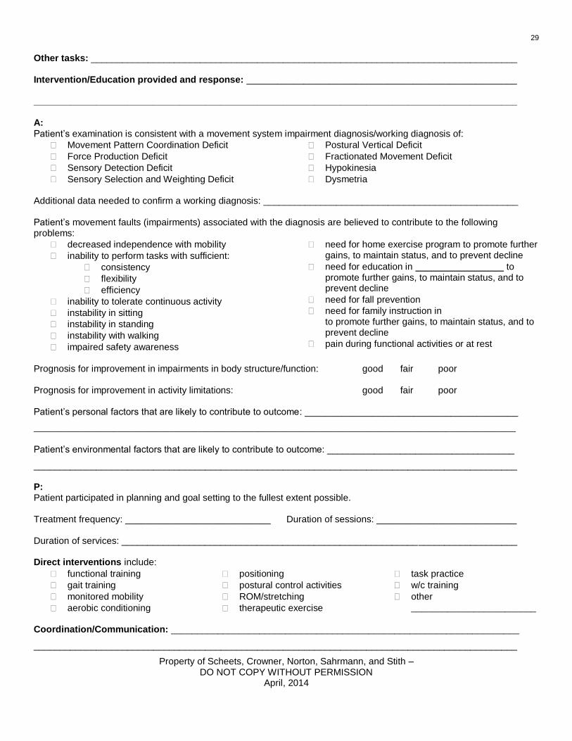
Other tasks: __________________________________________________________________________________
Intervention/Education provided and response: ____________________________________________________
_____________________________________________________________________________________________
A:
Patient’s examination is consistent with a movement system impairment diagnosis/working diagnosis of:
Movement Pattern Coordination Deficit
Force Production Deficit
Sensory Detection Deficit
Sensory Selection and Weighting Deficit
Postural Vertical Deficit
Fractionated Movement Deficit
Hypokinesia
Dysmetria Additional data needed to confirm a working diagnosis: _________________________________________________ Patient’s movement faults (impairments) associated with the diagnosis are believed to contribute to the following problems:
decreased independence with mobility
inability to perform tasks with sufficient:
consistency
flexibility
efficiency
inability to tolerate continuous activity
instability in sitting
instability in standing
instability with walking
impaired safety awareness
need for home exercise program to promote further gains, to maintain status, and to prevent decline
need for education in to promote further gains, to maintain status, and to prevent decline
need for fall prevention
need for family instruction in to promote further gains, to maintain status, and to prevent decline
pain during functional activities or at rest
Prognosis for improvement in impairments in body structure/function: good fair poor Prognosis for improvement in activity limitations: good fair poor Patient’s personal factors that are likely to contribute to outcome: _________________________________________
_______________________________________________________________________________________________________
Patient’s environmental factors that are likely to contribute to outcome: ____________________________________
_____________________________________________________________________________________________ P: Patient participated in planning and goal setting to the fullest extent possible. Treatment frequency: ____________________________ Duration of sessions: ___________________________ Duration of services: ____________________________________________________________________________ Direct interventions include:
functional training
gait training
monitored mobility
aerobic conditioning
positioning
postural control activities
ROM/stretching
therapeutic exercise
task practice
w/c training
other ________________________
Coordination/Communication: ___________________________________________________________________
_____________________________________________________________________________________________

Property of Scheets, Crowner, Norton, Sahrmann, and Stith – DO NOT COPY WITHOUT PERMISSION
April, 2014
30
Patient related instruction: ______________________________________________________________________
_____________________________________________________________________________________________ Patient to demonstrate the following: STG:
Activity or Task Performance Conditions Assistance Needed
Goal Date
LTG: (by final discharge from physical therapy services in ________________ wks or mos ):
Activity or Task Performance Conditions Assistance Needed
Goal Date
i Sahrmann SA and Norton BJ. The relationship of voluntary movement to spasticity in the upper motorneuron syndrome. Ann Neurol. 1977;2:460-465.
ii Scheets PL, Sahrmann SA, Norton BJ. Phys Ther. 2007;87:654-669.
iii Ashworth B. Preliminary trial of carisoprodol in multiple sclerosis. Practitioner. 1964;192:540-542.
iv Bohannon RW, Smith MD. Interrater reliability of a Modified Ashworth scale of muscle spasticity. Phys Ther. 1987;67:206-207.
v Mehrholz J Wagner K, MeiBner D et al. Reliability of the Modified Tardieu scale and the Modified Ashworth scale in adult patients with severe brain
injury: a comparison study. Clin Rehabil. 2005;19:751-759. vi
Bodin PG and Morris ME. Inter-rater reliability of the Modified Ashworth Scale for wrist flexors spasticity following stroke. World Federation of
Physiotherapy, 11th Congress. 1991:505-507.
vii Gregson JM, Leathley M, Moore P et al. Reliability of the Tone Assessment Scale and the Modified Ashworth Scale as clinical tools for assessing
poststroke spasticity. Arch Phys Med Rehabil. 1999;80:1013-1016. viii
Ansari NN, Naghdi S, Moammeri H, et al. Ashworth Scales are unreliable for the assessment of muscle spasticity. Physiotherapy Theory Practice.
2006;22:119-125. ix
Gregson JM, Leathley M, Moore P et al. Reliability of measurements of muscle tone and muscle power in stroke patients. Age and Ageing.
200;29:223-228. x Morris ME. Phys Ther. 2000;80(6):578-597.
xi Sahrmann SA and Norton BJ. The relationship of voluntary movement to spasticity in the upper motorneuron syndrome. Ann Neurol. 1977;2:460-465.
xii Snyder R et al. The relationship between the motorneuron response assessment and fractionated movement of the upper extremity in subjects with
hemiplegia. Phys Ther. 1994;74(5 Suppl):S45. xiii
Snyder et al. The relationship between the hemiparetic upper extremity and lower extremity using a tone assessment and fractionated movement.
Phys Ther. 1994;74(5 Suppl):S46. xiv
Sahrmann SA and Norton BJ. The relationship of voluntary movement to spasticity in the upper motorneuron syndrome. Ann Neurol. 1977;2:460-465. xv
Snyder R et al. The relationship between the motorneuron response assessment and fractionated movement of the upper extremity in subjects with
hemiplegia. Phys Ther. 1994;74(5 Suppl):S45.
Property of Scheets, Crowner, Norton, Sahrmann, and Stith – DO NOT COPY WITHOUT PERMISSION
April, 2014
31
xvi
Gregson JM, Leathley M, Moore P et al. Reliability of measurements of muscle tone and muscle power in stroke patients. Age and Ageing.
200;29:223-228. xvii
Fowler EG, Staudt LA, Breenberg MB et al. Selective control assessment of the lower extremity (SCALE): development, validation, and interrater
reliability of a clinical tool for patients with cerebral palsy. Dev Med Child Neurol. 2009;51:607-614. xviii
Kendall FP and McCreary EK. Muscle testing and function, 3rd ed. Baltimore, MD. Williams and Wilkins, 1983: 3-15.
xix Nolan MF. Introduction to the Neurologic Examination. Philadelphia, PA: F.A. Davis Company; 1996:134-135.
xx Karnath HO, Johannsen L, Broetz D et al. Prognosis for contraversive pushing. J Neurol. 2002;249:1250-1253.
xxi Karnath HO. Instructions for the clinical scale for contraversive pushing (SCP). Neurorehabil Neural Repair. 2007;21(4):370-371.
xxii Manckoundia P, Mourey F, Pfitzenmeyer P et al. Is backward disequilibrium in the elderly caused by an abnormal perception of verticality? A pilot
study. Clin Neurophysiol. 2007;118:786-793. xxiii
Baccini M, Paci M, Rinaldi LA. The scale for contraversive pushing: a reliability and validity study. Neurorehabil Neural Repair. 2006;20(4):468-472. xxiv
Carr JH and Shepherd RB. A Motor Relearning Programme for Stroke, second ed. Rockville, MD: Aspen Systems Corporation; 1982:91. xxv
Shumway-Cook A and Woollacott M. Motor Control Theory and Practical Application, second ed Philadelphia, PA: Williams & Wilkens; 2000:517-
537.