Motoneurons of Twitch and Nontwitch Extraocular Muscle ... · anti-synaptophysin (1:20) overnight...
18
Motoneurons of Twitch and Nontwitch Extraocular Muscle Fibers in the Abducens, Trochlear, and Oculomotor Nuclei of Monkeys JEAN A. BU ¨ TTNER-ENNEVER, 1 * ANJA K.E. HORN, 1 HANSJOERG SCHERBERGER, 3 AND PAOLA D’ASCANIO 3 1 Institute of Anatomy, Ludwig-Maximilian University, Munich, Germany 2 Division of Biology, California Institute of Technology, Pasadena, California 3 Department of Physiology and Biochemistry, University of Pisa, Pisa, Italy ABSTRACT Eye muscle fibers can be divided into two categories: nontwitch, multiply innervated muscle fibers (MIFs), and twitch, singly innervated muscle fibers (SIFs). We investigated the location of motoneurons supplying SIFs and MIFs in the six extraocular muscles of monkeys. Injections of retrograde tracers into eye muscles were placed either centrally, within the central SIF endplate zone; in an intermediate zone, outside the SIF endplate zone, targeting MIF endplates along the length of muscle fiber; or distally, into the myotendinous junction containing palisade endings. Central injections labeled large motoneurons within the abdu- cens, trochlear or oculomotor nucleus, and smaller motoneurons lying mainly around the periphery of the motor nuclei. Intermediate injections labeled some large motoneurons within the motor nuclei but also labeled many peripheral motoneurons. Distal injections labeled small and medium-large peripheral neurons strongly and almost exclusively. The peripheral neurons labeled from the lateral rectus muscle surround the medial half of the abducens nucleus: from superior oblique, they form a cap over the dorsal trochlear nucleus; from inferior oblique and superior rectus, they are scattered bilaterally around the midline, between the oculomotor nucleus; from both medial and inferior rectus, they lie mainly in the C-group, on the dorsomedial border of oculomotor nucleus. In the medial rectus distal injections, a “C-group extension” extended up to the Edinger-Westphal nucleus and labeled dendrites within the supraoculomotor area. We conclude that large motoneurons within the motor nuclei innervate twitch fibers, whereas smaller motoneurons around the periphery innervate nontwitch, MIF fibers. The peripheral subgroups also contain medium-large neu- rons which may be associated with the palisade endings of global MIFs. The role of MIFs in eye movements is unclear, but the concept of a final common pathway must now be recon- sidered. J. Comp. Neurol. 438:318 –335, 2001. © 2001 Wiley-Liss, Inc. Indexing terms: extraocular motoneurons; palisade endings; myotendinous junction; eye movements; medial rectus C-group; final common pathway Motoneurons of the extraocular muscles lie in three separate nuclei: the abducens nucleus (nVI), the trochlear nucleus (nIV), and the oculomotor nucleus (nIII). This pattern of innervation is extremely invariant throughout vertebrate evolution and probably reflects their derivation from separate rhombomeres of segmented prevertebrate ancestors (Baker, 1998). The location of the large mo- toneurons, characteristic of the oculomotor nuclei, has been the subject of several studies in monkey (Warwick, 1953; Augustine et al., 1981; Bu ¨ ttner-Ennever and Akert, 1981; Spencer and Porter, 1981). Single cell recordings, from behaving monkeys, show that all extraocular mo- Grant Sponsor: German Research Council; Grant number: SFB 462/B3. *Correspondence to: J.A. Bu ¨ ttner-Ennever, Institute of Anatomy, Ludwig-Maximilian University, Pettenkoferstr. 11, D-80336 Munich, Ger- many. E-mail: [email protected] Received 29 November 2000; Revised 29 March 2001; Accepted 20 June 2001 THE JOURNAL OF COMPARATIVE NEUROLOGY 438:318 –335 (2001) © 2001 WILEY-LISS, INC.
Transcript of Motoneurons of Twitch and Nontwitch Extraocular Muscle ... · anti-synaptophysin (1:20) overnight...
Motoneurons of Twitch and NontwitchExtraocular Muscle Fibers in the
Abducens, Trochlear, and OculomotorNuclei of Monkeys
JEAN A. BUTTNER-ENNEVER,1* ANJA K.E. HORN,1 HANSJOERG SCHERBERGER,3
AND PAOLA D’ASCANIO3
1Institute of Anatomy, Ludwig-Maximilian University, Munich, Germany2Division of Biology, California Institute of Technology, Pasadena, California3Department of Physiology and Biochemistry, University of Pisa, Pisa, Italy
ABSTRACTEye muscle fibers can be divided into two categories: nontwitch, multiply innervated
muscle fibers (MIFs), and twitch, singly innervated muscle fibers (SIFs). We investigated thelocation of motoneurons supplying SIFs and MIFs in the six extraocular muscles of monkeys.Injections of retrograde tracers into eye muscles were placed either centrally, within thecentral SIF endplate zone; in an intermediate zone, outside the SIF endplate zone, targetingMIF endplates along the length of muscle fiber; or distally, into the myotendinous junctioncontaining palisade endings. Central injections labeled large motoneurons within the abdu-cens, trochlear or oculomotor nucleus, and smaller motoneurons lying mainly around theperiphery of the motor nuclei. Intermediate injections labeled some large motoneurons withinthe motor nuclei but also labeled many peripheral motoneurons. Distal injections labeledsmall and medium-large peripheral neurons strongly and almost exclusively. The peripheralneurons labeled from the lateral rectus muscle surround the medial half of the abducensnucleus: from superior oblique, they form a cap over the dorsal trochlear nucleus; frominferior oblique and superior rectus, they are scattered bilaterally around the midline,between the oculomotor nucleus; from both medial and inferior rectus, they lie mainly in theC-group, on the dorsomedial border of oculomotor nucleus. In the medial rectus distalinjections, a “C-group extension” extended up to the Edinger-Westphal nucleus and labeleddendrites within the supraoculomotor area. We conclude that large motoneurons within themotor nuclei innervate twitch fibers, whereas smaller motoneurons around the peripheryinnervate nontwitch, MIF fibers. The peripheral subgroups also contain medium-large neu-rons which may be associated with the palisade endings of global MIFs. The role of MIFs ineye movements is unclear, but the concept of a final common pathway must now be recon-sidered. J. Comp. Neurol. 438:318–335, 2001. © 2001 Wiley-Liss, Inc.
Indexing terms: extraocular motoneurons; palisade endings; myotendinous junction; eye
movements; medial rectus C-group; final common pathway
Motoneurons of the extraocular muscles lie in threeseparate nuclei: the abducens nucleus (nVI), the trochlearnucleus (nIV), and the oculomotor nucleus (nIII). Thispattern of innervation is extremely invariant throughoutvertebrate evolution and probably reflects their derivationfrom separate rhombomeres of segmented prevertebrateancestors (Baker, 1998). The location of the large mo-toneurons, characteristic of the oculomotor nuclei, hasbeen the subject of several studies in monkey (Warwick,1953; Augustine et al., 1981; Buttner-Ennever and Akert,
1981; Spencer and Porter, 1981). Single cell recordings,from behaving monkeys, show that all extraocular mo-
Grant Sponsor: German Research Council; Grant number: SFB 462/B3.*Correspondence to: J.A. Buttner-Ennever, Institute of Anatomy,
Ludwig-Maximilian University, Pettenkoferstr. 11, D-80336 Munich, Ger-many. E-mail: [email protected]
Received 29 November 2000; Revised 29 March 2001; Accepted 20 June2001
THE JOURNAL OF COMPARATIVE NEUROLOGY 438:318–335 (2001)
© 2001 WILEY-LISS, INC.

toneurons participate in all types of eye movements: ver-gence, saccades, smooth pursuit, and both vestibulo-ocular and optokinetic nystagmus (Robinson, 1970; Maysand Porter, 1984). Furthermore, the motor unit dischargesare tightly linked to eye position (monkey: Keller andRobinson, 1972; Keller, 1973; man: Scott and Collins,1973). So it is currently assumed that oculomotor com-mands combine at the level of the motoneurons and acti-vate the muscle fibers through a “final common path.”
The targets of this “final common path” are among themost complicated muscles of the body (review: Spencerand Porter, 1988; Porter and Baker, 1998). Eye musclescan be divided into an outer orbital layer, which consists ofsmall caliber fibers, and an inner global layer, with largercaliber muscle fibers. There are also reports in humans ofa third layer outside the orbital layer, called the marginallayer (Wasicky et al., 2000). At least six different types ofmuscle fibers have been identified in eye muscles, andthese can be divided into two main categories: the nont-witch or multiply innervated muscle fibers (MIFs), andthe twitch or singly innervated muscle fibers (SIFs) (re-view: Mayr et al., 1975; Morgan and Proske 1984; Spencerand Porter, 1988). The original classification by Siebeckand Kruger (1955) used the terms “Fibrillenstruktur” and“Felderstruktur” fibers, for SIFs and MIFs, respectively.The orbital and global layers of all mammals examined todate contain both fiber types. The twitch fibers, or SIFs,are the type of muscle fibers that constitute all skeletalmuscles; they respond to electrical excitation with an all-or-nothing response that propagates along the wholelength of the fiber. They are innervated by relatively largenerves (7–11 mm), which terminate as large en plaquemotor endplates in an endplate zone occupying the centralthird of the muscle. The MIFs are highly unusual in mam-mals, occurring only in eye muscles, the larynx and mus-cles of the middle ear; but MIFs are common in amphibianmuscles, and most studies have been on these prepara-
tions (review: Morgan and Proske, 1984; Dieringer andPrecht, 1986). These fibers are fatigue resistant and re-spond to electrical stimulation with a slow tonic contrac-tion, which is not propagated along the muscle fiber(Bondi and Chirandini, 1983). They are innervated by amyelinated nerve fiber, which usually is of fine caliber(3–5 mm). The motor endplates are typically small and aredistributed all along the length of the fiber but have ahigher density in the distal half of the muscle. At thedistal tip of the eye muscle, as it inserts into the tendonthe global layer MIFs are capped by a tangle of nerveterminals called palisade endings, or myotendinous cylin-ders (Dogiel, 1906; cat: Alvarado-Mallart and Pincon-Raymond, 1979; monkey: Ruskell, 1978; human: Rich-mond et al., 1984; Lukas et al., 2000; review, Ruskell,1999). This characteristic is an exclusive property of theglobal layer MIFs, not possessed by the orbital MIFs orthe SIFs.
Despite the details that are known of the anatomicorganization of the eye muscle, it is not known how thesestructural features contribute to eye movements (Lenner-strand and Baker, 1987). The function of the palisadeendings is unclear, and they are at present the subject ofcontroversy, with some reports claiming them as sensorystructures and others describing motor-like properties(Lukas et al., 2000). In addition, the role of MIF musclefibers in eye movements is also not understood. No record-ings have been knowingly made from the motoneuronssupplying MIFs in awake mammals; neither is the loca-tion of motoneurons innervating MIF muscle fibersknown, although they are assumed to be in the nIII, nIV,and nVI motor nuclei (Shall et al., 1995). In this study, wehave attempted to locate the motoneurons of MIFs inmonkeys. Small deposits of retrograde tracer were placedinto distal regions of the eye muscles, avoiding the centralSIF endplate zone (Fig. 1). We found that the uptake of thetracer by structures such as the fine en grappe endplatesof the MIFs and possibly palisade endings of the globalMIFs, labeled a set of peripheral subgroups around theborders of the nIII, nIV, and nVI motor nuclei, separatefrom the classic motoneuron subgroups. Preliminary ac-counts of these results have been reported at meetings(Buttner-Ennever et al., 1998; Buttner-Ennever, 2000).
MATERIALS AND METHODS
All experimental procedures conformed to the state anduniversity regulations on Laboratory Animal Care, includ-ing the Principles of Laboratory Animal Care (NIH Pub-lication 85-23, Revised 1985), and were approved by theirAnimal Care Officers and Institutional Animal Care andUse Committees.
Macaque monkeys were anesthetized with sodium pen-tobarbital (30 mg/kg). Under sterile conditions, the ex-traocular eye muscles were exposed by retracting the eye-lids, making a conjunctival incision, and partiallycollapsing the eye ball. Large or small volumes of theneuronal tracers cholera toxin subunit B (CT, 1–10 ml, 1%from List Campbell, CA) or wheat germ agglutinin conju-gated to horseradish peroxidase (WGA:HRP, 5–30 ml,2.5% Sigma, St. Louis, MO), or iodinated WGA (125I-WGA,4–50 ml; Buttner-Ennever and Akert, 1981) were injectedthrough a Hamilton syringe into the belly or the distal tipof the eye muscle, respectively. In several cases, two mus-
Abbreviations
nIII oculomotor nucleusnIV trochlear nucleusnVI abducens nucleusnVII facial nucleusCT cholera toxin subunit Bd distal endEW Edinger-Wesphal nucleusF muscle fiberG global layeriC interstitial nucleus of CajalIO inferior oblique muscleIR inferior rectus muscleLP levator palpebraeLR lateral rectus muscleMIF multiply innervated (nontwitch) muscle fiberMLF medial longitudinal fasciculusMR medial rectus muscleMRF mesencephalic reticular formationNIII oculomotor nerveNIV trochlear nerveNVI abducens rootletsNVII facial nerveO orbital layerSIF singly innervated (twitch) muscle fibersSO superior oblique muscleSR superior rectus muscleWGA:HRP wheat germ agglutinin horseradish peroxidase complexWGA wheatgerm agglutinin
319EXTRAOCULAR MOTONEURONS
cles were injected with different tracers, and two indepen-dent series of brain sections processed for visualization.
After a survival time of 2 days (WGA:HRP), 3 days (CT),or 2–3 weeks (125I-WGA), the animals were killed with anoverdose of Nembutal (80 mg/kg body weight) and tran-scardially perfused with 0.9% saline (35°C) followed by 4%paraformaldehyde in a 0.1 M phosphate buffer solution(pH 7.4). Eye muscles were removed and stored in sucrosebuffer (pH 7.4) until they were cut at 15 mm on a cryostat.The brains were immersed in 10% sucrose in 0.1 M phos-phate buffer (pH7.4) and transferred to 30% sucrose for 4days. The brain was cut at 50 or 40 mm on a freezing-microtome in the transverse plane.
To estimate the borders of the central endplate zonewith respect to the injection sites, in one animal, a set ofsections was taken (every 60 mm) from each of the sixeye muscles; the series was mounted, and stained withanti-synaptophysin (monoclonal mouse DAKO MO776) tolabel endplates (Fig. 2). After suppressing endogenous per-oxidase activity with 10% methanol/3% H2O2, and preincu-bation in 5% normal-horse serum 1 0.3% Triton in 0.1 Mphosphate buffer (pH 7.4), the sections were incubated in theanti-synaptophysin (1:20) overnight at room temperature.The incubation with the second antibody (biotinylated anti-mouse 1:200), and the visualization by using ABC kit andthe diaminobenzidine (DAB) method, was similar to themethods described below for CT staining. A second set ofmuscle sections was stained to reveal the tracer uptake area(see below). Figure 2C shows CT-labeled palisade endings atthe myotendinous junction of a medial rectus muscle fiber.
The WGA:HRP was visualized with the tetramethyl-benzidine method (Mesulam, 1978). One series was usu-ally stabilized with diaminobenzidine cobalt (Horn andHoffmann, 1987).
For the immunocytochemical detection of CT, freefloating sections of brain, or mounted muscle sections,were processed. The sections were pretreated with 10%methanol/3% H2O2 to suppress endogenous peroxidaseactivity and then preincubated in 0.1 M phosphatebuffer at pH 7.4 (PB) containing 0.3% Triton X-100 with5% normal rabbit serum for 1 hour. Then, the sectionswere treated with a goat choleragenoid antibody (List;1:40,000) on a shaker for 48 hours at 4°C. Before use,the antibody was purified by a 2–12 hour absorptionwith chopped brain tissue of the monkey, which did notcontain the tracer. The sections were washed in 0.1 MPB three times and treated with biotinylated rabbitanti-goat (1:200; Vector Labs) for 1 hour at room tem-perature. Then, the sections were washed in 0.1 M PBthree times and incubated in avidin-biotin complex (1:50; Vector Labs) for 1 hour at room temperature. Aftertwo rinses in 0.1 M PB and one rinse in 0.05 M Trisbuffer solution (TBS) (pH 8.0), the antigenic site wasvisualized with a reaction in 0.05% DAB and 0.01%H2O2 in 0.05M TBS (pH 8.0) for 5–10 minutes. Thesections were mounted, air-dried, dehydrated, andcover-slipped in Depex. For orientation and analysis,alternate sets of sections were counterstained with 0.5%cresyl violet.
The 125I-WGA, sections were mounted on gelatinizedslides, de-fatted, rehydrated, and dried in the oven for 48hours at 40°C. In the darkroom, the slides were dipped inKodak NTB-3, or NTB-2, nuclear track emulsion diluted1:1 with distilled water and dried for 4 hours. After expo-sure of 4 or 8 weeks at 4°C, depending on the emulsionused, the slides were developed in Kodak D-19 developerfor 4 minutes at 12–15°C and fixed in Tetanal superfix(diluted 1:9 in distilled water) for 10 minutes. After wash-
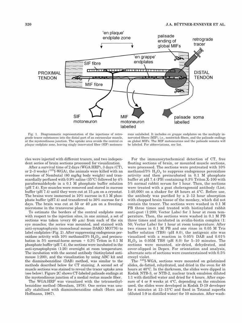
Fig. 1. Diagrammatic representation of the injections of retro-grade tracer substances into the distal part of an extraocular muscle,at the myotendinous junction. The uptake area avoids the central enplaque endplate zone, leaving singly innervated fiber (SIF) motoneu-
rons unlabeled. It includes en grappe endplates on the multiply in-nervated fibers (MIF), i.e., nontwitch fibers, and the palisade endingson global MIFs. The MIF motoneurons and the palisade somata willbe labeled. For abbreviations, see list.
320 J.A. BUTTNER-ENNEVER ET AL.
ing for 2 hours in running water, the sections were coun-terstained with cresyl violet, dehydrated, and cover-slipped with Depex. The sections were examined andphotographed by using a light microscope under darkfieldand brightfield illumination.
All injection sites are treated as left eye muscle injec-tions to facilitate the analysis. Images of brightfield pho-tographs were digitalized by using a 3-CCD videocameramounted on a microscope. The images were captured with4.0 Adobe Photoshop software. After conversion into blackand white, the sharpness, contrast, and brightness wereadjusted to reflect the appearance of the labeling seenthrough the microscope.
Data analysis of cell sizes
Initially measurements of cell area/cell diameter werecompiled by using a microscope and graphics board, con-
nected to a PDP 11/12. Only labeled neurons in which thenucleus was visible were selected. The population wasbimodal, and the exact location of each cell in the “smallmotoneuron cluster” was plotted by using black dots,whereas the location of the cells within the “large mo-toneuron cluster” were indicated by open circles (see Figs7, 9, 10, and 11). In later experiments (see histograms in Fig.13), the mean diameters (maximum diameter 1 minimumdiameter)/2 of labeled neurons were estimated by using animage analysis system (Optimas), from images capturedwith a 3-CCD videocamera mounted on a microscope.
RESULTS
Abducens nucleus
Large injections into the center, or belly, of the lateralrectus (expts: 81-25, 125I-WGA; H154, CT) retrogradely
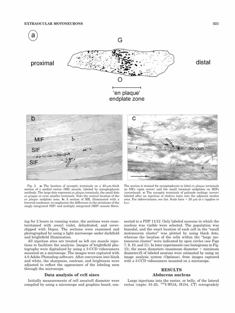
Fig. 2. a: The location of synaptic terminals on a 40-mm-thicksection of a medial rectus (MR) muscle, labeled by synaptophysinantibody. The large dots represent en plaque terminals, the small dotsen grappe or even smaller terminals. Note the central location of theen plaque endplate zone. b: A section of MR, illuminated with alowered condenser, to emphasize the difference in the striations of thesingly integrated (SIF) and multiply integrated (MIF) muscle fibers.
The section is stained for synaptophysin to label en plaque terminalson SIFs (open arrow) and the small terminal endplates on MIFs(arrowhead). c: The synaptic terminals of palisade endings (arrow)labeled after an injection of cholera toxin into the adjacent tendonarea. For abbreviations, see list. Scale bars 5 20 mm in c (applies tob,c).
321EXTRAOCULAR MOTONEURONS
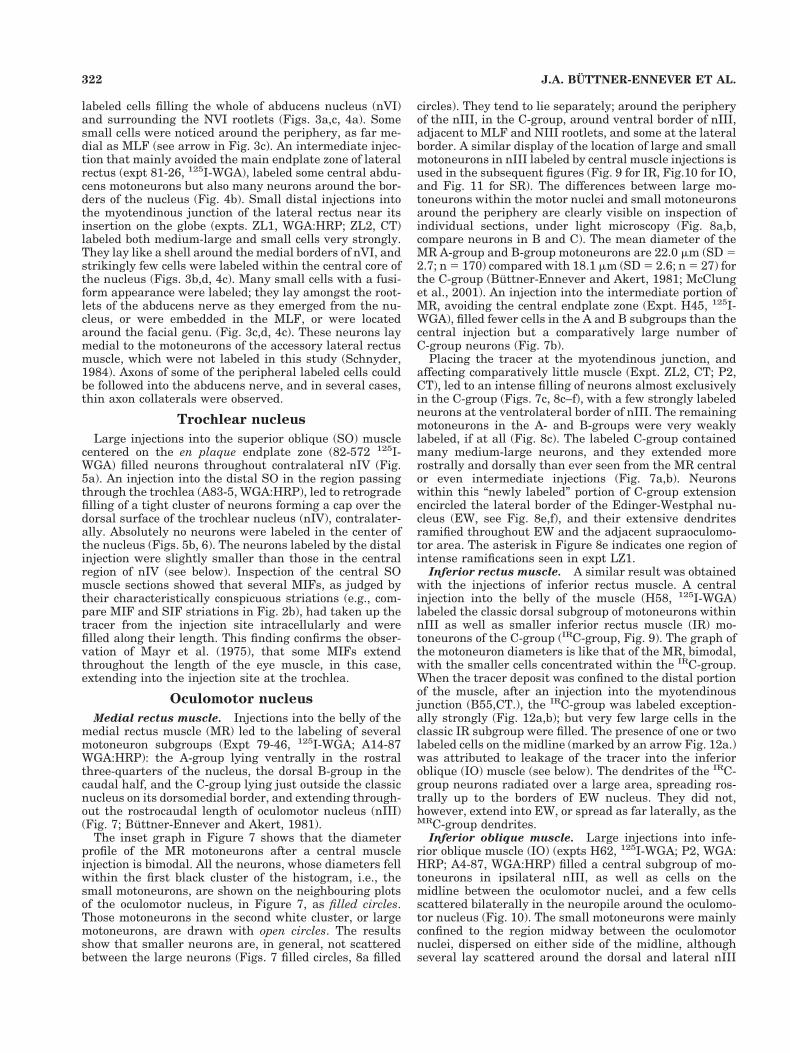
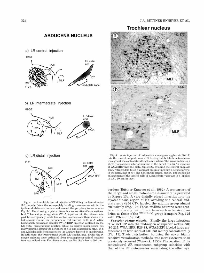
labeled cells filling the whole of abducens nucleus (nVI)and surrounding the NVI rootlets (Figs. 3a,c, 4a). Somesmall cells were noticed around the periphery, as far me-dial as MLF (see arrow in Fig. 3c). An intermediate injec-tion that mainly avoided the main endplate zone of lateralrectus (expt 81-26, 125I-WGA), labeled some central abdu-cens motoneurons but also many neurons around the bor-ders of the nucleus (Fig. 4b). Small distal injections intothe myotendinous junction of the lateral rectus near itsinsertion on the globe (expts. ZL1, WGA:HRP; ZL2, CT)labeled both medium-large and small cells very strongly.They lay like a shell around the medial borders of nVI, andstrikingly few cells were labeled within the central core ofthe nucleus (Figs. 3b,d, 4c). Many small cells with a fusi-form appearance were labeled; they lay amongst the root-lets of the abducens nerve as they emerged from the nu-cleus, or were embedded in the MLF, or were locatedaround the facial genu. (Fig. 3c,d, 4c). These neurons laymedial to the motoneurons of the accessory lateral rectusmuscle, which were not labeled in this study (Schnyder,1984). Axons of some of the peripheral labeled cells couldbe followed into the abducens nerve, and in several cases,thin axon collaterals were observed.
Trochlear nucleus
Large injections into the superior oblique (SO) musclecentered on the en plaque endplate zone (82-572 125I-WGA) filled neurons throughout contralateral nIV (Fig.5a). An injection into the distal SO in the region passingthrough the trochlea (A83-5, WGA:HRP), led to retrogradefilling of a tight cluster of neurons forming a cap over thedorsal surface of the trochlear nucleus (nIV), contralater-ally. Absolutely no neurons were labeled in the center ofthe nucleus (Figs. 5b, 6). The neurons labeled by the distalinjection were slightly smaller than those in the centralregion of nIV (see below). Inspection of the central SOmuscle sections showed that several MIFs, as judged bytheir characteristically conspicuous striations (e.g., com-pare MIF and SIF striations in Fig. 2b), had taken up thetracer from the injection site intracellularly and werefilled along their length. This finding confirms the obser-vation of Mayr et al. (1975), that some MIFs extendthroughout the length of the eye muscle, in this case,extending into the injection site at the trochlea.
Oculomotor nucleus
Medial rectus muscle. Injections into the belly of themedial rectus muscle (MR) led to the labeling of severalmotoneuron subgroups (Expt 79-46, 125I-WGA; A14-87WGA:HRP): the A-group lying ventrally in the rostralthree-quarters of the nucleus, the dorsal B-group in thecaudal half, and the C-group lying just outside the classicnucleus on its dorsomedial border, and extending through-out the rostrocaudal length of oculomotor nucleus (nIII)(Fig. 7; Buttner-Ennever and Akert, 1981).
The inset graph in Figure 7 shows that the diameterprofile of the MR motoneurons after a central muscleinjection is bimodal. All the neurons, whose diameters fellwithin the first black cluster of the histogram, i.e., thesmall motoneurons, are shown on the neighbouring plotsof the oculomotor nucleus, in Figure 7, as filled circles.Those motoneurons in the second white cluster, or largemotoneurons, are drawn with open circles. The resultsshow that smaller neurons are, in general, not scatteredbetween the large neurons (Figs. 7 filled circles, 8a filled
circles). They tend to lie separately; around the peripheryof the nIII, in the C-group, around ventral border of nIII,adjacent to MLF and NIII rootlets, and some at the lateralborder. A similar display of the location of large and smallmotoneurons in nIII labeled by central muscle injections isused in the subsequent figures (Fig. 9 for IR, Fig.10 for IO,and Fig. 11 for SR). The differences between large mo-toneurons within the motor nuclei and small motoneuronsaround the periphery are clearly visible on inspection ofindividual sections, under light microscopy (Fig. 8a,b,compare neurons in B and C). The mean diameter of theMR A-group and B-group motoneurons are 22.0 mm (SD 52.7; n 5 170) compared with 18.1 mm (SD 5 2.6; n 5 27) forthe C-group (Buttner-Ennever and Akert, 1981; McClunget al., 2001). An injection into the intermediate portion ofMR, avoiding the central endplate zone (Expt. H45, 125I-WGA), filled fewer cells in the A and B subgroups than thecentral injection but a comparatively large number ofC-group neurons (Fig. 7b).
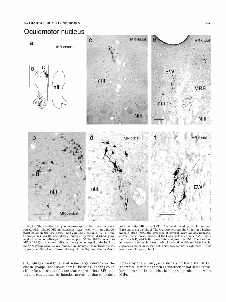
Placing the tracer at the myotendinous junction, andaffecting comparatively little muscle (Expt. ZL2, CT; P2,CT), led to an intense filling of neurons almost exclusivelyin the C-group (Figs. 7c, 8c–f), with a few strongly labeledneurons at the ventrolateral border of nIII. The remainingmotoneurons in the A- and B-groups were very weaklylabeled, if at all (Fig. 8c). The labeled C-group containedmany medium-large neurons, and they extended morerostrally and dorsally than ever seen from the MR centralor even intermediate injections (Fig. 7a,b). Neuronswithin this “newly labeled” portion of C-group extensionencircled the lateral border of the Edinger-Westphal nu-cleus (EW, see Fig. 8e,f), and their extensive dendritesramified throughout EW and the adjacent supraoculomo-tor area. The asterisk in Figure 8e indicates one region ofintense ramifications seen in expt LZ1.
Inferior rectus muscle. A similar result was obtainedwith the injections of inferior rectus muscle. A centralinjection into the belly of the muscle (H58, 125I-WGA)labeled the classic dorsal subgroup of motoneurons withinnIII as well as smaller inferior rectus muscle (IR) mo-toneurons of the C-group (IRC-group, Fig. 9). The graph ofthe motoneuron diameters is like that of the MR, bimodal,with the smaller cells concentrated within the IRC-group.When the tracer deposit was confined to the distal portionof the muscle, after an injection into the myotendinousjunction (B55,CT.), the IRC-group was labeled exception-ally strongly (Fig. 12a,b); but very few large cells in theclassic IR subgroup were filled. The presence of one or twolabeled cells on the midline (marked by an arrow Fig. 12a.)was attributed to leakage of the tracer into the inferioroblique (IO) muscle (see below). The dendrites of the IRC-group neurons radiated over a large area, spreading ros-trally up to the borders of EW nucleus. They did not,however, extend into EW, or spread as far laterally, as theMRC-group dendrites.
Inferior oblique muscle. Large injections into infe-rior oblique muscle (IO) (expts H62, 125I-WGA; P2, WGA:HRP; A4-87, WGA:HRP) filled a central subgroup of mo-toneurons in ipsilateral nIII, as well as cells on themidline between the oculomotor nuclei, and a few cellsscattered bilaterally in the neuropile around the oculomo-tor nucleus (Fig. 10). The small motoneurons were mainlyconfined to the region midway between the oculomotornuclei, dispersed on either side of the midline, althoughseveral lay scattered around the dorsal and lateral nIII
322 J.A. BUTTNER-ENNEVER ET AL.
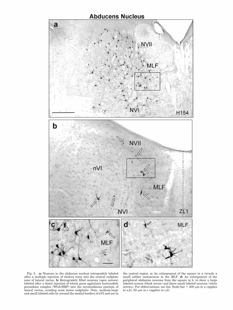
Fig. 3. a: Neurons in the abducens nucleus retrogradely labeledafter a multiple injection of cholera toxin into the central endplatezone of lateral rectus. b: Retrogradely filled neurons (open arrows)labeled after a distal injection of wheat germ agglutinin horseradishperoxidase complex (WGA:HRP) into the myotendinous junction oflateral rectus, avoiding most motor endplates. Note, medium-largeand small labeled cells lie around the medial borders of nVI and not in
the central region. c: An enlargement of the square in a reveals asmall caliber motoneuron in the MLF. d: An enlargement of theperipheral abducens neurons from the square in b, to show a largelabeled neuron (black arrow) and three small labeled neurons (whitearrows). For abbreviations, see list. Scale bar 5 500 mm in a (appliesto a,b), 50 mm in c (applies to c,d).
borders (Buttner-Ennever et al., 1982). A comparison ofthe large and small motoneuron diameters is providedby Figure 13a. A very distally placed injection into themyotendinous region of IO, avoiding the central end-plate zone (S54 CT), labeled the midline group almostexclusively (Fig. 10). These midline neurons were scat-tered bilaterally but did not have such extensive den-drites as those of the MR and IRC-group (compare Fig. 12dwith 12b and Fig. 8d).
Superior rectus muscle. Finally the large injectionsof WGA:HRP into the mid-region of superior rectus (SR)(80-217, WGA:HRP; B26-88, WGA:HRP) labeled large mo-toneurons on both sides of nIII but mainly contralaterally(Fig. 11). Their distribution, by using the newer highlysensitive visualization methods, was more extensive thanpreviously reported (Warwick, 1953). The location of thecontralateral SR motoneuron subgroup coincides withthat of the IO motoneurons innervating the other eye.
Fig. 4. a: A multiple central injection of CT filling the lateral rectus(LR) muscle. Note the retrogradely labeling motoneurons within theipsilateral abducens nucleus and around the periphery (same case asFig. 3a). The drawing is plotted from four consecutive 40-mm sections.b: A 125I-wheat germ agglutinin (WGA) injection into the intermediatepart LR retrogradely labels less central motoneurons than shown in abut several around the periphery of nVI (medial half). c: A WGA:horseradish peroxidase complex (WGA:HRP) injection centered on theLR distal myotendinous junction labels no central motoneurons butmany neurons around the periphery of nVI and scattered in MLF. In band c, labeled cells from six sections (40 mm) are depicted on one drawing.In both cases, the tracer spread within LR (shaded area) avoids the enplaque endplate zone estimated from synaptophysin-stained sectionsfrom a standard case. For abbreviations, see list. Scale bar 5 500 mm.
Fig. 5. a: An injection of radioactive wheat germ agglutinin (WGA)into the central endplate zone of SO retrogradely labels motoneuronsthroughout the contralateral trochlear nucleus. The arrow indicates aslightly separate cluster of neurons in the dorsal cap. b: An injectionof WGA:HRP into the distal tip of SO, avoiding the central endplatezone, retrogradely filled a compact group of medium neurons (arrow)in the dorsal cap of nIV and none in the central region. The inset is anenlargement of the labeled cells in b. Scale bars 5250 mm in a (appliesto a,b), 50 mm in inset.
324 J.A. BUTTNER-ENNEVER ET AL.
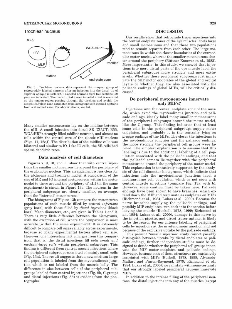
Many smaller motoneurons lay on the midline betweenthe nIII. A small injection into distal SR (ZU,CT; B55,WGA:HRP) strongly filled midline neurons, and almost nocells within the central core of the classic nIII nucleus(Figs. 11, 12e,f). The distribution of the midline cells wasbilateral and similar to IO. Like IO cells, the SR cells hadsparse dendritic trees.
Data analysis of cell diameters
Figures 7, 9, 10, and 11 show that with central injec-tions the smaller motoneurons lie around the periphery ofthe oculomotor nucleus. This arrangement is less clear forthe abducens and trochlear nuclei. A comparison of thesize of MR and IO internal motoneurons within the motornuclei to those around the periphery of nIII (in the sameexperiment) is shown in Figure 13a. The neurons in theperipheral subgroups are clearly smaller, on average,than the “internal” motoneurons.
The histograms of Figure 13b compare the motoneuronpopulations of each muscle filled by central injections(grey bars), with those filled by distal injections (blackbars). Mean diameters, etc., are given in Tables 1 and 2.There is very little difference between the histograms,with the exception of SO, where the comparison is mostaccurate (within the same experiment). It is extremelydifficult to compare cell sizes reliably across experiments,because so many experimental factors affect cell size.However, one interesting fact emerges from this compar-ison, that is, the distal injections fill both small andmedium-large cells within peripheral subgroups. Thisfinding is different from central muscle injections wherethe peripheral subgroups consisted of mainly small cells(Fig. 13a). The result suggests that a new medium-largecell population is labeled from the myotendinous junc-tion which is not labeled from the muscle belly. Thedifference in size between cells of the peripheral sub-groups labeled from central injections (Fig. 8b, C-group)and distal injections (Fig. 8d) is evident from the pho-tographs.
DISCUSSION
Our results show that retrograde tracer injections intothe central endplate zones of the eye muscles labels largeand small motoneurons and that these two populationstend to remain separate from each other. The large mo-toneurons lie within the classic boundaries of the extraoc-ular motor-nuclei, whereas the smaller motoneurons clus-ter around the periphery (Buttner-Ennever et al., 1982).More importantly, in this study, we showed that injec-tions into more distal parts of the eye muscle label theperipheral subgroups more strongly and more exclu-sively. Whether these peripheral subgroups just inner-vate the MIF motor endplates of the global and orbitallayers or whether they are also associated with thepalisade endings of global MIFs, will be critically dis-cussed.
Do peripheral motoneurons innervateonly MIFs?
Injections into the central endplate zone of the mus-cle, which avoid the myotendinous junction and pali-sade endings, clearly label many smaller motoneuronsof the peripheral subgroups around the motor nuclei,like the C-group. This finding indicates that at leastsome cells in the peripheral subgroups supply motorendplates, and probably it is the centrally lying engrappe endings of the MIFs. The closer the injections tothe myotendinous junction and the palisade endings,the more strongly the peripheral cell groups were la-beled. The simplest explanation is to assume that thislabeling is due to the additional labeling of a cell pop-ulation associated with the palisade endings, and thatthe ‘palisade’ somata lie together with the peripheralmotoneurons around the periphery of the motor nuclei.This explanation is tentatively supported by the analy-sis of the cell diameter histograms, which indicate thatinjections into the myotendinous junctions label amedium-large cell population which is not seen withcentral muscle injections (compare Fig. 13a with b).However, some caution must be taken here. Palisadeendings have been shown to have branches, which ex-tend down the MIF and terminate as endplates on MIFs(Richmond et al., 1984; Lukas et al., 2000). Because thenerve branches supplying the palisade endings, andpossibly MIF endplates, run back into the tendon beforeleaving the muscle (Ruskell, 1978, 1999; Richmond etal., 1984; Lukas et al., 2000), damage to this nerve bythe injection-pipette, and direct tracer uptake, is likelyto be the reason for our intense labeling of peripheralcells by injections at the myotendinous junction and notbecause of the exclusive uptake by the palisade endings.
This present “muscle injection” study cannot possiblydistinguish between uptake by distal endplates or pali-sade endings, further independent studies must be de-signed to decide whether the peripheral cell groups inner-vate the MIF motor-endplates and palisade endings.However, because both of these structures are exclusivelyassociated with MIFs (Ruskell, 1978, 1999; Alvarado-Mallart and Pincon-Raymond, 1979; Richmond et al.,1984; Lukas et al., 2000), we can state with some certaintythat our strongly labeled peripheral neurons innervateMIFs.
In addition to the intense filling of the peripheral neu-rons, the distal injections into any of the muscles (except
Fig. 6. Trochlear nucleus: dots represent the compact group ofretrogradely labeled neurons after an injection into the distal tip ofsuperior oblique muscle (SO). Labeled neurons from five sections (50mm) are indicated. The tracer uptake area (shaded area) is centeredon the tendon region passing through the trochlea and avoids thecentral endplate zone estimated from synaptophysin-stained sectionsfrom a standard case. For abbreviations, see list.
325EXTRAOCULAR MOTONEURONS
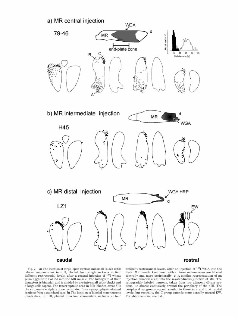
Fig. 7. a: The location of large (open circles) and small (black dots)labeled motoneurons in nIII, plotted from single sections at fourdifferent rostrocaudal levels, after a central injection of 125I-wheatgerm agglutinin (WGA) into the MR muscle. The histogram of theirdiameters is bimodal, and is divided by eye into small cells (black) anda large cells (open). The tracer-uptake area in MR (shaded area) fillsthe en plaque endplate zone, estimated from synaptophysin-stainedsections from a standard case. b: The location of labeled motoneurons(black dots) in nIII, plotted from four consecutive sections, at four
different rostrocaudal levels, after an injection of 125I-WGA into thedistal MR muscle. Compared with a, fewer motoneurons are labeledcentrally and more peripherally. c: A similar representation of aninjection (shaded area) into the myotendinous junction of MR. Theretrogradely labeled neurons, taken from two adjacent 40-mm sec-tions, lie almost exclusively around the periphery of the nIII. Theperipheral subgroups appear similar to those in a and b at caudallevels, but rostrally, the C-group extends more dorsally toward EW.For abbreviations, see list.
SO), always weakly labeled some large neurons in theclassic groups (not shown here). The weak labeling couldeither be the result of some tracer-spread into SIF end-plate areas, uptake by impaled nerves, or due to modest
uptake by the en grappe terminals on the distal MIFs.Therefore, it remains unclear whether or not some of thelarge neurons in the classic subgroups also innervateMIFs.
Fig. 8. The charting and photomicrographs in the upper row showretrogradely labeled MR motoneurons (a,c,e), each with an enlarge-ment below in the lower row (b,d,f). a: The location of A-, B-, andC-groups in mid-nIII labeled by a multiple injections of wheat germagglutinin horseradish peroxidase complex (WGA:HRP) tracer intoMR (A14-87); the square indicates the region enlarged in (b). b: Note,many C-group neurons are smaller in diameter than those in theB-group. c: Note the intense labeling of the C-group after a distal
injection into MR (case LZ1). The weak labeling of the A- andB-groups is not visible. d: The C-group neurons shown in c at a highermagnification. Note the presence of several large labeled neurons.e: The rostral-most neurons of the C-group labeled by a distal injec-tion into MR, which lie immediately adjacent to EW. The asteriskmarks one of the regions containing labeled dendritic ramifications insupraoculomotor area. For abbreviations, see list. Scale bars 5 300mm in c,e, 100 mm in b,d,f.
327EXTRAOCULAR MOTONEURONS
Are palisade endings sensory or motor?
Palisade endings are, at present, generally consideredto be sensory structures (Alvarado-Mallart RM andPincon-Raymond M, 1979; Billig et al., 1997; Ruskell,1999), and it is hard to imagine a motor role for them.However, Lukas et al. (2000) have re-examined palisadeendings in humans and decided that some aspects aremotor in character (e.g., branches with bungarotoxin-binding endplates terminating on distal MIF), whereas
other aspects seem sensory. Other lines of evidence alsosupport a motor role for palisade endings, although some-what indirectly. For example, palisade endings werecaused to degenerate by intracranial severance of NIII,NIV, or NVI in monkey (Tozer and Sherrrington, 1910),and again by stereotaxic lesions of the oculomotor nucleiin cat (Sas and Schab, 1952). Furthermore, Gentle andRuskell (1997) found too few cell bodies in the sensorytrigeminal ganglion of monkey to supply the necessary
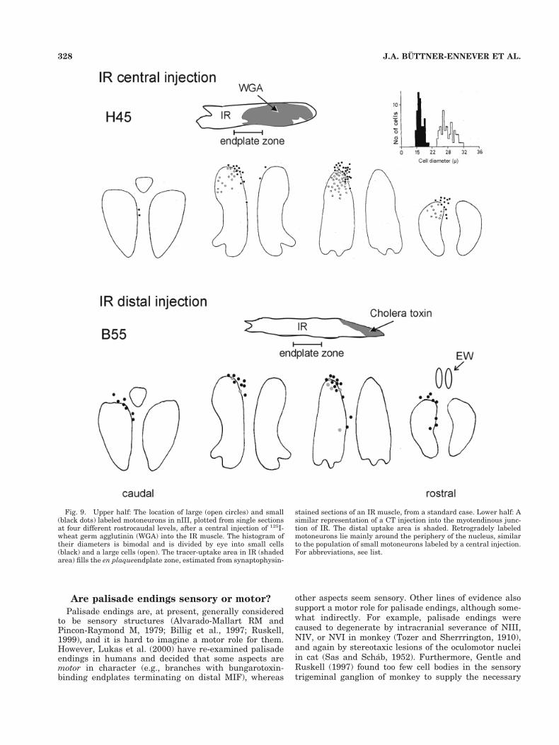
Fig. 9. Upper half: The location of large (open circles) and small(black dots) labeled motoneurons in nIII, plotted from single sectionsat four different rostrocaudal levels, after a central injection of 125I-wheat germ agglutinin (WGA) into the IR muscle. The histogram oftheir diameters is bimodal and is divided by eye into small cells(black) and a large cells (open). The tracer-uptake area in IR (shadedarea) fills the en plaqueendplate zone, estimated from synaptophysin-
stained sections of an IR muscle, from a standard case. Lower half: Asimilar representation of a CT injection into the myotendinous junc-tion of IR. The distal uptake area is shaded. Retrogradely labeledmotoneurons lie mainly around the periphery of the nucleus, similarto the population of small motoneurons labeled by a central injection.For abbreviations, see list.
328 J.A. BUTTNER-ENNEVER ET AL.
number of myelinated axons to innervate the palisadeendings. We did not systematically investigate the label-ing in the trigeminal ganglion in this study; but in the fewcases of myotendinous junction injections that we did ex-amine, a few trigeminal ganglion cells were retrogradelylabeled.
Our study cannot resolve the question of whether pali-sade endings are sensory or motor. The cells that wereintensely labeled by our injections directly into the pali-sade terminal region (see Figs. 4, 7, 8) lie immediatelyadjacent to the motor nuclei, they receive afferents similarto those of motoneurons and have the morphologic char-acteristics of g-motoneurons (May et al., 2000) not sensory
ganglion cells. However, if it is argued that these cellsinnervate only the MIF endplates and not the palisadeendings, then it is unclear why they are so intenselylabeled from the myotendinous junction, unless the MIFmotor nerves enter the muscle via the myotenolinous junc-tion.
Separation of large and small motoneurons
According to these experiments, it is a general feature ofall the three oculomotor nuclei that smaller motoneuronsare not intermingled with the large motoneurons. Theanatomic separation of the motoneuron subgroups hasimportant functional consequences. Each subset of mo-
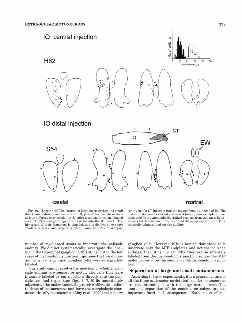
Fig. 10. Upper half: The location of large (open circles) and small(black dots) labeled motoneurons in nIII, plotted from single sectionsat four different rostrocaudal levels, after a central injection (shadedarea) of 125I-wheat germ agglutinin (WGA) into the IO muscle. Thehistogram of their diameters is bimodal, and is divided by eye intosmall cells (black) and large cells (open). Lower half: A similar repre-
sentation of a CT injection into the myotendinous junction of IO. Thedistal uptake area is shaded and avoids the en plaque endplate zone,estimated from synaptophysin-stained sections from this case. Retro-gradely labeled motoneurons lie around the periphery of the nucleus,especially bilaterally about the midline.
329EXTRAOCULAR MOTONEURONS
toneurons could be driven by different afferent pathways.Indeed this is known to be the case: injections of antero-grade tracers into the pretectum lead to terminal labelingover the peripheral borders of nIII, including the C-groupand midline cell groups, but no terminal labeling over theclassic larger motoneuron population of nIII (Buttner-
Ennever et al., 1996; Fig. 2b); moreover, only the dorsalcap of the contralateral trochlear nucleus is targeted byefferents of the superior vestibular nucleus, possibly car-rying otolith signals (Carpenter and Cowie, 1985). Otherafferent pathways to the oculomotor nucleus target boththe large and small motoneuron populations (Buttner-
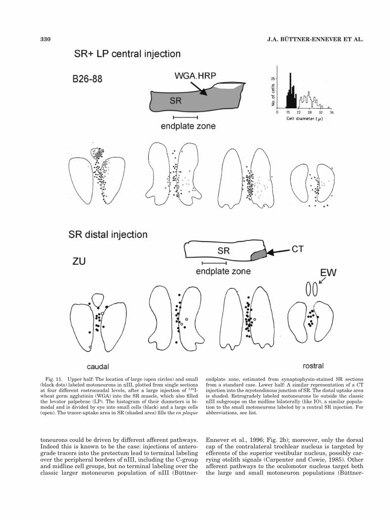
Fig. 11. Upper half: The location of large (open circles) and small(black dots) labeled motoneurons in nIII, plotted from single sectionsat four different rostrocaudal levels, after a large injection of 125I-wheat germ agglutinin (WGA) into the SR muscle, which also filledthe levator palpebrae (LP). The histogram of their diameters is bi-modal and is divided by eye into small cells (black) and a large cells(open). The tracer-uptake area in SR (shaded area) fills the en plaque
endplate zone, estimated from synaptophysin-stained SR sectionsfrom a standard case. Lower half: A similar representation of a CTinjection into the myotendinous junction of SR. The distal uptake areais shaded. Retrogradely labeled motoneurons lie outside the classicnIII subgroups on the midline bilaterally (like IO), a similar popula-tion to the small motoneurons labeled by a central SR injection. Forabbreviations, see list.
330 J.A. BUTTNER-ENNEVER ET AL.
Ennever and Akert, 1981). These findings underline theidea that there is a functional difference between the largeand small motoneuron population.
Organization of MIF motoneurons
The smaller motoneuron population of nIII has a com-pletely different organization than that of the large mo-toneurons. In the C-group, MR and IR motoneurons of thesame eye are intermingled; an afferent input to this sub-
group would lead to an adduction of the ipsilateral eye,with a downward component, and a bilateral activation ofthe C-group would produce vergence. At the midline, be-tween the nIII, where the bilaterally represented IO andSR “MIF” motoneurons are clustered together, an afferentinput onto these neurons would cause coactivation of theupward moving muscles of both eyes and produce upgaze.Close to the motoneurons lie a population of medium-largecells that may innervate the palisade endings of the global
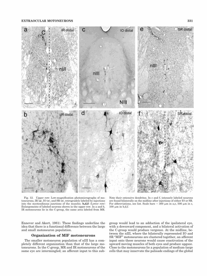
Fig. 12. Upper row: Low-magnification photomicrographs of mo-toneurons, IR (a), IO (c), and SR (e), retrogradely labeled by injectionsinto the myotendinous junctions of the muscles. b,d,f: (Lower row)Enlargements of labeled neurons shown in the upper row. In a and b,IR motoneurons lie in the C-group, the same area labeled from MR.
Note their extensive dendrites. In c and f, intensely labeled neuronsare found bilaterally on the midline after injections of either IO or SR.For abbreviations, see list. Scale bars 5 300 mm in a,e, 500 mm in c,100 mm in b,d,f.
331EXTRAOCULAR MOTONEURONS
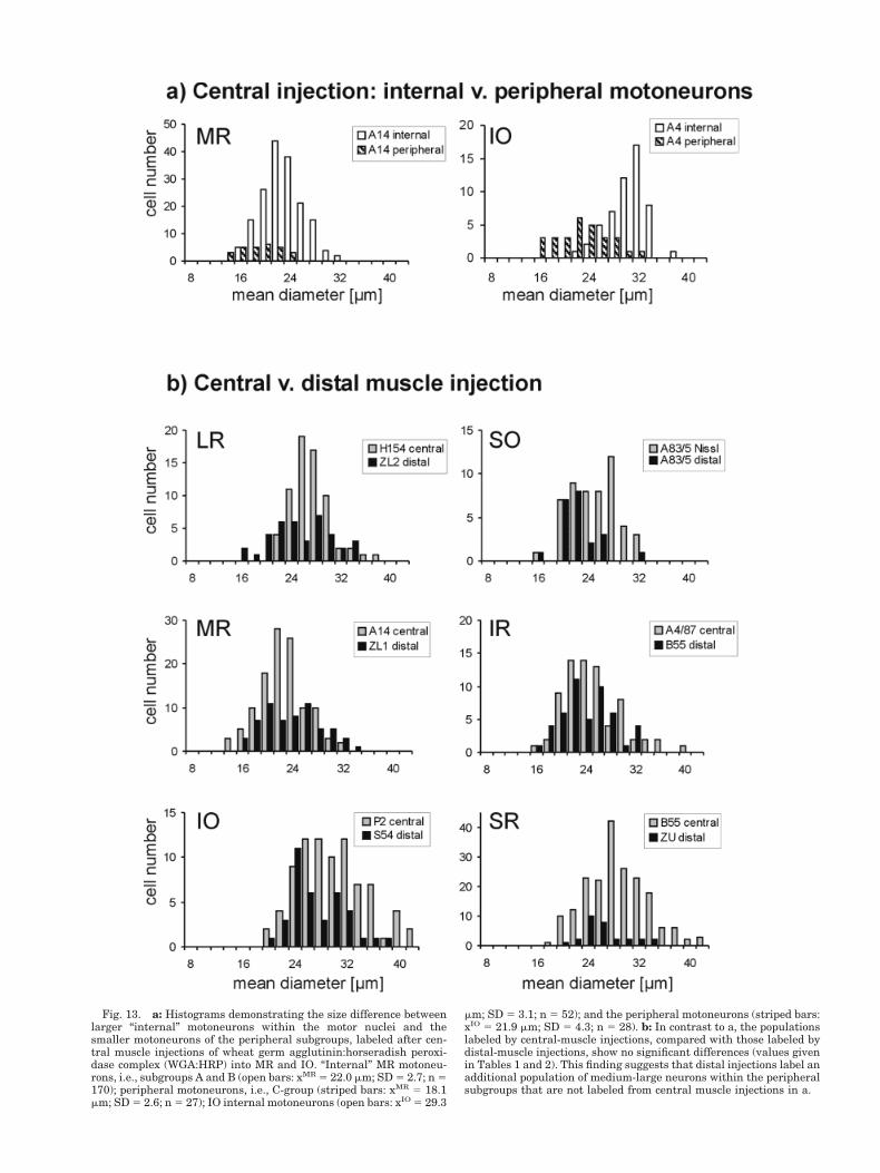
Fig. 13. a: Histograms demonstrating the size difference betweenlarger “internal” motoneurons within the motor nuclei and thesmaller motoneurons of the peripheral subgroups, labeled after cen-tral muscle injections of wheat germ agglutinin:horseradish peroxi-dase complex (WGA:HRP) into MR and IO. “Internal” MR motoneu-rons, i.e., subgroups A and B (open bars: xMR 5 22.0 mm; SD 5 2.7; n 5170); peripheral motoneurons, i.e., C-group (striped bars: xMR 5 18.1mm; SD 5 2.6; n 5 27); IO internal motoneurons (open bars: xIO 5 29.3
mm; SD 5 3.1; n 5 52); and the peripheral motoneurons (striped bars:xIO 5 21.9 mm; SD 5 4.3; n 5 28). b: In contrast to a, the populationslabeled by central-muscle injections, compared with those labeled bydistal-muscle injections, show no significant differences (values givenin Tables 1 and 2). This finding suggests that distal injections label anadditional population of medium-large neurons within the peripheralsubgroups that are not labeled from central muscle injections in a.
MIFs. These cells would receive a similar afferent input tothe MIF motoneurons.
Phylogenetically speaking, oculomotor neuroanatomy ishighly conservative, but nevertheless the C-group, inner-vating MR and IR, has only been described, up until now,in primates (Buttner-Ennever and Akert, 1981; Spencerand Porter, 1981; Sun and May, 1993; McCrea et al.,1986). Ishikawa et al. (1990) unfortunately referred to itas part of the Edinger-Westphal nucleus. Our central anddistal injections into MR and IR highlighted cells in theC-group, but MR injections also labeled an extension to-ward EW not seen with central MR muscle injections. TheC-group extension neurons were found to have very widelyspreading dendrites. Those of MR, but not IR, invadingEW and the supraoculomotor area. The results confirmthose of May et al. (2000), whose EM study revealed syn-aptic contacts on the MRC-group dendrites in EW. Theyconsidered the MRC-group neurons to have the structuralcharacteristics of g-motoneurons. Our results suggest thatthe C-group may contain several different populations ofneurons: MR MIF-motoneurons, IR MIF-motoneurons,and medium-large somata associated with the palisadeendings of the global MIFs.
Functional significance of peripheralsubgroups
The peripheral subgroups, including the C-group, inner-vate the MIFs. Robinson (1991) once suggested that theMIFs, with supposedly sensory palisade endings at bothends, resembled an inverted muscle spindle. However, thefunction of the nontwitch MIFs in extraocular eye musclesis still not known. Several lines of evidence point towardthe fact that MIFs do not play a significant role in devel-opment of fast muscle forces. First, on stimulation, thenontwitch MIFs produce tonic responses, with weak te-tanic tensions. However, they are extremely fatigue resis-tant and are innervated by slowly conducting axons (Nel-son et al., 1986; Dieringer and Precht, 1986). Second,muscle stimulation experiments carried out at the fusionfrequency of slow fibers (30/sec) produced no measurabletension changes in the muscle (Fuchs and Luschei, 1971).These features suggest that MIFs and, hence, the periph-eral motoneurons, are not suited to contribute signifi-cantly to the fast eye movement displacements, but are
more suited to playing a role in fixation and stabilizing theeyes around the primary position (Dean, 1996).
The supraoculomotor area and EW are parts of themidbrain near-response region (May et al., 1992). The EWnucleus contains preganglionic neurons driving accommo-dation of the lens, an essential part of the near-response(Gamlin et al., 1994; May et al., 2000 ); and recordingsmade in the supraoculomotor area, dorsal and lateral tonIII, demonstrate neurons carrying premotor vergencesignals (Mays, 1984), also an essential component of thenear-response and fixation. The close association of theMRC-group extension with midbrain near-response re-gions strengthens our previous hypothesis that theC-group may be specialized and play a role in vergencemovements (Buttner-Ennever and Akert, 1981).
A reassessment of our hypothesis on therole of the C-group and orbital layer
In our initial experiments on the oculomotor nucleus(Buttner-Ennever and Akert, 1981), we proposed that thesmall motoneurons of the C-group might innervate theorbital layer. We had placed small tracer injections, aimedat the orbital layer of the MR muscle and retrogradelylabeled the C-group, almost exclusively (Buttner-Enneverand Akert, 1981). The injections were mainly in the outerorbital layer, but they were also confined to the distal halfof the muscle, and, therefore, targeted predominantly theMIFs. They did not significantly involve the central end-plate zone of the orbital layer (Buttner-Ennever and Ak-ert, 1981; see Fig. 3). In addition, orbital MIFs do not havepalisade endings. Therefore, in the light of the presentexperiments, our original conclusion is wrong and shouldprobably be modified to: “. . . small motoneurons in theC-group innervate the MIFs of the orbital layer.”
In our present experiment, it is impossible to say howmuch of the orbital layer is involved in the injections.Morphologic studies report that the orbital layer of ex-traocular eye muscles does not extend as far distally intothe tendon as the global layer (Spencer and Porter, 1988),and conversely the global layer MIFs extend furthest intothe tendon (Mayr et al., 1975). Therefore, it seems that ourpresent injections into the distal myotendinous junctionhave primarily targeted the MIFs of the global layer, pos-sibly along with their palisade endings, whereas the pre-
TABLE 1. Diameter of Motoneurons Labeled by a Central Muscle Injection1
Muscle LR SO MR IR IO SR 1 LP
Case H154 83-5 A14 A4-87 P2 B55Tracer CT (Neutral red) WGA:HRP WGA:HRP WGA:HRP WGA:HRPMean diameter (mm) 26.4 26.0 21.6 24.1 28.9 27.7SD 3.2 2.4 3.7 4.7 5.0 4.8N 67 30 115 72 82 194
1See Figure 13b, gray bars. For abbreviations, see list.
TABLE 2. Diameter of Motoneurons Labeled by a Distal Injection1
Muscle LR SO MR IR IOSR 1
LP
Case ZL2 83-5 ZL1 B55 S54 ZUTracer WGA.HRP WGA.HRP WGA.HRP CT CT CTMean diameter (mm) 24.5 21.3 22.7 23.1 26.0 25.2SD 4.9 3.1 4.5 3.9 4.3 3.6N 38 23 61 48 37 29
1See figure 13b, striped bars. For abbreviations, see list.
333EXTRAOCULAR MOTONEURONS
vious injections (Buttner-Ennever and Akert, 1981)mainly involved the MIFs of the orbital layer. In both setsof experiments, the peripheral neurons of the C-groupwere labeled, suggesting that both orbital and global layerMIFs are innervated by the peripheral neurons.
The orbital layer MIFs appear to be more complicatedthan the global MIFs, because they change from a nont-witch MIF character at the fiber extremities to a twitch(SIF) morphology in the central region (Pachter, 1984;Jacoby et al., 1989). Recent reports by Demer et al. (2000)provide several lines of evidence showing that the orbitallayer terminates around the capsule of Tenon and controlsthe tension in the connective tissue “pulleys” surroundingthe eye muscles. Taken together, our results suggest thatthe peripheral motoneurons could control, amongst otherthings, the tension in the pulleys through the orbitalMIFs.
Clinical applications
Two lines of clinical evidence support the role of pali-sade endings and MIFs in determining eye position (Stein-bach, 1987, 2000). One line comes from strabismus pa-tients in which damage to the musculotendinous region,for example in marginal myotomy, was shown to affect theassessment of eye position (Steinbach and Smith, 1981;Steinbach et al., 1987). The second, is the experiments ofDell’Osso et al. (1999), who performed a tenotomy on theeye muscles of dogs with congenital nystagmus, followedby re-insertion at exactly the same position. Surprisingly,the operation led to the reduction of the nystagmus. Theseresults are interpreted by the authors in terms of the MIF-palisade endings being a sensory afferent system, which isdamaged by manipulations of the tendon. Even thoughthis interpretation may be in question, their results havefar reaching consequences for ophthalmic surgery, empha-sizing the need to protect the myotendinous region of theextraocular eye muscles, as do the results of this study.
In conclusion, we have shown that separate groups ofmotoneurons around the periphery of the motor nuclei,i.e., nIII nIV, and nVI, supply innervation to the nont-witch MIF of the eye muscles. These nerves are thought toinnervate the MIFs of both the orbital and the globallayers of the eye muscle. However, several questions re-main unclear: Do the peripheral subgroups also innervatethe palisade endings of the global MIFs? Are the palisadeendings sensory or motor structures? Because the periph-eral cell groups do not appear to be a homogeneous popu-lation, especially in the case of medial rectus, what is theinternal organization of the C-group? Further experi-ments are necessary to answer these questions and tounderstand the function of MIFs. A whole new set ofmotoneurons innervating the MIFs have now come tolight, and, at present, we have no knowledge of theirphysiological properties in awake animals. However, be-cause we now know their exact location, experiments in-vestigating their firing patterns, and their premotor in-puts are at last feasible. It is not unreasonable to expectthat these may well lead to an insight into the illusiveproblem of the sensory control of eye muscles.
ACKNOWLEDGMENTS
The authors thank Professor W. Lange for his supportand Ahmed Messoudi, M.Phil., and Ursula Schneider fortheir excellent technical support.
LITERATURE CITED
Alvarado-Mallart RM, Pincon-Raymond M. 1979. The palisade endings ofcat extraocular muscles: a light and electron microscope study. TissueCell 11:567–584.
Augustine JR, Deschamps EG, Ferguson JGJ. 1981. Functional organiza-tion of the oculomotor nucleus in the baboon. Am J Anat 161:393–403.
Baker R. 1998. From genes to behavior in the vestibular system. Otolar-yngol Head Neck Surg 119:263–275.
Billig I, Buisseret-Delmas C, Buisseret P. 1997. Identification of nerveendings in cat extraocular muscles. Anat Rec 248:566–575.
Bondi AY, Chiarandini DJ. 1983. Morphologic and electrophysiologic ver-ification of multiply innervated fibers in rat extraocular muscles. In-vest Ophthalmol Vis Sci 24:516–519.
Buttner-Ennever JA. 2000. A dual motor control of extraocular eye mus-cles. Neuro-ophthalmol 23:147–149.
Buttner-Ennever JA, Akert K. 1981. Medial rectus subgroups of the ocu-lomotor nucleus and their abducens internuclear input in the monkey.J Comp Neurol 197:17–27.
Buttner-Ennever JA, d’Ascanio P, Gysin R. 1982. The localization of largeand small motoneurons in the oculomotor complex of the monkey. InRoucoux A, Crommelinck M, editors. Physiological and pathologicalaspects of eye movements. The Haugue, Boston, London: Dr W. JunkPubl. p 345–349.
Buttner-Ennever JA, Cohen B, Horn AKE, Reisine H. 1996. Pretectalprojections to the oculomotor complex of the monkey and their role ineye movements. J Comp Neurol 366:348–359.
Buttner-Ennever JA, Horn AKE, Scherberger H-J, Henn V. 1998. Thelocalization of motoneurons innervating slow extraocular eye musclefibers in monkey. Soc Neurosci Abstr 24:145
Carpenter MB, Cowie RJ. 1985. Connections and oculomotor projections ofthe superior vestibular nucleus and cell group “y.” Brain Res 336:265–287.
Dean P. 1996. Motor unit recruitment in a distribution model of extraoc-ular muscle. J Neurophysiol 76:727–742.
Dell’Osso LF, Hertle RW, Williams RW, Jacobs JB. 1999. A new surgery forcongenital nystagmus: effects of tenotomy on an achiasmatic canineand the role of extraocular proprioception. J Am Acad Pediatr Ophthal-mol Strab 3:166–182.
Demer JL, Oh SY, Poukens V. 2000. Evidence for active control of rectusextraocular muscle pulleys. Invest Ophthalmol Vis Sci 41:1280–1290.
Dieringer N, Precht W. 1986. Functional organization of eye velocity andeye position signals in abducens motoneurons of the frog. J CompPhysiol 158:179–194.
Dogiel AS. 1906. Die Endigungen der sensiblen Nerven in den Augen-muskeln und deren Sehnen beim Menschen und den Saugetieren. ArchAnat Micros Morphol 6:501–526.
Fuchs AF, Luschei ES. 1971. Development of isometric tension in simianextraocular muscle. J Physiol (Lond) 219:155–166.
Gamlin PD, Zhang Y, Clendaniel RA, Mays LE. 1994. Behavior of identi-fied Edinger-Westphal neurons during ocular accommodation. J Neu-rophysiol 72:2368–2382.
Gentle A, Ruskell GL. 1997. Pathway of the primary afferent nerve fibersserving proprioception in monkey extraocular muscles. OphthalmicPhysiol Opt 17:225–231.
Horn AKE, Hoffmann K-P. 1987. Combined GABA-immunocytochemistryand TMB-HRP histochemistry of pretectal nuclei projecting to theinferior olive in rats, cats and monkeys. Brain Res 409:133–138.
Ishikawa S, Sekiya H, Kondo Y. 1990. The center for controlling the nearreflex in the midbrain of the monkey: a double labelling study. BrainRes 519:217–222.
Jacoby J, Chiarandini DJ, Stefani E. 1989. Electrical properties and in-nervation of fibers in the orbital layer of rat extraocular muscles.J Neurophysiol 61:116–125.
Keller EL. 1973. Accommodative vergence in the alert monkey: motor unitanalysis. Vision Res 13:1565–1575.
Keller EL, Robinson DA. 1972. Abducens unit behavior in the monkeyduring vergence movements. Vision Res 12:369–382.
Lennerstrand G, Baker R. 1987. Motoneural innervation and mechanicalproperties of extraocular muscles in the catfish, (Ictalurus punctatus).Acta Physiol Scand 131:361–369.
Lukas JR, Blumer R, Denk M, Baumgartner I, Neuhuber W, Mayr R. 2000.Innervated myotendinous cylinders in human extraocular muscles.Invest Ophthalmol Vis Sci 41:2422–2431.
334 J.A. BUTTNER-ENNEVER ET AL.
Mayr R, Gottschall J, Gruber H, Neuhuber W. 1975. Internal structure ofcat extraocular muscle. Anat Embryol (Berl) 148:25–34.
May PJ, Porter JD, Gamlin PD. 1992. Interconnections between the pri-mate cerebellum and midbrain near-response regions. J Comp Neurol315:98–116.
May PJ, Wright NF, Lin RCS, Erichsen JT. 2000. Light and electronmicroscopic features of medical rectus C-subgroup motoneurons inmacaques suggest near triad specializations (abstract). Invest Oph-thalmol Vis Sci 41:4353.
Mays LE. 1984. Neural control of vergence eye movements: convergenceand divergence neurons in midbrain. J Neurophysiol 51:1091–1108.
Mays LE, Porter JD. 1984. Neural control of vergence eye movements:activity of abducens and oculomotor neurons. J Neurophysiol 52:743–761.
McClung RJ, Shall MS, Goldberg SJ . 2001. Motoneurons of the lateralrectus and medial rectus extraocular muscles in squirrel monkey andcat. Cells Tissues Organs 168:220–227
McCrea R, Strassman A, Highstein SM. 1986. Morphology and physiologyof abducens motoneurons and internuclear neurons intracellularly in-jected with horseradish peroxidase in alert squirrel monkey. J CompNeurol 243:291–308.
Mesulam MM. 1978. Tetramethylbenzidine for horseradish peroxidasehistochemistry: a non-carcinogenic blue reaction product with superiorsensitivity for visualization of neuron afferents and efferents. J Histo-chem Cytochem 26:106–117.
Morgan DL, Proske U. 1984. Vertebrate slow muscle: its structure, patternof innervation, and mechanical properties. Physiol Rev 64:103–138.
Nelson JS, Goldberg SJ, McClung JR. 1986. Motoneuron electrophysiolog-ical and muscle contractile properties of superior oblique motor units incat. J Neurophysiol 55:715–726.
Pachter BR. 1984. Rat extraocular muscle. 3. Histochemical variabilityalong the length of multiply-innervated fibers of the orbital surfacelayer. Histochemistry 80:535–538.
Porter JD, Baker RS. 1998. Anatomy and embryology of the ocular motorsystem. In: Miller NR, Newman NJ, editors. Clinical neuro-ophthalmology. Baltimore, London, Paris, Munich: Williams &Wilkins. p 1043–1099.
Richmond FJR, Johnston W, Baker RS, Steinbach MJ. 1984. Palisadeendings in human extraocular muscle. Invest Ophthalmol Vis Sci 25:471–476.
Robinson DA. 1970. Oculomotor unit behavior in the monkey. J Neuro-physiol 38:393–404.
Robinson DA. 1991. Overview, in vision and vision dysfunction. In: Car-penter RHS, editor. Eye movements. Boca Raton: CRC Press. p 320–331.
Ruskell GL. 1978. The fine structure of innervated myotendinous cylindersin extraocular muscles of rhesus monkey. J Neurocytol 7:693–708.
Ruskell GL. 1999. Extraocular muscle proprioceptors and proprioception.Prog Retinal Eye Res 18:269–291.
Sas J, Schab R. 1952. Die sogennanten “Palisaden-Endigungen” der Au-genmuskeln. Acta Morph Acad Sci Hung 2:259–266.
Schnyder H. 1984. The innervation of the monkey accessory lateral rectusmuscle. Brain Res 296:139–144.
Scott AB, Collins CC. 1973. Division of labor in human extraocular muscle.Arch Ophthalmol 90:319–322.
Shall MS, Sorg PJ, McClung JR, Gilliam EE, Goldberg SJ. 1995. Relation-ship of the mechanical properties of the cat inferior oblique muscle tothe anatomy of its motoneurons and nerve branches. Acta Anat 153:151–160.
Siebeck R, Kruger P. 1955. Die histologische Struktur der auberen Augen-muskeln als Ausdruck der Funktion. Grafes Arch Ophthalmol 156:637–652.
Spencer RF, Porter JD. 1981. Innervation and structure of extraocularmuscles in the monkey in comparison to those of the cat. J CompNeurol 198:649–665.
Spencer RF, Porter JD. 1988. Structural organization of the extraocularmuscles. Rev Oculomot Res 2:33–79.
Steinbach MJ. 1987. Proprioceptive knowledge of eye position. Vision Res27:1737–1744.
Steinbach, MJ. 2000. The palisade ending: an afferent source for eyeposition information in humans. In: Lennerstrand G, Ygge J, LaurentT, editors. Advances in strabismus research: basic and clinical aspects.London: Portland Press. p 33–42.
Steinbach M, Smith D. 1981. Spatial localization after strabismus surgery:evidence for inflow. Science 213:1407–1409.
Steinbach MJ, Kirshner EL, Arstikaitis MJ. 1987. Recession vs marginalmyotomy surgery for strabismus: effects on spatial localization. InvestOphthalmol Vis Sci 28:1870–1872.
Sun W, May PJ. 1993. Organization of the extraocular and preganglionicmotoneurons supplying the orbit in the Lesser Galago. Anat Rec 237:89–103.
Tozer FM, Sherrington CS. 1910. Receptors and afferents of the third,fourth and sixth cranial nerve. Proc R Soc Lond 82:450–457.
Warwick R. 1953. Representation of the extraocular muscles in the oculo-motor nuclei of the monkey. J Comp Neurol 98:449–495.
Wasicky R, Zhya-Ghazvini F, Blumer R, Lukas JR, Mayr R. 2000. Musclefiber types of human extraocular muscles: a histochemical and immu-nohistochemical study. Invest Ophthalmol Vis Sci 41:980–990.
335EXTRAOCULAR MOTONEURONS