Morphology of Primary Teeth Dr Heba Mohd El khodary.
20
Morphology of Primary Teeth Dr Heba Mohd El khodary
-
Upload
buck-turner -
Category
Documents
-
view
228 -
download
0
Transcript of Morphology of Primary Teeth Dr Heba Mohd El khodary.

Morphology of Primary Teeth
Dr Heba Mohd El khodary

Objectives:
Identify and distinguish morphologic differences in primary teeth.
Apply the knowledge of morphology in clinical procedures for children
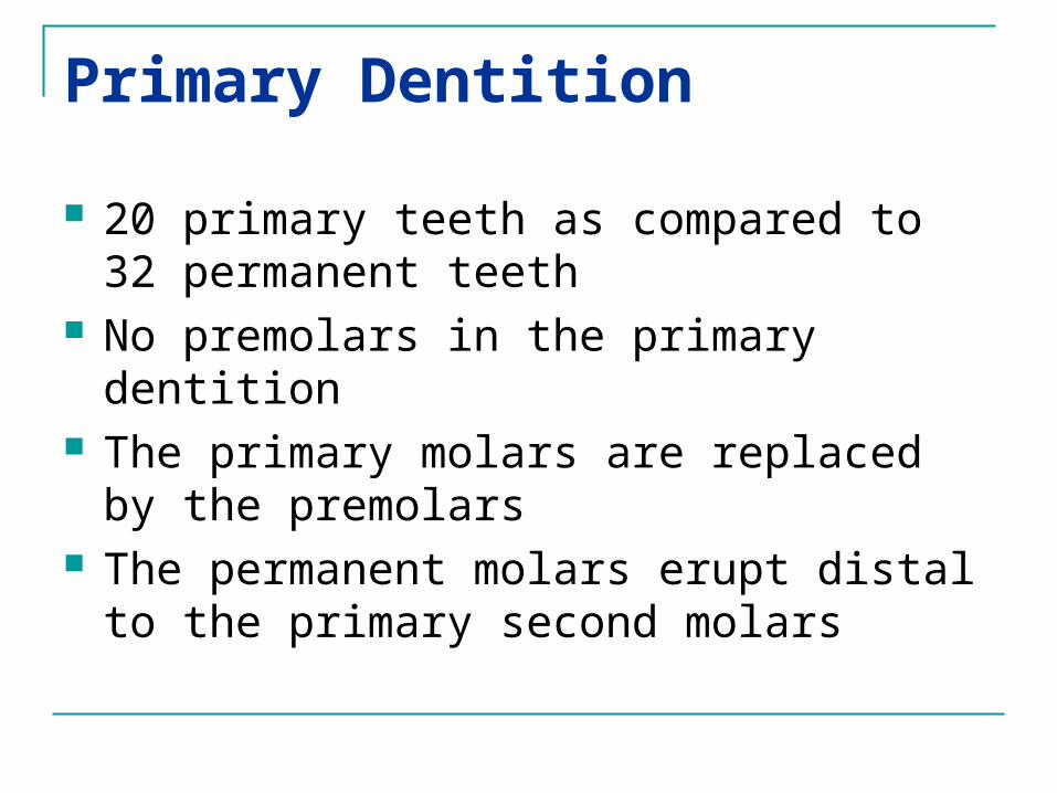
Primary Dentition
20 primary teeth as compared to 32 permanent teeth
No premolars in the primary dentition The primary molars are replaced by the
premolars The permanent molars erupt distal to
the primary second molars

General Morphologic considerations CrownCrown RootRoot PulpPulp

I- Size & Color
Smaller
MD width of ant. Primary < ant. Permanent teeth
MD width of Primary molars > Permanent premolars
Bluish-white (Primary) vs grayish-white or yellowish-white (Permanent) This has to be kept in mind during shade
selection for composite restoration or crown restoration.
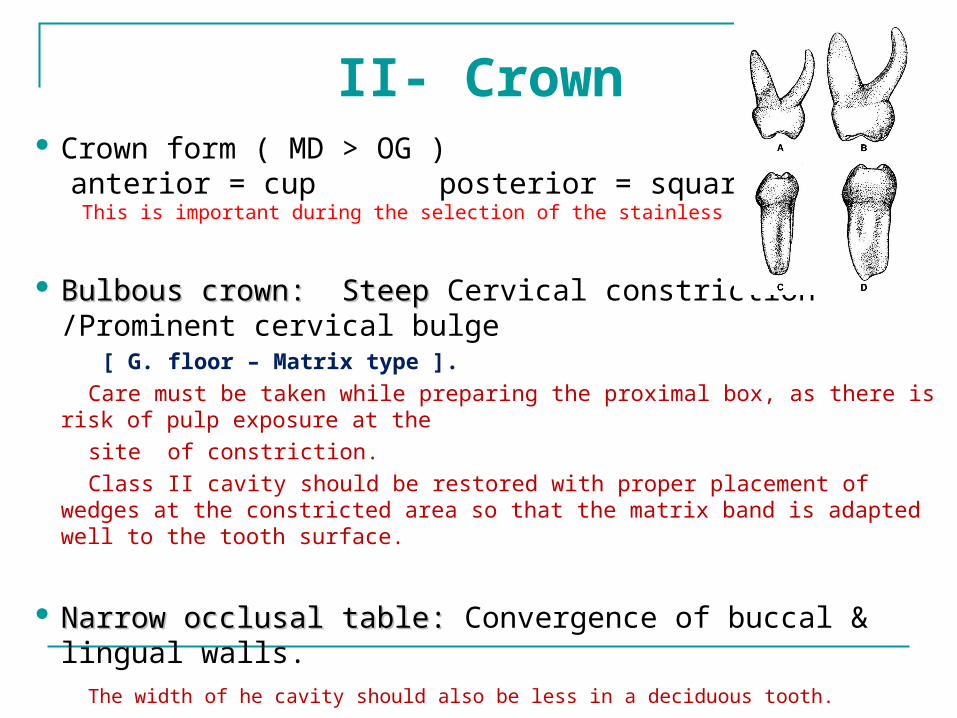
II- Crown Crown form ( MD > OG ) anterior = cup posterior = square This is important during the selection of the stainless steel crown.
Bulbous crown: Steep Bulbous crown: Steep Cervical constriction /Prominent cervical bulge
[ G. floor – Matrix type ].
Care must be taken while preparing the proximal box, as there is risk of pulp exposure at the
site of constriction. Class II cavity should be restored with proper placement of wedges at the
constricted area so that the matrix band is adapted well to the tooth surface.
Narrow occlusal table: Narrow occlusal table: Convergence of buccal & lingual walls.
The width of he cavity should also be less in a deciduous tooth.

Contact area Contact area in primary broader, flatter and more gingival:
[B&L wall of proximal box extend towards embrasure / gingival seat below contact area]
Primary

Cup
Cervical
constriction
Square
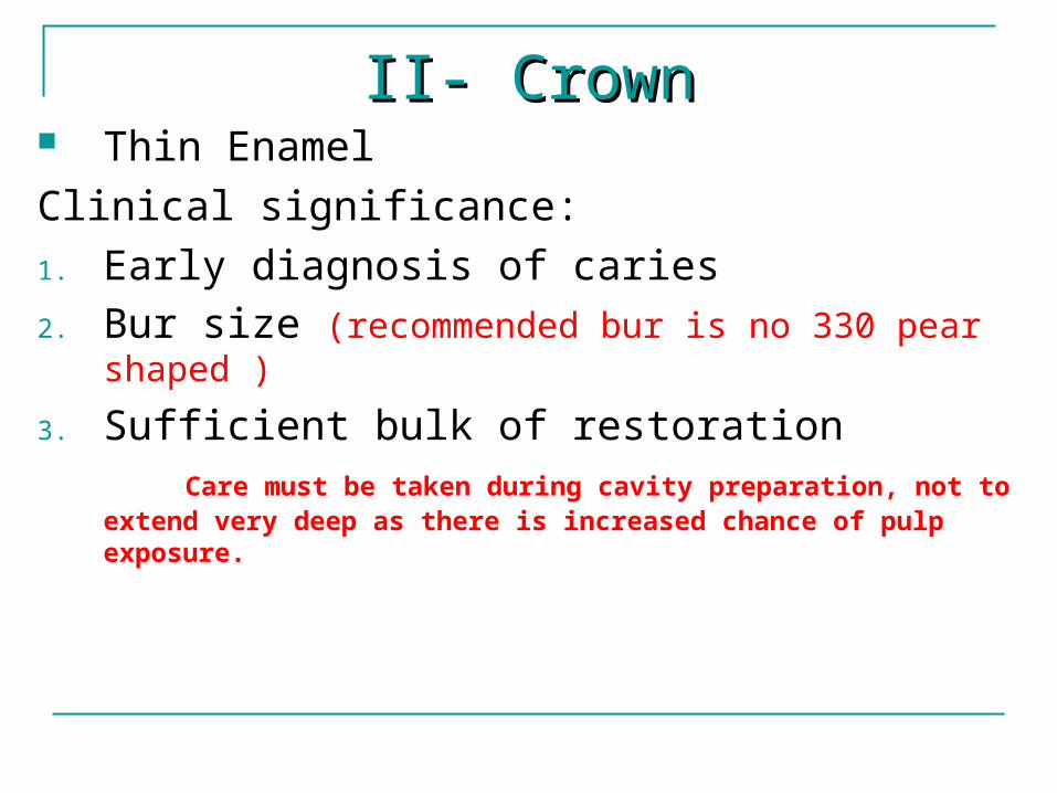
II- CrownII- Crown Thin EnamelClinical significance:1. Early diagnosis of caries2. Bur size (recommended bur is no 330 pear
shaped )
3. Sufficient bulk of restoration Care must be taken during cavity preparation, not to extend
very deep as there is increased chance of pulp exposure.

Enamel rods incline occlusally vs horizontal or apical in gingival 1/3
No beveling at the gingivo-cavo surface line angle is not required, as no enamel remain unsupported.
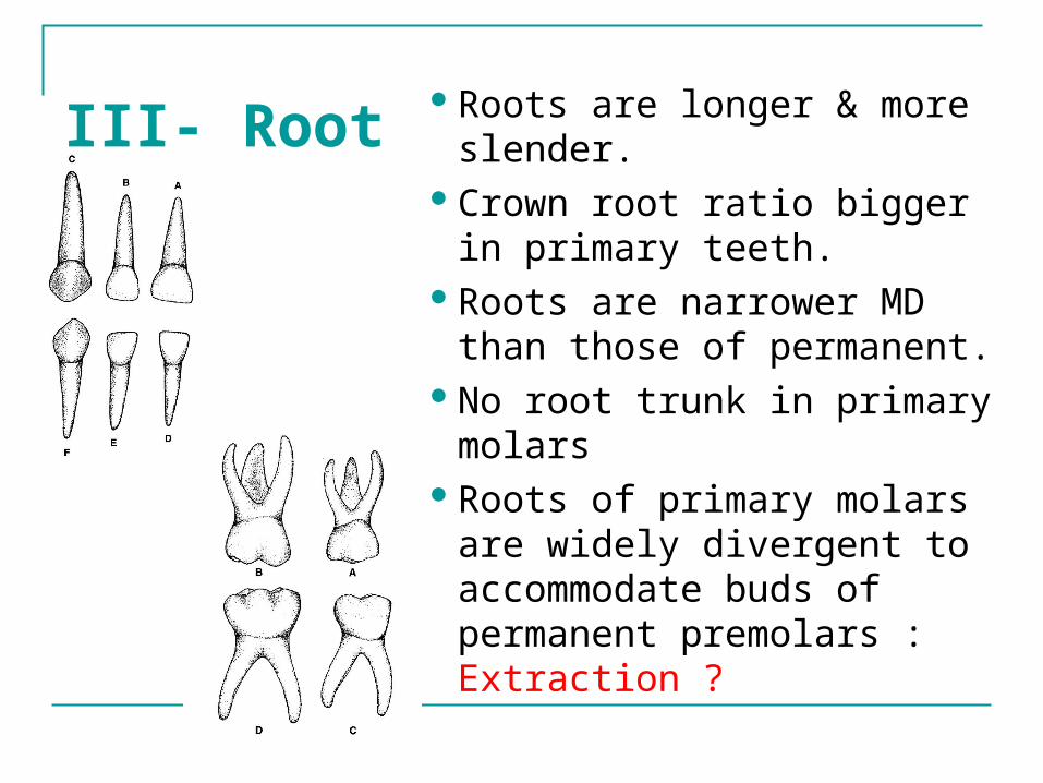
III- RootRoots are longer & more
slender.Crown root ratio bigger
in primary teeth.Roots are narrower MD
than those of permanent.No root trunk in primary
molarsRoots of primary molars
are widely divergent to accommodate buds of permanent premolars : Extraction ?

Roots are more Slender & longerCrown/Root ratio

No
Root
trunk
Widely
Divergent
roots

IV- Pulp• Follows the morphology of crown• Higher pulp horns / mesial• Pulp chamber is larger in relation to
crown size compared to permanent teeth / cavity depth
• Canals of primary molars have many lateral branches and apical ramifications
• Wide apical foramina• Increased blood supply : typical
inflammatory response• Less nerve fibers : less sensitivity to
pain

Summary
Primary teeth have Thinner enamel and dentin layers Pulp horns closer to the outer surface Mesial pulp horn much higher Relatively larger pulps Enamel rods direct slightly occlusally in the cervical
area Cervical area is constricted significantly Roots flare as they approach the apex More tortuous and irregular pulp canals


During Cavity Preparation: The depth of the cavity should be less. The width of the cavity should also be less in a deciduous
tooth. Care must be taken while preparing the proximal box,
as there is risk of pulp exposure at the site of constriction. Care must be taken during cavity preparation, not to
extend very deep as there is increased chance of pulp exposure.
Enamel beveling at the gingivocavo surface line angle is not required, as no enamel remain unsupported.
The proximal box preparation may have to be extended widely to break the contact free.

Thank you


![Heba Trabulsi - KSU...Heba Trabulsi Deanship of E-Transactions & Communications 2018 Heba Trabulsi 'l I n Il King Saud University EngW1 K Ing Saud University ä-ol.Q]l el-I-nil King](https://static.fdocuments.us/doc/165x107/5f123fbda31cbe095320accc/heba-trabulsi-ksu-heba-trabulsi-deanship-of-e-transactions-communications.jpg)