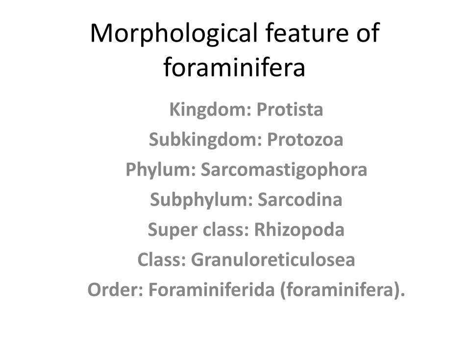
Morphological feature of foraminifera
14
Instituted of Geology 6th Semester (Morning) Submitted by: Khawar-u-zaman babar Roll No: 53 Submitted To: Dr.Munir Subject: Micropaleontology
Transcript of Morphological feature of foraminifera

Instituted of Geology
6th Semester (Morning)
Submitted by: Khawar-u-zaman babar
Roll No: 53
Submitted To: Dr.Munir
Subject: Micropaleontology

Topic
MORPHOLOGICAL FEATURES OF FORAMINIFERA
Morphological feature of foraminifera
Kingdom: Protista
Subkingdom: Protozoa
Phylum: Sarcomastigophora
Subphylum: Sarcodina
Super class: Rhizopoda
Class: Granuloreticulosea
Order: Foraminiferida (foraminifera).
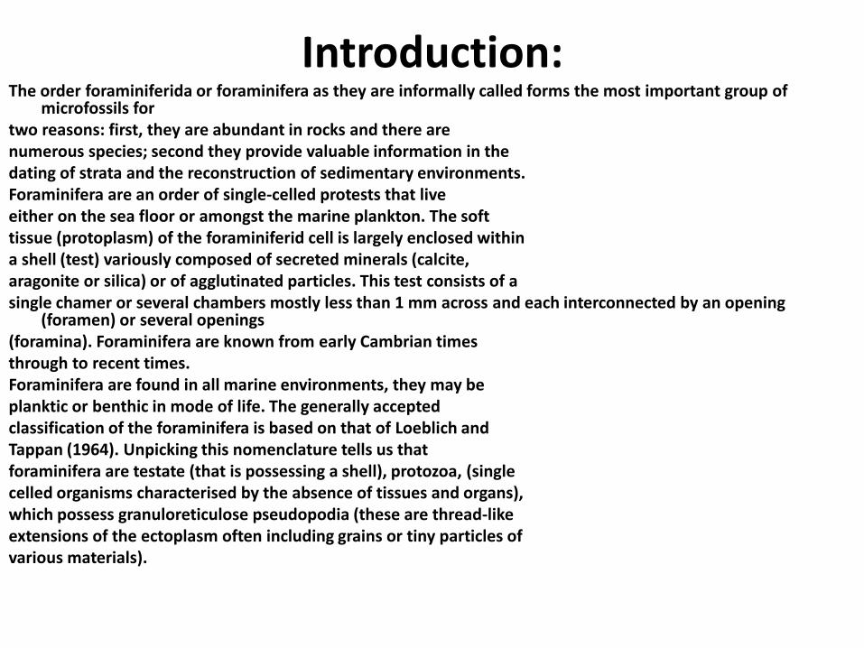
Introduction:The order foraminiferida or foraminifera as they are informally called forms the most important group of
microfossils fortwo reasons: first, they are abundant in rocks and there arenumerous species; second they provide valuable information in thedating of strata and the reconstruction of sedimentary environments.Foraminifera are an order of single-celled protests that liveeither on the sea floor or amongst the marine plankton. The softtissue (protoplasm) of the foraminiferid cell is largely enclosed withina shell (test) variously composed of secreted minerals (calcite,aragonite or silica) or of agglutinated particles. This test consists of asingle chamer or several chambers mostly less than 1 mm across and each interconnected by an opening
(foramen) or several openings(foramina). Foraminifera are known from early Cambrian timesthrough to recent times.Foraminifera are found in all marine environments, they may beplanktic or benthic in mode of life. The generally acceptedclassification of the foraminifera is based on that of Loeblich andTappan (1964). Unpicking this nomenclature tells us thatforaminifera are testate (that is possessing a shell), protozoa, (singlecelled organisms characterised by the absence of tissues and organs),which possess granuloreticulose pseudopodia (these are thread-likeextensions of the ectoplasm often including grains or tiny particles ofvarious materials).
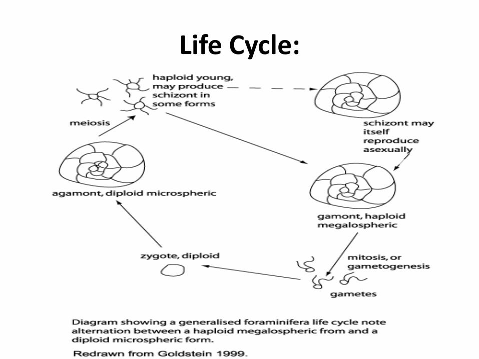
Life Cycle:
Classification
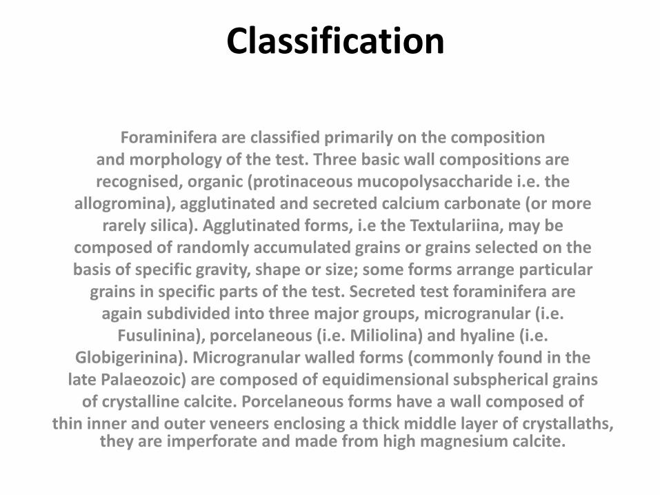
Foraminifera are classified primarily on the compositionand morphology of the test. Three basic wall compositions arerecognised, organic (protinaceous mucopolysaccharide i.e. the
allogromina), agglutinated and secreted calcium carbonate (or morerarely silica). Agglutinated forms, i.e the Textulariina, may be
composed of randomly accumulated grains or grains selected on thebasis of specific gravity, shape or size; some forms arrange particular
grains in specific parts of the test. Secreted test foraminifera areagain subdivided into three major groups, microgranular (i.e.
Fusulinina), porcelaneous (i.e. Miliolina) and hyaline (i.e.Globigerinina). Microgranular walled forms (commonly found in the
late Palaeozoic) are composed of equidimensional subspherical grainsof crystalline calcite. Porcelaneous forms have a wall composed of
thin inner and outer veneers enclosing a thick middle layer of crystallaths, they are imperforate and made from high magnesium calcite.
Test Morphology:Foraminifera are animals which build a shell; and for paleontologiststhe characterstics of the shell are the primary features which can be
used to distinguish one species from another.Wall Structure:
The most readily obvious featuredistingushing one foraminifer fromanother is its wall type. Whether the foraminifer builds its test wall
by cementing together exogenous grains, by carbonatemineralization, or by some combination of these two processes
separates the three primary foraminiferal groups, the agglutinated,the calcareous, and the microgranular foraminifera.
Agglutinated wall structureIn these, organic and mineral matter from the sea floor is bound
together by an organic, calcareous or ferric oxide cement. The grainsare commonly selected for size, texture or composition (e.g.
coccoliths, sponge, spicules and heavy minerals). Other agglutinatedforms are non-selective and will employ any particle from a substrate
as long as it lies in the appropriate size range.
Microgranular wallsMicrogranular walls evolved during the Paleozoic and are
consideredthe link between the agglutinated and the precipitated tests inforaminifera. Microgranular particles of calcite cemented by a
calcareous cement characterize this wall type and give it a sugary
appearance.Calcareous walls- hyaline type
Calcareous wall may be composed of either low or high Mg calcite, or
aragonite which is confined to only two foraminiferal families.Hyaline calcareous tests are characterized by the possession ofminute perforation in the test wall. The calcareous hyaline aregenerally glassy (hyaline) when viewed with reflected light and
greyto clear in transmitted light.
Chamber shape and chamber arrangement
:Foraminiferal tests may posses one or more chambers. The
initial chamber is most often spherical or oblate with an aperture.Later chambers range in shape from tubular, spherical, ovate to
several others. Additional chambers are added in a variety ofpatterns termed chamber arrangements:
1- Uniserial: the chamber arranged in a single row; if it forms acurved row, it is termed arcute; if a straight series, it is termed
rectilinear.2- Biserial: the chambers arranged in a double row.
3- Triserial: Chambers are added every 120o in a spiral fashion.4- Polyserial: the chambers arranged in a multiple row.
5- Planispiral: the chamber arranged spirally around an axis ofcoiling and the spiral lies in a single plane.
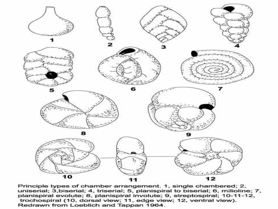
• 6- Trochospiral: when the spiral does not lie in one plane, but
• progresses up up the axis of coiling, the chamber arrangement
• becomes helicoidal.• 7- The Miliolidae have a streptospiral arrangement.
The arched• chambers, tangential at their two extremities with
the extension axis,• are arranged in cycles of five, three or two loculi or
one loculus. Each• new chamber has its aperture facing the aperture
of the the• preceding chamber.• When
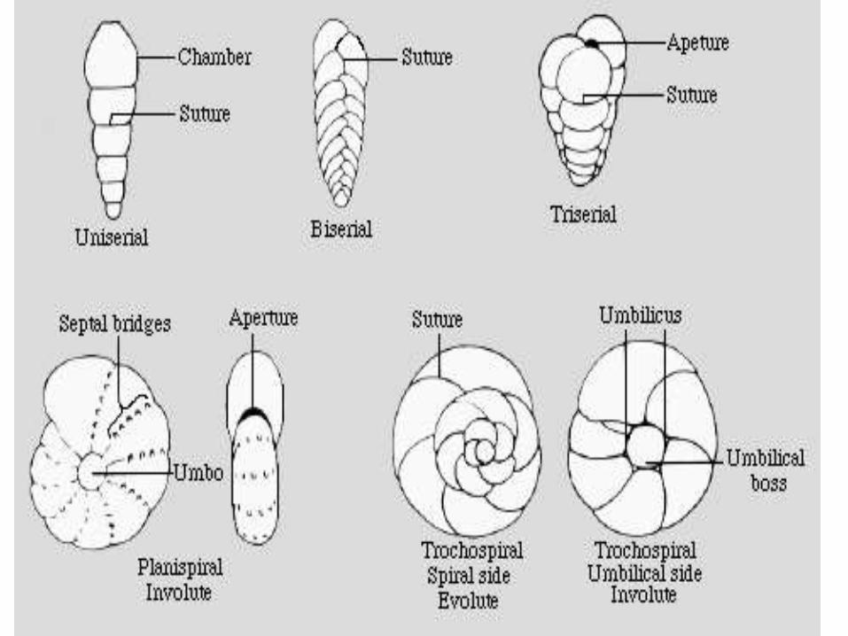
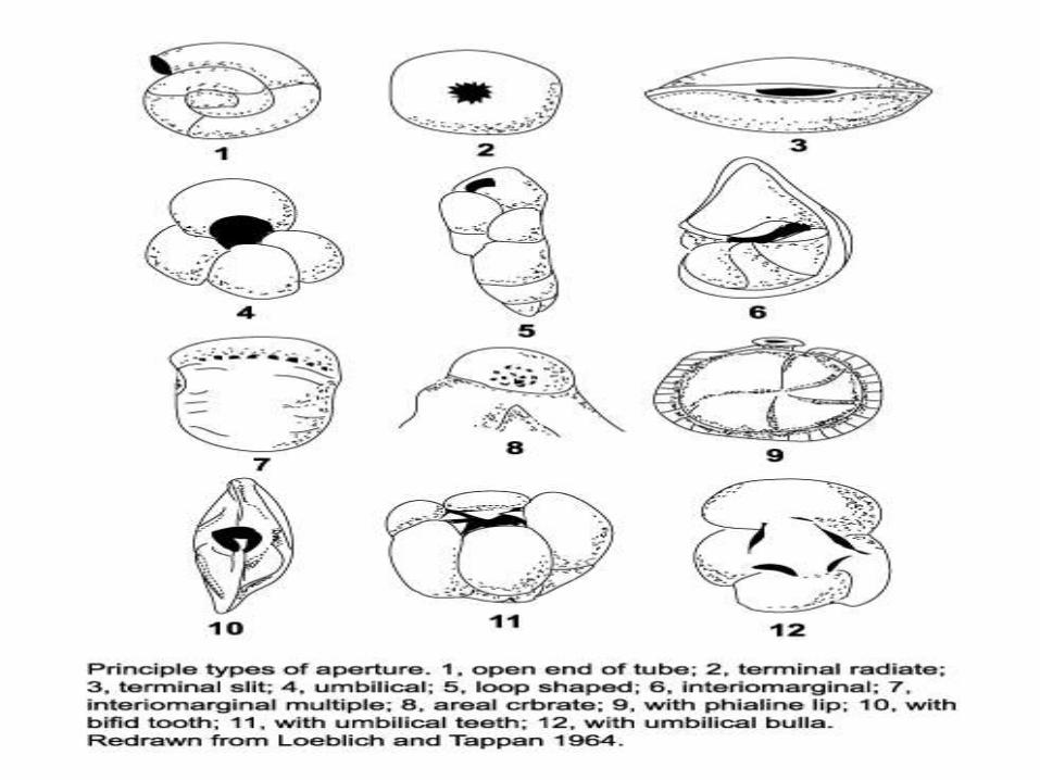
Apertures and openings:The aperture is the primary opening of the test to the outsideenvironment. Apertures vary in size and shape and the shape is mostoften a function of the shape of the chamber on which they arelocated. The aperture is found in the wall of the final chamber andserves to connect the external pseudopodia with the internalendoplasm, allowing passage of food and contractile vacuoles, nucleiand release of the dauther cells.Aperture may be single or multible in number and terminal, areal,basal extraumbilical, umbilical or sutural in position. Their shapevaries widely, e.g. rounded, bottle-necked (phialine), radiate,dendritic, sieve-like (cribrate), circular form, slit- or loop- shaped.