Mini Risk Assessment: Old World bollworm, Helicoverpa armigera ...
SYSTEMATICS, MORPHOLOGY AND PHYSIOLOGY
Morphological Characterization of Helicoverpa armigera (Hübner)(Lepidoptera: Noctuidae: Heliothinae)
L QUEIROZ-SANTOS1 , MM CASAGRANDE1, A SPECHT2
1Lab de Estudos de Lepidoptera Neotropical, Depto de Zoologia, Univ Federal do Paraná, Curitiba, PR( Brasil2Embrapa Cerrados, Planaltina, Brasil
AbstractKeywords
Adult, agricultural pest, chaetotaxy, egg,larva, old world bollworm, pupa
CorrespondenceL Queiroz-Santos, Depto de Zoologia, UnivFederal do Paraná, Zip Code 81531-980P.O. Box 19020, Curitiba, PR, Brasil;[email protected]
Edited by Takumasa Kondo – CORPOICA
Received 21 August 2017 and accepted 19December 2017
* Sociedade Entomológica do Brasil 2018
The cotton bollworm Helicoverpa armigera (Hübner) is a widespread lep-idopteran pest found in various crops worldwide. This highly polyphagousspecies, commonly found both in the Old and New World, has causedsignificant economic damage as an invasive agricultural pest in Brazil since2013. The goal of the present study is to provide a detailed morphologicalassessment of adults and immature stages of H. armigera, as this species isoften confused with H. zea (Boddie), a congeneric species that is native tothe NewWorld. The biology data were acquired during four full life cycles,and observations on general behavior, nocturnal habits of larvae andadults, and sensitivity of larvae to humidity were recorded. Larval chaeto-taxy differs between the first and the remaining instars, which bear L2 onthe meso- and metathorax and L3 on A3 through A6, along with conspic-uous chalazae and longitudinal bands. Important morphological charactersof this species include the following: eggs with four micropylar openings,lined with 12 cells arranged in the shape of a rosette; pupa adecticous andobtect, with prominent spiracles; adults with the distal antennomere stri-ate. Adults exhibit sexual dimorphism in the number of setae on thefrenulum and spines on the prothoracic leg. Illustrations of the criticalmorphological features of this species are provided.
Introduction
Following its introduction to South America, Helicoverpaarmigera (Hübner, [1808]) (Lepidoptera: Noctuidae:Heliothinae) has become one of the main noctuid pestsworldwide (Hardwick 1965; Matthews 1991; Tay et al 2013;Sosa-Gómez et al 2016, Bentivenha et al 2016).Characteristics of this species that likely contributed to itsbiological success include its dispersal capacity, high repro-ductive potential, polyphagy, rapid development, and op-tional diapause at higher latitudes (Hardwick 1965; Fitt1989; Matthews 1991).
The native distribution of H. armigera includes westernEurope, central Africa, Australia, Oceania, and parts of Asia(Hardwick 1965). It has been recorded in several localitiesattacking multiple host crops in Brazil since March 2013
(EMBRAPA 2013; Czepak et al 2013; Specht et al 2013; Tayet al 2013), as well as in other South American countries(SENAVE 2013; Murúa et al 2014; Castiglioni et al 2016;Arnemann et al 2016). Helicoverpa armigera is dispersingfrom South America towards North America and has recentlybeen recorded from Puerto Rico and Florida (CABI 2017).
Hübner described Noctua armigera between 1803 and1808 based on an illustrated plate (Hemming 1937).Subsequently, Hardwick (1965) erected the genusHelicoverpa, to which he transferred N. armigera and otherfive taxa, yielding a total of 11 species.
At the moment, several publications have described bio-logical (Hardwick 1965; Zalucki et al 1986; Karim 2000; Aliet al 2009) and behavioral (Sorensen et al 2006; Abbasiet al 2007; Feng et al 2010; Liu et al 2010; Nadda 2013;Jadhav et al 2013) aspects of this species, but no previous
Neotrop Entomolhttps://doi.org/10.1007/s13744-017-0581-4
study offers details on morphological features ofH. armigera.
Although much has been published concerning nocturnalspecies considered to be agricultural pests, works on generalmorphology, especially that of moths, are still scarce. Themain goal of this study is to provide morphological character-izations and natural history data on H. armigera, a speciesthat feeds on several crops, including soybeans, corn, cotton,green beans, tomatoes, citrus fruits, and pastures (Bueno &Sosa-Gómez 2014). Included here are descriptions of imma-ture stages and adults, emphasizing structures not previouslycharacterized. This information is intended to facilitate theaccurate identification of this species as it is often confusedwith other congeneric taxa, particularly cryptic species.
Materials and Methods
Insects were reared at room temperature under natural light.All immatures were obtained from the second generation oftwo individualized pairs and kept in PVC cages (80 cm diam-eter, 60 cm height). The cages were internally coated with akraft paper as a substrate for oviposition, and the top wascovered with voile fabric. This technique was developed bymodifying other methods described in the literature (Spechtet al 2006; Mironidis & Savopoulou-Soultani 2008; Wanget al 2008). Adults were fed 10% honey solution in distilledwater, which was provided in a hydrophilic cotton ball. Bothfood and kraft paper coating were changed every 48 h. Thisrearing procedure produced four batches of 24 male-femalepairs.
After eclosion, ten individual larvae of each instar werefixed in Kahle-Dietrich solution and then transferred to ethylalcohol at 70%. The remaining individuals were kept alive inorder to obtain subsequent developmental stages. The lar-vae were kept in plastic containers (2.5 cm diameter, 7 cmheight) coated with absorbent paper. Two groups were fedlettuce leaves with no additional humidity sources, and twogroups were given a bean-based artificial diet (Montezanoet al 2013). Head capsules were removed from the containersafter each molt and stored dry. For pupation, a small amount(3 cm height) of autoclaved expanded vermiculite was addedto each container, so that the caterpillars could constructpupal chambers. Ten pupae of each sex were fixed accordingto the method described above, after its cuticle hardened.Adult specimens were killed in a jar with ethyl acetate, whichkeeps exoskeletal structures intact for subsequent morpho-logical examination.
Wings were removed and bleached in order to study ve-nation patterns. Head, thorax, abdomen, and appendageswere immersed in a 10% potassium hydroxide solution(KOH) and placed in a warm bath until tissues softened andthe exoskeleton appeared semitransparent. Structures of
interest were then dissected for observation under a lightmicroscope. Voucher specimens are deposited at the PadreJesus Santiago Moure Collection, Lepidoptera, UniversidadeFederal do Paraná (DZUP), about numbers DZ 39.223, DZ39.233, DZ 39.243, DZ 39.253, DZ 39.263, DZ 39.273, DZ39.283, DZ 39.293, DZ 39.303, DZ 39.313, DZ 39.323, DZ39.363, DZ 39.373, DZ 39.383, DZ 39.393, DZ 39.403, DZ39.423, DZ 39.443, DZ 39.453, DZ 39.463, and DZUPIL 0141.
Measurements of immature stages (eggs, larvae, moltedhead capsules, and pupae) weremade using aWild Heerburgstereomicroscope with a micrometric scale. Head capsulewidth corresponds to the largest distance between genaein each instar. Body length is the distance from the frons tothe last abdominal segment in a pharate larva.Measurements are specified based on means and standarddeviation from multiple specimens (n).
Illustrations of specimens and structures of interest wereprepared with the aid of a camera lucida attached to a ZeissStemi SV6 stereomicroscope or a Zeiss Standard 20 micro-scope. Electron micrographs were produced with a JEOL JSM6360-LV scanning electron microscope. Ethanol-preservedspecimens were dehydrated in a series of 70, 80, 90%, andtwo 100% ethanol baths for 10 min each. These sampleswere then moved to a Thornton T14 ultrasonic cleaner fortwo 30-min baths and dried in a Bal-Tec® CPD-030 criticalpoint dryer. Dehydrated structures were mounted on metal-lic stubs with 3M® copper conductive double-sided tape andgold-coated with a Balzers® SCD030 – Union FL 9496 sputtercoater device. Dry samples were mounted directly on metal-lic stubs with 3M® copper double-sided tape and analyzed inlow-vacuummode. Illustrations were prepared using perma-nent black ink and were subsequently adjusted and format-ted using GIMP v. 2.8.2 (www.gimp.org).
Terminology and notation are based on the following con-tributions: Peterson (1961, 1964) for eggs; Stehr (1987) forchaetotaxy; Peterson (1962) for larval body area; Blaik &Malkiewickz (2003) for labral, maxillary, and thoracic legsetae; Grimes & Neunzing (1986) and Baker et al (1986) formaxillary palp sensillae; Mosher (1916) for pupae; Pierce(1909), Comstock (1918), Snodgrass (1935), Casagrande(1979), Scoble (1992), Speidel et al (1996), Hallberg et al(2003), Kristensen (2003), and Fibiger & Lafontaine (2004)for adult external morphology; and Butt & Cantu (1962) forsex determination. The taxonomic classification followsHemming (1937).
Results
Biology
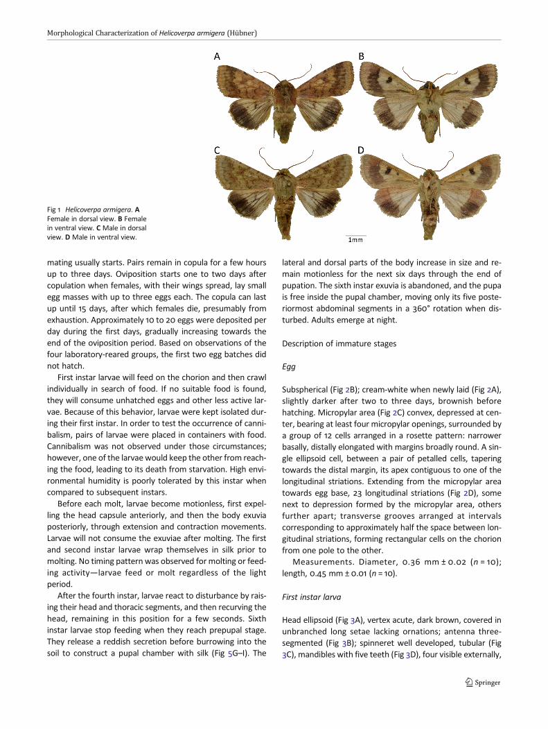
Adults (Fig 1A–D) were inactive during most of the day andwere active at night, after the evening twilight, during which
Queiroz-Santos et al
mating usually starts. Pairs remain in copula for a few hoursup to three days. Oviposition starts one to two days aftercopulation when females, with their wings spread, lay smallegg masses with up to three eggs each. The copula can lastup until 15 days, after which females die, presumably fromexhaustion. Approximately 10 to 20 eggs were deposited perday during the first days, gradually increasing towards theend of the oviposition period. Based on observations of thefour laboratory-reared groups, the first two egg batches didnot hatch.
First instar larvae will feed on the chorion and then crawlindividually in search of food. If no suitable food is found,they will consume unhatched eggs and other less active lar-vae. Because of this behavior, larvae were kept isolated dur-ing their first instar. In order to test the occurrence of canni-balism, pairs of larvae were placed in containers with food.Cannibalism was not observed under those circumstances;however, one of the larvaewould keep the other from reach-ing the food, leading to its death from starvation. High envi-ronmental humidity is poorly tolerated by this instar whencompared to subsequent instars.
Before each molt, larvae become motionless, first expel-ling the head capsule anteriorly, and then the body exuviaposteriorly, through extension and contraction movements.Larvae will not consume the exuviae after molting. The firstand second instar larvae wrap themselves in silk prior tomolting. No timing pattern was observed for molting or feed-ing activity—larvae feed or molt regardless of the lightperiod.
After the fourth instar, larvae react to disturbance by rais-ing their head and thoracic segments, and then recurving thehead, remaining in this position for a few seconds. Sixthinstar larvae stop feeding when they reach prepupal stage.They release a reddish secretion before burrowing into thesoil to construct a pupal chamber with silk (Fig 5G–I). The
lateral and dorsal parts of the body increase in size and re-main motionless for the next six days through the end ofpupation. The sixth instar exuvia is abandoned, and the pupais free inside the pupal chamber, moving only its five poste-riormost abdominal segments in a 360° rotation when dis-turbed. Adults emerge at night.
Description of immature stages
Egg
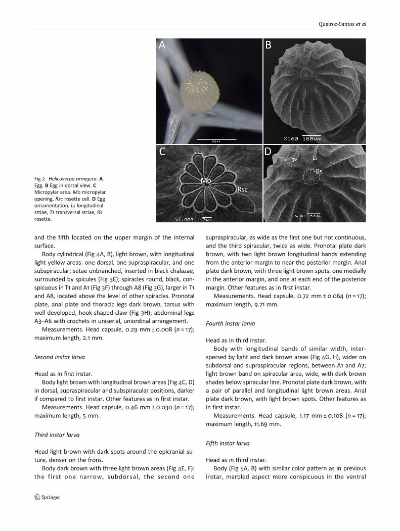
Subspherical (Fig 2B); cream-white when newly laid (Fig 2A),slightly darker after two to three days, brownish beforehatching. Micropylar area (Fig 2C) convex, depressed at cen-ter, bearing at least four micropylar openings, surrounded bya group of 12 cells arranged in a rosette pattern: narrowerbasally, distally elongated with margins broadly round. A sin-gle ellipsoid cell, between a pair of petalled cells, taperingtowards the distal margin, its apex contiguous to one of thelongitudinal striations. Extending from the micropylar areatowards egg base, 23 longitudinal striations (Fig 2D), somenext to depression formed by the micropylar area, othersfurther apart; transverse grooves arranged at intervalscorresponding to approximately half the space between lon-gitudinal striations, forming rectangular cells on the chorionfrom one pole to the other.
Measurements. Diameter, 0.36 mm ± 0.02 (n = 10);length, 0.45 mm ± 0.01 (n = 10).
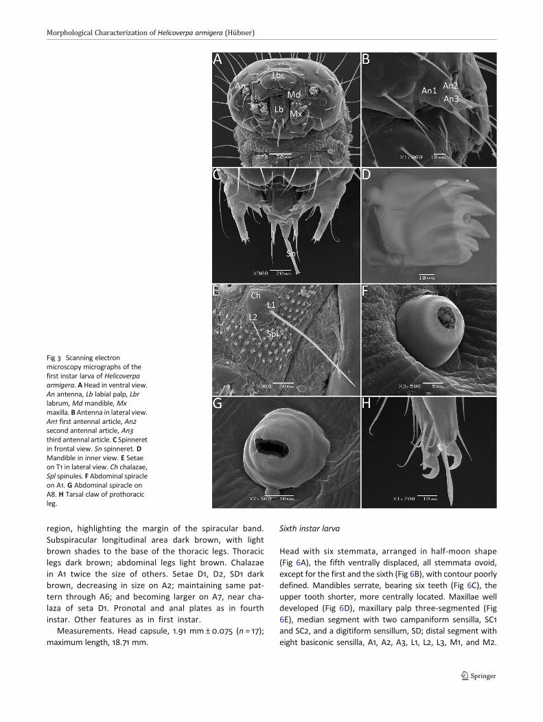
First instar larva
Head ellipsoid (Fig 3A), vertex acute, dark brown, covered inunbranched long setae lacking ornations; antenna three-segmented (Fig 3B); spinneret well developed, tubular (Fig3C), mandibles with five teeth (Fig 3D), four visible externally,
Fig 1 Helicoverpa armigera. AFemale in dorsal view. B Femalein ventral view. C Male in dorsalview. D Male in ventral view.
Morphological Characterization of Helicoverpa armigera (Hübner)
and the fifth located on the upper margin of the internalsurface.
Body cylindrical (Fig 4A, B), light brown, with longitudinallight yellow areas: one dorsal, one supraspiracular, and onesubspiracular; setae unbranched, inserted in black chalazae,surrounded by spicules (Fig 3E); spiracles round, black, con-spicuous in T1 and A1 (Fig 3F) through A8 (Fig 3G), larger in T1and A8, located above the level of other spiracles. Pronotalplate, anal plate and thoracic legs dark brown, tarsus withwell developed, hook-shaped claw (Fig 3H); abdominal legsA3–A6 with crochets in uniserial, uniordinal arrangement.
Measurements. Head capsule, 0.29 mm ± 0.008 (n = 17);maximum length, 2.1 mm.
Second instar larva
Head as in first instar.Body light brownwith longitudinal brown areas (Fig 4C, D)
in dorsal, supraspiracular and subspiracular positions, darkerif compared to first instar. Other features as in first instar.
Measurements. Head capsule, 0.46 mm ± 0.030 (n = 17);maximum length, 5 mm.
Third instar larva
Head light brown with dark spots around the epicranial su-ture, denser on the frons.
Body dark brown with three light brown areas (Fig 4E, F):the first one narrow, subdorsal, the second one
supraspiracular, as wide as the first one but not continuous,and the third spiracular, twice as wide. Pronotal plate darkbrown, with two light brown longitudinal bands extendingfrom the anterior margin to near the posterior margin. Analplate dark brown, with three light brown spots: one mediallyin the anterior margin, and one at each end of the posteriormargin. Other features as in first instar.
Measurements. Head capsule, 0.72 mm ± 0.064 (n = 17);maximum length, 9.71 mm.
Fourth instar larva
Head as in third instar.Body with longitudinal bands of similar width, inter-
spersed by light and dark brown areas (Fig 4G, H), wider onsubdorsal and supraspiracular regions, between A1 and A7;light brown band on spiracular area, wide, with dark brownshades below spiracular line. Pronotal plate dark brown, witha pair of parallel and longitudinal light brown areas. Analplate dark brown, with light brown spots. Other features asin first instar.
Measurements. Head capsule, 1.17 mm ± 0.108 (n = 17);maximum length, 11.69 mm.
Fifth instar larva
Head as in third instar.Body (Fig 5A, B) with similar color pattern as in previous
instar, marbled aspect more conspicuous in the ventral
Fig 2 Helicoverpa armigera. AEgg. B Egg in dorsal view. CMicropylar area. Mo micropylaropening, Rsc rosette cell. D Eggornamentation. Ls longitudinalstriae, Ts transversal striae, Rsrosette.
Queiroz-Santos et al
region, highlighting the margin of the spiracular band.Subspiracular longitudinal area dark brown, with lightbrown shades to the base of the thoracic legs. Thoraciclegs dark brown; abdominal legs light brown. Chalazaein A1 twice the size of others. Setae D1, D2, SD1 darkbrown, decreasing in size on A2; maintaining same pat-tern through A6; and becoming larger on A7, near cha-laza of seta D1. Pronotal and anal plates as in fourthinstar. Other features as in first instar.
Measurements. Head capsule, 1.91 mm ± 0.075 (n = 17);maximum length, 18.71 mm.
Sixth instar larva
Head with six stemmata, arranged in half-moon shape(Fig 6A), the fifth ventrally displaced, all stemmata ovoid,except for the first and the sixth (Fig 6B), with contour poorlydefined. Mandibles serrate, bearing six teeth (Fig 6C), theupper tooth shorter, more centrally located. Maxillae welldeveloped (Fig 6D), maxillary palp three-segmented (Fig6E), median segment with two campaniform sensilla, SC1and SC2, and a digitiform sensillum, SD; distal segment witheight basiconic sensilla, A1, A2, A3, L1, L2, L3, M1, and M2.
Fig 3 Scanning electronmicroscopy micrographs of thefirst instar larva of Helicoverpaarmigera. A Head in ventral view.An antenna, Lb labial palp, Lbrlabrum, Md mandible, Mxmaxilla. B Antenna in lateral view.An1 first antennal article, An2second antennal article, An3third antennal article. C Spinneretin frontal view. Sn spinneret. DMandible in inner view. E Setaeon T1 in lateral view. Ch chalazae,Spl spinules. F Abdominal spiracleon A1. G Abdominal spiracle onA8. H Tarsal claw of prothoracicleg.
Morphological Characterization of Helicoverpa armigera (Hübner)
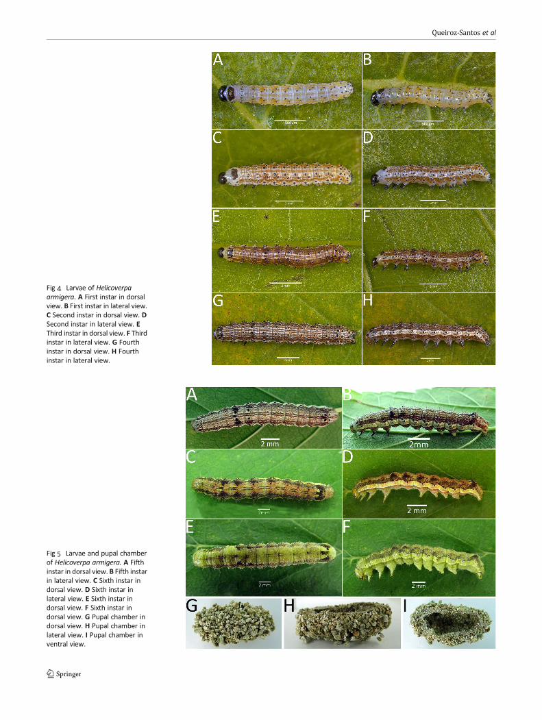
Fig 4 Larvae of Helicoverpaarmigera. A First instar in dorsalview. B First instar in lateral view.C Second instar in dorsal view. DSecond instar in lateral view. EThird instar in dorsal view. F Thirdinstar in lateral view. G Fourthinstar in dorsal view. H Fourthinstar in lateral view.
Fig 5 Larvae and pupal chamberof Helicoverpa armigera. A Fifthinstar in dorsal view. B Fifth instarin lateral view. C Sixth instar indorsal view. D Sixth instar inlateral view. E Sixth instar indorsal view. F Sixth instar indorsal view. G Pupal chamber indorsal view. H Pupal chamber inlateral view. I Pupal chamber inventral view.
Queiroz-Santos et al
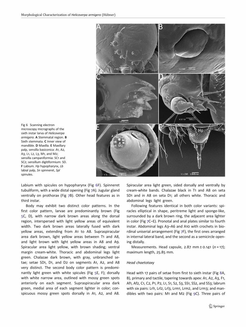
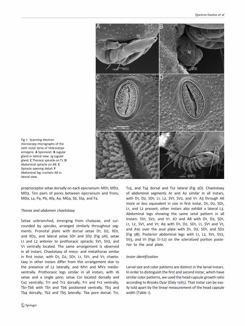
Labium with spicules on hypopharynx (Fig 6F). Spinnerettubuliform, with a wide distal opening (Fig 7A). Jugular glandventrally on prothorax (Fig 7B). Other head features as inthird instar.
Body may exhibit two distinct color patterns. In thefirst color pattern, larvae are predominantly brown (Fig5C, D), with narrow dark brown areas along the dorsalregion, interspersed with light yellow areas of equivalentwidth. Two dark brown areas laterally fused with darkyellow areas, extending from A1 to A8. Supraspiraculararea dark brown, light yellow areas between T1 and A8,and light brown with light yellow areas in A8 and A9.Spiracular area light yellow, with brown shading; ventralmargin cream-white. Thoracic and abdominal legs lightgreen. Chalazae dark brown, with gray, unbranched se-tae; setae SD1, D1, and D2 on segments A1, A2, and A8very distinct. The second body color pattern is predomi-nantly light green with white spicules (Fig 5E, F); dorsallywith white narrow area, outlined with mossy green spotsanteriorly on each segment. Supraspiracular area darkgreen, medial area of each segment lighter in color; con-spicuous mossy green spots dorsally in A1, A2, and A8.
Spiracular area light green, sided dorsally and ventrally bycream-white bands. Chalazae black in T1 and A8 on setaSD1 and in A8 on seta D1; all others white. Thoracic andabdominal legs light green.
Following features identical in both color variants: spi-racles elliptical in shape, peritreme light and sponge-like,surrounded by a dark brown ring, the adjacent area lighterin color (Fig 7C–E). Pronotal and anal plates similar to fourthinstar. Abdominal legs A3–A6 and A10 with crochets in bio-rdinal uniserial arrangement (Fig 7F), the first ones arrangedin internal lateral band, and the second as a semicircle open-ing distally.
Measurements. Head capsule, 2.87 mm ± 0.141 (n = 17);maximum length, 25.85 mm.
Head chaetotaxy
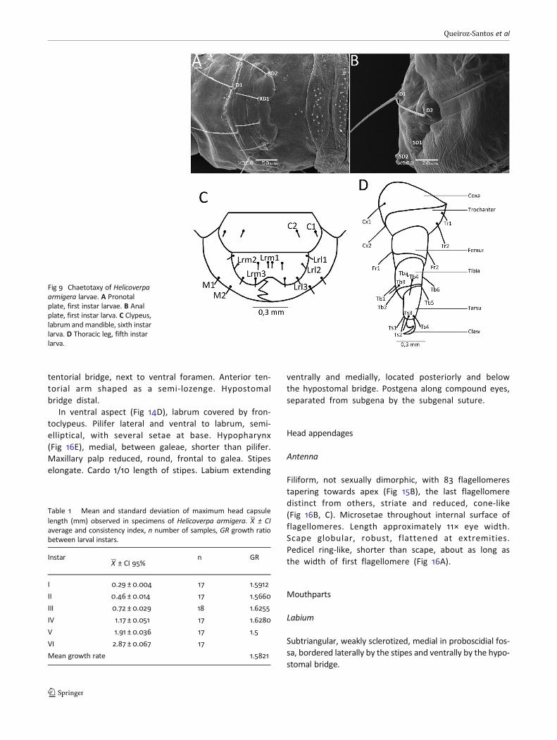
Head with 17 pairs of setae from first to sixth instar (Fig 8A,B), primary and tactile, tapering towards apex: A1, A2, A3, F1,Af1, Af2, C1, C2, P1, P2, L1, S1, S2, S3, SS1, SS2, and SS3; labrumwith six pairs: Lrl1, Lrl2, Lrl3, Lrm1, Lrm2, and Lrm3; and man-dibles with two pairs: M1 and M2 (Fig 9C). Three pairs of
Fig 6 Scanning electronmicroscopy micrographs of thesixth instar larva of Helicoverpaarmigera. A Stemmatal region. BSixth stemmata. C Inner view ofmandible. D Maxilla. E Maxillarypalp, sensilla basiconica: A1, A2,A3, L1, L2, L3, M1, and M2;sensilla campaniformia: SC1 andSC2; sensillum digitiformium: SD.F Labium. Hp hypopharynx, Lblabial palp, Sn spinneret, Splspinules.
Morphological Characterization of Helicoverpa armigera (Hübner)
proprioceptor setae dorsally on each epicranium: MD1, MD2,MD3. Ten pairs of pores between epicranium and frons,MDa, La, Pa, Pb, Afa, Aa, MGa, Sb, SSa, and Fa.
Thorax and abdomen chaetotaxy
Setae unbranched, emerging from chalazae, and sur-rounded by spicules, arranged similarly throughout seg-ments. Pronotal plate with dorsal setae D1, D2, XD1,and XD2, and lateral setae SD1 and SD2 (Fig 9A); setaeL1 and L2 anterior to prothoracic spiracle; SV1, SV2, andV1 ventrally located. The same arrangement is observedin all instars. Chaetotaxy of meso- and metathorax similarin first instar, with D1, D2, SD1, L1, SV1, and V1; chaeto-taxy in other instars differ from this arrangement due tothe presence of L2 laterally, and MV1 and MV2 medio-ventrally. Prothoracic legs similar in all instars, with 16setae and a single pore; setae Cx1 located dorsally andCx2 ventrally; Tr1 and Tr2 dorsally; Fr1 and Fr2 ventrally;Tb1-Tb6 with Tb1 and Tb6 positioned ventrally, Tb3 andTb4 dorsally, Tb2 and Tb5 laterally. Tba pore dorsal; Ts1,
Ts3, and Ts4 dorsal and Ts2 lateral (Fig 9D). Chaetotaxyof abdominal segments A1 and A2 similar in all instars,with D1, D2, SD1, L1, L2, SV1, SV2, and V1. A3 through A6more or less equivalent in size in first instar, D1, D2, SD1,L1, and L2 present; other instars also exhibit a lateral L3.Abdominal legs showing the same setal pattern in allinstars: SV1, SV2, and V1. A7 and A8 with D1, D2, SD1,L1, L2, SV1, and V1; A9 with D1, D2, SD1, L1, SV1 and V1,and A10 over the anal plate with D1, D2, SD1, and SD2(Fig 9B). Posterior abdominal legs with L1, L2, SV1, SV2,SV3, and V1 (Figs S1-S2) on the sclerotized portion poste-rior to the anal plate.
Instar identification
Larval size and color patterns are distinct in the larval instars.In order to distinguish the first and second instar, which havesimilar color patterns, we used the head capsule growth ratioaccording to Brooks-Dyar (Daly 1985). That instar can be eas-ily told apart by the linear measurement of the head capsulewidth (Table 1).
Fig 7 Scanning electronmicroscopy micrographs of thesixth instar larva of Helicoverpaarmigera. A Spinneret. B Jugulargland in lateral view. Jg jugulargland. C Thoracic spiracle on T1. DAbdominal spiracle on A8. ESpiracle opening detail. FAbdominal leg crochets A8 inlateral view.
Queiroz-Santos et al

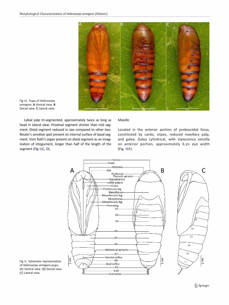
Pupa
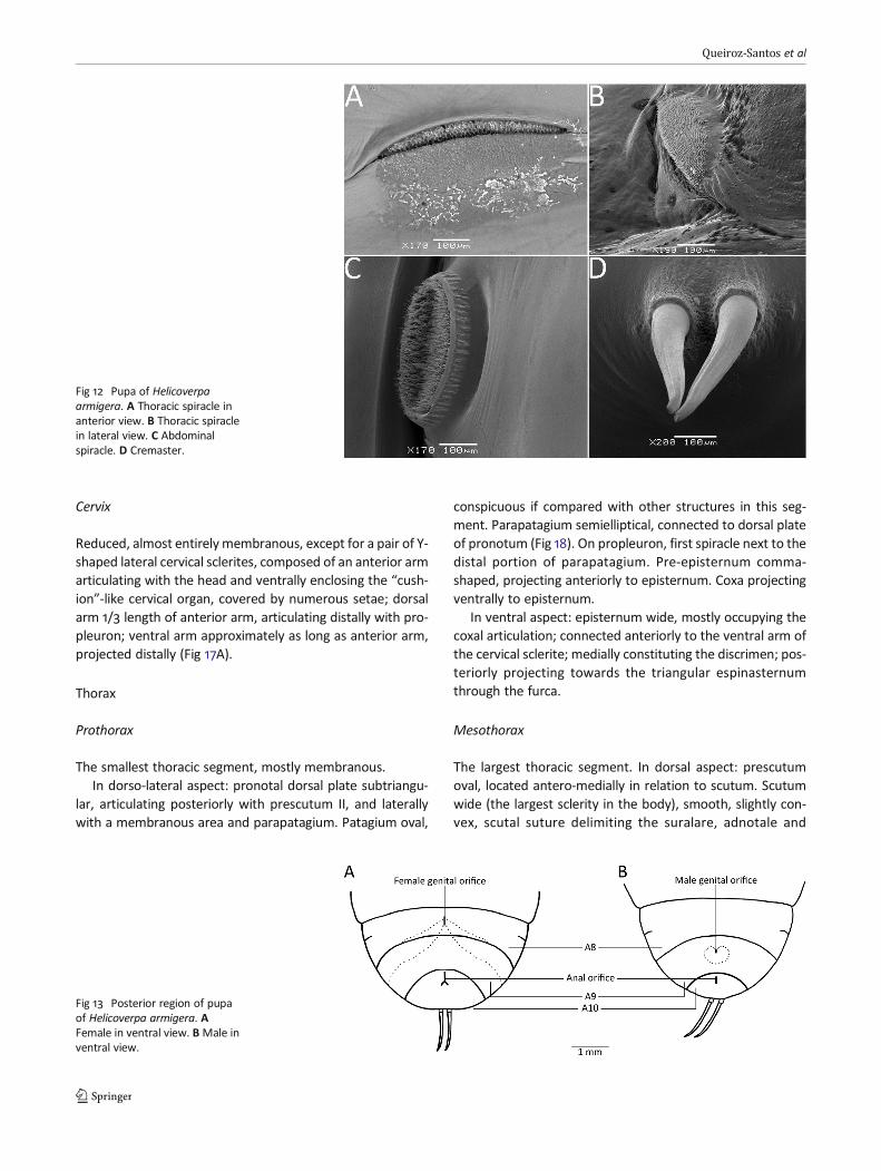
Adecticous and obtect, light brown immediately after pupa-tion, dark brown after five days; eyes noticeably dark justbefore emergence (Fig 10A–C). Elongate, cylindrical, headround, vertex extending towards the posterior region, alongthe prothorax and antenna; abdomen tapering after A4.Integument dense, smooth, edges of segments well marked(Fig 11(C)).
In ventral aspect (Figs 10A and 11(A)), frontoclypeusgoblet-like, between the compound eyes. Hypopharynxvisible, shaped like an inverted triangle, with short pos-terior labial palp. Antenna fine, long, reaching costalmargin of forewing, anterior to A5. Legs placed along
the antenna towards the galea in repose; metathoracicleg longest. Galea lanceolate, occupying most part ofthe anterior half of the ventral side; reaching posteriormargin of A4. Hind wing on top of forewing, bothending above anterior margin of A5.
In dorsal aspect (Figs 10B and 11(B)), prothoraxsmall, triangular, apex round, prothoracic spiracle nextto basal angle (Fig 12A, B). Mesothorax and metathoraxprojecting latero-ventrally, forming the pterotheca untilthe anterior margin of A5.
Abdomen divided in ten segments, spiracles elliptical, con-spicuous between A2–A7, reduced in A8 (Fig 12C). Abdomenslightly curved towards cremaster posterior to A5. Cremastercomposed of two articulate, parallel projections, taperingdistally, approximately as long as A10 (Fig 12D).
Anal opening scar elongate, longitudinal to body axis, lo-cated on distal ventral area of A10. Female genital openingscar (Fig 13A) medioventrally located on A8. Male genitalopening (Fig 13B) on A9, surrounded by small protuberances.
Measurements (male/female). Maximum width of thirdabdominal segment, 6.31 mm ± 0.33 (n = 9) (5.71–7; SE,0.03); body length, 20.47 mm ± 1.52 (n = 9) (18.57–22.41;SE, 0.16).
Adult
Head
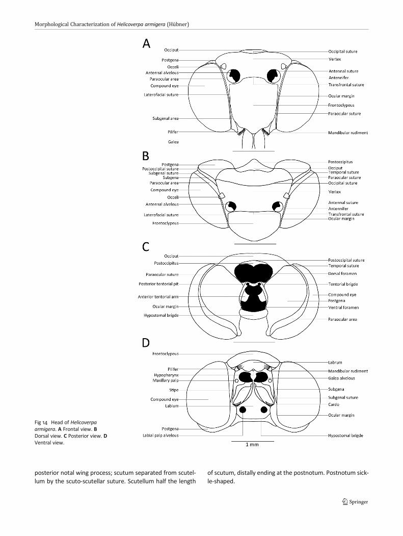
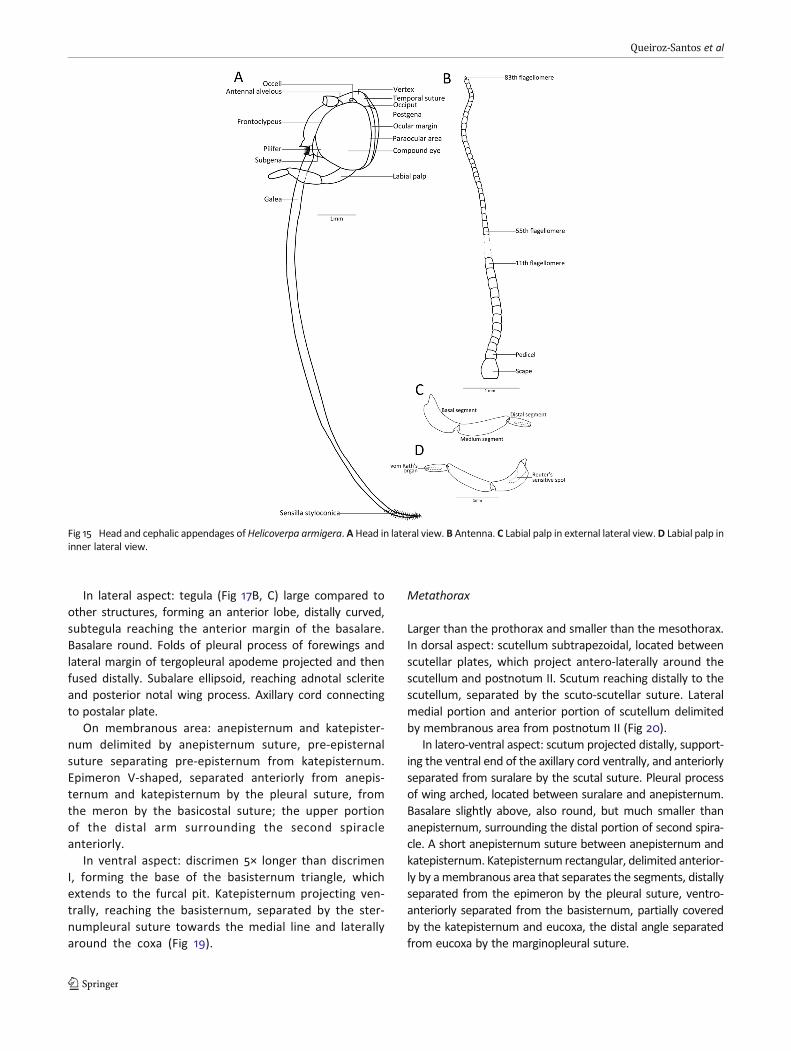
Hypognathous, entirely covered by scales. Compoundeyes semioval, glabrous, prominent, longer than wide.Paraocular area marked in parallel to eye orbit andparaocular suture; laterofacial suture reaching eye orbitslightly above its center. Frontoclypeus subquadrate,prominent, separated from vertex by the transfrontalsuture, which is close to, but not fused with, the an-tennal suture. Subgenal area fused laterally and basallywith mandibular rudiment, extending dorsally towardsthe pilifer. Pilifer setae present. Vertex wide, withchaetosemata throughout, enclosing pair of antennalsockets; antennifer surrounded by antennal suture,and a pair of dark ocelli laterad to the antennal sock-ets, separated posteriorly from the occiput by an oc-cipital suture (Figs 14A and 15A).
In dorsal and posterior aspects (Fig 14B, C), postoc-cipitus triangular, separated from occiput by postocci-pital suture. Occiput subrectangular, delimited laterallyfrom postgena by temporal sutures. Postgena semicir-cular, separated from subgena by subgenal suture.Postgena located posteriorly between eye orbit andtemporal suture, corresponding to most of hind portionof head. Postoccipitus located dorsally to the foramen,separated from ventral foramen by the tentorialbridge. Posterior tentorial pit at the posterior end of
Fig 8 Head capsule chaetotaxy of Helicoverpa armigera. A First instar infrontal view. B Sixth instar in lateral view.
Morphological Characterization of Helicoverpa armigera (Hübner)
tentorial bridge, next to ventral foramen. Anterior ten-torial arm shaped as a semi-lozenge. Hypostomalbridge distal.
In ventral aspect (Fig 14D), labrum covered by fron-toclypeus. Pilifer lateral and ventral to labrum, semi-elliptical, with several setae at base. Hypopharynx(Fig 16E), medial, between galeae, shorter than pilifer.Maxillary palp reduced, round, frontal to galea. Stipeselongate. Cardo 1/10 length of stipes. Labium extending
ventrally and medially, located posteriorly and belowthe hypostomal bridge. Postgena along compound eyes,separated from subgena by the subgenal suture.
Head appendages
Antenna
Filiform, not sexually dimorphic, with 83 flagellomerestapering towards apex (Fig 15B), the last flagellomeredistinct from others, striate and reduced, cone-like(Fig 16B, C). Microsetae throughout internal surface offlagellomeres. Length approximately 11× eye width.Scape globular, robust, flattened at extremities.Pedicel ring-like, shorter than scape, about as long asthe width of first flagellomere (Fig 16A).
Mouthparts
Labium
Subtriangular, weakly sclerotized, medial in proboscidial fos-sa, bordered laterally by the stipes and ventrally by the hypo-stomal bridge.
Fig 9 Chaetotaxy of Helicoverpaarmigera larvae. A Pronotalplate, first instar larvae. B Analplate, first instar larva. C Clypeus,labrum andmandible, sixth instarlarva. D Thoracic leg, fifth instarlarva.
Table 1 Mean and standard deviation of maximum head capsulelength (mm) observed in specimens of Helicoverpa armigera. X ± CIaverage and consistency index, n number of samples, GR growth ratiobetween larval instars.
InstarX ± CI 95%
n GR
I 0.29 ± 0.004 17 1.5912
II 0.46 ± 0.014 17 1.5660
III 0.72 ± 0.029 18 1.6255
IV 1.17 ± 0.051 17 1.6280
V 1.91 ± 0.036 17 1.5
VI 2.87 ± 0.067 17
Mean growth rate 1.5821
Queiroz-Santos et al
Labial palp tri-segmented, approximately twice as long ashead in lateral view. Proximal segment shorter than mid seg-ment. Distal segment reduced in size compared to other two.Reuter’s sensitive spot present on internal surface of basal seg-ment. Vom Rath’s organ present on distal segment as an invag-ination of integument, longer than half of the length of thesegment (Fig 15C, D).
Maxilla
Located in the anterior portion of proboscidial fossa,constituted by cardo, stipes, reduced maxillary palp,and galea. Galea cylindrical, with styloconica sensillaon anterior portion, approximately 6.3× eye width(Fig 16F).
Fig 10 Pupa of Helicoverpaarmigera. A Ventral view. BDorsal view. C Lateral view.
Fig 11 Schematic representationof Helicoverpa armigera pupa.(A) Ventral view. (B) Dorsal view.(C) Lateral view.
Morphological Characterization of Helicoverpa armigera (Hübner)
Cervix
Reduced, almost entirely membranous, except for a pair of Y-shaped lateral cervical sclerites, composed of an anterior armarticulating with the head and ventrally enclosing the “cush-ion”-like cervical organ, covered by numerous setae; dorsalarm 1/3 length of anterior arm, articulating distally with pro-pleuron; ventral arm approximately as long as anterior arm,projected distally (Fig 17A).
Thorax
Prothorax
The smallest thoracic segment, mostly membranous.In dorso-lateral aspect: pronotal dorsal plate subtriangu-
lar, articulating posteriorly with prescutum II, and laterallywith a membranous area and parapatagium. Patagium oval,
conspicuous if compared with other structures in this seg-ment. Parapatagium semielliptical, connected to dorsal plateof pronotum (Fig 18). On propleuron, first spiracle next to thedistal portion of parapatagium. Pre-episternum comma-shaped, projecting anteriorly to episternum. Coxa projectingventrally to episternum.
In ventral aspect: episternum wide, mostly occupying thecoxal articulation; connected anteriorly to the ventral arm ofthe cervical sclerite; medially constituting the discrimen; pos-teriorly projecting towards the triangular espinasternumthrough the furca.
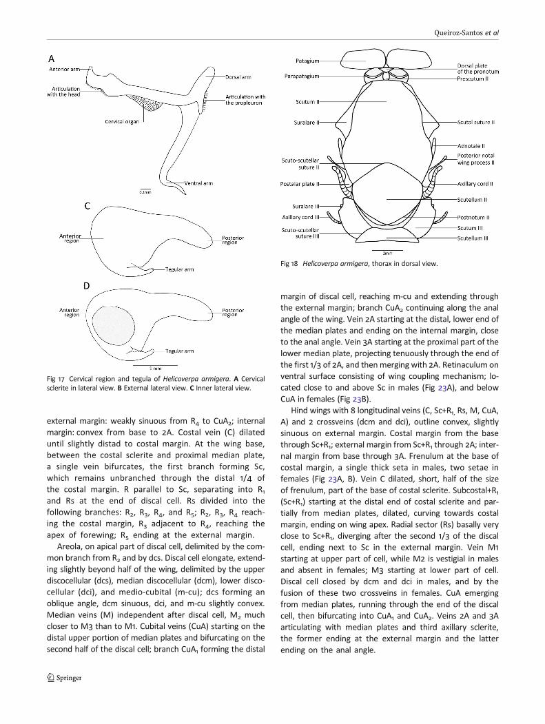
Mesothorax
The largest thoracic segment. In dorsal aspect: prescutumoval, located antero-medially in relation to scutum. Scutumwide (the largest sclerity in the body), smooth, slightly con-vex, scutal suture delimiting the suralare, adnotale and
Fig 12 Pupa of Helicoverpaarmigera. A Thoracic spiracle inanterior view. B Thoracic spiraclein lateral view. C Abdominalspiracle. D Cremaster.
Fig 13 Posterior region of pupaof Helicoverpa armigera. AFemale in ventral view. BMale inventral view.
Queiroz-Santos et al
posterior notal wing process; scutum separated from scutel-lum by the scuto-scutellar suture. Scutellum half the length
of scutum, distally ending at the postnotum. Postnotum sick-le-shaped.
Fig 14 Head of Helicoverpaarmigera. A Frontal view. BDorsal view. C Posterior view. DVentral view.
Morphological Characterization of Helicoverpa armigera (Hübner)
In lateral aspect: tegula (Fig 17B, C) large compared toother structures, forming an anterior lobe, distally curved,subtegula reaching the anterior margin of the basalare.Basalare round. Folds of pleural process of forewings andlateral margin of tergopleural apodeme projected and thenfused distally. Subalare ellipsoid, reaching adnotal scleriteand posterior notal wing process. Axillary cord connectingto postalar plate.
On membranous area: anepisternum and katepister-num delimited by anepisternum suture, pre-episternalsuture separating pre-episternum from katepisternum.Epimeron V-shaped, separated anteriorly from anepis-ternum and katepisternum by the pleural suture, fromthe meron by the basicostal suture; the upper portionof the distal arm surrounding the second spiracleanteriorly.
In ventral aspect: discrimen 5× longer than discrimenI, forming the base of the basisternum triangle, whichextends to the furcal pit. Katepisternum projecting ven-trally, reaching the basisternum, separated by the ster-numpleural suture towards the medial line and laterallyaround the coxa (Fig 19).
Metathorax
Larger than the prothorax and smaller than the mesothorax.In dorsal aspect: scutellum subtrapezoidal, located betweenscutellar plates, which project antero-laterally around thescutellum and postnotum II. Scutum reaching distally to thescutellum, separated by the scuto-scutellar suture. Lateralmedial portion and anterior portion of scutellum delimitedby membranous area from postnotum II (Fig 20).
In latero-ventral aspect: scutum projected distally, support-ing the ventral end of the axillary cord ventrally, and anteriorlyseparated from suralare by the scutal suture. Pleural processof wing arched, located between suralare and anepisternum.Basalare slightly above, also round, but much smaller thananepisternum, surrounding the distal portion of second spira-cle. A short anepisternum suture between anepisternum andkatepisternum. Katepisternum rectangular, delimited anterior-ly by amembranous area that separates the segments, distallyseparated from the epimeron by the pleural suture, ventro-anteriorly separated from the basisternum, partially coveredby the katepisternum and eucoxa, the distal angle separatedfrom eucoxa by the marginopleural suture.
Fig 15 Head and cephalic appendages ofHelicoverpa armigera. A Head in lateral view. B Antenna. C Labial palp in external lateral view.D Labial palp ininner lateral view.
Queiroz-Santos et al
Antero-posterior axis of coxa positioned obliquely to thediscrimen.
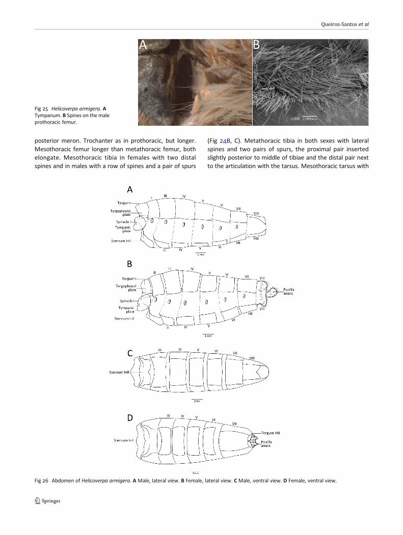
Subalare isolated, on membranous area, posterior to thedorsal portion of katepisternum and below the axillary cord.Epimeron U-shaped, the tympanic membrane at the end ofits distal arm (Fig 25A), and bearing a tympanal sclerite.
In ventral aspect: discrimen approximately the same sizeas discrimen I, extending medio-longitudinally from the an-terior margin of basisternum to furcal pit. Sternumpleuralsuture limiting the basisternum, which consists of a narrow,long sclerotized plate along the katepisternum.
Thoracic appendages
Axillary sclerites
Wings articulate with thorax through three irregular-shapedsclerites. On the anterior wing, first axillary sclerite locatedposterior to the adnotal and anterior to the second axillarysclerite. The second axillary sclerite articulating with the firstaxillary and with the proximal median plate. The third axillarysclerite connected anteriorly with the proximal median plate
and latero-posteriorly with anal veins. The costal scleritearticulates with the costal (C), subcostal (Sc), and radial (R)veins, latero-anteriorly to the humeral plate and distal to theproximal median plate. Dorsally, the distal median platemeets the median (M) vein, located anterior to the proximalmedial plate (Fig 21A).
On the hind wing, the costal sclerite articulates with C, Sc,and M, and is the base of the frenulum. The first axillarysclerite is projected into three arms: the anterior, basal,connected to the scutum, the second to the axillary cord,and the third to the second axillary sclerite and to the lateralmargin of one of the median plates. The third axillary scleritearticulates with the anal (A) veins posteriorly and anteriorlyto the medial plate (Fig 21B).
Wings
Forewing subrectangular (Fig 22A), with 14 longitudinal veins(C, Sc, R1-R5, M1-M3, Cua1 e Cua2, 2A e 3A) and 4 crossveins(dcs, dcm, dci, m-cu). Males and females with identical vena-tion pattern except for having slightly different outlines.Costal margin: slightly convex distally from base to R4;
Fig 16 Head and cephalicappendages of Helicoverpaarmigera. A Scape, pedicel, andproximal flagellomeres. B Distalportion of the antenna. C Detailof the last flagellomere. D Ocelliin frontal view. E Hypopharynx infrontal view. F Distal portion ofthe galea.
Morphological Characterization of Helicoverpa armigera (Hübner)
external margin: weakly sinuous from R4 to CuA2; internalmargin: convex from base to 2A. Costal vein (C) dilateduntil slightly distad to costal margin. At the wing base,between the costal sclerite and proximal median plate,a single vein bifurcates, the first branch forming Sc,which remains unbranched through the distal 1/4 ofthe costal margin. R parallel to Sc, separating into R1and Rs at the end of discal cell. Rs divided into thefollowing branches: R2, R3, R4, and R5; R2, R3, R4 reach-ing the costal margin, R3 adjacent to R4, reaching theapex of forewing; R5 ending at the external margin.
Areola, on apical part of discal cell, delimited by the com-mon branch from R2 and by dcs. Discal cell elongate, extend-ing slightly beyond half of the wing, delimited by the upperdiscocellular (dcs), median discocellular (dcm), lower disco-cellular (dci), and medio-cubital (m-cu); dcs forming anoblique angle, dcm sinuous, dci, and m-cu slightly convex.Median veins (M) independent after discal cell, M2 muchcloser to M3 than to M1. Cubital veins (CuA) starting on thedistal upper portion of median plates and bifurcating on thesecond half of the discal cell; branch CuA1 forming the distal
margin of discal cell, reaching m-cu and extending throughthe external margin; branch CuA2 continuing along the analangle of the wing. Vein 2A starting at the distal, lower end ofthe median plates and ending on the internal margin, closeto the anal angle. Vein 3A starting at the proximal part of thelower median plate, projecting tenuously through the end ofthe first 1/3 of 2A, and thenmerging with 2A. Retinaculum onventral surface consisting of wing coupling mechanism; lo-cated close to and above Sc in males (Fig 23A), and belowCuA in females (Fig 23B).
Hind wings with 8 longitudinal veins (C, Sc+R1, Rs, M, CuA,A) and 2 crossveins (dcm and dci), outline convex, slightlysinuous on external margin. Costal margin from the basethrough Sc+R1; external margin from Sc+R1 through 2A; inter-nal margin from base through 3A. Frenulum at the base ofcostal margin, a single thick seta in males, two setae infemales (Fig 23A, B). Vein C dilated, short, half of the sizeof frenulum, part of the base of costal sclerite. Subcostal+R1(Sc+R1) starting at the distal end of costal sclerite and par-tially from median plates, dilated, curving towards costalmargin, ending on wing apex. Radial sector (Rs) basally veryclose to Sc+R1, diverging after the second 1/3 of the discalcell, ending next to Sc in the external margin. Vein M1starting at upper part of cell, while M2 is vestigial in malesand absent in females; M3 starting at lower part of cell.Discal cell closed by dcm and dci in males, and by thefusion of these two crossveins in females. CuA emergingfrom median plates, running through the end of the discalcell, then bifurcating into CuA1 and CuA2. Veins 2A and 3Aarticulating with median plates and third axillary sclerite,the former ending at the external margin and the latterending on the anal angle.
Fig 17 Cervical region and tegula of Helicoverpa armigera. A Cervicalsclerite in lateral view. B External lateral view. C Inner lateral view.
Fig 18 Helicoverpa armigera, thorax in dorsal view.
Queiroz-Santos et al

Legs
Prothoracic leg sexually dimorphic, with a spiny area on theinternal surface in males (Fig 25B) which is absent in females.
Coxa elongate, cylindrical, articulating basally with the pro-pleuron and distally with the trochanter, which is triangular,1/5 coxa length. Femur cylindrical, the largest leg segment.Tibia slightly shorter than 1/2 femur length, with few spinesand an apical tibial spur; epiphises on upper 1/2 approximately
1/2 length of tibia. Tarsi with five tarsomeres, the proximal oneas long as the tibia, all others about the same size, the sum ofall four slightly longer than length of the basal tarsomeres. Alltarsomeres with spines present on internal and lateral surfa-ces. Tarsal claw bifid, with long, curved projections, the inter-nal projection slightly shorter; unguitractor plate with cushion-like arolium distally (Fig 24A).
Meso- and metathoracic legs similar. Coxa longitudinallydivided by the coxal suture into an anterior eucoxa and
Fig 19 Helicoverpa armigera,thorax in ventral view.
Fig 20 Helicoverpa armigera, thorax in lateral view.
Morphological Characterization of Helicoverpa armigera (Hübner)
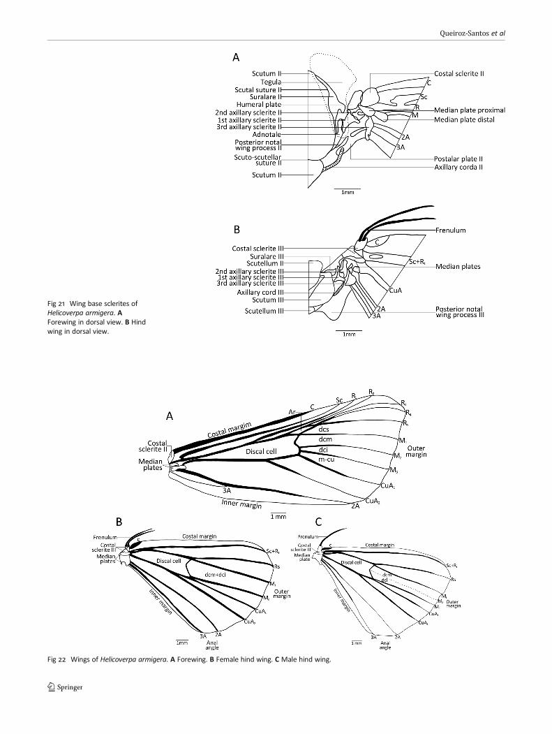
Fig 21 Wing base sclerites ofHelicoverpa armigera. AForewing in dorsal view. B Hindwing in dorsal view.
Fig 22 Wings of Helicoverpa armigera. A Forewing. B Female hind wing. C Male hind wing.
Queiroz-Santos et al
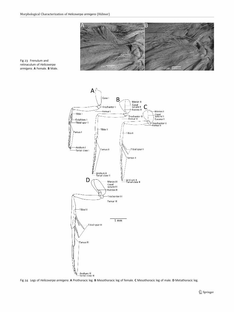
Fig 23 Frenulum andretinaculum of Helicoverpaarmigera. A Female. B Male.
Fig 24 Legs of Helicoverpa armigera. A Prothoracic leg. B Mesothoracic leg of female. C Mesothoracic leg of male. D Metathoracic leg.
Morphological Characterization of Helicoverpa armigera (Hübner)
posterior meron. Trochanter as in prothoracic, but longer.Mesothoracic femur longer than metathoracic femur, bothelongate. Mesothoracic tibia in females with two distalspines and in males with a row of spines and a pair of spurs
(Fig 24B, C). Metathoracic tibia in both sexes with lateralspines and two pairs of spurs, the proximal pair insertedslightly posterior to middle of tibiae and the distal pair nextto the articulation with the tarsus. Mesothoracic tarsus with
Fig 25 Helicoverpa armigera. ATympanum. B Spines on the maleprothoracic femur.
Fig 26 Abdomen of Helicoverpa armigera. A Male, lateral view. B Female, lateral view. C Male, ventral view. D Female, ventral view.
Queiroz-Santos et al
numerous spines laterally and internally, with five tar-someres, proximal tarsomeres larger, distal tarsomeresdecreasing in size. Metathoracic tarsus longer thanmesothracic (Fig 24B–D).
Abdomen
Pregenital segments
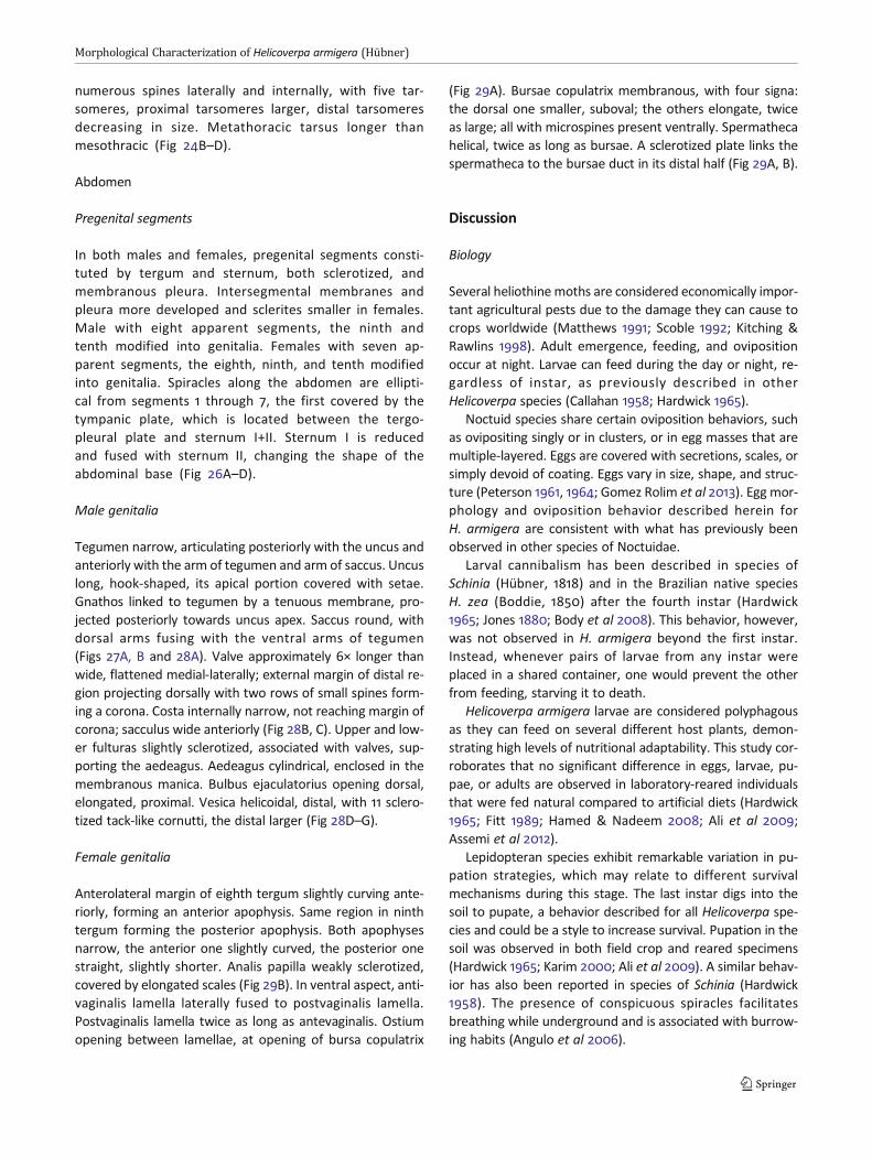
In both males and females, pregenital segments consti-tuted by tergum and sternum, both sclerotized, andmembranous pleura. Intersegmental membranes andpleura more developed and sclerites smaller in females.Male with eight apparent segments, the ninth andtenth modified into genitalia. Females with seven ap-parent segments, the eighth, ninth, and tenth modifiedinto genitalia. Spiracles along the abdomen are ellipti-cal from segments 1 through 7, the first covered by thetympanic plate, which is located between the tergo-pleural plate and sternum I+II. Sternum I is reducedand fused with sternum II, changing the shape of theabdominal base (Fig 26A–D).
Male genitalia
Tegumen narrow, articulating posteriorly with the uncus andanteriorly with the arm of tegumen and arm of saccus. Uncuslong, hook-shaped, its apical portion covered with setae.Gnathos linked to tegumen by a tenuous membrane, pro-jected posteriorly towards uncus apex. Saccus round, withdorsal arms fusing with the ventral arms of tegumen(Figs 27A, B and 28A). Valve approximately 6× longer thanwide, flattened medial-laterally; external margin of distal re-gion projecting dorsally with two rows of small spines form-ing a corona. Costa internally narrow, not reaching margin ofcorona; sacculus wide anteriorly (Fig 28B, C). Upper and low-er fulturas slightly sclerotized, associated with valves, sup-porting the aedeagus. Aedeagus cylindrical, enclosed in themembranous manica. Bulbus ejaculatorius opening dorsal,elongated, proximal. Vesica helicoidal, distal, with 11 sclero-tized tack-like cornutti, the distal larger (Fig 28D–G).
Female genitalia
Anterolateral margin of eighth tergum slightly curving ante-riorly, forming an anterior apophysis. Same region in ninthtergum forming the posterior apophysis. Both apophysesnarrow, the anterior one slightly curved, the posterior onestraight, slightly shorter. Analis papilla weakly sclerotized,covered by elongated scales (Fig 29B). In ventral aspect, anti-vaginalis lamella laterally fused to postvaginalis lamella.Postvaginalis lamella twice as long as antevaginalis. Ostiumopening between lamellae, at opening of bursa copulatrix
(Fig 29A). Bursae copulatrix membranous, with four signa:the dorsal one smaller, suboval; the others elongate, twiceas large; all with microspines present ventrally. Spermathecahelical, twice as long as bursae. A sclerotized plate links thespermatheca to the bursae duct in its distal half (Fig 29A, B).
Discussion
Biology
Several heliothine moths are considered economically impor-tant agricultural pests due to the damage they can cause tocrops worldwide (Matthews 1991; Scoble 1992; Kitching &Rawlins 1998). Adult emergence, feeding, and ovipositionoccur at night. Larvae can feed during the day or night, re-gardless of instar, as previously described in otherHelicoverpa species (Callahan 1958; Hardwick 1965).
Noctuid species share certain oviposition behaviors, suchas ovipositing singly or in clusters, or in egg masses that aremultiple-layered. Eggs are covered with secretions, scales, orsimply devoid of coating. Eggs vary in size, shape, and struc-ture (Peterson 1961, 1964; Gomez Rolim et al 2013). Egg mor-phology and oviposition behavior described herein forH. armigera are consistent with what has previously beenobserved in other species of Noctuidae.
Larval cannibalism has been described in species ofSchinia (Hübner, 1818) and in the Brazilian native speciesH. zea (Boddie, 1850) after the fourth instar (Hardwick1965; Jones 1880; Body et al 2008). This behavior, however,was not observed in H. armigera beyond the first instar.Instead, whenever pairs of larvae from any instar wereplaced in a shared container, one would prevent the otherfrom feeding, starving it to death.
Helicoverpa armigera larvae are considered polyphagousas they can feed on several different host plants, demon-strating high levels of nutritional adaptability. This study cor-roborates that no significant difference in eggs, larvae, pu-pae, or adults are observed in laboratory-reared individualsthat were fed natural compared to artificial diets (Hardwick1965; Fitt 1989; Hamed & Nadeem 2008; Ali et al 2009;Assemi et al 2012).
Lepidopteran species exhibit remarkable variation in pu-pation strategies, which may relate to different survivalmechanisms during this stage. The last instar digs into thesoil to pupate, a behavior described for all Helicoverpa spe-cies and could be a style to increase survival. Pupation in thesoil was observed in both field crop and reared specimens(Hardwick 1965; Karim 2000; Ali et al 2009). A similar behav-ior has also been reported in species of Schinia (Hardwick1958). The presence of conspicuous spiracles facilitatesbreathing while underground and is associated with burrow-ing habits (Angulo et al 2006).
Morphological Characterization of Helicoverpa armigera (Hübner)

Immature stages
Characters in the immature stages, such as the presence ofspicules in the larvae and the position of L2 and L2 in T1, aremajor synapomorphies of Heliothinae (Hardwick 1958;Kitching & Rawlins 1998). These features were also observedin H. armigera, whose larvae exhibit a larger number of spi-cules in early instars that decrease as the larva develops, aswell as a similar location of setae L1 and L2 on T1 inagreement with the description provided by Mitter et al(1993) for all instars. Immatures share additional morpholog-ical features, such as the presence of well-defined bands andspicules along the body, conspicuous chalazae, and un-branched setae lacking ornation. This tends to hinder therapid and accurate differentiation of Helicoverpa species inthe field.
The brown color pattern of third instar larvae is also ob-served in other congeneric species such as H. punctigera(Wallengren, 1860), H. gelotopoeon (Dyar, 1921), andH. zea. Other species, such as H. hawaiiensis (Quaintance& Brues, 1905) and H. assulta (Guenée, 1852), are mostlygreen (Hardwick 1965). The two-color pattern observed in
the sixth instar of H. armigera had not been reported forother species in this genus to date. This merits carefulinvestigation, as it might be associated with sexualdimorphism.
The presence of an exocrine jugular gland in thelast instar larvae of H. armigera agrees with previouscontributions concerning other species of Noctuidae, aswell as other Lepidoptera families, such as Hesperiidae,Notodontidae, Nymphalidae, and Pieridae. These glandsact as a defense mechanism (Peterson 1962, Borgeset al 2010, James et al 2012, Vegliante & Hasenfuss2012).
Fig 27 Male genitalia of Helicoverpa armigera. A Anterior view. BPosterior view.
Fig 28 Male genitalia of Helicoverpa armigera. A Lateral view. B Valvaein external view. C Valvae in inner view. D Aedeagus in dorsal view. EAedeagus in lateral view. F Aedeagus in ventral view. G Vesica in dorsalview.
Queiroz-Santos et al

The absence of abdominal legs on A3 in the trifinesubfamilies is associated with foraging, as it facilitatescrawling long distances, and does not match the lepidop-teran ground plan, in which abdominal legs are presenton A3 through A6 in all larval instars (Kitching & Rawlins1998). Even though H. armigera has great movementpotential within the host plant search scenario, thepresence of abdominal legs on A3 contrasts with previ-ous morphological characterizations of some trifinesubfamilies.
Adults
There are a variety of abiotic factors in Brazil that canfavor the dispersal of species with high reproductivecapacity, which directly affects economically importantcrops. Helicoverpa armigera adults are notable due totheir robust morphological structures, such as a widethorax, which enables them to fly long distances (Fenget al 2004, Ali et al 2009), an ability that can certainlyhelp them colonize new environments.
Most noctuids have filiform antenna that are similar ingeneral structure. These structures have several kinds of sen-sory setae, which can capture information on temperature,humidity, olfactory, and sexual pheromones (Jefferson et al1970; Hardwick 1970; Castrejón-Gómez et al 1999; Seada2015). No morphological differences were found betweenmale and female antennae, an aspect also observed byDiongue et al (2013) in their study of types and functions ofsensory setae of antennomeres. However, electron micros-copy analyses revealed structures such as the ones on the
last flagellomere (Fig 18), which lacks setae, indicating a pos-sible sensory function.
Certain insects have tympanic organs located in variousparts of their exoskeleton which can perceive acoustic infor-mation. The tympanic membranes of some moth species arefused to the metathorax and are located adjacent to theanterior margin of the abdomen, with an aperture that issometimes partially covered by a “lid”-like membrane(Forbes 1916; Kiriakoff 1963; Hoy & Robert 1996; Mhatreet al 2009). A tympanic sclerite covers the tympanic orificein H. armigera, and a tympanic plate also covers the firstabdominal spiracle.
Differences in the number of setae on the frenulum inmales and females have also been described in Tortricidae(Yang & Brown 2009; Rota et al 2009; Monsalve et al 2011)and Pseudobiston pinratanai Inoue, 1994 (Geometridae)(Rajaei et al 2015). In H. armigera, only one seta is present inmales, while females have a pair of setae. Besides the numberof setae on frenulum, their position on the retinaculum alsodiffers, a feature that is helpful in sex determination.
The sclerotized cornutti in male H. armigera vesica arealso observed in other species of the same genus, as wellas a few other species of Heliothinae (Hardwick 1965, 1970).
The position and presence of four signa in the bursa cop-ulatrix of female H. armigera have also been observed inHelicoverpa atacamae Hardwick, 1965, H. hawaiiensis, andH. punctigera. However, the pattern is distinct fromH. gelotopoeon (Hardwick 1965).
This contribution offers the first detailed morphologicalstudy of the economically important noctuid species,H. armigera. Several morphological structures describedherein will contribute to the taxonomic knowledge of this
Fig 29 Female genitalia of Helicoverpa armigera. A Ventral view. B Lateral view.
Morphological Characterization of Helicoverpa armigera (Hübner)
species and will also be useful for distinguishing males andfemales. Tools for the accurate identification of thiseconomically significant species are fundamental for fieldwork and crop management. Future comparative studiesmay help differentiate this species from H. zea, whoserepresentatives also occur in Brazil and are often confusedwith H. armigera due to close morphological resemblance. Asimilar case of misidentification with H. armigera was shownby Gillian & Passoa (2014) for H. gelotopoeon, a speciesfound in southern South America, which is also consideredan important economic pest.
Acknowledgments Illustrations presented in this paper were acquiredat the Electron Microscopy Center (CEM-UFPR) and Taxonline atUniversidade Federal do Paraná, Curitiba, Brazil. The authors thank Dr.Fernando Dias Maia for assisting with image editing and OliviaEvangelista, Ana Dal Molin, and Keith Bayless for translating and proof-reading an earlier version of this manuscript. We also thank Dr. Daniel R.Sosa-Gómez (Embrapa Soja, Londrina, Paraná, Brazil) for sending thepupae. Thanks again to Takumasa Kondo, and three anonymousreviewers, for detailed comments and corrections that improved thispaper.
Electronic supplementary material The online version of this article(https://doi.org/10.1007/s13744-017-0581-4) contains supplementarymaterial, which is available to authorized users.
Funding Information This research was funded by the NationalCouncil for Scientific and Technological Development (ConselhoNacional de Desenvolvimento Científico e Tecnológico – CNPq)(LQS proc. no. 130624/2014-1; MMC proc. no. 308247/2013-2;AS proc. no. 403376/2013-0, 476691/2013-3, 47304/2013-8) andEmbrapa (proc. SEG MP2 - 02.13.14.006.00.00).
References
Abbasi B, Ahmed K, Khalique F, Ayub N, Liu H, Kazmi S, Aftab M (2007)Rearing the cotton bollworm, Helicoverpa armigera, on a tapioca-based artificial diet. J Insect Sci 7(35):1–7. https://doi.org/10.1673/031.007.3501
Ali A, Choudhury RA, Ahmad Z, Rahman F, Khan FR, Ahmad SK (2009)Some biological characteristics of Helicoverpa armigera on chickpea.Tunis J Plant Prot 4:99–106
Angulo AO, Olivares TS, Weigert GT (2006) Estados inmaduros delepidópteros nóctuidos de importancia agrícola y forestal en Chile(Lepidoptera: Noctuidae). Universidad de Concepción, Concepción,p 154
Arnemann JA, James WJ, Walsh TK, Guedes JVC, Smagghe G, CastiglioniE, Tay WT (2016) Mitochondrial DNA COI characterization ofHelicoverpa armigera (Lepidoptera: Noctuidae) from Paraguay andUruguay. Genet Mol Res 15(2). doi:https://doi.org/10.4238/gmr.15028292
Assemi H, Rezapanah M, Vafaei-Shoushtari R, Mehrvar A (2012)Modified artificial diet for rearing of tobacco budworm, Helicoverpaarmigera, using the Taguchi method and derringer’s desirability func-tion. J Insect Sci 12(100):1–18. https://doi.org/10.1673/031.012.10001
Baker GT, Parrott WL, Jenkins JN (1986) Sensory receptors on the larvalmaxillae and labia of Heliothis zea (Boddie) and Heliothis virescens (F.)(Lepidoptera: Noctuidae). Int J Insect Morphol Embryol 15(3):227–232. https://doi.org/10.1016/0020-7322(86)90060-7
Bentivenha JPF, Paula-Moraes SV, Baldin ELL, Specht A, Silva IF, Hunt TE(2016) Battle in the New World: Helicoverpa armigera versusHelicoverpa zea (Lepidoptera: Noctuidae). PLoS One 11(12):e0167182.https://doi.org/10.1371/journal.pone.0167182
Blaik T, Malkiewicz A (2003) Morphology of larval and pupal stages ofIsturgia roraria (Fabricius, 1777) (Lepidoptera: Geometridae). Ann Zool53:245–258
Body BM, Daniels JC, Austin GT (2008) Predaceous behavior byHelicoverpa zea (Boddie) (Lepidoptera: Noctuidae: Heliothinae). JInsect Behav 21(3):143–146. https://doi.org/10.1007/s10905-007-9113-0
Borges E, Faccioni-Heuser M, Moreira G (2010) Morphology of the pros-ternal glands of Heliconius erato (Lepidoptera: Nymphalidae). Psyche1:8–8. https://doi.org/10.1155/2010/892960
Bueno AF, Sosa-Gómez DR (2014) The old world bollworm in the neo-tropical region: the experience of brazilian growers with Helicoverpaarmigera. Outlooks Pest Manag 25(4):261–265. https://doi.org/10.1564/v25_aug_04
Butt BA, Cantu E (1962) Sex determination of lepidopterous pupae.Washington. USDA, p 7
CABI (2017) Invasive species compendium datasheets Helicoverpaarmigera (cotton bollworm). Available in: http://www.cabi.org/isc/datasheet/26757. Accessed 8 May 2017
Callahan PS (1958) Serial morphology as a technique for determinationof reproductive patterns in the corn earworm, Heliothis zea Boddie.Ann Entomol Soc Am 51(5):413–428. https://doi.org/10.1093/aesa/51.5.413
Casagrande MM (1979) Sobre Caligo beltrao (Illiger). IV: Morfologiaexterna do adulto - abdome (Lepidoptera, Satyridae, Brassolinae).Rev Bras Biol 39:711–716
Castiglioni E, Perini CR, Chiaravalle W, Arnemann JA, Ugalde G, GuedesJVC (2016) Primer registro de ocurrencia de Helicoverpa armigera(Hübner, 1808) (Lepidoptera: Noctuidae) en soja, en Uruguay.Agrocien Urug 20:31–35
Castrejón-Gómez VR, Valdez-Carrasco J, Cibrian-Tovar J, Camino-LavinM, Osorio RO (1999) Morphology and distribution of the sense organson the antennae of Copitarsia consueta (Lepidoptera: Noctuidae). FlaEntomol 82(4):546–555. https://doi.org/10.2307/3496472
Comstock JH (1918) The wings of the insects. Comstock PublishingCompany, Nova Iorque, 430 p
Czepak C, Albernaz KC, Vivan LM, Guimarães HO, Carvalhais T (2013)Primeiro registro de ocorrência de Helicoverpa armigera (Hübner)(Lepidoptera: Noctuidae) no Brasil. Pesqu Agropecu Trop 43(1):110–113. https://doi.org/10.1590/S1983-40632013000100015
Daly HV (1985) Insect morphometrics. Annu Rev Entomol 30(1):415–438.https://doi.org/10.1146/annurev.en.30.010185.002215
Diongue A, Yang J, Lai P (2013) Biomorphometric characteristics of dif-ferent types of sensilla detected on the antenna of Helicoverpa armi-gera by scanning electronmicroscopy. J Asia Pac Entomol 16(1):23–28.https://doi.org/10.1016/j.aspen.2012.09.001
EMBRAPA (2013) Nota técnica sobre resultado do trabalho inicial delevantamento da lagarta do gênero Helicoverpa – detecção daespécie Helicoverpa armigera no Brasil. Technical Note 22 Mar2013. Planaltina: EMBRAPA CERRADOS. 2 p. doi: https://doi.org/10.13140/RG.2.1.2946.7685
Feng H, Wu K, Cheng D, Guo Y (2004) Northward migration ofHelicoverpa armigera (Lepidoptera: Noctuidae) and other moths inearly summer observed with radar in northern China. J Econ Entomol97:1874–1883. https://doi.org/10.1603/0022-0493-97.6.1874
Feng H, Gouldb F, Huang Y, Jiangd Y, Wua K (2010) Modeling the pop-ulation dynamics of cotton bollworm Helicoverpa armigera (Hübner)(Lepidoptera: Noctuidae) over a wide area in northern China. EcolModel 221:1819–1830. https://doi.org/10.1016/j.ecolmodel.2010.04.003
Fibiger M, Lafontaine JD (2004) A review of the higher classification ofthe Noctuoidea (Lepidoptera) with special reference to the Holarcticfauna. Esper 11:7–690
Queiroz-Santos et al
Fitt GP (1989) The ecology of Heliothis species in relation to agroecosys-tems. Annu Rev Entomol 34(1):17–52. https://doi.org/10.1146/annurev.en.34.010189.000313
Forbes WTM (1916) On the tympanum of certain Lepidoptera. Psyche23(6):183–192. https://doi.org/10.1155/1916/16801
Gilligan TM, Passoa SC (2014) LepIntercept—an identification resourcefor intercepted Lepidoptera larvae. Identification TechnologyProgram (ITP), Fort Collins, CO 1:3
Gomez Rolim AAS, Yano SAC, Specht A, Andrade CGTJ, Sosa-Gómez DR(2013) Morphological and molecular characterization of the eggs ofsome noctuid species associated with soybean in Brazil. Ann EntomolSoc Am 106(5):643–651. https://doi.org/10.1603/AN13049
Grimes LR, Neuzing HH (1986) Morphological survey of the maxillae inlast stage larvae of the suborder Ditrysia (Lepidoptera): Palpi. AnnEntomol Soc Am 79(3):491–509. https://doi.org/10.1093/aesa/79.3.491
Hallberg E, Hansson BS, Löfstedt C (2003) Sensilla and proprioceptors.In: Kristensen NP (ed) Lepidoptera, moths and butterflies. Vol.2: mor-phology, physiology and development. In: Fischer M (Ed.) Handbookof zoology. Vol, IV, Arthropoda:Insecta, vol 36. Walter de Gruyter,Berlin, New York, pp 267–288
Hamed M, Nadeem S (2008) Rearing of Helicoverpa Armigera (Hub.) onartificial diets in laboratory. Pak J Zool 40(6):447–450
Hardwick DF (1958) Taxonomy, life history and habits of the elliptoid-eyed species of Schinia, with notes on the Heliothidinae. Can Entomol90(Suppl.6):1–116. https://doi.org/10.4039/entm9006fv
Hardwick DF (1965) The corn earworm complex. Mem Entomol (Soc Can40):1–246
Hardwick DF (1970) A generic revision of the North AmericanHeliothidinae (Lepidoptera: Noctuidae). Mem Entomol 102(Soc Can73):1–59. https://doi.org/10.4039/entm10273fv
Hemming F (1937) Hübner: a bibliographical and systematic account ofthe entomological works of Jacob Hübner and of the supplementsthereto by Carl Geyer. Gottfried Franz von Frölich and GottliebAugust Wilhelm Herrich-Schäffer. Royal Entomological Society ofLondon, London, p 605
Hoy RR, Robert D (1996) Tympanal hearing in insects. Annu Rev Entomol41(1):433–450. https://doi.org/10.1146/annurev.en.41.010196.002245
Jadhav DR, Armes NJ, Bhatnagar VS (2013) Incidence of winter andsummer diapause in Helicoverpa armigera (Hübner) (Lepidoptera:Noctuidae) in Andhra Pradesh, India. Asian J Agric Sci 5(3):40–51
James DG, Hebert V, LePage J (2012) The prosternal gland in pacificnorthwest butterfly larvae with preliminary chemical analyses ofemissions. J Lepid Soc 66(3):137–142. https://doi.org/10.18473/lepi.v66i3.a3
Jefferson RN, Rubin RE, McFarland SU, Shorey HH (1970) Sex phero-mones of noctuid moths. XXII. The external morphology of the anten-nae of Trichoplusia ni, Heliothis zea, Prodenia ornithogalli, andSpodoptera exigua. Ann Entomol Soc Am 63(5):1227–1238. https://doi.org/10.1093/aesa/63.5.1227
Jones RW (1880) Boll worm devouring cotton worm. Am Entomol 3:253Karim S (2000) Management of Helicoverpa armigera: a review and
prospectus for Pakistan. Pak J Biol Sci 3(8):1213–1222Kiriakoff SG (1963) The tympanic structure of the Lepidoptera and the
taxonomy of the order. J Lepid Soc 17:1–20Kitching IJ, Rawlins JE (1998) The noctuoidea. In: Kristensen NP (Ed.)
Lepidoptera, moths and butterflies. Vol.1: evolution, systematic andbiogeography. In: Fischer M (Ed.) Handbook of zoology. Vol. IV,Arthropoda:Insecta 35. Walter de Gruyter, Berlin, New York, pp.355–401
Kristensen NP (2003) Reproductive organs. In: Kristensen, NP (Ed.)Lepidoptera, moths and butterflies. Vol.2: morphology, physiologyand development. In: Fischer M (Ed.) Handbook of zoology. Vol. IV,Arthropoda:Insecta 36.Walter de Gruyter, Berlin, New York, pp. 427–447
Liu Z, Gong P, Li D,WeiW (2010) Pupal diapause ofHelicoverpa armigera(Hübner) (Lepidoptera: Noctuidae) mediated by larval host plants:pupal weight is important. J Insect Physiol 56(12):1863–1870.https://doi.org/10.1016/j.jinsphys.2010.08.007
MatthewsM (1991) Classification of the Heliothinae. Bull Nat Resour Inst44:1–195
Mhatre N, Montealegre-Z F, Balakrishnan R, Robert D (2009)Mechanical response of the tympanal membranes of the tree cricketOecanthus henryi. J Comp Physiol A 195(5):453–462. https://doi.org/10.1007/s00359-009-0423-x
Mironidis GK, Savopoulou-Soultani M (2008) Development, survivor-ship, and reproduction of Helicoverpa armigera (Lepidoptera:Noctuidae) under constant and alternating temperatures. EnvironEntomol 37(1):16–28.
Mitter C, Poole RW, Matthews M (1993) Biosystematics of theHeliothinae (Lepidoptera: Noctuidae). Annu Rev Entomol 38(1):207–225. https://doi.org/10.1146/annurev.en.38.010193.001231
Monsalve S, Dombroskie JJ, LamWHY, Rota J, Brown JW (2011) Variationin the female frenulum in Tortricidae (Lepidoptera). Part 3.Tortricinae. Proc Entomol Soc Wash 113(3):335–370. https://doi.org/10.4289/0013-8797.113.3.335
Montezano DG, Specht A, Bortolin TM, Fronza E, Sosa-Gómez DR,Roque-Specht VF, Pezzi P, Luz PC, Barros NM (2013) Immature stagesof Spodoptera albula (Walker) (Lepidoptera: Noctuidae): develop-mental parameters and host plants. An Acad Bras Cienc 85(1):271–284. https://doi.org/10.1590/S0001-37652013000100013
Mosher E (1916) A classification of the Lepidoptera based on charactersof the pupae. Bull Ill State Lab Nat Hist 12:17–159
Murúa MG, Scalora FS, Navarro FR, Cazado LE, Casmuz A, Villagrán ME,Lobos E, Gastaminza G (2014) First record of Helicoverpa armigera(Lepidoptera: Noctuidae) in Argentina. Fla Entomo 97(2):854–856.https://doi.org/10.1653/024.097.0279
Nadda G (2013) Medicinal and aromatic crops as hosts of Helicoverpaarmigera Hübner (Lepidoptera: Noctuidae). Lepcey - J Trop AsianEntomol 02(1):44–46
Peterson A (1961) Some types of eggs deposited by moths, Heterocera-Lepidoptera. Fla Entomo 44(3):107–114. https://doi.org/10.2307/3492966
Peterson A (1962) Larvae of insects. An introduction to Neartic species.Part I. Lepidoptera and plant infesting Hymenoptera. EdwardsBrothers, Ann Arbor, Michigan, p. 315
Peterson A (1964) Egg types among moths of the Noctuidae(Lepidoptera). Fla Entomo 47(2):71–91. https://doi.org/10.2307/3493280
Pierce FN (1909) The genitalia of the group Noctuidae of the Lepidopteraof the British Islands. Liverpool, A.W. Duncan, p. 88
Rajaei H, Greve C, Letsch H, Stüning D, Wahlberg N, Minet J, Misof B(2015) Advances in Geometroidea phylogeny, with characterization ofa new family based on Pseudobiston pinratanai (Lepidoptera,Glossata). Zool Scr 44(4):418–436. https://doi.org/10.1111/zsc.12108
Rota J, Yang A, Brown JW (2009) Variation in the female frenulum inTortricidae (Lepidoptera). Part 2. Olethreutinae. Proc Entomol SocWash 111(4):826–866. https://doi.org/10.4289/0013-8797-111.4.826
Scoble M (1992) The Lepidoptera. Form, function and diversity. NaturalHistoryMuseum Publications. Oxford University Press, London, p 404
Seada MA (2015) Antennal morphology and sensillum distribution offemale cotton leaf worm Spodoptera littoralis (Lepidoptera:Noctuidae). J Basic Appl Zool 68:10–18. https://doi.org/10.1016/j.jobaz.2015.01.005
SENAVE (2013) Senave en alerta tras ingreso de peligrosa plaga agrícola.Available in: http://www.abc.com.py/edicion-impresa/economia/senave-en-alerta-tras-ingreso-de-peligrosa-plaga-agricola-629240.html. Accessed 17 Oct 2013
Snodgrass RE (1935) Principles of insect morphology. McGraw-Hill BookCompany, Nova York, p 665
Morphological Characterization of Helicoverpa armigera (Hübner)
Sorensen GS, Cribb BW, Merritt D, Johnson ML, Zaluck MP (2006)Structure and ultrastructure of the silk glands and spinneret ofHelicoverpa armigera (Hübner) (Lepidoptera: Noctuidae). ArthropodStruct Dev 35(1):3–13. https://doi.org/10.1016/j.asd.2005.10.002
Sosa-Gómez DR, Specht A, Paula-Moraes SV, Lopes-Lima A, Yano SAC,Micheli A, Morais EGF, Gallo P, Pereira PRVS, Salvadori JR, Botton M,Zenker MM, Azevedo-Filho WS (2016) Timeline and geographical dis-tribution of Helicoverpa armigera (Hübner) (Lepidoptera, Noctuidae:Heliothinae) in Brazil. Rev Bras Entomol 60(1):101–104. https://doi.org/10.1016/j.rbe.2015.09.008
Specht A, Formentini AC, Corseuil E (2006) Biologia de Automeris illustris(Walker) (Lepidoptera, Saturniidae, Hemileucinae). Rev Bras Zool23(2):537–546. https://doi.org/10.1590/S0101-81752006000200029
Specht A, Sosa-Gómez DR, Paula-Moraes SV, Yano SAC (2013)Helicoverpa armigera (Lepidoptera: Noctuidae: Heliothinae) noBrasil: Identificação morfológica e molecular. Pesqu AgropecuBras 48 (6 ) :689–692 . h t tps : / /do i .o rg / 10 . 1590/S0100-204X2013000600015
Speidel W, Fänger H, Naumann CM (1996) The phylogeny of theNoctuidae (Lepidoptera). Syst Entomol 21(3):219–251. https://doi.org/10.1046/j.1365-3113.1996.d01-14.x
Stehr FW (1987) Order Lepidoptera. In: Stehr FW (Ed.) Immature insects.Kendall/Hunt, Dubuque, Iowa, pp. 288–596
Tay WT, Soria MF, Walsh T, Thomazoni D, Silvie P, Behere GT, AndersonC, Downes S (2013) A brave new world for an old world pest:Helicoverpa armigera (Lepidoptera: Noctuidae) in Brazil. PLoS One8(11):e80134. https://doi.org/10.1371/journal.pone.0080134
Vegliante F, Hasenfuss I (2012) Morphology and diversity of exocrineglands in lepidopteran larvae. Annu Rev Entomol 57(1):187–204.https://doi.org/10.1146/annurev-ento-120710-100646
Wang HL, Ming QL, Zhao CH, Wang CZ (2008) Genetic basis of sexpheromone blend difference between Helicoverpa armigera(Hübner) and Helicoverpa assulta (Gueneée) (Lepidoptera:Noctuidae). J Insect Physiol 54(5):813–817. https://doi.org/10.1016/j.jinsphys.2008.02.011
Yang A, Brown JW (2009) Variation in the female frenulum in Tortricidae(Lepidoptera). Part 1. Chlidanotinae. Proc Entomol Soc Wash 111(3):742–750. https://doi.org/10.4289/0013-8797-111.3.743
Zalucki MP, Daglish G, Firempong S, Twine P (1986) The biology andecology of Heliothis armigera (Hübner) and H. punctigeraWallengren (Lepipoptera: Noctuidae) in Australia: what do we know?Aust J Zool 34(6):779–814. https://doi.org/10.1071/ZO9860779
Queiroz-Santos et al