Morphological Characterization of Emerging Cercariae among ...
14
Research Article Morphological Characterization of Emerging Cercariae among Lymnaeid Snails from Barangay Cawongan, Padre Garcia, Batangas, Philippines Gregorio L. Martin I 1 and Esperanza C. Cabrera 1,2 1 University of Santo Tomas, Graduate School, Manila, Philippines 2 Department of Biology, De La Salle University, Manila, Philippines Correspondence should be addressed to Gregorio L. Martin I; uklid [email protected] Received 7 May 2018; Revised 22 July 2018; Accepted 26 August 2018; Published 10 September 2018 Academic Editor: Bernard Marchand Copyright © 2018 Gregorio L. Martin I and Esperanza C. Cabrera. is is an open access article distributed under the Creative Commons Attribution License, which permits unrestricted use, distribution, and reproduction in any medium, provided the original work is properly cited. Background and Objectives. Lymnaeid snails are the known intermediate hosts of the liver fluke Fasciola spp. and therefore play an important role in the parasite’s life cycle. e study is conducted to determine specificity of snail host-parasite interaction and to determine the snail-trematode infection rate by cercarial emergence, characterizing the emerging larvae using standardized key. Materials and Methods. A total of 750 snails were collected from a rice field in Barangay Cawongan, Padre Garcia, Batangas, from November 2016 to March 2017 (n=150/month). Preliminary snail identification was based on morphological features of the shell. Each snail was acclimatized for 24 hours in a 50-ml capacity container before being exposed to strong artificial light. e 150 snails collected per month were grouped into 5 batches (n=30/batch) with each batch receiving different number of light exposures. Emerging cercariae were described and characterized using photo-referencing and standardized keys. All statistical tests were performed at p<0.05 level of significance using SPSS ver. 20. Results. e total cercarial shedding rate of the snails studied, as a measure of the infected snails, was found to be 35.6% and was positively associated with the length of the snail shell [OR = 1.809; 95% CI: 1.471–2.225; p<0.001], but not with the weight [OR = 0.003; 95% CI: 0.00–0.275; p=0.012] and width of the shell [OR = 0.937; 95% CI: 0.672–1.305]. e rates varied from 29.3% to 38.0% based on the frequency of 6-hour light exposure. Appearance of encysted forms increased with increasing number of light exposures [OR = 10.27, 95% CI: 3.04–34.76, p<0.001]. ree distinct cercariae were identified, namely, echinostome, longifurcate-pharyngeate distome cercariae (Strigea cercariae), and the virgulate xiphidiocercaria, with 26.4%, 2.27%, and 0.67% infection monitored by cercarial emergence, respectively. Conclusion. Local lymnaeid snails were infected with a single type of trematode larvae and coinfection with multiple larvae was rare but was encountered. 1. Introduction Freshwater gastropods exist as important components of aquatic food webs. ey also serve to balance the ecological niche by providing nutrients to both land and water ecosys- tems [1]. eir population increases, and their fecundity is affected by natural changes as seen in climate change [2], physical change as observed in creation of cemented damps, and chemical exposures through chemical sprays used in crop farming [3, 4]. In a different context, they are considered miniature nuisances that affect some agricultural products [4] and serve as intermediate hosts to human and animal parasites [3, 5]. Molluscs under the family Lymnaeidae are given much attention as they participate in the life cycles of many trema- todes with large-scale biomedical and veterinary significance [5, 6]. In fact, they are distributed worldwide and are known to act as intermediate hosts of more than 71 species of flukes belonging to 13 different families [7]. Larvae that are known to infect lymnaeid snails belong to superfamilies Schistoso- matoidea, Fasciolidae, Clinostomoidea, Paramphistomoidea, Echinostomatoidea, Diplostomoidea, and Pronocephaloidea [8], which cause diseases when transmitted to humans and animals [9]. Snails belong to the large group of class Gastropoda which has been recently reclassified by Bouchet and Rocroi (2017) Hindawi Journal of Parasitology Research Volume 2018, Article ID 5241217, 13 pages https://doi.org/10.1155/2018/5241217
Transcript of Morphological Characterization of Emerging Cercariae among ...

Research ArticleMorphological Characterization of EmergingCercariae among Lymnaeid Snails from Barangay Cawongan,Padre Garcia, Batangas, Philippines
Gregorio L. Martin I 1 and Esperanza C. Cabrera 1,2
1University of Santo Tomas, Graduate School, Manila, Philippines2Department of Biology, De La Salle University, Manila, Philippines
Correspondence should be addressed to Gregorio L. Martin I; uklid [email protected]
Received 7 May 2018; Revised 22 July 2018; Accepted 26 August 2018; Published 10 September 2018
Academic Editor: Bernard Marchand
Copyright © 2018 Gregorio L. Martin I and Esperanza C. Cabrera. This is an open access article distributed under the CreativeCommons Attribution License, which permits unrestricted use, distribution, and reproduction in any medium, provided theoriginal work is properly cited.
Background and Objectives. Lymnaeid snails are the known intermediate hosts of the liver fluke Fasciola spp. and therefore play animportant role in the parasite’s life cycle. The study is conducted to determine specificity of snail host-parasite interaction and todetermine the snail-trematode infection rate by cercarial emergence, characterizing the emerging larvae using standardized key.Materials and Methods. A total of 750 snails were collected from a rice field in Barangay Cawongan, Padre Garcia, Batangas, fromNovember 2016 to March 2017 (n=150/month). Preliminary snail identification was based on morphological features of the shell.Each snail was acclimatized for 24 hours in a 50-ml capacity container before being exposed to strong artificial light.The 150 snailscollected per month were grouped into 5 batches (n=30/batch) with each batch receiving different number of light exposures.Emerging cercariae were described and characterized using photo-referencing and standardized keys. All statistical tests wereperformed at p<0.05 level of significance using SPSS ver. 20. Results. The total cercarial shedding rate of the snails studied, as ameasure of the infected snails, was found to be 35.6% and was positively associated with the length of the snail shell [OR = 1.809;95%CI: 1.471–2.225; p<0.001], but not with theweight [OR= 0.003; 95%CI: 0.00–0.275; p=0.012] andwidth of the shell [OR= 0.937;95%CI: 0.672–1.305].The rates varied from29.3% to 38.0%based on the frequency of 6-hour light exposure.Appearance of encystedforms increased with increasing number of light exposures [OR = 10.27, 95% CI: 3.04–34.76, p<0.001].Three distinct cercariae wereidentified, namely, echinostome, longifurcate-pharyngeate distome cercariae (Strigea cercariae), and the virgulate xiphidiocercaria,with 26.4%, 2.27%, and 0.67% infection monitored by cercarial emergence, respectively. Conclusion. Local lymnaeid snails wereinfected with a single type of trematode larvae and coinfection with multiple larvae was rare but was encountered.
1. Introduction
Freshwater gastropods exist as important components ofaquatic food webs. They also serve to balance the ecologicalniche by providing nutrients to both land and water ecosys-tems [1]. Their population increases, and their fecundity isaffected by natural changes as seen in climate change [2],physical change as observed in creation of cemented damps,and chemical exposures through chemical sprays used in cropfarming [3, 4]. In a different context, they are consideredminiature nuisances that affect some agricultural products[4] and serve as intermediate hosts to human and animalparasites [3, 5].
Molluscs under the family Lymnaeidae are given muchattention as they participate in the life cycles of many trema-todes with large-scale biomedical and veterinary significance[5, 6]. In fact, they are distributed worldwide and are knownto act as intermediate hosts of more than 71 species of flukesbelonging to 13 different families [7]. Larvae that are knownto infect lymnaeid snails belong to superfamilies Schistoso-matoidea, Fasciolidae, Clinostomoidea, Paramphistomoidea,Echinostomatoidea, Diplostomoidea, and Pronocephaloidea[8], which cause diseases when transmitted to humans andanimals [9].
Snails belong to the large group of classGastropodawhichhas been recently reclassified by Bouchet and Rocroi (2017)
HindawiJournal of Parasitology ResearchVolume 2018, Article ID 5241217, 13 pageshttps://doi.org/10.1155/2018/5241217

2 Journal of Parasitology Research
1 32
Figure 1: Sampling site. (1) Overview of the rice field flooded with water scheduled for a new batch of plantation. (2) Portion of the waterdrainage system of the area. (3) Lymnaeid snails attached to a portion of the stem of Ipomea aquatica.
into eight (8) distinct subclasses namely: subclass Amphi-gastropoda, subclass Archaeobranchia, subclass Patellogas-tropoda, subclass Neomphaliones, subclass Vetigastropoda,subclass Neritimorpha, subclass Caenogastropoda, and sub-class Heterobranchia [10].
There are 11 different families of freshwater gastropodswith 26 genera reported in the Philippines [11]. Amongthe families are Neritidae (Rafinesque 1815), Viviparidae(Gray 1847), Ampullariidae (Guilding 1828), Bithyniidae(Gray 1857), Pomatiopsidae (Stimpson 1865), Stenothyri-dae (Fischer 1887), Paludomidae (Gill 1871), Thiaridae(Troschel 1857), Lymnaeidae (Rafinesque 1815), Planorbidae(Rafinesque 1815), and Ancylidae (Rafinesque 1815). Thesesnails belong to phylum Mollusca, which is considered to bean extremely diverse taxon [12], with about 100,000 speciesdescribed worldwide [13]. However, roughly less than 5%of the world’s gastropod fauna are freshwater snails, whichinhabit diverse forms of aquatic reservoir like rivers, streams,ponds, canals, and swampy rice fields [13].
Despite their abundance, freshwater molluscs are rarelystudied. To date, local studies have been limited to their focalbiogeographical distribution, biodiversity, and inventory [14–17] and to snail identification using varied conchologicalparameters and molecular attempts [18, 19]. Very few data onsnail host parasite interaction have been recovered, and thesewere mainly focused on Oncomelania hupensis quadrasi,due to the utmost interest in the nationwide control ofschistosomiasis caused by Schistosoma japonicum. Studieson infection rates of O. hupensis quadrasi have been usedas adjunct data for epidemiologic profiling of the extent ofthe blood fluke infection in man [20]. In this context is thestudy being conducted to explore parasite diversity in otherfreshwater gastropods like the lymnaeid snails, which areknown to harbor Fasciola sp. parasite, a medically importantparasite that causes liver fluke infection among humans andcattle. Pulmonates like planorbids and lymnaeids are amongthe most important intermediate hosts involved in a flukelife cycle where vertebrate hosts like mammals and birds arecommonly parasitized [21]. However, the Philippines lacksrecords on the varieties of parasites inhabiting different snailintermediate hosts. Currently, there are no recent or compre-hensive studies on diversity of trematode larvae inhabitingour local lymnaeid snails.
Snail-parasite interaction is a dynamic and highly specificphenomenon that is required for the development of the
larval stages of certain parasites, usually those belonging tothe class Trematoda. Trematodes, whether monogenetic ordigenetic, will always require snails in their complex lifecycles. The miracidium, upon release from the egg, willinfect a susceptible snail host and internally will continueto develop into sporocyst form, transforming into the rediawhere a mass of cercarial stages is enclosed. The motilecercariae will then be released from the snail and willinitiate encystment on a suitable second intermediate hostas infective metacercariae [22].These various intramolluscanstages undergo polyembryony and affect the growth patternof the infected host, influencing its physiology, metabolism,fecundity, and survival rate [23, 24]. These five larval stagesare commonly observed among nonschistosomal parasites.Infection is considered to be territorial as no other parasitespecies should and can infect a previously infected snailhost [25]. This concept of territory may therefore provide anavenue for control of the transmission pattern of potentiallyharmful parasites like that of the Schistosoma sp. and Fasciolasp. Nonhuman pathogen infecting the same intermediatehost may provide safe models for biodiversity and biocontrol[1]. Literature search showed that no study has yet beendone in the Philippines on parasite symbiosis or coinfection.Hence, this study aims to determine parasite symbiosis orcoinfection in the snail host and to determine trematodeinfection rate in the snails by cercarial emergence throughintense artificial light stimulation. Moreover, emerging larvaewere characterized to provide initial parasite grouping andidentification in order to provide impetus to future epi-demiological survey of snails with medical and veterinaryimportance.
2. Materials and Methods
2.1. Snail Sampling and Preliminary Identification. A total of750 lymnaeid snails (n=150/month for 5 months based on thecomputed sample size [26]) were collected from the watersof a rice field and areas near the water drainage in BarangayCawongan, Padre Garcia, Province of Batangas, Philippines(Figures 1(1) and 1(2)). Padre Garcia is located at 13.88∘ Northlatitude, 121.21∘ East longitude, and 182 meters elevationabove sea level. This fertile area also abounds with Ipomeaaquatica (or kangkong) which provides a favorable habitatfor the growth and survival of the snails. Some snails werecollected from the stem of the kangkong plants where they
Journal of Parasitology Research 3
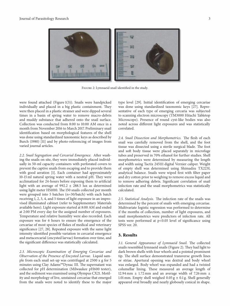
Figure 2: Lymnaeid snail identified in the study.
were found attached (Figure 1(3)). Snails were handpickedindividually and placed in a big plastic containment. Theywere then placed in a plastic strainer and were dipped severaltimes in a basin of spring water to remove macro-debrisand muddy substance that adhered onto the snail surface.Collection was conducted from 8:00 to 10:00 AM once in amonth fromNovember 2016 toMarch 2017. Preliminary snailidentification based on morphological features of the shellwas done using standardized taxonomic keys as described byBurch (1980) [11] and by photo-referencing of images fromvaried journal articles.
2.2. Snail Segregation and Cercarial Emergence. After wash-ing the snails on site, they were immediately placed individ-ually in 50-ml capacity containers with perforated covers toprevent the captive snails from escaping and to provide themwith good aeration [1]. Each container had approximately10-15ml natural spring water with a neutral pH. They wereacclimatized for 24-hours before exposing them to artificiallight with an average of 992.2 ± 288.5 lux as determinedusing light meter HS1010. The 150 snails collected per monthwere grouped into 5 batches (n=30/batch) with each batchreceiving 1, 2, 3, 4, and 5 times of light exposure in an impro-vised illuminated cabinet (refer to Supplementary Materialsavailable here). Light exposure started at 8:00 AM and endedat 2:00 PM every day for the assigned number of exposures.Temperature and relative humidity were also recorded. Eachexposure was for 6 hours to ensure the emergence of thecercariae of most species of flukes of medical and veterinarysignificance [27, 28]. Repeated exposure with the same lightintensity identified possible variation in cercarial emergenceandmetacercarial (encysted larvae) formation over time, andthe significant difference was statistically calculated.
2.3. Microscopic Examination of Emerging Cercariae andObservation of the Presence of Encysted Larvae. Liquid sam-ple from each snail set-up was centrifuged at 2500 x g for 5minutes using Clay Adams�Dynac III. The supernatant wascollected for pH determination (Milwaukee pH600 tester),and the sediment was examined using Olympus CX21. Motil-ity and morphology of live, unstained cercariae that emergedfrom the snails were noted to identify these to the major
type level [29]. Initial identification of emerging cercariaewas done using standardized taxonomic keys [27]. Repre-sentative of each type of emerging cercaria was subjectedto scanning electron microscopy (TM3000 Hitachi TabletopMicroscope). Presence of round cyst-like bodies was alsonoted across different light exposures and was statisticallycorrelated.
2.4. Snail Dissection and Morphometrics. The flesh of eachsnail was carefully removed from the shell, and the foottissue was dissected using a sterile surgical blade. The footand soft body tissue were placed separately in microfugetubes and preserved in 70% ethanol for further studies. Shellmorphometrics were determined by measuring the lengthand width using Tactix 245111 digital Vernier caliper. Weightof empty shell was determined using Shimadzu TX223Lanalytical balance. Snails were wiped first with filter paperand dry cotton prior to weighing to remove excess liquid andto remove adhering debris. Significant correlation of snailinfection rate and the snail morphometrics was statisticallycalculated.
2.5. Statistical Analysis. The infection rate of the snails wasdetermined by the percent of snails with emerging cercariae.Multivariate logistic regression was performed to determineif the months of collection, number of light exposures, andsnail morphometrics were predictors of infection rate. Alltests were performed at p<0.05 level of significance usingSPSS ver. 20.
3. Results
3.1. General Appearance of Lymnaeid Snail. The collectedsnails resembled lymnaeid snails (Figure 2).They had light todark brown shells with four whorls and a pointed prominenttip. The shell surface demonstrated transverse growth linesor striae. Apertural opening was dextral and body whorlwas enlarged. Body whorl was expanded and had a twistedcolumellar lining. These measured an average length of12.94mm ± 1.72mm and an average width of 7.26mm ±1.01mm. Empty shell weighed 0.05 g ± 0.1 g. Shell generallyappeared oval broadly and nearly globosely conical in shape.

4 Journal of Parasitology Research
Table 1: Types of cercariae and the frequency of recovery from lymnaeid snails (n=750).
Type of Cercariae Frequency %Single Infection
(i) Echinostome 198 26.4%(ii) Virgulate Xiphidiocercaria 5 0.67%(iii) longifurcate-pharyngeate distome cercariae (Strigea) 17 2.27%
Mixed infection(i) Echinostome and fork-tailed cercariae (Strigea) 2 0.27%(ii) Echinostome and Virgulate xiphidiocercaria 1 0.13%
Encysted forms only 44 5.9%
Figure 3: Microscopic images of different types of cercariae that emerged from lymnaeid snails (CX 21, 400x magnification). (1)-(2)Echinostome cercariae. A collar of spines in the oral sucker is noted. Upper image shows the complete larvae, while the lower image shows thede-tailed part of the body. (3)-(4) Virgulate xiphidiocercaria. Take note of defined stylet present in the anterior-most part of the oral sucker.(5)-(6) Longifurcate-pharyngeate distome cercariae (Strigea cercariae). Excretory pores are seen along the midportion of the bifurcated tail.
Despiteminor differences, the snails in the current studywereidentified as Lymnaea (Radix) quadrasi.
3.2. Emerging larval Forms. Cercariae are the terminalintramolluscan larval stages that emerge from an infectedsnail host. They may be readily infective to somemammalianhosts ormay encyst in a susceptible second intermediate host.Recovery from the samples is usually done through strongartificial light exposure that triggers their release, and theclassical method to identify them is through morphologicalcharacterization using standardized taxonomic keys [30].Our current set-up made use of an artificially illuminatedcabinet (refer to Supplementary Materials) with a light sourcethat emitted approximately 1000-lux intensity. This wasenough to allow release of cercariae from the snails. Resultsof the study showed that the actual rate of cercarial sheddingcan be established using a single 6-hour light exposure at this
intensity, as this did not differ among snails exposed for morethan once up to five times.
Three cercarial forms were observed to emerge fromthe snails (Figures 4 and 5). Table 1 shows the frequency ofrecovery of these different types from the lymnaeid snailsstudied.
3.2.1. Echinostome Cercariae. The echinostome type of cer-cariae was found in 26.4% of the 750 snails studied andwas the most common among the three types of cercariae(Figure 3: (1)-(2)). Freshly emerging larvae moved in atwitching manner using their powerful tails which eventuallydetached themselves from the body. The elongated oval-shaped body then moved in a slow crawling motion. Thewhole larvae measured an average size of 30𝜇m ± 0.6 𝜇m x11.4𝜇m ± 2.1𝜇m.The slender unforked tail was as long as thebody andmeasured on the average 31𝜇m ± 9.0𝜇mx 4.5 𝜇m ±

Journal of Parasitology Research 5
Figure 4: SEM images of three distinct cercariae that emerged from lymnaeid snails. (1) Echinostome cercaria showing a crown of spine(collar of spines). (2) Virgulate xiphidiocercaria. (3)-(4) Longifurcate-pharyngeate distome cercariae (Strigea cercariae). OS: oral sucker, VS:ventral sucker, FT: fork-tailed, CS: collar of spines.
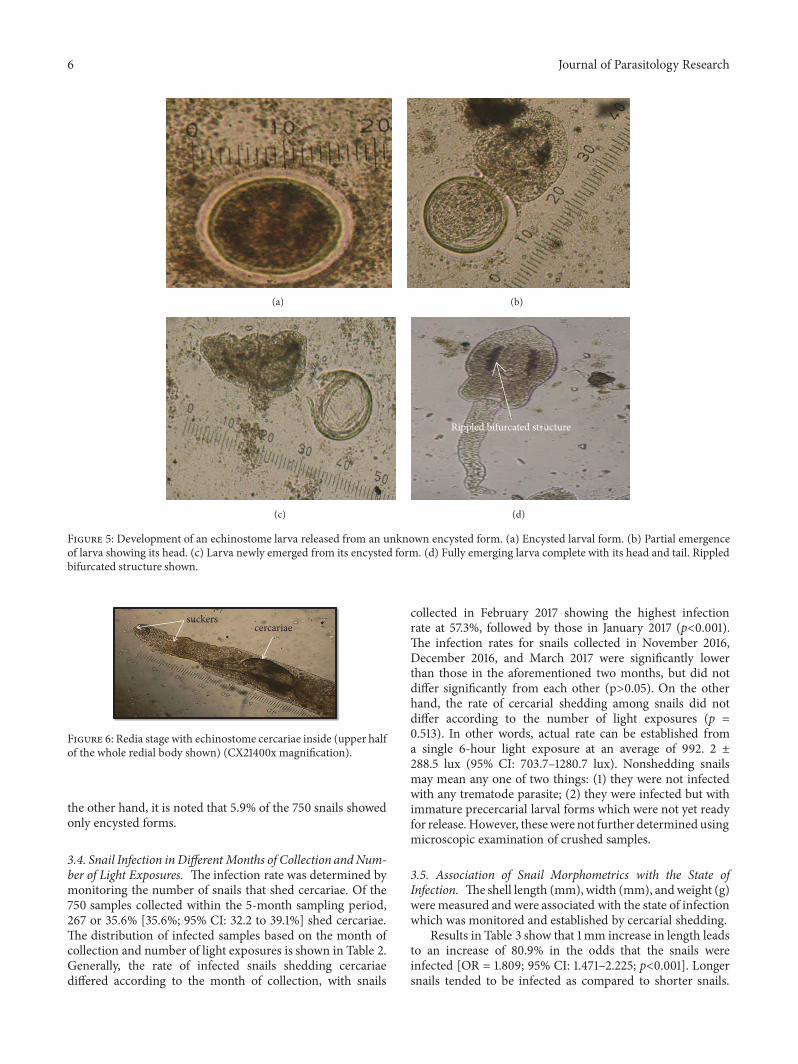
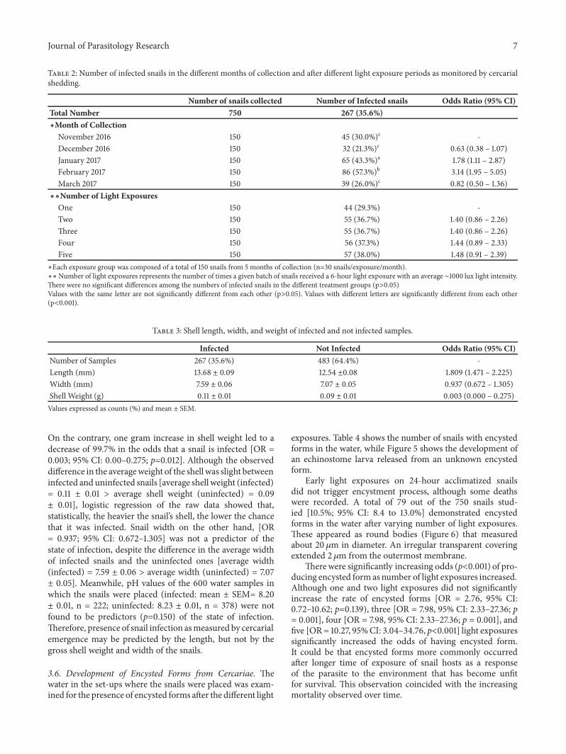
0.7𝜇m.Two prominent suckers were found to be present.Theoral sucker was in the anterior subterminal part of the bodyand was crowned with spinous processes as revealed fromthe scanning electron microscope (Figure 4). The numberof spines in the circular area was difficult to ascertain. Theventral sucker was slightly below the midportion of thebody. Dark rippled bifurcated structures (Figure 5(d)) wereobserved fromwithin the body, which were continuous fromthe oropharynx andmay represent esophageal tubing. Figures5(a) to 5(d) show echinostome larva that was found to beemerging from an unknown encysted form.This observationhas not been reported in published literature before. Figure 6is a mature redia with multiple live cercarial forms.
3.2.2. Virgulate Xiphidiocercaria. Virgulate xiphidiocercariaeis another type of cercaria that was recovered from thelymnaeid snails (Figure 3: (3)-(4)). However, this was onlyevident with the February 2017 and succeeding batchesof samples. The tail had no dorsoventral finfold, but thecercariae were observed tomove in a gentle sweeping motion.Bilobed or pyriform virgula organ was present in the oralsucker, and a distinct stylet was observed. Ventral sucker wassmaller than the oral sucker. Micrometry of representativesamples revealed that this kind of cercaria measured anaverage body size of 28𝜇m x 12𝜇m and an average tailsize of 25𝜇m x 4𝜇m. Parasites with this type of cercariabelong to the family Lecithodendriidae and are known toinfect intestines of bats, birds, and amphibians. Out of the
750 samples collected for the whole 5 months, 0.67% wereinfected with this kind of cercaria.
3.2.3. Longifurcate-Pharyngeate Distome Cercariae (StrigeaCercariae). The presence of fork-tailed cercariae (Figure 3:(5)-(6)) has not been reported yet among snails in thePhilippines other than Oncomelania hupensis quadrasi, aknown snail intermediate host for Schistosoma japonicum.The emerging cercaria was twice as long as the typical cercariaof Schistosoma japonicum. Its head measured an average of41 ± 1.4𝜇m x 9 ± 1.4𝜇m and the tail body measured anaverage of 34.7𝜇m ± 4𝜇m x 5𝜇m. Half of its tail measuredon the average 20.3𝜇m ± 0.6 𝜇m x 2 𝜇m. Two suckers wereobserved and these were subequal in size. It may be confusedwith a brevifurcate-distome apharyngeate cercaria, but thepresence of distinct excretory pores within the tail stemproved otherwise. Out of the 750 snails collected, 2.27% wereinfected with this type of larva. This type of cercaria belongsto the families Strigeidae andDiplostomidae which parasitizebirds and mammals.
3.3. Presence of Coinfection among Lymnaeid Snails. Exis-tence of coinfection is a very rare phenomenon amonglymnaeid snails. Snails coded 3FM35 and 1 M61 demonstratedthe presence of both echinostome and fork-tailed cercaria.This represented 0.27% of the total 750 snails collected. 1F94-coded snail showed an echinostome and xiphidiocercaria,and this represented 0.13%of the total 750 snails collected.On
6 Journal of Parasitology Research
(a) (b)
(c)
Rippled bifurcated structure
(d)
Figure 5: Development of an echinostome larva released from an unknown encysted form. (a) Encysted larval form. (b) Partial emergenceof larva showing its head. (c) Larva newly emerged from its encysted form. (d) Fully emerging larva complete with its head and tail. Rippledbifurcated structure shown.
suckerscercariae
Figure 6: Redia stage with echinostome cercariae inside (upper halfof the whole redial body shown) (CX21400x magnification).
the other hand, it is noted that 5.9% of the 750 snails showedonly encysted forms.
3.4. Snail Infection in Different Months of Collection andNum-ber of Light Exposures. The infection rate was determined bymonitoring the number of snails that shed cercariae. Of the750 samples collected within the 5-month sampling period,267 or 35.6% [35.6%; 95% CI: 32.2 to 39.1%] shed cercariae.The distribution of infected samples based on the month ofcollection and number of light exposures is shown in Table 2.Generally, the rate of infected snails shedding cercariaediffered according to the month of collection, with snails
collected in February 2017 showing the highest infectionrate at 57.3%, followed by those in January 2017 (p<0.001).The infection rates for snails collected in November 2016,December 2016, and March 2017 were significantly lowerthan those in the aforementioned two months, but did notdiffer significantly from each other (p>0.05). On the otherhand, the rate of cercarial shedding among snails did notdiffer according to the number of light exposures (p =0.513). In other words, actual rate can be established froma single 6-hour light exposure at an average of 992. 2 ±288.5 lux (95% CI: 703.7–1280.7 lux). Nonshedding snailsmay mean any one of two things: (1) they were not infectedwith any trematode parasite; (2) they were infected but withimmature precercarial larval forms which were not yet readyfor release. However, thesewere not further determined usingmicroscopic examination of crushed samples.
3.5. Association of Snail Morphometrics with the State ofInfection. Theshell length (mm),width (mm), andweight (g)weremeasured and were associated with the state of infectionwhich was monitored and established by cercarial shedding.
Results in Table 3 show that 1mm increase in length leadsto an increase of 80.9% in the odds that the snails wereinfected [OR = 1.809; 95% CI: 1.471–2.225; p<0.001]. Longersnails tended to be infected as compared to shorter snails.
Journal of Parasitology Research 7
Table 2: Number of infected snails in the different months of collection and after different light exposure periods as monitored by cercarialshedding.
Number of snails collected Number of Infected snails Odds Ratio (95% CI)Total Number 750 267 (35.6%)∗Month of Collection
November 2016 150 45 (30.0%)c -December 2016 150 32 (21.3%)c 0.63 (0.38 – 1.07)January 2017 150 65 (43.3%)a 1.78 (1.11 – 2.87)February 2017 150 86 (57.3%)b 3.14 (1.95 – 5.05)March 2017 150 39 (26.0%)c 0.82 (0.50 – 1.36)∗∗Number of Light Exposures
One 150 44 (29.3%) -Two 150 55 (36.7%) 1.40 (0.86 – 2.26)Three 150 55 (36.7%) 1.40 (0.86 – 2.26)Four 150 56 (37.3%) 1.44 (0.89 – 2.33)Five 150 57 (38.0%) 1.48 (0.91 – 2.39)∗Each exposure group was composed of a total of 150 snails from 5 months of collection (n=30 snails/exposure/month).∗∗Number of light exposures represents the number of times a given batch of snails received a 6-hour light exposure with an average ∼1000 lux light intensity.There were no significant differences among the numbers of infected snails in the different treatment groups (p>0.05)Values with the same letter are not significantly different from each other (p>0.05). Values with different letters are significantly different from each other(p<0.001).
Table 3: Shell length, width, and weight of infected and not infected samples.
Infected Not Infected Odds Ratio (95% CI)Number of Samples 267 (35.6%) 483 (64.4%) -Length (mm) 13.68 ± 0.09 12.54 ±0.08 1.809 (1.471 – 2.225)Width (mm) 7.59 ± 0.06 7.07 ± 0.05 0.937 (0.672 – 1.305)Shell Weight (g) 0.11 ± 0.01 0.09 ± 0.01 0.003 (0.000 – 0.275)Values expressed as counts (%) and mean ± SEM.
On the contrary, one gram increase in shell weight led to adecrease of 99.7% in the odds that a snail is infected [OR =0.003; 95% CI: 0.00–0.275; p=0.012]. Although the observeddifference in the averageweight of the shell was slight betweeninfected and uninfected snails [average shell weight (infected)= 0.11 ± 0.01 > average shell weight (uninfected) = 0.09± 0.01], logistic regression of the raw data showed that,statistically, the heavier the snail’s shell, the lower the chancethat it was infected. Snail width on the other hand, [OR= 0.937; 95% CI: 0.672–1.305] was not a predictor of thestate of infection, despite the difference in the average widthof infected snails and the uninfected ones [average width(infected) = 7.59 ± 0.06 > average width (uninfected) = 7.07± 0.05]. Meanwhile, pH values of the 600 water samples inwhich the snails were placed (infected: mean ± SEM= 8.20± 0.01, n = 222; uninfected: 8.23 ± 0.01, n = 378) were notfound to be predictors (p=0.150) of the state of infection.Therefore, presence of snail infection asmeasured by cercarialemergence may be predicted by the length, but not by thegross shell weight and width of the snails.
3.6. Development of Encysted Forms from Cercariae. Thewater in the set-ups where the snails were placed was exam-ined for the presence of encysted forms after the different light
exposures. Table 4 shows the number of snails with encystedforms in the water, while Figure 5 shows the development ofan echinostome larva released from an unknown encystedform.
Early light exposures on 24-hour acclimatized snailsdid not trigger encystment process, although some deathswere recorded. A total of 79 out of the 750 snails stud-ied [10.5%; 95% CI: 8.4 to 13.0%] demonstrated encystedforms in the water after varying number of light exposures.These appeared as round bodies (Figure 6) that measuredabout 20 𝜇m in diameter. An irregular transparent coveringextended 2𝜇m from the outermost membrane.
Therewere significantly increasing odds (p<0.001) of pro-ducing encysted formas number of light exposures increased.Although one and two light exposures did not significantlyincrease the rate of encysted forms [OR = 2.76, 95% CI:0.72–10.62; p=0.139), three [OR = 7.98, 95% CI: 2.33–27.36; p= 0.001], four [OR = 7.98, 95% CI: 2.33–27.36; p = 0.001], andfive [OR= 10.27, 95%CI: 3.04–34.76, p<0.001] light exposuressignificantly increased the odds of having encysted form.It could be that encysted forms more commonly occurredafter longer time of exposure of snail hosts as a responseof the parasite to the environment that has become unfitfor survival. This observation coincided with the increasingmortality observed over time.

8 Journal of Parasitology Research
Table 4: Counts of snails with encysted forms based on number of light exposures.
Number of Light ExposuresNumber and %
Snails with Encysted Form(n=150)
Odds Ratio (95% CI)
One 3 (2.0%)a -Two 8 (5.3%)a 2.76 (0.72 – 10.62)Three 21 (14.0%)b 7.98 (2.33 – 27.36)Four 21 (14.0%)b 7.98 (2.33 – 27.36)Five 26 (17.3%)b 10.27 (3.04 – 34.76)Values expressed as counts (%).∗Each exposure group was composed of a total of 150 snails from 5 months of collection (n=30 snails/exposure/month).∗Number of light exposures represents the number of times a given batch of snails received a 6-hour light exposure with an average ∼1000 lux light intensity.Values with the same letter are not significantly different from each other (p>0.001). Values with different letters are significantly different from each other(p<0.001).
4. Discussion
4.1. Identification of Lymnaeid Snails. The present studyexplored lymnaeid snails that thrived in the rice field ofBarangay Cawongan, Padre Garcia, Batangas. They have beenprimarily implicated in the transmission of liver flukes inthe Philippines [31], but have also been associated withlocal cases of human echinostomiasis [32]. They naturallysupport the life cycles of Fasciola spp. that cause diseases inmajority of the ruminants like cows and carabaos [33]. Theprevalence of human fascioliasis in the Philippines may below, but potential transmission of other parasitic infectionswith medical importance like echinostomiasis [32] and avianschistosomiasis [34] cannot be discounted, if cercariae ofthese parasites are likewise found in the same lymnaeidsnails. These non-Fasciola infections are caused by parasiteswhose other known intermediate hosts are nonlymnaeidsnails, which in the Philippines include Pila luzonica, secondintermediate host for Echinostoma ilocanum, and Oncomela-nia hupensis quadrasi, first intermediate host of Schistosomajaponicum.
There are three recognized genera under the familyLymnaeidae: genus Austropeplea (Cotton 1942), genus Bul-lastra (Bergh 1901), and genus Radix (Montfort 1810) [11].Lymnaea was not included among these. In many reports,all lymnaeid snails in the Western Pacific belong to a singleHolarctic genus, Lymnaea. Confusion in snail identificationand taxonomy has been known for a long time and iscommon among lymnaeids [35].
The snails in the study were tentatively identified as Lym-naea (Radix) quadrasi based on the morphological featuresof the shell using standardized taxonomic keys as describedby Burch (1980) [11] and photo-referenced with imagesfrom varied journal articles. They bore close resemblanceto those of Lymnaea (Radix) rubiginosa of Indonesia andThailand based on their morphological characteristics [36].Snail identification through gross description of the shellappearance and internal structure detailing is difficult andmay be extremely confusing [37].These taxonomic problemswere already evident in various studies which dealt withseveral stagnicoline snails [7, 38, 39]. Confusions had beenmade on adult Galba truncatula, which was mistaken as
preadult Lymnaea palustris or Lymnaea fuscus. The ever-changing environmental conditions have affected the generalpopulation of various living snails. Snails have their ownadaptive mechanisms that allow morphological variationsto happen among them. Consequently, this has made iden-tification of snails more difficult [40]. In addition, shellshapes may vary as a result of natural trematode infection,and dilemma in phenotypic characterization consequentlyhappens [41]. Despite this, there are still few attempts tostudy the phylogeny of lymnaeids in Asia. The same dilemmawas encountered in the identification of the 750 collectedsnails. Based on shell features in standardized keys and photovalidation, they were consistent with Lymnaea spp. However,other published references suggest that they belong to theGenus Radix. The snails demonstrated slight variations inshell coloration but were distinct in terms of apertural open-ing, number of whorls, and other significant morphometrics.Despite these inconsistencies in phenotypic identities, what isdefinite is that these snails belong to family Lymnaeidae, andmolecular confirmation is currently being undertaken usingCOX-1 gene primers to resolve this inherent problem [42].
4.2. Types of Cercariae from the Lymnaeid Snails. Three dif-ferent types of cercariae were observed to have emerged fromthe snails in the study.These were single infection of echinos-tome cercariae, which were predominant (26.4%), followedby longifurcate-pharyngeate distome cercariae (2.27%) andvirgulate xiphidiocercariae (0.67%), which was only evidentin snails from the latter part of the monthly collection.It is noted that no lymnaeid snail was found to harborthe gymnocephalus cercaria that is typical of Fasciola spp.,the parasite for which Lymnaea spp. are known to be thecommon intermediate hosts. The absence of the Fasciolacercariae from the lymnaeid snails is strongly suggestive ofthe absence of inherent fascioliasis in the area. The numberand types of definitive hosts that are present in the areacan influence the nature of infections. The possibility offascioliasis in the area is low because the number of definitivehosts not known to harbor the adult Fasciola spp. parasiteis high. That is, the presence of other significant hosts likeducks, chickens, wild bird-dwellers, and amphibians whichmay harbor different parasites is evident from the area and

Journal of Parasitology Research 9
may provide a rational explanation for the existence ofthese other types of cercariae. This supports the dilutioneffect hypothesis that underscores the relevance of negativecorrelation between disease incidence and host diversity[43]. Arguably, this hypothesis may not be always consistentin most human infections with great public health impact[44]. The finding of larvae other than Fasciola has likewisebeen reported by Faltynkova et al. [45], where they reported26.3% prevalence among Lymnaea stagnalis of various kindsof larvae identified to be Opisthioglyphe ranae, Plagiorchiselegans, and Echinoparyphium aconiatum.On the other hand,furcocercous type of cercariae identified as Diplostomumpseudospathaceum was reported at a rate of 18.8% in the samespecies of Lymnaea [46].
The abundant presence of these non-Fasciola cercariae inthe local lymnaeid snails may control Fasciola spp. transmis-sion as governed by larval competition and colonization, butmay be difficult to establish in this current study. On the otherhand, gymnocephalus cercaria of Fasciola spp. was found alsoamong nonlymnaeid snails in other studies. These includedBithynia siamensis of the family Bithyniidae [47] and array ofother snail genera like Biomphalaria, Bulinus, Ceratophallus,Gabbiella,Gyraulus, andMelanoides [27]. None as of this datehas reported presence of this type of cercariae among snailsother than Lymnaea spp. in the Philippines.
The results of the current study are not consistent withthose of other studies. Devkota et al. [48] showed onlygymnocephalus cercaria, which is typical of Fasciola spp.in 2.9% of the Lymnaea luteola snails studied, while Lukaand Mbaya [49] found distome cercaria of Fasciola spp. inLymnaea natalensis. Imani-Baran et al. [28], on the otherhand, showed xiphidiocercariae frequenting their sampledLymnaea gedrosiana snails at 81.98%, followed by furcocer-cariae (32.26%), echinostome cercariae (5.19%), and amonos-tome cercaria which belongs to the family Notocotylidae(1.24%). Ramitha and Vasandakumar [1] likewise recoveredfurcocercous, echinostome cercariae, and xiphidiocercariaeinhabiting L. luteola. Presence of these varieties of cercariaeclearly is a manifestation of a wide array of snail-parasiteinteraction and symbiosis. This abundance of trematodespecies that exploit snails as their mandatory first inter-mediate host has also been documented and reviewed inEurope [50]. In fact, richness of malacofauna in the areaalso demonstrated parasite diversity as shown commonly byvarious larvae infecting a single snail host one at a time orrarely by the occurrence of multiple infections.
There are ten different possible cercariae that may berecovered from lymnaeid snails [27], but only three typeswere found inhabiting the local lymnaeids in Batangas. Mostwere nonzoonotic parasites. However, the presence of echi-nostome type and forked-tailed cercariae may be exploredfurther as these two are known to be medically important.Echinostoma spp. are intestinal parasites of humans [51], andfork-tailed cercariae of avian origin can cause dermatitis.Echinostomes also are known to heavily infect domesticducks, thus affecting greatly poultry business [52]. In thePhilippines, twomedically important echinostomes, Echinos-toma ilocanum and Echinostoma malayanum, were recoveredfrom infected humans that required freshwater gastropods
belonging to the family Lymnaeidae as intermediate hosts[32].
Virgulate xiphidiocercaria is common among snailsbelonging to the genera Lymnaea and Melanoides [25], butwas also reported in the genus Thiara [53]. They are knownto be produced by intestinal parasites belonging to the familyLecithodendriidae which may infect animals like bats, birds,and amphibians. Poultry animals like ducks and chickenswere observed in the perimeter of the sampling site and mayprovide a rationale for the existence of this type of cercariae.No studies have been made on their pathology in theseanimals. Therefore, its veterinary impact on poultry animalsis yet to be explored.
Fork-tailed cercariae have never been reported in thePhilippines to occur in snails other than Oncomelaniahupensis quadrasi, the local snail hosts for Schistosomajaponicum. Longifurcate- pharyngeate distome cercariae(strigeid) were the second most common cercariae in thesamples collected and were twice as long as the cercariaeof Schistosoma japonicum. The size of the snail host wasalso twice longer than the Oncomelania snail. Severalsnail hosts can harbor any of the following differenttypes of fork-tailed cercariae: brevifurcate-apharyngeatedistome cercariae, brevifurcate-apharyngeate monostomecercariae, longifurcate-pharyngeate distome cercariae, andlongifurcate-pharyngeate monostome cercariae. Thesesnails include the following genera: Biomphalaria, Bulinus,Ceratophallus, Cleopatra, Gabbiella, Gyraulus, Melanoides,Melanopsis, Segmentorbis [27], Indoplanorbis, and Bithynia[47]. Among these, the brevifurcate-apharyngeate distomecercaria has been given medical attention because of itspublic health impact. None to date has explored the presenceof non-Schistosoma sp. fork-tailed cercariae in these widearray of snail intermediate hosts.The possibility of recoveringavian schistosomes causing dermatitis-like conditions fromthe sampling site is high because birds and other poultryanimals abound in the area. This has yet to be confirmed.
Encysted forms were observed as a result of light expo-sures over time. This natural mechanism of encystment isa protective response on the parasite which was exposedto unconducive environment [41]. Cercarial release in theabsence of suitable second intermediate host from a givenenvironment leads to death of the larval stage. In this context,encysted forms are mostly observed among snails that havebeen exposed to light longer during cercarial emergence.However, identity of the encysted forms was difficult toestablish but they may belong to any one of the threerecovered types of cercariae: echinostome, xiphidiocercaria,or the strigeid.
4.3. Existence of Double Infection in a Single LymnaeidSnail. Although single trematode parasitism is commonlydemonstrated by majority of snail infection cases, very fewhave noted coinfection in other snail species like that of Indo-planorbis exustus, where xiphidiocercaria and longifurcate-pharyngeate distome cercaria existed in a single snail host[48]. Once a parasite inhabits a snail host, it initiates chemicalchanges that alter the host’s attractiveness to other invadingparasites [54]. From there, it establishes a territory of its own

10 Journal of Parasitology Research
and creates its progenieswithin.Thus, any deviation from thisis a very rare phenomenon and is likely due to simultaneousinfection by two trematode species [54]. However, other fac-tors like chronological and spatial variations in the numberof eggs and miracidia limit the possibility of simultaneousmultiple trematode infections [55, 56].
Here in the Philippines, the finding of coinfection in ourlymnaeid snails with echinostome cercariae and fork-tailedcercariae (0.27%) and echinostome with xiphidiocercaria(0.13%) is first in the recorded history which can be eithera coincidence or a real coexisting phenomenon. Densityof each type of coexisting cercariae was not determined,so the degree of competition between them could not beduly determined. Moreover, the very low occurrence maybe due to increased snail mortalities which are associatedwith increased pathogenicity of double infections as com-pared to single infection [57]. Consequently, the samplesnails demonstrating simultaneous multiple infections maybe underrepresented. According to Sewell [58], only certaincombinations of trematode species could coexist togetheras double infections, and these double infections gener-ally involved the following cercarial groups: furcocercariae,xiphidiocercariae, and monostome cercariae. These findingsare consistent with our current results except for the existenceof echinostome cercariae in the pair. It may seem incidental,but if a given snail is home for a wide spectrum of trematodespecies, there is likelihood that coinfection happens such asin the case of B. siamensis [47].
4.4. Snail Infection Rates in Different Months of Collection.Monthly variations in snail infection rates were observedover the five-month collection period. Lowest infection ratewas seen in December 2016 (21.3%), and the highest was inFebruary 2017 (57.3%) with echinostome type of cercariaefrequenting the infected samples. No trend was observedover the 5-month period. The differences may be attributedto crop rotation and field practice of the nearby residents.Periodic sprays with chemicals against mollusc pests affectsnail density and therefore may affect parasite transmissionfrom one host to another. Monthly variations in infectionrates may have a trending pattern if practices of residentsare controlled and variations are all attributable to weatherconditions. As such, more snails will be expected to beinfected during rainy season as compared to those collectedduring summer [59–61].
4.5. Snail Morphometrics and Infection Rates. Changes inmorphology are associated with snail’s natural growth anddevelopment. It can also be an antipredatory adaptive mech-anism among parasite-induced snail hosts [62]. Burch [63]showed that shell length provided a rough estimate of theage of the snail, and since this was positively correlated withboth shell width (r = 0.797) and weight (r = 0.743), it wasdeduced that any of these three morphological features couldbe employed to determine the age of every snail. In fact, ahighly significant correlation of these morphometric traits(shell length and shell width: r = 0.757; shell length andtotal live weight: r = 0.770; p<0.01) was established among L.acuminate [64].
Our current findings showed that 35.6%of sampled snailsthat were naturally infected were longer [OR = 1.809; 95%CI: 1.471–2.225; p<0.001] as compared to the uninfectedgroup. Length coincides with the age of snails [65]. Althoughresistance to infection is observed among older snails [25],they may have been exposed to a number of miracidiaas they mature and may have been infected for a longerperiod. Snail infection acquired at younger stages persistsover time as snails develop [64]. Thus, increasing prevalenceor intensity of infection is associated with increasing sizevis-a-vis age of the snails [66–68]. Trematode-induced mor-phological changes can provide various consequences in theinfected snail hosts: enhanced growth rate [69], stunted ordecreased growth rate [70], and still or no effect in growth[71].
On the other hand, results of the study showed thatgross weight of empty shells differed slightly between theinfected and uninfected snails [average shell weight (infected)= 0.11 ± 0.01 > average shell weight (uninfected) = 0.09 ±0.01]. However, logistic regression of the raw data showedthat, statistically, the heavier the snail’s shell, the lower thechance that it was infected. The state of calcification process,whether hypercalcification or hypocalcification, may providea plausible and objective association to the state of infection,but this cannot be used to explain the results because thestudy did not measure calcium concentration of the emptyshell.The degree of calcium utilization and subsequent depo-sition on the shell is a parasite type-dependent occurrence. Inseveral reports, either shell hypercalcification [72, 73] or shellhypocalcification [74, 75] resulted after trematode-inducedinfection in selected snail hosts. In other words, the typeof infecting parasite is an influential factor in calcificationphenomenon in which this study did not determine any-more.
5. Conclusion
Lymnaeid snails that inhabit rice fields and man-made waterducts are potential carriers of zoonotic parasites that haveboth medical and veterinary public health significance. Inthis study, the total cercarial shedding rate of the snails asa measure of the infected snails was found to be 35.6%.Although single trematode infection was the common obser-vation, coinfection with two different larval forms was foundin less than 1% of the lymnaeid snails. The presence of anyof the three different cercariae, i.e., echinostome, fork-tailedstrigeid, and xiphidiocercariae, in the lymnaeid snails reflectsthe diversity of parasites and possibly of reservoir hosts,whichmay significantly pose potential health hazards to manand animals. Therefore, wide-scale surveillance of freshwatersnails is necessary, including a catalogue on the variety ofcercariae and their associated definitive hosts, to justify thenecessity of control measures against snail-borne parasiticinfections.
Data Availability
The data used to support the findings of this study areincluded within the article.

Journal of Parasitology Research 11
Conflicts of Interest
The authors have declared that no conflicts of interest exist.
Authors’ Contributions
Gregorio L. Martin I conceptualized the topic. He didthe field work and sample collection and performed thelaboratory experimentation at the University of Santo Tomas,Philippines. He prepared the initial and final draft of themanuscript. Esperanza C. Cabrera was involved in brain-storming during topic conceptualization and actively par-ticipated in consultative discussions. She also reviewed andedited the final manuscript.
Acknowledgments
The study was partially funded by the Philippine Depart-ment of Science and Technology-Science Education Institute(DOST-SEI). The authors acknowledge Mrs. Rona Escuetaand her family of Barangay Cawongan, Padre Garcia, Batan-gas, for providing the site for snail collection; AssistantProfessor Xandro Nieto for the statistical analysis; DeanAleth Therese Dacanay and the Faculty of Pharmacy forthe support in the conduct of the study; Dr. Henry Madsenof the University of Copenhagen and Dr. Bui Thi Dungfor sharing their expertise in snail research; Dr. Niels VanSteenkiste, Biodiversity Institute of Ontario, University ofGuelph, for sharing his articles on molecular assays; Prof.Maribel G. Nonato, Ph.D., Vice-Rector for Research andInnovation at UST, for providing assistance in the approvalof the dissertation grant under DOST-SEI; Dr. Ian KendrichC. Fontanilla, Associate Professor 5 of the University of thePhilippines through his research assistant, Mr. Raffy Fornil-los, for extending their expertise in molecular techniques.
Supplementary Materials
Supplemental Figure 1: illuminated cabinet devised andused for cercarial emergence. Distance of light source tothe top surface of the specimen container is 35 cm. Set-upincludes apparatus for temperature and relative humiditydetermination and a lux meter for measuring light intensity.(Supplementary Materials)
References
[1] U. C. Ramitha and M. V. Vasandakumar, “Survey of freshwatersnails in malabar, kerala and an account on the prevalence ofinfection by digenean (platyhelminth) parasites,” JCBPS, vol. 5,pp. 4065–4070, 2015.
[2] M. Cordellier, A. Pfenninger, B. Streit, and M. Pfenninger,“Assessing the effects of climate change on the distribution ofpulmonate freshwater snail biodiversity,” Marine Biology, vol.159, no. 11, pp. 2519–2531, 2012.
[3] B. C. Dazo, “Vector control of snail-mediated human trematodeinfections,”Arzneimittel-Forschung/Drug Research, vol. 34, no. 9B, pp. 1234–1236, 1984.
[4] S. D. Greene, “Extending integrated pest management to thegolden apple snail: Examining a community centre approach innortheast Thailand,” International Journal of Pest Management,vol. 54, no. 2, pp. 95–102, 2008.
[5] M. D. Bargues, P. Artigas, M. Khoubbane et al., “Lymnaeaschirazensis, an overlooked snail distorting fascioliasis data:Genotype, phenotype, ecology, worldwide spread, susceptibil-ity, applicability,” PLoS ONE, vol. 6, no. 9, 2011.
[6] N. A. I. Mohammed, H. Madsen, and A. A. A. R. M. Ahmed,“Types of trematodes infecting freshwater snails found inirrigation canals in the East Nile locality, Khartoum, Sudan,”Infectious Diseases of Poverty, vol. 5, no. 1, article no. 16, 2016.
[7] M. D. Bargues, M. Vigo, P. Horak et al., “European lymnaeidae(Mollusca: Gastropoda), intermediate hosts of trematodiases,based on nuclear ribosomal DNA ITS-2 sequences,” Infection,Genetics and Evolution, vol. 1, no. 2, pp. 85–107, 2001.
[8] Z. Islam, M. Alam, S. Akter, B. Roy, and M. Mondal, “Dis-tribution patterns of vector snails and trematode cercaria intheir vectors in some selected areas of mymensingh,” Journal ofEnvironmental Science and Natural Resources, vol. 5, no. 2, 2013.
[9] S. Ukong, D. Krallas, T. Dangprasert, and P. Channgarm, “Stud-ies on the morphology of cercariae obtained from freshwatersnails at Erawan Waterfall, Erawan National Park, Thailand,”Southeast Asian Journal of Tropical Medicine and Public Health,vol. 38, no. 2, pp. 302–312, 2007.
[10] P. Bouchet, J.-P. Rocroi, B. Hausdorf et al., “Revised classifica-tion, nomenclator and typification of gastropod and monopla-cophoran families,”Malacologia, vol. 61, no. 1-2, pp. 1–526, 2017.
[11] J. B. Burch, “A guide to the freshwater snails of the philippines,”Malacological Review, vol. 13, pp. 121–143, 1980.
[12] R. T. Abbott, “Snail invaders,” Journal of Natural History, vol. 59,p. 85, 1950.
[13] E. E. Strong, O. Gargominy, W. F. Ponder, and P. Bouchet,“Global diversity of gastropods (Gastropoda; Mollusca) infreshwater,”Hydrobiologia, vol. 595, no. 1, pp. 149–166, 2008.
[14] I. F. Pagulayan, R. C. Pagulayan, S. S. Pimentel, and C. C. Buer-ano, “Freshwater snails in Central Sierra Madre , Philippines,”The Technical Journal of Philippine Ecosystems and NaturalResources, 1998.
[15] P. N. Young and I. K. C. Fontanilla, “Biodistribution of theinformal group basommatophora in the Philippines,” ScienceDilliman, vol. 26, pp. 53–76, 2014.
[16] G. L. Galan, M. M. Ediza, M. S. Servasques, and H. C. Porquis,“Diversity of gastropods in the selected rivers and lakes inbukidnon,” International Journal of Environmental Science andDevelopment, vol. 6, no. 8, pp. 615–619, 2015.
[17] A.-G. A. Adorable-Asis, G. A. Cauyan, R. C. Pagulayan, F. S.Magbanua, andR. D. S. Papa, “Themacro-gastropod communi-ties of aquaculture-intensive lakes in the Philippines,”MolluscanResearch, vol. 36, no. 4, pp. 223–230, 2016.
[18] A. J. Cukingnan and R. C. Pagulayan, “Anatomy of the digestivesystem of radixsp sp. (Basommatophora: Lymnaeidae) fromLake Taal, Batangas,” Science Dilliman, vol. 7-8, pp. 48–52, 1996.
[19] E. A. Remigio and D. Blair, “Molecular systematics of the fresh-water snail family lymnaeidae (Pulmonata: Basommatophora)utilising mitochondrial ribosomal DNA sequences,” Journal ofMolluscan Studies, vol. 63, no. 2, pp. 173–185, 1997.
[20] L. Leonardo, P. Rivera, O. Saniel et al., “New endemic foci ofschistosomiasis infections in the Philippines,” Acta Tropica, vol.141, pp. 354–360, 2015.

12 Journal of Parasitology Research
[21] W. H. Ewers, “An analysis of the molluscan hosts of thetrematodes of birds and mammals and some speculations onhost-specificity,” Parasitology, vol. 54, no. 3, pp. 571–578, 1964.
[22] R. Poulin and K. N. Mouritsen, “Large-scale determinants oftrematode infections in intertidal gastropods,” Marine EcologyProgress Series, vol. 254, pp. 187–198, 2003.
[23] N. J. Morley, M. E. Adam, and J. W. Lewis, “The role of Bithyniatentaculata in the transmission of larval digeneans from a gravelpit in the Lower Thames Valley,” Journal of Helminthology, vol.78, no. 2, pp. 129–135, 2004.
[24] E. Zbikowska, “Infection of snails with bird schistosomesand the threat of swimmer’s itch in selected Polish lakes,”Parasitology Research, vol. 92, no. 1, pp. 30–35, 2004.
[25] A. E. Lockyer, C. S. Jones, L. R. Noble, and D. Rollinson,“Trematodes and snails: an intimate association,” CanadianJournal of Zoology, vol. 82, no. 2, pp. 251–269, 2004.
[26] P. Wessa, “Minimum Sample Size (Testing Proportion) (v1.0.4)in Free Statistics Software (v1.1.23-r7), Office for ResearchDevelopment and Education,” http://www.wessa.net/rwasp.sample.wasp/.
[27] F. Frandsen and N. O. Christensen, “An introductory guide tothe identification of cercariae from African freshwater snailswith special reference to cercariae of trematode species ofmedical and veterinary importance,” Acta Tropica, vol. 41, no.2, pp. 181–202, 1984.
[28] A. Imani-Baran, M. Yakhchali, R. Malekzadeh-Viayeh, and A.Farahnak, “Seasonal and geographic distribution of cercarialinfection in Lymnaea gedrosiana (Pulmunata: Lymnaeidae) innorth west Iran,” Iranian Journal of Parasitology, vol. 8, no. 3, pp.423–429, 2013.
[29] A. A. M. Ahmed, N. A. Ibrahim, and M. A. Idris, “Laboratorystudies on the prevalence and cercarial rhythms of trematodesfrom Bulinus truncatus and Biomphalaria pfeifferi snails fromKhartoum State, Sudan,” Sultan Qaboos University MedicalSciences Journal, vol. 6, no. 2, pp. 65–69, 2006.
[30] S. Anucherngchai, T. Tejangkura, and T. Chontananarth, “Epi-demiological situation and molecular identification of cercarialstage in freshwater snails in Chao-Phraya Basin, Central Thai-land,”Asian Pacific Journal of Tropical Biomedicine, vol. 6, no. 6,pp. 539–545, 2016.
[31] E. C. Molina, G. A. Navarra, and P. O. Aloot, “Fasciolosis incattle and carabaos in selected barangays of Pikit and Kabacan,Cotabato, Philippines,” USMR& D, vol. 18, pp. 129–132, 2010.
[32] V. Y. Belizario Jr., G. G. Geronilla, M. B. M. Anastacio et al.,“Echinostomamalayanum infection, the Philippines,” EmergingInfectious Diseases, vol. 13, no. 7, pp. 1130-1131, 2007.
[33] D. B. Copeman and R. S. Copland, “Importance and potentialimpact of liver fluke in cattle and buffalo,” in Overcoming LiverFluke as a Constraint to Ruminant Production in South-EastAsia, G. D, R. S. Gray, D. B. Copland, and Copeman., Eds., pp.1–34, AustralianCentre for InternationalAgricultural Research,Canberra, Australia, 2008.
[34] P. Horak, L. Mikes, L. Lichtenbergova, V. Skala, M. Soldanova,and S. V. Brant, “Avian schistosomes and outbreaks of cercarialdermatitis,”ClinicalMicrobiology Reviews, vol. 28, no. 1, pp. 165–190, 2015.
[35] C. A. Wright, Flukes and Snails, vol. 168, George Allen andUnwin, London, 1971.
[36] R. B. Monzon, V. Kitikoon, N. Thammapalerd, P. Temcharoen,S. Sornmani, and V. Viyanant, “Comparative shell morphologyof Lymnaea (Bullastra) cumingiana (Pulmonata: Lymnaeidae)
and related taxa in the Indo-Pacific region.,”TheSoutheast AsianJournal of Tropical Medicine and Public Health, vol. 24, no. 3, pp.554–562, 1993.
[37] A. C. Correa, J. S. Escobar, P. Durand et al., “Bridging gapsin the molecular phylogeny of the Lymnaeidae (Gastropoda:Pulmonata), vectors of Fascioliasis,” BMC Evolutionary Biology,vol. 10, no. 1, 2010.
[38] M. D. Bargues, P. Horak, R. A. Patzner et al., “Insights into therelationships of palearctic and nearctic lymnaeids (mollusca:gastropoda) by rDNA ITS-2 sequencing and phylogeny ofstagnicoline intermediate host species of Fasciola hepatica,”Parasite, vol. 10, no. 3, pp. 243–255, 2003.
[39] A. Novobilsky, M. Kasny, L. Beran, D. Rondelaud, and J.Hoglund, “Lymnaea palustris and Lymnaea fuscus are potentialbut uncommon intermediate hosts of Fasciola hepatica inSweden,” Parasites & Vectors, vol. 6, no. 1, article no. 251, 2013.
[40] B. T. Dung, P. N. Doanh, D. T. The, H. T. Loan, B. Losson,and Y. Caron, “Morphological and molecular characterizationof lymnaeid snails and their potential role in transmission ofFasciola spp. in Vietnam,” The Korean Journal of Parasitology,vol. 51, no. 6, pp. 657–662, 2013.
[41] E. Zbikowska, “One snail - three digenea species, differentstrategies in host-parasite interaction,” Animal Biology, vol. 61,no. 1, pp. 1–19, 2011.
[42] H.-Y. Kim, I.-W. Choi, Y.-R. Kim et al., “Fasciola hepaticain snails collected from water-dropwort fields using pcr,” TheKorean Journal of Parasitology, vol. 52, no. 6, pp. 645–652, 2014.
[43] P. T. J. Johnson, D. L. Preston, J. T. Hoverman et al., “Speciesdiversity reduces parasite infection through crossgenerationaleffects on host abundance,” Ecology, vol. 93, no. 1, pp. 56–64,2012.
[44] C. L. Wood, K. D. Lafferty, G. DeLeo, H. S. Young, P. J. Hudson,and A. M. Kuris, “Does biodiversity protect humans againstinfectious disease?” Ecology, vol. 95, no. 4, pp. 817–832, 2014.
[45] A. Faltynkova, V. Nasincova, and L. Kablaskova, “Larval trema-todes (Digenea) of the great pond snail, lymnaea stagnalis(L.), (Gastropoda, pulmonata) in Central Europe: A survey ofspecies and key to their identification,” Parasite, vol. 14, no. 1,pp. 39–51, 2007.
[46] A. Faltynkova, K. Niewiadomska, M. J. Santos, and E. T. Val-tonen, “Furcocercous cercariae (Trematoda) from freshwatersnails in Central Finland,” Acta Parasitologica, vol. 52, no. 4, pp.310–317, 2007.
[47] T. Chontananarth and C. Wongsawad, “Epidemiology of cer-carial stage of trematodes in freshwater snails from ChiangMai province, Thailand,” Asian Pacific Journal of TropicalBiomedicine, vol. 3, no. 3, pp. 237–243, 2013.
[48] R. Devkota, P. B. Budha, and R. Gupta, “Trematode cercariaeinfections in freshwater snails of Chitwan district, centralNepal,”Himalayan Journal of Sciences, vol. 7, no. 9, 2011.
[49] J. Luka and A. W. Mbaya, “Cercarial shedding of trematodesand their associated snail intermediate hosts in Borno State,Nigeria,” Asian Pacific Journal of Tropical Disease, vol. 5, no. 4,pp. 293–298, 2015.
[50] E. Zbikowska andA.Nowak, “One hundred years of research onthe natural infection of freshwater snails by trematode larvae inEurope,” Parasitology Research, vol. 105, no. 2, pp. 301–311, 2009.
[51] W. Saijuntha, C. Tantrawatpan, P. Sithithaworn, R. H. Andrews,and T. N. Petney, “Spatial and temporal genetic variation ofEchinostoma revolutum (Trematoda: Echinostomatidae) fromThailand and the Lao PDR,”Acta Tropica, vol. 118, no. 2, pp. 105–109, 2011.

Journal of Parasitology Research 13
[52] W. Saijuntha, K. Duenngai, and C. Tantrawatpan, “Zoonoticechinostome infections in free-grazing ducks in Thailand,”TheKorean Journal of Parasitology, vol. 51, no. 6, pp. 663–667, 2013.
[53] R. Brinesh and K. P. Janardanan, “Three new species ofXiphidiocercariae from the thiarid snail Thiara tuberculata inPalakkad, Kerala, India,” Journal of Parasitic Diseases, vol. 35,no. 1, pp. 42–49, 2011.
[54] A. M. Kuris and K. D. Lafferty, “Community structure: Larvaltrematodes in snail hosts,” Annual Review of Ecology, Evolution,and Systematics, vol. 25, pp. 189–217, 1994.
[55] J. A. Williams and G. W. Esch, “Infra- and component com-munity dynamics in the pulmonate snail Helisoma anceps,with special emphasis on the hemiurid trematode Halipegusoccidualis,” Journal of Parasitology, vol. 77, no. 2, pp. 246–253,1991.
[56] W. P. Sousa, “Interspecific antagonism and species coexistencein a diverse guild of larval trematode parasites,” EcologicalMonographs, vol. 63, no. 2, pp. 103–128, 1993.
[57] W. P. Sousa, “Interspecific interactions among larval trematodeparasites of freshwater and marine snails,” American Zoologist,vol. 32, no. 4, pp. 583–592, 1992.
[58] R. B. Sewell, “Cercariae Indica,” The Indian Journal of MedicalResearch, vol. X, pp. 1–370, 1922.
[59] C. L. Yadav, S. Vatsya, R. Garg, R. R. Kumar, P. S. Banerjee, andG. Rajesh, “Seasonal prevalence of Fasciola gigantica infectionin sheep and goats inwesternUttar Pradesh,” Journal of ParasiticDiseases, pp. 31–141, 2007.
[60] A. Singh, S. Srivastava, C. Shekhar, and J. Singh, “Prevalenceof trematodes in bovines and snails,” Indian Veterinary Journal,vol. 86, no. 2, pp. 206-207, 2009.
[61] M. N. Tigga, R. K. Bauri, A. R. Deb, and S. S. Kullu, “Prevalenceof snail’s intermediate host infected with different trematodescercariae in and around Ranchi,”Veterinary World, vol. 7, no. 8,pp. 630–634, 2014.
[62] E. G. Vasallo, M. A. Torres, and C. G. Demayo, “Relative warpanalysis of parasite–induced plasticity in the shell shape of theO. quadrasi,” Journal of Medical and Bioengineering, vol. 2, no.2, pp. 120–125, 2013.
[63] J. B. Burch, Freshwater Snails (Mollusca:Gastropoda) of NorthAmerica, 1982.
[64] P. Shejwal, D. Wagh, and M. Patil, “Shell length study for aprediction of morphometric traits in freshwater snail Lymnaeaacuminate,” Journal of Entomology and Zoology Studies, vol. 4,pp. 252–255, 2016.
[65] G. Smith, “The relationship between the size of Lymnaea trun-catula naturally infectedwith Fasciola hepatica and the intensityand maturity of the redial infection,” Journal of Helminthology,vol. 58, no. 2, pp. 123–127, 1984.
[66] R. M. Anderson and J. Crombie, “Experimental studies of age-intensity and age-prevalence profiles of infection: Schistosomamansoni,” in Snails and Mice in Ecology and Genetics of Host-Parasite Interactions, D. Rollinson and R. M. Anderson, Eds.,vol. 11, pp. 111–146, Academic Press, London, U.K, 1985.
[67] M. E. J. Woolhouse, “On the interpretation of age–prevalencecurves for schistosome infections of host snails,” Parasitology,vol. 99, no. 1, pp. 47–56, 1989.
[68] A. L. Graham, “Effects of snail size and age on the prevalenceand intensity of avian schistosome infection: Relating labora-tory to field studies,” Journal of Parasitology, vol. 89, no. 3, pp.458–463, 2003.
[69] A. C. Krist, “Effect of the digenean parasite Proterometramacrostoma on host morphology in the freshwater snail Elimialivescens,” Journal of Parasitology, vol. 86, no. 2, pp. 262–267,2000.
[70] A. C. Krist andC.M. Lively, “Experimental exposure of juvenilesnails (Potamopyrgus antipodarum) to infection by trematodelarvae (Microphallus sp.): Infectivity, fecundity compensationand growth,” Oecologia, vol. 116, no. 4, pp. 575–582, 1998.
[71] J. Fernandez and G. W. Esch, “Effect of parasitism on thegrowth rate of the pulmonate snail Helisoma anceps,” Journalof Parasitology, vol. 77, no. 6, pp. 937–944, 1991.
[72] T. C. Cheng,Enhanced growth asmanifestation of parasitism andshell depositing in parasitized molluscs.), Aspects of the Biology ofSymbiosis, University Park Press, Baltimore, 1971.
[73] J. Pinheiro and S. Amato, “Calcium determination in theshell of Lymnaea columella(Mollusca, Gastropoda) infectedwith Fasciola hepatica(Platyhelminthes, Digenea),” BrazilianArchives of Biology and Technology, vol. 38, pp. 761–767, 1995.
[74] M. M. White, M. Chejlava, B. Fried, and J. Sherma, “Effects ofvarious larval digeneans on the calciumcarbonate content of theshells of Helisoma trivolvis, Biomphalaria glabrata, and Physasp.,” Parasitology Research, vol. 95, no. 4, pp. 252–255, 2005.
[75] O. M. S. Mostafa, “Effects of Schistosoma mansoni and Schis-tosoma haematobium infections on calcium content in theirintermediate hosts,” Parasitology Research, vol. 101, no. 4, pp.963–966, 2007.

Hindawiwww.hindawi.com
International Journal of
Volume 2018
Zoology
Hindawiwww.hindawi.com Volume 2018
Anatomy Research International
PeptidesInternational Journal of
Hindawiwww.hindawi.com Volume 2018
Hindawiwww.hindawi.com Volume 2018
Journal of Parasitology Research
GenomicsInternational Journal of
Hindawiwww.hindawi.com Volume 2018
Hindawi Publishing Corporation http://www.hindawi.com Volume 2013Hindawiwww.hindawi.com
The Scientific World Journal
Volume 2018
Hindawiwww.hindawi.com Volume 2018
BioinformaticsAdvances in
Marine BiologyJournal of
Hindawiwww.hindawi.com Volume 2018
Hindawiwww.hindawi.com Volume 2018
Neuroscience Journal
Hindawiwww.hindawi.com Volume 2018
BioMed Research International
Cell BiologyInternational Journal of
Hindawiwww.hindawi.com Volume 2018
Hindawiwww.hindawi.com Volume 2018
Biochemistry Research International
ArchaeaHindawiwww.hindawi.com Volume 2018
Hindawiwww.hindawi.com Volume 2018
Genetics Research International
Hindawiwww.hindawi.com Volume 2018
Advances in
Virolog y Stem Cells International
Hindawiwww.hindawi.com Volume 2018
Hindawiwww.hindawi.com Volume 2018
Enzyme Research
Hindawiwww.hindawi.com Volume 2018
International Journal of
MicrobiologyHindawiwww.hindawi.com
Nucleic AcidsJournal of
Volume 2018
Submit your manuscripts atwww.hindawi.com