Morphological and molecular characterisation of a mixed Cryptosporidium muris/Cryptosporidium felis...
5
Veterinary Parasitology 175 (2011) 160–164 Contents lists available at ScienceDirect Veterinary Parasitology journal homepage: www.elsevier.com/locate/vetpar Short Communication Morphological and molecular characterisation of a mixed Cryptosporidium muris/Cryptosporidium felis infection in a cat Louise FitzGerald a , Mark Bennett a , Josephine Ng a , Philip Nicholls a , Fleur James b , Aileen Elliot a , Mike Slaven a , Una Ryan a,∗ a School of Veterinary and Biomedical Sciences, Murdoch University, Murdoch Drive, Murdoch, WA, 6150, Australia b School of Veterinary Clinical Sciences, Murdoch University, Murdoch, WA, 6150, Australia article info Article history: Received 1 July 2010 Received in revised form 24 September 2010 Accepted 4 October 2010 Keywords: Cryptosporidium muris Cryptosporidium felis Cat Mixed infection Morphology Genotyping abstract To date Cryptosporidium muris has been identified by microscopy and genotyping in cats in two studies. We report morphological and genetic evidence of a mixed C. muris and C. felis infection in a cat and provide the first histological, immunohistochemical, in situ hybridisation and genetic confirmation of a C. muris infection in the stomach of a cat. The cat suffered persistent diarrhoea after the initial consultation, which remained unresolved, despite several medical interventions. Further studies are required to determine the range, prevalence and clinical impact of Cryptosporidium species infecting cats. © 2010 Elsevier B.V. All rights reserved. 1. Introduction Cryptosporidium is a genus of protozoan parasites whose members can cause diarrhoea in many hosts including humans and domestic animals. Currently 23 species of Cryptosporidium are accepted as valid including Cryp- tosporidium muris, which infects rodents as its primary host and Cryptosporidium felis in cats (Xiao, 2010; Fayer et al., in press). Cryptosporidium spp. infection is relatively common in cats and epidemiological surveys conducted worldwide have reported that the prevalence in cats ranges from 0 to 29% (Lucio-Forster et al., in press). This apparent variation in the rate of infection might be due, in part, to the method of detection (e.g. concentration of oocysts and direct light ∗ Corresponding author. Tel.: +61 8 9360 2482. E-mail address: [email protected] (U. Ryan). microscopy versus microscopy of stained smears or PCR), as well as the population being sampled (animal age differ- ences, owned animals, stray populations, shelter animals) and symptomatic versus asymptomatic animals (Lucio- Forster et al., in press). Genetic characterisation of oocysts recovered from fae- cal samples of cats have identified C. felis (Ballweber et al., 2009; Palmer et al., 2008; Huber et al., 2007; Thomaz et al., 2007; Fayer et al., 2006; Santín et al., 2006; Morgan et al., 1998; Sargent et al., 1998; Gasser et al., 2001; Ryan et al., 2003; Hajdusek et al., 2004) and C. muris in two studies (Santín et al., 2006; Pavlasek and Ryan, 2007). The iden- tification of C. muris in cats in the latter two studies was based on genotyping of oocysts recovered from faeces. No histological studies were conducted and therefore it was not possible to determine if the cats were actually infected with C. muris or were merely acting as mechanical vectors. In the present study, we report on genetic, morphological and histological characterisation a mixed C. muris/C. felis infection in a cat. 0304-4017/$ – see front matter © 2010 Elsevier B.V. All rights reserved. doi:10.1016/j.vetpar.2010.10.003
-
Upload
louise-fitzgerald -
Category
Documents
-
view
227 -
download
5
Transcript of Morphological and molecular characterisation of a mixed Cryptosporidium muris/Cryptosporidium felis...

S
MC
LAa
b
a
ARR2A
KCCCMMG
1
mhCtap
ch2io
0d
Veterinary Parasitology 175 (2011) 160–164
Contents lists available at ScienceDirect
Veterinary Parasitology
journa l homepage: www.e lsev ier .com/ locate /vetpar
hort Communication
orphological and molecular characterisation of a mixedryptosporidium muris/Cryptosporidium felis infection in a cat
ouise FitzGeralda, Mark Bennetta, Josephine Nga, Philip Nichollsa, Fleur Jamesb,ileen Elliota, Mike Slavena, Una Ryana,∗
School of Veterinary and Biomedical Sciences, Murdoch University, Murdoch Drive, Murdoch, WA, 6150, AustraliaSchool of Veterinary Clinical Sciences, Murdoch University, Murdoch, WA, 6150, Australia
r t i c l e i n f o
rticle history:eceived 1 July 2010eceived in revised form4 September 2010ccepted 4 October 2010
a b s t r a c t
To date Cryptosporidium muris has been identified by microscopy and genotyping in catsin two studies. We report morphological and genetic evidence of a mixed C. muris andC. felis infection in a cat and provide the first histological, immunohistochemical, in situhybridisation and genetic confirmation of a C. muris infection in the stomach of a cat. Thecat suffered persistent diarrhoea after the initial consultation, which remained unresolved,
eywords:ryptosporidium murisryptosporidium felisatixed infectionorphology
despite several medical interventions. Further studies are required to determine the range,prevalence and clinical impact of Cryptosporidium species infecting cats.
© 2010 Elsevier B.V. All rights reserved.
enotyping
. Introduction
Cryptosporidium is a genus of protozoan parasites whoseembers can cause diarrhoea in many hosts including
umans and domestic animals. Currently 23 species ofryptosporidium are accepted as valid including Cryp-osporidium muris, which infects rodents as its primary hostnd Cryptosporidium felis in cats (Xiao, 2010; Fayer et al., inress).
Cryptosporidium spp. infection is relatively common in
ats and epidemiological surveys conducted worldwideave reported that the prevalence in cats ranges from 0 to9% (Lucio-Forster et al., in press). This apparent variationn the rate of infection might be due, in part, to the methodf detection (e.g. concentration of oocysts and direct light
∗ Corresponding author. Tel.: +61 8 9360 2482.E-mail address: [email protected] (U. Ryan).
304-4017/$ – see front matter © 2010 Elsevier B.V. All rights reserved.oi:10.1016/j.vetpar.2010.10.003
microscopy versus microscopy of stained smears or PCR),as well as the population being sampled (animal age differ-ences, owned animals, stray populations, shelter animals)and symptomatic versus asymptomatic animals (Lucio-Forster et al., in press).
Genetic characterisation of oocysts recovered from fae-cal samples of cats have identified C. felis (Ballweber et al.,2009; Palmer et al., 2008; Huber et al., 2007; Thomaz etal., 2007; Fayer et al., 2006; Santín et al., 2006; Morgan etal., 1998; Sargent et al., 1998; Gasser et al., 2001; Ryan etal., 2003; Hajdusek et al., 2004) and C. muris in two studies(Santín et al., 2006; Pavlasek and Ryan, 2007). The iden-tification of C. muris in cats in the latter two studies wasbased on genotyping of oocysts recovered from faeces. Nohistological studies were conducted and therefore it was
not possible to determine if the cats were actually infectedwith C. muris or were merely acting as mechanical vectors.In the present study, we report on genetic, morphologicaland histological characterisation a mixed C. muris/C. felisinfection in a cat.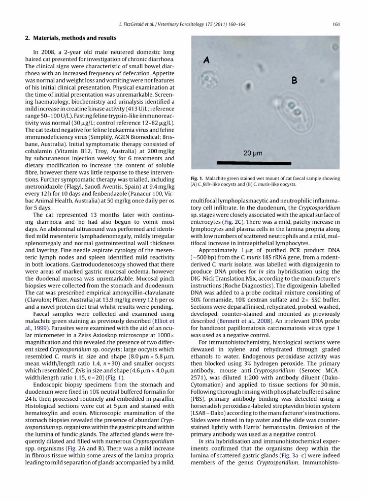
ry Parasitology 175 (2011) 160–164 161
L. FitzGerald et al. / Veterina2. Materials, methods and results
In 2008, a 2-year old male neutered domestic longhaired cat presented for investigation of chronic diarrhoea.The clinical signs were characteristic of small bowel diar-rhoea with an increased frequency of defecation. Appetitewas normal and weight loss and vomiting were not featuresof his initial clinical presentation. Physical examination atthe time of initial presentation was unremarkable. Screen-ing haematology, biochemistry and urinalysis identified amild increase in creatine kinase activity (413 U/L; referencerange 50–100 U/L). Fasting feline trypsin-like immunoreac-tivity was normal (30 �g/L; control reference 12–82 �g/L).The cat tested negative for feline leukaemia virus and felineimmunodeficiency virus (Simplify, AGEN Biomedical; Bris-bane, Australia). Initial symptomatic therapy consisted ofcobalamin (Vitamin B12, Troy, Australia) at 200 mg/kgby subcutaneous injection weekly for 6 treatments anddietary modification to increase the content of solublefibre, however there was little response to these interven-tions. Further symptomatic therapy was trialled, includingmetronidazole (Flagyl, Sanofi Aventis, Spain) at 9.4 mg/kgevery 12 h for 10 days and fenbendazole (Panacur 100, Vir-bac Animal Health, Australia) at 50 mg/kg once daily per osfor 5 days.
The cat represented 13 months later with continu-ing diarrhoea and he had also begun to vomit mostdays. An abdominal ultrasound was performed and identi-fied mild mesenteric lymphadenomegaly, mildly irregularsplenomegaly and normal gastrointestinal wall thicknessand layering. Fine needle aspirate cytology of the mesen-teric lymph nodes and spleen identified mild reactivityin both locations. Gastroduodenoscopy showed that therewere areas of marked gastric mucosal oedema, howeverthe duodenal mucosa was unremarkable. Mucosal pinchbiopsies were collected from the stomach and duodenum.The cat was prescribed empirical amoxycillin-clavulanate(Clavulox; Pfizer, Australia) at 13.9 mg/kg every 12 h per osand a novel protein diet trial whilst results were pending.
Faecal samples were collected and examined usingmalachite green staining as previously described (Elliot etal., 1999). Parasites were examined with the aid of an ocu-lar micrometer in a Zeiss Axioskop microscope at 1000×magnification and this revealed the presence of two differ-ent sized Cryptosporidium sp. oocysts; large oocysts whichresembled C. muris in size and shape (8.0 �m × 5.8 �m,mean width/length ratio 1.4, n = 30) and smaller oocystswhich resembled C. felis in size and shape (4.6 �m × 4.0 �mwidth/length ratio 1.15, n = 20) (Fig. 1).
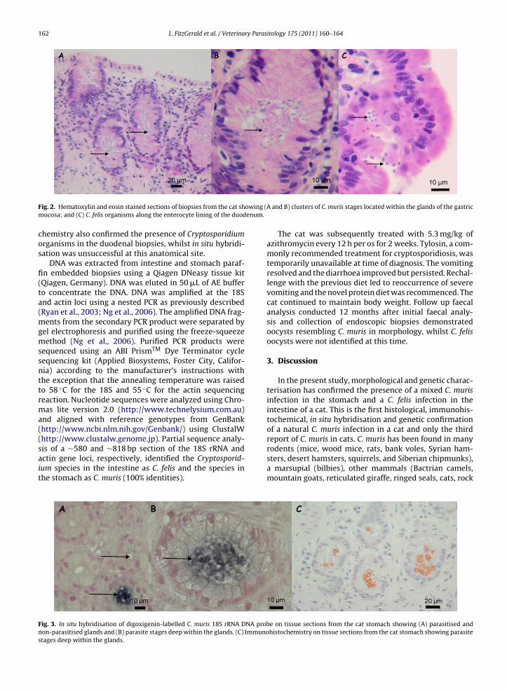
Endoscopic biopsy specimens from the stomach andduodenum were fixed in 10% neutral buffered formalin for24 h, then processed routinely and embedded in paraffin.Histological sections were cut at 5 �m and stained withhematoxylin and eosin. Microscopic examination of thestomach biopsies revealed the presence of abundant Cryp-tosporidium sp. organisms within the gastric pits and within
the lumina of fundic glands. The affected glands were fre-quently dilated and filled with numerous Cryptosporidiumspp. organisms (Fig. 2A and B). There was a mild increasein fibrous tissue within some areas of the lamina propria,leading to mild separation of glands accompanied by a mild,Fig. 1. Malachite green stained wet mount of cat faecal sample showing(A) C. felis-like oocysts and (B) C. muris-like oocysts.
multifocal lymphoplasmacytic and neutrophilic inflamma-tory cell infiltrate. In the duodenum, the Cryptosporidiumsp. stages were closely associated with the apical surface ofenterocytes (Fig. 2C). There was a mild, patchy increase inlymphocytes and plasma cells in the lamina propria alongwith low numbers of scattered neutrophils and a mild, mul-tifocal increase in intraepithelial lymphocytes.
Approximately 1 �g of purified PCR product DNA(∼500 bp) from the C. muris 18S rRNA gene, from a rodent-derived C. muris isolate, was labelled with digoxigenin toproduce DNA probes for in situ hybridisation using theDIG-Nick Translation Mix, according to the manufacturer’sinstructions (Roche Diagnostics). The digoxigenin-labelledDNA was added to a probe cocktail mixture consisting of50% formamide, 10% dextran sulfate and 2× SSC buffer.Sections were deparaffinised, rehydrated, probed, washed,developed, counter-stained and mounted as previouslydescribed (Bennett et al., 2008). An irrelevant DNA probefor bandicoot papillomatosis carcinomatosis virus type 1was used as a negative control.
For immunohistochemistry, histological sections weredewaxed in xylene and rehydrated through gradedethanols to water. Endogenous peroxidase activity wasthen blocked using 3% hydrogen peroxide. The primaryantibody, mouse anti-Cryptosporidium (Serotec MCA-2571), was diluted 1:200 with antibody diluent (Dako-Cytomation) and applied to tissue sections for 30 min.Following thorough rinsing with phosphate buffered saline(PBS), primary antibody binding was detected using ahorseradish peroxidase-labeled streptavidin biotin system(LSAB – Dako) according to the manufacturer’s instructions.Slides were rinsed in tap water and the slide was counter-stained lightly with Harris’ hematoxylin. Omission of the
primary antibody was used as a negative control.In situ hybridisation and immunohistochemical exper-iments confirmed that the organisms deep within thelumina of scattered gastric glands (Fig. 3a–c) were indeedmembers of the genus Cryptosporidium. Immunohisto-
162 L. FitzGerald et al. / Veterinary Parasitology 175 (2011) 160–164
F owing (Am enum.
cos
fi(ta(mgmssnttrma((sait
Fns
ig. 2. Hematoxylin and eosin stained sections of biopsies from the cat shucosa; and (C) C. felis organisms along the enterocyte lining of the duod
hemistry also confirmed the presence of Cryptosporidiumrganisms in the duodenal biopsies, whilst in situ hybridi-ation was unsuccessful at this anatomical site.
DNA was extracted from intestine and stomach paraf-n embedded biopsies using a Qiagen DNeasy tissue kitQiagen, Germany). DNA was eluted in 50 �L of AE buffero concentrate the DNA. DNA was amplified at the 18Snd actin loci using a nested PCR as previously describedRyan et al., 2003; Ng et al., 2006). The amplified DNA frag-
ents from the secondary PCR product were separated byel electrophoresis and purified using the freeze-squeezeethod (Ng et al., 2006). Purified PCR products were
equenced using an ABI PrismTM Dye Terminator cycleequencing kit (Applied Biosystems, Foster City, Califor-ia) according to the manufacturer’s instructions withhe exception that the annealing temperature was raisedo 58 ◦C for the 18S and 55 ◦C for the actin sequencingeaction. Nucleotide sequences were analyzed using Chro-as lite version 2.0 (http://www.technelysium.com.au)
nd aligned with reference genotypes from GenBankhttp://www.ncbi.nlm.nih.gov/Genbank/) using ClustalW
http://www.clustalw.genome.jp). Partial sequence analy-is of a ∼580 and ∼818 bp section of the 18S rRNA andctin gene loci, respectively, identified the Cryptosporid-um species in the intestine as C. felis and the species inhe stomach as C. muris (100% identities).ig. 3. In situ hybridisation of digoxigenin-labelled C. muris 18S rRNA DNA probon-parasitised glands and (B) parasite stages deep within the glands. (C) Immunotages deep within the glands.
and B) clusters of C. muris stages located within the glands of the gastric
The cat was subsequently treated with 5.3 mg/kg ofazithromycin every 12 h per os for 2 weeks. Tylosin, a com-monly recommended treatment for cryptosporidiosis, wastemporarily unavailable at time of diagnosis. The vomitingresolved and the diarrhoea improved but persisted. Rechal-lenge with the previous diet led to reoccurrence of severevomiting and the novel protein diet was recommenced. Thecat continued to maintain body weight. Follow up faecalanalysis conducted 12 months after initial faecal analy-sis and collection of endoscopic biopsies demonstratedoocysts resembling C. muris in morphology, whilst C. felisoocysts were not identified at this time.
3. Discussion
In the present study, morphological and genetic charac-terisation has confirmed the presence of a mixed C. murisinfection in the stomach and a C. felis infection in theintestine of a cat. This is the first histological, immunohis-tochemical, in situ hybridisation and genetic confirmationof a natural C. muris infection in a cat and only the third
report of C. muris in cats. C. muris has been found in manyrodents (mice, wood mice, rats, bank voles, Syrian ham-sters, desert hamsters, squirrels, and Siberian chipmunks),a marsupial (bilbies), other mammals (Bactrian camels,mountain goats, reticulated giraffe, ringed seals, cats, rocke on tissue sections from the cat stomach showing (A) parasitised andhistochemistry on tissue sections from the cat stomach showing parasite

ry Paras
L. FitzGerald et al. / Veterinahyraxes, cynomolgus monkeys, dogs, and pigs) (Warren etal., 2003; Santín et al., 2006; Pavlasek and Ryan, 2007; Lupoet al., 2008; Kodadkova et al., 2009; Kvac et al., 2009; Feng,2010), and birds (tawny frogmouth) (Ng et al., 2006). It hasalso been identified in a few humans in developing coun-tries (Palmer et al., 2003; Gatei et al., 2006; Muthusamyet al., 2006). Experimental C. muris infections have beenreported in dogs, rabbits, lambs and cats (Iseki et al., 1989).
C. felis has a much more restricted host range andhas been confirmed using molecular techniques in cats,immunocompetent and immunocompromised humansand a cow (Bornay-Llinares et al., 1999; Lucio-Forster etal., in press). In children in developing countries, C. felis isresponsible for as much as 3.3% of overall cryptosporid-iosis cases (Lucio-Forster et al., in press). However, mosthuman cases of cryptosporidiosis, worldwide, are asso-ciated with Cryptosporidium hominis and Cryptosporidiumparvum (Xiao, 2010) and therefore C. muris and C. felis incats are likely to be of low zoonotic risk to humans. Ithas also been suggested that some C. felis infections inhumans were anthroponotically transmitted (Cama et al.,2006). In the present study, the source of infection in thecat is unknown as the cat was acquired as a stray and therewere several other pets in the household. There was noclinical evidence of diarrhoea in any other household mem-bers.
As the cat was infected with both C. muris and C. felis, itis difficult to attribute the clinical presentations to eitherspecies. The presence of mild inflammation accompaniedby mild fibrosis in the stomach and inflammation withinthe duodenum in association with the Cryptosporidium spp.is suggestive of an ongoing host response secondary tothe presence of the organisms, however contribution fromother factors (such as concurrent food hypersensitivity)cannot be ruled out especially given the partial responseto a novel protein diet.
In the present study, azithromycin was unsuccessful inresolving the diarrhoea. Tylosin, which was temporarilyunavailable at time of diagnosis, has been used success-fully in cats but requires a long course of treatment (Barrand Bowman, 2006). Nitazoxanide has also been shown toreduce oocyst shedding in cats (Barr and Bowman, 2006).There was no overt evidence of immunosuppression in thiscat as it was feline leukemia virus and feline immunodefi-ciency virus negative, yet 9 months after the initial faecalanalysis, the cat was still shedding C. muris but apparentlynot C. felis indicating a persistent infection, however thepossibility of reinfection cannot be discounted.
The present study has confirmed that C. muris natu-rally infects the stomach of cats and therefore cats are notmerely acting as mechanical vectors. Further studies arerequired to determine the range, prevalence and clinicalimpact of Cryptosporidium species infecting cats, and thestatus of the host immune system in persistent or recurrentCryptosporidium spp. infections.
Acknowledgements
We are grateful to staff at the Murdoch University Vet-erinary Hospital’s Internal Medicine, Clinical Pathology,Histology and Diagnostic Imaging sections for professional
itology 175 (2011) 160–164 163
services and to the owner of the cat for provision ofsamples.
References
Ballweber, L.R., Panuska, C., Huston, C.L., Vasilopulos, R., Pharr, G.T.,Mackin, A., 2009. Prevalence of and risk factors associated with shed-ding of Cryptosporidium felis in domestic cats of Mississippi andAlabama. Vet. Parasitol. 160, 306–310.
Barr, S.C., Bowman, D.D., 2006. Cryptosporidiosis. In: Barr, S.C., Bowman,D.D. (Eds.), The 5-Minute Veterinary Consult Clinical Companion:Canine and Feline Infectious Diseases and Parasitology. Blackwell Pub-lishing, pp. 157–161.
Bennett, M.D., Woolford, L., O’Hara, A.J., Warren, K.S., Nicholls, P.K., 2008.In situ hybridization to detect bandicoot papillomatosis carcino-matosis virus type 1 in biopsies from endangered western barredbandicoots (Perameles bougainville). J Gen. Virol. 89, 419–423.
Bornay-Llinares, F.J., da Silva, A.J., Mourna, I.N., Myjap, P., Pietkiewicz, H.,Kruminis-Lozowska, W., Graczak, T.K., Pieniazek, N.J., 1999. Identifi-cation of Cryptosporidium felis in a cow by morphologic and molecularmethods. Appl. Environ. Microbiol. 65, 1455–1458.
Cama, V., Gilman, R.H., Vivar, A., Ticona, E., Ortega, Y., Bern, C., Xiao, L.,2006. Mixed Cryptosporidium infections and HIV. Emerg. Infect. Dis.12, 1025–1028.
Elliot, A., Morgan, U.M., Thompson, R.C.A., 1999. Improved stainingmethod for detecting Cryptosporidium oocysts in stools using Mala-chite Green. J. Gen. Appl. Microbiol. 45, 139–142.
Fayer, R., Santín, M., Trout, J.M., Dubey, J.P., 2006. Detection of Cryp-tosporidium felis and Giardia duodenalis Assemblage F in a cat colony.Vet. Parasitol. 140, 44–53.
Fayer, R., Santín, M., Macarisin, D., in press. Cryptosporidium ubiquitumn.sp. in animals and humans. Vet. Parasitol. 172, 23-32.
Feng, Y., 2010. Cryptosporidium in wild placental mammals. Exp. Parasitol.124, 128–137.
Gasser, R.B., Zhu, X., Caccio, S., Chalmers, R., Widmer, G., Morgan, U.M.,Thompson, R.C., Pozio, E., Browning, G.F., 2001. Genotyping Cryp-tosporidium parvum by single-strand conformation polymorphismanalysis of ribosomal and heat shock gene regions. Electrophoresis22, 433–437.
Gatei, W., Wamae, C.N., Mbae, C., Waruru, A., Mulinge, E., Waithera, T.,Gatika, S.M., Kamwati, S.K., Revathi, G., Hart, C.A., 2006. Cryptosporid-iosis: prevalence, genotype analysis, and symptoms associated withinfections in children in Kenya. Am. J. Trop. Med. Hyg. 75, 78–82.
Hajdusek, O., Ditrich, O., Slapeta, J., 2004. Molecular identification of Cryp-tosporidium spp. in animal and human hosts from the Czech Republic.Vet. Parasitol. 122, 183–192.
Huber, F., da Silva, S., Bomfim, T.C., Teixeira, K.R., Bello, A.R., 2007. Geno-typic characterization and phylogenetic analysis of Cryptosporidiumsp. from domestic animals in Brazil. Vet. Parasitol. 150, 65–74.
Iseki, M., Maekawa, T., Moriya, K., Uni, S., Takada, S., 1989. Infectivity ofCryptosporidium muris (strain RN 66) in various laboratory animals.Parasitol. Res. 75, 218–222.
Kodadkova, A., Kvac, M., Ditrich, O., Sak, B., Xiao, L., 2009. Cryptosporid-ium muris in a reticulated giraffe (Giraffa camelopardalis reticulata). J.Parasitol. 96, 211–212.
Kvac, M., Hanzlikova, D., Sak, B., Kvetonova, D., 2009. Prevalence and age-related infection of Cryptosporidium suis, C. muris and Cryptosporidiumpig genotype II in pigs on a farm complex in the Czech Republic. Vet.Parasitol. 160, 319–322.
Lucio-Forster, A., Griffiths, J.K., Cama, V.A., Xiao, L., Bowman, D.D., in press.Minimal zoonotic risk of cryptosporidiosis from pet dogs and cats.Trends Parasitol. 26, 174-179.
Lupo, P.J., Langer-Curry, R.C., Robinson, M., Okhuysen, P.C., Chappell, C.L.,2008. Cryptosporidium muris in a Texas canine population. Am. J. Trop.Med. Hyg. 78, 917–921.
Morgan, U.M., Sargent, K.D., Elliot, A., Thompson, R.C., 1998. Cryptosporid-ium in cats—additional evidence for C. felis. Vet. J. 156, 159–161.
Muthusamy, D., Rao, S.S., Ramani, S., Monica, B., Banerjee, I., Abraham, O.C.,Mathai, D.C., Primrose, B., Muliyil, J., Wanke, C.A., Ward, H.D., Kang,G., 2006. Multilocus genotyping of Cryptosporidium sp. isolates fromhuman immunodeficiency virus-infected individuals in South India. J.Clin. Microbiol. 44, 632–634.
Ng, J., Pavlasek, I., Ryan, U., 2006. Identification of novel Cryptosporidiumgenotypes from avian hosts. Appl. Environ. Microbiol. 72, 7548–7553.
Palmer, C.J., Xiao, L., Terashima, A., Guerra, H., Gotuzzo, E., Saldías, G.,Bonilla, J.A., Zhou, L., Lindquist, A., Upton, S.J., 2003. Cryptosporidiummuris, a rodent pathogen, recovered from a human in Peru. Emerg.Infect. Dis. 9, 1174–1176.

1 ry Paras
P
P
R
S
64 L. FitzGerald et al. / Veterina
almer, C.S., Traub, R.J., Robertson, I.D., Devlin, G., Rees, R., Thompson,R.C., 2008. Determining the zoonotic significance of Giardia and Cryp-tosporidium in Australian dogs and cats. Vet. Parasitol. 154, 142–147.
avlasek, I., Ryan, U., 2007. The first finding of a natural infection of Cryp-tosporidium muris in a cat. Vet. Parasitol. 144, 349–352.
yan, U., Xiao, L., Read, C., Zhou, L., Lal, A.A., Pavlasek, I., 2003. Identification
of novel Cryptosporidium genotypes from the Czech Republic. Appl.Environ. Microbiol. 69, 4302–4307.antín, M., Trout, J.M., Vecino, J.A., Dubey, J.P., Fayer, R., 2006.Cryptosporidium, Giardia and Enterocytozoon bieneusi in cats fromBogota (Colombia) and genotyping of isolates. Vet. Parasitol. 141,334–339.
itology 175 (2011) 160–164
Sargent, K.D., Morgan, U.M., Elliot, A., Thompson, R.C., 1998. Morpho-logical and genetic characterisation of Cryptosporidium oocysts fromdomestic cats. Vet. Parasitol. 77, 221–227.
Thomaz, A., Meireles, M.V., Soares, R.M., Pena, H.F., Gennari, S.M., 2007.Molecular identification of Cryptosporidium spp. from fecal samplesof felines, canines and bovines in the state of São Paulo, Brazil. Vet.
Parasitol. 150, 291–296.Warren, K.S., Swan, R.A., Morgan-Ryan, U.M., Friend, J.A., Elliot, A., 2003.Cryptosporidium muris infection in bilbies (Macrotis lagotis). Aust. Vet.J. 81, 739–741.
Xiao, L., 2010. Molecular epidemiology of cryptosporidiosis: an update.Exp. Parasitol. 124, 80–89.