Morphogenic and genetic differences between strains are ... · Morphogenic and genetic differences...
9
Morphogenic and genetic differences between Candida albicans strains are associated with keratomycosis virulence Xia Hua, 1 Xiaoyong Yuan, 1 Bradley M. Mitchell, 1 Michael C. Lorenz, 2 Denis M. O’Day, 3 Kirk R. Wilhelmus 1 1 Department of Ophthalmology, Baylor College of Medicine, Houston, TX; 2 Department of Microbiology and Molecular Genetics, The University of Texas Health Science Center at Houston, Houston, TX; 3 Department of Ophthalmology and Visual Sciences, Vanderbilt University School of Medicine, Nashville, TN Purpose: To correlate the morphogenic and molecular traits that affect fungal virulence in human corneas. Methods: C. albicans wild-type strains SC5314 and VE175 were compared using in vitro growth kinetics, filamentation assays, and microarray analysis. Corneal virulence was assessed ex vivo by inoculating C. albicans onto superficially scarified human corneas that were processed after 1 and 3 days to measure hyphal penetration. For comparison, DSY459, a C. albicans homozygous deletion mutant deficient in secreted aspartyl proteinases (SAP) 4, 5, and 6, was evaluated. Results: C. albicans strain SC5314 was highly filamentous in vitro and more invasive in human corneal explants while VE175 demonstrated limited filamentation and less corneal invasion. Among 6,655 C. albicans genes, 9.0% significantly (p<.05) differed by 2 fold or more between SC5314 and VE175. Genes involved in fungal filamentation that were upregulated in strain SC5314 compared to VE175 included SAP5, SAP6, and other hypha-associated genes. Compared to wild-type strains, DSY459 had intermediate filamentation and stromal penetration. Conclusions: Fungal genes involved in filamentation likely contribute to virulence differences between wild-type strains of C. albicans. The corneal pathogenicity of C. albicans involves the morphogenic transformation of yeasts into hyphae. Candida albicans is a commensal fungus of the ocular surface [1], an occasional contaminant of ophthalmic devices [2-4], and an opportunistic pathogen of the compromised cornea [5]. In recent clinical series, Candida species were isolated from 1% to 5% of eyes with microbial keratitis [6-11]. Improved prevention and therapy of C. albicans infections require better understanding of fungal metabolism and growth. O’Day and associates [12] first suggested using C. albicans strains having divergent ocular pathogenicity to help identify the mechanisms of fungal virulence for the eye. SC5314, a pathogenic C. albicans strain widely used for experimental and genomic studies, rapidly penetrated into the cornea after topical inoculation in immunocompetent rabbits [12,13]. In contrast, another human isolate of C. albicans, strain VE175, was significantly less virulent and formed pseudohyphae that were largely limited to the superficial stroma [12]. O’Day [12] hypothesized that “fungal genes that control morphogenesis may be involved”. Using these same fungal strains in an immunocompetent mouse model of experimental fungal keratitis, Mitchell and associates [14] confirmed that strain SC5314 produced significantly worse keratomycosis than VE175 [15]. Additional studies with fungal mutants provided evidence that fungal filamentation is an important virulence factor for Correspondence to: Dr. Kirk R. Wilhelmus, Sid W. Richardson Ocular Microbiology Laboratory, Cullen Eye Institute, 6565 Fannin Street, Houston, TX, 77030; Phone: (713) 798 5952; FAX: (713) 798-4142 email: [email protected] keratomycosis [15-17]. Fungal genes associated with hyphal morphogenesis may account for differences in corneal disease severity produced by disparate strains of C. albicans [18]. Our present study further evaluated these two wild-type strains of C. albicans. We examined their phenotypic growth characteristics under various environmental conditions and used comparative genomics to explore genetic differences between the two strains. We then developed an ex vivo model of human corneal infection to test the ability of each fungal strain to invade explanted tissue. Finally, we used a C. albicans mutant to demonstrate that fungal genes associated with hyphal morphogenesis are important to the pathogenesis of fungal corneal infection. METHODS Fungal strains: C. albicans strains included SC5314, a human clinical isolate recovered from a patient with generalized candidiasis [19,20], and VE175, a human corneal isolate obtained from a patient with candidal keratitis [21]. After reviewing the genomic differences between SC5314 and VE175, a mutant strain DSY459 deficient in secreted aspartyl proteinase (SAP) SAP4, SAP5, and SAP6 genes ( Δsap4-6 ) was obtained from Sanglard and associates who derived this triple-null homozygote from SC5314 using a Ura-blaster cassette [22]. In vitro growth kinetics: Yeast strains were grown in 1% yeast-extract, 2% peptone, and 2% dextrose (YPD) liquid medium at 30 °C, harvested during exponential growth, and suspended in sterile phosphate-buffered saline (PBS). Optical Molecular Vision 2009; 15:1476-1484 <http://www.molvis.org/molvis/v15/a158> Received 23 June 2009 | Accepted 27 July 2009 | Published 30 July 2009 © 2009 Molecular Vision 1476
Transcript of Morphogenic and genetic differences between strains are ... · Morphogenic and genetic differences...

Morphogenic and genetic differences between Candida albicansstrains are associated with keratomycosis virulence
Xia Hua,1 Xiaoyong Yuan,1 Bradley M. Mitchell,1 Michael C. Lorenz,2 Denis M. O’Day,3 Kirk R. Wilhelmus1
1Department of Ophthalmology, Baylor College of Medicine, Houston, TX; 2Department of Microbiology and Molecular Genetics,The University of Texas Health Science Center at Houston, Houston, TX; 3Department of Ophthalmology and Visual Sciences,Vanderbilt University School of Medicine, Nashville, TN
Purpose: To correlate the morphogenic and molecular traits that affect fungal virulence in human corneas.Methods: C. albicans wild-type strains SC5314 and VE175 were compared using in vitro growth kinetics, filamentationassays, and microarray analysis. Corneal virulence was assessed ex vivo by inoculating C. albicans onto superficiallyscarified human corneas that were processed after 1 and 3 days to measure hyphal penetration. For comparison, DSY459, aC. albicans homozygous deletion mutant deficient in secreted aspartyl proteinases (SAP) 4, 5, and 6, was evaluated.Results: C. albicans strain SC5314 was highly filamentous in vitro and more invasive in human corneal explants whileVE175 demonstrated limited filamentation and less corneal invasion. Among 6,655 C. albicans genes, 9.0% significantly(p<.05) differed by 2 fold or more between SC5314 and VE175. Genes involved in fungal filamentation that wereupregulated in strain SC5314 compared to VE175 included SAP5, SAP6, and other hypha-associated genes. Compared towild-type strains, DSY459 had intermediate filamentation and stromal penetration.Conclusions: Fungal genes involved in filamentation likely contribute to virulence differences between wild-type strainsof C. albicans. The corneal pathogenicity of C. albicans involves the morphogenic transformation of yeasts into hyphae.
Candida albicans is a commensal fungus of the ocularsurface [1], an occasional contaminant of ophthalmic devices[2-4], and an opportunistic pathogen of the compromisedcornea [5]. In recent clinical series, Candida species wereisolated from 1% to 5% of eyes with microbial keratitis[6-11].
Improved prevention and therapy of C. albicansinfections require better understanding of fungal metabolismand growth. O’Day and associates [12] first suggested usingC. albicans strains having divergent ocular pathogenicity tohelp identify the mechanisms of fungal virulence for the eye.SC5314, a pathogenic C. albicans strain widely used forexperimental and genomic studies, rapidly penetrated into thecornea after topical inoculation in immunocompetent rabbits[12,13]. In contrast, another human isolate of C. albicans,strain VE175, was significantly less virulent and formedpseudohyphae that were largely limited to the superficialstroma [12]. O’Day [12] hypothesized that “fungal genes thatcontrol morphogenesis may be involved”.
Using these same fungal strains in an immunocompetentmouse model of experimental fungal keratitis, Mitchell andassociates [14] confirmed that strain SC5314 producedsignificantly worse keratomycosis than VE175 [15].Additional studies with fungal mutants provided evidence thatfungal filamentation is an important virulence factor for
Correspondence to: Dr. Kirk R. Wilhelmus, Sid W. RichardsonOcular Microbiology Laboratory, Cullen Eye Institute, 6565 FanninStreet, Houston, TX, 77030; Phone: (713) 798 5952; FAX: (713)798-4142 email: [email protected]
keratomycosis [15-17]. Fungal genes associated with hyphalmorphogenesis may account for differences in corneal diseaseseverity produced by disparate strains of C. albicans [18].
Our present study further evaluated these two wild-typestrains of C. albicans. We examined their phenotypic growthcharacteristics under various environmental conditions andused comparative genomics to explore genetic differencesbetween the two strains. We then developed an ex vivo modelof human corneal infection to test the ability of each fungalstrain to invade explanted tissue. Finally, we used a C.albicans mutant to demonstrate that fungal genes associatedwith hyphal morphogenesis are important to the pathogenesisof fungal corneal infection.
METHODSFungal strains: C. albicans strains included SC5314, a humanclinical isolate recovered from a patient with generalizedcandidiasis [19,20], and VE175, a human corneal isolateobtained from a patient with candidal keratitis [21]. Afterreviewing the genomic differences between SC5314 andVE175, a mutant strain DSY459 deficient in secreted aspartylproteinase (SAP) SAP4, SAP5, and SAP6 genes ( Δsap4-6 )was obtained from Sanglard and associates who derived thistriple-null homozygote from SC5314 using a Ura-blastercassette [22].In vitro growth kinetics: Yeast strains were grown in 1%yeast-extract, 2% peptone, and 2% dextrose (YPD) liquidmedium at 30 °C, harvested during exponential growth, andsuspended in sterile phosphate-buffered saline (PBS). Optical
Molecular Vision 2009; 15:1476-1484 <http://www.molvis.org/molvis/v15/a158>Received 23 June 2009 | Accepted 27 July 2009 | Published 30 July 2009
© 2009 Molecular Vision
1476
density (OD) was measured with an Ultraspec 2000spectrophotometer (Pharmacia Biotech, Princeton, NJ) at awavelength of 600 nm (OD600). A conversion factor of oneOD600 unit equivalent to 3×10⁷ colony-forming units (CFU)/ml was used to determine fungal concentration [23]. Triplicatesamples of 3×10⁵ CFU for each strain were inoculated into 25ml M199 liquid media (Invitrogen, Grand Island, NY) at pH6.0, pH 7.3, and pH 8.0 and incubated at 27 °C with continuousshaking. C. albicans concentrations were determinedspectrophotometrically at 1.5, 3, 4.5, 6, 9, 12, 15, 21, 27, 35,and 48 h post inoculation (pi).Filamentation assay: C. albicans strains were grownovernight on Sabouraud dextrose agar (Difco, Detroit, MI) for3 days at 25 ºC. Yeasts were harvested, diluted in sterile PBSto yield 1 CFU/μl, inoculated onto M199 agar platescontaining Earle’s salts and glutamine but lacking sodiumbicarbonate that were buffered with 2 M Tris-HCl to yieldmedia pH values of 7.3 and 8.0, and incubated at 37 ºC.Colonies were observed daily by inverted microscopy for 10days.Genomic microarray: C. albicans strains SC5314 and VE175were separately inoculated into 10 ml of YPD media andincubated in a room-temperature (22 ºC) shaker until the lateexponential growth phase. Ten replicate 1 ml fungal
suspensions in 50 ml M199 media at pH 8.0 were held at 37°C in a 180 rpm incubator, and fungi were harvested after 4 h[24]. Hot acidic phenol extraction was performed to prepareyeast ribonucleic acid (RNA) [25]. The RNA concentrationwas measured with a ND-1000 spectrophotometer (NanoDropTechnologies, Wilmington, DE), followed by gelelectrophoresis to confirm purity and integrity.
Twenty µg of total RNA were labeled by an aminoallyl-derivative protocol using the Micromax RNA labeling kit(PerkinElmer, Waltham, MA). Samples were mixed andhybridized to a glass-slide microarray in the C. albicans ArrayReady Oligo Set (Operon Biotechnology, Huntsville, AL)composed of 8,117 70-mer oligonucleotides representing6,655 genes from Assembly 19 of the C. albicans genome[26], plus specific and non-specific controls, resulting inslightly more than the predicted 6,354 protein-coding genesdue to the inclusion of some tRNA genes and spurious genespredicted in earlier assemblies [27]. The array was printed induplicate on Codelink II (GE Healthcare, Princeton, NJ) slides(Microarrays, Nashville, TN). Labeled samples were mixedand hybridized to the arrays. Two technical replicates and twobiological replicates were processed for each sample for a totalof eight spots for each gene. Arrays were scanned using anAxon 4000B scanner (Molecular Devices, Sunnywale, CA),
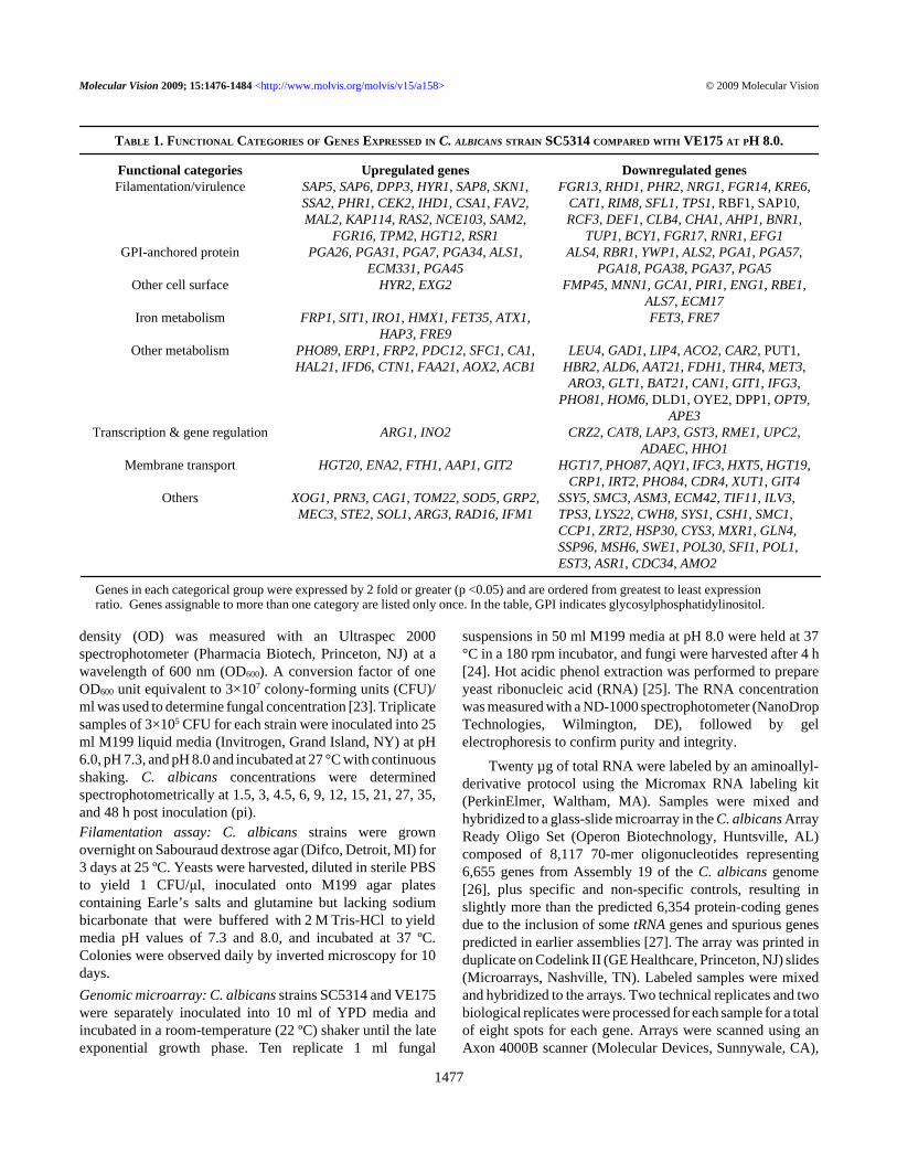
TABLE 1. FUNCTIONAL CATEGORIES OF GENES EXPRESSED IN C. ALBICANS STRAIN SC5314 COMPARED WITH VE175 AT PH 8.0.
Functional categories Upregulated genes Downregulated genesFilamentation/virulence SAP5, SAP6, DPP3, HYR1, SAP8, SKN1,
SSA2, PHR1, CEK2, IHD1, CSA1, FAV2,MAL2, KAP114, RAS2, NCE103, SAM2,
FGR16, TPM2, HGT12, RSR1
FGR13, RHD1, PHR2, NRG1, FGR14, KRE6,CAT1, RIM8, SFL1, TPS1, RBF1, SAP10,RCF3, DEF1, CLB4, CHA1, AHP1, BNR1,
TUP1, BCY1, FGR17, RNR1, EFG1GPI-anchored protein PGA26, PGA31, PGA7, PGA34, ALS1,
ECM331, PGA45ALS4, RBR1, YWP1, ALS2, PGA1, PGA57,
PGA18, PGA38, PGA37, PGA5Other cell surface HYR2, EXG2 FMP45, MNN1, GCA1, PIR1, ENG1, RBE1,
ALS7, ECM17Iron metabolism FRP1, SIT1, IRO1, HMX1, FET35, ATX1,
HAP3, FRE9FET3, FRE7
Other metabolism PHO89, ERP1, FRP2, PDC12, SFC1, CA1,HAL21, IFD6, CTN1, FAA21, AOX2, ACB1
LEU4, GAD1, LIP4, ACO2, CAR2, PUT1,HBR2, ALD6, AAT21, FDH1, THR4, MET3,ARO3, GLT1, BAT21, CAN1, GIT1, IFG3,
PHO81, HOM6, DLD1, OYE2, DPP1, OPT9,APE3
Transcription & gene regulation ARG1, INO2 CRZ2, CAT8, LAP3, GST3, RME1, UPC2,ADAEC, HHO1
Membrane transport HGT20, ENA2, FTH1, AAP1, GIT2 HGT17, PHO87, AQY1, IFC3, HXT5, HGT19,CRP1, IRT2, PHO84, CDR4, XUT1, GIT4
Others XOG1, PRN3, CAG1, TOM22, SOD5, GRP2,MEC3, STE2, SOL1, ARG3, RAD16, IFM1
SSY5, SMC3, ASM3, ECM42, TIF11, ILV3,TPS3, LYS22, CWH8, SYS1, CSH1, SMC1,CCP1, ZRT2, HSP30, CYS3, MXR1, GLN4,SSP96, MSH6, SWE1, POL30, SFI1, POL1,EST3, ASR1, CDC34, AMO2
Genes in each categorical group were expressed by 2 fold or greater (p <0.05) and are ordered from greatest to least expressionratio. Genes assignable to more than one category are listed only once. In the table, GPI indicates glycosylphosphatidylinositol.
Molecular Vision 2009; 15:1476-1484 <http://www.molvis.org/molvis/v15/a158> © 2009 Molecular Vision
1477

and data were analyzed using GenePix Pro 6.0 software(Molecular Devices, Downingtown, PA) as previouslydescribed [28]. Inconsistent or saturated spots were filtered,and spot values were calculated by subtracting the backgroundintensity from the median pixel intensity of each spot. Valueswere then normalized to equalize median intensities betweenthe two channels across the entire array. From the eightreplicate spots, geometric means of the ratios and p-valueswere estimated. Genes having an expression ratio of at least2 fold difference and p <0.05 between the two strains weregrouped into functional categories using information curatedby the Candida Genome Database [29].Reverse transcription (RT) of RNA and RT-polymerase chainreaction (RT-PCR): Total RNA was separately isolated fromC. albicans strains SC5314 and VE175 following cultivationfor 4 h at pH 7.3 and pH 8.0, respectively. First-strand cDNAwas synthesized from 1 μg total RNA with Ready-To-GoYou-Prime First-Strand Beads (GE Healthcare) and randomhexamers (Applied Biosystems, Foster City, CA). RT-PCRwas performed using PuReTaq Ready-To-Go PCR beads (GEHealthcare). Primers for SAP4 (P1: GTC AAC GCT GGTGTC CTC TT; P2: GCA GGA ACG GAA ATC TTG AG,197 bp), SAP5 (P2: CCG AAT TCC TTT TCC AAA CA; P2:TGG AGC CAT GGA GAT TTT CT, 158 bp), and SAP6 (P1:GTC AAC GCT GGT GTC CTC TT; P2: TTC ACG AACACG AAT TTC ACA; 270 bp) were synthesized (Sigma, St.Louis, MO), with ACT1 (TGC TGA ACG TAT GCA AAAGG; P2: TGA ACA ATG GAT GGA CCA GA; 186 bp) asthe housekeeping gene. Semiquantitative RT-PCR wasestablished by terminating reactions at intervals of 18, 20, 22,24, 26, 28, 30, 32, 34, 36, and 40 cycles for each primer pair
to ensure that PCR products were within the linear portion ofthe amplification curve. All products were separated by 2%agarose gel electrophoresis and visualized with 0.5 mg/mlethidium bromide. Fidelity of RT-PCR products wasconfirmed by comparing cDNA bands and by sequencingPCR products.Ex vivo human cornea model: Thirty-six human corneas wereobtained from the Lions Eye Bank of Texas, Houston, TXafter informed consent for research use was obtained from thedecedent donors’ next-of-kin. Donor corneas were maintainedat 4 ºC in Optisol-GS (Bausch & Lomb, Irvine, CA) beforebeing transferred to modified supplemented hormonalepithelial medium (SHEM), consisting of equal volumes ofDulbecco’s modified Eagle’s medium and Ham’s F12medium that contained 5 ng/ml epidermal growth factor, 5μg/ml insulin, 5 μg/ml transferrin, 5 μg/ml sodium selenite,0.5 μg/ml hydrocortisone, 30 ng/ml cholera toxin A, 0.5%dimethylsulfoxide, 50 μg/ml gentamicin, and 5% fetal bovineserum, and was buffered with 2 M Tris-HCL to pH 7.3 or 8.0.Corneas were superficially scarified using a 22 gauge needleto produce a 15x15 cross-hatch pattern, similar to a protocolpreviously described for an experimental fungal keratitismodel [14]. A Horizon artificial anterior chamber (RefractiveTechnologies, Cleveland, OH) fixated the corneal buttonduring superficial scarification, followed by topicalapplication of 10 μl of 1×10⁵ CFU C. albicans per cornea.Inoculated corneas were put epithelial side up into a 6 wellculture dish (Corning, Corning, NY) so that the sclera wasimmersed in modified SHEM and the central cornea vaultedupward. Tissues were incubated at 34 °C in 5% CO2 with 95%humidity, and culture medium was changed daily. After 24
Figure 1. Differential gene regulationcomparing strain SC5314 to strainVE175 of 44 C. albicans genes involvedin fungal filamentation.
Molecular Vision 2009; 15:1476-1484 <http://www.molvis.org/molvis/v15/a158> © 2009 Molecular Vision
1478

and 72 h, corneas were embedded in OCT compound (SakuraFinetec, Torrance, CA) and frozen at -80 °C for subsequenthistopathologic processing.Hyphal penetration: Frozen sections (10 µm) were cut, andthree sections were examined for each cornea at 10 μmintervals for 100 µm from the corneal mid-point. Sectionswere stained with periodic acid-Schiff (PAS) reagent (Sigma-Aldrich, St. Louis, MO). Images were captured of an entirelimbus-to-limbus section from each cornea with a DS-Fildigital camera (Nikon, Tokyo, Japan) attached to a Nikon Y-FL microscope. We measured the overall hyphal depth with10X magnification at 5 different points at equidistantintervals. The entire corneal thickness was then estimated at5X magnification at the same positions for each section usingthe NIS-Element 3.0 image analysis system (Nikon). Theoverall average depth of penetration was calculated as thepercentage of corneal thickness. The maximal percentage ofhyphal penetration was also estimated from measurementstaken at regions of each corneal section demonstrating thegreatest depth of corneal penetration. To assure that points ofmaximum hyphal penetration were selected, results from thethree largest hyphal-depth percentages were averaged from 5
measurements taken for each histological section andcalculated as percentage of corneal thickness. Results werecompared by the Student t-test.
RESULTSIn vitro comparison of C. albicans strains: Strains SC5314and VE175 demonstrated similar lag, log-growth, and plateauphases. The doubling time of both C. albicans strains use thesame at pH 6.0 (1.94±0.04 h for SC5314 and 1.92±0.04 h forVE175) and similar at pH 7.3 (2.21±0.04 h for SC5314 and1.94±0.01 h for VE175) but showed that SC5314 grew slightlyfaster (2.27±0.03 h) than VE175 (2.72±0.05 h) at alkaline pH(p=0.02). Mock-inoculated control cultures remainednegative for growth throughout 24 h of observation. On M199agar, numerous filaments surrounded SC5314 colonies 24 hafter inoculation and continued growing during the following10 days. Few filaments emerged from VE175 colonies, andwhile dissimilarity was present at physiologic pH, thedifference with in vitro filamentation was more apparent atpH 8.0.Genomic comparison of C. albicans strains: Among 6,655genes detected by microarray, 601 (9.03%) genes in SC5314
Figure 2. RT-PCR analysis of SAP4,SAP5, and SAP6 expression in C.albicans strains SC5314 and VE175cultured on pH 7.3 and pH 8.0 media.SAP5 and SAP6 bands were moreapparent from SC5314 than VE175.ACT1 gene expression appeared similarbetween both strains at each pH. SAP4was not detected.
Molecular Vision 2009; 15:1476-1484 <http://www.molvis.org/molvis/v15/a158> © 2009 Molecular Vision
1479

were significantly (p<0.05) and differently (≥2 fold change)regulated compared to VE175. Of these, 69 namedupregulated genes and 116 named downregulated genes werecategorized into functional groups (Table 1), including 44genes involved in hyphal filamentation and virulence (Figure1). mRNA expression of SAP4, SAP5, and SAP6 was furtherexamined by RT-PCR (Figure 2). No difference of ACT1expression occurred between SC5314 and VE175 at either pH7.3 or pH 8.0. mRNA expression of SAP4 was not detected ineither strain at either pH. SAP5 and SAP6 mRNA were
expressed higher in SC5314 than VE175 at both pHconditions.
Corneal virulence: One day after inoculation of strainsSC5314 and VE175 onto human corneas incubated at pH 7.3,fungal hyphae were present in the anterior corneal stroma. Bythe third day SC5314 invaded toward the central stroma(Figure 3A). VE175, however, failed to continue hyphalinvasion between 1 and 3 days pi and remained limited to theanterior cornea. At pH 8.0, hyphal penetration into cornealtissue was consistently greater for SC5314 than VE175. Theoverall and maximal penetration percentage are shown in
Figure 3. Histopathology of humankeratomycosis induced by C. albicansstrains SC5314 and VE175. A: At pH7.3 SC5314 established superficialinfection by day 1 post inoculation, andhyphae proliferated and invadedcentrally by day 3. Strain VE175produced few pesudohyphae in thesuperficial cornea that did not penetratethrough anterior lamellae by day 3. B:At pH 8.0 strain SC5314 formed hyphaethat extended around superficialscarification site on day 1 postinoculation and continued to invade byday 3. Strain VE175 produced fewfilamentous forms on day 1 or day 3.Periodic acid-Schiff stain. Scale bars,50µm.
Molecular Vision 2009; 15:1476-1484 <http://www.molvis.org/molvis/v15/a158> © 2009 Molecular Vision
1480

Table 2 and Table 3. Initially, few conidia of VE175 werepresent on the corneal surface and no hyphal formation wasobserved (Figure 3B). Pseudohyphae subsequently formedwith VE175 but did not penetrate to the same extent asSC5314.Δsap4-6 deletion mutant: Fungal mutant DSY459 grew wellat pH 6.0 and pH 7.3 and slightly more slowly at pH 8.0.Moderate filamentation was found in vitro at pH 7.3 and pH8.0 (Figure 4). In explanted corneas at 1 day pi (Figure 4), theoverall penetration was 5.2%±4.7% at pH 7.3 and 1.0%±2.1%at pH 8.0 (p=0.24), and the maximal penetration was 5.6%±8.5% at pH 7.3 and 2.7% ± 4.1% at pH 8.0 (p=0.38). At day3 pi, the overall penetration was 11.1%±3.0% at pH 7.3 and
7.2%±2.1% at pH 8.0 (p=0.14), and maximal penetration was23.8%±5.1% at pH 7.3 and 16.0%±11.3% at pH 8.0(p=0.049). Compared to SC5314, the maximal penetration ofDSY459 was significantly less at pH 7.3 at day 1 (p<0.001)and at day 3 (p<0.001) and was significantly less at pH 8.0 atday 1 (p<0.001) and at day 3 (p=0.003), but there was nodifference at the overall hyphal penetration between these twostrains at both pH conditions at day 1 and day 3.
DISCUSSIONC. albicans is a common symbiont of mammalian microfloraand an important infectious agent. Impaired immunity andaltered defenses predispose a host to C. albicans ocular
TABLE 2. OVERALL PENETRATION (%) OF C. ALBICANS STRAINS INTO EXPLANTED HUMAN CORNEAS UNDER PHYSIOLOGIC ANDALKALINE PH CONDITIONS.
pH 7.3 pH 8.0Time SC5314 VE175 p value SC5314 VE175 p valueDay 1 8.1±4.9 2.0±2.4 0.12 1.7±0.8 0 0.02Day 3 19.9±4.6 6.9±7.2 0.06 10.8±4.7 7.6±8.6 0.60
TABLE 3. MAXIMAL PENETRATION (%) OF C. ALBICANS STRAINS INTO EXPLANTED HUMAN CORNEAS UNDER PHYSIOLOGIC ANDALKALINE PH CONDITIONS.
pH 7.3 pH 8.0Time SC5314 VE175 p value SC5314 VE175 p valueDay 1 21.8±5.6 16.0±7.6 0.11 17.0±6.0 0 <0.0001Day 3 41.9±6.1 16.0±6.7 0.0007 33.7±12.3 18.9±12.5 0.02
Figure 4. In vitro and ex vivofilamentation of C. albicans ∆sap4-6mutant strain. Compared with SC5314and VE175, this mutant strain exhibitedlow but intermediate capabilities toproduce filaments around fungalcolonies. At day 1 postinoculation, themutant strain formed hyphae in thesuperficial stroma at pH 7.3 but did notproduce filamentous forms at pH 8.0.
Molecular Vision 2009; 15:1476-1484 <http://www.molvis.org/molvis/v15/a158> © 2009 Molecular Vision
1481

infection [5,14], but fungal virulence traits are also involved[16]. Pathogenicity involves a dynamic interaction betweensusceptible hosts and fungal pathogens [30].
The infectious nature of C. albicans and other fungi ispartly a result of their morphological plasticity. Microbialsurvival depends on the ability to detect and to respond to localconditions. During tissue invasion C. albicans transformsfrom blastospores into invasive filamentous forms [16]. Thistransition from commensal yeasts to pseudohyphae andhyphae results from interrelated pathways that respond tolocal cues [31,32]. Changes that bring about alterations infungal morphology include a rise from room to bodytemperature and a shift in pH.
Adaptation to neutral and alkaline environments by fungiuses the Rim101/PacC signal transduction pathway, amechanism conserved throughout yeasts and moulds [32-34].This pathway regulates gene expression via transcriptionfactor Rim101/PacC during the pathogenesis of fungalinfection and links the ability to respond to environmental pHwith virulence and disease [35].
The Rim101/PacC protein, available as a full-length orprocessed form, is inactive under acidic conditions and isactivated at neutral or alkaline pH by proteolytic processing,becoming capable of inducing hyphal formation andfilamentation [33,34]. Proteolysis is regulated in part byupstream members of the pathway including Rim8/PalF,Rim13/PalB, Rim20/PalA, Rim21/PalH, and Snf7 proteins[33,36] and downstream genes including EFG1,PHR1, andPHR2 [37-39]. Mechanisms governing fungal morphogenesisare closely related to the development of keratomycosis [16].
Fungi differ in virulence, even among strains of the samespecies. C. albicans strains can produce differing levels ofinfection severity [40,41]. C. albicans SC5314, a widely usedstrain, shows substantially higher virulence for rabbit andmouse eyes than C. albicans VE175, a human corneal isolate[12,15]. Our ex vivo model of human corneal infectionconfirms that SC5314 rapidly forms hyphae that penetrate intothe corneal stroma. On the other hand, based on overallpenetration and maximal fungal invasion, VE175 is lessinvasive and is largely limited to epithelial adherence ofblastospores with few pseudohyphae in the superficial stroma.
Both SC5314 and VE175 have similar replicationkinetics. However, strain SC5314 readily producedfilamentary growth in vitro at neutral and alkaline pH whileVE175 had limited filamentation. VE175 also demonstratedless hyphal penetration into corneal tissue compared to pH7.3. We hypothesize that a defect in filamentation regulatedthrough the Rim101/PacC signal transduction pathway mightexplain the dissimilarity in corneal pathogenicity produced bythese C. albicans strains.
Phenotypic differences in virulence can be due togenotypic variations among C. albicans strains [42,43].O’Day [12] pointed out that identifying “the intrinsic genetic
differences between such strains may help identify factorsresponsible for fungal virulence”. Since the genome of C.albicans has been sequenced [26], we compared the relativegenetic expression of SC5314 and VE175 to seek possiblereasons for their disparate pathogenicity.
C. albicans has numerous genes involved in nutrientuptake, metabolism, cellular structure, and morphogenesisthat are needed for growth and survival. We found that apreponderance of C. albicans genes to be similarly expressedin SC5314 and VE175 strains, suggesting that relatively fewgenes may contribute to virulence differences between theseclinical isolates. Nearly one fourth of named genes that weredifferentially expressed were involved in hyphal formation.Because VE175 displayed reduced filamentation in vitro andwithin the explanted cornea, the formation of fungal hyphaeappears to be part of a causal pathway during infection by C.albicans.
The ability of C. albicans to produce hyphae in vitrocorrelates with the production of filamentous forms duringexperimental corneal infection [16]. Mutations in fungalgenes involved in the yeast-to-hyphal morphogenesis of C.albicans affect the development of C. albicans keratitis [15,44]. Since a mild environmental shift activates a pathwayleading to fungal filamentation [31,33], we examined theeffect of pH in this study.
The expression levels of genes involved in the pH-dependent Rim101 pathway were largely similar between thetwo strains, but downstream differences were found. PHR1, apH-responsive gene that encodes a cell-wall glycosidaseacting on polysaccharide cross-linking during hyphalmorphogenesis [38,45], was significantly downregulated instrain VE175. We also noted that VE175 tended to produceround blastospores that were larger than SC5314 when grownin vitro, resembling a PHR1-deletion mutant grown in analkaline medium [38]. In a neutral-to-alkaline milieu PHR1 isinduced following Rim101p activation, and deletion of theRIM13 gene attenuates corneal virulence of C. albicans [15].In addition, the relative upregulation of the transcriptionalrepressor TUP1 in VE175 could contribute to this strain’sattenuated virulence [16]. These findings indicate that thedimorphic transformation from yeasts to hyphae isincriminated in the onset and progression of C. albicanskeratitis.
Another virulence factor associated with hyphalmorphogenesis is the family of hypha-associated hydrolases[46]. Among secreted aspartyl proteinases [47] Sap6p isrequired for fungal invasion of the parenchyma and of thecornea [44,48]. Downregulation of SAP5 and SAP6 in the lessvirulent strain VE175 compared to the pathogenic strainSC5314 may partly underlie their different abilities to invade.To explore the role of these proteinases, we used a Δsap4-6deletion mutant of C. albicans. This knockout strain wasintermediate between wild-type strains in the ability to
Molecular Vision 2009; 15:1476-1484 <http://www.molvis.org/molvis/v15/a158> © 2009 Molecular Vision
1482

penetrate corneal tissue at a physiologic pH. Proteinasesencoded by SAP5 and SAP6 may facilitate the penetration ofC. albicans hyphae through the epithelium and extracellularmatrix [49,50].
The molecular mechanisms responsible for myoticinfections are emerging. This study used phenotypicscreening, comparative genomics, and ex vivo cornealinfection to gain better insight into the molecular biology offungal keratitis. By demonstrating genetic variations betweenfungal strains having different capabilities of producingkeratomycosis, this study supports the key roles of fungalfilamentation and hypha-associated proteins in thepathogenesis of C. albicans keratitis.
ACKNOWLEDGMENTSDominique Sanglard, Institut de Microbiologie, UniversityHospital Lausanne, Switzerland, and Bernhard Hube,Abteilung Mikrobielle Pathogenität, Leibniz-Institut fürNaturstoff-Forschung and Infektionsbiologie, Hans Knöll-Institut, Friedrich Schiller Universtität, Jena, Germany,provided the DSY459 mutant strain. The Lions Eye Bank ofTexas, Houston, TX, provided donor research tissue.
REFERENCES1. Wu T, Mitchell B, Carothers T, Coats D, Brady-McCreery K,
Paysse E, Wilhelmus K. Molecular analysis of the pediatricocular surface for fungi. Curr Eye Res 2003; 26:33-6. [PMID:12789534]
2. Kercher L, Wardwell SA, Wilhelmus KR, Mitchell BM.Molecular screening of donor corneas for fungi beforeexcision. Invest Ophthalmol Vis Sci 2001; 42:2578-83.[PMID: 11581202]
3. Marqués-Calvo MS. Colonization of hydrophilic contact lensesby yeast. J Ind Microbiol Biotechnol 2004; 31:255-60.[PMID: 15221665]
4. Barnes SD, Dohlman CH, Durand ML. Fungal colonization andinfection in Boston keratoprosthesis. Cornea 2007; 26:9-15.[PMID: 17198007]
5. Sun RL, Jones DB, Wilhelmus KR. Clinical characteristics andoutcome of Candida keratitis. Am J Ophthalmol 2007;143:1043-5. [PMID: 17524775]
6. Dahlgren MA, Lingappan A, Wilhelmus KR. The clinicaldiagnosis of microbial keratitis. Am J Ophthalmol 2007;143:940-4. [PMID: 17408586]
7. Pachigolla G, Blomquist P, Cavanagh HD. Microbial keratitispathogens and antibiotic susceptibilities: a 5-year review ofcases at an urban county hospital in north Texas. Eye ContactLens 2007; 33:45-9. [PMID: 17224678]
8. Panda A, Satpathy G, Nayak N, Kumar S, Kumar A.Demographic pattern, predisposing factors and managementof ulcerative keratitis: evaluation of one thousand unilateralcases at a tertiary care centre. Clin Experiment Ophthalmol2007; 35:44-50. [PMID: 17300570]
9. Toshida H, Kogure N, Inoue N, Murakami A. Trends inmicrobial keratitis in Japan. Eye Contact Lens 2007;33:70-3. [PMID: 17496698]
10. Sharma S, Taneja M, Gupta R, Upponi A, Gopinathan U,Nutheti R, Garg P. Comparison of clinical and
microbiological profiles in smear-positive and smear-negative cases of suspected microbial keratitis. Indian JOphthalmol 2007; 55:21-5. [PMID: 17189882]
11. Yilmaz S, Ozturk I, Maden A. Microbial keratitis in WestAnatolia, Turkey: a retrospective review. Int Ophthalmol2007; 27:261-8. [PMID: 17453152]
12. O'Day DM, Head WS, Csank C, Shetlar DJ, Robinson RD,McCollum GW, Yang R, Zhu TL, Wang MX. Differences invirulence between two Candida albicans strains inexperimental keratitis. Invest Ophthalmol Vis Sci 2000;41:1116-21. [PMID: 10752949]
13. O'Day DM, Head WS, Robinson RD, Yang R, Shetlar D, WangMX. Contact lens-induced infection--a new model of Candidaalbicans keratitis. Invest Ophthalmol Vis Sci 1999;40:1607-11. [PMID: 10359345]
14. Wu TG, Wilhelmus KR, Mitchell BM. Experimentalkeratomycosis in a mouse model. Invest Ophthalmol Vis Sci2003; 44:210-6. [PMID: 12506077]
15. Mitchell BM, Wu TG, Jackson BE, Wilhelmus KR. Candidaalbicans strain-dependent virulence and Rim13p-mediatedfilamentation in experimental keratomycosis. InvestOphthalmol Vis Sci 2007; 48:774-80. [PMID: 17251477]
16. Jackson BE, Wilhelmus KR, Mitchell BM. Geneticallyregulated filamentation contributes to Candida albicansvirulence during corneal infection. Microb Pathog 2007;42:88-93. [PMID: 17241762]
17. Jackson BE, Mitchell BM, Wilhelmus KR. Corneal virulenceof Candida albicans strains deficient in Tup1-regulated genes.Invest Ophthalmol Vis Sci 2007; 48:2535-9. [PMID:17525181]
18. Rooney PJ, Klein BS. Linking fungal morphogenesis withvirulence. Cell Microbiol 2002; 4:127-37. [PMID: 11906450]
19. Gillum AM, Tsay EY, Kirsch DR. Isolation of the Candidaalbicans gene for orotidine-5'-phosphate decarboxylase bycomplementation of S. cerevisiae ura3 and E. coli pyrFmutations. Mol Gen Genet 1984; 198:179-82. [PMID:6394964]
20. Odds FC, Brown AJ, Gow NA. Candida albicans genomesequence: a platform for genomics in the absence of genetics.Genome Biol 2004; 5:230. [PMID: 15239821]
21. O'Day DM, Robinson R, Head WS. Efficacy of antifungalagents in the cornea. I. A comparative study. InvestOphthalmol Vis Sci 1983; 24:1098-102. [PMID: 6874272]
22. Sanglard D, Hube B, Monod M, Odds FC, Gow NA. A tripledeletion of the secreted aspartyl proteinase genes SAP4,SAP5, and SAP6 of Candida albicans causes attenuatedvirulence. Infect Immun 1997; 65:3539-46. [PMID: 9284117]
23. Yuan X, Mitchell BM, Wilhelmus KR. Gene profiling andsignaling pathways of Candida albicans keratitis. Mol Vis2008; 14:1792-8. [PMID: 18843377]
24. Bensen ES, Martin SJ, Li M, Berman J, Davis DA.Transcriptional profiling in Candida albicans reveals newadaptive responses to extracellular pH and functions forRim101p. Mol Microbiol 2004; 54:1335-51. [PMID:15554973]
25. Collart MA, Oliviero S. Preparation of yeast RNA. Curr ProtocMol Biol 2001; 13:13.12. [PubMed: 18265096]
26. Jones T, Federspiel NA, Chibana H, Dungan J, Kalman S,Magee BB, Newport G, Thorstenson YR, Agabian N, MageePT, Davis RW, Scherer S. The diploid genome sequence of
Molecular Vision 2009; 15:1476-1484 <http://www.molvis.org/molvis/v15/a158> © 2009 Molecular Vision
1483
http://www.ncbi.nlm.nih.gov/entrez/query.fcgi?cmd=Retrieve&db=PubMed&dopt=abstract&list_uids=6394964
http://www.ncbi.nlm.nih.gov/entrez/query.fcgi?cmd=Retrieve&db=PubMed&dopt=abstract&list_uids=6394964
http://www.ncbi.nlm.nih.gov/entrez/query.fcgi?cmd=Retrieve&db=PubMed&dopt=abstract&list_uids=6874272
Candida albicans. Proc Natl Acad Sci USA 2004;101:7329-34. [PMID: 15123810]
27. Braun BR, van Het Hoog M, d'Enfert C, Martchenko M,Dungan J, Kuo A, Inglis DO, Uhl MA, Hogues H, BerrimanM, Lorenz M, Levitin A, Oberholzer U, Bachewich C, HarcusD, Marcil A, Dignard D, Iouk T, Zito R, Frangeul L, TekaiaF, Rutherford K, Wang E, Munro CA, Bates S, Gow NA,Hoyer LL, Köhler G, Morschhäuser J, Newport G, Znaidi S,Raymond M, Turcotte B, Sherlock G, Costanzo M, Ihmels J,Berman J, Sanglard D, Agabian N, Mitchell AP, Johnson AD,Whiteway M, Nantel A. A human-curated annotation of theCandida albicans genome. PLoS Genet 2005; 1:36-57.[PMID: 16103911]
28. Lorenz MC, Bender JA, Fink GR. Transcriptional response ofCandida albicans upon internalization by macrophages.Eukaryot Cell 2004; 3:1076-87. [PMID: 15470236]
29. Costanzo MC, Arnaud MB, Skrzypek MS, Binkley G, Lane C,Miyasato SR, Sherlock G. The Candida Genome Database:facilitating research on Candida albicans molecular biology.FEMS Yeast Res 2006; 6:671-84. [PMID: 16879419]
30. Rozell B, Ljungdahl PO, Martinez P. Host-pathogeninteractions and the pathological consequences of acutesystemic Candida albicans infections in mice. Curr DrugTargets 2006; 7:483-94. [PMID: 16611036]
31. Hube B. From commensal to pathogen: stage- and tissue-specific gene expression of Candida albicans. Curr OpinMicrobiol 2004; 7:336-41. [PMID: 15288621]
32. Brown AJ, Odds FC, Gow NA. Infection-related geneexpression in Candida albicans. Curr Opin Microbiol 2007;10:307-13. [PMID: 17707687]
33. Davis D. Adaptation to environmental pH in Candida albicansand its relation to pathogenesis. Curr Genet 2003; 44:1-7.[PMID: 12819929]
34. Penalva MA, Arst HN Jr. Regulation of gene expression byambient pH in filamentous fungi and yeasts. Microbiol MolBiol Rev 2002; 66:426-46. [PMID: 12208998]table ofcontents.12208998
35. Davis D, Edwards JE Jr, Mitchell AP, Ibrahim AS. Candidaalbicans RIM101 pH response pathway is required for host-pathogen interactions. Infect Immun 2000; 68:5953-9.[PMID: 10992507]
36. Li M, Martin SJ, Bruno VM, Mitchell AP, Davis DA. Candidaalbicans Rim13p, a protease required for Rim101p processingat acidic and alkaline pHs. Eukaryot Cell 2004; 3:741-51.[PMID: 15189995]
37. Muhlschlegel FA, Fonzi WA. PHR2 of Candida albicansencodes a functional homolog of the pH-regulated gene PHR1with an inverted pattern of pH-dependent expression. MolCell Biol 1997; 17:5960-7. [PMID: 9315654]
38. Saporito-Irwin SM, Birse CE, Sypherd PS, Fonzi WA. PHR1,a pH-regulated gene of Candida albicans, is required formorphogenesis. Mol Cell Biol 1995; 15:601-13. [PMID:7823929]
39. Sentandreu M, Elorza MV, Sentandreu R, Fonzi WA. Cloningand characterization of PRA1, a gene encoding a novel pH-regulated antigen of Candida albicans. J Bacteriol 1998;180:282-9. [PMID: 9440517]
40. Bartie KL, Williams DW, Wilson MJ, Potts AJ, Lewis MA.Differential invasion of Candida albicans isolates in an invitro model of oral candidosis. Oral Microbiol Immunol 2004;19:293-6. [PMID: 15327640]
41. Thewes S, Kretschmar M, Park H, Schaller M, Filler SG, HubeB. In vivo and ex vivo comparative transcriptional profilingof invasive and non-invasive Candida albicans isolatesidentifies genes associated with tissue invasion. MolMicrobiol 2007; 63:1606-28. [PMID: 17367383]
42. Thewes S, Moran GP, Magee BB, Schaller M, Sullivan DJ,Hube B. Phenotypic screening, transcriptional profiling, andcomparative genomic analysis of an invasive and non-invasive strain of Candida albicans. BMC Microbiol 2008;8:187. [PMID: 18950481]
43. Tuch BB, Galgoczy DJ, Hernday AD, Li H, Johnson AD. Theevolution of combinatorial gene regulation in fungi. PLoSBiol 2008; 6:e38. [PMID: 18303948]
44. Jackson BE, Wilhelmus KR, Hube B. The role of secretedaspartyl proteinases in Candida albicans keratitis. InvestOphthalmol Vis Sci 2007; 48:3559-65. [PMID: 17652724]
45. Fonzi WA. PHR1 and PHR2 of Candida albicans encodeputative glycosidases required for proper cross-linking ofbeta-1,3- and beta-1,6-glucans. J Bacteriol 1999;181:7070-9. [PMID: 10559174]
46. Schaller M, Borelli C, Korting HC, Hube B. Hydrolyticenzymes as virulence factors of Candida albicans. Mycoses2005; 48:365-77. [PMID: 16262871]
47. Naglik JR, Challacombe SJ, Hube B. Candida albicans secretedaspartyl proteinases in virulence and pathogenesis. MicrobiolMol Biol Rev 2003; 67:400-28. [PMID: 12966142]
48. Felk A, Kretschmar M, Albrecht A, Schaller M, Beinhauer S,Nichterlein T, Sanglard D, Korting HC, Schäfer W, Hube B.Candida albicans hyphal formation and the expression of theEfg1-regulated proteinases Sap4 to Sap6 are required for theinvasion of parenchymal organs. Infect Immun 2002;70:3689-700. [PMID: 12065511]
49. Moran G, Stokes C, Thewes S, Hube B, Coleman DC, SullivanD. Comparative genomics using Candida albicans DNAmicroarrays reveals absence and divergence of virulence-associated genes in Candida dubliniensis. Microbiology2004; 150:3363-82. [PMID: 15470115]
50. Villar CC, Kashleva H, Nobile CJ, Mitchell AP, Dongari-Bagtzoglou A. Mucosal tissue invasion by Candida albicansis associated with E-cadherin degradation, mediated bytranscription factor Rim101p and protease Sap5p. InfectImmun 2007; 75:2126-35. [PMID: 17339363]
Molecular Vision 2009; 15:1476-1484 <http://www.molvis.org/molvis/v15/a158> © 2009 Molecular Vision
The print version of this article was created on 27 July 2009. This reflects all typographical corrections and errata to the articlethrough that date. Details of any changes may be found in the online version of the article.
1484
http://www.ncbi.nlm.nih.gov/entrez/query.fcgi?cmd=Retrieve&db=PubMed&dopt=abstract&list_uids=9315654
http://www.ncbi.nlm.nih.gov/entrez/query.fcgi?cmd=Retrieve&db=PubMed&dopt=abstract&list_uids=7823929
http://www.ncbi.nlm.nih.gov/entrez/query.fcgi?cmd=Retrieve&db=PubMed&dopt=abstract&list_uids=7823929