m.o.r.e M.O.R.E. Journal - Medacta M.O.R.E. Journal... · dr. Alberto Siccardi ... Summary AmIS...
40
M.O.R.E. Journal The official Journal of the 15 TH ANNIVERSARY APRIL 2014 M.O.R.E Journal - April 2014, 15 th ANNIVERSARY SPECIAL EDITION
Transcript of m.o.r.e M.O.R.E. Journal - Medacta M.O.R.E. Journal... · dr. Alberto Siccardi ... Summary AmIS...

M.O.R.E. Journal
The official Journal of the
1 5 t h A N N I V E R S A R Y APRIL 2014
m.o
.r.e Journal - A
pril 2014, 15th A
NN
IVer
SArY
SpecIAl edItIoN

2
special edition.O.R.E.I n s t I t u t e
3
m.o.r.e. Journal - march 2013, SupplementProven accuracy and effectiveneSS of myKnee®M.O.r.e. JOurnal 15TH annIVerSarY
Medacta Internationalcelebrating 15 years of transformational care
medacta was founded in 1999 with a vision of creating a better patient experience for people needing joint replacement. our vision and our passion resulted from my own experience and a desire to improve care and
return patients to the life they once lived – free from pain and disability.medacta knows that improving lives requires meticulous design and responsible innovation. We design with the patient in mind since our technology becomes part of their life experience. We invest in ongoing medical education and proctorship, more than $57 million in the last five years, to enhance surgical proficiency and patient outcomes.
We recognize the profound effect our products have on human lives.
We are committed to responsible innovation through systematic and exhaustive testing of new technologies. We take a leadership role by committing to healthcare economic stability through initiatives like the anterior minimally invasvie surgery for total hip replacement that speeds recovery and streamlines costs.
Safety and restorative responsibility is our focus and passion.
our focus and passion are expressed in design excellence that reflects an in-depth understanding of human anatomy and function. It also shows in our proven patient matched solutions, which reduce surgical steps by as much as 60% while improving placement accuracy. We also offer experienced and personalized support through our expert technicians and interactive tools, all helping surgeons to deliver a more precise and reproducible outcome.
We are committed to economic sustainability and patient access.
We embody this by pioneering tissue and bone-sparing breakthroughs like the AmIS platform that is designed to reduce treatment and post-operative costs. We are reducing procedural complexity and costs with myKnee instruments and our single-use GmK efficiency system that streamline surgical technique and optimize o.r. turnover. We are taking these significant steps toward long-term health system sustainability so that our advanced solutions are accessible to the patients who need them.the global community has embraced medacta’s technology and approach with over 110,000 AmIS patients and over 15,000 myKnee patients enjoying the restorative benefits of our solutions. embracing AmIS and the anterior approach is good for business as well, consistently enabling practices to realize growth while making a major impact on their community. medacta’s focus on education and proctorship is evident with over 1,600 surgeons trained on the AmIS procedure worldwide.
We are celebrating a number of milestones this year, and we are delighted that you can join us in this celebration. Not only is this our 15th anniversary as a company, it is also the 10th anniversary of AMIS and the 5th anniversary of MyKnee. We have planned an excellent clinical agenda for you, and we hope it will provide opportunities for collaboration for you and others who are transforming the standard of orthopedic and neurosurgical care. In the following pages you will learn more about medacta and our cutting edge technologies – welcome and thank you for joining us.
Best regards,
dr. Alberto Siccardipresident – medacta International

4
special edition.O.R.E.I n s t I t u t e
5
m.o.r.e. Journal - march 2013, SupplementProven accuracy and effectiveneSS of myKnee®M.O.r.e. JOurnal 15TH annIVerSarY
Summary
AmIS past, present and future 6 AMIS and implants: a proven synergy 8 QuAdrA: a complete range of straight stems 8 AMIStem: a logical evolution of hip stem design 9 AMIS clinical benefits 10
the beginning of the medacta’s patient matched technology 14 What works and what doesn’t? 16 This one works: proven accuracy and effectiveness of MyKnee 17 The origin of MyHip 20 MyHip: accurate 3d preoperative planning, kinematic simulation of the roM and patient-specific guides 21
GmK Sphere: Stability for life 24 GMK Sphere heritage 27 Stable medial compartment 29 Anatomical fit 30 Innovative patellar tracking design 31 Minimized wear rate 32 Preliminary results 32
efficiency in tKr instrumentation 34 GMK Efficiency: innovation that delivers 35 GMK Efficiency improves the quality of the o.r. enviroment 36 GMK Efficiency promotes a sustainable o.r. management 36 GMK Efficiency adds value! 37

6
special edition.O.R.E.I n s t I t u t e
the anterior approach was first described by a German surgeon named carl Hueter
(1838-1882), on his work entitled “Grundriss der chirurgie” (the compendium of Surgery) published in 18811. the first anterior approach for hip arthroplasty was performed in France in 1947 by robert Judet. He operated on a patient positioned supine on the so-called orthopaedic “Judet table” (designed by Henry Judet, his father, in 1940)2. robert Judet died in 1981, and after this date the anterior approach continued to be known only in paris, to the point that in the nineties it was even impossible to buy a “Judet table”. the history of AmIS is strictly connected to the history of the anterior approach and the experience of a French orthopedic surgeon, dr. Frédéric laude. In 1987 dr. laude, when was a resident in paris, performed his first total hip arthroplasty through the anterior approach. In 1996 he moved to private practice, where he had no access to an orthopedic table dedicated to the anterior approach. As he strongly believed that Anterior minimally Invasive Surgery is the best approach for patient well-being, he decided to build his own personal orthopedic table to continue performing the anterior approach.
In 2004 dr. laude presented his concept for the anterior approach to medacta. His core idea was to focus on patient well-being, trying to perform the most tissue preserving surgical technique for total Hip replacement, the minimally Invasive
AMIS past, present and future
Anterior Approach. dr. laude asked medacta for its commitment to optimize and enhance the reproducibility of the anterior approach, developing new dedicated implants and instruments. this idea perfectly matched the vision of medacta of creating a better patient experience for people needing joint replacement.
dr. laude and medacta decided to create a new word for this synergy: AmIS, which means Anterior minimally Invasive Surgery, but also “friends” in French. the first step was to develop a mobile leg positioner to correctly perform the AmIS approach. In fact, stable and reproducible leg positioning is essential to enable an easier exposure and a simple and reproducible surgical procedure to restore patient anatomy and function. Almost since the beginning it has been clear that AmIS approach can potentially bring relevant clinical advantages to the patient, but also that minimally Invasive Surgery is difficult to adapt to, and that a steep learning curve is often encountered during the first cases. this learning curve has discouraged many surgeons and caused them to abandon mIS/lIS for other techniques.
AMIS (Anterior Minimally Invasive Surgery) is a minimally invasive surgical technique for hip replacement which follows a path both intermuscular and internervous, therefore considerably reducing the risk of damage to periarticular structures such as muscles, tendons, vessels and nerves. AMIS is not only a surgical technique, but a complete set of services for a surgeon who wants to perform the anterior approach, including ongoing medical education, dedicated implants and instruments.
over the last 10 years, the M.o.r.E. AMIS Education Program has enabled successful diffusion of the AMIS approach all over the world and made Medacta the “Best in Class” on anterior approach education. It benefits from a strong commitment to keep evolving always in the direction of providing the best support, the best solutions and to maintain the effort of adding value to surgical practices around the world.
Dr. Laude’s core idea was to focus on patient well-being, trying to perform the most tissue preserving surgical technique.

7
m.o.r.e. Journal - march 2013, SupplementProven accuracy and effectiveneSS of myKnee®M.O.r.e. JOurnal 15TH annIVerSarY
medacta’s mission is to reduce learning curve difficulties by providing unconditional support, through an ongoing process of medical education, to surgeons who seek to adopt AmIS. For this reason, in 2005 six international AmIS pioneers – dr. Frédéric laude (clinique paris V – paris, France), prof. claudio dora (Uniklinik Balgrist – Zurich, Switzerland), prof. André Gächter (Berit Klinik – St. Gallen, Switzerland), dr. Fabian Kalberer (Kantonsspital Winterthur – Winterthur, Switzerland), dr. pascal moreau (polyclinique de montier la celle – troyes, France) and dr. pascal Vié (clinique du cèdre – Bois Guillaume, France) – created the AmIS International education Board, responsible for the development and the continuous improvement of the m.o.r.e. AmIS education program. medacta constantly invests in this education program, to provide ongoing surgeon training and proctorship.
In the following years, the concept of AmIS evolved to provide a complete system of tools and services to support the surgeons willing to begin the process of adopting the anterior approach. Nowadays, the m.o.r.e. AmIS education program is a comprehensive set of courses for continuous medical education. It has been developed to help the surgeon mastering the AmIS approach through different levels of learning which are: • the Instructional level, designed
to allow the surgeon to avoid predictable complications, minimize the learning curve, and also provide some important “pearls” to ease the transition to AmIS.
• the Advanced level, introduced to improve the AmIS technique and widen patient selection. It focuses on detailed scientific topics to stimulate expert-to-expert open discussion, increasing confidence in AmIS and widening patient selection to almost all primary cases.
• the master level, recently created to allow surgeons to master the AmIS technique, focusing on difficult cases and revisions. the revision AmIS learning center offers the opportunity to learn and try in cadaver workshops different strategies for complex revision arthroplasties.
The M.O.R.E. AMIS Education Program is a comprehensive set of courses for
continuous medical education.
After 10 years of experience, the m.o.r.e. AmIS education program has allowed successful diffusion of the AmIS approach all over the world and made medacta the “Best in class” on anterior approach education, with significant results: • the AmIS education Board now encompasses
more than 100 expert reference surgeons worldwide. each reference center is glad to welcome surgeons to visit and be introduced to the AmIS technique, and possibly assist them for their first surgeries.
• thanks to the m.o.r.e. AmIS education program, more than 2,000 reference center visits have been arranged and there have been more than 1,600 participants attending learning centers, from which 80% of surgeons have continued to use the AmIS approach.
• more than 150 learning centers have been conducted, and medacta is organizing about 30 learning centers worldwide each year.
• Globally, there are more than 850 hospitals using AmIS and more than 110,000 hip replacements have been performed through the AmIS approach.
AmIS has had a major impact on the global orthopaedic market as the numbers indicate. the success of AmIS is directly linked to its added value. AmIS offers a complete set of services for healthcare efficiency with no additional cost. the cost is zero for:• dedicated instrumentation.• the AmIS mobile leg positioner: the most
effective table extension for the anterior approach.
• potential reduced surgical team.• Surgeon training provided by the m.o.r.e.
Institute.• Support for initial surgeries, with AmIS expert
surgeons ready to assist.• the medacta mypractice development plan
comprising services to promote both the surgeon and the hospital to enhance their appeal to patients and to increase surgical volume.
Healthcare efficiency provided by AmIS also allows major cost savings: a reduction in hospital stay3,4 provides additional profitability for the hospital, the reduced risk of dislocation3,5 reduces the costs associated with other related treatments, reduced blood loss3,6,7 reduces the cost inherent in blood transfusion, reduced post-operative pain7,8
8
special edition.O.R.E.I n s t I t u t e
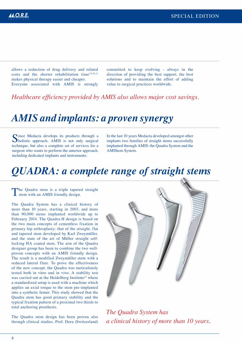
the Quadra stem is a triple tapered straight stem with an AmIS friendly design.
the Quadra System has a clinical history of more than 10 years, starting in 2003, and more than 90,000 stems implanted worldwide up to February 2014. the Quadra-H design is based on the two main concepts of cementless fixation in primary hip arthroplasty: that of the straight, flat and tapered stem developed by Karl Zweymüller, and the state of the art of müller straight self-locking HA coated stem. the aim of the Quadra designer group has been to combine the two well-proven concepts with an AmIS friendly design. the result is a modified Zweymüller stem with a reduced lateral flare. to prove the effectiveness of the new concept, the Quadra was meticulously tested both in vitro and in vivo. A stability test was carried out at the Heidelberg Institute12 where a standardized setup is used with a machine which applies an axial torque to the stem pre-implanted into a synthetic femur. this study showed that the Quadra stem has good primary stability and the typical fixation pattern of a proximal two thirds to total anchoring prosthesis.
the Quadra stem design has been proven also through clinical studies. prof. dora (Switzerland)
Amis and implants: a proven synergy
QUADRA: a complete range of straight stems
Healthcare efficiency provided by AMIS also allows major cost savings.
allows a reduction of drug delivery and related costs and the shorter rehabilitation time3,9,10,11
makes physical therapy easier and cheaper.everyone associated with AmIS is strongly
committed to keep evolving - always in the direction of providing the best support, the best solutions and to maintain the effort of adding value to surgical practices worldwide.
The Quadra System has a clinical history of more than 10 years.
Since medacta develops its products through a holistic approach, AmIS is not only surgical
technique, but also a complete set of services for a surgeon who wants to perform the anterior approach, including dedicated implants and instruments.
In the last 10 years medacta developed amongst other implants two families of straight stems successfully implanted through AmIS: the Quadra System and the AmIStem System.

9
m.o.r.e. Journal - march 2013, SupplementProven accuracy and effectiveneSS of myKnee®M.O.r.e. JOurnal 15TH annIVerSarY
Based on the clinical experience of Quadra (straight, rectangular, cementless hip stem
by medacta), in 2009 medacta developed the AmIStem System. the AmIStem is the first stem specifically designed for AmIS. the AmIStem differs from the Quadra in offering a reduced shoulder and a reduction in length of 15%, facilitating the processes of broaching and stem insertion. Following the principles of responsible innovation, the AmIStem has been meticulously designed and tested both in vitro and in vivo. A stability test was carried out at the Heidelberg Institute16 where a standardized setup is used with a machine which applies an axial torque to the stem pre-implanted into a synthetic femur. According to this test, the AmIStem shows good primary stability and the typical fixation pattern of a proximal two thirds anchorage stem. the reduction of the shoulder allows for easier implantation with the AmIS approach. to prove the comparability of Quadra and AmIStem, dr. Nolde17 (orthopraxx - mümchen, Germany)
Amistem: a logical evolutionof hip stem design
published his personal series of over 100 cases at the SGo (Société Suisse d’orthopédie et de traumatologie), demonstrating that at a minimum follow up of 5 years the survival rate is 100% considering aseptic loosening as an end point13. dr. moreau collected clinical outcomes from his series of 97 stems, implanted between January 2003 and January 2005. results demonstrate that at 7 years follow up the survival rate of the Quadra stem is 100% considering aseptic loosening as an end point14. the Australian orthopaedic Association NJrr report 201315 also shows excellent results with the Quadra stem. the Australian data shows:• Quadra-H is the 2nd most implanted cementless
stem in Australia for 2011 and 2012 (with more than 1,800 stems implanted in 2012, 95%
More than 60,000 AMIStem have been implanted worldwide.
through the AmIS approach), only 5 years after its introduction in the Australian market.
• Quadra is the 3rd most implanted stem in primary total conventional hip replacement when cemented stems are included.
Quadra stem numbers are increasing year by year, with a growth of 33% from 2011 to 2012. considering that Quadra was introduced in Australia together with the AmIS approach in late 2007, these results clearly demonstrate the appeal and effectiveness of medacta hip implants and the AmIS approach with its unique education program. these positive outcomes are the welcome consequence of all the efforts of medacta to invest in responsible innovation and effective ongoing surgeon training and proctorship.
performed a randomized clinical study on 40 patients. the aim was to compare the bone mineral density of the AmIStem and Quadra femoral components through a deXA analysis at one year. the study, presented at the european Hip Society in 2012, demonstrated that the two stems are statistically equivalent in all zones at any time-point, with the exception of Gruen zone r2 and r7, in which a positive trend for AmIStem was recorded at 6 months.AmIStem has been on the market since 2009 and more than 60,000 AmIStem have been implanted worldwide up to April 2014.the AmIStem implant perfectly matches with the AmIS dedicated instruments, designed to facilitate bone preparation for prosthesis implantation and to optimize the approach. Approach-dedicated instruments and self-retaining retractors have been introduced in order to avoid the surrounding soft tissues and potentially reduce the risk of nerve or muscle damage18. medacta constantly strives to improve its instrument design and effectiveness, to adapt to surgeon’s needs.
10
special edition.O.R.E.I n s t I t u t e
Amis clinical benefits
As demonstrated in the literature, the anterior approach is the only technique which follows
a path both intermuscular and internervous therefore considerably reducing the risk of damaging periarticular structures such as muscles, tendons, vessels and nerves. For this reason, the AmIS technique is the ideal approach for the patient, offering a less traumatic surgical procedure, fundamental for a painless fast recovery and a quick return to daily activities. thanks to the fact that potentially no muscles are cut, the patient can benefit from: shorter hospitalization time1,2,19-21, significantly shorter rehabilitation20-23, less blood loss2,20,23, reduced post-operative pain19,20,23, faster return to daily activities19-21,24 and improved cosmesis2,19,20,23. In addition, hospitals and surgeons can benefit from multiple savings. It has been proven that AmIS can effectively provide short-term clinical benefits claimed for mIS surgery when compared to standard approaches11. many studies have been successfully conducted and others are still ongoing to prove the real benefits of AmIS for patient well-being.
moreover, the literature shows evidence that the advantages of using the AmIS approach are not limited to the short term.
An article published on the Journal of Bone & Joint Surgery (Br) (now called the Bone & Joint Journal) by dora c. et al. showed that at one year after surgery there is less symptomatic and asymptomatic muscle degeneration for patients operated using AmIS compared with other conventional approaches. this means for the patients no residual trochanteric pain and limping in daily activities and advantages in case of revision. other studies have also confirmed these findings23,24,25.
revision through AmIS approach has been extensively investigated by laude F. et al. in an article published in the Journal of Bone & Joint Surgery (Am)5. A preserved soft-tissue structure could be advantageous in case of revision.
Soft-tissue changes in hip abductor muscles and tendons after total hip replacementBremer AK, Kalberer F, Pfirrmann CWA, Dora CJ Bone Joint Surg (Br) 2011–July; 93-B:886-9.
Overall, the AMIS group presented significantly less alterations of the abductor structures. There was no register of full-thickness tears or detachments on the AMIS group while the control group presented them twice involving the tendon of gluteus minimus, and four times involving the lateral part of the gluteus medius. According to the odds ratio calculated, there was an increased risk seven times higher of abductor insertion alterations and an increased risk five times higher of collecting bursal fluid within the trochanteric region, when a transgluteal approach was used. The AMIS group displayed significantly less fatty atrophy of the gluteus medius and gluteus minimus.
It is important to recognize that although many MR imaging findings such as altered signal intensity and abductor tendon diameter, bursal fluid collections, and fatty atrophy of the anterior gluteus minimus muscle are more frequent in symptomatic patients, they are also frequently found in asymptomatic patients after lateral transgluteal THR14.
These results confirms that the AMIS approach allows for less damage of the pelvitrochanteric muscles and tendons and no residual trochanteric pain and limping when compared to the transgluteal approach.
Residual pain and damage to soft tissues after total hip replacement are strictly connected14. This publication aims to compare, through MRI, the muscle and tendons damage produced by the AMIS approach compared to the transgluteal approach. Two groups of patients underwent primary THR: 25 patients were operated through the AMIS approach, and others 25 using a transgluteal approach. All patients underwent a post-operatively MRI at one year using the same 1.5-T system and protocol. The goal was to assess the presence of bursal fluid, the amount of damage to the abductor tendons and the grade of fatty atrophy of the abductor muscles.
The literature shows evidence that the advantages of using the AMIS approach are not limited to the short term.

11
m.o.r.e. Journal - march 2013, SupplementProven accuracy and effectiveneSS of myKnee®M.O.r.e. JOurnal 15TH annIVerSarY
results of the anterior approach are supported by good preliminary results of the AmIStem.
the AmIStem was implanted in the University hospital of Geneva, Switzerland (HUG – Hopital Universitaire Genève) beginning in April 2009 for total hip replacement (tHr). dr. christofilopoulos, prof. Hoffmeyer and prof. peter conducted a prospective cohort study18 comparing 617 AmIStem with 364 Spotorno stems at 2 years follow-up. the AmIStem showed short-term complication rates comparable to or slightly lower than the Spotorno stem. At 2 years good radiological and clinical results were observed.dr. Kalberer (Kantonsspital Winterthur – Winterthur, Switzerland) is performing a clinical
study on AmIStem-H26 to evaluate at one year follow-up any sign of radiolucency on the femur according to Gruen classification and the possible presence of pain. to date dr. Kalberer (who has performed more than 2000 AmIS procedures) and his junior consultant dr. Schmied (during her AmIS learning curve) have collected data on 266 patients with at least one year follow up. preliminary analysis reports radiological results similar to the Quadra stem results reported at five years by prof. dora13.
Improved radiological results have been achieved by:• minimizing bone removal from the Great
trochanter compared to traditional flat tapered wedge implants (Quadra by medacta, corail by depuy, taperloc by Biomet,…)
• Adopting a less aggressive rehabilitation protocol, to facilitate the bone-implant osteointegration process, by extending the use of crutches during first post-operative weeks.
After 10 years of success, a uniquely holistic approach has encouraged successful diffusion of the AmIS technique all over the world and made medacta the “Best in class” in anterior approach education thanks to the m.o.r.e. Institute.
Revision Total Hip Arthroplasty Performed Through the Hueter Interval Mast NH, Laude FJBJS Am. 2011; 93(Suppl.2):143-148.
Mast NH and Laude F15 investigate the possible benefits of anterior approach in case of complex or revision total hip arthroplasties, eventually using anterior approach extensions when needed. The study also evaluates the advantages of AMIS approach for the THR prior to revision. A consecutive series of fifty-one patients who underwent revision THR through the AMIS approach has been retrospectively reviewed, with a mean follow-up of 54.5 months (range of 8 to 160). The cases reported involve revision of acetabular components alone (41%), femoral components alone (2%), acetabular and femoral components (41%) and resurfacing systems (16%).
For all the revision cases reported, a good post-operative function was generally obtained (medium WOMAC score of 83), especially in case of revision of failed resurfacing systems (WOMAC>95). No post-operative dislocations have been reported.
A revision through the anterior approach after a primary posterior or anterolateral approaches can results in less scare removal during the approach, making the surgical procedure easier. In case of primary total hip replacement performed through an anterior approach, the revision can potentially access through the same anatomic path, preserving major structures of the
hip joint and consequently allowing for possible quicker recovery. An acetabular-only revision can usually be treated with a standard exposure for a primary anterior approach, with a similar post-operative recovery and rehabilitation process.
In conclusion, the study demonstrates that the anterior approach for revision THR provides advantages for the patient and for the surgeon. The anterior approach can be used for complex or revision THR with good outcomes in terms of WOMAC score and hip stability.
After 10 years of success, a uniquely holistic approach has encouraged
successful diffusion of the AMIS technique all over the world and made
Medacta the “Best in Class” in anterior approach education.

12
special edition.O.R.E.I n s t I t u t e
1. Rachbauer F, Kain MSH, Leunig M. The History of the Anterior Approach to the Hip. Orthop Clin North Am. 2009 Jul;40(3):311-20.2. Kreuzer S, Matta JM. Single-incision anterior approach for total hip arthroplasty: Smith-Petersen approach. O’Connor MI, Griffin LY, eds., Limited Incisions for Total Hip
Arthroplasty, AAOS, Rosemont, 1-14, 2007.3. Laude F, Moreau P, Vié P. Arthroplastietotale de hanche par voieantérieure de Hueter mini-invasive. Maîtrise Orthopédique 2008( 179):6-11.4. Laude F. Total hip arthroplasty through an anterior Hueter minimally invasive approach. Interact Surg (2006) 1: 5-11.5. Mast NH, Laude F. Revision Total Hip Arthroplasty Performed Through the Hueter Interval. JBJS Am. 2011; 93(Suppl.2):143-148.6. F Laude, E Lesur. The minimally invasive trend in total hip arthroplasty through the anterior approach. Encyclopédie Médico-Chirurgicale (2004) 44-667-B.7. F Rachbauer. Minimally Invasive total hip arthroplasty: anterior approach. Orthopade. 2005 Nov;34(11):1103-4, 1106-8, 1110. 8. Goebel S, Steinert AF, Schillinger J, Eulert J, Broscheit J, Rudert M, Nöth U. Reduced postoperative pain in total hip arthroplasty after minimal-invasive anterior approach.
Int Orthop. 2012 Mar;36(3):491-8.9. Siguier T, Siguier M, Brumpt B. Mini-incision anterior approach does not increase dislocation rate: a study of 1037 consecutive total hip replacements. Clin Orthop
Relat Res. 2004 Sep;(426):164-73.10. Bremer AK, Kalberer F, Pfirrmann CWA, Dora C. Soft-tissue changes in hip abductor muscles and tendons after total hip replacement: Comparison between the direct
anterior approach and the transgluteal approaches approaches. J Bone Joint Surg Br. 2011 Jul;93(7):886-9.11. Christofilopoulos P. Economical aspect of the AMIS Procedure. Presentation. World Conference on Anterior Hip Approach, 10-12 Sept 2009, Zurich, Switzerland.12. Heidelberg Lab-Report, Orthopädische Universitätsklinik Heidelberg, 2005. Data on file Medacta.13. Müller DA, Zingg P, Dora C. 5 year survival and radiological outcome of minimally invasive total hip replacements using a relatively new implant (Quadra®/
Versafitcup®, Medacta®, Switzerland. 71. Congrès annuel de la Société Suisse d’Orthopédie et de Traumatologie, Lausanne, 22– 24 June 2011.14. Moreau P. Cementless HA coated Quadra® stem – 7 years clinical outcomes. M.O.R.E. Journal 2012, Vol. 2: 3-6.15. AOA NJRR Annual Report, 2013.16. Bernardoni M, Siccardi F, Quagliana I, Grimoldi G. Mechanical stability of the AMIStem a standardized in-vitro analysis. M.O.R.E. Journal, 2011, May; Vol.1: 8-11.17. Petridis G, Scherer M, Gradinger R, Beck J, Nolde M. Prospective Randomized Study comparing Bone Mineral Density (DEXA) using the AMIStem or the Quadra stem.
10th European Hip Society Meeting, Milan, 2012.18. HUG – Hopital Universitaire Genève Internal report.19. Pfirrmann CWA, Notzli HP, Dora C, Hodler J, Zanetti M. Abductor tendons and muscles assessed at MR Imaging after total hip arthroplasty in asymptomatic and
symptomatic patient. Radiology. 2005 Jun;235(3):969-76.20. Bremer AK, Kalberer F, Pfirrmann CWA, Dora C. Soft-tissue changes in hip abductor muscles and tendons after total hip replacement: Comparison between the direct
anterior approach and the transgluteal approaches approaches. J Bone Joint Surg Br. 2011 Jul;93(7):886-9.21. C Dora, C Pfirmann, H Nötzli, J Hodler, M Zanetti. MR imaging of the abductor tendons and muscles after total hip replacement in asymptomatic and symptomatic
patients. EFORT 2007, Italy, Florence, 11-15 May 2007.22. Müller DA, Zingg PO, Dora C. Anterior minimally invasive approach for total hip replacement: five-year survivorship and learning curve. Hip Int. 2014. 23. Dora C. Der anteriore Zugang für die minimal-invasive HTPE. Leading Opinions, Orthopädie 1, 2006. 24. Dora C. Minimalinvasive Zugängean an der Hüfte (Minimally invasive approaches in hip surgery). Orthopäedie Mitteilungen 6/07, 574-576.25. Dora C, Bremer A, Kalberer F, Pfirrmann C. Muscular damage after total hip arthroplasty: conventional versus minimally invasive anterior approach. Podium Presentation
at the 68th Annual Scientific Meeting of the AOA 2008, Hobart, Australia, October 12-16, 2008. 26. F. Kalberer, “Preliminary Results on AMIStem Radiographic analysis”, Medacta White Paper, 2013
References

13
m.o.r.e. Journal - march 2013, SupplementProven accuracy and effectiveneSS of myKnee®M.O.r.e. JOurnal 15TH annIVerSarY
Learn AMIS and MyKneewith full confidence
The M.O.R.E. Institute has been created to provide continuous support to healthcare
professionals in the field of Research and Education.
The M.O.R.E. Institute offers Surgeon to Surgeon educational opportunities to share
experience and improve patient outcome.

14
special edition.O.R.E.I n s t I t u t e
Joint replacements are well-proven and established procedures. However, their outcomes
are strongly dependent on the technical success of the surgery, and component malalignment may lead to early failure of the procedure.over recent years, medical technology has tried to address this problem by proposing various solutions to optimize the surgical procedure and improve outcomes and patient satisfaction. In the 1990s computer Assisted Surgery, either robotics or navigation systems, enjoyed some popularity as they were proven to be extremely accurate in implant positioning, which correlates with improved survival rates of prosthetic joint replacement and better functional outcomes. despite the undisputed benefits of providing accurate implant positioning, cAS systems have some limitations that have limited adoption on large scale: intraoperative set up of the system potentially increasing the operative time, troubleshooting in the operative room, significant learning curve and high cost.In 2000s the advances of fast prototyping technology and 3d printing, opened the door to patient matched technology with the goal of replicating the anatomy of human bones thanks to 3d printing technology that can melt powder of medical grade polyamide or similar materials thanks to a laser sintering beam.the potential of this technology was exploited and adopted in orthopaedics. Starting with a 3d bone model, patient specific templates can be manufactured to assist the surgeon in positioning conventional metal instruments accurately on reference surfaces of joints.
The beginning of Medacta’s patient matched technology
medacta recognised that this technology perfectly matches its vision of improving patient well-being and healthcare sustainability, providing precision and good outcomes and, at the same time, allowing for time and cost savings. In 2008 myKnee, patient matched cutting blocks for tKr, were born. the technology was completely developed in house by medacta in cooperation with an international team of designer surgeons from europe and the United States.After an incredibly rapid development period, in September 2009 the first GmK primary surgery with myKnee patient specific cutting blocks took place in Switzerland, initiating an extensive clinical evaluation aimed at testing and validating the outcomes of a promising, but new technology.
New technology adoption without extensive testing and validation can expose patients to the risk of unintended consequences and new modes of failure. medacta’s belief in rigorous and continuous medical education, confirmed by the success of m.o.r.e. AmIS education program, was adopted in the launch of the myKnee System. A comprehensive and continuous education program is offered to myKnee users, including worldwide proctorship, wetlab courses and live surgeries, with the aim to allowing surgeons to smoothly adopt the myKnee technology and take advantage of its accuracy and effectiveness.
Medacta’s patient matched technology was conceived to provide assured precision and consistent outcomes to patients and surgeons along with delivering enhanced cost-effectiveness to hospitals and healthcare systems.MyKnee is the core of Medacta’s patient matched technology. 5 years of clinical success and 15,000 surgeries have established it as one of the most frequently used patient matched system in TKr, able to deliver proven accuracy in component alignment with demonstrated cost-effectiveness.MyKnee was the beginning of a comprehensive patient matched offering from Medacta, including MyHip solutions for hip replacement, MySpine for spinal surgery and MyShoulder for shoulder replacement.
This technology perfectly matches Medacta’s vision of improving patient well-being and healthcare sustainability.

15
m.o.r.e. Journal - march 2013, SupplementProven accuracy and effectiveneSS of myKnee®M.O.r.e. JOurnal 15TH annIVerSarY
Since the first surgeries the accuracy and effectiveness of myKnee was clear and myKnee has enjoyed a continuous increase in popularity, becoming one of the most frequently used patient matched technology in tKr, with more than 15,000 cases performed across europe, the USA, Australia, Japan and other markets.
the significant knowledge in patient matched technology gained from the myKnee program and the data and clinical evidence that it provided, convinced medacta that it would be appropriate to enlarge the range of solutions for patient matched knee surgery.
5 years of clinical success and 15,000 surgeries have established
MyKnee as one of the most frequently used patient matched system in TKR.
In 2011, myKnee for minimally invasive approaches, called myKnee mIS and myKnee for unicompartmental knee replacement, called myKnee UNI were developed. In 2012, medacta launched myKnee lBS - the first and only patient matched technology with an integrated ligament balancing system. this allows surgeons to combine the precision and the effectiveness of patient matched instrumentation with the opportunity to make intra-operative adjustments to balance the knee.
the experience gained over several years in patient matched technology and the excellent results of the various myKnee options encouraged medacta export the technology into other specialties where it is now being validated, such as Hip, Spine and Shoulder surgery.

16
special edition.O.R.E.I n s t I t u t e
What works and what doesn’t?
the advantages of patient matched technology persuaded almost every major orthopaedic
company to offer a competing system. However, published results after nearly 5 years of patient matched technology in tKr surgery are inconsistent and the majority of the published results do not support the claims of an increase in accuracy that patient matched technology was developed to provide2-22. Some systems do not achieve statistical significance in reducing the number of outliers in restoring natural alignment of the lower limb and this has discouraged market adoption of patient specific technology despite its initial promise.By contrast, myKnee has proven to be clinically successful23-35, proving yet again that in orthopaedic technology, what appear to be small differences can create significant variations in efficacy. Some technical choices in the design of myKnee have made the difference and can explain the reason for its consistent success.
the first of these is the choice of imaging technology, mainly ct rather than mrI based. ct has been proven to be an accurate and straightforward technology to achieve consistent and reproducible results in patient matched technology1 thanks to clear image processing, limited examination time minimizing potential artefacts, wide availability and possible application also where there is pre-existing metal hardware around the joint. mrI is a more demanding technology, with comparable outcomes only achievable if high-performing mrI devices (3t) are used and set up is meticulous.As well as scanning technology, the alignment reconstruction algorithm has a big influence on success. Aligning the prosthesis to the mechanical Hip-Knee-Ankle (HKA) axis, calculated by computer simulation helps in obtaining a reliable axis to assure implant longevity.
methods such as kinematic alignment and the adjunctive use of long leg X-rays have exhibited poor results that have discouraged adoption of a sound base-technology.
myKnee was developed to be used either as an actual resection block or a pin placement guide. the possibility of cutting directly through the myKnee block further reduces the margin of error as it minimizes the number of surgical steps to perform and potential for “stacked errors.”
In addition, a close interaction between the surgeon and his own personal myKnee technician is another key factor. this constant relationship helps a specific technician to develop an understanding of the preferences and needs of each surgeon, leading to preoperative planning that is a faithful reproduction of what the surgeon wants to achieve. From a logistical point of view, the myKnee process is highly efficient as it ensures the shortest lead time in the current market for this technology: only 3 weeks between the receipt of a ct scan and the day of surgery.
myKnee is an innovation that delivers excellent results to the Health care System, helping the surgeon to achieve assured accuracy and patient satisfaction.
MyKnee is an innovation that delivers excellent results to the Health Care System, helping the surgeon to achieve assured accuracy and patient satisfaction.

17
m.o.r.e. Journal - march 2013, SupplementProven accuracy and effectiveneSS of myKnee®M.O.r.e. JOurnal 15TH annIVerSarY
From the beginning of the myKnee project, medacta committed to extensive and rigorous
clinical evaluation, aimed at testing and validating the intentions, claims and outcomes of the technology.It appears that amongst all patient matched technologies available in the market, myKnee is the only one that consistently reports positive outcomes in its studies, showing proven accuracy and effectiveness in more than 10 publications23-35. most of these studies radiologically assess the accuracy of implant positioning achieved with myKnee. Some address the economic advantages anticipated with the technology.
the obvious way to assess the accuracy of final implant positioning is to compare the results of patient matched technology to those obtained by other known techniques, such as conventionally instrumented or cAS procedures. Four studies compared myKnee cutting blocks with conventional and navigated instrumentation. Koch et al. reported that ct-based patient-specific myKnee cutting blocks provide accurate and constant radiological data with the number of outliers >3° deviated from the coronal mechanical axis comparable to the results achieved and published with computer-assisted tKr and clearly better than that obtained using conventional instrumentation. these results don’t vary with the learning curve, demonstrating that myKnee seems to provide a safe implantation technique not only for experienced but also for less experienced surgeons performing fewer tKr’s23.léon et al. were able to demonstrate the accuracy and reliability of the myKnee system comparing the outcomes with those obtained with conventional and cAS procedures. In their study, myKnee was shown to be most accurate of the three instrumentation options, particularly with regard to outliers24.Nabavi et al. reported that myKnee ct-based patient specific cutting blocks produced statistically improved clinical outcomes compared to a conventional approach in the short term after
the total Knee replacement surgery (12 months). the study was also observed that the transfusion rate in the ct based cutting blocks group was lower than in standard technique25.Finally, ponziani et al. investigated two main preoperative procedures: a ct-based protocol with myKnee system and an mrI-based protocol with the Visionaire system by analysing their critical points, advantages and disadvantages. cases of missing congruence were observed with the mrI-based preoperative procedure. In general short term outcomes from the pmt technique and conventional technique were almost comparable26.
Another parameter to evaluate the accuracy of patient matched technology is the comparison between the planned and the actual resections. Koch et al. reported 98% accuracy of the planned component size23. positive results were also achieved by dussault et al. the authors describe satisfying results related not only with implant size matching, but also with the planned resections27.
postoperative analysis allows the ultimate evaluation of surgical precision. Baldo et al. evaluated the preoperative planning reliability with ct scans in the postoperative period. preliminary results revealed a greater preoperative planning reliability with higher accuracy of anatomical reconstruction with myKnee28. the same positive feedback was reported by müller et al. their preliminary radiological results reported an accurate match between the preoperative plan and postoperative results. Good implant alignment was achieved, with a mean HKA angle of 179.6°. the authors concluded that myKnee technology is a reliable and straightforward technique, with high possibility of reducing operative time29. Another prospective study with the myKnee was reported by Goldberg et al.30 the study analyses the accuracy of myKnee (100 tKr’s) through a prospective analysis of postoperative standard long-standing X-ray. the results of the study show that ct-based myKnee cutting blocks provide a
This one works: proven accuracy and effectiveness of myKnee
Amongst all patient matched technologies available in the market, MyKnee is the only one that consistently reports positive outcomes in its studies,
showing proven accuracy and effectiveness in more than 10 publications.
18
special edition.O.R.E.I n s t I t u t e
post-operative neutral alignment in 95.9% cases, with an average of 179.36°. the same authors analysed the accuracy of myKnee ct-based cutting blocks (12 tKr’s) when operating on patients with different types of hardware in or in proximity to the knee joint (staples, plates, screws, rods).31 they experienced a short tourniquet time (42 min on average) with all patients within 3° neutral alignment, showing that ct-based patient matched technology, unlike that using mrI, can be successfully used to tackle cases with pre-existing hardware adjacent to the knee.
Finally, accuracy of the myKnee UNI cutting block has been analysed by dr. Helmy through the analysis of postoperative ct scans. 28 medial UKAs (Unicompartmental Knee Arthroplasty)were analysed by comparing the tibial resections and tibial implant positioning with the preoperative planning. the results show excellent accuracy of tibial position in terms of varus/valgus (∆0.3° ± 1.7°), posterior slope (∆1.1° ± 2.6°) and external rotation (∆1.5° ± 3.3°). Moreover the authors suggested that UKAs with patient-specific cutting blocks provided good outcomes comparable with those obtained with robotic-guided UKAs32.
other studies published on myKnee aim to demonstrate the economic benefits of patient matched technology. most authors agree that this novel technology has the potential of reducing surgical steps and operative time, therefore improving o.r. logistics and turnover in addition to the reduction of costs associated with instrument sterilization. one other theoretical benefit concerns the potential increase of cases per day or session due to enhanced efficiencies.
dr. Goldberg addressed potential economic benefits from utilizing myKnee technology. In addition to the alignment advantages, reduction of outliers (more homogeneous results), accuracy, and very good implant size matching, the author reported that with reduced operative and set-up times with myKnee, the possibility of an increase of 2 cases per week would exist. reporting a profit of $2,500 per case, a potential profit of $230,000 could be realized at this Hospital33. Koch demonstrated myKnee economic benefits as well. In a description of myKnee technique, the author
discusses the reduced set-up times and turnover, which allow a decrease in the costs of sterilization, operative time, and o.r. utilization. moreover, the author claims that myKnee enables a more efficient surgery, simpler not only for the surgeon but also for his operative team34. the economic benefits of myKnee were also studied by dr. Gagna. He revealed that a well-planned surgery with myKnee will allow the hospital to reduce costs associated with sterilization (-60%), o.r. time usage (-10min), hospital stay (-20%) and transfusions (-43%)35.
In conclusion, myKnee technology, with a preference to ct image acquisition, has proven to allow precise preoperative planning, correct alignment, excellent implant size matching, less instrumentation trays needed, reduced surgical step and surgical time resulting in improved o.r. efficiency. the use of myKnee technology appears to be universally supported by all publications which describe its use. these include improvement of the postoperative mechanical alignment, cost reduction and effective clinical results. myKnee, after 5 years of successful outcomes with more than 15,000 procedures, appears to prove that it can achieve the intended goals that the industry has identified for patient matched technology.
MyKnee technology, with a preference to CT image acquisition, has proven to allow precise preoperative planning, correct alignment, excellent implant size matching, less instrumentation trays needed, reduced surgical step and surgical time resulting in improved O. R. efficiency.

19
m.o.r.e. Journal - march 2013, SupplementProven accuracy and effectiveneSS of myKnee®M.O.r.e. JOurnal 15TH annIVerSarY
1. White D et al. Accuracy of MRI vs CT imaging with particular reference to patient matched templates for total knee replacement surgery, IJMRCAS 2008; 4: 224 -23.2. Daniilidis K et al., Frontal plane alignment after total knee arthroplasty using patient-specific instruments, Int Orthop. 2013 Jan;37(1):45-50.3. Nam D et al, Patient-specific instrumentation in total knee arthroplasty: A review, J Knee Surg 2012; 25: 213-220.4. Webb J et al., Early experience with customized patient instrumentation in knee arthroplasty. 12th EFORT Annual Meeting, Copenhagen, June 1-4, 2011.5. Stronach B et al., Patient-Specific Instrumentation in TKR Required Frequent Surgeon Directed Intraoperative Changes. AAOS Annual Meeting, San Francisco, CA,
February 2012.6. Ng VY et al., Improved accuracy of alignment with patient-specific positioning guides compared with manual instrumentation in TKR, Clin Orthop Relat Res (2012)
470:99-107.7. Boonen B et al., Preliminary experience with the patient-specific templating total knee arthroplasty: 40 cases compared with a matched control group, Acta Orthopaedica
2012; 83 (4): 387-393.8. Nam D et al., Patient Specific Instrumentation versus Large-Console, Computer-Assisted Navigation in Total Knee Arthroplasty, 22nd AAHKS Annual Meeting, Dallas,
USA, November 2-4 2012 .9. Hamadouche M et al., RCT Multicenter Comparison of Primary TKR using Patient Specific versus Conventional Instrumentation, 22nd AAHKS Annual Meeting, Dallas,
USA, November 2-4 201210. Stronach B et al., Patient-Specific Instrumentation in Total Knee Arthroplasty Provides No Improvement in Component Alignment. AAOS Annual Meeting, San Francisco,
CA, February 2012.11. Lustig S et al., Unsatisfactory Accuracy as Determined by Computer Navigation of VISIONAIRE Patient-Specific Instrumentation for Total Knee Arthroplasty. J Arthroplasty.
2013 Mar;28(3):469-73. 12. Misur P et al., The accuracy of implant positioning using the Visionaire patient matched knee arthroplasty system. J Bone Joint Surg Br 2012, 94-B (XLI 38). 13. Klatt BA et al., Custom-Fit Total Knee Arthroplasty (OtisKnee) Results in Malalignment. J Arthroplasty 2008, 23(1): 26-29.14. Coolican M et al., Evaluation of the Zimmer PSI system for total knee arthroplasty using computer navigation, Arthroscopic 2013 Oct; 29(10):e169.15. Nunley RM et al., Do patient-specific Guides Improve Coronal Alignment in Total Knee Arthroplasty? Clin Orthop Relat Res (2012) 470:895-902.16. Nunley RM et al., Are patient-specific cutting blocks cost-effective for total knee arthroplasty? Clin Orthop Relat Res (2012), 470:889-89417. Hilliard L et al., Malalignment after patient matched, 125th AOA Annual Meeting – Washington DC, USA, June 27-30, 2012 .18. Parker D et al., Evaluation of the Visionaire instrumentation for total knee arthroplasty using computer navigation. J Bone Joint Surg Br 2012; 94-B(XLI 37). 19. Conteduca F et al., Are MRI-based, patient matched cutting jigs as accurate as the tibial guides? Int Orthop. 2012 Aug;36(8):1589-93.20. Conteduca F et al., Evaluation of the accuracy of a patient-specific instrumentation by navigation, Knee Surg Sports Traumatol Arthrosc. 2013 Oct;21(10):2194-9.21. Noble J W et al., The Value of Patient-Matched Instrumentation in Total Knee Arthroplasty, J Arthroplasty. 2012 Jan;27(1):153-5.22. Spencer B A et al., Initial experience with custom-fit total knee replacement: intra-operative events and long-leg coronal alignment, Int Orthop. 2009 Dec;33(6):1571-5. 23. Koch PP, Müller D, Pisan M, Fucentese SF, Radiographic accuracy in TKR with a CT-based patient-specific cutting block technique, Knee Surg Sports Traumatol Arthrosc.
2013 Oct;21(10):2200-5. 24. Leon V, Patient matched technology vs conventional instrumentation and CAS, 13th EFORT Annual Meeting, Berlin, May 23-25, 2012.25. Nabavi A. A prospective study comparing early results of TKR performed utilizing CT-based custom made cutting blocks with a standard technique, 14th EFORT Annual
Meeting, Istanbul, Turkey, June 5-8, 201326. Ponziani L et al. Patient matched technology nella chirurgia protesica del ginocchio, Tecniche a confronto, Sphera medical journal, 2013.27. Dussault M, Goldberg T, Greenhow R, Hampton D, Parry S, Slimack M - Preoperative planning accuracy of MyKnee system. M.O.R.E. Journal 2012; 2:22-25.28. Baldo F, Boniforti B – Patient-specific cutting blocks for total knee arthroplasty: preoperative planning reliability. J Orthopaed Traumatol 2011; 12 (Suppl 1): S23-S88. 29. Müller DA et al., CT based patient-specific cutting blocks for total knee arthroplasty: technique and preliminary radiological results. 71st SSOT Annual Congress,
Lausanne, Switzerland, June 22-24, 2011.30. Goldberg TD et al., CT-Based Patient-Specific Instrumentation Is Accurate for TKR: A Single-Surgeon, Prospective Trial. Bone Joint Journal 2013; 95-B(SUPP 34): 325. 31. Goldberg TD et al, Ct-Based Patient-Specific Instrumentation Is Effective in Patients With Pre-Existing Hardware About the Knee. Bone Joint Journal 2013; 95-B(SUPP
34): 326.32. Dao Trong ML, Diezi C, Goerres G, Helmy N. Improved positioning of the tibial component in unicompartmental knee arthroplasty with patient-specific cutting blocks,
Knee Surg Sports Traumatol Arthrosc. 2014 Jan 17. [Epub ahead of print].33. Goldberg TD - MyKnee economical and clinical results. 6th M.O.R.E. International Symposium, Stresa, Italy, May 13-14, 2011.34. Koch P - MyKnee System : A new vision in total knee replacement. Leading Opinions - Orthopädie & Rheumatologie 2, 2011: 32-35.35. Gagna G - Aspects economiques de la technologie sur mesure MyKnee en chirurgie prothetique du genou, Maitrise Orthopedique 2012 Nov :14-15.
References

20
special edition.O.R.E.I n s t I t u t e
The origin of myHipperformed freehand or by the use of standard mechanical alignment guides (assembled in the shell impactor), even if it has been demonstrated that these might not be reliable methods14,15,16 to achieve adequate implant positioning. Various navigation systems have been developed to improve tHr outcomes, but the need for navigation equipment set-up, additional surgical time, associated learning curve, difficulties in incorporating pelvic tilting, and the high costs have impeded broad acceptance of these systems, despite the improvement in reported outcomes16,17. the femoral neck osteotomy is usually done freehand, based only on intraoperative landmarks and on preoperative radiographic templates, which can be difficult to do accurately18.
Uncompromising commitment towards responsible innovation devoted to increased patient well-being, motivated medacta to apply patient matched technology (successfully developed with the myKnee system) to the issue of component alignment in tHr.
medacta designed the myHip system to help the surgeon to address the continuing challenges
in total Hip replacement (tHr), focusing on patient well-being and improving accuracy in implant positioning and sizing, through:• precise 3d preoperative planning1,2 which can
halve the number of alignment outliers when compared to 2d templating3,4.
• patient-specific guides5,6,7 which can reproduce the 3d preoperative plan.
Appropriate final implant positioning is one of the most important factors to achieve good patient outcomes in tHr. Inaccurate acetabular component positioning could lead to an increased risk of dislocation, wear, neck/cup impingement and decreased range of motion (rom)8,9,10,11,12. Inappropriate positioning of the femoral and acetabular components can lead to leg length discrepancy (potentially associated with limping, neuro-muscular dysfunction, aseptic component loosening, less walking capacity and hip pain) 1,13 and femoral offset alteration (which could affect abductor strength, range of motion and hip pain and function)2. these problems could eventually lead to early implant failure and patient dissatisfaction.the positioning of the acetabular shell is usually
21
m.o.r.e. Journal - march 2013, SupplementProven accuracy and effectiveneSS of myKnee®M.O.r.e. JOurnal 15TH annIVerSarY
Uncompromising commitment towards responsible innovation devoted to
increased patient well-being, motivated Medacta to apply Patient Matched
Technology to the issue of component alignment in THR.
the myHip technology uses patient ct scans to enable the use of a tool to perform 3d
preoperative planning and 3d cinematic simulation of the rom of the patient’s hip procedure. the preoperative planning process, validated by the surgeon, is then used as the basis for the production of patient-specific femoral and acetabular guides.
the preoperative planning provides one of the greatest advantages of the myHip system. 3d preoperative planning greatly improves the accuracy of the final implant position, allowing also for an accurate restoration of leg length and of femoral offset19,20. It considers all the standard parameters used in the tHr practice, including rom assessment and pelvic tilt, thanks to a validated image processing protocol (ct and X-ray based).
the first step is the definition of the anatomical landmarks which will be used to perform the planning. these landmarks are the ones used during conventional surgery and are confirmed by today’s literature21,22,23,24,25,26. With the initial landmarks defined, it is possible to define implant related parameters, taking into account the patient’s anatomy and the surgeon’s preferences. pelvic tilt is considered at this step to minimise the risk of inaccurate implant positioning.
the implant parameters defined will be used by a myHip technician to finalize the first proposition for a preoperative plan submitted to the surgeon. A myHip Surgical planning report is produced each time a preoperative plan is proposed to the surgeon. this report provides a summary of all the information needed to perform a tHr: implant size expected (acetabular shell, femoral stem and femoral head neck length), ideal implant positioning (acetabular shell inclination and anteversion, femoral neck osteotomy level and inclination, femoral stem anteversion) and leg parameters (leg length and leg lateralization discrepancy). At this stage, a 3d kinematic simulation model is provided. this simulation shows the hip’s mobility through its full rom to effectively predict the performance of the in vivo implants. this way, it is possible to optimize patient’s functional outcomes by correctly selecting the size and position of the implant which, in turn, potentially avoids the risk of: impingement, squeaking with ceramic-on-ceramic bearing and edge loading during the patient’s daily activity. At this time, the surgeon has the possibility to review the 3d preoperative plan and, if desired, to adjust not only the implant characteristics but also the positioning parameters. After the surgeon’s approval of the 3d preoperative plan, patient specific guides can be manufactured.
myHip: accurate 3D preoperative planning, kinematic simulation of the Rom
and patient-specific guides
this technology could in fact also bring additional value to hip arthroplasty, enabling better patient outcomes though a more accurate implant position, optimizing overall procedure cost. these objectives led to the initiation of the myHip project in 2011, completely developed in-house in collaboration with an international team of designer surgeons. this technology can potentially help improve the accuracy of the femoral neck osteotomy and of acetabular shell positioning. trials have already commenced with very promising results in selected centers of excellence worldwide.

22
special edition.O.R.E.I n s t I t u t e
Medacta designed the MyHip system to help the surgeon address the continuing challenges in Total Hip Replacement, focusing on patient well-being and improving accuracy in implant positioning and sizing.
the use of patient-specific guides are expected to improve the implant positioning accuracy27,28,29. myHip has been designed to help the surgeon to restore patient anatomy and function, trying to minimize the potential for complications during the surgery. myHip guides are available both for anterior and posterior approach, according to surgeon preferences.
the myHip Femoral Guide is designed to define the level of the femoral neck osteotomy, allowing the surgeon to accurately resect the femoral neck, improving accuracy. the anatomical shape of the guide and its unique and stable positioning profile potentially enhances the reproducibility of the preoperative planning, guaranteeing an accurate femoral neck osteotomy. An accurate resection of the femoral neck potentially reduces the risk of leg length discrepancy and improves femoral offset restoration.
the myHip Acetabular Guide has been designed to define the correct orientation for acetabular reaming and implantation of the final acetabular shell. the myHip Acetabular Guide is placed inside the acetabulum and thanks to its unique and stable positioning potentially reproduces the parameters set during the preoperative planning. the guide allows placement of a rod that will be used as a local visual reference to indicate the correct orientation of acetabulum reaming and shell positioning. the reference rod provides orientation uncompromised by patient movement (because it is integral to the patient itself) and recognizes the variability of the patient’s pelvic tilt, in contrast to standard technique where the movement of the pelvis may mislead the surgeon.
M L
A P
Implant position in: A. Preoperative planning B. Original CT C. 3D kinematic simulation
C.
A.
B.

23
m.o.r.e. Journal - march 2013, SupplementProven accuracy and effectiveneSS of myKnee®M.O.r.e. JOurnal 15TH annIVerSarY
1. Austin MS, Hozack WJ, Sharkey PF, Rothman RH. Stability and leg length equality in Total Hip Arthroplasty. J Arthroplasty. 2003 Apr;18(3 Suppl 1):88-90.2. Cassidy KA, Noticewala MS, Macaulay W, Lee JH, Geller JA. Effect of femoral offset on pain and function after total hip arthroplasty. J Arthroplasty. 2012
Dec;27(10):1863-9.3. Koch PP, Müller D, Pisan M, Fucentese SF. Radiographic accuracy in TKA with a CT-based patient-specific cutting block technique. Knee Surg Sports Traumatol Arthrosc.
2013 Oct;21(10):2200-5.4. Schmalzried TP, Guttmann D, Grecula M, Amstutz HC. The relationship between the design, position, and articular wear of acetabular components inserted without
cement and the development of pelvic osteolysis. J Bone Joint Surg Am. 1994 May;76(5):677-88.5. Robinson RP, Simonian PT, Gradisar IM, Ching RP. Joint motion and surface contact area related to component position in total hip arthroplasty. J Bone Joint Surg Br.
1997 Jan;79(1):140-6.6. Kennedy JG, Rogers WB, Soffe KE, Sullivan RJ, Griffen DG, Sheehan LJ. Effect of acetabular component orientation on recurrent dislocation, pelvic osteolysis,
polyethylene wear, and component migration. J Arthroplasty. 1998 Aug;13(5):530-4.7. D’Lima DD, Urquhart AG, Buehler KO, Walker RH, Colwell CW Jr. The effect of the orientation of the acetabular and femoral components on the range of motion of
the hip at different head-neck ratios. J Bone Joint Surg Am. 2000 Mar;82(3):315-21.8. Lewinnek GE, Lewis JL, Tarr R, Compere CL, Zimmerman JR. Dislocations after total hip replacement arthroplasties. J Bone Joint Surg Am. 1978 Mar;60(2):217-20.9. Nishii T, Sugano N, Miki H, Koyama T, Takao M, Yoshikawa H. Influence of component positions on dislocation: computed tomographic evaluations in a consecutive
series of total hip arthroplasty. J Arthroplasty. 2004 Feb;19(2):162-6.10. Jolles BM, Zangger P, Leyvraz PF. Factors predisposing to dislocation after primary total hip arthroplasty: a multivariate analysis. J Arthroplasty. 2002 Apr;17(3):282-8.11. Del Schutte H Jr, Lipman AJ, Bannar SM, Livermore JT, Ilstrup D, Morrey BF. Effects of acetabular abduction on cup wear rates in total hip arthroplasty. J Arthroplasty.
1998 Sep;13(6):621-6.12. Widmer KH, Zurfluh B. Compliant positioning of total hip components for optimal range of motion. J Orthop Res. 2004 Jul;22(4):815-21.13. Röder C, Vogel R, Burri L, Dietrich D, Staub LP. Total hip arthroplasty: leg length inequality impairs functional outcomes and patient satisfaction. BMC Musculoskelet
Disord. 2012 Jun 11;13:95.14. Bosker BH, Verheyen CCPM, Horstmann WG, Tulp NJA. Poor accuracy of freehand cup positioning during total hip arthroplasty. Arch Orthop Trauma Surg. 2007
Jul;127(5):375-9. 15. Minoda Y, Ohzono K, Aihara M, Umeda N, Tomita M, Hayakawa K. Are acetabular component alignment guides for total hip arthroplasty accurate? J Arthroplasty.
2010 Sep;25(6):986-9. 16. Kelley TC, Swank ML. Role of navigation in total hip arthroplasty. J Bone Joint Surg Am. 2009 Feb;91 Suppl 1:153-8.17. Lin F, Lim D, Wixson RL, Milos S, Hendrix RW, Makhsous M. Limitations of imageless computer-assisted navigation for total hip arthroplasty. J Arthroplasty. 2011
Jun;26(4):596-605.18. Jeffers R, Ponneru R, Vanderstraeten L, Binns MS. A simple technique for translating the planned template to the intraoperative femoral neck cut using posterior head
resection during hip arthroplasty. J Bone Joint Surg Br 2008 vol. 90-B no. SUPP III 549-550.19. Sariali E, Mouttet A, Pasquier G, Durante E, Catone Y. Accuracy of reconstruction of the hip using computerised three-dimensional pre-operative planning and a
cementless modular neck. J Bone Joint Surg Br. 2009 Mar;91(3):333-40.20. Sariali E, Mauprivez R, Khiami F, Pascal-Mousselard H, Catonné Y. Accuracy of the preoperative planning for cementless total hip arthroplasty. A randomised
comparison between three-dimensional computerised planning and conventional templating. Orthop Traumatol Surg Res. 2012 Apr;98(2):151-8.21. Bittersohl B, Freitas J, Zaps D, Schmitz MR, Bomar JD, Muhamad AR, Hosalkar HS. EOS Imaging of the Human Pelvis: Reliability, Validity, and Controlled Comparison
with Radiography. J Bone Joint Surg Am. 2013 May 1;95(9):e581-9.22. Srimahi T, Muthukumar T, Anandarani VS, Sembian U, Subramanian R. A study on femoral neck anteversion and its clinical correlation. Journal of Clinical and
Diagnostic Research. 2012 April, Vol-6(2): 155-158.23. Zalawadia A, Ruparelia S, Shah S, Parekh D, Patel S, Rathod SP, Patel SV. Study of femoral neck anteversion of adult dry femora in Gujarat region. NJIRM 2010; Vol.
1(3). July-Sept.24. Murray DW. The definition and measurement of acetabular orientation. J Bone Joint Surg Br. 1993 Mar;75(2):228-32.25. Mayr E, Kessler O, Prassl A, Rachbauer F, Krismer M, Nogler M. The frontal pelvic plane provides a valid reference system for implantation of the acetabular cup.
Special orientation of the pelvis in different positions. Acta Orthop. 2005 Dec;76(6):848-53.26. Abe H, Sakai T, Hamasaki T, Takao M, Nishii T, Nakamura N, Sugano N. Is the transverse acetabular ligament a reliable cup orientation guide? Computer simulation
in 160 hips. Acta Orthop. Oct 2012; 83(5): 474–480.27. Small T, Krebs V, Molloy R, Bryan J. Comparison of Acetabular Shell Position Using Patient Specific Instruments vs. Standard Surgical Instruments: A Randomized Clinical
Trial. J Arthroplasty. 2013 Oct 16.28. Buller L, Smith T, Bryan J, Klika A, Barsoum W. The use of patient-specific instrumentation improves the accuracy of acetabular component placement. J Arthroplasty.
2013 Apr;28(4):631-6.29. Hananouchi T, Saito M, Koyama T, Sugano N, Yoshikawa H. Tailor-made Surgical Guide Reduces Incidence of Outliers of Cup Placement. Clin Orthop Relat Res.
2010 Apr;468(4):1088-95.
References
24
special edition.O.R.E.I n s t I t u t e
GMK Sphere: Stability for LifeTotal Knee Arthroplasty offers potential for significant improvement in order to improve patient satisfaction and meet expectations, to help them to forget the artificial joint and to restore normal knee function and stability. TKr patients perform significantly worse than THr patients in terms of recovery, post-operative pain and function.The GMK Sphere is an innovative implant designed to deliver maximum functional stability to the replaced knee with the goal of increasing TKr patient satisfaction and decreasing post-operative knee pain. The GMK Sphere provides a fully conforming medial compartment which has been shown in literature to provide high stability and enhanced patient satisfaction when compared to conventional knee designs. This implant combines the successful features of its predicate devices with many innovative characteristics which have the potential to provide advantages in the patello-femoral joint and could make the replaced knee move and feel more like a healthy knee.
Although knee replacement is considered a successful treatment, tKr patients are
not universally satisfied with the procedure and there is still room for improvement. Bullens et al. (2001) identified a poor correlation between the
traditional objective scores (Knee Society Score and radiologic assessment) and a subjective patient-assessed satisfaction score (Visual Analogue Scale score)1.
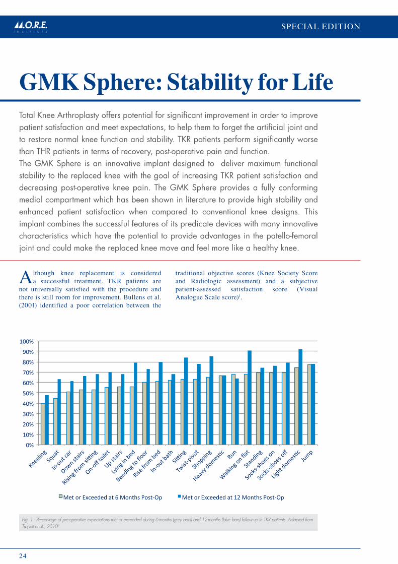
Fig. 1 - Percentage of pre-operative expectations met or exceeded during 6-months (grey bars) and 12-months (blue bars) follow-up in TKR patients. Adapted from Tippett et al., 20104.
0%
10%
20%
30%
40%
50%
60%
70%
80%
90%
100%
Kneeling
Squat
In-‐out car
Down stairs
Rising from siDng
On-‐off toilet
Up stairs
Lying in bed
Bending to floor
Rise from bed
In-‐out bath
SiDng
Twist-‐pivot
Shopping
Heavy domesSc Run
Walking on flat
Standing
Socks-‐shoes on
Socks-‐shoes off
Light domesSc Jump
Met or Exceeded at 6 Months Post-‐Op Met or Exceeded at 12 Months Post-‐Op

25
m.o.r.e. Journal - march 2013, SupplementProven accuracy and effectiveneSS of myKnee®M.O.r.e. JOurnal 15TH annIVerSarY
A UK study evaluated patient satisfaction using the oxford knee score on a cohort of 10,000 patients more than one year following total knee replacement: almost 20% were not satisfied after their tKr2.
recent studies show that patients’ pre-operative expectations are higher than their post-operative ability3. 98% of tKr patients expected to have major improvement in pain; at 12 months 93% reported less pain but this percentage decreased to 63% at the 5-years follow-up. 96% of tKr patients expected improvements in function during activities of daily living, but 90% and 61% of patients experienced improvements at 12-months and 5-years respectively. Also, expectations regarding leisure activities are higher than the outcomes: at 1-year only 24% of patients are able to go dancing and golfing although 41% had this expectation pre-operatively3.
A comparison between pre-operative expectations and post-operative satisfaction, using the Knee injury and osteoarthritis outcome Score (KooS), was performed by tippett et al. in 20104. patients’ expectations are not always realised after tKr, and many patients still report difficulties during normal daily living activities such as ascending and descending stairs, squatting, getting in/out of a car, getting in/out of the bath, running and performing domestic duties (Fig. 1).
patient satisfaction following tHr is higher when compared to tKr and a forgotten Hip is much more common than a forgotten Knee. In the study published by Bourne et al. tHr patients expressed greater overall satisfaction than tKr patients (tHr 89%, tKr 81%): in particular tHr patients expressed higher satisfaction in the ability to perform daily activities compared to tKr patients5. Following hip replacement patients are more satisfied compared to knee replacement (69% vs 51% declared themselves “very satisfied”) and patients are more willing to undergo another surgery5. moreover, the patient’s ability to forget the artificial joint in everyday life is greater following hip arthroplasty compared to tKr6
(Fig.2).
Fig. 2 - Mean Forgotten Joint score following Total Hip Arthroplasty (Grey bar) and Total Knee Arthroplasty (Blue bar). A higher score indicates greater patient ability to forget the artificial joint during daily life activities. Adapted from Behrend et al., 20126.
Fig. 3 - GMK Sphere: an innovative implant designed to deliver maximum functional stability to the replaced knee with the goal of increasing TKR patient satisfaction and decreasing post-operative knee pain.
TKR patients are not universally satisfied with the procedure
and there is still room for improvement.
44 46 48 50 52 54 56 58 60 62
Mean Forgo1en Joint Score
THA
TKA

26
special edition.O.R.E.I n s t I t u t e
0
5
10
15
20
25
30
Dancing
Sta/onary bik
ing
Swimming
Golfin
g
Stretc
hing
Moving
later
ally
Turning/cu
Bng
Carry
ing he
avy o
bjects
Sexual ac/vity
Leg-‐s
trengthe
ning e
xcercises
SquaBng
Garde
ning
Kneeling
Expe
rienc
ing sig
nific
ant d
ifficu
lty (%
)
TKA pa/ents Control subjects
Fig. 4 - Comparison between the percentage of TKR patients reporting significant difficulty in performing some daily life activities and the corresponding percentage in the control group (age- and gender-matched controls). Adapted from Noble et al., 20059.
Bachmeier et al. compared the outcomes following hip joint surgery and knee joint surgery and found that the improvement in pain and physical function was significantly greater in tHr patients7. Higher functional ability, especially in managing stairs, and less pain was reported after hip replacement compared with total knee replacement. Improvements occur more rapidly following tHr8.
Noble et al. wondered if tKr can restore normal knee function: patients who received a total knee implant still experienced significant difficulties during activities in daily life when compared to their age- and gender-matched peers9 (Fig. 4).
Improvement in pain and physical function was significantly greater following THR compared to TKR.
published data suggests that total Knee Arthroplasty offers potential for significant improvement in both the procedure and prosthesis design in order to increase patient satisfaction and meet expectations, to restore normal knee function and stability and to deliver a “forgotten joint.”

27
m.o.r.e. Journal - march 2013, SupplementProven accuracy and effectiveneSS of myKnee®M.O.r.e. JOurnal 15TH annIVerSarY
professor michael Freeman started working on knee replacement in the 1960’s when he
founded, together with professor Alan Swanson, a Biomechanics Unit in the department of mechanical engineering at the Imperial college in london. At that time, knee prostheses were mainly full cobalt chrome hinges that allowed flexion-extension only. prof Freeman and prof Swanson started working on the first condylar replacement and developed the first ever condylar metal/polyethylene total knee for implantation, the Freeman-Swanson knee (Fig. 5). At the time, it was believed that the femur rolled backwards on the tibia during flexion and this movement was generated by the action of the 4-bar link in the knee. In order to avoid roll-back, the knee was designed as a roller in a trough with a cylindrical femoral component and a polyethylene baseplate with same radius as the femoral radius. this first modern knee prostheses was manufactured by Howmedica and available in just one size.
the Freeman-Swanson knee evolved in the IclH (Imperial college london Hospital) knee in the mid 70’s. the IclH Knee added extra sizes, cementless fixation, an anterior flange and a polyethylene patellar button10.
In the late 1970’s, learning from the experience gained with these knee designs, prof Freeman and Kent Samuelson md designed the successful Freeman-Samuelson knee, manufactured by protek in Bern. the design added a midline gap between the two femoral condyles to better remove cement from the posterior side and a defined trochlea grove which enhanced patellar stability. the Swedish Knee Arthroplasty register reported a 10 year rate of survival (aseptic loosening) of 96.6% for the cemented Freeman-Samuelson knee (2,695 prosthesis)11.
the designers started realizing that the two sides of the knee behave in a different way and believed it was necessary to allow longitudinal rotation in the prosthetic design. In the late 80’s the mrK (medial rotation Knee) was designed as a modification of the Freeman Samuelson knee and was characterised by a congruent and spherical medial femoro-tibial articulation combined with a lateral roller-in-trough articulation (remaining unchanged from the Freeman-Samuelson knee). Finsbury orthopaedics limited began the commercialisation of the mrK knee in 2001. this was the first introduction of an asymmetric knee prosthesis which aimed to reproduce the medial stability inherent in the native knee while allowing for rotation around a medial axis. the mrK was successful and was the knee implant with the lowest revision rate in the 2012 UK National Joint register for the third consecutive year 12. However, some criticism of the design suggested that the sagittal congruency of the lateral compartment inhibited rotation and limited flexion. the anterior flange of the prosthesis was also longer and more bulky than other successful contemporary knee designs.In the late 1990’s, prof. Freeman began collaboration with professor Vera pinskerova (University of prague, czech republic) and a group of Japanese surgeons to study and better understand knee joint anatomy and movements.they published a number of papers on these two subjects by studying mrI of cadaveric and living knees and a collection of these studies is published in “tHe ANAtomY ANd moVemeNtS oF tHe tIBIo-FemorAl JoINt” published by Freeman and pinskerova in April 2014.
the results showed that the medial condyle experiences minimal antero-posterior translation
GmK sphere heritage
Fig. 5 - The Freeman-Swanson knee. Source: Robinson, 200510.
In the 1960’s knee prostheses were mainly full cobalt chrome hinges.

28
special edition.O.R.E.I n s t I t u t e
at least until 120°, while the lateral condyle tends to exhibit considerable anterior-posterior motion during flexion-extension. the medial stability characterizes every type of movement, while the anterior-posterior movement in the lateral compartment is not constant, but does occur in certain activities13.these new observations were used for a further design refinement of the knee prosthesis by removing constraint on the lateral side and allowing the knee to move in a way sympathetic to the kinematics of each individual patient. this was felt to be preferable to imposing “guided motion” based on an average of the translation and rotation observed in a healthy knee.In 2010, prof michael Freeman was introduced to medacta by professor richard Field (elective orthopaedic centre, epsom, UK) establishing a productive collaboration that resulted in an innovative project that reflected the mission of medacta of providing safe and effective solutions to improve patient well-being. together, they embarked upon development of a new prosthetic design intended to provide maximum functional stability throughout the range of motion resulting in increased patient satisfaction and less anterior knee pain: GmK Sphere. (Fig. 6)
GMK Sphere: a new prosthetic design intended to provide maximum functional stability throughout the range of motion resulting in increased patient satisfaction and less anterior knee pain.the four key attributes of the GmK Sphere design:• Ap stability in mid-flexion and throughout the
range of motion with a fully conforming medial compartment;
• A design which can replicate the motion of a healthy un-replaced knee, characterised by stability on the medial side and freedom of translation on the lateral side;
• An anatomic design with a full range of femoral profiles, anatomically shaped tibial baseplates and inserts with 1 mm increments intended to optimise fit and balance while minimising the risk of overhang;
• An innovative patellar tracking design which in combination with allowing the lateral condyle to translate posteriorly could reduce stresses associated with anterior knee pain.
Fig. 6 - The GMK Sphere total knee implant: stable and fully conforming medial compartment combined with a sagittally unconstrained lateral compartment (the lateral compartment offers curve-on-curve congruency in the coronal plane)
“Throughout my clinical
life I had suspected that,
although I was replacing
the abnormal knee, I did
not fully understand
how the normal knee
moved.”
Professor Michael Freeman

29
m.o.r.e. Journal - march 2013, SupplementProven accuracy and effectiveneSS of myKnee®M.O.r.e. JOurnal 15TH annIVerSarY
conventional knee designs, both cruciate retaining and posterior-stabilised, commonly exhibit paradoxical motion, which is the unnatural anterior translation of the femur during flexion. this paradoxical motion may make knee patients feel insecure, especially when rising from a low seat, ascending/descending stairs or walking on uneven surfaces. moreover, patients may be obliged to stabilise the knee through positioning or changes in muscle contraction (e.g. quadriceps avoidance gait)14.
the GmK Sphere is designed with a spherical and fully congruent medial compartment which provides anterior-posterior stability in mid-flexion and during the range of motion (Fig. 7) – effectively a “ball-in-socket mechanism”15.
the fully conforming design of the GmK Sphere medial compartment provides stability reducing paradoxical motion and noise (i.e. pops, clicks, and clunks) that may be generated by the replaced knee 15,16.
Studies on bilateral patients have shown that more stable knee prostheses (designs with a ball-in-socket mechanism or Acl-pcl preserving) are preferred by approximately 76% of patients compared to other conventional knee designs15,17,18.
patients gave the following reasons for their preference:• the replaced knee feels more like a normal knee
than the one on the other side;• the knee is stronger during ascending stairs;• the replaced knee feels more stable during
flexion and overall;• they can hear fewer clunks, pops and clicks;• the knee shows superior single-leg weight
bearing abilities.
stable medial compartmentStudies on bilateral patients have shown that more stable knee prostheses are preferred by approximately 76% of patients.
Fig. 7 - The GMK Sphere medial ball-in-socket mechanism.

30
special edition.O.R.E.I n s t I t u t e
Anatomical fit
more than 15,000 ct and mrI knee scans have been analysed to design a total knee implant that reflects a diverse range of patient anatomy.19
the GmK Sphere provides 13 different femoral profiles with an increase of 2 mm in the ml and Ap dimension of each adjacent size (Fig. 8).
An anatomically shaped tibial baseplate which fits the asymmetrical profile of the tibia is associated with a range of tibial inserts with 1 mm increments to optimize ligament balancing (Fig. 9).
Fig. 8 - The GMK Sphere range of 13 femoral profiles: 2 mm increment in AP and ML dimension.
Fig. 9 - Anatomically shaped tibial baseplate combined with a range of inserts with 1 mm increments.
1mm
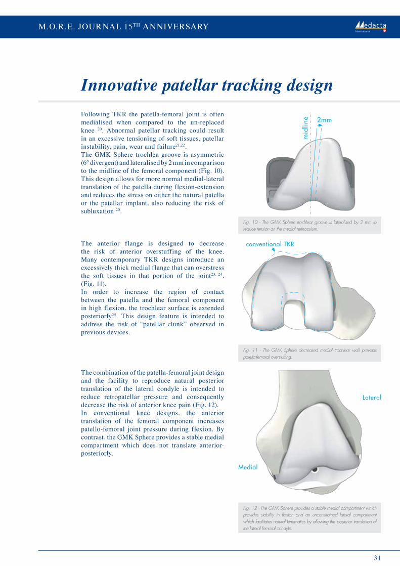
31
m.o.r.e. Journal - march 2013, SupplementProven accuracy and effectiveneSS of myKnee®M.O.r.e. JOurnal 15TH annIVerSarY
Following tKr the patella-femoral joint is often medialised when compared to the un-replaced knee 20. Abnormal patellar tracking could result in an excessive tensioning of soft tissues, patellar instability, pain, wear and failure21,22.the GmK Sphere trochlea groove is asymmetric (6° divergent) and lateralised by 2 mm in comparison to the midline of the femoral component (Fig. 10). this design allows for more normal medial-lateral translation of the patella during flexion-extension and reduces the stress on either the natural patella or the patellar implant, also reducing the risk of subluxation 20.
the anterior flange is designed to decrease the risk of anterior overstuffing of the knee. many contemporary tKr designs introduce an excessively thick medial flange that can overstress the soft tissues in that portion of the joint23, 24. (Fig. 11).In order to increase the region of contact between the patella and the femoral component in high flexion, the trochlear surface is extended posteriorly25. this design feature is intended to address the risk of “patellar clunk” observed in previous devices.
the combination of the patella-femoral joint design and the facility to reproduce natural posterior translation of the lateral condyle is intended to reduce retropatellar pressure and consequently decrease the risk of anterior knee pain (Fig. 12).In conventional knee designs, the anterior translation of the femoral component increases patello-femoral joint pressure during flexion. By contrast, the GmK Sphere provides a stable medial compartment which does not translate anterior-posteriorly.
innovative patellar tracking design
Fig. 10 - The GMK Sphere trochlear groove is lateralised by 2 mm to reduce tension on the medial retinaculum.
Fig. 11 - The GMK Sphere decreased medial trochlear wall prevents patella-femoral overstuffing.
Fig. 12 - The GMK Sphere provides a stable medial compartment which provides stability in flexion and an unconstrained lateral compartment which facilitates natural kinematics by allowing the posterior translation of the lateral femoral condyle.
Medial
Lateral
©Medacta
conventional TKR
2mm
mid
line

32
special edition.O.R.E.I n s t I t u t e
minimized wear rate
Preliminary results
laboratory evaluation has demonstrated that the principles of the GmK Sphere design provide for maximal contact area in the medial compartment throughout the range of loaded motion26. Generally,
the majority of load is applied through the medial compartment and extensive spherical contact in the medial femoro-tibial joint is desirable. Although the lateral femoro-tibial compartment is not congruent on the sagittal plane, it is congruent in the coronal plan, minimizing contact stress levels19,26
(Fig. 13 and 14).
medacta is committed to providing innovative and safe solutions with a responsible and
evidence-based approach. GmK Sphere was tested over 2 years prior to launch through an intensive evaluation programme including in vitro and in vivo trials and laboratory tests.the first implantation took place in london in November 2011 and during the first 2 years of implantation a few selected centres were involved for a careful evaluation of the preliminary data. A total of 1,000 implants were implanted in those 2 years and both surgeons and patients provided encouraging feedback.the GmK Sphere is intensively studied in a large number of in vivo and in vitro studies which are investigating different aspects of knee replacement. one key study will involve follow-up of GmK Sphere in more than 1,200 knees in a number of centres located in different parts of world. As part of an odep (orthopaedic data evaluation panel) multicentre study a UK co-ordinating centre is collecting the Knee Society Score, radiographic analysis, oxford knee score and euroQol score of 500 knees for up to 10 years in several centres in the UK and Belgium. other centres are collecting
the oxford score and the Forgotten Joint Score, as well as the scores collected in a standard follow-up. In addition, many single-site rct’s are comparing the ability to achieve a “forgotten joint” comparing GmK Sphere and other contemporary knee designs.
the GmK Sphere’s performance and kinematics have also been investigated in an in vivo kinematic analysis co-ordinated by prof Scott Banks phd (University of Florida, US) and a retropatellar pressure analysis in collaboration with the university of munich (Germany). the preliminary results of these studies confirm the GmK Sphere claims. other studies will be added to make the clinical planning even more detailed.
The GMK Sphere is intensively studied in a large number of in vivo and in vitro studies which are investigating different aspects of knee replacement.
Fig. 13 - TKR wear results (mg per million cycles) from widely used and clinically successful TKR implants. Dara from University of Nebraska Medical Center 27.
Fig. 14 - GMK Sphere wear results compared with the mean value ofall fixed bearing TKR implants tested by Endolab GmbH19.
Wea
r rat
e (m
g/M
C) 20
15
10
5
0
Fixed Bearings Mobile Bearings
Wea
r rat
e (m
g/M
C) 20
15
10
5
0
Fixed Bearings GMK® Sphere
33
m.o.r.e. Journal - march 2013, SupplementProven accuracy and effectiveneSS of myKnee®M.O.r.e. JOurnal 15TH annIVerSarY
1. Bullens PHJ, van Loon CJM, de Waal Malefijt MC, Laan R, Veth R. Patient Satisfaction After Total Knee Arthroplasty A Comparison Between Subjective and Objective Outcome Assessments. J Arthroplasty. 2001 Sep;16(6):740-7.
2. Baker PN, van der Meulen JH, Lewsey J, Gregg PJ. The role of pain and function in determining patient satisfaction after total knee replacement. J Bone Joint Surg Br. 2007 Jul;89(7):893-900.
3. Nilsdotter AK, Toksvig-Larsen S, Roos EM. Knee arthroplasty: are patients’ expectations fulfilled? A prospective study of pain and function in 102 patients with 5-year follow-up. Acta Orthopaedica 2009; 80 (1): 55–61.
4. Tippett SR, Mang J, Dwyer KA, Lesko J, O’Dell T. Collecting Data with Palm Technology: Comparing Preoperative Expectations and Postoperative Satisfaction in Patients Undergoing Total Knee Arthroplasty. J Bone Joint Surg Am. 2010 Dec;92 Suppl 2:59-67.
5. Bourne RB, Chesworth B, Davis A, Mahomed N, Charron K. Comparing patient outcomes after THA and TKA: is there a difference? Clin Orthop Relat Res. 2010 Feb;468(2):542-6.
6. Behrend H, Giesinger K, Giesinger JM, Kuster MS. The “forgotten joint” as the ultimate goal in joint arthroplasty: validation of a new patient-reported outcome measure. J Arthroplasty. 2012 Mar;27(3):430-436.
7. Bachmeier CJ, March LM, Cross MJ, Lapsley HM, Tribe KL, Courtenay BG, Brooks PM. A comparison of outcomes in osteoarthritis patients undergoing total hip and knee replacement surgery. Osteoarthritis Cartilage. 2001 Feb;9(2):137-46.
8. Wylde V, Blom AW, Whitehouse SL, Taylor AH, Pattison GT, Bannister GC. Patient-reported outcomes after total hip and knee arthroplasty: comparison of midterm results. Arthroplasty. 2009 Feb;24(2):210-6.
9. Noble PC, Gordon MJ, Weiss JM, Reddix RN, Conditt MA, Mathis KB. Does total knee replacement restore normal knee function? Clin Orthop Relat Res. 2005 Feb;(431):157-65.
10. Robinson RP. The early innovators of today’s resurfacing condylar knees. J Arthroplasty. 2005 Jan;20(1 Suppl 1):2-26.11. Robertsson O, Scott G, Freeman MA. Ten-year survival of the cemented Freeman-Samuelson primary knee arthroplasty. Data from the Swedish Knee Arthroplasty Register
and the Royal London Hospital. J Bone Joint Surg Br. 2000 May;82(4):506-7.12. National Joint Registry for England and Wales, 9th Annual Report (2012).13. Freeman MA, Pinskerova V. The movement of the normal tibio-femoral joint. J Biomech. 2005 Feb;38(2):197-208.14. Blaha JD. The rationale for a total knee implant that confers anteroposterior stability throughout range of motion. J Arthroplasty. 2004 Jun;19(4 Suppl 1):22-6.15. Hossain F, Patel S, Rhee SJ, Haddad FS. Knee arthroplasty with a medially conforming ball-and-socket tibiofemoral articulation provides better function. Clin Orthop
Relat Res. 2011 Jan;469(1):55-63.16. Sharkey PF, Miller AJ. Noise, numbness, and kneeling difficulties after total knee arthroplasty: is the outcome affected? J Arthroplasty. 2011 Dec;26(8):1427-31.17. Pritchett JW. Patient preferences in knee prostheses. J Bone Joint Surg Br. 2004 Sep;86(7):979-82.18. Pritchett JW. Patients prefer a bicruciate-retaining or the medial pivot total knee prosthesis. J Arthroplasty. 2011 Feb;26(2):224-8.19. Data on file: Medacta. 20. Meijerink HJ, Barink M, van Loon CJ, Schwering PJ, Donk RD, Verdonschot N, de Waal Malefijt MC. The trochlea is medialized by total knee arthroplasty: an intraoperative assessment in 61 patients. Acta Orthop. 2007 Feb;78(1):123-7.21. Armstrong AD, Brien HJ, Dunning CE, King GJ, Johnson JA, Chess DG. Patellar position after total knee arthroplasty: influence of femoral component malposition. J Arthroplasty. 2003 Jun;18(4):458.22. D’Lima DD, Chen PC, Kester MA, Colwell CW Jr. Impact of patellofemoral design on patellofemoral forces and polyethylene stresses. J Bone Joint Surg Am. 2003;85-A
Suppl 4:85-93.23. Belvedere C, Catani F, Ensini A, Moctezuma de la Barrera JL, Leardini A. Patellar tracking during total knee arthroplasty: an in vitro feasibility study. Knee Surg Sports
Traumatol Arthrosc. 2007 Aug;15(8):985-93. 24. Yamada Y, Toritsuka Y, Horibe S, Sugamoto K, Yoshikawa H, Shino K. In vivo movement analysis of the patella using a three-dimensional computer model. J Bone Joint
Surg Br. 2007 Jun;89(6):752-60.25. Kulkarni SK, Freeman MA, Poal-Manresa JC, Asencio JI, Rodriguez JJ. The patellofemoral joint in total knee arthroplasty: is the design of the trochlea the critical factor?
J Arthroplasty. 2000 Jun;15(4):424-9.26. Minoda Y, Kobayashi A, Iwaki H, Iwakiri K, Inori F, Sugama R, Ikebuchi M, Kadoya Y, Takaoka K. In vivo analysis of polyethylene wear particles after total knee
arthroplasty: the influence of improved materials and designs. J Bone Joint Surg Am. 2009 Nov;91 Suppl 6:67-73.27. Haider H, Kaddick C, Wear of Mobile Bearing Knees: is it Necessarily Less?, Journal of ASTM International, 2012.
References

34
special edition.O.R.E.I n s t I t u t e
Efficiency in TKr instrumentationThe number of Total Knee replacements has increased dramatically over the last decade and the number of patients requiring TKr is projected to increase at an accelerating rate in many key economies1. An obvious problem is that the demand for healthcare is growing faster than the supply of resource and funding. Surgeons and hospitals are facing the threat of cuts in reimbursement. orthopaedic suppliers are being faced with constant demands to cut prices to levels that threaten profitability and ongoing investment in innovation and quality. Somehow, overall costs of treatment have to be reduced and productivity improved. Medacta has always respected the need for healthcare economic sustainability with innovation focused on providing additional benefit to patients but with an emphasis on reducing overall costs of treatment.
If we focus on the tKr surgical procedure, a decrease of intraoperative time can be
beneficial to increase efficiency, but surgical time is multifactorial and dependent on surgeon habits, patient variables and the behaviour and availability of other personnel, therefore it is not easy to control or standardize. An analysis of more global aspects of a tKr procedure, reveals that a large impact on operating room efficiency lies in the management of the tKr instrumentation.
there have been reports published on increasing operative efficiency by decreasing turnover time and making draping, prepping and processing of instrumentation easier to set up and turnover2,3.
Single use instruments have been already adopted in different medical fields, including ophthalmology, cardiothoracic surgery and urologic surgery4-7.
Single use instruments can be a valid alternative to conventional metal reusable sets to decrease or even eliminate a lot of procedural costs linked to tKr, thus optimizing its efficiency.A partial application of single use instruments in joint replacement can be found in patient matched technology, where part of the instrumentation is replaced by single use patient matched guides. Studies show that the use of single use patient matched guides can have an important economic impact on the surgical procedure, allowing for decreased time and costs dedicated to wash, assemble and sterilize the instrument sets8-11. In 2012, aware of the economic benefits that its patient matched technology myKnee brought to the hospital, medacta committed to radically increasing the benefit of cost reduction associated with instrument management and handling by developing a complete offering: GmK efficiency.

35
m.o.r.e. Journal - march 2013, SupplementProven accuracy and effectiveneSS of myKnee®M.O.r.e. JOurnal 15TH annIVerSarY
GmK efficiency is a complete single use instrument solution used to implant both GmK
Sphere and GmK primary, conceived to optimize instrument management, providing significant benefits in the o.r. and throughout the hospital supply chain. GmK efficiency instrumentation consists of 3 main instrument sets: a general set including spacers, impactors and resection and drilling guides, a size-specific femoral set including trials femoral components and resection blocks and a size-specific tibial set including trial inlays and baseplates. GmK efficiency can be used in combination with myKnee patient matched instrumentation, maximizing its benefits. As an alternative to myKnee, an additional GmK efficiency set can be provided including alignment guides and sizers. A GmK efficiency patella instrument set is also available as an option.
GmK Efficiency: innovation that delivers
Single use instruments can be a valid alternative to conventional metal reusable sets to decrease
or even eliminate a lot of procedural costs linked to TKR,
thus optimizing its efficiency.
GmK efficiency instruments are manufactured using special medical grade technopolymers, able to give high fatigue and abrasion resistance alongside form versatility. Bulky and complicated connections found in metal reusable instruments are replaced by ergonomic and fast coupling mechanisms, allowing for more straightforward surgical procedures. Impaction instruments have the same solidity as the metal ones, without unwanted weight. the overall lightness of the GmK efficiency instrumentation, just 2 kg for a complete set – 10 times less than a conventional instrument set, represents just one of the several benefits to the or environment that this innovative instrumentation brings.

36
special edition.O.R.E.I n s t I t u t e
GmK Efficiency improves the quality of the o.R. environment
GmK Efficiency promotes sustainable o.R. management
the value of GmK efficiency is significant. o.r. staff are able to work with extremely light instrumentation, provided already packed and sterile and just dispose it after use, without the need
to post-process it. the size specific layout allows easy selection of the most suitable composition for the surgery, minimizing instrument volume and simplifying backtable preparation.
All GmK efficiency instruments are ready to use, always sterile and brand new. this simplifies o.r. scheduling, avoiding surgery cancellation or delay due to missing, non-sterile or not functional instruments. GmK efficiency promotes an easier o.r. environment to enhance productivity.
the value of the efficiency can be translated into significant savings for hospitals. medacta
has conducted a survey in different hospitals and practices in europe to verify the potential economic impact of GmK efficiency, single use instrumentation12.
different aspects related to tKr instrumentation management have been considered:• occurrence of surgery cancellation or
rescheduling because of sterilization failure;• peri-operative management of the
instrumentation;• Intra-operative management of the
instrumentation;• Impact of sterilization procedures;• Infection management.
occurrence of surgery cancellation or rescheduling because of sterilization failure
on average, this has been estimated to be 2% of the total annual volume of surgery, with an impact of 30 minutes lost per postponed procedure. this does not take into account the stress and work imposed upon staff that have to manage the episode.
Peri-operative management of the instrumentation
As GmK efficiency is provided already assembled and sterile, the instrumentation preparation for the surgery is minimal, eliminating the need for a completeness check. Surveyed centers estimated 30 minutes as the time dedicated to the preoperative check of the instrumentation, 20 minutes for backtable preparation and 30 minutes to check the instruments and prepare them for collection after surgery. that suggests that potentially the use of GmK efficiency could potentially allow savings of more than 60 minutes per surgery.
intra-operative management of the instrumentation
concerns in intraoperative management of metal reusable instruments are associated with the incidence of missing or not functional instruments. the survey showed that around 4% of surgeries in a year experience these problems, resulting in a potential loss of 15 minutes per procedure.the ease-of-use and ergonomics of the instruments result in more reproducible and stress-free surgical procedures. this potentially decreases the surgical time and related costs and may minimize complications.
The GMK Efficiency instruments are ready to use, always sterile and brand new.
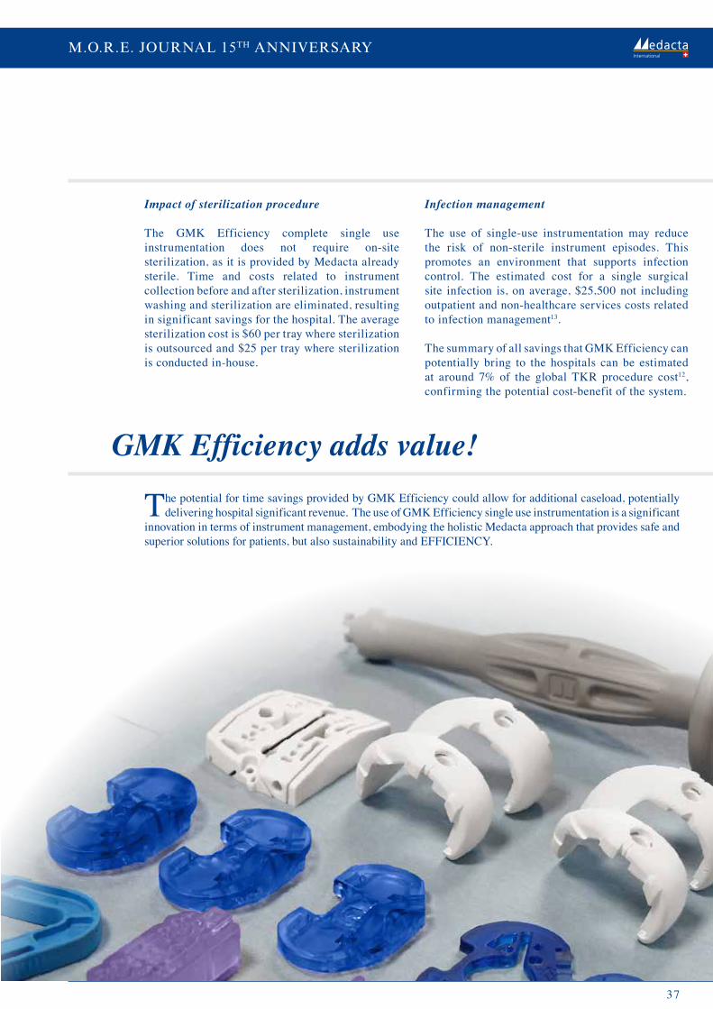
37
m.o.r.e. Journal - march 2013, SupplementProven accuracy and effectiveneSS of myKnee®M.O.r.e. JOurnal 15TH annIVerSarY
impact of sterilization procedure
the GmK efficiency complete single use instrumentation does not require on-site sterilization, as it is provided by medacta already sterile. time and costs related to instrument collection before and after sterilization, instrument washing and sterilization are eliminated, resulting in significant savings for the hospital. the average sterilization cost is $60 per tray where sterilization is outsourced and $25 per tray where sterilization is conducted in-house.
GmK Efficiency adds value!
the potential for time savings provided by GmK efficiency could allow for additional caseload, potentially delivering hospital significant revenue. the use of GmK efficiency single use instrumentation is a significant
innovation in terms of instrument management, embodying the holistic medacta approach that provides safe and superior solutions for patients, but also sustainability and eFFIcIeNcY.
infection management
the use of single-use instrumentation may reduce the risk of non-sterile instrument episodes. this promotes an environment that supports infection control. the estimated cost for a single surgical site infection is, on average, $25,500 not including outpatient and non-healthcare services costs related to infection management13.
the summary of all savings that GmK efficiency can potentially bring to the hospitals can be estimated at around 7% of the global tKr procedure cost12, confirming the potential cost-benefit of the system.
38
special edition.O.R.E.I n s t I t u t e
1. Kurtz S, Ong K, Lau E, et al. Projections of primary and revision hip and knee arthroplasty in the United States from 2005 to 2030. J Bone Joint Surg Am 2007;89(4):780
2. Cendan JC, Good M. Interdisciplinary workflow assessment and redesign decreases operating room turnover time and allows for additional caseload. Arch Surg 2006; 141:65.
3. Jacofsky D, Lyman J. The efficient delivery of elective orthopedics. J Arthroplasty 2007;22(Suppl 3):2.4. Galinski M, Catineau J, Rayeh F, et al. Laryngoscope plastic blades in scheduled general anesthesia patients: a comparative randomized study. J Clin Anesth
2011;23(2):107.5. Gupta A, Mercieca K, Fahad B. Biswas. The effectiveness and safety of single-use disposable instruments in cataract surgery— a clinical study using a surgeon-based
survey. J Perioper Pract 2009;19:148.6. Slover JD, Rubash HE, Malchau H, et al. Cost-effectiveness analysis of custom total knee cutting blocks. J Arthroplasty 2012;27(2):180.7. Stiehl JB, Jackson S, Szabo A. Multi-factorial analysis of time efficiency in total knee arthroplasty. Comput Aided Surg 2009;14(1–3):58.8. Goldberg TD - MyKnee economical and clinical results. Podium presentation at the 6th M.O.R.E. International symposium, Stresa, Italy, May 13-14, 2011.9. Koch P - MyKnee System: A new vision in total knee replacement. Leading Opinions - Orthopädie & Rheumatologie 2, 2011: 32-35.10. Gagna G - Aspects economiques de la tecnologie sur mesure MyKnee en chirurgie prothetique du genou. Podium presentatiuon at the 87th Annual Congress of the
SOFCOT, Paris, November 11-14, 2012.”11. Noble J W, Moore C A, Liu N - The value of patient-matched instrumentation in total knee arthroplasty. J Arthroplasty, Jan 2012, 27(1):153-5.12. Data on file: Medacta13. Stone at al, American Journal of infection control, 2005.
References
Copyright © 2014 by Medacta® International SA, all rights reserved

39
m.o.r.e. Journal - march 2013, SupplementProven accuracy and effectiveneSS of myKnee®M.O.r.e. JOurnal 15TH annIVerSarY
Medacta International SA Strada regina - 6874 castel San pietro - Switzerlandphone +41 91 696 60 60 - Fax +41 91 696 60 66 - [email protected]
Headquarters
representativeSwitzerland - FrauenfeldGewerbestrasse 3 - 8500 Frauenfeldphone +41 (0) 848 423 423 - Fax +41 (0) 848 423 424 - [email protected]
distributors
SubsidiariesAustralia - Medacta Australia PTY.LTDUnit F37, 16 mars road - lane cove Business park - NSWphone +61 (2) 94202944 - Fax +61 (2) 94202578 - [email protected]
Belgium - Medacta Belgium B.V.B.A./S.P.r.L.5a rue de la maîtrise - 1400 Nivellesphone +32 (0) 67 555 482 - Fax +32 (0) 67 555 483 - [email protected]
Canada - Medacta Canada Inc.31 mcBrine drive, Unit 11- N2r 1J1 - Kitchener, ontariophone +1 519 279 1934 - Fax +1 519 279 1938 - [email protected]
China - Medacta Chinaroom B, 32/F, New SH Intl tower - No. 360 pudong South road - Shanghai 200120, chinaphone +86 21 5835 1149 - [email protected]
France - Medacta France SAS6 rue du commandant d’estienne d’orves - parc des chanteraines - 92390 Villeneuve - la Garennephone +33 147 39 07 22 - Fax +33 147 39 73 17 - [email protected]
Germany - Medacta Ortho GmbHJahnstrasse 86 - d - 73037 Göppingenphone +49 (0) 7161 50 44 312 - Fax +49 (0) 7161 50 44 320 - [email protected]
Italy - Medacta Italia SrlVia G. Stephenson, 94 - 20157 milanophone +39 02 390 181 - Fax +39 02 390 00 704 - [email protected]
Japan - Medacta Japan CO. LTD100-0014 chiyoda House 201 - 2 - 17-8, Nagatacho, chiyoda-ku, tokyophone +81 (0) 3 5510 8883 - Fax +81 (0) 3 5510 8884 - [email protected]
Spain - Medacta España SLAvda de las Jacarandas - 2 - edificio creA oficina 631- 46100 - Burjassotphone +34 (0) 963 484 688 - Fax +34 (0) 963 484 688 - [email protected]
UK - Medacta UK Limited9 cartwright court - cartwright Way - Forest Business park Bardon Hill - leicestershire - le67 1Uephone +44 (0) 1530 830451 - Fax +44 (0) 1530 839326 - [email protected]
USA - Medacta USA, Inc.1556 West carroll Avenue - chicago - Il 60607phone +1 312 878 2381 - Fax +1 312 546 6881 - [email protected]
Argentina
South AfricaNew ZealandMexicoMalaysiaIndonesia
GreeceBulgariaBrazilAustria Belarus Colombia
Kuwait Vietnam

* D a t a a s a t D e c e m b e r 2 0 1 3
We want to thank all of you all over the world.
Your trust in Medacta has resulted in treatment of more than 110,000* AMIS and 15,000*
MyKnee Patients, helping to make Medacta the largest European provider of Hip and
Knee arthroplasty solutions with growing success around the world.
MEDACTA INTERNATIONAL1999-201415 YEARS OF SUCCESSTHANK YOU for supporting us in our commitment to advance:- Patient Well-Being- Ongoing Medical Education- Healthcare Economic Efficiency
99.9
9.jo
urna
lAN
rev.
00