Monocyte-Astrocyte Networks and the Regulation of Networks and the Regulation of Chemokine Secretion...
10
of May 25, 2018. This information is current as Neurocysticercosis Regulation of Chemokine Secretion in Monocyte-Astrocyte Networks and the E. Gonzalez and Jon S. Friedland Jasim Uddin, Hector H. Garcia, Robert H. Gilman, Armando http://www.jimmunol.org/content/175/5/3273 doi: 10.4049/jimmunol.175.5.3273 2005; 175:3273-3281; ; J Immunol References http://www.jimmunol.org/content/175/5/3273.full#ref-list-1 , 27 of which you can access for free at: cites 66 articles This article average * 4 weeks from acceptance to publication Fast Publication! • Every submission reviewed by practicing scientists No Triage! • from submission to initial decision Rapid Reviews! 30 days* • Submit online. ? The JI Why Subscription http://jimmunol.org/subscription is online at: The Journal of Immunology Information about subscribing to Permissions http://www.aai.org/About/Publications/JI/copyright.html Submit copyright permission requests at: Email Alerts http://jimmunol.org/alerts Receive free email-alerts when new articles cite this article. Sign up at: Print ISSN: 0022-1767 Online ISSN: 1550-6606. Immunologists All rights reserved. Copyright © 2005 by The American Association of 1451 Rockville Pike, Suite 650, Rockville, MD 20852 The American Association of Immunologists, Inc., is published twice each month by The Journal of Immunology by guest on May 25, 2018 http://www.jimmunol.org/ Downloaded from by guest on May 25, 2018 http://www.jimmunol.org/ Downloaded from
Transcript of Monocyte-Astrocyte Networks and the Regulation of Networks and the Regulation of Chemokine Secretion...

of May 25, 2018.This information is current as
NeurocysticercosisRegulation of Chemokine Secretion in Monocyte-Astrocyte Networks and the
E. Gonzalez and Jon S. FriedlandJasim Uddin, Hector H. Garcia, Robert H. Gilman, Armando
http://www.jimmunol.org/content/175/5/3273doi: 10.4049/jimmunol.175.5.3273
2005; 175:3273-3281; ;J Immunol
Referenceshttp://www.jimmunol.org/content/175/5/3273.full#ref-list-1
, 27 of which you can access for free at: cites 66 articlesThis article
average*
4 weeks from acceptance to publicationFast Publication! •
Every submission reviewed by practicing scientistsNo Triage! •
from submission to initial decisionRapid Reviews! 30 days* •
Submit online. ?The JIWhy
Subscriptionhttp://jimmunol.org/subscription
is online at: The Journal of ImmunologyInformation about subscribing to
Permissionshttp://www.aai.org/About/Publications/JI/copyright.htmlSubmit copyright permission requests at:
Email Alertshttp://jimmunol.org/alertsReceive free email-alerts when new articles cite this article. Sign up at:
Print ISSN: 0022-1767 Online ISSN: 1550-6606. Immunologists All rights reserved.Copyright © 2005 by The American Association of1451 Rockville Pike, Suite 650, Rockville, MD 20852The American Association of Immunologists, Inc.,
is published twice each month byThe Journal of Immunology
by guest on May 25, 2018
http://ww
w.jim
munol.org/
Dow
nloaded from
by guest on May 25, 2018
http://ww
w.jim
munol.org/
Dow
nloaded from

Monocyte-Astrocyte Networks and the Regulation ofChemokine Secretion in Neurocysticercosis1
Jasim Uddin,* Hector H. Garcia,†‡ Robert H. Gilman,†§ Armando E. Gonzalez,¶ andJon S. Friedland2*
Neurocysticercosis, caused by infection with larval Taenia solium, is a major cause of epilepsy worldwide. Larval degeneration,which is symptomatic, results in inflammatory cell influx. Astrocytes, the most abundant cell type and major cytokine-producingcell within the CNS, may be important in orchestrating inflammatory responses after larval degeneration. We investigated theeffects of direct stimulation and of conditioned medium from T. solium larval Ag (TsAg)-stimulated monocytes (CoMTsAg) onneutrophil and astrocyte chemokine release. CoMTsAg, but not control conditioned medium, stimulated astrocyte CCL2/MCP-1(161.5 � 16 ng/ml), CXCL8/IL-8 (416 � 6.2 ng/ml), and CXCL10/IFN-�-inducible protein (9.07 � 0.6 ng/ml) secretion after 24 h,whereas direct astrocyte or neutrophil stimulation with TsAg had no effect. There was rapid accumulation of CCL2 and CXCL8mRNA within 1 h, with somewhat delayed expression of CXCL10 mRNA initially detected 8 h poststimulation. Neutralizinganti-TNF-� inhibited CoMTsAg-induced CCL2 mRNA accumulation by up to 99%, causing total abolition of CXCL10 and up to77% reduction in CXCL8 mRNA. CoMTsAg induced maximal nuclear binding of NF-�B p65 and p50 by 1 h, with I�B� andI�B� decay within 15 min. In addition, CoMTsAg induced transient nuclear binding of AP-1, which peaked 4 h poststimu-lation. In NF-�B blocking experiments using pyrrolidine dithiocarbamate, CoMTsAg-induced CCL2 secretion was reducedby up to 80% (p � 0.0006), whereas CXCL8 was inhibited by up to 75% (p � 0.0003). In summary, the data show thatastrocytes are an important source of chemokines following larval Ag stimulation. Such chemokine secretion is NF-�Bdependent, likely to involve AP-1, and is regulated in a paracrine loop by monocyte-derived TNF-�. The Journal ofImmunology, 2005, 175: 3273–3281.
N eurocysticercosis (NCC),3 caused by CNS infection withlarvae of the parasite Taenia solium, affects 50 millionpeople worldwide (1, 2). Infection is endemic in South
America, Asia, and sub-Saharan Africa, with increasing preva-lence in countries such as the United States as a result of emigra-tion from endemic areas (3–5). Infection is acquired via the fecal-oral route following ingestion of microscopic Taenia eggs (6).People with no direct contact with infected pigs are at risk ofinfection due to contamination of food or water by human tape-worm carriers (7, 8). In the human gut, Taenia eggs differentiate toinvasive oncospheres, which penetrate the mucosal lining and en-ter the general circulation from where they disseminate and accu-mulate preferentially in the CNS forming fluid-filled cysticerci (4).
Clinical symptoms usually present after prolonged asymptom-atic periods and depend on the size, location, and burden of infec-tion (3). Epilepsy is the most common manifestation; in diseaseendemic regions, 25% of epilepsy may be due to NCC (9–12). Thetransition from asymptomatic to symptomatic disease is thought todepend on cyst degeneration, a process that may be accelerated byantiparasite therapy (13). Studies in animal models suggest that theintact parasite maintains a Th2-permissive environment. The in-flammatory response that leads to larval degeneration is charac-terized by a switch to a Th1-type cytokine profile, overexaggera-tion of which results in a chronic inflammatory response involvinggranuloma formation and tissue destruction (14, 15). Brain gran-ulomas from NCC patients consist of mononuclear cells, granulo-cytes, and CNS microglial cells (16, 17). A prerequisite for gran-uloma formation is cell influx to sites of larval degeneration, andthis will involve chemokines, which are fundamental in controllingcell trafficking (18). The source of such chemokines in NCC hasnot been investigated, although astrocytes that surround the perim-eter of granulomas (17) are able to secrete a range of cytokines andmay be important in orchestrating granuloma formation.
Astrocytes are the most abundant cell type in the CNS, consti-tuting 50–75% of the total cell number (19, 20). Astrocytes andmicroglia function as primary immune effector cells of the CNS(21, 22). In addition, astrocytes have a central function in the main-tenance of the blood-brain barrier (BBB), thereby indirectly con-tributing to control of CNS leukocyte trafficking (23). Astrocytesdirectly regulate CNS leukocyte invasion, secreting and expressinga number of chemokines and their associated receptors (24–26).TNF-� is an important stimulus for CNS chemokine expression(27). The role of chemokines in CNS infection has been mostwidely studied in viral infections, particularly HIV, where astro-cyte chemokine secretion and chemokine receptor expression is
*Department of Infectious Diseases, Faculty of Medicine and the Wellcome TrustCentre for Clinical Tropical Medicine, Imperial College (Hammersmith Campus),London, United Kingdom; †Departments of Microbiology and Pathology, Univer-sidad Peruana Cayetano Heredia, Lima, Peru; ‡Cysticercosis Unit, Instituto Nacionalde Ciencias Neurologicas, Lima, Peru; §Department of International Health, JohnsHopkins University Bloomberg School of Public Health, Baltimore, MD 21205; and¶School of Veterinary Medicine, Universidad Nacional Mayor de San Marcos, Lima,Peru
Received for publication January 19, 2005. Accepted for publication June 13, 2005.
The costs of publication of this article were defrayed in part by the payment of pagecharges. This article must therefore be hereby marked advertisement in accordancewith 18 U.S.C. Section 1734 solely to indicate this fact.1 This study was funded by the National Institutes of Health Grants AI-42037-01 andAI-35894, International Training in Research in Emerging Diseases Training GrantTW00910, and The Wellcome Trust of Great Britain.2 Address correspondence and reprint requests to Prof. Jon S. Friedland, Departmentof Infectious Diseases, Imperial College London, Hammersmith Campus, Du CaneRoad, London W12 ONN, U.K. E-mail address: [email protected] Abbreviations used in this paper: NCC, neurocysticercosis; BBB, blood-brain bar-rier; CoM, conditioned medium; CoMCon, control conditioned medium; PDTC, pyr-rolidine dithiocarbamate; PMNL, polymorphonuclear leukocyte; TsAg, Taenia so-lium larval Ag.
The Journal of Immunology
Copyright © 2005 by The American Association of Immunologists, Inc. 0022-1767/05/$02.00
by guest on May 25, 2018
http://ww
w.jim
munol.org/
Dow
nloaded from
important in mediating initial cellular entry and subsequent CNSpathology (25, 28, 29). Astrocyte-derived chemokines also con-tribute to pathology after CNS infection with the intracellular par-asites Trypanosoma brucei and Toxoplasma gondii (30, 31).
We have investigated astrocyte chemokine secretion in NCC.Our data demonstrate for the first time that astrocytes but not neu-trophils are a major source of the chemokines CCL2, CXCL8, andCXCL10. Chemokine secretion does not follow direct exposure toneurocysticercal (T. solium) Ags (TsAg) but is dependent onTNF-� derived from monocytes stimulated with TsAg and is reg-ulated at the transcriptional level by an NF-�B-dependent mech-anism. Such CNS networks may be important in increasing in-flammatory cell influx and contributing to the pathology of NCC.
Materials and MethodsReagents
RPMI 1640, HBSS, Eagle’s MEM, L-glutamine, and sodium pyruvate wereobtained from Invitrogen Life Technologies. FCS (endotoxin level �0.06ng/ml) was obtained from Labtech International. Ficoll-Paque, nitrocellu-lose membrane (Hybond-C), nylon membrane (Hybond-N), ECL Hyper-film, and [�-32P]ATP were obtained from Amersham Biosciences. Recom-binant human TNF-�, IL-1 receptor antagonist, and neutralizing polyclonalrabbit anti-human TNF-� were obtained from PeproTech. The RNase pro-tection assay system was from BD Pharmingen, and the DIG Wash andBlot set and CDP-Star were from Roche. The avidin-phosphatase was ob-tained from Tropix. The NF-�B, AP-1, and SP-1 consensus oligos and theT4 polynucleotide kinase were purchased from Promega. Premade 30%bisacrylamide stock used for PAGE was obtained from Anachem. Rabbitanti-human p65, p50, p52, c-Rel, rel-B, c-Fos, and I�B�� Abs were ob-tained from Santa Cruz Biotechnology. HRP for chemokine ELISAs waspurchased from BioSource International. All other materials and reagentswere purchased from Sigma-Aldrich.
Preparation of TsAg
T. solium metacestodes (cysticerci) were dissected at postmortem fromnaturally infected pigs obtained from endemic areas in the Peruvian high-lands. Metacestodes were homogenized in cold-buffered PBS using a glasshomogenizer. Ag suspensions were subsequently prepared by sonication at70 Hz for 3 min before storage at �70°C. Concentration of protein in theAg preparation, which was used as a suspension, was quantified using aBradford assay (Bio-Rad). Endotoxin contamination, measured using theLimulus amebocyte lysate assay, was minimal (between 0.11 and 5.46 pg/ml) and did not induce chemokine secretion in vitro.
Cell culture of U373MG cells, monocytes, and neutrophils
The human astrocytic U373MG cell line (no. 89081403; European Collec-tion of Cell Cultures) was maintained in Eagle’s MEM supplemented with10% FCS, 2 mM L-glutamine, 1 mM sodium pyruvate, 1% nonessentialamino acids, and 100 �g/ml ampicillin, in a humidified 5% CO2 atmo-sphere at 37°C. Confluent cultures were passaged with 0.25% trypsin-EDTA, seeding at a density of 3 � 10,000 cells/cm2. Primary human pe-ripheral blood monocytes and polymorphonuclear leukocytes (PMNLs)were prepared from pooled buffy-coat residues from healthy donors (NorthLondon Blood Transfusion Service). Briefly, mononuclear cells andPMNLs were isolated by density gradient centrifugation over Ficoll-Paque.Monocytes were adhesion purified on tissue culture plastic for 1 h beforebeing washed three times with sterile HBSS to remove nonadherent lym-phocytes. PMNLs were separated from RBCs by hypertonic lysis followedby two washes using HBSS. Purified cells were maintained in RPMI 1640medium supplemented with 10% FCS, 2 mM glutamine, and 100 �g/mlampicillin, at 37°C in a humidified 5% CO2 atmosphere. Giemsa stainingconfirmed that �90% of the PMNLs were neutrophils.
Experimental protocol
Monocytes, U373MG cells, and PMNLs were seeded at a density of 1 �105, 1 � 105, or 2 � 105 cells/cm2, respectively, in 6-well tissue cultureplates. Cells were stimulated in triplicate with 100 �g/ml TsAg or variousdoses of LPS (from Escherichia coli serotype 0127:B8) for 24 h (mono-cytes and U373MG cells) or 14 h (neutrophils). Cell-free supernatants weresubsequently collected and stored at �20°C before assay.
In experiments investigating monocyte-astrocyte networks, conditionedmedium was prepared by stimulating monocytes with 100 �g/ml TsAg(CoMTsAg) for 8 h, after which sterile, cell-free supernatants were re-
moved, aliquoted, and stored. Conditioned medium from unstimulated hu-man monocytes cultured for 8 h (control conditioned medium; CoMCon)was the negative control. U373MG cells (1 � 105 cells/cm2) were seededin 6-well tissue culture plates, 100-mm- or 150-mm-diameter dishes asappropriate for preparation of cell supernatants, RNA, or nuclear proteins.Cells were stimulated in triplicate for each experimental condition witheither CoMTsAg or CoMCon at dilutions of 1/10, 1/50, or 1/100 for de-fined time points. Cell-free supernatants, cellular RNA, whole cell lysates,or nuclear extracts were subsequently collected and stored at either �20°Cor �80°C (as appropriate) before being assayed.
In cytokine-neutralizing experiments, U373MG cells were preincubatedwith various doses of IL-1 receptor antagonist for 2 h before stimulationwith CoMTsAg. The role of TNF-� was investigated by preincubatingCoMTsAg for 2 h with various concentrations of neutralizing anti-TNF-�Ab before cellular stimulation. In NF-�B blocking experiments, U373MGcells were preincubated with 1, 10, or 100 �M pyrrolidine dithiocarbamate(PDTC; a broad spectrum NF-�B inhibitor) for 2 h before being stimulatedwith either CoMTsAg or TNF-� (10 ng/ml) for 24 h.
Chemokine ELISAs
CXCL8/IL-8 and CCL2/MCP-1 protein concentrations were measured us-ing ELISAs based on matched Ab pairs (R&D Systems). The lower limitof sensitivity of the CCL2 and CXCL8 assays was 15 pg/ml. The level ofCXCL10 (IFN-�-inducible protein) was measured using cytosets, accord-ing to manufacturer’s protocols (BioSource International). The lower limitof detection was 30 pg/ml.
RNA isolation and RNase protection assay
Total cellular RNA was extracted from U373MG cells using a guanidinethiocyanate and phenol mixture (Tri-reagent; Sigma-Aldrich). RNA wasprecipitated with isopropanol and washed with 70% ethanol before beingdissolved in RNase-free water. mRNA analysis was conducted using amodified version of the RiboQuant multiprobe RNase protection assay pro-tocol (BD Pharmingen). RNA (15 �g) was hybridized overnight with amultiprobe set containing cDNA templates for human XCL1, CCL5,CXCL10, CCL4, CCL3, CCL2, CXCL8, CCL1, and housekeeping genesL32 and GAPDH (hCK-5; BD Pharmingen), which had been biotinylatedand in vitro transcribed using T7 RNA polymerase. After hybridization,RNA mixtures were treated with an RNase mixture at 30°C for 45 min, andprotected mRNA species were then precipitated and separated on a 8 Murea-5% polyacrylamide gel before being transferred to Hybond-N by elec-troblotting and fixed by UV exposure (UV Stratalinker 1800; Stratagene).Blots were blocked overnight before incubation with alkaline phosphatase.After three washes, the substrate CDP-Star was added, and blots wereexposed to enhanced chemiluminescence Hyperfilm for up to 1 h. Imageswere scanned and analyzed using Scion Image vBeta 4.0.2 (Scion). Datawere normalized using housekeeping genes L32 and GAPDH.
EMSA
Nuclear extracts were prepared from U373MG cells grown in 150-mm-diameter tissue culture dishes, according to Clarke et al. (32). Briefly, cellswere washed with ice-cold HBSS before extraction with a hypotonic buffer(5 mM HEPES, pH 7.9, 10 mM KCl, 1.5 mM MgCl2 containing proteaseinhibitors leupeptin, aprotinin, pepstatin, bestatin, and PMSF, all at 1 �g/ml). After addition of 0.25% Nonidet P-40, nuclei were pelleted, superna-tant was removed, and nuclei were resuspended in a hypertonic buffer (5mM HEPES, pH 7.9, 0.5 M NaCl, 1.5 mM MgCl2, 0.2 mM EDTA, 25%glycerol). After equilibration for 2 h at 4°C, nuclei were pelleted and sol-uble nuclear extract was aspirated and stored at �80°C. After protein con-centrations were quantified by the Bradford assay, EMSAs were performedas follows. Double-stranded NF-�B or AP-1 consensus oligonucleotideswere end labeled using [�-32P]ATP and T4 polynucleotide kinase. Nuclearextracts (5–7 �g) and labeled oligo probes (specific activity �1 � 108
cpm) were mixed in binding buffer (20% glycerol, 5 mM MgCl2, 2.5 mMEDTA, 2.5 mM DTT, 250 mM NaCl, 50 mM Tris-HCl, pH 7.5, 0.25mg/ml poly(dI:dC)-poly(dI:dC)) for 20 min at room temperature beforebeing subjected to 5% nondenaturing PAGE and autoradiography. Probe-binding specificity was confirmed in competition assays using 50-fold mo-lar excess of cold, unlabeled NF-�B, AP-1, or SP-1 oligos. Supershiftanalysis to determine NF-�B subunit binding was performed by adding 1�g of specific Abs raised against human p65, p50, p52, c-Rel, rel-B, orc-Fos (a subunit of AP-1) to the binding mixture 40 min before addition ofradiolabeled oligo.
3274 CHEMOKINE SECRETION IN NCC
by guest on May 25, 2018
http://ww
w.jim
munol.org/
Dow
nloaded from

Western blotting for I�B
Western blot analysis was performed according to standard procedures(33). Briefly, cell lysates were prepared by adding ice-cold lysis buffer(PBS containing 0.1% SDS, 0.1% Nonidet P-40, 0.5% deoxycholate, 10mM NaF, 1 mM sodium orthovanadate, 170 �g/ml PMSF, and proteaseinhibitors leupeptin, pepstatin, bestatin, and aprotinin all at 1 �g/ml) to 5 �106 U373MG cells, followed by centrifugation at 800 � g for 5 min at 4°C.Protein concentration was determined by the Bradford assay; equal vol-umes of loading buffer (containing 50 mM HEPES, 10% glycerol, 5%DTT, 2% SDS, and bromphenol blue) were added to 50 �g of protein, andsamples were boiled for 5 min before being frozen at �80°C. Sampleswere resolved on a 10% SDS-PAGE gel, transferred by electroblotting toa nitrocellulose membrane, and probed with 0.5 �g of rabbit anti-humanI�B� or 0.8 �g of rabbit anti-human I�B�. After incubation with perox-idase-conjugated goat anti-rabbit IgG, protein bands were visualized bychemiluminescence.
Data presentation and statistics
Data from ELISAs are means � SEM of a triplicate experiment performedon at least two independent occasions. Data were analyzed using an un-paired Student’s t test, where p � 0.05 was taken as significant. RNaseprotection assays (together with densitometric analysis) and EMSAs shownare representative of at least three independent experiments.
ResultsTsAg stimulates cell-specific secretion of CCL2 and CXCL8 inmonocytes but not in PMNLs or astrocytes
We first determined which cell types secreted chemokines in re-sponse to direct stimulation with TsAg. Human neutrophils arecapable of responding to parasitic and proinflammatory microbial
stimuli by producing chemokines (34, 35). At 24 h, monocytessecreted significant amounts of CCL2 and CXCL8 after directstimulation with TsAg, whereas astrocytes and PMNLs did not(Fig. 1a). Compared with control, in unstimulated cells that se-creted 1.84 � 0.002 ng/ml CCL2 and 45.3 � 6.4 ng/ml CXCL8,100 �g/ml TsAg caused a 212-fold induction in the secretion ofCCL2 (390 � 13.5 ng/ml) and a 11-fold induction in CXCL8(513 � 43 ng/ml) in monocytes. There was a minimal induction inCXCL8 secretion following stimulation of PMNLs with TsAg,whereas the positive control, LPS (10 �g/ml), induced a relativelyhigh level of CXCL8 secretion from PMNLs (17.1 � 1.7 ng/ml;data not shown). We did not analyze PMNL CXCL8 secretionbeyond 14 h because of significant decreases in cell viability (datanot shown). TsAg-stimulated U373MG cells did not secrete moreCCL2 or CXCL8 than control cells. CCL2 and CXCL8 increasedin a dose-dependent manner after LPS stimulation, demonstratingthat these cells can secrete chemokines in response to microbialstimuli (Fig. 1b). The possibility that these and subsequent datawere due to LPS contamination was excluded by the limulus assay.
CoMTsAg induces secretion of CCL2, CXCL8, and CXCL10from astrocytic U373MG cells
Because TsAg had no direct effect on astrocyte chemokine secre-tion, we examined whether a cytokine network might be active.U373MG cells stimulated with CoMTsAg at a 1/10 dilution se-creted 28.7 � 3.3 ng/ml CCL2 within 4 h (Fig. 2a). This increase
FIGURE 1. Human astrocytic U373MG cells do not secrete CXCL8 orCCL2 in response to direct stimulation with TsAg. a, Monolayers ofU373MG cells (astrocytes; 1 � 105/cm2), primary human neutrophils(PMNLs; 2 � 105/cm2), or primary human monocytes (1 � 105/cm2) werestimulated with medium (control) or 100 �g/ml TsAg (�) for 24 h. Cell-free supernatants were subsequently harvested and analyzed for CCL2 andCXCL8 protein by ELISA. b, U373MG cells were stimulated with medium(control; Con) or with 0.01, 0.1, 1, or 10 �g/ml LPS for 24 h, and CCL2and CXCL8 protein concentrations in culture supernatants were measuredby specific ELISA. Data are means � SEM of a representative triplicateexperiment performed on at least three independent occasions.
FIGURE 2. Kinetics of CoMTsAg-induced CCL2, CXCL8, andCXCL10 secretion from human astrocytic U373MG cells. CoMTsAg wasused to stimulate monolayers of U373MG cells (1 � 105/cm2) over 48 h atdilutions of 1/10, 1/50, and 1/100. CoMCon was used at a dilution of 1/10.CCL2 (a), CXCL8 (b), and CXCL10 (c) concentrations (indicated bybrackets) were measured by ELISA in cell-free culture supernatants at 0, 2,4, 8, 24, and 48 h after stimulation. Chemokine concentrations at 0 hrepresent basal monocyte-derived chemokine present in CoMTsAg. Re-sults are mean values � SEM of a triplicate experiment representative ofthree independent experiments.
3275The Journal of Immunology
by guest on May 25, 2018
http://ww
w.jim
munol.org/
Dow
nloaded from

reached maximal amounts of 161.53 � 16.02 ng/ml 24 h post-stimulation. CoMTsAg was a potent stimulus for CCL2 secretion,as dilutions as low as 1/100 caused 27 � 0.7 ng/ml CCL2 to besecreted within 8 h. Cells stimulated with CoMTsAg at a dilutionof 1/100 did not drive CXCL8 secretion greater than that in CoM-Con-stimulated cells (Fig. 2b). CoMTsAg at a 1/10 dilution re-sulted in 276.5 � 36.7 ng/ml CXCL8 secretion at 8 h with con-centrations increasing over 48 h (452.40 � 7.39 ng/ml; Fig. 2b).The higher concentration of CoMTsAg (1/10) was required to in-duce CXCL10 secretion, and the absolute amounts were low com-pared with CCL2 and CXCL8 (Fig. 2c). Chemokine values quotedwere taken after accounting for monocyte-derived chemokine lev-els present in the CoMTsAg; this was represented by the baselinechemokine secretion at 0 h.
CoMTsAg induces differential CCL2, CXCL8, and CXCL10mRNA accumulation in astrocytic U373MG cells
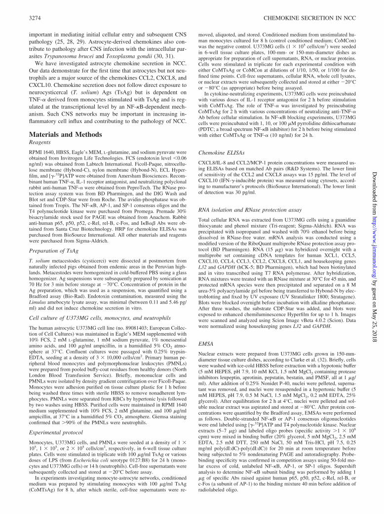
The kinetics of CCL2, CXCL8, and CXCL10 gene expression de-termined by RNase protection assay were consistent with proteinsecretion data. CCL2 mRNA appeared within 1-h stimulation withCoMTsAg and peaked after 4 h before it began to fall after 8 h(Fig. 3, a and b). Kinetics of CXCL8 mRNA accumulation fol-lowed a similar pattern, although there was constitutive expressionof CXCL8 mRNA at baseline and in CoMCon-stimulated cells(Fig. 3, a and c). In contrast to CCL2 and CXCL8, the overallaccumulation of CXCL10 mRNA was modest, with initial detec-tion of gene expression occurring 8 h poststimulation and levelsrising a further 33% by 24 h (Fig. 3, a and d).
Monocyte-derived TNF-� is essential for driving CoMTsAg-induced chemokine secretion from U373MG cells
To investigate mediators potentially important in CoMTsAg, wefocused on the proinflammatory cytokines TNF-� and IL-1, whichare secreted by human monocytes exposed to a diverse range ofpathogens (36–38) and may regulate chemokine secretion in hu-man astrocytic cells (39, 40). Preincubation of CoMTsAg withanti-TNF-� caused dose-dependent inhibition of CCL2, CXCL8,and CXCL10 secretion from U373MG cells. CCL2 secretion wasmost sensitive to anti-TNF-�, and preincubation with 0.1, 1, and10 �g/ml caused 36, 57, and 73% decreases in CoMTsAg-inducedCCL2 secretion, respectively ( p � 0.04, 0.005, and 0.002, respec-tively; Fig. 4). In contrast, CXCL8 and CXCL10 secretion was lesssensitive to anti-TNF-� treatment, which at maximal concentra-tions caused 46 and 69% decreases in CXCL8 and CXCL10 ( p �0.005 and 0.001, respectively; data not shown). Anti-TNF-� alsoinhibited CoMTsAg-induced chemokine mRNA accumulation(Fig. 5). On densitometric analysis, anti-TNF-� at a concentrationof 10 �g/ml caused an �84% reduction in CCL2 gene expressionwith almost total abolition of mRNA accumulation after preincu-bation with 100 �g/ml. Anti-TNF-� caused �77% inhibition inCXCL8 mRNA accumulation. CXCL10 gene expression is themost sensitive to anti-TNF-� as preincubation of CoMTsAg with10 �g/ml caused a 100% inhibition. Because anti-TNF-� inhibitedchemokine secretion, we first investigated whether TNF-� couldinduce astrocyte chemokine secretion. U373MG cells stimulatedwith 10 ng/ml TNF-� for 24 h secreted 185.3 � 24.4 ng/ml CCL2,910.5 � 52.3 ng/ml CXCL8, and 1132 � 383 pg/ml CXCL10(data not shown). Neutralization of this TNF-� bioactivity using50 �g/ml anti-TNF-� caused levels of CCL2, CXCL8, andCXCL10 to return to control (data not shown). In addition, weconfirmed that TsAg-stimulated monocytes secrete active TNF-�into CoMTsAg using the WEHI 164 bioassay. TNF-� secretionoccurs in the first 24 h, with concentrations peaking at 130.1 �44.9 pg/ml at 8 h. Finally, we showed that TNF-� at this concen-
FIGURE 3. Kinetics of CoMTsAg-induced CCL2, CXCL8, andCXCL10 mRNA accumulation in astrocytic U373MG cells. Total cellularRNA was extracted at 0, 1, 2, 4, 8, and 24 h from 1 � 107 U373MG cellsstimulated with either a 1/10 dilution of CoMTsAg or CoMCon (Con; for24 h). Chemokine mRNA was assessed using a biotinylated multiprobe RNaseprotection assay. After computerized analysis, chemokine mRNA densitome-try was normalized for loading using L32 and GAPDH densitometry and ex-pressed as relative units. Shown are a representative autoradiograph (a) to-gether with densitometric analysis of CCL2 (b), CXCL8 (c), and CXCL10 (d)mRNA. Data are representative of three independent experiments.
3276 CHEMOKINE SECRETION IN NCC
by guest on May 25, 2018
http://ww
w.jim
munol.org/
Dow
nloaded from

tration drove CCL2 and CXCL8 secretion to a similar order ofmagnitude to that described above (data not shown). In contrast, nodetectable TNF-� was secreted by TsAg-stimulated astrocytes.Preincubation of U373MG cells with IL-1 receptor antagonistcaused no significant inhibition of CoMTsAg-induced chemokinesecretion (data not shown).
CoMTsAg induces nuclear binding of NF-�B and AP-1 inU373MG cells
Next, mechanisms important in regulating CoMTsAg-inducedchemokine secretion were investigated, focusing on the role of
NF-�B and AP-1. As shown in Fig. 6a, there was rapid NF-�Bactivation in U373MG cells 30 min after stimulation with CoMT-sAg, which reached maximal levels within 1 h before returning toundetectable levels by 8 h. CoMTsAg-stimulated NF-�B binding
FIGURE 4. TNF-� mediates CoMTsAg-induced CCL2 secretion fromU373MG cells. Aliquots of 1/10 diluted CoMTsAg were preincubated for2 h at 37°C in the absence or presence of 0.01, 0.1, 1, or 10 �g/ml rabbitanti-human neutralizing anti-TNF-�, before stimulation of U373MG cellmonolayers (1 � 105/cm2). Cell-free culture supernatants were collectedafter 24 h and analyzed for CCL2 concentrations by ELISA. Differencesbetween groups were analyzed for statistical significance using an un-paired, two-tailed Student’s t test. Results are means � SEM of a triplicateexperiment conducted on at least two separate occasions. Unstim,unstimulated.
FIGURE 5. TNF-� mediates CoMTsAg-induced CCL2, CXCL8, andCXCL10 mRNA accumulation in U373MG cells. Aliquots of 1/10 dilutedCoMTsAg were preincubated for 2 h at 37°C in the absence or presence of1, 10, or 100 �g/ml rabbit anti-human neutralizing anti-TNF-�, beforestimulation of U373MG cell monolayers (1 � 105/cm2). Control cells werestimulated with a 1/10 dilution of CoMCon. After 24 h culture, total cellularRNA was extracted and purified, and chemokine mRNA was assessed bybiotinylated multiprobe RNase protection assay. A representative autoradio-graph is shown from three independent experiments, and the densitometricanalysis normalized using L32 and GAPDH expression is presented in the text.
FIGURE 6. CoMTsAg stimulates nuclear translocation of NF-�B andAP-1 in astrocytic U373MG cells. Nuclear extracts were prepared from2 � 107 U373MG cells stimulated with 1/10 diluted CoMTsAg for 0, 0.5,1, 2, 4, 8, and 24 h. Equal amounts of nuclear protein (7 �g for NF-�B and5 �g for AP-1) were incubated with 32P-end labeled NF-�B (a) or AP-1consensus oligonucleotides (c), and protein-DNA complexes were sepa-rated by PAGE and visualized by autoradiography. Specificity of NF-�Band AP-1 binding was demonstrated by incubating extracts obtained either1 h (NF-�B) or 4 h (AP-1) poststimulation with a 50-fold molar excess ofcold, unlabeled specific or nonspecific probe. b, To determine involvementof specific NF-�B-family members, supershift analysis was performed onextracts obtained from 2-h CoMTsAg-stimulated cells. Extracts were in-cubated with 1 �g of rabbit anti-human Ab specific to NF-�B subunits p65,p50, p52, c-Rel, and rel-B (or c-Fos, an unrelated Ab to the AP-1 subunit)before addition of 32P-labeled NF-�B probe. Shown are data from a rep-resentative experiment repeated on at least three separate occasions.
FIGURE 7. Kinetics of I�B� and I�B� degradation and resynthesis inCoMTsAg-stimulated U373MG cells. Whole cell lysates were preparedfrom CoMCon (1/10 dilution)-, CoMTsAg (1/10 dilution)-, and TNF-� (10ng/ml)-stimulated U373MG cells. After Bradford analysis, 50 �g of pro-tein (with appropriate molecular mass markers) were separated by SDS-PAGE, transferred to nitrocellulose membrane, and analyzed for I�B� (a) andI�B� (b) protein by immunoblotting using specific Abs. Blots shown are rep-resentative of experiments performed on at least three separate occasions.
3277The Journal of Immunology
by guest on May 25, 2018
http://ww
w.jim
munol.org/
Dow
nloaded from

was similar to that induced by TNF-� (data not shown). This wasspecific and competed out with a 50-fold molar excess of coldunlabeled NF-�B probe but not by the unrelated AP-1 oligonucle-otide (Fig. 6b, right panel). Supershift analysis showed that p65and p50 NF-�B subunits were specifically involved in CoMTsAg-stimulated NF-�B activation (Fig. 6b). Supershift data after stim-ulation with 10 ng/ml TNF-� were almost identical (data notshown). NF-�B activation is regulated in the cytoplasm by inhib-itory I�B proteins released upon stimulus-specific, phosphoryla-tion-dependent proteolysis (41). CoMTsAg induced rapid degra-dation of I�B� within 15 min, with maximal degradation in 2 h,before complete resynthesis at 4 h (Fig. 7a, middle panel). Deg-radation of I�B�, usually associated with more prolonged NF-�Bactivation (42), was transient, occurring in 15 min, with maximaldecay by 30 min, before resynthesis within 1 h (Fig. 7b, middlepanel). TNF-�-induced I�B� degradation was more short-livedand biphasic, whereas I�B� degradation was prolonged relative tothat induced by CoMTsAg (Fig. 7). CoMCon did not alter I�B�expression over 24 h (Fig. 7b, top panel), although it did causelimited I�B� decay at 4–24 h, which was consistent with weak lateNF-�B activation (Fig. 7a, top panel, and data not shown).
CoMTsAg resulted in a delayed AP-1 activation in U373MGcells first detected at 1h, with maximal nuclear binding at 4 h (Fig.6c), whereas CoMCon induced no detectable AP-1 nuclear bind-ing (data not shown). Specificity of AP-1 binding was confirmedin competition experiments (Fig. 6c). Kinetics of AP-1 nuclear
binding after TNF-� stimulation was similar to that induced byCoMTsAg although the magnitude of signal was greater.
CoMTsAg-induced chemokine secretion is NF-�B dependent
To further investigate the role of NF-�B, U373MG cells were pre-treated for 2 h with PDTC, a broad-spectrum NF-�B inhibitor.CoMTsAg-induced CCL2 secretion was highly sensitive to PDTCtreatment, and 1 �M caused 41% reduction in secretion ( p �0.007), with 100 �M PDTC causing CCL2 concentrations to fallby 80% to levels seen in control cells ( p � 0.0006; Fig. 8a).CoMTsAg-induced CXCL8 secretion was similarly sensitive toPDTC. Pretreatment with 1 �M and 100 �M PDTC resulted inCXCL8 secretion decreasing 30% and 75%, respectively ( p �0.007 and 0.0003; Fig. 8c). In comparison, TNF-�-induced CCL2and CXCL8 secretion was somewhat less sensitive to PDTC (Fig.8, b and d).
DiscussionCCL2, CXCL8, and CXCL10 are chemokines that play key rolesin CNS inflammation (43–45). This study demonstrates that para-crine networks between human monocytes and astrocytes afterTsAg stimulation are important regulators of CCL2, CXCL8, andCXCL10 gene expression and secretion. Such chemokine secretionis critically regulated at the transcriptional level by NF-�B and
FIGURE 8. PDTC inhibits CoMTsAg- and TNF-�-induced CCL2 and CXCL8 secretion from U373MG cells. Monolayers of U373MG cells (1 �105/cm2) were preincubated with increasing concentrations of PDTC (1, 10, or 100 �M) for 2 h before stimulation with CoMTsAg (1/10 dilution; a andc) or TNF-� (10 ng/ml; b and d) for 24 h, after which cell-free culture supernatants were collected and assayed for CCL2 (a and b) and CXCL8 (c andd) by specific ELISA. Results are expressed as means � SEM of a triplicate experiment, which is representative of two separate experiments. Differencesbetween groups were analyzed for statistical significance using an unpaired, two-tailed Student’s t test.
3278 CHEMOKINE SECRETION IN NCC
by guest on May 25, 2018
http://ww
w.jim
munol.org/
Dow
nloaded from

involves AP-1 binding to gene promoters. The network is depen-dent on TNF-�. The data suggest that astrocytes may have a cen-tral role in mediating cell influx after larval degeneration in NCC.
Astrocytes have a multiplicity of functions (46) and form anessential part of the BBB (47). Such anatomical positioning allowscommunication between astrocytes and circulating peripheral im-mune cells. Interactions between monocyte and astrocytic cellshave been implicated in regulating CCL2 secretion in both celltypes (48). Our data identify a TNF-�-dependent monocyte-astro-cyte network that causes transcription-dependent secretion ofCCL2, CXCL8, and CXCL10 from astrocytic cells. Astrocytes arethus activated in NCC, and astrogliosis, a reflection of astrocyticcell activation, has been detected in patients with active NCC (17,49). In contrast, direct stimulation with TsAg did not cause CCL2or CXCL8 secretion from astrocytic cells, whereas stimulationwith even low LPS concentrations (0.01 �g/ml) was able to inducehigh-level secretion of both chemokines, indicating that the bio-chemical pathways activated by TsAg and LPS are distinct. Sim-ilarly, neutrophils did not respond to TsAg with chemokine secre-tion. The exact mechanisms by which TsAg drives monocyteTNF-� secretion are the subject of ongoing research, but prelim-inary data indicate that signaling is not via either TLR 2 or 4.
CCL2, important in monocytic cell recruitment (50), was po-tently up-regulated in astrocytes after stimulation with CoMTsAg.This may increase influx of blood-derived monocytes into the CNSand migration of resident microglial cells after exposure to cystic-ercal Ags. Consistent with these results, astrocytes have previouslybeen found to be the major source of CCL2 within the CNS (24).Studies in CCL2�/� mice suggested that CCL2 secretion is im-portant in mediating both entry of monocytic cells and develop-ment of Th2 immune responses during CNS inflammation (51).However, in NCC inflammation is associated with a switch to aTh1 phenotype. CCL2 is likely have additional direct affects onbrain endothelial cells (which express CCR2) and therefore onBBB permeability (52).
CXCL8, a potent neutrophil chemoattractant also able to attractmonocytes and lymphocytes (53–55), was up-regulated in re-sponse to CoMTsAg, and the absolute concentrations were higherwhen compared with CCL2. In the normal CNS, there are fewPMNLs. CNS neutrophilia is a relatively acute occurrence and isassociated with clinically serious brain injury due to raised intra-cranial pressure, cerebral infarction, and encephalitis, all of whichare potential complications of anti-helminthic therapy in patientswith NCC (4, 56). Neutrophils have been detected in brain lesionsfrom NCC patients (16) as well in brain parenchyma early duringthe course of experimental NCC in a mouse model of disease (57).CNS neutrophil recruitment mediated primarily by CXCL8 mayexplain the relatively acute affects of treatment-associated deteri-oration observed in some NCC patients.
In contrast to CCL2 and CXCL8, CXCL10 gene expression andsecretion by astrocytes in response to CoMTsAg stimulation wasdelayed and of a lower order of magnitude. The delayed produc-tion of CXCL10, a chemoattractant for activated T cells (58, 59),is consistent with the relatively late recruitment of lymphocytes tosites of infection and inflammation. T cells are important for gran-uloma development and have been detected in large numbers inlate-stage brain granulomas in NCC patients (6, 60). Evidencefrom a murine model of NCC suggests that �� T cells are one ofthe predominant cell types in NCC, and knock-out mice have re-duced neurological symptomatology (61). Although CXCL10 isable to recruit activated T cells in general, this chemokine prefer-entially recruits T cells expressing the CXCR3 receptor, which ispredominantly expressed on Th1 cells (62). In addition, CXCL10may block the recruitment of CCR3 expressing Th2 cells (63).
Delayed CXCL10 production by astrocytes may be important inpromoting the shift from a protective Th2 profile to the develop-ment of Th1 responses associated with progression to symptomaticNCC (14, 57, 60).
TNF-�-mediated secretion of CCL2 and CXCL8 from astro-cytes was dependent on NF-�B, as blockade with PDTC, whichblocks the dissociation of I�B from cytoplasmic NF-�B (64), re-sulted in a significant reduction in secretion of both chemokines. Inaddition, AP-1 nuclear activation, although not directly correlatedto chemokine secretion, was also observed in CoMTsAg-stimu-lated astrocytes. The activation of this transcription factor was rel-atively delayed when compared with NF-�B. Such findings areconsistent with the presence of binding sites for both transcriptionfactors in the promoters of CCL2, CXCL8, and CXCL10 genes(65, 66) and with previous work indicating that TNF-� may induceactivation of NF-�B and AP-1 in astrocytes (67, 68). CoMTsAginduced activation of NF-�B and AP-1 with a kinetic profileroughly equivalent to that induced by 10 ng/ml TNF-�, althoughthe magnitude of activation in particular for AP-1 was greater afterTNF-� stimulation. Such differences may simply reflect differ-ences in TNF-� concentrations because CoMTsAg was found tocontain TNF-� levels of 130 � 26 pg/ml (data not shown). CoMT-sAg induced activation of p65/p50 subunits as did TNF-� stimu-lation, which is the most potent NF-�B family gene transactivatorcomplex (41).
I�B degradation kinetics revealed distinct differences in re-sponses to TNF-� and CoMTsAg stimulation. I�B� degradation inresponse to TNF-� was relatively short-lived and showed a bipha-sic profile compared with kinetics obtained after CoMTsAg stim-ulation. I�B� degradation was relatively prolonged in response toTNF-�. Such differences suggest that TNF-� is not solely respon-sible for mediating the CoMTsAg effects.
In conclusion the data are consistent with a model in whichlarval degeneration causes the release of previously masked im-munogenic Ags that initially activate microglial cells and the smallnumber of resident macrophages (present as a result of low-leveltransient transendothelial migration through the BBB), which arestimulated to produce and secrete CCL2, CXCL8, and CCL3. Se-cretion of these chemokines causes transendothelial migration ofperipheral monocytes and neutrophils across the BBB via chemo-tactic activity and direct effects on BBB permeability. Cellularinflux of monocytes and neutrophils would be further amplified byTNF-�-dependent monocyte-astrocyte networks that increaseCCL2 and CXCL8 secretion as well as initiate secretion ofCXCL10 in a NF-�B- and/or AP-1-dependent fashion, whichdrives CNS lymphocyte influx. Such lymphocyte influx and che-mokine secretion likely contribute to shaping Th1-type cytokineprofiles, which may be important in augmenting larval degenera-tion and in mounting Ag-Ab immune responses. The net result iscell influx, chronic granulomatous inflammation, tissue damage,and clinical symptomatology.
DisclosuresThe authors have no financial conflict of interest.
References1. White, A. C., Jr. 1997. Neurocysticercosis: a major cause of neurological disease
worldwide. Clin. Infect. Dis. 24: 101–113.2. Roman, G., J. Sotelo, O. Del Brutto, A. Flisser, M. Dumas, N. Wadia, D. Botero,
M. Cruz, H. Garcia, P. R. de Bittencourt, et al. 2000. A proposal to declareneurocysticercosis an international reportable disease. Bull. World Health Organ.78: 399–406.
3. Carpio, A. 2002. Neurocysticercosis: an update. Lancet Infect. Dis. 2: 751–762.4. White, A. C., Jr. 2000. Neurocysticercosis: updates on epidemiology, pathogen-
esis, diagnosis, and management. Annu. Rev. Med. 51: 187–206.
3279The Journal of Immunology
by guest on May 25, 2018
http://ww
w.jim
munol.org/
Dow
nloaded from

5. Schantz, P. M., P. P. Wilkins, and V. C. Tsang. 1999. Taenia solium cysticercosisas an imported disease. In Taenia solium: Taeniasis/Cysticercosis. H. H. Garcia,and S. M. Martinez, eds. Editorial Universo, Lima, p. 263.
6. Sotelo, J., and O. H. Del Brutto. 2000. Brain cysticercosis. Arch. Med. Res. 31:3–14.
7. Evans, C., H. H. Garcia, R. H. Gilman, and J. S. Friedland. 1997. Controversiesin the management of cysticercosis. Emerg. Infect. Dis. 3: 403–405.
8. Schantz, P. M., A. C. Moore, J. L. Munoz, B. J. Hartman, J. A. Schaefer,A. M. Aron, D. Persaud, E. Sarti, M. Wilson, and A. Flisser. 1992. Neurocys-ticercosis in an Orthodox Jewish community in New York City. N. Engl. J. Med.327: 692–695.
9. Pal, D. K., A. Carpio, and J. W. Sander. 2000. Neurocysticercosis and epilepsyin developing countries. J. Neurol. Neurosurg. Psychiatry. 68: 137–143.
10. Carpio, A., A. Escobar, and W. A. Hauser. 1998. Cysticercosis and epilepsy: acritical review. Epilepsia 39: 1025–1040.
11. Commission on Tropical Diseases of the International League Against Epilepsy.1994. Relationship between epilepsy and tropical diseases. Epilepsia 35: 89–93.
12. Garcia, H. H., R. Gilman, M. Martinez, V. C. W. Tsang, J. B. Pilcher, G. Herrara,F. Diaz, M. Alvardo, and E. Miranda. 1993. Cysticercosis as a major cause ofepilepsy in Peru. The Cysticercosis Working Group in Peru (CWG). Lancet 341:197–200.
13. Garcia, H. H., C. A. Evans, T. E. Nash, O. M. Takayanagui, A. C. White, Jr.,D. Botero, V. Rajshekhar, V. C. Tsang, P. M. Schantz, J. C. Allan, et al. 2002.Current consensus guidelines for treatment of neurocysticercosis. Clin. Micro-biol. Rev. 15: 747–756.
14. Villa, O. F., and R. E. Kuhn. 1996. Mice infected with the larvae of Taeniacrassiceps exhibit a Th2-like immune response with concomitant anergy anddownregulation of Th1-associated phenomena. Parasitology 112: 561–570.
15. Robinson, P., R. L. Atmar, D. E. Lewis, and A. C. White, Jr. 1997. Granulomacytokines in murine cysticercosis. Infect. Immun. 65: 2925–2931.
16. Restrepo, B. I., P. Llaguno, M. A. Sandoval, J. A. Enciso, and J. M. Teale. 1998.Analysis of immune lesions in neurocysticercosis patients: central nervous sys-tem response to helminth appears Th1-like instead of Th2. J. Neuroimmunol. 89:64–72.
17. Alvarez, J. I., C. H. Colegial, C. A. Castano, J. Trujillo, J. M. Teale, andB. I. Restrepo. 2002. The human nervous tissue in proximity to granulomatouslesions induced by Taenia solium metacestodes displays an active response.J. Neuroimmunol. 127: 139–144.
18. Baggiolini, M. 1998. Chemokines and leukocyte traffic. Nature 392: 565–568.19. Korzus, E., H. Nagase, R. Rydell, and J. Travis. 1997. The mitogen-activated
protein kinase and JAK-STAT signaling pathways are required for an oncostatinM-responsive element-mediated activation of matrix metalloproteinase 1 geneexpression. J. Biol. Chem. 272: 1188–1196.
20. Peterson, P. K., S. Hu, J. Salak-Johnson, T. W. Molitor, and C. C. Chao. 1997.Differential production of and migratory response to � chemokines by humanmicroglia and astrocytes. J. Infect. Dis. 175: 478–481.
21. Shrikant, P., and E. N. Benveniste. 1996. The central nervous system as an im-munocompetent organ: role of glial cells in antigen presentation. J. Immunol.157: 1819–1822.
22. Carson, M. J., and J. G. Sutcliffe. 1999. Balancing function vs. self defense: theCNS as an active regulator of immune responses. J. Neurosci. Res. 55: 1–8.
23. Bush, T. G., N. Puvanachandra, C. H. Horner, A. Polito, T. Ostenfeld,C. N. Svendsen, L. Mucke, M. H. Johnson, and M. V. Sofroniew. 1999. Leuko-cyte infiltration, neuronal degeneration, and neurite outgrowth after ablation ofscar-forming, reactive astrocytes in adult transgenic mice. Neuron 23: 297–308.
24. Huang, D., Y. Han, M. R. Rani, A. Glabinski, C. Trebst, T. Sorensen, M. Tani,J. Wang, P. Chien, S. O’Bryan, et al. 2000. Chemokines and chemokine receptorsin inflammation of the nervous system: manifold roles and exquisite regulation.Immunol. Rev. 177: 52–67.
25. Hesselgesser, J., and R. Horuk. 1999. Chemokine and chemokine receptor ex-pression in the central nervous system. J. Neurovirol. 5: 13–26.
26. Oh, J. W., L. M. Schwiebert, and E. N. Benveniste. 1999. Cytokine regulation ofCC and CXC chemokine expression by human astrocytes. J. Neurovirol. 5:82–94.
27. Murphy, C. A., R. M. Hoek, M. T. Wiekowski, S. A. Lira, and J. D. Sedgwick.2002. Interactions between hemopoietically derived TNF and central nervoussystem-resident glial chemokines underlie initiation of autoimmune inflammationin the brain. J. Immunol. 169: 7054–7062.
28. Cota, M., A. Kleinschmidt, F. Ceccherini-Silberstein, F. Aloisi, M. Mengozzi,A. Mantovani, R. Brack-Werner, and G. Poli. 2000. Upregulated expression ofinterleukin-8, RANTES and chemokine receptors in human astrocytic cells in-fected with HIV-1. J. Neurovirol. 6: 75–83.
29. Persidsky, Y., A. Chorpade, J. Rasmussen, J. Limoges, X. Liu, M. Stins, M. Fiala,D. Way, K. Sik Kim, M. H. Witte, et al. 1999. Microglial and astrocyte chemo-kines regulate monocyte migration through the blood-brain barrier in human im-munodeficiency virus-1 encephalitis. Am. J. Pathol. 155: 1599–1611.
30. Sharafeldin, A., R. Eltayeb, M. Pashenkov, and M. Bakhiet. 2000. Chemokinesare produced in the brain early during the course of experimental Africantrypanosomiasis. J. Neuroimmunol. 103: 165–170.
31. Strack, A., V. C. Asensio, I. L. Campbell, D. Schluter, and M. Deckert. 2002.Chemokines are differentially expressed by astrocytes, microglia and inflamma-tory leukocytes in Toxoplasma encephalitis and critically regulated by interfer-on-�. Acta Neuropathol. (Berl.) 103: 458–468.
32. Clarke, C. J., A. Hales, A. Hunt, and B. M. Foxwell. 1998. IL-10-mediatedsuppression of TNF-� production is independent of its ability to inhibit NF-�Bactivity. Eur. J. Immunol. 28: 1719–1726.
33. Thomas, L. H., M. I. Y. Wickremasinghe, M. Sharland, and J. S. Friedland. 2000.Synergistic upregulation of interleukin-8 secretion from pulmonary epithelialcells by direct and monocyte-dependent effects of respiratory syncytial virus in-fection. J. Virol. 74: 8425–8433.
34. Bliss, S. K., A. J. Marshall, Y. Zhang, and E. Y. Denkers. 1999. Human poly-morphonuclear leukocytes produce IL-12, TNF�, and the chemokines MIP-1�and -1� in response to Toxoplasma gondii antigens. J. Immunol. 162: 7369–7375.
35. Hachicha, M., P. Rathanaswami, P. H. Naccache, and S. R. McColl. 1998. Reg-ulation of chemokine gene expression in human peripheral blood neutrophilsphagocytosing microbial pathogens. J. Immunol. 160: 449–454.
36. Wallis, R. S., M. Amir-Tahmasseb, and J. J. Ellner. 1990. Induction of interleukin1 and tumor necrosis factor by mycobacterial proteins: the monocyte Westernblot. Proc. Natl. Acad. Sci. USA 87: 3348–3352.
37. Suzuki, T., S. Hashimoto, N. Toyoda, S. Nagai, N. Yamazaki, H. Y. Dong,J. Sakai, T. Yamashita, T. Nukiwa, and K. Matsushima. 2000. Comprehensivegene expression profile of LPS-stimulated human monocytes by SAGE. Blood96: 2584–2591.
38. Brattig, N. W., U. Rathjens, M. Ernst, F. Geisinger, A. Renz, andF. W. Tischendorf. 2000. Lipopolysaccharide-like molecules derived from Wol-bachia endobacteria of the filaria Onchocerca volvulus are candidate mediatorsin the sequence of inflammatory and antiinflammatory responses of human mono-cytes. Microbes. Infect. 2: 1147–1157.
39. Kasahara, T., N. Mukaida, K. Yamashita, H. Yagisawa, T. Akahoshi, andK. Matsushima. 1991. IL-1 and TNF-� induction of IL-8 and monocyte chemo-tactic and activating factor (MCAF) mRNA expression in a human astrocytomacell line. Immunology 74: 60–67.
40. Barna, B. P., J. Pettay, G. H. Barnett, P. Zhou, K. Iwasaki, and M. L. Estes. 1994.Regulation of monocyte chemoattractant protein-1 expression in adult humannon-neoplastic astrocytes is sensitive to tumor necrosis factor (TNF) or antibodyto the 55-kDa TNF receptor. J. Neuroimmunol. 50: 101-.107
41. Li, Q., and I. M. Verma. 2002. NF-�B regulation in the immune system. Nat. Rev.Immunol. 2: 725–734.
42. Thompson, J., R. Phillips, H. Erdjument-Bromage, P. Tempst, and S. Ghosh.1995. I�B� regulates the persistent response in a biphasic activation of NF-�B.Cell 80: 573–582.
43. Bacon, K. B., and J. K. Harrison. 2000. Chemokines and their receptors in neu-robiology: perspectives in physiology and homeostasis. J. Neuroimmunol. 104:92–97.
44. Glabinski, A. R., and R. M. Ransohoff. 1999. Chemokines and chemokine re-ceptors in CNS pathology. J. Neurovirol. 5: 3–12.
45. Asensio, V. C., and I. L. Campbell. 1999. Chemokines in the CNS: plurifunc-tional mediators in diverse states. Trends Neurosci. 22: 504–512.
46. Dong, Y., and E. N. Benveniste. 2001. Immune function of astrocytes. Glia 36:180–190.
47. Abbott, N. J., P. A. Revest, and I. A. Romero. 1992. Astrocyte-endothelial in-teraction: physiology and pathology. Neuropathol. Appl. Neurobiol. 18:424–433.
48. Andjelkovic, A. V., D. Kerkovich, and J. S. Pachter. 2000. Monocyte:astrocyteinteractions regulate MCP-1 expression in both cell types. J. Leukocyte Biol. 68:545–552.
49. Gupta, R. K., M. K. Kathuria, and S. Pradhan. 1999. Magnetisation transfermagnetic resonance imaging demonstration of perilesional gliosis: relation withepilepsy in treated or healed neurocysticercosis. Lancet 354: 44–45.
50. Lu, B., B. J. Rutledge, L. Gu, J. Fiorillo, N. W. Lukacs, S. L. Kunkel, R. North,C. Gerard, and B. J. Rollins. 1998. Abnormalities in monocyte recruitment andcytokine expression in monocyte chemoattractant protein 1-deficient mice.J. Exp. Med. 187: 601–608.
51. Huang, D. R., J. Wang, P. Kivisakk, B. J. Rollins, and R. M. Ransohoff. 2001.Absence of monocyte chemoattractant protein 1 in mice leads to decreased localmacrophage recruitment and antigen-specific T helper cell type 1 immune re-sponse in experimental autoimmune encephalomyelitis. J. Exp. Med. 193:713–726.
52. Andjelkovic, A. V., and J. S. Pachter. 2000. Characterization of binding sites forchemokines MCP-1 and MIP-1� on human brain microvessels. J. Neurochem.75: 1898–1906.
53. Yoshimura, T., K. Matsushima, J. J. Oppenheim, and E. J. Leonard. 1987. Neu-trophil chemotactic factor produced by lipopolysaccharide (LPS)-stimulated hu-man blood mononuclear leukocytes: partial characterization and separation frominterleukin 1 (IL 1). J. Immunol. 139: 788–793.
54. Larsen, C. G., A. O. Anderson, E. Appella, J. J. Oppenheim, and K. Matsushima.1989. The neutrophil-activating protein (NAP-1) is also chemotactic for lympho-cytes. Science 243: 1464–1466.
55. Gerszten, R. E., E. A. Garcia-Zepeda, Y. C. Lim, M. Yoshida, H. A. Ding,M. A. Gimbrone, Jr., A. D. Luster, F. W. Luscinskas, and A. Rosenzweig. 1999.MCP-1 and IL-8 trigger firm adhesion of monocytes to vascular endotheliumunder flow conditions. Nature 398: 718–723.
56. Martinez, M. A., J. M. Martinez, C. Padilla, H. Saavedra, M. Alvarado, andS. M. M. Mendoza. 1999. Clinical aspects and unsolved questions in neurocys-ticercosis. In Taenia solium: Taeniasis/Cysticercosis. H. H. Garcia andS. M. Martinez, eds. Editorial Universo Lima, Lima.
57. Cardona, A. E., B. I. Restrepo, J. M. Jaramillo, and J. M. Teale. 1999. Devel-opment of an animal model for neurocysticercosis: immune response in the cen-tral nervous system is characterized by a predominance of � � T cells. J. Immunol.162: 995–1002.
58. Dufour, J. H., M. Dziejman, M. T. Liu, J. H. Leung, T. E. Lane, and A. D. Luster.2002. IFN-�-inducible protein 10 (IP-10; CXCL10)-deficient mice reveal a role
3280 CHEMOKINE SECRETION IN NCC
by guest on May 25, 2018
http://ww
w.jim
munol.org/
Dow
nloaded from

for IP-10 in effector T cell generation and trafficking. J. Immunol. 168:3195–3204.
59. Romagnani, P., F. Annunziato, E. Lazzeri, L. Cosmi, C. Beltrame, L. Lasagni,G. Galli, M. Francalanci, R. Manetti, F. Marra, et al. 2001. Interferon-inducibleprotein 10, monokine induced by interferon �, and interferon-inducible T-cell �chemoattractant are produced by thymic epithelial cells and attract T-cell receptor(TCR) ��� CD8� single-positive T cells, TCR��� T cells, and natural killer-type cells in human thymus. Blood 97: 601–607.
60. Restrepo, B. I., J. I. Alvarez, J. A. Castano, L. F. Arias, M. Restrepo, J. Trujillo,C. H. Colegial, and J. M. Teale. 2001. Brain granulomas in neurocysticercosispatients are associated with a Th1 and Th2 profile. Infect. Immun. 69:4554–4560.
61. Cardona, A. E., and J. M. Teale. 2002. �/� T cell-deficient mice exhibit reduceddisease severity and decreased inflammatory response in the brain in murineneurocysticercosis. J. Immunol. 169: 3163–3171.
62. Loetscher, M., B. Gerber, P. Loetscher, S. A. Jones, L. Piali, I. Clark-Lewis,M. Baggiolini, and B. Moser. 1996. Chemokine receptor specific for IP10 andmig: structure, function, and expression in activated T-lymphocytes. J. Exp. Med.184: 963–969.
63. Loetscher, P., A. Pellegrino, J. H. Gong, I. Mattioli, M. Loetscher, G. Bardi,M. Baggiolini, and I. Clark-Lewis. 2001. The ligands of CXC chemokine recep-tor 3, I-TAC, Mig, and IP10, are natural antagonists for CCR3. J. Biol. Chem.276: 2986–2991.
64. Schreck, R., B. Meier, D. N. Mannel, W. Droge, and P. A. Baeuerle. 1992.Dithiocarbamates as potent inhibitors of nuclear factor �B activation in intactcells. J. Exp. Med. 175: 1181–1194.
65. Roebuck, K. A., L. R. Carpenter, V. Lakshminarayanan, S. M. Page, J. N. Moy,and L. L. Thomas. 1999. Stimulus-specific regulation of chemokine expressioninvolves differential activation of the redox-responsive transcription factors AP-1and NF-�B. J. Leukocyte Biol. 65: 291–298.
66. Ohmori, Y., and T. A. Hamilton. 1995. The interferon-stimulated response ele-ment and a �B site mediate synergistic induction of murine IP-10 gene transcrip-tion by IFN-� and TNF-�. J. Immunol. 154: 5235–5244.
67. Li, Q. Q., C. T. Bever, D. R. Burt, S. I. Judge, and G. D. Trisler. 2001. Inductionof RANTES chemokine expression in human astrocytic cells is dependent uponactivation of NF-�B transcription factor. Int. J. Mol. Med. 7: 527–533.
68. Kordula, T., M. Bugno, R. E. Rydel, and J. Travis. 2000. Mechanism of inter-leukin-1- and tumor necrosis factor �-dependent regulation of the � 1-antichy-motrypsin gene in human astrocytes. J. Neurosci. 20: 7510–7516.
3281The Journal of Immunology
by guest on May 25, 2018
http://ww
w.jim
munol.org/
Dow
nloaded from