Monoclonal antibody R2D5 reveals midsagittal radial glial system in postnatally developing and
5
Proc. Nati. Acad. Sci. USA Vol. 87, pp. 5489-5493, July 1990 Neurobiology Monoclonal antibody R2D5 reveals midsagittal radial glial system in postnatally developing and adult brainstem (raphe nuclei/tanycytes/cerebrospinal fluid) KENSAKU MORI, JUN IKEDA, AND OSAMU HAYAISHI Department of Neuroscience, Osaka Bioscience Institute, Furuedai, Suita, Osaka 565, Japan Contributed by Osamu Hayaishi, May 11, 1990 ABSTRACT Radial glial cells and their processes play critical roles in organizing the spatial arrangement of the nervous system in the embryonic brain. It has been thought that following completion of their roles in the embryo, most of the radial glial processes disappear before or shortly after birth. Here we use R2D5, a monoclonal antibody to a soluble cytosolic protein, to demonstrate that a specific system of midsagittal radial glial cells persists in postnatal and adult brain. In the brainstem of postnatally developing and adult rabbits and cats, the R2D5-positive processes of radial glial cells were observed to be arranged in a precisely parallel array at the midsagittal seam. These radial glial processes formed a continuous palisade separating the right and left brainstem. In early postnatal animals, R2D5-positive radial processes were found to reach the pial surface and to cover the entire mid- sagittal seam of the brainstem. These processes embraced dendrites and somata of neurons in almost all of the midsagittal nuclei, including the raphe nuclei, suggesting that the radial glial cells may interact with the midsagittal groups of neurons. In addition, the palisade of R2D5-positive radial processes formed loose openings for crossing axonal bundles at the midline decussations of fiber tracts. In more mature brains, somata of R2D5-positive radial glial cells that had migrated ventrally were observed within the palisades, and in adult cats, most of the R2D5-positive radial processes were found to have retracted from the ventral parts of the midsagittal seam. The spatial arrangement of R2D5-positive processes suggests that they may have persistent functional roles as an interface between ventricular humoral signals and midsagittal groups of neurons in the postnatally developing brainstem and in the adult brainstem. The structure of the midline glial system suggests also that it plays a role in organizing the spatial arrangement of decussating axons during development. Radial glial cells are process-bearing ependymal cells lining the surface of brain ventricles and the central canal of the spinal cord. These are the first class of glial cells to appear during embryonic development and they are thought to play critical roles in organizing the spatial arrangement of the nervous system (1, 2). In lower vertebrates, for example, radial processes of ependymal cells have been reported to form channels for growing axons during both regeneration and embryonic development and thus have been suggested to display on their surfaces trace pathways that the axons follow toward their destination (3, 4). In the mammalian embryo, ependymal cells project their processes (radial glial fibers) to almost all regions of the brain and are thought to supply pathways guiding the migration of newly generated neurons (1, 5). Most of these mammalian radial glial cells disappear before birth (5) and process-bearing ependymal cells (tany- cytes) have been reported to be present only in restricted portions of the adult brain (6). Using an antibody to S-100 protein, Van Hartesveldt et al. (7) found a massive radial glial structure distributed in the midline raphe of the midbrain, hindbrain, and cervical spinal cord of newborn rat. This midline radial glial structure was thought to be transient and to regress because the immuno- reactivity to S-100 begins to disappear at postnatal day 5 (P5) and is no longer visible by P7 and P8 (7). We demonstrate here using monoclonal antibody (mAb) R2D5 that the midline radial glial structure persists in postnatally developing and adult brain and that it undergoes a structural change during postnatal development. Since mAb R2D5 labels even fine processes of radial glial cells, it enabled us to examine in detail the distribution and spatial arrangement of the radial glial processes in the whole brain and spinal cord of rabbits and cats. We report here a characteristic organization of the R2D5-positive radial glial system in the brainstem and de- scribe its relation to midsagittal nuclear structures (e.g., raphe nuclei) and axonal decussations. MATERIALS AND METHODS mAb. The procedure for production of the hybridoma secreting mAb R2D5 has been described (8, 9). R2D5 is a mouse IgG1 and binds to a soluble cytosolic protein in olfactory receptor cells and ependymal cells in rabbits (9). R2D5 reacts also with olfactory receptor cells of cats, but not with those of rats. Immunohistochemistry. Ten rabbits (Japanese White, rang- ing in age from P2 to adult) and six cats (from P8 to adult) were deeply anesthetized with urethane (>1.5 g/kg) and sodium pentobarbital (Nembutal, >50 mg/kg), respectively. The animals were then perfused via the left ventricle, first with isotonic saline and then with 10% formalin in 0.07 M sodium phosphate buffer (pH 7.4). Brains and spinal cords were removed, postfixed for about 10 hr in the same fixative, and then kept in cold (4°C) 0.07 M phosphate buffer (pH 7.4) containing 30% sucrose. Coronal or sagittal sections of the brainstem (40 ,um in thickness) were cut using a freezing microtome. For immunofluorescence staining, sections were incubated overnight (at 40C) with undiluted hybridoma culture super- natant and then washed for 20 min with 10 mM sodium phosphate-buffered saline (Pi/NaCl, pH 7.4). They were then incubated for 20 min with fluorescein-conjugated goat anti- mouse IgG (reactive with both heavy and light chains; Cappel Laboratories) diluted 1:70 in Pi/NaCl. Sections were washed with Pi/NaCl, mounted on gelatinized glass slides, and cov- erslipped with 90% glycerol. For immunoperoxidase staining, sections were incubated for 3 hr at room temperature or overnight at 4°C in hybridoma culture supernatant. Sections were washed in P-/NaCl and Abbreviations: mAb, monoclonal antibody; Pn, postnatal day n. 5489 The publication costs of this article were defrayed in part by page charge payment. This article must therefore be hereby marked "advertisement" in accordance with 18 U.S.C. §1734 solely to indicate this fact.
Transcript of Monoclonal antibody R2D5 reveals midsagittal radial glial system in postnatally developing and
Proc. Nati. Acad. Sci. USAVol. 87, pp. 5489-5493, July 1990Neurobiology
Monoclonal antibody R2D5 reveals midsagittal radial glial systemin postnatally developing and adult brainstem
(raphe nuclei/tanycytes/cerebrospinal fluid)
KENSAKU MORI, JUN IKEDA, AND OSAMU HAYAISHIDepartment of Neuroscience, Osaka Bioscience Institute, Furuedai, Suita, Osaka 565, Japan
Contributed by Osamu Hayaishi, May 11, 1990
ABSTRACT Radial glial cells and their processes playcritical roles in organizing the spatial arrangement of thenervous system in the embryonic brain. It has been thoughtthat following completion of their roles in the embryo, most ofthe radial glial processes disappear before or shortly afterbirth. Here we use R2D5, a monoclonal antibody to a solublecytosolic protein, to demonstrate that a specific system ofmidsagittal radial glial cells persists in postnatal and adultbrain. In the brainstem of postnatally developing and adultrabbits and cats, the R2D5-positive processes of radial glialcells were observed to be arranged in a precisely parallel arrayat the midsagittal seam. These radial glial processes formed acontinuous palisade separating the right and left brainstem. Inearly postnatal animals, R2D5-positive radial processes werefound to reach the pial surface and to cover the entire mid-sagittal seam of the brainstem. These processes embraceddendrites and somata of neurons in almost all of the midsagittalnuclei, including the raphe nuclei, suggesting that the radialglial cells may interact with the midsagittal groups of neurons.In addition, the palisade of R2D5-positive radial processesformed loose openings for crossing axonal bundles at themidline decussations of fiber tracts. In more mature brains,somata of R2D5-positive radial glial cells that had migratedventrally were observed within the palisades, and in adult cats,most of the R2D5-positive radial processes were found to haveretracted from the ventral parts of the midsagittal seam. Thespatial arrangement of R2D5-positive processes suggests thatthey may have persistent functional roles as an interfacebetween ventricular humoral signals and midsagittal groups ofneurons in the postnatally developing brainstem and in theadult brainstem. The structure of the midline glial systemsuggests also that it plays a role in organizing the spatialarrangement of decussating axons during development.
Radial glial cells are process-bearing ependymal cells liningthe surface of brain ventricles and the central canal of thespinal cord. These are the first class of glial cells to appearduring embryonic development and they are thought to playcritical roles in organizing the spatial arrangement of thenervous system (1, 2). In lower vertebrates, for example,radial processes of ependymal cells have been reported toform channels for growing axons during both regenerationand embryonic development and thus have been suggested todisplay on their surfaces trace pathways that the axons followtoward their destination (3, 4). In the mammalian embryo,ependymal cells project their processes (radial glial fibers) toalmost all regions of the brain and are thought to supplypathways guiding the migration of newly generated neurons(1, 5). Most of these mammalian radial glial cells disappearbefore birth (5) and process-bearing ependymal cells (tany-
cytes) have been reported to be present only in restrictedportions of the adult brain (6).Using an antibody to S-100 protein, Van Hartesveldt et al.
(7) found a massive radial glial structure distributed in themidline raphe of the midbrain, hindbrain, and cervical spinalcord of newborn rat. This midline radial glial structure wasthought to be transient and to regress because the immuno-reactivity to S-100 begins to disappear at postnatal day 5 (P5)and is no longer visible by P7 and P8 (7). We demonstrate hereusing monoclonal antibody (mAb) R2D5 that the midlineradial glial structure persists in postnatally developing andadult brain and that it undergoes a structural change duringpostnatal development. Since mAb R2D5 labels even fineprocesses of radial glial cells, it enabled us to examine indetail the distribution and spatial arrangement of the radialglial processes in the whole brain and spinal cord of rabbitsand cats. We report here a characteristic organization of theR2D5-positive radial glial system in the brainstem and de-scribe its relation to midsagittal nuclear structures (e.g.,raphe nuclei) and axonal decussations.
MATERIALS AND METHODSmAb. The procedure for production of the hybridoma
secreting mAb R2D5 has been described (8, 9). R2D5 is amouse IgG1 and binds to a soluble cytosolic protein inolfactory receptor cells and ependymal cells in rabbits (9).R2D5 reacts also with olfactory receptor cells of cats, but notwith those of rats.
Immunohistochemistry. Ten rabbits (Japanese White, rang-ing in age from P2 to adult) and six cats (from P8 to adult)were deeply anesthetized with urethane (>1.5 g/kg) andsodium pentobarbital (Nembutal, >50 mg/kg), respectively.The animals were then perfused via the left ventricle, firstwith isotonic saline and then with 10% formalin in 0.07 Msodium phosphate buffer (pH 7.4). Brains and spinal cordswere removed, postfixed for about 10 hr in the same fixative,and then kept in cold (4°C) 0.07 M phosphate buffer (pH 7.4)containing 30% sucrose. Coronal or sagittal sections of thebrainstem (40 ,um in thickness) were cut using a freezingmicrotome.For immunofluorescence staining, sections were incubated
overnight (at 40C) with undiluted hybridoma culture super-natant and then washed for 20 min with 10 mM sodiumphosphate-buffered saline (Pi/NaCl, pH 7.4). They were thenincubated for 20 min with fluorescein-conjugated goat anti-mouse IgG (reactive with both heavy and light chains; CappelLaboratories) diluted 1:70 in Pi/NaCl. Sections were washedwith Pi/NaCl, mounted on gelatinized glass slides, and cov-erslipped with 90% glycerol.For immunoperoxidase staining, sections were incubated
for 3 hr at room temperature or overnight at 4°C in hybridomaculture supernatant. Sections were washed in P-/NaCl and
Abbreviations: mAb, monoclonal antibody; Pn, postnatal day n.
5489
The publication costs of this article were defrayed in part by page chargepayment. This article must therefore be hereby marked "advertisement"in accordance with 18 U.S.C. §1734 solely to indicate this fact.
Proc. Natl. Acad. Sci. USA 87 (1990)
processed with the avidin-biotin-peroxidase complex kit(Vector Laboratories). The peroxidase was visualized byimmersing the sections for 10-15 min in 0.07% 3,3'-diaminobenzidine tetrahydrochloride solution containing0.005% H202.For immunofluorescence and immunoperoxidase proce-
dures, control sections were treated as described above,except that hybridoma culture supernatant was omitted orreplaced with myeloma culture supernatant. No staining wasobserved under these conditions.
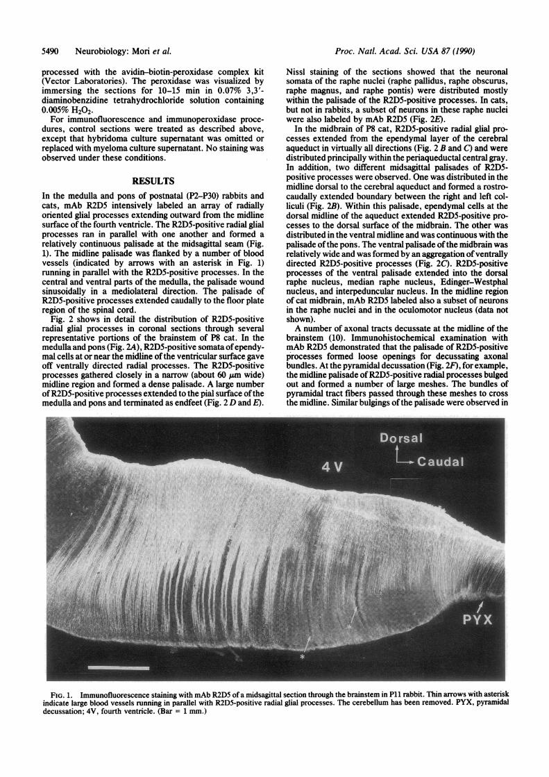
RESULTSIn the medulla and pons of postnatal (P2-P30) rabbits andcats, mAb R2D5 intensively labeled an array of radiallyoriented glial processes extending outward from the midlinesurface of the fourth ventricle. The R2D5-positive radial glialprocesses ran in parallel with one another and formed arelatively continuous palisade at the midsagittal seam (Fig.1). The midline palisade was flanked by a number of bloodvessels (indicated by arrows with an asterisk in Fig. 1)running in parallel with the R2D5-positive processes. In thecentral and ventral parts of the medulla, the palisade woundsinusoidally in a mediolateral direction. The palisade ofR2D5-positive processes extended caudally to the floor plateregion of the spinal cord.
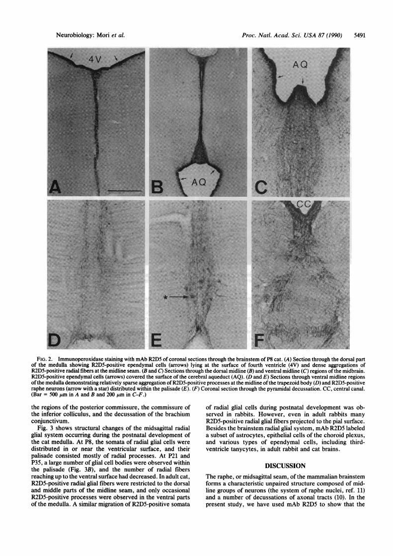
Fig. 2 shows in detail the distribution of R2D5-positiveradial glial processes in coronal sections through severalrepresentative portions of the brainstem of P8 cat. In themedulla and pons (Fig. 2A), R2D5-positive somata ofependy-mal cells at or near the midline ofthe ventricular surface gaveoff ventrally directed radial processes. The R2D5-positiveprocesses gathered closely in a narrow (about 60 ,gm wide)midline region and formed a dense palisade. A large numberofR2D5-positive processes extended to the pial surface ofthemedulla and pons and terminated as endfeet (Fig. 2 D and E).
Nissl staining of the sections showed that the neuronalsomata of the raphe nuclei (raphe pallidus, raphe obscurus,raphe magnus, and raphe pontis) were distributed mostlywithin the palisade of the R2D5-positive processes. In cats,but not in rabbits, a subset of neurons in these raphe nucleiwere also labeled by mAb R2D5 (Fig. 2E).
In the midbrain of P8 cat, R2D5-positive radial glial pro-cesses extended from the ependymal layer of the cerebralaqueduct in virtually all directions (Fig. 2 B and C) and weredistributed principally within the periaqueductal central gray.In addition, two different midsagittal palisades of R2D5-positive processes were observed. One was distributed in themidline dorsal to the cerebral aqueduct and formed a rostro-caudally extended boundary between the right and left col-liculi (Fig. 2B). Within this palisade, ependymal cells at thedorsal midline of the aqueduct extended R2D5-positive pro-cesses to the dorsal surface of the midbrain. The other wasdistributed in the ventral midline and was continuous with thepalisade ofthe pons. The ventral palisade ofthe midbrain wasrelatively wide and was formed by an aggregation ofventrallydirected R2D5-positive processes (Fig. 2C). R2D5-positiveprocesses of the ventral palisade extended into the dorsalraphe nucleus, median raphe nucleus, Edinger-Westphalnucleus, and interpeduncular nucleus. In the midline regionof cat midbrain, mAb R2D5 labeled also a subset of neuronsin the raphe nuclei and in the oculomotor nucleus (data notshown).A number of axonal tracts decussate at the midline of the
brainstem (10). Immunohistochemical examination withmAb R2D5 demonstrated that the palisade of R2D5-positiveprocesses formed loose openings for decussating axonalbundles. At the pyramidal decussation (Fig. 2F), for example,the midline palisade ofR2D5-positive radial processes bulgedout and formed a number of large meshes. The bundles ofpyramidal tract fibers passed through these meshes to crossthe midline. Similar bulgings ofthe palisade were observed in
I ,. ii
I,#
FIG. 1. Immunofluorescence staining with mAb R2D5 of a midsagittal section through the brainstem in P11 rabbit. Thin arrows with asteriskindicate large blood vessels running in parallel with R2D5-positive radial glial processes. The cerebellum has been removed. PYX, pyramidaldecussation; 4V, fourth ventricle. (Bar = 1 mm.)
5490 Neurobiology: Mori et al.
Proc. Natl. Acad. Sci. USA 87 (1990) 5491
FIG. 2. Immunoperoxidase staining with mAb R2D5 of coronal sections through the brainstem of P8 cat. (A) Section through the dorsal partof the medulla showing R2D5-positive ependymal cells (arrows) lying at the surface of fourth ventricle (4V) and dense aggregations ofR2D5-positive radial fibers at the midline seam. (B and C) Sections through the dorsal midline (B) and ventral midline (C) regions ofthe midbrain.R2D5-positive ependymal cells (arrows) covered the surface of the cerebral aqueduct (AQ). (D and E) Sections through ventral midline regionsofthe medulla demonstrating relatively sparse aggregation ofR2D5-positive processes at the midline of the trapezoid body (D) and R2D5-positiveraphe neurons (arrow with a star) distributed within the palisade (E). (F) Coronal section through the pyramidal decussation. CC, central canal.(Bar = 500 Aim in A and B and 200 jum in C-F.)
the regions of the posterior commissure, the commissure ofthe inferior colliculus, and the decussation of the brachiumconjunctivum.
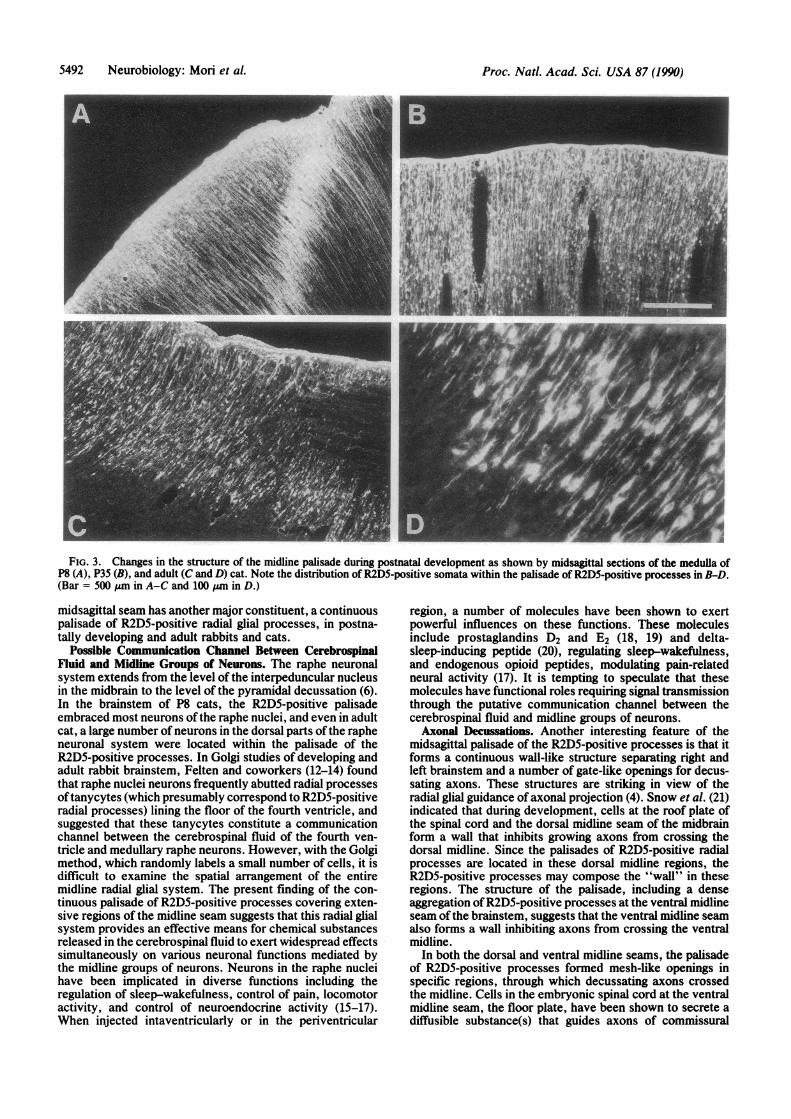
Fig. 3 shows structural changes of the midsagittal radialglial system occurring during the postnatal development ofthe cat medulla. At P8, the somata of radial glial cells weredistributed in or near the ventricular surface, and theirpalisade consisted mostly of radial processes. At P21 andP35, a large number of glial cell bodies were observed withinthe palisade (Fig. 3B), and the number of radial fibersreaching up to the ventral surface had decreased. In adult cat,R2D5-positive radial glial fibers were restricted to the dorsaland middle parts of the midline seam, and only occasionalR2D5-positive processes were observed in the ventral partsof the medulla. A similar migration of R2D5-positive somata
of radial glial cells during postnatal development was ob-served in rabbits. However, even in adult rabbits manyR2D5-positive radial glial fibers projected to the pial surface.Besides the brainstem radial glial system, mAb R2D5 labeleda subset of astrocytes, epithelial cells of the choroid plexus,and various types of ependymal cells, including third-ventricle tanycytes, in adult rabbit and cat brains.
DISCUSSION
The raphe, or midsagittal seam, of the mammalian brainstemforms a characteristic unpaired structure composed of mid-line groups of neurons (the system of raphe nuclei, ref. 11)and a number of decussations of axonal tracts (10). In thepresent study, we have used mAb R2D5 to show that the
Neurobiology: Mori et al.
Proc. Natl. Acad. Sci. USA 87 (1990)
FIG. 3. Changes in the structure of the midline palisade during postnatal development as shown by midsagittal sections of the medulla ofP8 (A), P35 (B), and adult (C and D) cat. Note the distribution of R2D5-positive somata within the palisade of R2D5-positive processes in B-D.(Bar = 500 ,um in A-C and 100 ,um in D.)
midsagittal seam has another major constituent, a continuouspalisade of R2D5-positive radial glial processes, in postna-tally developing and adult rabbits and cats.
Possible Communication Channel Between CerebrospinalFluid and Midline Groups of Neurons. The raphe neuronalsystem extends from the level of the interpeduncular nucleusin the midbrain to the level of the pyramidal decussation (6).In the brainstem of P8 cats, the R2D5-positive palisadeembraced most neurons ofthe raphe nuclei, and even in adultcat, a large number of neurons in the dorsal parts of the rapheneuronal system were located within the palisade of theR2D5-positive processes. In Golgi studies of developing andadult rabbit brainstem, Felten and coworkers (12-14) foundthat raphe nuclei neurons frequently abutted radial processesoftanycytes (which presumably correspond to R2D5-positiveradial processes) lining the floor of the fourth ventricle, andsuggested that these tanycytes constitute a communicationchannel between the cerebrospinal fluid of the fourth ven-tricle and medullary raphe neurons. However, with the Golgimethod, which randomly labels a small number of cells, it isdifficult to examine the spatial arrangement of the entiremidline radial glial system. The present finding of the con-tinuous palisade of R2D5-positive processes covering exten-sive regions of the midline seam suggests that this radial glialsystem provides an effective means for chemical substancesreleased in the cerebrospinal fluid to exert widespread effectssimultaneously on various neuronal functions mediated bythe midline groups of neurons. Neurons in the raphe nucleihave been implicated in diverse functions including theregulation of sleep-wakefulness, control of pain, locomotoractivity, and control of neuroendocrine activity (15-17).When injected intaventricularly or in the periventricular
region, a number of molecules have been shown to exertpowerful influences on these functions. These moleculesinclude prostaglandins D2 and E2 (18, 19) and delta-sleep-inducing peptide (20), regulating sleep-wakefulness,and endogenous opioid peptides, modulating pain-relatedneural activity (17). It is tempting to speculate that thesemolecules have functional roles requiring signal transmissionthrough the putative communication channel between thecerebrospinal fluid and midline groups of neurons.Axonal Decussations. Another interesting feature of the
midsagittal palisade of the R2D5-positive processes is that itforms a continuous wall-like structure separating right andleft brainstem and a number of gate-like openings for decus-sating axons. These structures are striking in view of theradial glial guidance of axonal projection (4). Snow et al. (21)indicated that during development, cells at the roof plate ofthe spinal cord and the dorsal midline seam of the midbrainform a wall that inhibits growing axons from crossing thedorsal midline. Since the palisades of R2D5-positive radialprocesses are located in these dorsal midline regions, theR2D5-positive processes may compose the "wall" in theseregions. The structure of the palisade, including a denseaggregation ofR2D5-positive processes at the ventral midlineseam of the brainstem, suggests that the ventral midline seamalso forms a wall inhibiting axons from crossing the ventralmidline.
In both the dorsal and ventral midline seams, the palisadeof R2D5-positive processes formed mesh-like openings inspecific regions, through which decussating axons crossedthe midline. Cells in the embryonic spinal cord at the ventralmidline seam, the floor plate, have been shown to secrete adiffusible substance(s) that guides axons of commissural
5492 Neurobiology: Mori et al.
Proc. Natl. Acad. Sci. USA 87 (1990) 5493
neurons toward the floor plate (22). It has been suggested thatcells in the floor plate interact with the decussating axons ofcommissural neurons and control the expression of commis-sural axon surface glycoproteins TAG1 and L (23, 24). Thestructure and the strategic position of the R2D5-positivedorsal and ventral palisades seem to be advantageous forguiding the decussation ofgrowing axons at specific positionsof the midline of the brainstem.Topographic Arrangements of the Radial Glial System. The
parallel trajectory of R2D5-positive processes within themidsagittal plane of the brainstem (cf. Fig. 1) clearly dem-onstrates a topographic relationship between the radial glialprocesses and various structures, including the raphe nucleiand axonal decussations, located within the midline seam.Structures located at different places in the rostrocaudal axisreceive projections from different groups of radial glial cells.For example, the pyramidal decussation received R2D5-positive radial processes from a specific group of ependymalcells lying at the surface ofthe most rostral part of the centralcanal (Fig. 2F). Neurons in the dorsal raphe nucleus receivedradial processes selectively from those ependymal cells thatwere distributed at the ventral midline of the cerebral aque-duct (Fig. 2C). It is thought that in the developing cerebralcortex, radial glial fibers provide a scaffold for migration ofneurons and specify the spatial arrangement of somata anddendrites of cortical neurons (1, 2, 25). Similarly, the midlineradial glial processes in the brainstem may serve as guides formigration of midline groups of neurons and for their spatialarrangement in the midsagittal seam (26). The midline radialglial system may also provide a scaffold not only for instruct-ing growing axons to decussate at specific positions but alsofor reorganizing transient axonal projections during embry-onic and postnatal development (27). In contrast to mostother radial glial fibers in the central nervous system, whichdisappear before or shortly after birth (5), the midline radialglial system persists in the postnatally developing and in theadult brain, although undergoing structural changes duringdevelopment. This suggests that the midsagittal radial glialsystem has functions required in postnatal and adult brain aswell as in embryonic brain. Use of mAb R2D5 and charac-terization of the R2D5 protein may provide insights into theinteractions ofthe radial glia and neurons at the midline seamof the brainstem.
We thank Dr. M. Connolly and Dr. K. Imamura for reading themanuscript. This work was supported in part by Special Coordina-tion Funds from the Science and Technology Agency of the JapaneseGovernment.
1. Rakic, P. (1981) in The Organization of Cerebral Cortex, eds.Schmitt, F. O., Worden, F. G., Adelman, G. & Dennis, S. G.(MIT Press, Cambridge, MA), pp. 7-28.
2. Rakic, P. (1988) Science 241, 170-176.3. Nordlander, R. H. & Singer, M. (1978) J. Comp. Neurol. 180,
349-374.4. Singer, M., Nordlander, R. H. & Egar, M. (1979) J. Comp.
Neurol. 185, 1-22.5. Levitt, P. & Rakic, P. (1980) J. Comp. Neurol. 193, 815-840.6. Flament-Durand, J. & Brion, J. P. (1985) Int. Rev. Cytol. 9,
121-155.7. Van Hartesveldt, C., Moore, B. & Hartman, B. (1986) J. Comp.
Neurol. 253, 175-184.8. Fujita, S. C., Mori, K., Imamura, K. & Obata, K. (1985) Brain
Res. 326, 192-196.9. Mori, K., Fujita, K., Imamura, K. & Obata, K. (1985) J. Comp.
Neurol. 242, 214-229.10. Berman, A. L. (1968) The Brainstem ofthe Cat-A Cytoarchi-
tectonic Atlas with Stereotaxic Coordinates (Univ. Wis. Press,Madison, WI).
11. Tork, I. (1985) in Rat Nervous System, ed. Paxinos, G. (Aca-demic, Sydney), pp. 43-78.
12. Felten, D. L., Harrigan, P., Burnett, B. T. & Cummings, J. P.(1981) Brain Res. Bull. 6, 427-436.
13. Cummings, J. P. & Felten, D. L. (1979) J. Comp. Neurol. 183,1-24.
14. Burnett, B. T. & Felten, D. L. (1981) Anat. Rec. 200, 337-347.15. Chase, T. N. & Murphy, D. L. (1973) Annu. Rev. Pharmacol.
13, 181-197.16. Jouvet, M. (1983) in Functions ofthe Nervous System: Psycho-
Neurobiology, eds. Monnier, M. & Meulders, M. (Elsevier,Amsterdam), Vol. 4, pp. 249-274.
17. Basbaum, A. I. & Fields, H. L. (1984) Annu. Rev. Neurosci. 7,309-338.
18. Hayaishi, 0. (1988) J. Biol. Chem. 263, 14593-14596.19. Matsumura, H., Goh, Y., Ueno, R., Sakai, T. & Hayaishi, 0.
(1988) Brain Res. 362, 114-121.20. Schoenenberger, G. A. & Monnier, M. (1977) Proc. Natl.
Acad. Sci. USA 74, 1282-1286.21. Snow, D. M., Steindler, D. A. & Silver, J. (1990) Dev. Biol.
137, 359-376.22. Tessier-Lavigne, M., Placzek, M., Lumsden, A. G. S., Dodd,
J. & Jessell, T. M. (1988) Nature (London) 336, 775-778.23. Dodd, J., Morton, S. B., Karagogeos, D., Yamamoto, M. &
Jessell, T. M. (1988) Neuron 1, 105-116.24. Furley, A. J., Morton, S. B., Manalo, D., Karagogeos, D.,
Dodd, J. & Jessell, T. M. (1990) Cell 61, 157-170.25. Steindler, D. A., Cooper, N. G. F., Faissner, A. & Schachner,
M. (1989) Dev. Biol. 131, 243-260.26. Wallace, J. A. & Lauder, J. M. (1983) Brain Res. Bull. 10,
459-479.27. Cowan, W. M., Fawcett, J. W., O'Leary, D. D. M. & Stan-
field, B. B. (1984) Science 225, 1258-1265.
Neurobiology: Mori et al.