Monkey,(Baboon,(or(Orangutan(?( - HKSCCMhksccm.org/.../Monkey_Baboon_or_Orangutan_3_3_2_3.pdf ·...
100
Monkey, Baboon, or Orangutan ? Presented by Dr Sinn Ting Ting Maria ICU, TKOH
-
Upload
hoangthien -
Category
Documents
-
view
217 -
download
1
Transcript of Monkey,(Baboon,(or(Orangutan(?( - HKSCCMhksccm.org/.../Monkey_Baboon_or_Orangutan_3_3_2_3.pdf ·...

Monkey, Baboon, or Orangutan ?
Presented by Dr Sinn Ting Ting Maria ICU, TKOH

Case History
• M/ 24 • born in China, came to Hong Kong at age 10
Past Health: • History of transient frothy urine at age 2? , subsided with medicaHons, unknown diagnosis
• Alpha thal trait

AdmiLed On 26/11/13
• Presented with R facial and periorbital swelling. • Mild R sided headache • ExerHonal SOB++, generalized malaise • No limb weakness • No dysuria/ urinary frequency/ âurine output • No frothy urine/ haematuria • No fever/ chest pain • Yellowish loose stool x 1

• Taken TCM on 21-‐ 22/11/2-‐13 for flu like symptoms, claimed periorbital swelling occurred before intake of TCM.

Physical ExaminaHon
• BP at AED: 262/151, P 136/ min • Rechecked: 221/139. P 100/ min • Conscious and alert • Periorbital and eyelid swelling, LMN R facial nerve palsy
• Fundi: no papilloedema • No ankle edema • Chest , CV, abdo exam: unremarkable • PR : empty

InvesHgaHons
• CBC: Hb 6.1(McHc), WBC 6.6, Plt 60; Schistocytes+
• RFT: Na 139 K 4.2 Cr 918 Urea 31.8 • LFT: normal, Alb 37.7 • INR: normal • TnT: 37 • SG: 6.1 • VBG: Bicarb 17.1 • LDH 529

• ECG: SR 132/min, tall T waves, LVH paLern • CXR: cardiomegaly
• Imp: Hypertensive crisis, microangiopathic haemolyHc anaemia, thrombocytopenia, renal failure
• Transferred to ICU for further care.

Progress
1. MAHA, thrombocytopenia : • Plasmapheresis given, replacement with FFP since 28/11/2013.
• Developed urHcaria, desaturaHon whilst on plasmapheresis. Responded to piriton, hydrocorHsone and ventolin.
• Changed replacement to cryo-‐reduced plasma during plasmapheresis since 29/11/13. Rash and itchiness controlled with regular iv piriton and hydrocorHsone.
• Completed 5 cycles of plasmapheresis in total(28/11-‐ 2/12). PLT count slowly improving to 179 , Cr 804.

ADAMTS 13: • AcHvity: â 31 (70-‐160) % • AnHgen: 315.8 (253-‐2238) ng/ml • AutoanHbody: – ve
Comment: • Moderate reducHon in ADAMTS 13 acHvity. The level is not
low enough to be specific for TTP but post treatment effect has to be considered. Normal ADAMTS 13 anHgen level and negaHve for the corresponding autoanHbody.
• Overall the laboratory results do not support a diagnosis of TTP.

Progress
• 2. Hypertensive crisis: • Given labetalol for BP control iniHally. Later BP stabilized with norvasc, prazosin and betaloc.
• Transferred to general ward on 2/12/2013.

Progress
• 3. Renal Failure: • US kidney ( 28/11/13): Renal outlines are smooth. R kidney measures 11.2 cm and L 11.0 cm. Renal parenchymal echogeniciHes are increased, compaHble with parenchymal disease. CollecHng systems are not dilated. No renal stone is idenHfied. Urinary bladder is normal in appearance.
• Impression: Renal parenchymal disease.
• Toxicology screen for TCM( TCM list faxed): no nephrotoxic substance idenHfied.

• Thrombosed arteriole with fibrin thrombi in the lumen

• Marked inHmal thickening with myxoid material in media accompanied by proliferaHon of fibroblasHc cells
• Arteriole with onion-‐skin change, edema of endothelial cells and myxoid degeneraHon.

• Interlobular artery with inHmal thickening • Arteriole with thrombosis and recanalizaHon

• Renal Biopsy was done on 12/12: • 5/7 glomerular globally sclerosed, the remaining two shows collapse and
wrinkling of glomerular tums and basement membrane. Crescent, karyorrhexis or fibrinoid necrosis is not evident. There is endothelial swelling in majority of the arterioles and interlobular arteries. MyoinHmal thickening and onion-‐skin changes are present in the arterioles, accompanied by thrombosis and mucoid degeneraHon. Moderate inHmal thickening is present in some interlobular arteries as well. Silver impregnaHon does not show duplicaHon or epimembranous spike. There is 30% intersHHal fibrosis with proporHonal intersHHal inflammatory infiltrate. The tubular cells are largely unremarkable. No intratubular cast, crystal or cells are present. Direct immunofluorescene study shows negaHve staining for IgG, IgA, IgM , C3 and C1q. There is no light chain restricHon.
• The overall pictures are consistent with thromboHc microangiopathy + changes of chronic HT nephropathy.

Further Work up
• C3: â 0.85 ( N: 0.9-‐1.8 g/L) • C4/ANA/ANCA/ anH-‐GBM/APL Ab : NAD • Ig paLern: IgA 3.88, IgG â 6.79, IgM â0.29; SPE: no paraproteins
Echo: conc LVH with mildly impaired LV, EF 50%. LV wall thickness ~ 1.4 cm. No RWMA. Mild TR.

Overall diagnosis: thromboHc microangiopathy, possible TTP, undiagnosed long standing HT with end-‐organ damage (LVH/CKD).
• RFT: urea / Cr 31.2/ 758 • TP: 1.61 g/day, CrCl: 13 ml/min • Hb: 7.9, Plt: 137, film: no schizocytes • LDH: 175

MedicaHon on Discharge 20/12/13 • Prednisolone 60 mg daily for 11 days ( later taper to 45 mg
daily x 2 wks, 30 mg daily x 2 weeks)
• Betaloc 150 mg BD • Norvasc 10 mg daily • Prazosin 5 mg q 8 h • Hydralazine 75 mg q 8 h • Lasix 40 mg BD
• NaHCO3 300 mg TDS • CaCO3 2000 mg before lunch and 2000 mg before dinner • Pepcidine 20 mg BD

??? • Microangiopathic haemolyHc anaemia + • Thrombocytopenia+ • Acute on chronic renal impairment+ • Hypertensive crisis+ • R LML facial nerve palsy , mild headache • = • ? TTP • ? HUS • ? TTP/ HUS • ? Secondary to malignant HT


Second Admission on 24/1/2014
• Presented with central neck swelling for 2 days, associated with sorethroat and mild dysphagia.
• No fever • No drooling of saliva/ toothache/ SOB

Physical ExaminaHon
• Afebrile • BP 176/ 107, P 92/min • SpO2 100 % on 2 L O2 • Central neck swelling + ve, mildly tender, no definite crepitus
• Chest/CV/abdo exam: NAD
• XR lateral neck: ? thumb sign

Prelim Blood Results
• Hb: 8.8 staHc • WBC: 13.7, Plt: 63 • Blood film: rare schistocytes • INR 1.2 , APTT 36.6 • Cr: 602 • LFT: normal
• Transferred to ICU for further management.

Progress
• 1. ENT was consulted: • Lem submandibular infecHon/ abscess. Suggested to CT neck
with contrast . Upgrade augmenHn to tazocin + flagyl
• CT neck and upper thorax 24/1/14: • MulHple submandibular and cervical lymphadenopathy is
seen. • Upper lungs are clear. No mass is noted. • No retroperitoneal or mediasHnal mass is seen. • Impression: No frank abscess. ? CelluliHs.
• Was treated as submandibular celluliHs, likely dental source of infecHon.
• Responded to anHbioHc. • Blood culture 24/1/14: -‐ ve growth

Progress
• 2. Suspected TTP relapse. • Plasmapheresis was given for 5 days from 25-‐ 29/1/14 , replacement with cryo-‐reduced plasma, covered with hydrocorHsone and piriton for FFP allergy.
• LDH 28/1/14 : 218 U/L


Progress
• 3. A session of HD was given on 30/1/14. • Urea / Cr 34.6/ 653 (pre-‐dialysis)à 19.5/422

Progress
• 4. Hypertension • Initally put on isoket for BP control, later controlled with hydralazine 100 mg TDS, adalat retard 40 mg BD, prazosin 10 mg BD and minoxidil 2.5 mg daily, in addiHon to betaloc 150 mg BD
• US kidney and CT angiogram of renal arteries: no evidence of renal artery stenosis

On Discharge 7/2/14
• Hb 7.4 , Plt 95 • Urea / Cr 28.5/538

MedicaHon on Discharge on 7/2/2014
• Prednisolone 25 mg daily for 4 days
• Betaloc 150 mg BD • Adalat retard 40 mg BD • Prazosin 10 mg BD • Hydralazine 100 mg q 8 h • Minoxidil 2.5 mg daily • Lasix 40 mg BD
• CaCO3 2000 mg before lunch and 2000 mg before dinner • NaHCO3 300 mg BD • Pepcidine 20 mg BD

Clues:
• Recurrent microangiopathic haemolyHc anaemia +
• Thrombocytopenia + • Acute on chronic renal impairment + • SubopHmal control blood pressure + • Submandibular celluliHs
=

DDx • 1. TTP ( ADAMTS 13 acHvity not very low, anHbody – ve )
• 2. HUS ( no diarrhoea , but ARF +ve ) • 3. TTP/HUS • 4. high blood pressure induced thromboHc microangiopathy
• 5. Sepsis induced thromboHc microangiopathy


ThromboHc Microangiopathies(TMA)
• TMA comprises a spectrum of disorders characterized by microangiopathic hemolyHc anaemia, peripheral thrombocytopenia and organ failure of variable severity caused by microvascular occlusion, associated with mulHple pathogenic factors.
• For a long Hme, TMA remained a heterogenous group of poorly differenHated diseases with obscure pathophysiologies.
• Advances in recent years have delineated the molecular mechanisms of most of the TMA syndromes and it is now clear that the TMA syndromes are caused by several disHnct molecular defects

ThromboHc Thrombocytopenic Purpura(TTP)
• Rare, reported incidence of 6 cases per million per year in UK ( Scully et al, 2008)
• Untreated mortality 90 %
• Diagnosis: originally characterized by a pentad of thrombocytopenia, MAHA, fluctuaHng neurological signs, renal impairment and fever, omen with insidious onset.

TTP : PresentaHon
• Median platelet count is typically 10-‐30 x 10 9/ l at presentaHon
• Median Hb levels on admission are typically 80-‐100 g/l.

TTP: PresentaHon Central neurological impairment, omen fliqng and variable 70-‐80%:
– Altered personality – Headache – Confusion – Fits – Paresis, aphasis, dysarthria, visual problems – Encephalopathy – Coma ( 10%)
• Up to 35 % of paHents do not have neurological signs at presentaHon.
• ( Scully et al. Br J Haem 2012, 158, 323-‐335)

TTP: PresentaHon
• Renal abnormaliHes and fever are not prominent features.
• Renal abnormaliHes: proteinuria, haematuria, but no or minimal renal clearance impairment
• Acute renal failure requiring haemodialysis is rare in TTP and more indicaHve of HUS.

ADAMTS 13 assays
• In 1998, Tsai and Furlan idenHfied that the severe deficiency in the von Willebrand factor cleaving protease ADAMTS 13 could explain the accumulaHon of unusually large VWF mulHmers in the plasma of paHents with chronic relapsing TTP first reported by Moake in the 80s.
• ADAMTS13 deficiency can be due to mutaHons of the encoding gene or autoanHbodies directed against various epitopes of the protein.

ADAMTS 13 assays • Measure in acute phase of illness. Blood must be taken prior
to treatment to assess baseline ADAMTS 13 acHvity.
• Severely reduced ADAMTS 13 acHvity (< 5 %) ± the presence of an inhibitor or IgG anHbodies, confirms the diagnosis.
• The specificity of severe ADAMTS 13 deficiency (<5%) in disHnguishing acute TTP from HUS is 90 % .
• Decreased ADAMTS 13 acHvity (< 40 % but > 5 %) has been reported in a wide variety of non-‐TTP condiHons such as uraemia, inflammatory states, post-‐operaHvely, and during pregnancy.

Subgroups of TTP • 1. Congenital TTP : • Dx : ADAMTS 13 acHvity < 5% in the absence of anHbodies to
ADAMTS 13 .
• MutaHons in ADAMTS 13 gene, autosomal recessive
• Incidence: 1: 1,000, 000
• Variable phenotype and can present at any age. But most omen at birth or during childhood.
• Can be asymptomaHc unHl a precipitaHng event results in frank TTP episode, eg pregnancy.

Subgroups of TTP • 2. Acute idiopathic TTP – Most common form of TTP – Characterized by anHbodies, usually IgG, directed against ADAMTS 13 .
• 3. HIV-‐ associated TTP
• 4. Pregnancy-‐ associated TTP
• 5. Drug-‐ associated TTP: quinine, , Hclodipine, oestrogen-‐containing hormone preparaHons, , trimethoprim, peglyated interferon, simvastaHn.

Subgroups of TTP
• 6. Transplant-‐ associated microangiopathy • 7. Malignancy-‐ associated thromboHc microangiopathy
• 8. PancreaHHs-‐ associated TTP

HemolyHc Uremic Syndrome(HUS)
• Comprises the triad of microangiopathic hemolyHc anaemia, thrombocytopenia, and acute kidney injury.
• About 90 % of pediatric paHents develop this syndrome amer infecHon with Shigella dysenteriae, which produces true Shiga toxins, or Escherichia coli, some strains produce Shiga-‐like toxins.
• Self –limiHng disease, with > 90% of childhood cases recover independent renal funcHon spontaneously.

• HUS that is not related to Shiga toxins is called atypical HUS (aHUS), accounts for ~ 10 % of all HUS cases.

aHUS
• Occurs in individuals of all ages ( extremes : 1 day to 83 years )
• About 60 % onset ≤ 18 years • 70% of children have first episode before age 2 .
• Omen familial, can be sporadic
• Incidence in US: ~ 2 per million populaHon per year

Prognosis is poor: – IniHal mortality higher in children(6.7% vs 0.8% at 1 year)
– Adults progress to end-‐stage renal disease (ESRD) more frequently at iniHal presentaHon( 46% vs 16%).
– At 3 to 5 years amer onset, 36 % to 48% of children and 64% to 67% of adults had died or reached (ESRD)
• (Fremeaux –Bacchi et al, Clin J Am Soc Nephrol. 2013) • (Noris M et al, Clin J Am Soc Nephrol. 2010)

PresenHng Features • Onset generally sudden
• Most complete triad of HUS at first invesHgaHon: – Hb < 10 g/dl – Plt generally between 30 000 and 60 000/mm3
– Renal insufficiency
• Hypertension is frequent and omen severe , due both to hyperreninaemia secondary to renal TMA and volume overload in case of oliguria/ anuria. Cardiac failure or neurological complicaHons due to hypertension possible.
• Extra renal manifestaHons in 20 % of paHents. Most frequent is CNS involvement( 10 % ) : irritability, drowsiness, seizures, diplopia, corHcal blindness, hemiparesis or hemiplegia, stupor, coma.

Triggering Events
• 1. InfecHon, mainly URTI or GE, in at least 50 % of paHents, up to 80 % in pediatric cohorts
• 2. Pregnancy

• David et al . Semiin Nephr 2013

aHUS • aHUS is the prototypic disorder of complement regulaHon.
• Since 1974, reduced serum C3 levels with normal C4 have been reported in paHents with aHUS, indicaHng a selecHve acHvaHon of the alternaHve pathway.
• The discovery of mutaHons in the complement system in aHUS by Warwicker et al (1998) set the train in research in idenHfying the geneHc basis of uncontrolled alternaHve pathway acHvaHon in aHUS.
• IdenHficaHon of the specific geneHc defect carries crucial prognosHc informaHon.

The Complement System
• Ancient defence mechanism that sHmulates the inflammatory response and destroys pathogens through opsonizaHon and lysis.
• It bridges innate and adapHve immunity and it disposes of immune complexes and injured Hssues and cells.


• Three pathways of acAvaAon: – Classical – LecHn – AlternaHve
• These pathways converge at the point of cleavage of C3 .
• AcHvaHon of the classical and the lecHn pathways occurs amer binding to immune complexes or microorganisms respecHvely, the alternaHve pathway is conHnually acHvated by the spontaneous hydrolysis of C3. It generates C3b which binds indiscriminately to pathogens and host cells.

• Four regulatory proteins of the alternaHve pathway: – Complement factor H (CFH) (most important) – Complement Factor I (CFI) – Membrane cofactor protein ( MCP or CD46) – Thrombomodulin (THBD)
• And complement acHvators , 2 proteins of the C3 convertase : C3 and factor B (CFB)
• àHad a role in pathogenesis of aHUS.
[humoral factors/ fluid phase]
[membrane-‐bound factors]

• A loss-‐of-‐ funcHon mutaHon in a complement-‐inhibiHng gene or a gain-‐of –funcHon mutaHon in a gene that encodes a complement acHvator will lead to an unopposed acHvaHon of the complement system
• à formaHon of membrane aLack complex ( C5b-‐9) on cell surfaces of especially endothelial cells in kidney à destroyed cells by forming transmembrane pores.
• àendothelial cells are damaged and leucocytes are aLracted, releasing oxygen radicals and proteinases, which can further damage the endothelium
• à result in increased platelet adherence and formaHon of microthrombi in kidney
• à ARF, thrombocytopenia and haemolyHc anaemia

• Between 6 % and 10% of aHUS paHents have anHbodies which bind to the C terminal region of factor H.

• About 60% of individuals affected have an inherited and/or acquired abnormality affecHng components of complement pathway.
• Penetrance of geneHc forms of aHUS is around 50 % in carriers of CFH, MCP,CFI, THBD and C3 mutaHons.
• à mulAple concurrent geneAc and environmental hits are needed to determine disease expression.

What is the significance of knowing all these molecular/
geneHc defects?

Significance
• 1. Clinical course • 2. Management: – respond to plasmapheresis – outcome of kidney transplant – selecHon of living donors
• 3. Prognosis
Pa$ent care should be individualized.

Clinical course/ Prognosis
• 1. 80-‐90% of paHents with an MCP mutaHon will develop a remission, although recurrences omen occur.
• 2.60-‐70% of paHents with a CFH, CFI, or C3 mutaHon will develop ESRF within one year amer diagnosis.
• 3. 88% of paHents with CFB mutaHon develop ESRF within one year amer diagnosis.

Renal Transplant • Overall risk of aHUS recurrence amer renal transplant is 50 % . • Risk of gram loss 80-‐90% in paHents with recurrence.
• Risk of post-‐transplant aHUS recurrence: – 75-‐90% with CFH mutaHon – 45-‐80% with CFI mutaHon – 40-‐70% with C3 mutaHon – 100 % with CFB mutaHon(3 paHents) – 30 % with THBD mutaHon – 0-‐20% with MCP mutaHon – 40-‐60% with anH-‐CFH Ab

Management 1. Plasmapheresis 2. Eculizumab 3. TransplantaHon 4. General measures

Plasmapheresis • 1. Aim: – Replacement of non-‐funcHoning complement proteins – Removal of CFH autoanHbodies and hyperfuncHonal complement components
• 2. Plasmapheresis vs Plasma infusion – Plasma infusion is sufficient with a missing or defecHve complement-‐regulaHng protein( majority of cases) .

Plasmapheresis • 3. Efficacy of plasmapheresis: – Cohort studies have shown the mortality rate has decreased from 50% to 25 % since the introducHon of plasma therapy.
– Despite this, majority of paHents need long term renal replacement therapy within 2 years of presentaHon. (Noris et al, 2010)
– AbnormaliHes in soluble regulators such as factor H respond beLer to plasma exchange than paHents with abnormaliHes in the transmembrane regulator CD46 ( Caprioli et al, 2006)
– Those with MCP mutaHon alone ( membrane-‐bound protein), plasma therapy is of limited value: remission in 80-‐90% without plasma treatment.

Plasmapheresis
• 4. Regime: ( consensus-‐ based guidelines) • Start within 24 hours of diagnosis – Adult: 1-‐2 plasma volumes (60-‐75 ml/kg) per session with FFP
– Children: 50-‐100 ml /kg – Performed daily iniHally unHl hemolysis is controlled. – Individuals with geneHc defects in complement system are frequently plasma dependent ( weekly/biweekly) to maintain remission.
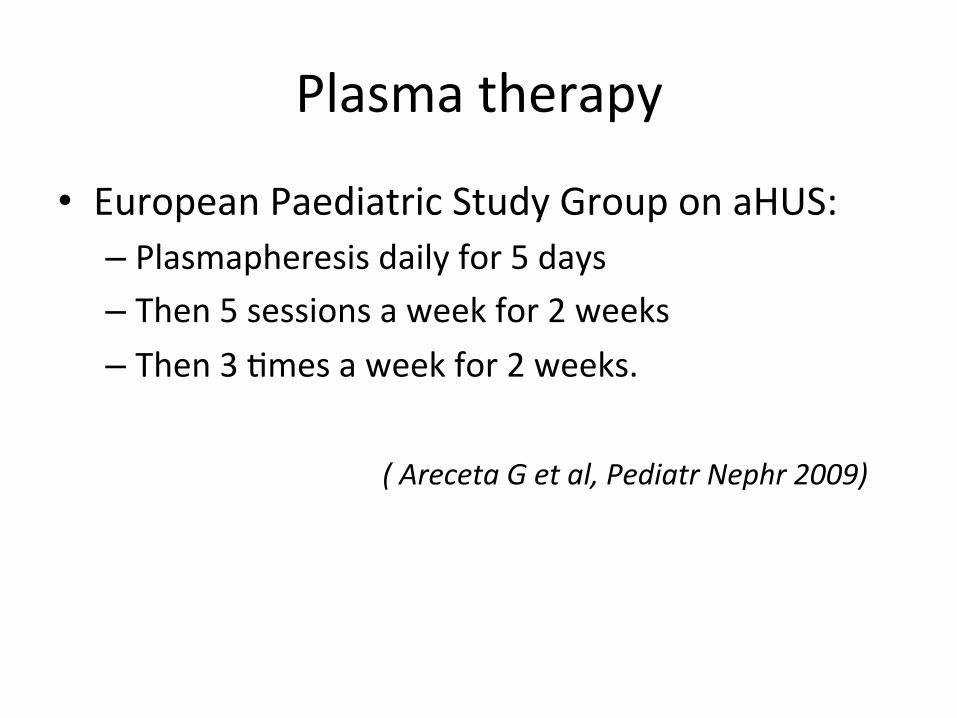
Plasma therapy
• European Paediatric Study Group on aHUS: – Plasmapheresis daily for 5 days – Then 5 sessions a week for 2 weeks – Then 3 Hmes a week for 2 weeks.
( Areceta G et al, Pediatr Nephr 2009)

Plasma Infusion
• When plasma infusion is used, suggested dosage is 30-‐40ml/kg iniHally and 10-‐20ml/kg per day thereamer. The dose and frequency may be reduced to weekly or biweekly intervals according to response.
• (Kavanagh D et al, Br Med Bull, 2006)

Eculizumab
• A recombinant humanized monoclonal Ig G anHbody directed against C5, blocks the cleavage of C5 into its effecHve components C5a and C5b, ulHmately prevenHng the generaHon of the proinflammatory pepHde C5b and the cytotoxic membrane aLack complex C5b-‐9.
• Approved worldwide for the treatment of paroxysmal nocturnal hemoglobinuria (PNH).

Eculizumab
• The use of Eculizumab in aHUS was first reported by Gruppo and Rother ( 2009) and Nurnberger et al. ( 2009) as two separate cases. ( NEJM 2009).

Eculizumab • Highly effecHve, with ~ 85 % paHents becoming disease-‐free in both plasma-‐resistant and plasma-‐dependent aHUS.
• EffecHve in paHents with and without idenHfied complement mutaHons.
• The earlier eculizumab is commenced, the greater preservaHon of kidney funcHon.
(Wong et al, Molecul. Immun 2013)

ProspecHve Phase 2 trial • Trial 1: Eligible paHents had evidence of progressive
thromboHc microangiopathy amer >= 4 sessions of plasma exchange or infusion in the prior week : Plasma – resistant group
• Trial 2: Eligible paHents had no decrease in platelet count of > 25% for at least 8 weeks before they received the first dose of eculizumab and were being treated with plasma exchange or infusion at least once every 2 weeks but no more than 3 Hmes/ week : Plasma –dependent group
• Legendre et al. NEJM 2013; 368:2169-‐2181

-‐Mean increase in Plt count 73 x 10 9/ L , p < 0.001 -‐87 % of paVents had normal platelet count at weeks 26 and 64

Mean increase in GFR at week 26 : 32 ml / min / 1.73m 2, p = 0.001

-‐Mean increase in GFR at week 26 : 6 ml/min/1.73m 2, p=0.003 -‐In both trials, earlier iniVaVon of ecu. was ass. with a sig.greater
improvement in GFR throughout the treatment period


Eculizumab
• 1. Treatment should begin as soon as Stx-‐HUS and ADAMTS 13 deficiency can be eliminated.
• 2. Current protocols suggest life-‐long therapy.

Protocol
• Recommended doses for aHUS are 30% higher than for PNH, in order to completely block the complement terminal acHvaHon. (trough > 35 ug/ml) In adults: 900 mg iv weekly for 4 weeks Then 1200 mg for the 5th injecHon Every 14 days as maintenance long term

Eculizumab • Downside: increased risk of Neisseria meningiHs infecHon. – Receive vaccinaHon against Neisseria meningiHs before treatment ( but cannot protect against B serotype)
– Permanent anHbioHc prophylaxis ( penicillin or erythromycin ) during treatment in addiHon to vaccinaHon
• Adverse effects: HT, URTI, diarrhoea

Eculizumab
• In 2011, eculizumab (Soliris R) was approved as a new drug for the treatment of aHUS in Europe and the US.

Eculizumab
• Very expensive. • Up to Euro 300, 000 per treatment year

• NHS England has published a Clinical Commissioning Policy Statement in 2013 : states that it will commission eculizumab for new paHents with aHUS and for paHents who are on dialysis and are suitable for a kidney transplant.

‘The price of eculizumab will mean that plasma exchange will remain the only
currently available opHon in many countries’
David Kavanagh et at, Sem in Nephro, 2013

Renal TransplantaHon
• 1. Eculizumab as prophylaxis before transplantaHon in those with a known mutaHon, also to treat and prevent recurrent disease post-‐transplant.
• 2. Because CFH, CFI, C3 and CFB are synthesized predominantly in liver, combined liver/kidney transplantaHon may correct the underlying geneHc deficiency. Short term mortality up to 14% .

General Measures
• Avoid triggers of endothelial injury , such as HT, hypercholesteraemia, by adequate blood pressure control and use of staHns.


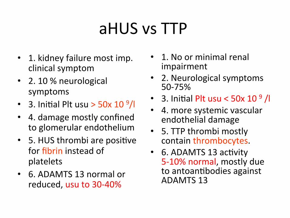
aHUS vs TTP • 1. kidney failure most imp.
clinical symptom • 2. 10 % neurological
symptoms • 3. IniHal Plt usu > 50x 10 9/l • 4. damage mostly confined
to glomerular endothelium • 5. HUS thrombi are posiHve
for fibrin instead of platelets
• 6. ADAMTS 13 normal or reduced, usu to 30-‐40%
• 1. No or minimal renal impairment
• 2. Neurological symptoms 50-‐75%
• 3. IniHal Plt usu < 50x 10 9 /l • 4. more systemic vascular
endothelial damage • 5. TTP thrombi mostly
contain thrombocytes. • 6. ADAMTS 13 acHvity
5-‐10% normal, mostly due to antoanHbodies against ADAMTS 13


Dx of aHUS
• 1. Serum levels of C3, C4, CFH and CFI, and a complement anHbody screen.
• 2. FACS analysis of peripheral blood mononuclear cells for MCP expression.
• 3. GeneHc tesHng including a method to detect copy number variaHon should be undertaken.


Complement InvesHgaHons: Pi|alls
• 1. Most assays measure the presence of complement protein, not the acHvity
• MutaHonal screening should be performed in the complement genes( CFH, CFI, MCP, C3, CFB, and THBD), irrespecHve of serum C3, CFH, or CFI levels.
• 2. AbnormaliHes in complement regulaHon may occur at the level of endothelial cell surface, not systemically.


Progress of Our PaHent • 1. Tapering dose of prednisolone , last seen at OPD on 9 June 2014 : – Urea / Cr 26.3/ 616 – Proteinuria: 1.15g/ day – CrCl 16 ml / min – Hb 9.2 – Plt 150 – LDH 208
• Further decrease prednisolone to 10 mg daily

Take Home Message • 1. HUS is characterized by a triad of microangiopathic
hemolyHc anaemia, thrombocytopenia and acute kidney injury.
• 2. The diagnosis aHUS requires the exclusion of TTP, STEC-‐HUS, and other causes of renal thromboHc microangiopathy.
• 3. aHUS is a disease caused by complement dysregulaHon in the alternaHve pathway.
• 4. First line of therapy : eculizumab
• 5. For pracHcal purpose, plasmapheresis/ plasma therapy is the iniHal treatment of choice, and it should be started within 24 hours of presentaHon of acute aLack.

• 6. It is a disease with poor prognosis: – About 50 % died or reach ESRD 3-‐5 years amer disease onset.
– Overall risk of aHUS recurrence amer renal transplant is 50 % . Risk of gram loss 80-‐90% in paHents with recurrence.
• 7. Knowledge of underlying complement defect will help to individualize paHent care.

THANK YOU !