Monitoring Bacterial Diversity of the Marine Sponge...
11
APPLIED AND ENVIRONMENTAL MICROBIOLOGY, July 2008, p. 4133–4143 Vol. 74, No. 13 0099-2240/08/$08.000 doi:10.1128/AEM.00454-08 Copyright © 2008, American Society for Microbiology. All Rights Reserved. Monitoring Bacterial Diversity of the Marine Sponge Ircinia strobilina upon Transfer into Aquaculture †‡ Naglaa M. Mohamed, 1 Venkateswara Rao, 2 Mark T. Hamann, 2 Michelle Kelly, 3 and Russell T. Hill 1 * Center of Marine Biotechnology, University of Maryland Biotechnology Institute, Baltimore, Maryland 21202 1 ; Department of Pharmacognosy, Pharmacology, Chemistry and Biochemistry and National Center for the Development of Natural Products, The University of Mississippi, Oxford, Mississippi 38655 2 ; and National Centre for Aquatic Biodiversity and Biosecurity, National Institute of Water and Atmospheric Research (NIWA) Ltd., Auckland, New Zealand 3 Received 25 February 2008/Accepted 28 April 2008 Marine sponges in the genus Ircinia are known to be good sources of secondary metabolites with biological activities. A major obstacle in the development of sponge-derived metabolites is the difficulty in ensuring an economic, sustainable supply of the metabolites. A promising strategy is the ex situ culture of sponges in closed or semiclosed aquaculture systems. In this study, the marine sponge Ircinia strobilina (order Dictyoceratida: family Irciniidae) was collected from the wild and maintained for a year in a recirculating aquaculture system. Microbiological and molecular community analyses were performed on freshly collected sponges and sponges maintained in aquaculture for 3 months and 9 months. Chemical analyses were performed on wild collected sponges and individuals maintained in aquaculture for 3 months and 1 year. Denaturing gradient gel elec- trophoresis was used to assess the complexity of and to monitor changes in the microbial communities associated with I. strobilina. Culture-based and molecular techniques showed an increase in the Bacteroidetes and Alpha- and Gammaproteobacteria components of the bacterial community in aquaculture. Populations affiliated with Beta- and Deltaproteobacteria, Clostridia, and Planctomycetes emerged in sponges maintained in aquaculture. The diversity of bacterial communities increased upon transfer into aquaculture. Sponges harbor diverse microorganisms, and various symbi- otic relationships between sponges and microorganisms may contribute to the sponge’s health and nutrition. Sponge-asso- ciated microbes can constitute up to 60% of the sponge bio- mass (24, 66, 67, 72). Important roles of the symbionts include photosynthetic carbon fixation (73), nitrification (6, 9), nitro- gen fixation (59, 74, 75), and anaerobic metabolism (27). An- other important role of sponge-associated bacteria is the pro- duction of potential secondary metabolites, such as antibiotics, antifungal compounds, and compounds that prevent predation or fouling (45). Sponges are sessile filter feeders with numer- ous tiny pores on the body surface and channels within the body that allow water to enter and circulate, with microorgan- isms and organic matter being removed through filtration (35, 68). Sponges contain assemblages of symbiotic bacteria that are distinct from the bacteria that are being filtered out of the surrounding water during the sponges’ feeding process (62). Marine sponges are a rich source of pharmacologically ac- tive compounds that can potentially be used as medicines to cure human diseases, and the isolation of bioactive compounds from sponges has been reviewed extensively (17, 25, 30, 35, 49, 55, 58). Bacteria isolated from sponges have also been sources of novel bioactive compounds. In some cases, symbiotic bac- teria may be the producers of promising compounds first found in the extracts of sponges. Circumstantial evidence for the microbial origin of a sponge-derived compound may be sup- plied by the structure of the compound (31). The onnamides and theopederins are polyketides that structurally resemble pederin, the defensive polyketide in Paederus fuscipes beetles. Strong evidence was provided for a bacterial producer of the onnamides and theopederins found in the sponge Theonella swinhoei (49). Genes closely resembling those encoding ped- erin were found in the complex metagenome of T. swinhoei and had characteristic prokaryotic signatures (49). Several bioactive compounds from sponges have passed the preclinical stage (44). The low yields of these compounds, known as the supply problem, are a major obstacle limiting successful transition of marine-derived compounds through clinical studies and into commercial production (42, 46). Har- vesting sponges from the environment generally will not pro- vide a reliable, large-scale supply and could lead to the extinc- tion of the particular sponge species. Several techniques may produce large quantities of sponge biomass needed for the extraction of bioactive metabolites that are not viable candi- dates for total synthesis. Such techniques include sponge farm- ing (10, 11) and the culture of primmorphs. Primmorphs are aggregates of sponge cells that still contain bacteria (reviewed by Mu ¨ller et al. [41]). The NOMATEC (Novel Marine Tech- nologies) project showed that Ircinia variabilis is suitable for mariculture (69). Furthermore, De Rosa et al. (7) reported the development of cell cultures from Ircinia muscarum. There were major differences in the composition of secondary me- tabolites between the wild sponge and its cell cultures, with a lower concentration of lipids and a loss of sterols and volatile compounds in cell cultures (8). When the bioactive compounds are produced by a sponge-associated bacterium, options in- * Corresponding author. Mailing address: Center of Marine Bio- technology, Columbus Center Suite 236, 701 E. Pratt Street, Balti- more, MD 21202. Phone: (410) 234-8883. Fax: (410) 234-8896. E-mail: [email protected]. † Supplemental material for this article may be found at http://aem .asm.org/. ‡ Contribution no. 06-143 from the Center of Marine Biotechnology. Published ahead of print on 9 May 2008. 4133 on July 13, 2018 by guest http://aem.asm.org/ Downloaded from
Transcript of Monitoring Bacterial Diversity of the Marine Sponge...

APPLIED AND ENVIRONMENTAL MICROBIOLOGY, July 2008, p. 4133–4143 Vol. 74, No. 130099-2240/08/$08.00�0 doi:10.1128/AEM.00454-08Copyright © 2008, American Society for Microbiology. All Rights Reserved.
Monitoring Bacterial Diversity of the Marine Sponge Ircinia strobilinaupon Transfer into Aquaculture�†‡
Naglaa M. Mohamed,1 Venkateswara Rao,2 Mark T. Hamann,2 Michelle Kelly,3 and Russell T. Hill1*Center of Marine Biotechnology, University of Maryland Biotechnology Institute, Baltimore, Maryland 212021; Department ofPharmacognosy, Pharmacology, Chemistry and Biochemistry and National Center for the Development of Natural Products,
The University of Mississippi, Oxford, Mississippi 386552; and National Centre for Aquatic Biodiversity and Biosecurity,National Institute of Water and Atmospheric Research (NIWA) Ltd., Auckland, New Zealand3
Received 25 February 2008/Accepted 28 April 2008
Marine sponges in the genus Ircinia are known to be good sources of secondary metabolites with biologicalactivities. A major obstacle in the development of sponge-derived metabolites is the difficulty in ensuring aneconomic, sustainable supply of the metabolites. A promising strategy is the ex situ culture of sponges in closedor semiclosed aquaculture systems. In this study, the marine sponge Ircinia strobilina (order Dictyoceratida:family Irciniidae) was collected from the wild and maintained for a year in a recirculating aquaculture system.Microbiological and molecular community analyses were performed on freshly collected sponges and spongesmaintained in aquaculture for 3 months and 9 months. Chemical analyses were performed on wild collectedsponges and individuals maintained in aquaculture for 3 months and 1 year. Denaturing gradient gel elec-trophoresis was used to assess the complexity of and to monitor changes in the microbial communitiesassociated with I. strobilina. Culture-based and molecular techniques showed an increase in the Bacteroidetesand Alpha- and Gammaproteobacteria components of the bacterial community in aquaculture. Populationsaffiliated with Beta- and Deltaproteobacteria, Clostridia, and Planctomycetes emerged in sponges maintained inaquaculture. The diversity of bacterial communities increased upon transfer into aquaculture.
Sponges harbor diverse microorganisms, and various symbi-otic relationships between sponges and microorganisms maycontribute to the sponge’s health and nutrition. Sponge-asso-ciated microbes can constitute up to 60% of the sponge bio-mass (24, 66, 67, 72). Important roles of the symbionts includephotosynthetic carbon fixation (73), nitrification (6, 9), nitro-gen fixation (59, 74, 75), and anaerobic metabolism (27). An-other important role of sponge-associated bacteria is the pro-duction of potential secondary metabolites, such as antibiotics,antifungal compounds, and compounds that prevent predationor fouling (45). Sponges are sessile filter feeders with numer-ous tiny pores on the body surface and channels within thebody that allow water to enter and circulate, with microorgan-isms and organic matter being removed through filtration (35,68). Sponges contain assemblages of symbiotic bacteria thatare distinct from the bacteria that are being filtered out of thesurrounding water during the sponges’ feeding process (62).
Marine sponges are a rich source of pharmacologically ac-tive compounds that can potentially be used as medicines tocure human diseases, and the isolation of bioactive compoundsfrom sponges has been reviewed extensively (17, 25, 30, 35, 49,55, 58). Bacteria isolated from sponges have also been sourcesof novel bioactive compounds. In some cases, symbiotic bac-teria may be the producers of promising compounds first found
in the extracts of sponges. Circumstantial evidence for themicrobial origin of a sponge-derived compound may be sup-plied by the structure of the compound (31). The onnamidesand theopederins are polyketides that structurally resemblepederin, the defensive polyketide in Paederus fuscipes beetles.Strong evidence was provided for a bacterial producer of theonnamides and theopederins found in the sponge Theonellaswinhoei (49). Genes closely resembling those encoding ped-erin were found in the complex metagenome of T. swinhoei andhad characteristic prokaryotic signatures (49).
Several bioactive compounds from sponges have passed thepreclinical stage (44). The low yields of these compounds,known as the supply problem, are a major obstacle limitingsuccessful transition of marine-derived compounds throughclinical studies and into commercial production (42, 46). Har-vesting sponges from the environment generally will not pro-vide a reliable, large-scale supply and could lead to the extinc-tion of the particular sponge species. Several techniques mayproduce large quantities of sponge biomass needed for theextraction of bioactive metabolites that are not viable candi-dates for total synthesis. Such techniques include sponge farm-ing (10, 11) and the culture of primmorphs. Primmorphs areaggregates of sponge cells that still contain bacteria (reviewedby Muller et al. [41]). The NOMATEC (Novel Marine Tech-nologies) project showed that Ircinia variabilis is suitable formariculture (69). Furthermore, De Rosa et al. (7) reported thedevelopment of cell cultures from Ircinia muscarum. Therewere major differences in the composition of secondary me-tabolites between the wild sponge and its cell cultures, with alower concentration of lipids and a loss of sterols and volatilecompounds in cell cultures (8). When the bioactive compoundsare produced by a sponge-associated bacterium, options in-
* Corresponding author. Mailing address: Center of Marine Bio-technology, Columbus Center Suite 236, 701 E. Pratt Street, Balti-more, MD 21202. Phone: (410) 234-8883. Fax: (410) 234-8896. E-mail:[email protected].
† Supplemental material for this article may be found at http://aem.asm.org/.
‡ Contribution no. 06-143 from the Center of Marine Biotechnology.� Published ahead of print on 9 May 2008.
4133
on July 13, 2018 by guesthttp://aem
.asm.org/
Dow
nloaded from
clude isolation and cultivation of this producer bacterium aswell as using molecular approaches, including transfer of sym-biont biosynthetic genes into cultivable bacteria (20, 48). An-other possibility is to grow the entire sponge and its microbialcommunity in self-contained aquaculture systems for the eco-nomic, sustainable supply of important metabolites. The ad-vantage of the latter strategy compared with growth of spongesin the wild or in open-water mariculture is better control ofenvironmental conditions, such as temperature, light, foodsupply, and possibly precursors of important bioactive metab-olites. In addition, aquaculture of sponges provides less per-turbation of the bacterium-host association over growth ofbacterial “producer” strains in pure culture, which could bevery important for maintaining production of compounds ofinterest.
Aquaculture in tanks might be preferable to open-watermariculture because it is generally more reliable and may alsobe less expensive, partly because it is possible to shift fromseasonal growth to continuous growth during the year (10–12,46, 60). To examine the potential of ex situ culture of spongesin closed aquaculture systems, it is crucial to determinewhether the microbial communities change upon transfer intoaquaculture. The aim of our study was to address the followingtwo questions. (i) Does transferring the sponge into aquacul-ture affect the stability of sponge-microbe associations? (ii)Are changes in the microbial communities correlated withchanges in the chemistry of the sponge? Ircinia strobilina(Lamarck, 1816) was chosen for this study as a representativeof the genus Ircinia (order Dictyoceratida: family Irciniidae).This genus includes several species that are very rich sources ofsecondary metabolites with a variety of biological activities andstructural classes (18). I. strobilina contains variabilin, a fura-nosesterterpene that has been identified as a fish feeding de-terrent (16). A diverse group of metabolites has been foundfrom other sponges in the genus Ircinia and includes othersesterterpenes (1, 3, 29, 38, 52, 65, 76). The compounds ircinalA and B, precursors of antimalarial manzamine alkaloids, wereisolated from the Okinawan marine sponge Ircinia sp. (32).Ircinamine, an alkaloid with moderate activity against the mu-rine leukemia cell line P388, was purified from a marine Irciniasp. (33). Tedanolide C, a cytotoxic macrolide, was isolatedfrom the Papua New Guinea sponge Ircinia sp. (5). Othercompounds of potential biomedical importance from the genusIrcinia include two murine and human cancer cell growth in-hibitors, irciniastatin A and B (47), and a ceramide (77).
MATERIALS AND METHODS
Sponge and water sample collection. The marine sponge I. strobilina wascollected by scuba diving at Conch Reef, Key Largo, FL, in June 2004 at a depthof ca. 18 m (latitude, 24°57.11�N; longitude, 80°27.57�W). The water salinity was36 ppt, and the temperature was 26.7°C. Voucher samples were preserved in 70%ethanol immediately after collection for taxonomic identification. Sponge sam-ples were frozen at �20°C for later molecular and chemical characterization.Three water samples were collected from the vicinity of the sponge in sterile20-liter containers and filtered through 0.22-�m-pore-size Sterivex filters (Mil-lipore, Billerica, MA). The Sterivex filters were frozen immediately and stored at�20°C for isolation of nucleic acids. Three individuals of I. strobilina werecollected for the aquaculture study and kept in containers filled with seawaterthat was replaced every 2 to 6 h during road transportation from Florida toBaltimore. Histology sections were examined using light microscopy of thinsections for taxonomic identification. In the wild under moderate current con-ditions, the sponge generally forms a squat rubbery mass with distinctive webs
extending between large blunt conules set well apart on the sponge surface. Theoscules are typically grouped together on the apex of the sponge. In reef areaswith high current activity, sponges are taller and more cylindrical, with osculesraised on an apical ridge. The sponge is dark brownish black under full illumi-nation; shaded portions are cream. The consistency is tough and spongy, and itis very difficult to tear or cut; upon being cut, the sponge emits a fetid odor. Theprimary fibers are large and form coarse trellises with only rare connecting fibers.
Sponge aquaculture. In an attempt to maintain healthy sponges in captivity, arecirculating aquaculture system was designed to house the collected I. strobilinasponges as described by Mohamed et al. (39). Sponges were fed the microalgaNanochloropsis sp., with the addition of 40 ml of culture (4 � 106 cells ml�1)every 2 to 3 days. Three I. strobilina sponges were maintained in the aquaculturesystem. Sponges were inspected visually during this period to monitor theirhealth. Viability assays (2, 40) were used to check that the sponges were aliveimmediately before sacrificing them for microbiological studies. Manually dis-persed sponge tissues reaggregated spontaneously, indicating the viability ofsponge cells. Two sponges were sacrificed after 3 months. A third sponge wassubsampled for microbiology at 9 months and retained in aquaculture for anadditional 3 months prior to sacrifice at 1 year for chemical analysis.
Sponge processing for isolation of culturable bacteria. Sponge samples wererinsed with sterile artificial seawater immediately after collection from the fieldand after being harvested from the aquaculture system to remove any transientbacteria. Sponge tissue (1 cm3) was ground in artificial seawater, and 10-foldserial dilutions were plated in triplicate onto Difco marine agar 2216 (BDBiosciences, Franklin Lakes, NJ). Plates were incubated at 30°C for 1 week, atwhich time the plate counts were determined. To determine the total bacterialcounts, a defined volume of the tissue homogenate was fixed with 37% (wt/vol)paraformaldehyde to a final concentration of 2 to 4% (wt/vol) and stored at 4°Cuntil use. DAPI (4�,6�-diamidino-2-phenylindole) was added to the fixed samplesto a final concentration of 20 �g ml�1, as described previously (51). Ten milli-liters of DAPI-stained homogenates was filtered under slow vacuum onto a25-mm-diameter, 0.1-�m polycarbonate membrane (GE Osmonics, Minneapolis,MN) that was supported with a 45-�m GF-F-type membrane (Whatman Inter-national Ltd., Maidstone, England). The filters were air dried and mounted withimmersion oil onto a microscope slide. Bacterial numbers were determined usingan epifluorescence microscope (Axioplan microscope; Zeiss, Germany). Three,two, and one independent sample was processed from wild, 3-month, and9-month sponges, respectively. For each sample, an average bacterial numberwas determined by counting 10 fields.
Identification of isolates by 16S rRNA gene sequence analysis. All culturedbacteria from initial isolation plates were subcultured to obtain isolates for eachdistinctive morphology. A representative of each colony type was selected fromeach sample for sequencing. Single pure colonies of each isolate were transferredto 20 ml of marine broth and incubated overnight at 30°C in a shaking incubator.DNAs were extracted from isolates by use of an Ultra-Clean microbial kit(MoBio Laboratories, Carlsbad, CA). Isolates were cryopreserved at �80°C inmarine broth 2216 supplemented with 30% glycerol for long-term storage. 16SrRNA gene fragments were PCR amplified using universal primers 27F and1492R (34).
DNA extraction from sponges and water samples. To extract DNA fromsponges, sponge tissue (1 cm3) was lyophilized and ground by use of a sterilemortar and pestle. Total genomic DNA was extracted mechanically using themethod described by Pitcher et al. (50), modified for sponge tissues according tothe method of Enticknap et al. (15). To recover nucleic acids from seawater andaquarium water samples, the protocol described by Somerville et al. (61) wasused. Briefly, bacteria were concentrated by filtration of water through Sterivexfilters, stored frozen in SET buffer (20% sucrose, 50 mM EDTA, 50 mM Tris-HCl, pH 7.6), and processed later in the laboratory for DNA extraction withinthe intact filters (61). The extracted supernatant was precipitated with 2 volumesof 100% ethanol on ice, followed by centrifugation at 10,000 � g for 30 min. Thepellets were washed with 70% ethanol, air dried, and suspended in 100 �l ofTris-EDTA buffer.
DGGE. 16S rRNA denaturing gradient gel electrophoresis (DGGE) was usedto analyze total bacterial communities present in sponges. A 195-bp regioncorresponding to positions 341 to 534 in the 16S rRNA gene of Escherichia coliwas amplified from the genomic DNAs extracted from sponges and water sam-ples, using the P2 and P3 primers (43). DGGE was performed using a Bio-RadDCode system (Bio-Rad, Hercules, CA) and a 6% (wt/vol) polyacrylamide gelwith a denaturing gradient of 40 to 70% in 1� Tris-acetate-EDTA buffer.Electrophoresis was performed for 17 h at 60 V and 60°C, and gels were stainedin a staining bath of SYBR green in 1� Tris-acetate-EDTA. DGGE gels wererun twice to confirm the reproducibility of the overall pattern.
4134 MOHAMED ET AL. APPL. ENVIRON. MICROBIOL.
on July 13, 2018 by guesthttp://aem
.asm.org/
Dow
nloaded from

PCR amplification of genomic DNA, cloning, and sequencing. 16S rRNA genefragments from the total genomic DNA were PCR amplified using the samegeneral protocol described for the culturable isolates. PCR was terminated after15, 20, 25, and 30 cycles, with 30 cycles used for the negative control sample.Amplification products were visualized by agarose gel electrophoresis. Visiblebands of approximately 1,500 bp, corresponding in size to the expected 16SrRNA gene products, were excised from the reaction products with the smallestnumber of cycles that gave visible bands and then gel purified. The correspondingposition of the negative control was also excised. PCR products were ligated intoPCR-XL-TOPO vector and transformed into OneShot TOP 10 chemically com-petent E. coli cells, using a TOPO XL PCR cloning kit (Invitrogen Life Tech-nologies, Carlsbad, CA). Plasmid DNA was isolated from individual clones andpurified using a SprintPrep 384 HC kit (Agencourt Bioscience, Beverly, MA).Sequencing was done using an ABI Prism 3130xl genetic analyzer (AppliedBiosystems, Foster City, CA) and the universal M13 forward primer.
Phylogenetic analysis. 16S rRNA gene sequences derived from isolates andthe three clone libraries were edited using PreGap4 and Gap4 in the StadenPackage and analyzed initially using the BLASTN tool at the National Center forBiotechnology Information website to aid in the selection of the closest referencesequences. Chimeric sequences were identified using the CHECK_CHIMERAprogram of the Ribosomal Database Project (37). All of the sequences wereimported into the ARB software package (36), which was used to align homol-ogous regions of 16S rRNA gene sequences, using the PT server, with a data setcontaining the nearest relative matches. This database was supplemented withrelevant environmental sequences that were submitted recently to GenBank.Multiple alignments were checked manually and improved by the ARB editortool. Phylogenetic trees including novel sequences and reference taxa wereconstructed using the neighbor-joining algorithm (Jukes-Cantor correction) im-plemented in ARB (53). The robustness of the inferred tree topologies wasevaluated after 1,000 bootstrap replicates of the neighbor-joining data. Phylip,version 3.6, was used to generate bootstrap values (19). Short sequences (�500bp) were analyzed with the BLASTN algorithm for initial identification. Theidentification of partial sequences was confirmed by adding them to the inferredtree without changing the tree topology by using the ARB parsimony interactivemethod.
Estimation of microbial diversity and statistical analysis of clone libraries. Tocompare libraries statistically, S-LIBSHUFF was used (57). S-LIBSHUFF com-pares more than two libraries at once with the same distance matrix to determinewhether two libraries were drawn from the same population. DOTUR (distance-based OTU and richness) was used to assign sequences to operational taxonomicunits (OTUs) (56). It was also used to calculate collector’s curves for observedunique OTUs, Chao1 and abundance-base coverage estimator (ACE) richnessestimators, and Shannon’s and Simpson’s indices. Rarefaction analysis was doneto determine the number of observed OTUs as a function of the distancebetween sequences and the number of sequences sampled.
Profiles of small molecules. Overall profiles of small molecules, includingprimary and secondary metabolites extracted from sponge samples, were deter-mined in order to detect any gross shifts in the chemistry of sponges upontransfer into aquaculture. Three I. strobilina individuals were used as controlsamples, whereas test samples comprised two I. strobilina individuals maintainedfor 3 months and one individual maintained for 1 year in the aquaculture system.Two grams of frozen sponge tissue was lyophilized and extracted with ethanol.The dried ethanol extract (100 mg) was dissolved in methanol and passedthrough a C18 column. Liquid chromatography-mass spectrometry (LC-MS)analysis was performed on a Bruker micro-time-of-flight spectrometer with elec-trospray ionization. Sample solutions were prepared in methanol and subjectedto LC-MS analysis using a reverse-phase C18 column (5 �m by 4.6 mm by 150mm; Phenomenex, Torrance, CA) eluting at 0.4 ml/minute with a 15-min lineargradient from 20% to 100% phase B. Phase A was water and phase B wasacetonitrile. Electrospray ionization-MS of the samples (eluates) was carried outin positive mode on a mass spectrometer equipped with an electrospray ionsource and a micro-time-of-flight data system.
Nucleotide sequence accession numbers. 16S rRNA gene sequences fromisolates were submitted to GenBank under accession numbers EF629549 toEF629580. 16S rRNA gene sequences from clone libraries were submitted toGenBank under accession numbers EF629581 to EF629828.
RESULTS
Sponge aquaculture. The marine sponge I. strobilina wasmaintained in the recirculating aquaculture system for 1 year.The growth of the sponges was observed visually in aquacul-
ture. Growth rates of the sponges were not quantified, butsignificant growth was not visually apparent. The health ofsponges was assessed visually and judged to be good becauseno necrosis was observed and all three Ircinia individuals re-mained unfouled for the entire course of the study. In addition,when sponges were removed for analysis, the area beneath andimmediately adjacent to each sponge was unfouled, whereasthe rest of the sediment in tanks was covered by a thin algalfilm. All sponges in aquaculture also retained the black col-oration typical of these sponges in the wild.
Bacterial enumeration. Total (DAPI-stained) and culturable(plate) bacterial counts were determined for samples from wildI. strobilina sponges and sponges maintained in a closed aqua-culture system for 3 months and 9 months. For the three wildsponges, the total count was 2.8 � 109 � 0.4 � 109 cells ml�1
(mean � standard error) and the plate count was 1.1 � 106 �0.4 � 106 CFU ml�1. In the case of the two 3-month individ-uals, the total count was 5.8 � 109 � 0.8 � 109 cells ml�1 andthe plate count was 9.7 � 106 � 3.3 � 106 CFU ml�1. In thecase of the 9-month sponge, the total count was 7.3 � 109 cellsml�1 and the plate count was 8.0 � 105 CFU ml�1. Based onthese counts, the percentages of culturable bacteria in thesponge samples ranged from 0.01% to 0.17%, indicating theimportance of assessing these communities by using moleculartechniques.
DGGE. Bacterial communities varied substantially betweenwild sponges and surrounding seawater and water in the aqua-culture system (Fig. 1). This indicates that the sponges harbordifferent assemblages of bacteria from those found in the sur-rounding seawater. The diversity of the microbial community,inferred by the complexity of the banding patterns, increasedupon maintenance of I. strobilina in aquaculture for 3 months(see Fig. S1 in the supplemental material). A replicate 3-monthsample gave a similar banding pattern to that of the 3-monthsample shown in Fig. S1, lane 2, in the supplemental material
FIG. 1. Dendrogram constructed from DGGE profiling of PCR-amplified 16S rRNA genes of bacterial communities associated with I.strobilina sponges collected from the wild (lanes IS1 to -3), from sea-water (lanes SW1 to -3), and from water samples from the aquaculturesystem (lanes AW1 to -3). Jukes and Cantor’s model was used fordistance calculation, and the unweighted-pair group method usingaverage linkages was used for dendrogram construction.
VOL. 74, 2008 BACTERIAL DIVERSITY OF A SPONGE IN AQUACULTURE 4135
on July 13, 2018 by guesthttp://aem
.asm.org/
Dow
nloaded from

(data not shown). The amount of community diversity thendecreased in the sample from the sponge maintained for 9months in aquaculture, reverting to a similar pattern (judgedon the basis of the four dominant bands for the 9-monthsample, marked by black arrows in Fig. S1, lane 3, in the
supplemental material) to that of the wild sponge, althoughthis should be interpreted with caution because the weak band-ing pattern of the 9-month sponge sample might be due topoor PCR amplification and only a single sponge was pro-cessed after 9 months in aquaculture, so it was not possible to
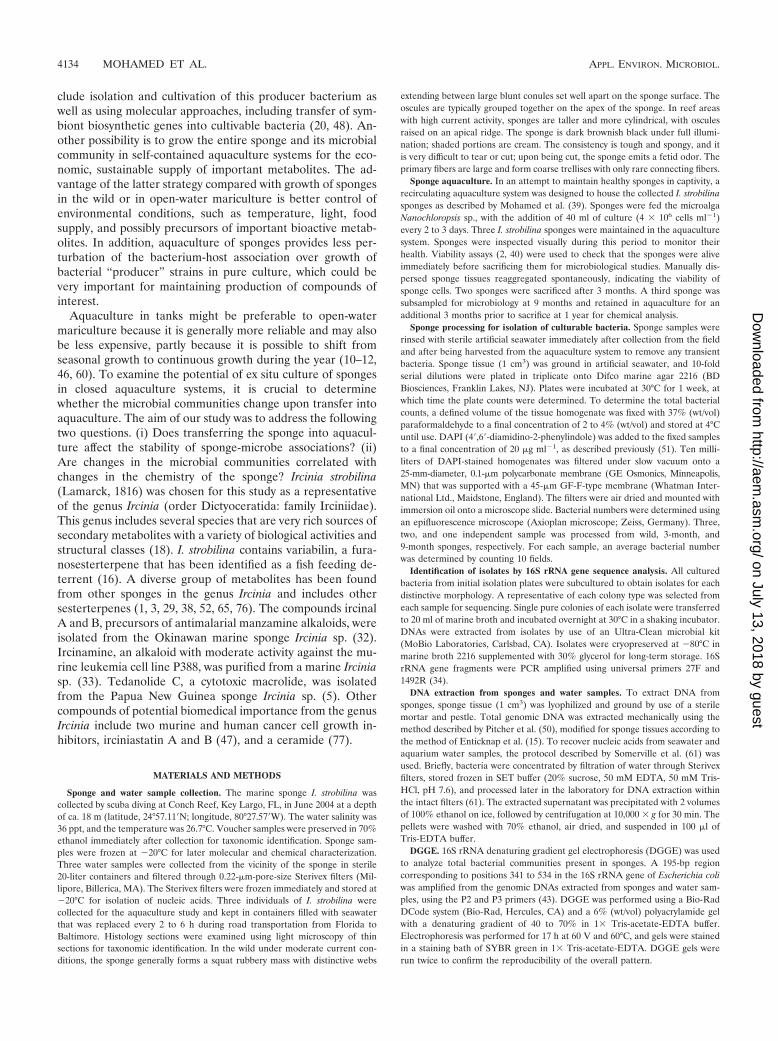
FIG. 2. Rooted neighbor-joining phylogenetic trees of partial 16S rRNA genes of isolates and clones in the Bacteroidetes, Chloroflexi, andClostridia (A); Acidobacteria, Actinobacteria, Cyanobacteria, and Planctomycetes (B); Alphaproteobacteria (C); and Beta- and Gammaproteobacteria(D). Isolates and clones were recovered from I. strobilina sponges collected from the wild (prefixed N04IS for isolates and W04IS for clones andshown in green) and maintained for 3 months (prefixed N3mIS for isolates and 3m04AIS for clones and shown in blue) and 9 months (prefixedN9mIS for isolates and 9m05AIS for clones and shown in red) in the aquaculture system. Bootstrap confidence values of �50% are shown at thenodes. The polygons represent clones that are �98% similar. The composition of each of these groups is shown in Table S1 in the supplementalmaterial. The numbers listed in bold before the group names indicate the numbers of clones. Thermotoga maritima was used as an outgroup. Thescale bar indicates 0.10 substitution per nucleotide position. Reference sequences are shown in bold, with GenBank accession numbers listed aftereach sequence name.
4136 MOHAMED ET AL. APPL. ENVIRON. MICROBIOL.
on July 13, 2018 by guesthttp://aem
.asm.org/
Dow
nloaded from

obtain a replicate for this time point. The shifts that occurredin sponge-associated bacterial communities, as indicated byDGGE analysis, are consistent with the findings from statisticalanalysis of clone library data (below).
Phylogenetic analysis of bacterial isolates. Traditional cul-turing techniques were used to isolate heterotrophic bacteriafrom sponges. Ten strains from wild sponges and spongesmaintained for 3 months in aquaculture were characterized by16S rRNA sequence analysis, and 12 strains were characterizedfrom sponges maintained for 9 months. Alpha- and gamma-proteobacterial strains dominated the culturable bacterial as-semblages of both wild sponges and those maintained inaquaculture (Fig. 2C and D). Bacteria belonging to the Bacte-roidetes appeared only in the culturable bacterial communitiesof sponges maintained in aquaculture (Fig. 2A).
Phylogenetic analysis of 16S rRNA gene clone libraries. Todetermine the stability of the microbial community upontransfer of I. strobilina into aquaculture, 16S rRNA geneclone libraries were generated from community DNAs ob-tained from a representative wild sponge and sponges main-tained for 3 months and 9 months in aquaculture. Afterelimination of a small number of chimeric clones from eachlibrary, 100, 74, and 74 clones were analyzed from the wild,3-month, and 9-month libraries, respectively. The 16S rRNAgene clones from the wild sponge corresponded to 35 uniqueOTUs, which fell into the following five bacterial lineages:Acidobacteria, Actinobacteria, Bacteroidetes, Chloroflexi, andCyanobacteria (Fig. 2). The 16S rRNA gene clones from the3-month sponge corresponded to 48 unique OTUs that en-compassed the following eight bacterial lineages: Bacte-
FIG. 2—Continued.
VOL. 74, 2008 BACTERIAL DIVERSITY OF A SPONGE IN AQUACULTURE 4137
on July 13, 2018 by guesthttp://aem
.asm.org/
Dow
nloaded from
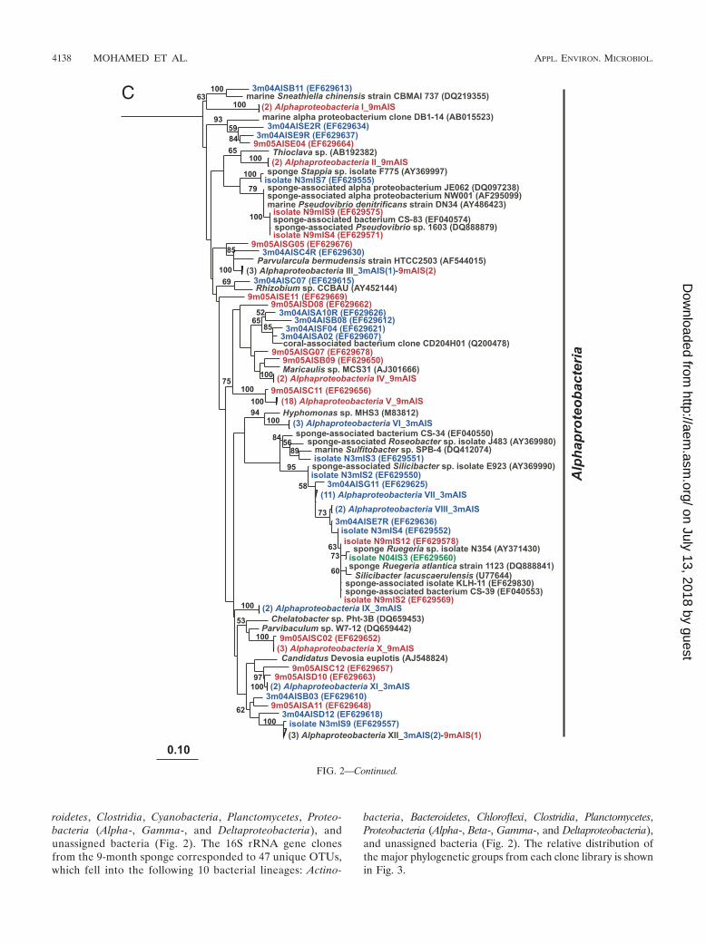
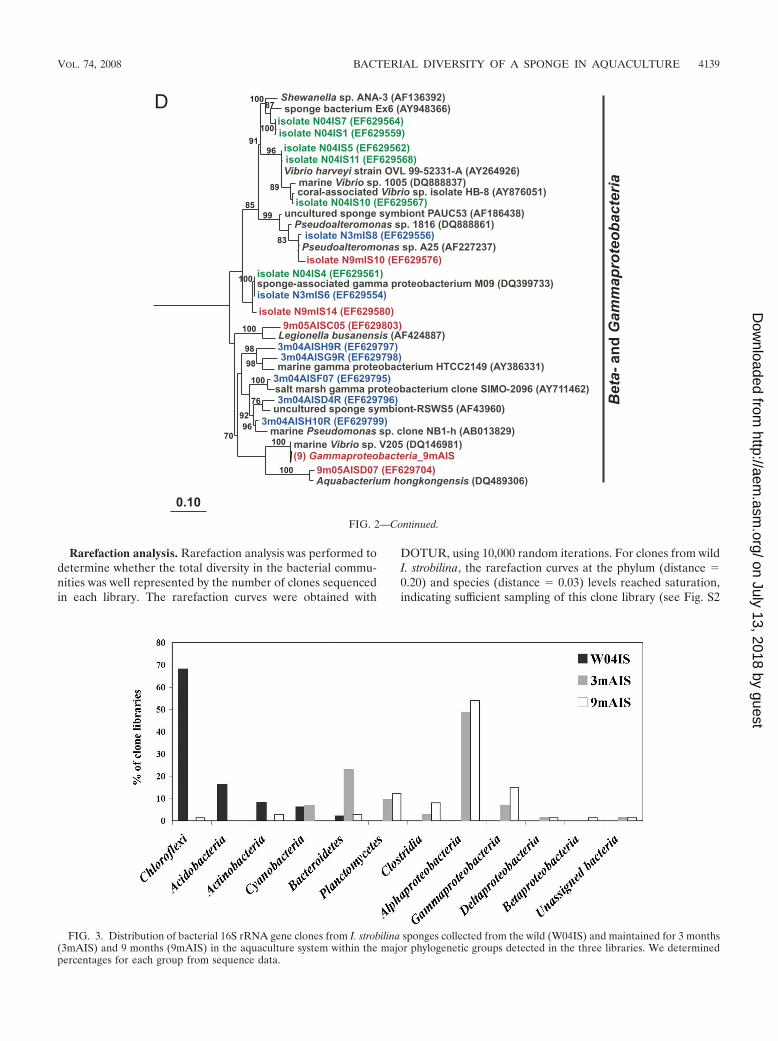
roidetes, Clostridia, Cyanobacteria, Planctomycetes, Proteo-bacteria (Alpha-, Gamma-, and Deltaproteobacteria), andunassigned bacteria (Fig. 2). The 16S rRNA gene clonesfrom the 9-month sponge corresponded to 47 unique OTUs,which fell into the following 10 bacterial lineages: Actino-
bacteria, Bacteroidetes, Chloroflexi, Clostridia, Planctomycetes,Proteobacteria (Alpha-, Beta-, Gamma-, and Deltaproteobacteria),and unassigned bacteria (Fig. 2). The relative distribution ofthe major phylogenetic groups from each clone library is shownin Fig. 3.
FIG. 2—Continued.
4138 MOHAMED ET AL. APPL. ENVIRON. MICROBIOL.
on July 13, 2018 by guesthttp://aem
.asm.org/
Dow
nloaded from
Rarefaction analysis. Rarefaction analysis was performed todetermine whether the total diversity in the bacterial commu-nities was well represented by the number of clones sequencedin each library. The rarefaction curves were obtained with
DOTUR, using 10,000 random iterations. For clones from wildI. strobilina, the rarefaction curves at the phylum (distance 0.20) and species (distance 0.03) levels reached saturation,indicating sufficient sampling of this clone library (see Fig. S2
FIG. 2—Continued.
FIG. 3. Distribution of bacterial 16S rRNA gene clones from I. strobilina sponges collected from the wild (W04IS) and maintained for 3 months(3mAIS) and 9 months (9mAIS) in the aquaculture system within the major phylogenetic groups detected in the three libraries. We determinedpercentages for each group from sequence data.
VOL. 74, 2008 BACTERIAL DIVERSITY OF A SPONGE IN AQUACULTURE 4139
on July 13, 2018 by guesthttp://aem
.asm.org/
Dow
nloaded from

in the supplemental material). The rarefaction curves for the3-month and 9-month sponges reached saturation at the phy-lum level, where richness reached an asymptotic maximum, butnot at the species level, indicating that further sampling of theclone library would have revealed additional diversity. The wildsponge had less bacterial richness than sponges maintained inaquaculture did, especially at the species level. Maintaining I.strobilina in aquaculture clearly increased the bacterial rich-ness, as demonstrated by steeper rarefaction curves than thosefor wild sponges.
Statistical analysis of bacterial diversity. Additional mea-sures of diversity and richness were obtained with the statisticalrichness estimators and diversity indices shown in Table 1.These indices were calculated using DOTUR. The input fileswere in the form of distance matrices generated by ARB. Thetotal number of OTUs in a bacterial population was calculatedusing nonparametric estimators. Chao1 richness estimateswere based on singletons and doubletons, as described by Chao(4), while ACE was based on the distribution of abundant(�10) and rare (�10) species. Shannon’s index and the recip-rocal of Simpson’s index were used as diversity indices, wherehigher numbers indicate greater diversity. Consistent with therarefaction curves, both statistical indices suggested that thecommunity diversity in sponges maintained in aquaculture wasgreater than that in the wild sponge. LIBSHUFF was used toquantitatively compare the three libraries. Evolutionary dis-tances were calculated using the neighbor-joining algorithm inARB, and the three libraries were significantly different (P �0.0001/0.0100).
LC-MS profiles of small molecules. LC-MS analysis wasperformed to determine whether gross changes in overall met-abolic profiles of I. strobilina occurred upon transfer intoaquaculture. Minor changes in profiles of small molecules wereobserved in the sponges maintained in aquaculture comparedto control wild samples, but overall patterns remained consis-tent, indicating no major shift in the profiles of secondarymetabolites (see Fig. S3 in the supplemental material).
DISCUSSION
The aquaculture of sponges in closed or semiclosed systemsis a promising strategy to overcome the supply problem forsponge-derived natural products. Although it might be possible
in some cases to make bioactive metabolites produced bysponges by chemical synthesis and transgenic techniques, di-rect extraction from the sponge biomass may be the best optionin other cases (43). A recirculating aquaculture system wasused to examine the possibility of maintaining I. strobilina in exsitu closed systems under controlled environmental conditionswith ecological parameters similar to those in the sponge’snatural habitat. Using different biotechnological methods toproduce sponge biomass may affect the microbial assemblagesof the sponge host. In cases where microbes are the producersof compounds of interest, changes in the microbial communi-ties may affect the production of natural products. The bacteriaassociated with sponges are also likely to be important for thehealth of the sponges (26). For these reasons, it is important tostudy the effects of these methods on the microbial communi-ties associated with sponges. The present study is one of fewstudies that have looked at the changes in microbial commu-nities associated with sponges following aquaculture or spongetransplantation. In this long-term study of the bacterial com-munities associated with I. strobilina in a self-contained, recir-culating aquaculture system, an increase in diversity of thebacterial communities after 3 months and a return to an inter-mediate level of diversity after 9 months was found, suggestiveof acclimation to aquaculture conditions.
We previously showed that the bacterial community of themarine sponge Mycale laxissima changed substantially upontransfer into flowthrough and recirculating aquaculture sys-tems (39). Hoffmann and coworkers (28) used fluorescence insitu hybridization to study the stability and specificity of mi-crobes associated with the cold-water marine sponge Geodiabarretti during cultivation for 8 months in an open recirculationsystem where members of the Alpha- and Gammaproteobacteriawere maintained during the period of cultivation. Friedrichand coworkers (21) found that a large fraction of the microbialcommunity of the Mediterranean sponge Aplysina aerophobaremained stable upon starvation of sponges or after antibioticexposure for 11 days in recirculating seawater aquaria.
Phylogenetic and statistical analyses of small-subunit-rRNAgene libraries were used to monitor structural shifts in thebacterial communities associated with I. strobilina followingcultivation in aquaculture. Bacterial communities associatedwith I. strobilina were different from bacterioplankton commu-nities found in the surrounding seawater. DGGE analysis of
TABLE 1. Richness and diversity estimates for bacterial 16S rRNA gene clone libraries from I. strobilina samples collected from the wild andmaintained in aquaculture
Sponge (I. strobilina) source (n)a Distanceb Richnessc ACEd Chao1estimatore
Shannonindexf
1/Simpsonindexf
Wild (100) 0.2 7 8 7 1.2 2.30.03 14 15 14 2.2 8.3
3 Mo of aquaculture (74) 0.2 12 17 17 1.8 40.03 41 200 107 3.3 17.8
9 Mo of aquaculture (74) 0.2 14 21 19 1.8 3.80.03 37 130 67 3.1 12.8
a n, number of 16S rRNA gene sequences analyzed.b Eighty percent identity was estimated as the phylum-level distance (D 0.20), and 97% identity was estimated as the species-level distance (D 0.03).c Richness is based on observed unique OTUs.d Nonparametric statistical predictions of the total richness of different OTUs were based on the distribution of abundant (�10) and rare (�10) OTUs.e Nonparametric statistical predictions of the total richness of OTUs were based on the distribution of singletons and doubletons.f A higher number represents more diversity.
4140 MOHAMED ET AL. APPL. ENVIRON. MICROBIOL.
on July 13, 2018 by guesthttp://aem
.asm.org/
Dow
nloaded from

sponges maintained in aquaculture and the surrounding waterindicated substantial differences in these bacterial communi-ties. This was confirmed by community analysis of these sam-ples by 16S rRNA gene sequencing studies. The data for bac-terial community analysis of the I. strobilina samples arepresented here, and those for bacterial community analysis ofthe surrounding water were reported by Mohamed et al. (39).This suggests that the bacterial community associated with wildI. strobilina is sponge specific rather than simply comprisingtransient bacteria from the water column. The fact that marinesponges harbor different bacteria from those in the water col-umn has been shown in previous reports (23, 39, 54, 62–64, 71).
The culturable bacteria isolated from wild sponges includedisolates found only in marine sponges, which indicates thatthey might be sponge specific (14, 23, 70). A comparison wasmade between a subset of bacteria isolated from I. strobilinaand the surrounding seawater. Sixty percent of the top BLASThits of isolates from I. strobilina were for bacteria found only insponges. Bacteria isolated from the seawater had only 16% oftop BLAST hits matching sponge bacteria (data not shown).
Total communities included representatives primarily clus-tered within the Acidobacteria, Actinobacteria, Bacteroidetes,Chloroflexi, and Cyanobacteria groups. Interestingly, a largenumber of the clones were affiliated with uncultured Chloro-flexi (59% of the total wild sponge library). Phylotypes relatedto Actinobacteria were no longer detectable in sponges main-tained for 3 months in aquaculture and then were detectedagain in the 9-month sponge. Since Actinobacteria organismsassociated with marine sponges may be sources of bioactivecompounds, the resumption of actinobacterial diversity mayplay a role in the production of specific bioactive compoundsthat are derived from this group.
Both culture-based and molecular techniques showed anincrease in the Bacteroidetes community in aquaculture, withthe highest representation in the 3-month sponge. Populationsaffiliated with Clostridia, Planctomycetes, and Proteobacteria(Alpha-, Gamma-, and Deltaproteobacteria) emerged in aqua-culture. This indicates that adaptation to aquaculture conditionsfavored the abundance of these populations. These increas-ingly large populations may have originally existed in lowerabundances in the wild sponge and/or have been acquired fromthe surrounding water in the aquaculture system. The diversityof the bacterial community associated with I. strobilina in-creased in aquaculture. This trend of increasing complexity ofthe bacterial community upon transfer to aquaculture tankswas observed with the marine sponge Mycale laxissima in ourprevious study (36). Statistical analyses revealed a significantshift in the bacterial communities in sponges maintained forboth 3 and 9 months in aquaculture compared to the commu-nity in wild sponges. Based on the observed diversity indices,more bacterial diversity was present in the 3-month spongesamples than in wild sponges, possibly due to the stress of thesponge when it was first transferred from its natural habitatinto aquaculture. It is not clear whether this is a result of stress,the presence of different bacteria in the surrounding water, thelight regimen, or some other parameter that differs in the wildand in aquaculture. On the basis of all five statistical tests usedto compare clone libraries at the species level, the level ofdiversity of the bacterial community associated with the spongemaintained for 9 months in aquaculture was intermediate be-
tween those for wild and three-month sponges. This may indi-cate an acclimation in this bacterial community after the9-month period in aquaculture.
Metabolomics, defined as the study of the nonprotein-aceous, endogenously synthesized small molecules present inan organism, is an emerging strategy in drug discovery anddevelopment (13, 22). The combination of chromatographyand mass spectrometry allows the separation of individual me-tabolites and their identification based on mass. The change inthe metabolome of an organism can be used to understandwhat has changed in the system. Applying this tool to our studyshowed that the environmental stress following the transfer ofI. strobilina into aquaculture produced no detectable effect onthe overall profile of small molecules associated with thesponges. LC-MS chemical fingerprinting revealed no majorchanges in the natural product profiles of I. strobilina, althoughthe composition of the bacterial community changed substan-tially following transfer into aquaculture. This suggests thatbacterial symbionts associated with I. strobilina may not beinvolved in the production of the major metabolites or thatthese metabolites are produced by a stable bacterial fractionthat was maintained in aquaculture. Candidates are membersof the Actinobacteria, Bacteroidetes, and Chloroflexi. In thiscase, the stability of the metabolites in aquaculture may implythat these symbionts constitutively produce essential metabo-lites. Another possible explanation for the stability of metab-olites in aquaculture is that they could have been synthesizedby the sponge or its associated microbes while the sponge wasin the wild, stored, and remained undegraded when the spongewas transferred into aquaculture.
In this study, the possibility of maintaining the marinesponge I. strobilina alive in a recirculating aquaculture systemis demonstrated. Further optimization of the aquaculture sys-tem is required for it to be useful in terms of production ofsponge biomass for harvesting natural products. Our key find-ing is that bacterial communities changed upon transfer of thesponge to aquaculture but showed signs of returning to thecommunity present in wild sponges after 9 months of mainte-nance in aquaculture. This highlights the importance of mon-itoring the bacterial communities associated with marinesponges when maintaining sponges in aquaculture systems byshowing that profound changes may occur in these bacterialcommunities. Concomitant changes in the overall chemicalprofile of the sponge were not detected. Additional detailedstudies of this type are needed to determine on a case-by-casebasis whether changes in sponge-associated microbial commu-nities are linked with changes in overall chemical profiles orspecific compounds of interest.
ACKNOWLEDGMENTS
We give special thanks to Julie Enticknap for her invaluable adviceand assistance and to Matthew Anderson for his help in maintainingthe sponges in aquaculture and assistance in the statistical analyses.We thank Scott McIntosh for designing and implementing the recir-culating aquaculture system. We thank Marcelino Suzuki and NaomiMontalvo for help using ARB. We acknowledge the National Under-sea Research Center (NURC), University of North Carolina at Wil-mington, for providing sampling opportunities in Key Largo, FL, andthe Aquaculture Research Center (ARC) at the Center of MarineBiotechnology for assistance in maintaining sponges in aquaculture.
VOL. 74, 2008 BACTERIAL DIVERSITY OF A SPONGE IN AQUACULTURE 4141
on July 13, 2018 by guesthttp://aem
.asm.org/
Dow
nloaded from
Funding for this research was provided by the Microbial Observa-tories Program, National Science Foundation (MCB-0238515), toR.T.H. and by NIH grant RO1 AI 036596 to M.T.H. and R.T.H.
REFERENCES
1. Alfano, G., G. Cimino, and S. De Stefano. 1979. Palinurin, a new linearsesterterpene from a marine sponge. Cell. Mol. Life Sci. 35:1136–1137.
2. Blumbach, B., Z. Pancer, B. Diehl-Seifert, R. Steffen, J. Munkner, I. Muller,and W. E. Muller. 1998. The putative sponge aggregation receptor. Isolationand characterization of a molecule composed of scavenger receptor cysteine-rich domains and short consensus repeats. J. Cell Sci. 111:2635–2644.
3. Buchanan, M. S., A. Edser, G. King, J. Whitmore, and R. J. Quinn. 2001.Cheilanthane sesterterpenes, protein kinase inhibitors, from a marinesponge of the genus Ircinia. J. Nat. Prod. 64:300–303.
4. Chao, A. 1984. Non-parametric estimation of the number of classes in apopulation. Scand. J. Stat. 11:783–791.
5. Chevallier, C., T. S. Bugni, X. Feng, M. K. Harper, A. M. Orendt, and C. M.Ireland. 2006. Tedanolide C: a potent new 18-membered-ring cytotoxic mac-rolide isolated from the Papua New Guinea marine sponge Ircinia sp. J. Org.Chem. 71:2510–2513.
6. Corredor, J. E., C. R. Wilkinson, V. P. Vicente, J. M. Morell, and E. Otero.1988. Nitrate release by Caribbean reef sponges. Limnol. Oceanogr. 33:114–120.
7. De Rosa, S., S. De Caro, G. Tommonaro, K. Slantchev, K. Stefanov, and S.Popov. 2001. Development in a primary cell culture of the marine spongeIrcinia muscarum and analysis of the polar compounds. Mar. Biotechnol.3:281–286.
8. De Rosa, S., G. Tommonaro, K. Slantchev, K. Stefanov, and S. Popov. 2002.Lipophylic metabolites from the marine sponge Ircinia muscarum and its cellculture. Mar. Biol. 140:465–470.
9. Diaz, M. C., and B. B. Ward. 1997. Sponge-mediated nitrification in tropicalbenthic communities. Mar. Ecol. Prog. Ser. 156:97–109.
10. Duckworth, A. R., and C. N. Battershill. 2003. Developing farming structuresfor production of biologically active sponge metabolites. Aquaculture 217:139–156.
11. Duckworth, A. R., and C. N. Battershill. 2003. Sponge aquaculture for theproduction of biologically active metabolites: the influence of farming pro-tocols and environment. Aquaculture 221:311–329.
12. Duckworth, A. R., G. A. Samples, A. E. Wright, and S. A. Pomponi. 2003. Invitro culture of the tropical sponge Axinella corrugata (Demospongia): effectof food cell concentration on growth, clearance rate and biosynthesis ofstevensine. Mar. Biotechnol. 5:519–527.
13. Dunn, W. B., and D. I. Ellis. 2005. Metabolomics: current analytical platformand methodologies. Trends Anal. Chem. 24:285–294.
14. Enticknap, J. J., M. K. Shanks, O. Peraud, and R. T. Hill. 2006. Character-ization of a culturable alphaproteobacterial symbiont common to many ma-rine sponges and evidence for vertical transmission via sponge larvae. Appl.Environ. Microbiol. 72:4105–4119.
15. Enticknap, J. J., R. Thompson, O. Peraud, J. E. Lohr, M. T. Hamann, andR. T. Hill. 2004. Molecular analysis of a Florida Keys sponge: implicationsfor natural products discovery. Mar. Biotechnol. 6:S288–S293.
16. Epifanio, R. D. A., D. L. Martins, and G. Muricy. 1999. The sesterterpenevariabilin as a fish-predation deterrent in the western Atlantic sponge Irciniastrobilina. J. Chem. Ecol. 25:2247–2254.
17. Faulkner, D. J. 2002. Marine natural products. Nat. Prod. Rep. 19:1–48.18. Faulkner, D. J., M. K. Harper, M. G. Haygood, C. E. Salomon, and E. W.
Schmidt. 2000. Symbiotic bacteria in sponges: sources of bioactive sub-stances, p. 107–119. In N. Fusetani (ed.), Drugs from the sea. Karger, Basel,Switzerland.
19. Felsenstein, J. 2004. PHYLIP (phylogenetic inference package), version 3.6.Department of Genetics, University of Washington, Seattle, WA.
20. Fortman, J. L., and D. H. Sherman. 2005. Utilizing the power of microbialgenetics to bridge the gap between the promise and the application of marinenatural products. Chem. Biochem. 6:960–978.
21. Friedrich, A. B., I. Fischer, P. Proksch, J. Hacker, and U. Hentschel. 2001.Temporal variation of the microbial community associated with the Medi-terranean sponge Aplysina aerophoba. FEMS Microbiol. Ecol. 38:105–113.
22. Harrigan, G. G., and L. A. Yates. 2006. High-throughput screening, metabo-lomics and drug discovery. IDrugs 9:188–192.
23. Hentschel, U., J. Hopke, M. Horn, A. B. Friedrich, M. Wagner, J. Hacker,and B. S. Moore. 2002. Molecular evidence for a uniform microbial commu-nity in sponges from different oceans. Appl. Environ. Microbiol. 68:4431–4440.
24. Hentschel, U., K. M. Usher, and M. W. Taylor. 2006. Marine sponges asmicrobial fermenters. FEMS Microbiol. Ecol. 55:167–177.
25. Hildebrand, M., L. E. Waggoner, G. E. Lim, K. H. Sharp, C. P. Ridley, andM. G. Haygood. 2004. Approaches to identify, clone, and express symbiontbioactive metabolite genes. Nat. Prod. Rep. 21:122–142.
26. Hill, R. T. 2004. Microbes from marine sponges: a treasure trove of biodi-versity for natural products discovery, p. 177–190. In A. T. Bull (ed.), Mi-crobial diversity and bioprospecting. ASM Press, Washington, DC.
27. Hoffmann, F., O. Larsen, V. Thiel, H. T. Rapp, and J. Reitner. 2005. Ananaerobic world in sponges. Geomicrobiol. J. 22:1–10.
28. Hoffmann, F., H. Rapp, and J. Reitner. 2006. Monitoring microbial commu-nity composition by fluorescence in situ hybridization during cultivation ofthe marine cold-water sponge Geodia barretti. Mar. Biotechnol. 8:373–379.
29. Issa, H. H., J. Tanaka, and T. Higa. 2003. New cytotoxic furanosesterterpenesfrom an Okinawan marine sponge, Ircinia sp. J. Nat. Prod. 66:251–254.
30. Kobayashi, J., and M. Ishibashi. 1993. Bioactive metabolites of symbioticmarine microorganisms. Chem. Rev. 93:8305–8308.
31. Kobayashi, M. 2000. Search for biologically active substances from marinesponges, p. 46–58. In N. Fusetani (ed.), Drugs from the sea. Karger, Basel,Switzerland.
32. Kondo, K., H. Shigemori, Y. Kikuchi, M. Ishibashi, T. Sasaki, and J. Koba-yashi. 1992. Ircinals A and B from the Okinawan marine sponge Ircinia sp.:plausible biogenetic precursors of manzamine alkaloids. J. Org. Chem. 57:2480–2483.
33. Kuramoto, M., H. Arimoto, and D. Uemura. 2004. Bioactive alkaloids fromthe sea: a review. Mar. Drugs 1:39–54.
34. Lane, D. J. 1991. 16S/23S rRNA sequencing, p. 115–175. In E. Stackebrandtand M. Goodfellow (ed.), Nucleic acid techniques in bacterial systematics.John Wiley & Sons, Chichester, United Kingdom.
35. Lee, Y. K., J.-H. Lee, and H. K. Lee. 2001. Microbial symbiosis in marinesponges. J. Microbiol. 39:254–264.
36. Ludwig, W., O. Strunk, R. Westram, L. Richter, H. Meier, Yadhukumar, A.Buchner, T. Lai, S. Steppi, G. Jobb, W. Forster, I. Brettske, S. Gerber, A. W.Ginhart, O. Gross, S. Grumann, S. Hermann, R. Jost, A. Konig, T. Liss, R.Lussmann, M. May, B. Nonhoff, B. Reichel, R. Strehlow, A. Stamatakis, N.Stuckmann, A. Vilbig, M. Lenke, T. Ludwig, A. Bode, and K. H. Schleifer.2004. ARB: a software environment for sequence data. Nucleic Acids Res.32:1363–1371.
37. Maidak, B. L., J. R. Cole, C. T. Parker, Jr., G. M. Garrity, N. Larsen, B. Li,T. G. Lilburn, M. J. McCaughey, G. J. Olsen, R. Overbeek, S. Pramanik,T. M. Schmidt, J. M. Tiedje, and C. R. Woese. 1999. A new version of theRDP (Ribosomal Database Project). Nucleic Acids Res. 27:171–173.
38. Martınez, A., S. Robledo, D. L. Munoz, S. Blair, E. Higuita, E. Echeverri, J.Bedoya, S. Zea, and I. Vitae. 2001. Antiparasitic activity of methanol extractsand isolated fractions from Caribbean sponges. Rev. Fac. Quım. Farm.8:71–81.
39. Mohamed, N. M., J. J. Enticknap, J. E. Lohr, S. M. McIntosh, and R. T. Hill.2008. Changes in bacterial communities of the marine sponge Mycale laxis-sima on transfer into aquaculture. Appl. Environ. Microbiol. 74:1209–1222.
40. Muller, W. E., I. Muller, and R. K. Zahn. 1974. Two different aggregationprinciples in reaggregation process of dissociated sponge cells (Geodiacydonium). Experientia 30:899–902.
41. Muller, W. E. G., V. A. Grebenjuk, G. Le Pennec, H. C. Schroder, F. Brum-mer, U. Hentschel, I. M. Muller, and H. J. Breter. 2004. Sustainable pro-duction of bioactive compounds by sponges—cell culture and gene clusterapproach: a review. Mar. Biotechnol. 6:105–117.
42. Munro, M. H., J. W. Blunt, E. J. Dumdei, S. J. Hickford, R. E. Lill, S. Li,C. N. Battershill, and A. R. Duckworth. 1999. The discovery and develop-ment of marine compounds with pharmaceutical potential. J. Biotechnol.70:15–25.
43. Muyzer, G., E. C. de Waal, and A. G. Uitterlinden. 1993. Profiling of complexmicrobial populations by denaturing gradient gel electrophoresis analysis ofpolymerase chain reaction-amplified genes coding for 16S rRNA. Appl.Environ. Microbiol. 59:695–700.
44. Newman, D. J., and G. M. Cragg. 2004. Marine natural products and relatedcompounds in clinical and advanced preclinical trials. J. Nat. Prod. 67:1216–1238.
45. Osinga, R., E. Armstrong, J. G. Burgess, F. Hofmann, J. Reitner, and G.Schumann-Kindel. 2001. Sponge-microbe associations and their importancefor sponge bioprocess engineering. Hydrobiologia 461:55–62.
46. Osinga, R., J. Tramper, and R. H. Wijffels. 1999. Cultivation of marinesponges. Mar. Biotechnol. 1:509–532.
47. Pettit, G. R., H. Hoffmann, D. L. Herald, J. McNulty, A. Murphy, K. C.Higgs, E. Hamel, N. E. Lewin, L. V. Pearce, P. M. Blumberg, R. K. Pettit, andJ. C. Knight. 2004. Antineoplastic agents 491. Synthetic conversion ofaaptamine to isoaaptamine, 9-demethylaaptamine, and 4-methylaaptamine.J. Org. Chem. 69:2251–2256.
48. Piel, J. 2006. Bacterial symbionts: prospects for the sustainable production ofinvertebrate-derived pharmaceuticals. Curr. Med. Chem. 13:39–50.
49. Piel, J., D. Hui, G. Wen, D. Butzke, M. Platzer, N. Fusetani, and S. Matsu-naga. 2004. Antitumor polyketide biosynthesis by an uncultivated bacterialsymbiont of the marine sponge Theonella swinhoei. Proc. Natl. Acad. Sci.USA 101:16222–16227.
50. Pitcher, D. G., N. A. Saunders, and R. J. Owen. 1989. Rapid extraction ofbacterial genomic DNA with guanidium thiocyanate. Lett. Appl. Microbiol.8:151–156.
51. Porter, K. G., and Y. S. Feig. 1980. The use of DAPI for identifying andcounting aquatic microflora. Limnol. Oceanogr. 25:943–948.
52. Rifai, S., A. Fassouane, P. M. Pinho, A. Kijjoa, N. Nazareth, M. Sao, J.Nascimento, and W. Herz. 2005. Cytotoxicity and inhibition of lymphocyte
4142 MOHAMED ET AL. APPL. ENVIRON. MICROBIOL.
on July 13, 2018 by guesthttp://aem
.asm.org/
Dow
nloaded from

proliferation of fasciculatin, a linear furanosesterterpene isolated fromIrcinia variabilis collected from the Atlantic Coast of Morocco. Mar. Drugs3:15–21.
53. Saitou, N., and M. Nei. 1987. The neighbor-joining method: a new methodfor reconstructing phylogenetic trees. Mol. Biol. Evol. 4:406–425.
54. Santavy, D. L., P. Willenz, and R. R. Colwell. 1990. Phenotypic study ofbacteria associated with the Caribbean sclerosponge Ceratoporella nichol-soni. Appl. Environ. Microbiol. 56:1750–1762.
55. Schirmer, A., R. Gadkari, C. D. Reeves, F. Ibrahim, E. F. DeLong, and C. R.Hutchinson. 2005. Metagenomic analysis reveals diverse polyketide synthasegene clusters in microorganisms associated with the marine sponge Disco-dermia dissoluta. Appl. Environ. Microbiol. 71:4840–4849.
56. Schloss, P. D., and J. Handelsman. 2005. Introducing DOTUR, a computerprogram for defining operational taxonomic units and estimating speciesrichness. Appl. Environ. Microbiol. 71:1501–1506.
57. Schloss, P. D., B. R. Larget, and J. Handelsman. 2004. Integration of mi-crobial ecology and statistics: a test to compare gene libraries. Appl. Environ.Microbiol. 70:5485–5492.
58. Schmidt, E. W., J. T. Nelson, D. A. Rasko, S. Sudek, J. A. Eisen, M. G.Haygood, and J. Ravel. 2005. Patellamide A and C biosynthesis by a micro-cin-like pathway in Prochloron didemni, the cyanobacterial symbiont of Lis-soclinum patella. Proc. Natl. Acad. Sci. USA 102:7315–7320.
59. Shieh, W. Y., and Y. M. Lin. 1994. Association of heterotrophic nitrogen-fixing bacteria with a marine sponge of Halichondria sp. Bull. Mar. Sci.54:557–564.
60. Sipkema, D., R. Osinga, W. Schatton, D. Mendola, J. Tramper, and R. H.Wijffels. 2005. Large-scale production of pharmaceuticals by marine sponges:sea, cell, or synthesis? Biotechnol. Bioeng. 90:201–222.
61. Somerville, C. C., I. T. Knight, W. L. Straube, and R. R. Colwell. 1989.Simple, rapid method for direct isolation of nucleic acids from aquaticenvironments. Appl. Environ. Microbiol. 55:548–554.
62. Taylor, M. W., R. Radax, D. Steger, and M. Wagner. 2007. Sponge-associ-ated microorganisms: evolution, ecology, and biotechnological potential. Mi-crobiol. Mol. Biol. Rev. 71:295–347.
63. Taylor, M. W., P. J. Schupp, I. Dahllof, S. Kjelleberg, and P. D. Steinberg.2004. Host specificity in marine sponge-associated bacteria, and potentialimplications for marine microbial diversity. Environ. Microbiol. 6:121–130.
64. Taylor, M. W., P. J. Schupp, R. de Nys, S. Kjelleberg, and P. D. Steinberg. 2005.Biogeography of bacteria associated with the marine sponge Cymbastela con-centrica. Environ. Microbiol. 7:419–433.
65. Tziveleka, L. A., A. P. Kourounakisb, P. N. Kourounakisb, V. Roussisa, andC. Vagias. 2002. Antioxidant potential of natural and synthesised poly-prenylated hydroquinones. Bioorg. Med. Chem. 10:935–939.
66. Vacelet, J. 1975. Etude en microscopie electronique de l’association entrebacteries et spongiaires du genre Verongia (Dictyoceratida). J. Microsc. Biol.Cell 23:271–288.
67. Vacelet, J., and C. Donadey. 1977. Electron microscope study of the associ-ation between some sponges and bacteria. J. Exp. Mar. Biol. Ecol. 30:301–314.
68. Van Soest, R. 1996. Porifera, Schwamme, p. 98–119. In W. Westheide and R.Rieger (ed.), Spezielle Zoologie. Teil 1. Einzeller and wirbellose Tiere.Gustav Fischer Verlag, Stuttgart, Germany.
69. van Treeck, P., M. Eisinger, J. Muller, M. Paster, and H. Schuhmacher.2003. Mariculture trials with Mediterranean sponge species: the exploitationof an old natural resource with sustainable and novel methods. Aquaculture218:439–455.
70. Webster, N. S., and R. T. Hill. 2001. The culturable microbial community ofthe Great Barrier Reef sponge Rhopaloeides odorabile is dominated by an-proteobacterium. Mar. Biol. 138:843–851.
71. Wilkinson, C. R. 1978. Microbial associations in sponges. I. Ecology, physi-ology and microbial populations of coral reef sponges. Mar. Biol. 49:161–167.
72. Wilkinson, C. R. 1978. Microbial associations in sponges. II. Numericalanalysis of sponge and water bacterial populations. Mar. Biol. 49:169–176.
73. Wilkinson, C. R. 1983. Net primary productivity in coral reef sponges. Sci-ence 219:410–412.
74. Wilkinson, C. R., and P. Fay. 1979. Nitrogen fixation in coral reef spongeswith symbiotic cyanobacteria. Nature 279:527–529.
75. Wilkinson, C. R., R. Summons, and E. Evans. 1999. Nitrogen fixation insymbiotic marine sponges: ecological significance and difficulties in detec-tion. Mem. Queensl. Mus. 44:667–673.
76. Yang, S. W., T. M. Chan, S. A. Pomponi, W. Gonsiorek, G. Chen, A. E.Wright, W. Hipkin, M. Patel, V. Gullo, B. Pramanik, P. Zavodny, and M.Chu. 2003. A new sesterterpene, Sch 599473, from a marine sponge, Irciniasp. J. Antibiot. 56:783–786.
77. Zhang, G.-W., X.-Q. Ma, C.-X. Zhang, J.-Y. Su, W.-C. Ye, X.-Q. Zhang, X.-S.Yao, and L.-M. Zeng. 2005. Two new ceramides from the marine spongeIrcinia fasciculata. Helv. Chim. Acta 88:885–890.
VOL. 74, 2008 BACTERIAL DIVERSITY OF A SPONGE IN AQUACULTURE 4143
on July 13, 2018 by guesthttp://aem
.asm.org/
Dow
nloaded from