Monitoring and - download.e-bookshelf.de · vii Contributors, ix Preface, xi Acknowledgments, xiii...
30
Transcript of Monitoring and - download.e-bookshelf.de · vii Contributors, ix Preface, xi Acknowledgments, xiii...


Monitoring and Intervention for the Critically Ill Small AnimalThe Rule of 20

Monitoring and Intervention for the Critically Ill Small AnimalThe Rule of 20
Edited by
Rebecca Kirby, DVM, DACVIM, DACVECC
and
Andrew Linklater, DVM, DACVECC

This edition first published 2017 © 2017 by John Wiley & Sons, Inc.
Editorial offices: 1606 Golden Aspen Drive, Suites 103 and 104, Ames, Iowa 50010, USA The Atrium, Southern Gate, Chichester, West Sussex, PO19 8SQ, UK 9600 Garsington Road, Oxford, OX4 2DQ, UK
For details of our global editorial offices, for customer services and for information about how to apply for permission to reuse the copyright material in this book please see our website at www.wiley.com/wiley‐blackwell.
Authorization to photocopy items for internal or personal use, or the internal or personal use of specific clients, is granted by Blackwell Publishing, provided that the base fee is paid directly to the Copyright Clearance Center, 222 Rosewood Drive, Danvers, MA 01923. For those organizations that have been granted a photocopy license by CCC, a separate system of payments has been arranged. The fee codes for users of the Transactional Reporting Service are ISBN‐13: 9781118900833/2016.
Designations used by companies to distinguish their products are often claimed as trademarks. All brand names and product names used in this book are trade names, service marks, trademarks or registered trademarks of their respective owners. The publisher is not associated with any product or vendor mentioned in this book.
The contents of this work are intended to further general scientific research, understanding, and discussion only and are not intended and should not be relied upon as recommending or promoting a specific method, diagnosis, or treatment by health science practitioners for any particular patient. The publisher and the author make no representations or warranties with respect to the accuracy or completeness of the contents of this work and specifically disclaim all warranties, including without limitation any implied warranties of fitness for a particular purpose. In view of ongoing research, equipment modifications, changes in governmental regulations, and the constant flow of information relating to the use of medicines, equipment, and devices, the reader is urged to review and evaluate the information provided in the package insert or instructions for each medicine, equipment, or device for, among other things, any changes in the instructions or indication of usage and for added warnings and precautions. Readers should consult with a specialist where appropriate. The fact that an organization or Website is referred to in this work as a citation and/or a potential source of further information does not mean that the author or the publisher endorses the information the organization or Website may provide or recommendations it may make. Further, readers should be aware that Internet Websites listed in this work may have changed or disappeared between when this work was written and when it is read. No warranty may be created or extended by any promotional statements for this work. Neither the publisher nor the author shall be liable for any damages arising herefrom.
Library of Congress Cataloging‐in‐Publication Data
Names: Kirby, Rebecca, editor. | Linklater, Andrew K. J., editor.Title: Monitoring and intervention for the critically ill small animal : the rule of 20 / [edited by] Rebecca Kirby, Andrew Linklater.Description: Ames, Iowa : John Wiley & Sons, Inc, 2016. | Includes bibliographical references and index.Identifiers: LCCN 2016018620 (print) | LCCN 2016030761 (ebook) | ISBN 9781118900833 (pbk.) | ISBN 9781118900963 (pdf) | ISBN 9781118900840 (epub)Subjects: LCSH: Dogs–Diseases–Treatment. | Cats–Diseases–Treatment. | Veterinary critical care. | Veterinary emergencies. | MESH: Emergencies–veterinary | Critical Care | Dog Diseases | Cat DiseasesClassification: LCC SF991 .M65 2016 (print) | LCC SF991 (ebook) | NLM SF 778 | DDC 636.0896028–dc23LC record available at https://lccn.loc.gov/2016018620
A catalogue record for this book is available from the British Library.
Wiley also publishes its books in a variety of electronic formats. Some content that appears in print may not be available in electronic books.
Set in 9/11pt Minion by SPi Global, Pondicherry, India
1 2017

Dedication
In loving memory of Douglass K. Macintire and Lesley G. King … whose beauty, grace, compassion, and wisdom bless our lives and the lives of thousands of animals. We miss you …

vii
Contributors, ix
Preface, xi
Acknowledgments, xiii
Conversion table, xv
1 An introduction to SIRS and the Rule of 20, 1Rebecca Kirby
2 Fluid balance, 9Rebecca Kirby and Elke Rudloff
3 Blood pressure, 29Lauren Sullivan
4 Albumin and colloid osmotic pressure, 43Adesola Odunayo
5 Glucose, 55Natara Loose
6 Electrolytes, 73Linda Barton and Rebecca Kirby
7 Acid–base status, 95Ryan Wheeler and Jan Kovacic
8 Oxygenation and ventilation, 109Christin Reminga and Lesley G. King
9 Coagulation, 137Andrew Linklater
10 Red blood cells and hemoglobin, 157Andrew Linklater and Veronica Higgs
11 Heart rate, rhythm, and contractility, 177Dennis E. Burkett
12 Neurological status, 207Christine Iacovetta
13 The renal system, 225Lee Herold
14 White blood cells, immune status, and antimicrobial stewardship, 247Carol E. Haak
15 Gastrointestinal system motility and integrity, 267Jennifer Klaus
16 Nutritional status, 285Caroline Tonozzi
17 Temperature, 303Conni Wehausen
18 Drug selection and dosing regimens, 319Dawn Merton Boothe
19 Pain management, 333Armi Pigott
20 Veterinary nursing care, 349Heather Darbo and Cheryl Page
21 Wounds and bandages, 373Jennifer J. Devey, Andrew Linklater and Rebecca Kirby
22 Anesthesia of the critical patient, 389Susan E. Leonard
Index, 401
Contents

ix
Linda Barton, DVM, DACVECCBluePearl Specialty and Emergency Medicine for PetsRenton, Washington
Dawn Merton Boothe, DVM, MS, PhD, DACVIM, DACVCPDirector, Clinical Pharmacology LaboratoryProfessor of Physiology and PharmacologyDepartment of Anatomy, Physiology, and PharmacologyCollege of Veterinary Medicine, Auburn UniversityAuburn, Alabama
Dennis E. Burkett, VMD, PhD, DACVECC, DACVIM (CArDIology)Hope Veterinary SpecialistsMalvern, Pennsylvania
Heather Darbo, CVT, VTS (ECC)Veterinary TechnicianLakeshore Veterinary SpecialistsGlendale, Wisconsin
Jennifer J. Devey, DVM, DACVECCConsultantFox Valley Animal Referral CenterAppleton, Wisconsin
Carol E. Haak, DVM, DACVECCMilwaukee, Wisconsin
Lee Herold, DVM, DACVECCChief Medical OfficerDoveLewis Emergency Animal HospitalPortland, Oregon
Veronica Higgs, DVMMetropolitan Veterinary Specialists and Emergency ServicesLouisville, Kentucky
Christine Iacovetta, DVM, DACVECCBluePearl Veterinary PartnersQueens, New York
Lesley G. King, MVB, DACVIM, DACVECC, DECVIM (CoMPAnIon AnIMAlS)†
Professor, Clinical EducatorDirector, Intensive Care UnitDirector, Emergency and Critical Care Residency ProgramMedical Director, Wards NursingSchool of Veterinary MedicineUniversity of PennsylvaniaPhiladelphia, Pennsylvania
Rebecca Kirby, DVM, DACVIM, DACVECC(Formerly) Animal Emergency CenterGainesville, Florida
Jennifer Klaus, DVM, DACVECCDirectorBlue Pearl Veterinary PartnersPhoenix, Arizona
Jan Kovacic, DVM, DACVECCHorizon Veterinary ServicesFour Seasons Veterinary SpecialistsLafayette, California
Susan E. Leonard, DVM, DACVECCEmergency VeterinarianNortheast Veterinary Referral HospitalPlains, Pennsylvania
Andrew Linklater, DVM, DACVECCClinical Specialist and InstructorLakeshore Veterinary SpecialistsMilwaukee, Wisconsin
Natara Loose, DVM, DACVECCBrooklyn, New York
Adesola Odunayo, DVM, MS, DACVECCClinical Assistant Professor of Emergency and Critical CareUniversity of TennesseeKnoxville, Tennessee
Cheryl Page, CVT, VTS (ECC)Veterinary TechnicianLakeshore Veterinary SpecialistsGlendale, Wisconsin
Armi Pigott, DVM, DACVECCClinical Specialist and InstructorLakeshore Veterinary SpecialistsMilwaukee, Wisconsin
Christin Reminga, DVMUniversity of PennsylvaniaPhiladelphia, Pennsylvania
Elke Rudloff, DVM, DACVECCClinical Specialist and InstructorLakeshore Veterinary SpecialistsMilwaukee, Wisconsin
Contributors
†Deceased.

x Contributors
Lauren Sullivan, DVM, MS, DACVECCAssistant ProfessorSmall Animal Emergency and Critical CareDepartment of Clinical SciencesColorado State UniversityFort Collins, Colorado
Caroline Tonozzi, DVM, DACVECCVeterinary SpecialistVCA Aurora Animal HospitalAurora, Illinois
Conni Wehausen, DVM, DACVECCBoard‐Certified CriticalistAnimal Emergency and Referral Center of MinnesotaSt Paul, Minnesota
Ryan Wheeler, DVMFour Seasons Veterinary SpecialistsLafayette, California
xi
The Rule of 20: how to keep them alive in the ICU
It is 4:00 in the morning and finally time to go home after a long day. A last look is given to Maggie, the Toy Poodle that had gram‐positive rods circulating in her neutrophils, Junior, the Poodle with diabetes, pancreatitis, and seizures, and Shooter, the postoperative gastric dilation‐torsion Great Dane. The care of these and other critical patients must be passed on to the on‐duty critical care team. How can I be confident that I have addressed the immediate problems of each of these animals? Do I have a plan in place that is sufficient to antici-pate, detect, and minimize complications? Will intervention be early enough to make a positive difference in the outcome? These heartfelt concerns led to the creation of the Rule of 20.
The Rule of 20, in its simplest form, is a check‐off list. It was created to remind doctors and ICU staff to examine the status of critical organ systems, clinical and laboratory parameters, and treatment goals that are essential for patient survival. While the inciting cause of the critical illness will vary, a common denomi-nator often exists between the most critical patients – systemic inflammation. Most of the pathophysiology is occurring at the capillary level, making every organ at risk for deleterious changes. Damage to one organ can rapidly initiate a cascade of events resulting in multiple organ dysfunction or failure. A multiorgan approach to patient assessment and treatment is required and can be effectively guided by using the Rule of 20.
Monitoring and Intervention for the Critically Ill Small Animal Patient: The Rule of 20 is focused on dogs and cats admitted for intensive care. Each of the 20 parameters of the Rule of 20 has a
dedicated chapter, with the final chapter focused on the selection and administration of anesthesia for the critical small animal patient. The authors are veterinary specialists devoted to the critical care of dogs and cats and share their expertise and experiences related to their topic. The book is designed for use as a reference as well as at the cage side, with tables, schematics, algorithms, and drawings provided for quick reference. Common problems and complications encountered in the ICU patient are highlighted within each chapter. A review of the basic physiology and patho-physiology relevant to the topic is presented, with recommendations for the diagnosis, monitoring, and treatment of common disorders. While the book is not meant to be an emergency manual, the basic life‐saving concepts and procedures for the emergency patient and the ICU patient are often identical and are highlighted throughout the book.
Veterinary specialists, practicing veterinarians, residents, interns, veterinary nurse technicians, assistants, and veterinary students will find valuable and potentially life‐saving ideas, procedures, and tips in this comprehensive patient care book. It is ideal to review the book beginning with the Introduction to SIRS and the Rule of 20 (Chapter 1) to become acquainted with the concepts. You will also gain insight into the consequences of systemic inflammation and the systemic inflammatory response syndrome (SIRS). Chapter 2 (Fluid Balance) should immediately follow since the formulation of an effective fluid resuscitation and maintenance plan is at the core of every ICU treatment sheet. Subsequently, careful review of each chapter in the book is warranted to acquire the knowledge base essential for understanding the complex nature of critical illness.
Preface


xiii
The editors wish to thank the contributing authors for their exper-tise, as well as Nancy Turner, Erica Judisch, and Susan Engelken at Wiley Publishing for their help and patience through this project. We want to recognize the important contributions of Jan Kovacic, DVM, DACVECC, and Dennis T. Crowe, DVM, DACVS, DACVECCS, for their roles in establishing the Animal Emergency
Center as an important training center, setting the stage for the development of the Rule of 20. We also wish to thank each of our mentors for guiding our professional development. Finally, a big thank you to our friends and family for their patience and support.
Becky and Andrew
Acknowledgments
Kiris (left) and Ella (right) Linklater, both rescues from Wisconsin, providing endless support
Izzy (left) and Dolly (right) Kirby, hand raised since birth, providing love daily.

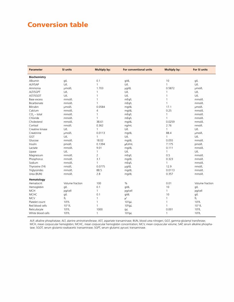
Parameter SI units Multiply by: For conventional units Multiply by: For SI units
BiochemistryAlbumin g/L 0.1 g/dL 10 g/LALP/SAP U/L 1 U/L 1 U/LAmmonia µmol/L 1.703 µg/dL 0.5872 µmol/LALT/SGPT U/L 1 U/L 1 U/LAST/SGOT U/L 1 U/L 1 U/LBase excess mmol/L 1 mEq/L 1 mmol/LBicarbonate mmol/L 1 mEq/L 1 mmol/LBilirubin µmol/L 0.0584 mg/dL 17.1 µmol/LCalcium mmol/L 4 mg/dL 0.25 mmol/LCO2 – total mmol/L 1 mEq/L 1 mmol/LChloride mmol/L 1 mEq/L 1 mmol/LCholesterol mmol/L 38.61 mg/dL 0.0259 mmol/LCortisol nmol/L 0.362 ng/mL 2.76 nmol/LCreatine kinase U/L 1 U/L 1 U/LCreatinine µmol/L 0.0113 mg/dL 88.4 µmol/LGGT U/L 1 U/L 1 U/LGlucose mmol/L 18.02 mg/dL 0.055 mmol/LInsulin pmol/L 0.1394 μIU/mL 7.175 pmol/LLactate mmol/L 9.01 mg/dL 0.111 mmol/LLipase U/L 1 U/L 1 U/LMagnesium mmol/L 2 mEq/L 0.5 mmol/LPhosphorus mmol/L 3.1 mg/dL 0.323 mmol/LSodium mmol/L 1 mEq/L 1 mmol/LThyroxine (T4) nmol/L 0.0775 µg/dL 12.9 nmol/LTriglycerides mmol/L 88.5 mg/dL 0.0113 mmol/LUrea (BUN) mmol/L 2.8 mg/dL 0.357 mmol/L
HematologyHematocrit Volume fraction 100 % 0.01 Volume fractionHemoglobin g/L 0.1 g/dL 10 g/LMCH pg/cell 1 pg/cell 1 pg/cellMCHC g/L 0.1 g/dL 10 g/LMCV fL 1 μ3 1 fLPlatelet count 109/L 1 103/μL 1 109/LRed blood cells 1012/L 1 106/μL 1 1012/LReticulocyte 109/L 1000 /μL 0.001 109/LWhite blood cells 109/L 1 103/μL 1 109/L
ALP, alkaline phosphatase; ALT, alanine aminotransferase; AST, aspartate transaminase; BUN, blood urea nitrogen; GGT, gamma‐glutamyl transferase; MCH, mean corpuscular hemoglobin; MCHC, mean corpuscular hemoglobin concentration; MCV, mean corpuscular volume; SAP, serum alkaline phospha-tase; SGOT, serum glutamic‐oxaloacetic transaminase; SGPT, serum glutamic pyruvic transaminase.
Conversion table

1
Monitoring and Intervention for the Critically Ill Small Animal: The Rule of 20, First Edition. Edited by Rebecca Kirby and Andrew Linklater. © 2017 John Wiley & Sons, Inc. Published 2017 by John Wiley & Sons, Inc.
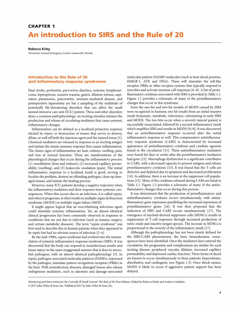
Introduction to the Rule of 20 and inflammatory response syndromes
Heat stroke, peritonitis, parvovirus diarrhea, systemic lymphosar-coma, leptospirosis, massive trauma, gastric dilation‐torsion, aspi-ration pneumonia, pancreatitis, immune‐mediated disease, and postoperative laparotomy are but a sampling of the multitude of potentially life‐threatening disorders that can affect the small animal intensive care unit (ICU) patient. These and other disorders share a common pathophysiology: an inciting stimulus initiates the production and release of circulating mediators that cause systemic inflammatory changes.
Inflammation can be defined as a localized protective response elicited by injury or destruction of tissues that serves to destroy, dilute, or wall off both the injurious agent and the injured tissue [1]. Chemical mediators are released in response to an inciting antigen and initiate the innate immune response that causes inflammation. The classic signs of inflammation are heat, redness, swelling, pain, and loss of normal function. These are manifestations of the physiological changes that occur during the inflammatory process: (1) vasodilation (heat and redness), (2) increased capillary perme-ability (swelling), and (3) leukocytic exudation (pain). The initial inflammatory response to a localized insult is good, serving to localize the problem, destroy an offending pathogen, clean up dam-aged tissues, and initiate the healing process.
However, many ICU patients develop a negative trajectory when the inflammatory mediators and their response have systemic con-sequences. When this occurs due to an infection, it is called sepsis, and when it progresses, it often results in multiple organ dysfunction syndrome (MODS) or multiple organ failure (MOF).
It might appear logical that an overwhelming infectious agent could stimulate systemic inflammation. Yet, an almost identical clinical progression has been commonly observed in response to conditions that are not due to infection (such as trauma, surgery, and certain metabolic diseases). The term “sepsis syndrome” was first used to describe this in human patients when they appeared to be septic but had no obvious source of infection [2–4].
By the mid‐1990s, sepsis syndrome had evolved into the nomen-clature of systemic inflammatory response syndrome (SIRS). It was discovered that the body can respond to noninfectious insults and tissue injury in the same exaggerated manner that it does to micro-bial pathogens, with an almost identical pathophysiology [5]. In sepsis, pathogen‐associated molecular patterns (PAMPs), expressed by the pathogen, stimulate pattern recognition receptors (PRRs) in the host. With noninfectious diseases, damaged tissues also release endogenous mediators, such as alarmins and damage‐associated
molecular pattern (DAMP) molecules (such as heat shock proteins, HMGB‐1, ATP, and DNA). These will stimulate the toll‐like receptor, PRRs or other receptor systems that typically respond to microbes and activate immune cell responses [6–8]. A list of proin-flammatory cytokines associated with SIRS is provided in Table 1.1. Figure 1.1 provides a schematic of many of the proinflammatory changes that occur in this syndrome.
Soon the one‐hit and two‐hit models of MODS caused by SIRS were recognized in humans; one hit results from an initial massive insult (traumatic, metabolic, infectious), culminating in early SIRS and MODS. The two hits occur when a severely injured patient is successfully resuscitated, followed by a second inflammatory insult which amplifies SIRS and results in MODS [9,10]. It was discovered that an antiinflammatory response occurred after the initial inflammatory response as well. This compensatory antiinflamma-tory response syndrome (CARS) is characterized by increased appearance of antiinflammatory cytokines and cytokine agonists found in the circulation [11]. These antiinflammatory mediators were found for days or weeks after the proinflammatory mediators had gone [12]. Macrophage dysfunction is a significant contributor to CARS, with a decreased capacity to present antigens and release proinflammatory cytokines [13]. It was found that the T‐cells are defective and depleted due to apoptosis and decreased proliferation [14]. In addition, there is an increase in the suppressor cell popula-tions [15]. Many of the cytokines released during CARS are listed in Table 1.1. Figure 1.2 provides a schematic of many of the antiin-flammatory changes that occur during this process.
It was determined that the production of proinflammatory and antiinflammatory cytokines occurs simultaneously, with antiin-flammatory gene expression paralleling the increased expression of proinflammatory genes [16]. It was then proposed that the induction of SIRS and CARS occurs simultaneously [17]. The emergence of myeloid‐derived suppressor cells (MDSCs) results in suppression of T‐cell responses through increased production of nitric oxide and reactive oxygen species. The increase in MDSCs is proportional to the severity of the inflammatory insult [17].
Although the pathophysiology has not been clearly defined for the SIRS‐CARS phenomenon, the basic hemodynamic conse-quences have been identified. Once the mediators have entered the circulation, the progression and complications are similar for each inciting disease: peripheral vascular dilation, increased capillary permeability, and depressed cardiac function. Three forms of shock are known to occur simultaneously in these patients: hypovolemic, distributive, and cardiogenic (see Figure 1.3). Once shock ensues, MODS is likely to occur if aggressive patient support has been delayed.
An introduction to SIRS and the Rule of 20
Rebecca Kirby(Formerly) Animal Emergency Center, Gainesville, Florida
ChApteR 1

2 Monitoring and Intervention for the Critically Ill Small Animal: The Rule of 20
Ag
Cytokine/Receptor blockade
IFN
TGF-βPD-1
protein
Apoptosis IL-10Monocyte Cytokines
VasodilationiNOS
O2.
TNFTN
FR
IL-1R
IL-4IL-1
Killing
TLR Impairedkilling
Figure 1.2 A schematic of some of the major consequences of the antiinflam-matory component of the compensatory antiinflammatory response syndrome (CARS). Red dotted lines depict inhibitory actions, blue solid lines depict stimulatory action. T‐cells, monocytes, and macrophages are the primary cells affected. The same antigens (microbial or tissue based) that stimulate the proinflammatory response can also stimulate the antiinflamma-tory cascades. The antiinflammatory mediators will block the production of many of the proinflammatory cytokines (red triangle and red dotted lines). TNF and IL‐1 receptors are found in the circulation and will bind and inactivate TNF and IL‐1 proinflammatory mediators. Ag, antigen; IFN, interferon; IL, interleukin; IL‐1R, interleukin‐1 receptor; iNOS, inducible nitric oxide synthetase; O2
., superoxide radicals; PD‐1, programmed death‐1; TGF‐β, tissue growth factor‐beta; TLR, toll‐like receptor; TNF, tumor necrosis factor; TNFR, tumor necrosis factor receptor.
Table 1.1 Inflammatory and hemostatic mediators of severe sepsis and their effects. Adapted from: Balk RA, Ely EW, Goyette RE. Stages of infection in patients with severe sepsis. In: Sepsis Handbook, 2nd edn. Thomson Advanced Therapeutics Communication, 2004, pp 24–31.
Proinflammatory mediatorsTumor necrosis factor
IL‐6 induction, TF expression, downregulation of TM gene expression and increased catabolism, activation of fibrinolysis, cytotoxicity, upregulation of endothelial cell adhesion molecules, induction of NO synthase, neutrophil activation, antiviral activity, fever, and other effects; circulating soluble receptor is antagonist
Interleukin‐1Fever, synthesis of acute‐phase proteins, induction of IL‐6 synthesis, upregulation of TF expression, decreased TM expression, activation of fibrinolysis, and other effects
Interleukin‐6Induction of acute‐phase response, induces B‐cell growth and T‐cell differentiation, enhances NK‐cell activity, promotes maturation of megakaryocytes, can inhibit endotoxin‐induced IL‐1 and TNF‐alpha; circulating soluble receptor is agonist
Interleukin‐8Release stimulated by TNF, IL‐1, IL‐2, promotes chemotaxis, enhances neutrophil function, upregulates adhesion molecule expression, level correlates with severity of systemic manifestation of pathology
Interferon‐gammaInduction of IgG production, potentiation of activity of IL‐12, macrophage activation
Antiinflammatory mediatorsInterleukin‐4
Stimulation and inhibition of various classes of T‐cells, suppression of TNF and IL‐1 secretion, upregulation of IgE and IgG secretion
Interleukin‐10Inhibition of inflammatory cytokine production by mononuclear cells, suppression of monocyte procoagulant activity, downregulation of monocyte killing, share receptor homology with interferon
Transforming growth factor‐betaTissue development and repair, other antiinflammatory properties
Soluble receptor and receptor antagonistsSoluble TNF‐1 receptor and IL‐1 soluble receptor inhibit function
Hemostatic factorsTissue factor
Upregulates expression on monocytes and subset of endothelial cells by TNF and IL‐1 leading to stimulation of extrinsic coagulation cascade
ThrombinProtein CProtein SAntithrombinPlasminogen activator inhibitor‐1Tissue factor pathway inhibitorPlasminThrombin activatable fibrinolysis inhibitor
Other mediatorsNitric oxideBradykininLipopolysaccharide binding proteinComplementLeukotrienesProstaglandinsSuperoxide radicalsPlatelet activating factorMyocardial depressant factor
Cytokine/Receptor
TLR
Proteinkinase C
Phagocytosis
Bloodclot
Capillary
Chemotaxisadherence
TTP-VIIaEndothelial damage
Plateletaggregation
Degranulation
IFN IL-1TNF
PAF
PG
O2.
Ag
Figure 1.1 A schematic of some of the major consequences of the proinflam-matory component of systemic inflammatory response syndrome (SIRS). Many cells produce proinflammatory mediators, including monocytes, macrophages, and endothelial cells. The interaction of an antigen (microbial or tissue based) with its receptor will cause the stimulation of protein kinase C and the production of cytokines. Cytokines in the circulation will interact with their specific receptor on other cells and stimulate the production of more cytokines. In addition to the release of cytokines (IFN, IL‐1, TNF), the arachidonic acid cascade is stimulated and produces PG, PAF, and leukotrienes. Reactive oxygen species are produced as well. Some of the consequences include degranulation of white cells, endothelial damage, stimulation of coagulation, white blood cell chemotaxis and adherence in capillaries, and increased phagocytosis of Ags. Ag, antigen; IFN, interferon; IL, interleukin; O2
., superoxide radicals; PAF, platelet activating factor; PG, prostaglandins; TNF, tumor necrosis factor; TLR, toll‐like receptor; TTP, tissue thromboplastin; VIIa, activated factor VII.

Chapter 1: An introduction to SIRS and the Rule of 20 3
Many research and clinical trials have been conducted in labora-tory animals and humans looking for a single best therapy that would be effective in treating most patients with the SIRS‐CARS phenomenon, with minimal success. Since inflammation and immune suppression have been found to be occurring simulta-neously, each patient is more likely to be experiencing their own unique combination of immune stimulation and suppression. This makes a standardized protocol for therapy extremely difficult to formulate until further knowledge is acquired. Emphasis is no longer primarily directed at methods to stop exaggerated proin-flammatory responses but is instead placed on supporting the patient and searching for new methods that prevent prolonged immunosuppression or restore immune function [18].
Sepsis, the SIRS‐CARS phenomenon (referred to simply as SIRS from here on), and MODS remain tremendous obstacles to the successful treatment of critically ill small animals. A “back to basics” approach is critical for any patient with the potential for inflammatory changes. Several basic yet key principles that can be used to guide patient assessment and care are listed in Box 1.1. Problems within the major organ systems should be anticipated in advance, with appropriate diagnostic, therapeutic, and monitoring efforts employed early, rather than waiting for a problem to surface and reacting to it. The Rule of 20 was developed to assist the criti-cal care team in thoughtfully and carefully assessing these patients. Table 1.2 lists common problems to anticipate under each parameter of the Rule of 20 in patients with SIRS. Sample forms that can be used when applying the Rule of 20 are provided in Figures 1.4 and 1.5.
The critical care team must remain open to options for the diag-nosis and care of the patient when changes in patient status occur and must reconsider differentials when a patient is not progressing
Pro-in�ammatory Anti-in�ammatoryMediators
Inciting event
Increased capillarypermeabilityvasodilation
Cardiogenicshock
Hypovolemicshock
Distributiveshock
Myocardialdepression
Peripheralvasodilation
Figure 1.3 A schematic depicting the presence of proinflammatory (blue) and antiinflammatory (yellow) mediators released concurrently (green), causing hemodynamic changes that result in three simultaneous forms of shock.
Box 1.1 Key principles to guide the care of the small animal ICU patient.
• Treat the most life‐threatening problem first• Treat the patient, not the numbers• Anticipate the worst and be ready for it• Provide the right treatment, at the right time, in the right amount• Examine the cause of the problem and the effect on the patient• Weigh the pros and cons of every drug and procedure• There is not a drug for every problem – less is best• If it has not been written down, it has not been done• Never ignore your gut feeling• Things are done in the order of importance
Table 1.2 Common problems to anticipate for each parameter of the Rule of 20 in the SIRS patient.
Rule of 20 parameter Anticipated problems
Fluid balance Hypovolemia; vasodilation, increased capillary permeabilityAlbumin, COP Hypoalbuminemia; loss through capillaries and catabolic state; reduced COP and extravasation of fluid from vasculatureBlood pressure Hypotension; hypovolemia and impaired cardiac performance, peripheral vasodilationGlucose Hypoglycemia; increased consumption, decreased intake; can affect vascular tone and cardiac performanceElectrolytes Hypokalemia; any disorder is possible with fluid imbalances, catabolic state, and depressed nutritional intakeAcid–base Metabolic acidosis associated with poor perfusion and elevated lactate is commonOxygenation/ventilation Hypoxemia possible if pulmonary edema from inflammatory vascular changes or poor cardiac performanceCoagulation Thrombocytopenia (declining trend); some form of DIC due to mediator‐induced vasculitis, activation of serine
proteases, and insufficient antithrombinRed blood cells Anemia from disease or erythrocytosis from dehydration; frequent blood sampling can result in anemiaHeart rate, rhythm, contractility
Tachycardia in dogs with poor perfusion, bradycardia in cats with poor perfusion; arrhythmias if poor perfusion; depressed myocardial contractility with circulating mediators
Neurological status Depressed level of consciousness from perfusion changes, hypoxemia if present, circulating mediators, metabolic changes or underlying disease
Urinary tract status Azotemia if poor perfusion or dehydration; impaired renal function if prolonged hypotension or nephrotoxic drugsWBC, immune status Impaired immune function, lymphopenia possible, susceptible to nosocomial infectionsGastrointestinal status Gastric paresis, ileus; third body fluid spacing into bowel; bacterial translocation if no enteral feedingNutrition Catabolic state and early malnutrition anticipated; bacterial translocation without enteral feedingDrugs Altered volume of distribution, metabolism and excretionBody temperature High if active inflammation; low in cats with poor perfusion or dogs with difficult‐to‐resuscitate shockPain control Pain is anticipated with all critical illness and deserves analgesic support; critical early in shock resuscitationWound and bandage care Possible source of pathogens requiring close monitoring of wound sites and bandage changesNursing care and TLC Must receive the right treatment at the right time in the right amount; anticipate problems, be ready; provide TLC
COP, colloidal osmotic pressure; DIC, disseminated intravascular coagulation; TLC, tender loving care; WBC, white blood cell.

4 Monitoring and Intervention for the Critically Ill Small Animal: The Rule of 20
as expected. A problems list for the patient should be established and revised at least daily, with options for diagnostic, therapeutic, and monitoring plans for each problem outlined and considered (Figure 1.6). A differential diagnosis list is prepared for each problem and frequently reevaluated with the goal of finding one diagnosis that could be responsible for all the listed problems.
There are many aspects of critical care that are unique to the cat. Challenges occur when treating the cat due to species differences such as their physiological response to shock, the specific methods required for shock resuscitation and the different drug responses, metabolism, and dosing requirements. Knowledge of the traits specific to the cat is mandatory for optimizing their ability to recover from critical illness. These differences are highlighted in each chapter throughout the Rule of 20.
The successful treatment of SIRS and MODS has lead to the emergence of a new syndrome identified in human medicine, the persistent inflammation/immunosuppression catabolism syn-drome (PICS) [17]. Secondary nosocomial infections and severe protein catabolism are hallmarks of PICS. This syndrome presents
the simultaneous challenge of managing chronic inflammation and immunosuppression. These patients are identified in the surgical ICU after >10 days and have persistent inflammation defined by findings such as elevated C‐reactive protein, lymphopenia (<800/mm3), serum albumin < 3 g/dL, and weight loss >10%.
A study of adult humans suffering severe blunt trauma found that patients with complicated clinical outcomes are exhibiting PICS [19]. These patients were reported as being significantly older and sicker, with persistent leukocytosis but low lymphocyte and albumin levels compared with uncomplicated patients. They expressed significant suppression of myeloid cell differentiation, increased inflammation, decreased chemotaxis, and defective innate immunity compared with uncomplicated patients. Genomic analysis found changes consistent with defects in the adaptive
Parameter Patient Target Intervention Parameter Patient Target InterventionFluid balance Heart rate,
rhythm,contractility
Bloodpressure
Neurologicalstatus
Oncotic pull/albumin
Urinary tractstatus
Glucose WBC, immunestatus, antibiotics
Electrolytes Gastrointestinalstatus
Acid–base Nutritionalstatus
Oxygenation &ventilation
Drugs, dosage,metabolism
Coagulation Pain
RBCs Wounds,bandages
Temperature Nursing care
Figure 1.5 Rule of 20 form for recording current patient status, targeted endpoints, and proposed intervention. RBC, red blood cell; WBC, white blood cell.
Rule of 20 Parameters
Fluid balance
Blood pressure
Albumin, COP
Glucose
Electrolytes
Acid – base
Oxygenation/ventilation
Coagulation
Red blood cell status
Heart rate, rhythm,contractility
Neurological status
Urinary tract status
WBC, Immune status
GI tract status
Nutritional status
Drug dosing, metabolism
Body temperature
Pain control
Wound and bandage care
Nursing care, TLC
Figure 1.4 The Rule of 20. Each parameter should be assessed regularly in any critically ill dog or cat. The order of importance will vary between individual patients. COP, colloidal osmotic pressure; GI, gastrointestinal, TLC, tender loving care; WBC, white blood cell.
Problemslist
Poor perfusion Tachycardia, Pale MM, CRT 3 sec
Doppler BPCVP whenstable
Crystalloids, HEShigh normalend-points;large volumetechnique
Doppler BPCVP, UOphysical perfusionparameters
PE for hydrationand perfusionfrequency of vomiting
POC database,CBC, UA,biochemistry,radiographswhen stable
Vomiting Yellow liquid
Fluid therapyNPOanti-emetics± NG tube
Dx plan Rx plan Mx plan
Figure 1.6 An example of a worksheet to ensure that each patient problem has a diagnostic, therapeutic, and monitoring plan. The worksheet has some examples of problems to demonstrate the intention of the form. Each of the problems that the patient has that day should be listed in the left‐hand column. New and unresponsive problems deserve a diagnostic, therapeutic, and monitoring plan written down. After assessing each problem and possible plan, the task of choosing the most efficient means for patient diagnosis and care can be performed. Dx, diagnostic; Mx, monitoring; Rx, therapeutic.
Chapter 1: An introduction to SIRS and the Rule of 20 5
immune response and increased inflammation. Clinical data showed persistent inflammation, immunosuppression, and protein depletion.
Unfortunately, at this time, when PICS is recognized, the course correction is difficult. Therapeutic interventions are geared towards supportive care and treating secondary infections. Further research is needed to identify appropriate multimodal therapies that target specific components of the syndrome [16]. The Rule of 20 now becomes even more important for thoroughly assessing and sup-porting these critical patients.
Diagnostic and monitoring procedures
The practice of medicine is an art that depends on the ability to suc-cessfully acquire and integrate the findings from the patient history, physical examination (PE), and cage‐side point of care (POC) labo-ratory database. Patients continuously give important information through their physical changes, progression of illness, and clinical signs. Additional diagnostic testing is done to confirm, deny or better define the clinical impressions gained from evaluation of the patient status and underlying disease(s). It is not uncommon for life‐threatening problems to require stabilization before time‐consuming or invasive diagnostic and monitoring procedures are employed.
the history and physical examinationThe key to taking a great history is organization. A sample format for obtaining a sequential history pertaining to the small animal ICU patient is presented in Table 1.3. The order in which the topics are addressed is specifically arranged to better direct information gathering while allowing the owner to describe their concerns about their pet.
Frequently reported complaints elicited from the history (such as vomiting, inability to walk, diarrhea) require further characteriza-tion to localize the disease or indicate the severity of the problem. Discussions about significant historical data pertaining to each of the Rule of 20 topics can be found in the corresponding topic chapter.
Each member of the critical care team will develop his or her own style and routine for performing a PE for individual patients. The key is to be consistent and thorough. A rapid evaluation of the ABCs (Airways, Breathing, Bleeding, Circulation, Consciousness) is the first priority, with intervention provided when potentially life‐threatening problems are identified. Developing a head‐to‐tail system of examination helps to maintain a routine and remain focused. Saving the examination of the body parts most likely related to the presenting complaint to the end of the PE can help prevent distraction and failure to complete the remainder of the PE. It is best to perform the equipment‐dependent examinations at the end of the PE to avoid distraction.
As the PE progresses from head to tail, neurological and ortho-pedic evaluations are done along with the general PE. Any animal that has had head trauma, loss of consciousness, prolonged seizures, or other indication of intracranial edema or hemorrhage must maintain a normal head position throughout the examination. When an area of pain is identified, examination of that area is post-poned and the general PE is continued, followed by a closer assessment of the painful region. Significant PE findings relative to each of the Rule of 20 topics are discussed in the corresponding topic chapter.
point of care testingThe most important and immediate laboratory assessment of the ICU small animal patient is done at the cage side with POC testing. The minimum database should include the packed cell volume (PCV), plasma total protein (TP), blood glucose, blood urea nitrogen (BUN or creatinine), electrolytes, acid–base status, blood lactate, coagulation profile, blood smear for platelet estimate and red blood cell (RBC) morphology, and urinalysis.
There are several POC data points that, when abnormal, warrant immediate investigation and intervention (indicated by red check-marks in Table 1.4). The significance of the abnormalities with the possible cause(s), intervention options, and monitoring recom-mendations are available under the corresponding topic chapters.
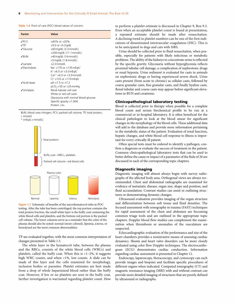
The microhematocrit tube provides a great deal of data. Figure 1.7 illustrates information that can be obtained from the spun tube, including the PCV, TP, buffy coat, and serum color. The PCV and
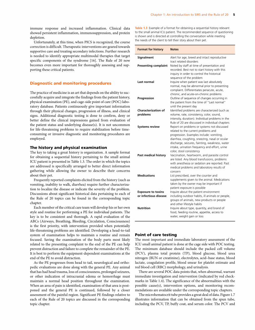
Table 1.3 Example of a format for obtaining a sequential history relevant to the small animal ICU patient. The recommended sequence of questioning is shown and is directed at controlling the conversation while meeting the needs of the client to tell their story about their pet.
Format for history Notes
Signalment Alert for age, breed and intact reproductive tract related disorders
Presenting complaint Noted by staff at time of presentation and recorded. Best not to start history with this inquiry in order to control the historical sequence of the problem
Last normal Inquire when patient was last absolutely normal, may be abnormal prior to presenting complaint. Differentiates peracute, acute, chronic, and acute‐on‐chronic problems
Progression Outline of sequence of changes occurring in the patient from the time of “Last normal” until the present day
Characterization of problems
Identified problems are characterized (such as volume, rate, consistency, color, sound, intensity, duration). Individual problems in the Rule of 20 are discussed in individual chapters
Systems review Report on problems or systems not discussed related to the current problems and progression. Examples include: vomiting, diarrhea, coughing, sneezing, nasal or ocular discharge, seizures, fainting, weakness, water intake, urination frequency and effort, urine color, stool consistency
Past medical history Vaccination, heartworm, and parasite control are listed. Any blood transfusions, problems with anesthesia or sedation are reported. Past medical problems and laboratory results of concern
Medications List prescribed, over the counter and supplements given to the animal. Medications taken by the owner may be important if patient exposure is possible
Exposure to toxins or infectious disease
Inquire about the patient environment including outdoor habits, ill animals or people, groups of animals, new products or people and other lifestyle habits
Nutrition Inquire about type, quantity, and brand of food, feeding routine, appetite, access to water, weight gain or loss
6 Monitoring and Intervention for the Critically Ill Small Animal: The Rule of 20
TP are evaluated together, with the most common interpretation of changes presented in Table 1.5.
The white layer in the hematocrit tube, between the plasma and the RBCs, consists of the white blood cells (WBCs) and platelets, called the buffy coat. When this is >1–2%, it suggests high WBC counts, and when <1%, low counts. A slide can be made of this layer and the cells examined for morphology, inclusion bodies or parasites. Platelet estimates are best made from a drop of whole heparinized blood rather than the buffy coat. However, if few or no platelets are seen in the buffy coat, further investigation is warranted regarding platelet count. How
to perform a platelet estimate is discussed in Chapter 9, Box 9.3. Even when an acceptable platelet count is found at presentation, a repeated estimate should be made after resuscitation. A declining trend in platelet numbers can be one of the first indi-cations of disseminated intravascular coagulation (DIC). This is to be anticipated in dogs and cats with SIRS.
Urine should be collected prior to fluid resuscitation, when pos-sible, especially for patients with likely infectious or metabolic problems. The ability of the kidneys to concentrate urine is reflected by the specific gravity. Glycosuria without hyperglycemia reflects proximal tubular cell damage, a complication of nephrotoxic drugs or renal hypoxia. Urine sediment is evaluated for casts in animals on nephrotoxic drugs or having experienced severe shock. Urine casts present (from acute to chronic) as cellular casts, followed by coarse granular casts, fine granular casts, and finally hyaline casts. Renal tubular and coarse casts may appear before significant eleva-tions in BUN and creatinine.
Clinicopathological laboratory testingBlood is collected prior to therapy when possible for a complete blood count and serum biochemical profile to be run at a commercial or in‐hospital laboratory. It is often beneficial for the clinical pathologist to look at the blood smear for significant changes in the morphology of the blood cells. These additional data will add to the database and provide more information pertaining to the metabolic status of the patient. Evaluation of renal function, hepatic changes, and white blood cell response to illness is impor-tant for every critically ill patient.
Often special tests must be ordered to identify a pathogen, con-firm a diagnosis or evaluate the success of treatment in the patient. Common clinicopathological laboratory tests that can be used to better define the cause or impact of a parameter of the Rule of 20 are discussed in each of the corresponding topic chapters.
Diagnostic imagingDiagnostic imaging will almost always begin with survey radio-graphs of the affected body area. Orthogonal views are always rec-ommended. Chest and abdominal radiographs are examined for evidence of metastatic disease, organ size, shape and position, and fluid accumulation. Contrast studies can assist in outlining struc-tures or demonstrating dynamic changes.
Ultrasound evaluation provides imaging of the organ structure and differentiation between soft tissue and fluid densities. The focused assessment with sonography in trauma (FAST) techniques for rapid assessment of the chest and abdomen are becoming common triage tools and are outlined in the appropriate topic chapters. Doppler blood flow studies can complement the exami-nation when thrombosis or anomalies of the vasculature are suspected.
Echocardiographic evaluation of the performance and size of the heart chambers provides a noninvasive means of assessing cardiac dynamics. Shunts and heart valve disorders can be more closely evaluated using color flow Doppler techniques. The electrocardio-gram (ECG) demonstrates cardiac conduction. Information regarding cardiac assessment is presented in Chapter 11.
Endoscopy, laparoscopy, thoracoscopy, and cystoscopy can each provide images and biopsies and facilitate specific procedures of different organs when indicated. Computed tomography (CT) and magnetic resonance imaging (MRI) with and without contrast can provide more detailed imaging of structures that are poorly defined by ultrasound or radiographs.
Total proteins
Buffy coat – WBCs, platelets
Packed cell volume – red blood cells
Plasma color
Normal Lipemia Icterus Hemolysis
Figure 1.7 Schematic of benefits of the microhematocrit tube in POC testing. After the tube has been centrifuged, the top portion contains the total protein fraction, the small white layer is the buffy coat containing the white blood cells and platelets, and the bottom red portion is the packed cell volume. The lower columns serve as a reminder that the color of the plasma should also be noted: normal (straw colored), lipemia, icterus, or hemolyzed are the most common abnormalities.
Table 1.4 Point of care (POC) blood values of concern.
Factor Value
✓PCV >60% or <20%✓TP >9.0 or <5.0 g/dL✓Glucose <60 mg/dL (3.3 mmol/L)
>200 mg/dL (11.1 mmol/L)✓BUN >40 mg/dL (14 mmol/L)
<5 mg/dL (1.8 mmol/L)✓Lactate >2.0 mmol/L✓Electrolytes Na+ >170 or <135 mEq/L*
K+ >6.0 or <3.0 mEq/L*
Cai++ >6.0 or <3.0 mmol/LCl– >125 or <110 mEq/L
✓Acid–base pH >7.5 or <7.2pCO2 > 50 or <25 mmHg
✓Urinalysis Renal tubular cell castWhite or red cell castsGlycosuria with normal blood glucoseSpecific gravity <1.004Protein ≥3+
BUN, blood urea nitrogen; PCV, packed cell volume; TP, total protein; i, ionized.* (mEq/L = mmol/L).

Chapter 1: An introduction to SIRS and the Rule of 20 7
Recommendations for diagnostic imaging procedures with suggested techniques (such as contrast studies, FAST examination) are presented for each topic of the Rule of 20 in the corresponding topic chapter.
Monitoring proceduresThe PE findings will always provide the most important data regarding the status of the patient. Following the trend of change in every monitored parameter affords more accurate information than assessing a single value. Equipment‐based monitoring can include indirect and direct blood pressure, ECG, pulse oximetry, end‐tidal CO2, central venous pressure, urine output, body tem-perature, and body weight and is readily available for the small animal patient. Serial assessment of blood values, such as PCV, TP, acid–base status, coagulation times, electrolytes and lactate, reflects patient progress and can guide therapy. More sophisticated procedures, such as pulmonary artery catheters, ScvO2, and calo-rimetry, are presented as options in the appropriate chapters, with known advantages and disadvantages highlighted. Each topic in the Rule of 20 will require patient monitoring. The recommended monitoring procedures are discussed in each corresponding topic chapter.
Communications and the Rule of 20Exceptional communication skills are needed to quickly build a good rapport with the pet owner under very stressful and
emotional circumstances. From first contact by telephone to final discharge of the patient and follow‐up care, each member of the critical care team must develop a caring and trusting relationship with the pet owner (client). It is important to create an open forum that includes a gentle tone of voice, body language that projects an approachable demeanor, open‐ended questions when taking information, attentive listening to owner concerns, and establishing realistic medical and financial expectations. When successful, the decisions made regarding the medical care of the patient can be a shared process between the owner(s) and the critical care team. More information can be found in the Further reading list at the end of the chapter.
The Rule of 20 is a fluid and dynamic monitoring tool that can be utilized to treat any critical patient. As the knowledge pertain-ing to the pathophysiology of disease expands, new drugs, new treatments, additional diagnostic tools, and state‐of‐the‐art monitoring methods can be easily inserted into the format. The information gained from the Rule of 20 provides a solid foundation for patient care, as well as for communications among staff and with clients. The Rule of 20 assists the critical care team in providing the structured, thorough, and complete evaluation needed for small animal patients with complex medical problems.
Human medicine has coined the term hospital medicine to describe the discipline concerned with the medical care of acutely ill hospitalized patients. Physicians whose primary professional focus is hospital medicine are called hospitalists [20]. The term
Table 1.5 Changes in packed cell volume and total protein and their significance.
Variable value Cause Interpretation Plan
Packed cell volume>60%* General concerns:
HypoxiaHemoconcentrationOverproduction
HyperviscosityPulmonary diseaseLoss of plasma waterPolycythemia vera
Oxygen supplementationTreat causeFluids+ phlebotomy
<20% General concerns:Blood lossLack of productionRBC destruction
Tissue hypoxiaHemorrhageBone marrow problemImmune mediated
+ transfusionHemostasis if warranted,treat underlying cause
Total protein>9.0 g/dL General concerns:
Loss of fluidsOverproduction
HyperviscosityHemoconcentrationInflammation, cancer
Promote blood flowFluidsTreat cause
<5.0 g/dL General concerns:Dilution of plasmaLack of productionLoss of proteins
Loss of COPExcessive waterGI or liver diseaseVasculitis, glomerular, hemorrhage
Give colloidsAdjust fluidsTreat causeHemostasis
PCV/TP↑ ↑ Hemoconcentration Loss of plasma water Fluids↑ ↓ or N Blood loss
Hemoconcentration with protein loss or poor production
Splenic contractionSIRS, liver or glomerular disease
HemostasisColloids, fluidsTreat underlying cause
↓ ↓ Blood lossChronic disease
Acute hemorrhageLiver, glomerular
Hemostasis, + transfusionTreat cause, + transfusion
N ↓ Protein lossPoor production
Liver, glomerular, GI Colloids, treat cause
COP, colloidal osmotic pressure; GI, gastrointestinal; PCV, packed cell volume; SIRS, systemic inflammatory response syndrome; TP, total protein (plasma).* PCV between 60% and 70% can be normal for sight hounds, ferrets, and animals at high altitudes.

8 Monitoring and Intervention for the Critically Ill Small Animal: The Rule of 20
criticalist has been used in a similar capacity in veterinary medicine. The hospital medicine concept in some human studies has been associated with decreased mortality and fewer adverse events [21,22]. The Rule of 20 provides an important tool for the critical care team to facilitate reaching similar goals for the veterinary small animal ICU.
References
1. Miller‐Keane Encyclopedia and Dictionary of Medicine, Nursing, and Allied Health, 7th edn. St Louis: Saunders, 2003.
2. Waydhas C, Nast‐Kolb D, et al. Inflammatory mediators, infection, sepsis, and multiple organ failure after severe trauma. Arch Surg. 1992;127(4);460–7.
3. Nuytinck HK, Offermans XJ, et al. Whole body inflammation in trauma patients: an autopsy study. Prog Clin Biol Res. 1987;236A:55–61.
4. Faist E, Baue AE, et al. Multiple organ failure in polytrauma patients. J Trauma. 1983;23(9);775–87.
5. Matzinger P. The danger model: a renewed sense of self. Science. 2002;296(5566);301–5.
6. Zhang Q, Raoof M, et al. Circulating mitochondrial DAMPs cause inflammatory responses to injury. Nature. 2010;464(7285);104–7.
7. Pugin J. Dear SIRS, the concept of ‘alarmins’ makes a lot of sense! Intensive Care Med. 2008;34(2);218–21.
8. Tang D, Kang R, et al. PAMPs and DAMPs: signals that spur autophagy and immu-nity. Immunol Rev. 2012;249(1);158–75.
9. Moore FA, Moore EE. Evolving concepts in the pathogenesis of postinjury multiple organ failure. Surg Clin North Am. 1995;75(2):2577.
10. Moore FA, Sauaia A, et al. Postinjury multiple organ failure: a bimodal phenomenon. J Trauma. 1996;40(4);501–10.
11. Bone RC. Toward a theory regarding the pathogenesis of the systemic inflammatory response syndrome: what we do and do not know about cytokine regulation. Crit Care Med. 1996;24(1):163–72.
12. Rogy MA, Coyle SM, et al. Persistently elevated soluble tumor necrosis factor receptor and interleukin‐1 receptor antagonist levels in critically ill patients. J Am Coll Surg. 1994;178(2):132–8.
13. Munoz C, Carlet J, et al. Dysregulation of in vitro cytokine production by mono-cytes during sepsis. J Clin Invest. 1991;88(5):1747–54.
14. Hotchkiss RS, Osmon SB, et al. Accelerated lymphocyte death in sepsis occurring by both the death receptor and mitochondrial pathways. J Immunol. 2005;174(8): 5110–18.
15. Fehervari A, Sakaguchi S.CD4+ Tregs and immune control. J Clin Invest. 2004;114(9):1209–17.
16. Xiao W, Mindrinos MN, et al. A genomic storm in critically injured humans. J Exp Med. 2011;208(13):2581–90.
17. Gentile LF, Cuenca AG, et al. Persistent inflammation and immunosuppression: a common syndrome and new horizon for surgical intensive care. J Trauma Acute Care Surg. 2012;72(6):1491–501.
18. Hotchkiss RS, Coopersmith SM, et al. The sepsis seesaw: tilting toward immuno-suppression. Nat Med. 2009;15(5);496–7.
19. Vanzant EL, Lopez CM, et al. Persistent inflammation, immunosuppression, and catabolism syndrome after severe blunt trauma. J Trauma Acute Care Surg. 2014;76(1):21–9.
20. Vazirani S, Lankarani‐Fard A, et al. Perioperative processes and outcomes after implementation of a hospitalist‐run preoperative clinic. J Hosp Med. 2012 7(9):697–701.
21. Raghavendra M, Hoeg RT, et al. Management of neutrophic fever during a transition from traditional hematology/oncology service to hospitalist care. World Med J. 2014;113(2):53–8.
22. Tadros RO, Raries, PL, et al. The effect of a hospitalist co‐management service on vascular surgery inpatients. J Vasc Surg. 2015;61(6):1550–5.
Further reading
Silverman J, Kurtz S, Draper J. Skills for Communicating with Patients, 3rd edn. London: Radcliffe Publishing, 2013.
9
Monitoring and Intervention for the Critically Ill Small Animal: The Rule of 20, First Edition. Edited by Rebecca Kirby and Andrew Linklater. © 2017 John Wiley & Sons, Inc. Published 2017 by John Wiley & Sons, Inc.
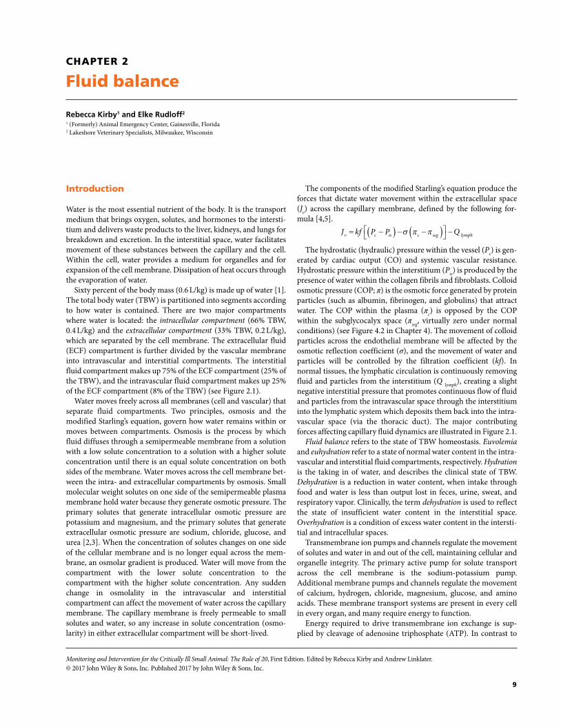
Introduction
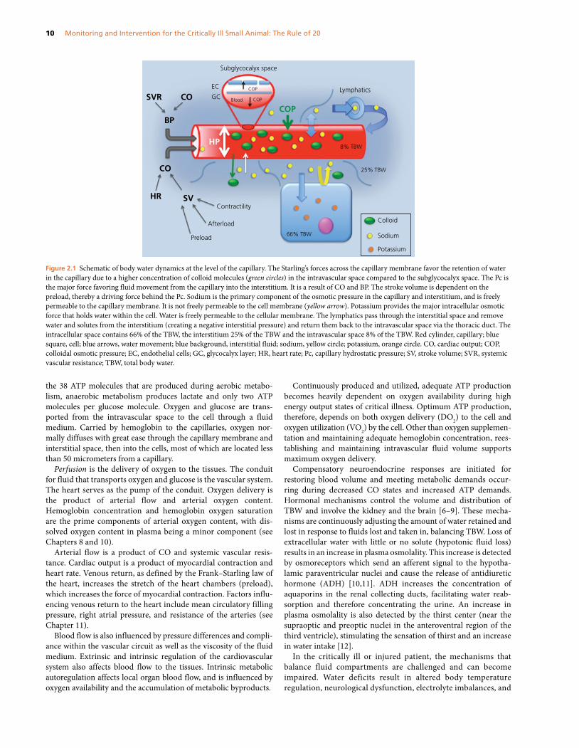
Water is the most essential nutrient of the body. It is the transport medium that brings oxygen, solutes, and hormones to the interstitium and delivers waste products to the liver, kidneys, and lungs for breakdown and excretion. In the interstitial space, water facilitates movement of these substances between the capillary and the cell. Within the cell, water provides a medium for organelles and for expansion of the cell membrane. Dissipation of heat occurs through the evaporation of water.
Sixty percent of the body mass (0.6 L/kg) is made up of water [1]. The total body water (TBW) is partitioned into segments according to how water is contained. There are two major compartments where water is located: the intracellular compartment (66% TBW, 0.4 L/kg) and the extracellular compartment (33% TBW, 0.2 L/kg), which are separated by the cell membrane. The extracellular fluid (ECF) compartment is further divided by the vascular membrane into intravascular and interstitial compartments. The interstitial fluid compartment makes up 75% of the ECF compartment (25% of the TBW), and the intravascular fluid compartment makes up 25% of the ECF compartment (8% of the TBW) (see Figure 2.1).
Water moves freely across all membranes (cell and vascular) that separate fluid compartments. Two principles, osmosis and the modified Starling’s equation, govern how water remains within or moves between compartments. Osmosis is the process by which fluid diffuses through a semipermeable membrane from a solution with a low solute concentration to a solution with a higher solute concentration until there is an equal solute concentration on both sides of the membrane. Water moves across the cell membrane between the intra‐ and extracellular compartments by osmosis. Small molecular weight solutes on one side of the semipermeable plasma membrane hold water because they generate osmotic pressure. The primary solutes that generate intracellular osmotic pressure are potassium and magnesium, and the primary solutes that generate extracellular osmotic pressure are sodium, chloride, glucose, and urea [2,3]. When the concentration of solutes changes on one side of the cellular membrane and is no longer equal across the membrane, an osmolar gradient is produced. Water will move from the compartment with the lower solute concentration to the compartment with the higher solute concentration. Any sudden change in osmolality in the intravascular and interstitial compartment can affect the movement of water across the capillary membrane. The capillary membrane is freely permeable to small solutes and water, so any increase in solute concentration (osmolarity) in either extracellular compartment will be short‐lived.
The components of the modified Starling’s equation produce the forces that dictate water movement within the extracellular space (Jv) across the capillary membrane, defined by the following formula [4,5].
J kf P P Qv c is c seg lymph
The hydrostatic (hydraulic) pressure within the vessel (Pc) is generated by cardiac output (CO) and systemic vascular resistance. Hydrostatic pressure within the interstitium (Pis) is produced by the presence of water within the collagen fibrils and fibroblasts. Colloid osmotic pressure (COP; π) is the osmotic force generated by protein particles (such as albumin, fibrinogen, and globulins) that attract water. The COP within the plasma (πc) is opposed by the COP within the subglycocalyx space (πseg, virtually zero under normal conditions) (see Figure 4.2 in Chapter 4). The movement of colloid particles across the endothelial membrane will be affected by the osmotic reflection coefficient (σ), and the movement of water and particles will be controlled by the filtration coefficient (kf). In normal tissues, the lymphatic circulation is continuously removing fluid and particles from the interstitium (Q lymph), creating a slight negative interstitial pressure that promotes continuous flow of fluid and particles from the intravascular space through the interstitium into the lymphatic system which deposits them back into the intravascular space (via the thoracic duct). The major contributing forces affecting capillary fluid dynamics are illustrated in Figure 2.1.
Fluid balance refers to the state of TBW homeostasis. Euvolemia and euhydration refer to a state of normal water content in the intravascular and interstitial fluid compartments, respectively. Hydration is the taking in of water, and describes the clinical state of TBW. Dehydration is a reduction in water content, when intake through food and water is less than output lost in feces, urine, sweat, and respiratory vapor. Clinically, the term dehydration is used to reflect the state of insufficient water content in the interstitial space. Overhydration is a condition of excess water content in the interstitial and intracellular spaces.
Transmembrane ion pumps and channels regulate the movement of solutes and water in and out of the cell, maintaining cellular and organelle integrity. The primary active pump for solute transport across the cell membrane is the sodium‐potassium pump. Additional membrane pumps and channels regulate the movement of calcium, hydrogen, chloride, magnesium, glucose, and amino acids. These membrane transport systems are present in every cell in every organ, and many require energy to function.
Energy required to drive transmembrane ion exchange is supplied by cleavage of adenosine triphosphate (ATP). In contrast to
Fluid balance
Rebecca Kirby1 and Elke Rudloff2
1 (Formerly) Animal Emergency Center, Gainesville, Florida2 Lakeshore Veterinary Specialists, Milwaukee, Wisconsin
Chapter 2
10 Monitoring and Intervention for the Critically Ill Small Animal: The Rule of 20
the 38 ATP molecules that are produced during aerobic metabolism, anaerobic metabolism produces lactate and only two ATP molecules per glucose molecule. Oxygen and glucose are transported from the intravascular space to the cell through a fluid medium. Carried by hemoglobin to the capillaries, oxygen normally diffuses with great ease through the capillary membrane and interstitial space, then into the cells, most of which are located less than 50 micrometers from a capillary.
Perfusion is the delivery of oxygen to the tissues. The conduit for fluid that transports oxygen and glucose is the vascular system. The heart serves as the pump of the conduit. Oxygen delivery is the product of arterial flow and arterial oxygen content. Hemoglobin concentration and hemoglobin oxygen saturation are the prime components of arterial oxygen content, with dissolved oxygen content in plasma being a minor component (see Chapters 8 and 10).
Arterial flow is a product of CO and systemic vascular resistance. Cardiac output is a product of myocardial contraction and heart rate. Venous return, as defined by the Frank–Starling law of the heart, increases the stretch of the heart chambers (preload), which increases the force of myocardial contraction. Factors influencing venous return to the heart include mean circulatory filling pressure, right atrial pressure, and resistance of the arteries (see Chapter 11).
Blood flow is also influenced by pressure differences and compliance within the vascular circuit as well as the viscosity of the fluid medium. Extrinsic and intrinsic regulation of the cardiovascular system also affects blood flow to the tissues. Intrinsic metabolic autoregulation affects local organ blood flow, and is influenced by oxygen availability and the accumulation of metabolic byproducts.
Continuously produced and utilized, adequate ATP production becomes heavily dependent on oxygen availability during high energy output states of critical illness. Optimum ATP production, therefore, depends on both oxygen delivery (DO2) to the cell and oxygen utilization (VO2) by the cell. Other than oxygen supplementation and maintaining adequate hemoglobin concentration, reestablishing and maintaining intravascular fluid volume supports maximum oxygen delivery.
Compensatory neuroendocrine responses are initiated for restoring blood volume and meeting metabolic demands occurring during decreased CO states and increased ATP demands. Hormonal mechanisms control the volume and distribution of TBW and involve the kidney and the brain [6–9]. These mechanisms are continuously adjusting the amount of water retained and lost in response to fluids lost and taken in, balancing TBW. Loss of extracellular water with little or no solute (hypotonic fluid loss) results in an increase in plasma osmolality. This increase is detected by osmoreceptors which send an afferent signal to the hypothalamic paraventricular nuclei and cause the release of antidiuretic hormone (ADH) [10,11]. ADH increases the concentration of aquaporins in the renal collecting ducts, facilitating water reabsorption and therefore concentrating the urine. An increase in plasma osmolality is also detected by the thirst center (near the supraoptic and preoptic nuclei in the anteroventral region of the third ventricle), stimulating the sensation of thirst and an increase in water intake [12].
In the critically ill or injured patient, the mechanisms that balance fluid compartments are challenged and can become impaired. Water deficits result in altered body temperature r egu lation, neurological dysfunction, electrolyte imbalances, and
Subglycocalyx space
EC
GCCO
CO
HP
SVHRContractility
Afterload
66% TBW
Colloid
25% TBW
8% TBW
Lymphatics
COP
COP
COPBlood
Sodium
Potassium
Preload
BP
SVR
Figure 2.1 Schematic of body water dynamics at the level of the capillary. The Starling’s forces across the capillary membrane favor the retention of water in the capillary due to a higher concentration of colloid molecules (green circles) in the intravascular space compared to the subglycocalyx space. The Pc is the major force favoring fluid movement from the capillary into the interstitium. It is a result of CO and BP. The stroke volume is dependent on the preload, thereby a driving force behind the Pc. Sodium is the primary component of the osmotic pressure in the capillary and interstitium, and is freely permeable to the capillary membrane. It is not freely permeable to the cell membrane (yellow arrow). Potassium provides the major intracellular osmotic force that holds water within the cell. Water is freely permeable to the cellular membrane. The lymphatics pass through the interstitial space and remove water and solutes from the interstitium (creating a negative interstitial pressure) and return them back to the intravascular space via the thoracic duct. The intracellular space contains 66% of the TBW, the interstitium 25% of the TBW and the intravascular space 8% of the TBW. Red cylinder, capillary; blue square, cell; blue arrows, water movement; blue background, interstitial fluid; sodium, yellow circle; potassium, orange circle. CO, cardiac output; COP, colloidal osmotic pressure; EC, endothelial cells; GC, glycocalyx layer; HR, heart rate; Pc, capillary hydrostatic pressure; SV, stroke volume; SVR, systemic vascular resistance; TBW, total body water.
Chapter 2: Fluid balance 11
hypovolemia causing reduced organ perfusion, acute kidney injury, and eventually, death [13,14]. Excess water can result in altered ventilation and lung function, gastrointestinal dysfunction, electrolyte imbalances, and cerebral edema [15]. Movement of water between compartments and into and out of the body can occur rapidly, and is not tolerated in critically ill patients. The normal compensatory responses might not occur as quickly as needed and can be incomplete, inappropriate or ineffective due to end‐organ dysfunction. Frequent assessment of the fluid balance in the critical small animal patient is necessary throughout hospitalization to identify the cause(s) of fluid imbalance, and to make appropriate adjustments in the treatment plan.
Diagnostic and monitoring procedures
Monitoring the fluid balance of a patient should occur on a regular basis since changes can be very drastic over relatively short periods of time. The history and physical examination provide initial insight into problems that can affect or be affected by an altered fluid balance. In hospital point of care (POC) testing, clinicopathological laboratory tests, diagnostic imaging, and monitoring procedures are each assessed in light of the physical status of the patient.
history and physical examinationHistorical and presenting problems reported by the owner that can be associated with or affect assessment of water balance include vomiting, diarrhea, respiratory problems, blood loss, trauma, toxin exposure, fever, nasal discharge, heart failure, alterations in water intake and urine output, and exposure to environmental heat extremes. A list of prescribed and over‐the‐counter medications should be reviewed for drugs that might affect water balance (such as diuretics or vasodilators). Recently administered medication (such as ketamine or atropine) can alter mucous membrane moisture without being a reflection of TBW changes.
The physical examination will identify abnormal parameters associated with fluid imbalance, in particular those associated with perfusion and hydration. Parameters that reflect peripheral perfusion include heart rate, mucous membrane (MM) color, capillary refill time (CRT), pulse quality, level of consciousness, and body temperature. Reduction in intravascular volume leads to clinical signs of hypovolemic shock. A progression of shock manifests in alterations in perfusion parameters, listed in Table 2.1. Rectal temperatures represent peripheral body temperatures and are an indirect indicator of the status of peripheral blood flow. Redistribution of blood flow from the periphery to the core with vasoconstriction can give a differential core‐to‐peripheral temperature (see Chapter 17).
Clinical signs of compensatory shock in the dog can be easily overlooked. The signs of hyperemic mucous membranes, tachycardia, rapid capillary refill time, and normal to increased arterial blood pressure should not be interpreted as normal. The arterial blood pressure is being maintained at the expense of the increased heart rate and mild vasoconstriction. The cat does not typically manifest a compensatory shock response since this stage lasts only seconds to minutes after the initiation of shock in the cat [16].
The state of hydration is in continuous flux, and there is no single parameter that reflects hydration status accurately. Physical examination findings and laboratory indices are used to estimate current hydration status [17]. Physical parameters used to assess hydration include mucous membrane and corneal moisture, skin turgor, and eye position within the orbit (Table 2.2). Two important qualifications must be considered: (1) acute changes in tissue hydration may not be evident on physical examination, since there has not been time for compensatory fluid shifting (underestimating the percentage of dehydration), and (2) older or emaciated animals have poor skin turgor and sunken eyes in their orbit unrelated to their hydration status (overestimating the percentage of dehydration).
Up to 90% of acute changes in body mass can be attributed to a change in TBW, so that in the critically ill patient, a 1 kg change in
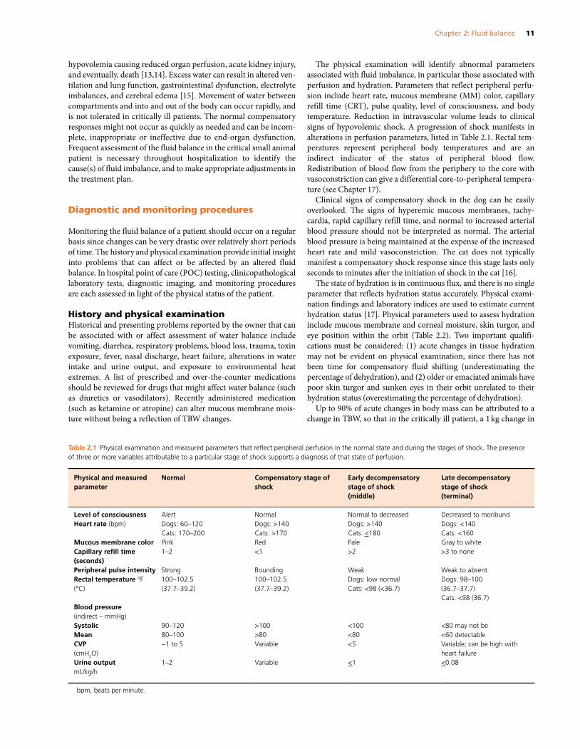
Table 2.1 Physical examination and measured parameters that reflect peripheral perfusion in the normal state and during the stages of shock. The presence of three or more variables attributable to a particular stage of shock supports a diagnosis of that state of perfusion.
Physical and measured parameter
Normal Compensatory stage of shock
Early decompensatory stage of shock(middle)
Late decompensatory stage of shock(terminal)
Level of consciousness Alert Normal Normal to decreased Decreased to moribundHeart rate (bpm) Dogs: 60–120
Cats: 170–200Dogs: >140Cats: >170
Dogs: >140Cats: <180
Dogs: <140Cats: <160
Mucous membrane color Pink Red Pale Gray to whiteCapillary refill time (seconds)
1–2 <1 >2 >3 to none
Peripheral pulse intensity Strong Bounding Weak Weak to absentRectal temperature °F(°C)
100–102.5(37.7–39.2)
100–102.5(37.7–39.2)
Dogs: low normalCats: <98 (<36.7)
Dogs: 98–100(36.7–37.7)Cats: <98 (36.7)
Blood pressure(indirect – mmHg)SystolicMean
90–12080–100
>100>80
<100<80
<80 may not be<60 detectable
CVP(cmH2O)
−1 to 5 Variable <5 Variable; can be high with heart failure
Urine outputmL/kg/h
1–2 Variable <1 <0.08
bpm, beats per minute.

12 Monitoring and Intervention for the Critically Ill Small Animal: The Rule of 20
body weight may be equivalent to a 1 L change in TBW [18–22]. However, due to third body fluid space accumulation (in the abdominal or pleural spaces, or intestines or uterus) or tissue edema, a change in body weight may not occur even though there is loss of fluids from the intravascular or interstitial space(s). Weighing patients, particularly small patients, every six hours is recommended and is a simple monitoring tool to use when assessing trends of change in fluid balance.
Any systemic inflammatory response syndrome (SIRS) disease (such as sepsis, pancreatitis, trauma, immune‐mediated disease, neoplasia) will have increased capillary permeability as part of the syndrome that can result in fluid extravasation. This can lead to complications associated with hypovolemia, interstitial edema, and third body space fluid accumulation. Abdominal distension, reduced bowel sounds, and short or shallow breaths due to increased pressure on the diaphragm can provide physical evidence of a large accumulation of abdominal fluid. Fluid waves might be felt during abdominal palpation. Pleural fluid accumulation can produce an elevated respiratory rate and effort, and muffled lungs sounds. Pulmonary edema may cause respiratory distress with a rapid, shallow breathing pattern and moist lung sounds heard on auscultation. Edema of the intestines may result in poor gastrointestinal function, altered borborygmi, vomiting or diarrhea.
Areas of the body that have thin membranes, low muscle mass, and/or lack of fat will demonstrate fluid accumulation first. Conjunctival edema (chemosis) is an early sign of fluid intolerance, followed quickly by subcutaneous fluid accumulation around the common calcaneal tendon, intermandibular space, head and neck, and the distal limbs. Box 2.1 lists common clinical signs of overhydration. Causes of interstitial edema or cavitary effusion unrelated to increased capillary permeability include oliguric kidney failure, right‐sided heart failure, and portal hypertension. Hypovolemia may or may not be associated with these problems.
point of care testingThe minimum POC laboratory database that should be assessed before and during fluid therapy includes the packed cell volume (PCV), total protein (TP) measured by refractometer, creatinine, blood glucose, plasma lactate, serum electrolytes, acid–base status, coagulation times, platelet estimate, and urine specific gravity (USG). Following the trend of change over time is necessary since the infusion of fluids and other forms of therapy will result in changes that may require intervention.
Loss and gain of extracellular water can result in hemoconcentration (increased PCV and TP) or hemodilution (decreased PCV and TP). Hemorrhage should be considered when the PCV and TP are reduced, although patients with acute hemorrhage can have normal or increased PCV as a result of sympathetic‐induced splenic contraction releasing red cells back into the circulation. In that situation, a reduction in the initial TP can be a tell‐tale sign of hemorrhage in a patient with a compatible history. An initial low TP without hemorrhage might reflect hypoalbuminemia and fluid shifts due to a loss of intravascular COP. As fluid resuscitation and rehydration are performed, hemodilution will result in a decrease in both PCV and TP.
Decreased DO2 due to inadequate blood flow caused by hypovolemic shock can result in anaerobic metabolism. Hyperlactatemia, metabolic acidosis, and increased base deficit are usually associated with conditions causing tissue hypoxemia.
Intracellular volume changes cannot be identified on physical examination. The clinician must rely on changes in the effective osmolality of ECF (primarily recognized by changes in sodium concentration) to mark changes in cell volume. Hyponatremia will be associated with movement of water from the ECF into the intracellular fluid (ICF) compartment, and a subsequent increase in intracellular volume. Hypernatremia will be associated with a decrease in intracellular volume (see Chapter 6).
Changes in blood glucose can affect water balance. Hyperglycemia increases plasma osmolarity and can result in increased extracellular
Table 2.2 Physical examination findings and the associated estimated percent dehydration (interstitial fluid deficits).
Estimated % dehydration
Physical exam findings
0–4 No physical exam changes4–6 Tacky mucous membranes6–8 Loss of skin turgor
Dry mucous membranesSlight hemoconcentration
8–10 Loss of skin turgorDry mucous membranesRetracted globes within orbitsModerate hemoconcentration
10–12 Persistent skin tent due to complete loss of skin elasticityDry mucous membranesRetracted globesDull corneasSevere hemoconcentration
>12 Persistent skin tentDry mucous membranesRetracted globesDull corneasSigns of perfusion deficits (see Table 2.1)Extreme hemoconcentration
Box 2.1 Common clinical signs associated with excess interstitial fluid (overhydration) in the dog and cat.
Mild signs
Serous nasal dischargeChemosisPolyuriaHypothermia, shivering
Moderate signs
Increased skin turgor (gelatinous nature)Tissue edemaIncreased respiratory rate and effortPleural effusionAbdominal fluid wave, ascitesDiarrheaNausea and vomitingAcute weight gain
Severe signs
ExophthalmosHypertensionPulmonary moist cracklesCoughTachy‐ or bradycardiaDepressed level of consciousness