Monash Vision Group 2013 Annual Report · Monash Vision Group 2013 Annual Report Australian...
59
Monash Vision Group 2013 Annual Report Australian Research Council Research in Bionic Vision Science and Technology Initiative Monash Vision Group Monash Vision Group is an A R C-funded Special Initiative with collaborative partners Monash University, Grey Innovation, MiniFAB and Alfred Health. This unique cross-sector consortium has two key goals: • to develop a commercially and clinically viable cortical vision prosthesis or "direct - to-brain" bionic eye; • to build upon existing knowledge to create outstanding research capabilities in bionic vision science and technology in Australia. www.monash.edu/bioniceye The Alfred Alfred Health is a leader in healthcare delivery and improvement, striving to achieve the best possible health outcomes for patients and the community by integrating clinical practice with research and education. Alfred Health is recognised as a pacesetter in the national healthcare arena, is consistently linked to progressive developments in healthcare and services, medical research and healthcare teaching. Alfred Health is providing expertise in MVG's clinical program, including the recruitment, testing and after-care of patients. www.alfred.org.au Grey Innovation Grey Innovation is a cutting edge engineering company with experience in complex software, hardware and mechanical architectures across a number of industries and markets. Grey Innovation provides a unique product development and commercialisation service from initial strategy through design, testing, technology innovation, manufacture and market penetration. Grey Innovation is providing expertise for the development of MVG's external electronics and processing components of the vision system. www.greyinnovation.com
Transcript of Monash Vision Group 2013 Annual Report · Monash Vision Group 2013 Annual Report Australian...

Monash Vision Group 2013 Annual Report
Australian Research Council Research in Bionic Vision Science and Technology Initiative
Monash Vision Group Monash Vision Group is an A R C-funded Special Initiative with collaborative partners Monash University, Grey Innovation, MiniFAB and Alfred Health. This unique cross-sector consortium has two key goals: • to develop a commercially and clinically viable cortical vision prosthesis or "direct-to-brain" bionic eye; • to build upon existing knowledge to create outstanding research capabilities in bionic vision science and technology in Australia. www.monash.edu/bioniceye
The Alfred Alfred Health is a leader in healthcare delivery and improvement, striving to achieve the best possible health outcomes for patients and the community by integrating clinical practice with research and education. Alfred Health is recognised as a pacesetter in the national healthcare arena, is consistently linked to progressive developments in healthcare and services, medical research and healthcare teaching. Alfred Health is providing expertise in MVG's clinical program, including the recruitment, testing and after-care of patients. www.alfred.org.au
Grey Innovation Grey Innovation is a cutting edge engineering company with experience in complex software, hardware and mechanical architectures across a number of industries and markets. Grey Innovation provides a unique product development and commercialisation service from initial strategy through design, testing, technology innovation, manufacture and market penetration. Grey Innovation is providing expertise for the development of MVG's external electronics and processing components of the vision system. www.greyinnovation.com

MiniFAB MiniFAB is a privately-held Melbourne-based contract engineering firm providing custom development and manufacture of disposable polymer micro-engineered products. Established in 2002, MiniFAB's product development process covers the entire spectrum, from converting early stage product concepts into prototypes, through to full-scale, high throughput OEM & ODM manufacturing. MiniFAB is providing expertise in the design and manufacture of MVG's implantable devices and tooling. www.minifab.com.au
Monash University Monash University is one of Australia's leading universities with an enviable record for research and development leading to commercialisation. MVG has Chief Investigators from departments within the Faculties of Engineering, Science and Medicine, Nursing and Health Sciences, with key inputs into all aspects of the Monash Vision project. www.monash.edu <pp> 2
Contents
Introduction to the Monash Vision Group Message from the Chair Director's Report Governance & Management Product Development, Supporting Research and Testing 2013 Highlights Research Training and Skills Development Commercial Program Visitors, Events and Public Engagement MVG in the Media Journal Publications and Submissions Conference Proceedings Financial Statement Key Performance Indicators Acknowledgements

Introduction to the Monash Vision Group
Monash Vision Group (MVG) is a consortium of engineers, vision scientists, industrial designers and medical researchers from Monash University, clinicians from Alfred Health and industry partners Grey Innovation and MiniFAB, with the primary goal of realising and demonstrating in patients a cortical vision prosthesis - or "direct to brain" bionic eye. MVG was established in 2010 with funding for four years from the Australian Research Council (A R C) Research in Bionic Vision Science and Technology Initiative. In July 2013, the A R C announced a one year extension, taking the Initiative to the end of 2014. MVG's bionic vision system "Gennaris" bypasses damage to the eye and optic nerve and may therefore address conditions that cause up to 85% of currently untreatable blindness including glaucoma, age-related macular degeneration, acquired retinal disease and traumatic injury. Gennaris comprises a miniature camera worn externally by the user. High-resolution images from the camera are fed to a custom-designed vision processor, which applies a number of signal processing techniques to extract the most useful features from the camera images. This new signal is fed - via a wireless transmitter - to up to 11 tiles that have been surgically implanted in the primary visual cortex of the brain. Each tile houses its own microchip or A S I C (Application Specific Integrated Circuit), wireless receiver and 43 hair-thin microelectrodes that stimulate the neurons in the primary visual cortex. This stimulation evokes brief flashes of light known as "phosphenes" in the visual field of the user, which the brain learns to interpret as vision. The number of phosphenes depends upon the number of implanted electrodes, in our case this will be up to 473. MVG aims to implant Gennaris in first patients in 2015; this 2013 Annual Report presents progress by MVG over the past twelve months towards this key goal. <pp> 3
Message from the Chair Professor David De Kretser AC
2013 was always going to be a critical year for the Monash Vision Group. With funding from the Special Research Initiative (S R I) due to finish, it was imperative for

MVG to demonstrate successes in its technical and commercial programs and to secure funding for beyond 2013. We enjoyed an excellent start to the year with the A R C Mid-Term Review site visit to Monash University in February. The panel spoke with many members of the MVG team, including board members, management, technical staff, students and also Monash University Executive. Panel members also enjoyed a tour of the MVG engineering and physiology laboratories. MVG received positive and encouraging feedback, with the panel highlighting the excellent progress that has been made since project launch in 2010 and a recommendation for this first phase of the S R I to continue. In MVG's 2012 Annual Report, I commented on the importance of fundraising and my close interactions with Professor David Penington, former Chair of Bionic Vision Australia (BVA), towards attracting funding for both groups. During 2013, David and I continued with our approaches to Federal and State Governments, the Australian Research Council and National Health and Medical Research Council to highlight the benefits of continuing the S R I beyond the first phase. The discussions culminated in the S R I being extended for a further year, providing MVG and BVA with funding until the end of 2014. My thanks go to the A R C for allocating $1.9M to MVG on top of the $8M already awarded, which has enabled us to retain our team and continue on our path to producing a commercially attractive device to restore some sight to many people with profound vision impairment. My sincere thanks also go to David for his dedication and proactive approach to fundraising and I wish him all the very best in his future endeavours. We have many successes to report but on a personal note, I would like to highlight the well-deserved achievements of two team members in particular. Our Director, Professor Arthur Lowery, was awarded a prestigious Australian Laureate Fellowship by the A R C in July, which is one of our nation's highest academic achievements. In June, Professor Jeffrey Rosenfeld was awarded an Officer of the Order of the British Empire for his services to neurosurgery and the University of Papua New Guinea's School of Medicine. Many congratulations to both Arthur and Jeffrey for these outstanding accomplishments. I am delighted to report that each of our valued Advisory Board members has agreed to continue with MVG for the extension period. I would like to thank them for their important contributions, enthusiasm and insights and I look forward to working with them over the next year to provide guidance to MVG management and in securing resources for beyond 2014. My thanks also go to our industry and clinical partners - MiniFAB, Grey Innovation and Alfred Health - and to Monash University. Their support is central to the success of MVG as we continue on our exciting journey towards first patient implantation and a commercial device. Professor David De Kretser AC Advisory Board Chair, Monash Vision Group <pp> 4

Director's Report Professor Arthur Lowery
This has been a team-building experience for all of us, and more importantly, a capability building endeavour. Awarded $9.9M of Government funding. Over $12M in-kind contributions from MVG partners. Over $3M cash contributions from Monash University. I am delighted to present the fourth report of the Monash Vision Group. We are in the final stages of manufacturing a complete bionic vision system prototype, which may benefit hundreds of thousands of people who are vision impaired. We are aiming to have full testing completed within the next 18 months, which will enable a First in Human (F I H) trial to be conducted. The initial project funding was for 4 years, from 2010 to 2013 inclusive, under the Australian Research Council's "Research in Bionic Vision Science and Technology Initiative". We are thankful to the A R C for extending the funding for a further year, until the end of 2014, and to the NHMRC for awarding a $1.46M Development Grant to fund further human trials following the F I H. The extension followed a successful A R C review in February 2013 and presentations from Professors David de Kretser (MVG) and David Penington (Bionic Vision Australia) to senior figures in Australia. I would like to personally thank Senator Kim Carr for his passionate belief and support of scientific and engineering research over many years, without which we would not be equipped to take on challenges of a global scale. Our system is designed to benefit the majority of people with untreatable blindness, as it stimulates the brain directly with several hundred electrodes, each potentially producing a "phosphene" or sensation of light. This approach does not require a functional optic nerve or eye, so is the only solution for those who have lost their sight through trauma to these parts of the visual pathway. The project has been extremely challenging. To elicit several hundred points of light, we are planning to implant up to 11 tiles onto the surface of the brain. Each tile is a miniaturised electronic system that converts radio waves into discrete stimulation pulses on 43 electrodes. The system includes a radio receiver, which also harvests power from the waves; decoding and error correcting circuits to ensure the integrity of the received data; 43 digital to analogue converters, to provide 32 different stimulation levels; 43 voltage to current converters, to provide stimulation pulses, and 43 micro-electrodes that conduct these pulses to the correct layer of the visual cortex of the brain. This complexity is designed into 500,000 transistors per tile, forming a complex "system on a chip". Importantly, the electronics is sealed within an hermetic package, to ensure no unwanted interaction with the tissue around it.

The materials engineering is complex, because we have only a limited pallet of FDA-approved materials with which to make the tile. The process of constructing the electronics and sealing into the tile is complex and requires over 70 assembly steps. Thus, experience of our partner, MiniFAB, in certified manufacturing has been critical in the development and manufacture of the tiles. On the outside of the head is a wireless transmitter, camera and another set <pp> 5 of complex electronics to convert the camera's complex images into a useful representation of the real world on a few hundred electrodes. In 2012, Monash Art, Design and Architecture joined our project to provide the industrial design of the packaging that contains these components. Based on consultations with Vision Australia and its clients, prototype headgear has been developed in conjunction with Grey Innovation and with help from the project workshop in Monash Electrical Engineering. This prototype was exhibited as part of the Melbourne Now exhibition over the summer of 2013/14. The vision processing software, developed by Monash's computer vision group and ported onto the processor in collaboration with Grey Innovation, has also been demonstrated at several conferences and events. The challenge in 2014 is to bring the F I H prototype system together for extensive testing. In preparation, we have spent much of 2013 developing methods to test the implant tiles and external processor during its assembly, and as a full system after manufacture. This has involved the creation of a set of electronic test boards, into which the partially-assembled parts plug, and a suite of testing software. We have also developed bench-top test rigs for the insertion tool, which is somewhat like a pinball machine, and thermal test jigs for the tiles, to ensure that they can be cooled sufficiently by blood flow. Our bench-top testing is backed by extensive computer simulations and library research. We have also developed a set of psychophysics tests, to explore the capabilities of our potential patients before and after bionic vision has been turned on. This is critical as regulatory approval is dependent upon showing an improvement in quality of life. We were pleased to host Doctor Bob Greenberg, CEO of Second Sight, to our laboratories in August 2013. He was very helpful in detailing their experiences with the FDA in obtaining humanitarian approval for their retinal implant device. The remainder of this report will detail the technology and successes of 2013. This includes detailed technical reports, presented as sub-projects, which have been written by the scientists and engineers undertaking the work. I would like to commend all of our technical staff for their outstanding effort in the past year. A key to our confidence in being able to deliver a F I H implant in 2015 has been the extremely detailed planning and reporting of the whole team on a weekly basis. The weekly Technical Architecture meetings and reporting have been driven by Peter Bettonvil, of MiniFAB, and attended by all groups across Monash and the partners. This has been a team-building experience for all of us, and more importantly, a capability building endeavour. We are one of the few places in the world that is able to develop micro-miniaturised electronic systems to meet medical challenges. This

capability could underpin a new industry sector in Australia with a large export potential. I should like to congratulate our team on some of their personal successes. Prof Jeffrey Rosenfeld A M, was awarded an O B E for his outstanding efforts to improve the lives of patients in PNG over the last 30 years. Professor Gary Egan (Monash Biomedical Imaging) has led a successful Centre of Excellence bid in Integrative Brain Function, which includes MVG's Marcello Rosa and Ramesh Rajan. MVG Honours student Ross Anderson was recognised and received an award for his outstanding thesis, which explored the expectations, motivations, concerns and decision-making of people with profound vision impairment when considering receiving a cortical vision prosthesis. Finally, I thank our whole team including our support staff at all levels, but especially Ms Vicki Tutungi who has skilfully chaired the Steering Committee, which has directed resource to critical paths of the project and ensured that the project progresses quickly and on budget. Doctor Jeanette Pritchard, General Manager of MVG, has integrated the project over many timescales and locations, to ensure quick resolution of many issues and is now focusing on commercialisation. Prof David de Kretser has chaired our wonderful Advisory Board, who provide an interface to stakeholders, technical wisdom and strategic direction, and has tirelessly sought new funding. Professor Arthur Lowery Director and Project Leader, Monash Vision Group <pp> 6
Governance & Management
Advisory Board The Advisory Board met at The Alfred Centre, Prahran on 23rd April and at the Monash Conference Centre, Melbourne CBD on 5th December. Professors David de Kretser and Lyn Beazley and Mr Gerard Menses also attended the A R C Review Panel interviews at Monash University on 12th February.
Professor David de Kretser AC Chair, MVG Advisory Board

Sir John Monash Distinguished Professor, Monash University
Professor Lyn Beazley Chief Scientist, Western Australia (2006 - 2013) and A R C Nominee
Professor Jim Patrick Chief Scientist, Cochlear Ltd
Mr Gerard Menses CEO Make a Wish Foundation
Professor Arthur Lowery Director, Monash Vision Group
Doctor David Lyster Manager, Research Partnerships, Monash University <pp> 7
Steering Committee
Ms Vicki Tutungi Independent Chair, MVG Steering Committee Managing Director, ProLearn

Professor Arthur Lowery Director, Monash Vision Group
Professor Jeffrey V Rosenfeld AM Director of Neurosurgery, Alfred Hospital and Head, Division of Clinical Sciences and Department Surgery, Central Clinical School, Monash University
Ms Halina Oswald Director, Monash Research Office, Monash University
Doctor David Lyster Manager, Research Partnerships, Monash University
Professor Marcello Rosa Deputy Head of Physiology, Monash University
Ms Fiona Miles Legal Counsel, Alfred Health
Doctor Erol Harvey CEO,

MiniFAB (AUST) Pty Ltd
Mr Jefferson Harcourt Managing Director, Grey Innovation Pty Ltd
Executive Team
Professor Arthur Lowery Director
Doctor Jeanette Pritchard General Manager
Ms Suzanne Hayster Administrative Assistant <pp> 8
Senior Investigators
Professor Arthur Lowery Director and Project Leader, MVG
Professor Marcello Rosa Chief Investigator, Department of Physiology, Monash University

Professor Jeffrey V. Rosenfeld AM Chief Investigator, Division of Clinical Sciences and Department of Surgery, Monash University
Associate Professor Ramesh Rajan Chief Investigator, Department of Physiology, Monash University
Associate Professor Lindsay Kleeman Chief Investigator, Department of Electrical and Computer Systems Engineering, Monash University
Doctor Wai Ho Li Chief Investigator, Department of Electrical and Computer Systems Engineering, Monash University
Associate Professor John Forsythe Chief Investigator, Department of Materials Engineering, Monash University
Professor Kate Smith-Miles Chief Investigator, School of Mathematical Sciences, Monash University
Professor Ben Adler Chief Investigator, Department of Microbiology,

Monash University
Professor Julian Rood Chief Investigator, Department of Microbiology, Monash University
Doctor Erol Harvey Partner Investigator and CEO, MiniFAB
Associate Professor Anthony Hall Partner Investigator and Director of Ophthalmology, Alfred Hospital "As an engineer, I like building things that solve novel, difficult and complex problems. It is a rare opportunity where one gets to do this and make something that has the potential to bring light back into a person's world. Working alongside a group of motivated and like-minded individuals is a fantastic cherry on top." Doctor Wai Ho Li <pp> 9
Technical Personnel and Researchers
Monash University
Electrical and Computer Systems Engineering
Doctor Anand Mohan Research Engineer Doctor Damien Browne Research Engineer Doctor David Fitrio Research Engineer

Doctor Dennis Lui Research Engineer Mr Andrew Stephens Contract Engineer Ms Collette Mann Clinical and Psychophysics Coordinator Mr Horace Josh PhD Candidate Mr Titus Tang PhD Candidate Ms Emma Brunton PhD Candidate Ms Jamila Bird Honours Student Mr Akshay Kumar Undergraduate Summer Student Ms Gillian Gan Undergraduate Summer Student Mr Daniel Jong Undergraduate Summer Student Ms Ami Pasricha Undergraduate Summer Student
Physiology
Doctor Nic Price Lecturer Doctor Chun Wang Research Scientist Doctor Leo Lui Research Fellow Doctor Saman Haghgooie Biomedical Engineer Doctor Edwin Yan

Research Scientist Doctor Kostas Chatzidimitrakis Research Fellow Ms Kahli Cassells Technician Ms Simone Carron PhD Candidate Ms Amanda Davies PhD Candidate Ms Francesca Vigo Honours Student Ms Marina De Oliveira Montibeller Undergraduate Student
Materials Engineering
Doctor Kun Zhou Research Engineer
Monash Art, Design and Architecture
Associate Professor Arthur de Bono Head, Department of Design, Associate Dean, Research Professor Mark Armstrong Practice Professor Industrial Design; Eva and Marc Besen International Research Chair in Design Mr Kieran John Industrial Designer
Monash Biomedical Imaging
Professor Gary Egan Director

Ms Amanda Ng Computational Biomedical Imaging Scientist
Monash Alfred Psychiatry Research
Professor Jayashri Kulkarni Director Professor Paul Fitzgerald Deputy Director Doctor Jerome Maller Research Fellow Doctor Richard Thomson Research Fellow
Monash Medical Centre Doctor Sanjeeva Ramasundara MRI Consultant
Alfred Hospital Associate Professor Harvey Newnham Associate Professor of Medicine Doctor Helen Ackland Research Fellow
Grey Innovation Mr Mike Smith Chief Technical Officer Mr George Greenall Project Manager Mr Graham Lyford Software Engineer Ms Nicola Andrews

Industrial Designer Mr Nathan Lee Industrial Designer Mr Alistair Webster Engineer Mr Simon Kennedy Engineer Mr Warwick Fifeld Quality Systems Manager
MiniFAB Doctor Micah Atkin Manager & Technical Lead Mr Peter Bettonvil Project Manager Mr Matthew Solomon Senior Engineer Mr Brody Payne Project Engineer Mr Brendan Ruys Project Engineer Mr Alexander Stankovic Production Worker Mr Michael Packiasamy Technician <pp> 10
Product Development, Supporting Research and Testing

The following section describes the progress that has been made by MVG during 2013 on device and component manufacture for the Gennaris bionic vision system, including bench and preclinical testing and verification of the design. Updates on clinical research programs are also provided. The schematic below shows the flow of the sub-projects that have been established by MVG, from external electronics and wearable headgear, through implantable components to patient testing and clinical work. <Transcriber's note> The diagram has been omitted. </Transcriber's note> The implant tile constitutes a titanium ring welded to a ceramic package, 43 stimulating electrodes through the package base and a cap that is welded over the electronics. The electronics, including the A S I C, will sit upon a circuit board sub-assembly within the implant package and the receive coil, which receives power and stimulation commands is attached to this circuit board. The external electronics powers and controls the stimulation via a transmitter, which is positioned on the back of the user's head. The small size of the autonomous implant tiles is critical to optimise the number of tiles that can be implanted in each patient, thereby improving the likelihood of providing more phosphenes to the user and ultimately improving the quality of their vision. Device development, manufacture and testing were managed through weekly technical architecture meetings and monthly risk and harm meetings, both of which involved engineering and physiology staff from all MVG partner organisations. Furthermore, meetings were held fortnightly to discuss specific issues relating to the electrode array design, coating and fabrication methodologies. <pp> 11
Useful MVG Definitions Algorithm: Step-by-step instructions telling a computer how to do something, such as turning an image into phosphenes. A S I C: Application Specific Integrated Circuit, also referred to as microchip. A S I C1: MVG's first prototype. A S I C: fully wired electrode stimulator. A S I C1B: Second prototype, incorporating wireless capability (low voltage). A S I C2: Third prototype, incorporating wireless capability (high voltage). A S I C3: Final MVG A S I C, to be used in First In Human implant tile. CAD: Computer-Aided Design. CMOS: Complementary Metal Oxide Semiconductor. CPGA: Ceramic Pin Grid Array. E E G: Electroencephalography. Gennaris: MVG's bionic vision system. GFAP+: Glial Fibrillary Acidic Protein Positive. Hatpack: Portable headwear designed and built by MVG engineers to simulate the bionic vision experience. Hermetic: Impermeable to fluid ingress/leak resistant.

F I H: First In Human. Implant tile: MVG implantable device, including A S I C, wireless receiver and 43 electrodes. L E D: Light Emitting Diode. MAC1: Fully wired MVG implant tile (43 electrodes, 43 wires). MAC2: Partially wired (43 electrodes, 14 wires) MVG implant tile incorporating A S I C1B. M A D A: Monash Art Design and Architecture. M E G: Magnetoencephalography. MRI: Magnetic Resonance Imaging. Open CV: Open source computer vision PCB: Printed Circuit Board Phosphene: Spots of light generated through electrical stimulation of the visual cortex. Psychophysics: Quantitative assessment of the relationship between physical stimuli and the sensations/perceptions they affect. S E M: Scanning electron microscope. TMS: Transcranial Magnetic Stimulation. <pp> 12
Camera and External Electronics As part of the Technical Architecture team, Grey Innovation worked through manufacturing, testing and test support for MAC1 and MAC2 during 2013. This included manufacturing and testing sub-systems such as the vision processor, cameras, test targeted variants of A S I C interface boards, development test hardware and many jigs and cables to connect MVG equipment to off-the-shelf test equipment required for testing the A S I C1 in vitro and in vivo. Included within this sub-project was the integration of Open CV libraries for running algorithms on the vision processor, which now use these libraries for image processing primitive functions. The challenge was in integrating this open library into the framework that supports the algorithms. Extra support tools for the algorithms were developed, including: • profiling that allows algorithm time costs to be measured • simulation and regression test functions, allowing pre-recorded input frames to be processed and the electrode outputs saved. The vision processor software was enhanced to enable the one software set to support all three versions of communication protocols with the A S I C tiles. Thus A S I C protocol variant support was achieved. <pp> 13 Substantial design and testing of hardware for A S I C2 and A S I C3 were also undertaken, including analogue and digital test harnesses for packaged and partially packaged A S I Cs. This micron-level manufacturing required substantial design and engineering, working with extremely fine tolerance PCB manufacturers and

component bonding partners. This process was largely managed by Grey Innovation with input from other members of the Technical Architecture team where required. Ongoing through 2013, Grey Innovation worked closely with the Monash team to enhance and extend the stimulator software to meet new and developing requirements of the preclinical team. One example of this was to remove the 3 second trigger-stimulation delay to increase the success of trials. This was achieved by reconfiguring the software architecture of the stimulator and integrating it with a digital interface to a desktop PC. The year also included manufacture and demonstration of a sample F I H handset, headset, spectacles concept and other mechanical design in association with M A D A; demonstration of external electronics including the production and testing of four variants of a wired camera, mechanical housings purpose-built for development, demonstration and physiology testing. Caption: Exploded view of the vision processor showing the PCB, battery and other components. Caption: Vision processor front panel artwork. <pp> 14
Product Design MVG has built a strong relationship with the industrial design team at Monash University - Faculty of Art Design & Architecture. M A D A implements research and industrial design capability that is critical to humanise the technology. The primary goal during 2013 was to deliver a design to house the external electronics, integrate mechanical adjustment capable of altering dimensions for anthropometric needs and re-position the coil to accurately transmit a wireless signal to the implant tiles. The team focused on providing an outcome to facilitate artificial vision and equally a user-oriented experience that prioritises usability, comfort, adjustability and long term reliability. An in-depth research report was compiled to analyse existing products, market trends and competitors with intent to design a series of initial prototypes. Further ideation and development prioritised two designs; an eyewear and non-eyewear device each exhibiting unique features and mechanical characteristics in wearable technology. Capitalising on originality and increasing public interest, MVG successfully sought a US design registration for the non-eyewear design and following public release of both designs mid-year, these featured heavily in print, radio and televised media garnering national and international attention. With two viable concepts, the team engaged an ergonomist to conduct a preliminary assessment to substantiate the design rationale, which revealed a need for further analysis with end-user input. This resulted in the team conducting a user focus group in conjunction with Vision Australia and its clients. The participant's responses produced valuable opinions, feedback and information that built scenarios detailing
daily habits and routines pertinent to functional needs and requirements of the designs. The latest design iterations have been updated according to these focus group findings and key indicators derived from preliminary ergonomic and usability analysis and prototype evaluation. With further assessment of test rig prototypes, reliable and simpler solutions were identified for various mechanical components used to control adjustment and coil positioning. Through M A D A, the National Gallery of Victoria (NGV) invited MVG to display the prototype Gennaris bionic vision system at the much publicised "Melbourne Now" exhibition. With the intent to highlight and celebrate the industrial design contribution, this presented a unique opportunity for M A D A to display its capability on an international stage. Running between 22nd November 2013 and 23rd March 2014, Melbourne Now was the largest and most ambitious exhibition in the gallery's 152 year history. During 2014, the industrial design team will initiate a thorough usability and ergonomic analysis for the revised designs with the aim of generating a commercially viable outcome for use in patient testing. <pp> 15 Caption: Non-eyewear concept generated from CAD data. Rear right perspective (off head). Caption: Non-eyewear concept generated from CAD data. Front left perspective. Caption: Eyewear concept generated from CAD data. Front left perspective. <pp> 16
Signal Processing A major challenge for signal processing is to deal with "irregular" phosphene patterns: visual patterns of dots experienced by the patient that do not follow a simple grid-like structure. Both retinal and cortical vision prostheses are expected to produce irregular phosphene patterns; an expectation confirmed by previously conducted clinical studies. For example, phosphenes produced by cortical stimulation are expected to have an irregularity where phosphenes are larger towards the periphery of vision; this is due to a physiological phenomenon called "cortical magnification". Thus far, signal processing approaches have ignored the problem of irregular phosphene patterns. A key achievement in 2013 was the development of a signal processing approach that is flexible enough to handle arbitrary irregular phosphene patterns. This patent-pending approach also lends itself to fast software implementations and allows for a range of sensor and system configurations; providing flexibility in how the external electronics of the device can be reconfigured non-invasively via software in order to tailor it for an individual patient. Given that each patient will have a unique phosphene pattern and preferences to how signal processing is used to display the world around them as phosphenes, the

development of this new flexible algorithmic technology is key in helping maximise post-implant results. Another signal processing achievement of 2013 was the development and preliminary testing of improved software for the vision processor. In 2012, the signal processing team delivered software that can operate in real time on the limited computational and power budget afforded by the highly portable vision processor. The system was designed around regular phosphene patterns and met the engineering target of 10 images processed per second to allow real world use. Taking advantage of this development, the vision processor software was updated to deal with irregular phosphene patterns. Computational performance was improved and power usage was reduced dramatically. The new image processing software, which includes real time visualisations of phosphene patterns for clinicians and engineers, only takes a few milliseconds to execute for each camera frame. <pp> 17 Preliminary tests suggest that image processing takes an average of two milliseconds - an improvement of at least an order of magnitude over 2012's already fast software. The improved software processing speed will allow more head room for additional processing and algorithms to be used in parallel, which may improve the patient's visual experience, lead to improved battery life and open up more options for hardware choices in future products. Much of 2014 will be spent on the systematic and detailed testing of the vision processor prototype and signal processing software to ensure that they are both robust and operate according to engineering and medical requirements. Several test sequences of daily activities as well as synthetic videos will be used to exercise the vision processor and software over many hours of operation. An initial overnight software test of the vision processor conducted in 2013 showed that the system is robust and stable over a period of operation beyond the typical intended usage length. Further tests will be performed in 2014 followed by close interaction with MVG system engineers to adapt and integrate the vision processor hardware and software into the Gennaris bionic vision system. Caption: Prototype of the wearable vision processor co-developed by Grey Innovation and Monash University. The signal processing software runs on this processor and is tested on live images coming from a small camera module. This camera module can be head-mounted to simulate the patient's visual experience in order to help optimise signal processing algorithms and algorithmic parameters. Preliminary robustness tests have started for the vision processor to ensure that it is able to withstand the daily activities of patients. <pp> 18
Wireless Link 2013 saw the successful completion of most aspects of the wireless link: the receiver circuit, receiver coil, transmitter circuit output stage and transmitter coil. Initial

characterisation of the complete wireless link was also performed. The PCB that A S I C3 and the receiver components are assembled on was also completed. All that remains is the development of the transmitter driver circuitry and full characterisation of the wireless link, to determine the region of operation. Within each implant tile, a coil and receiver circuit recovers the power and data. Key design criteria for the receiver and coil were reliability, size and efficiency. The design also needs to minimise the variation in received power versus variation in transmitter-to-receiver coil separation to accommodate the different positions of tiles from patient to patient. Several voltage regulator topologies were considered and several receiver designs were developed, prototyped and tested, in part due to the varying requirements of A S I C1B, A S I C2 and A S I C3. The receiver coil was also designed, tested and optimised. The final design provides the A S I C with four regulated voltages and a differential data signal. It features a low loss coil (high-Q) that maximises received power and minimises power dissipation. The development of the transmitter also reached the final stages. A high efficiency class-E output stage with a very high-Q Litz-wire coil has been developed and tested. The transmitter was optimised for minimal variation in received power versus distance, link efficiency, received data integrity and minimal power dissipation. The final design can power multiple tiles up to 25 mm away and consume less than 400 mW to maximise battery life. With the transmitter output stage and receiver complete, work progressed on fully characterising the complete wireless link and determining the region of operation. A micrometer-based test fixture allowing accurate and repeatable measurements of all the received voltages and signals, at different coil separations (parallel distance), offsets (off-axis distance) and relative angles was developed. Preliminary measurements suggest the working range will be 10-25 mm parallel distance, ±20 mm offset and ±25° relative angle from parallel. It was found that angles up to +90° or -90° are also possible, depending on the offset distance. During the early part of 2014, the wireless link development will be finalised. This involves completing the transmitter driver - the interface between the vision processor and the output stage - and determining the complete region of operation, detailing all viable distances and angles from the transmitter to implant. This region of operation allows the surgeon to know exactly where the tiles may be implanted in order to receive sufficient power to function. Once complete, the wireless link hardware and software will be incorporated in the implant tile package for bench testing. <pp> 19 Caption: Thermal testing of wireless transmission system. Caption: Wireless link testing showing transmitted signal (green), demodulated data (pink/yellow) and recovered data stream (blue). Caption: A S I C3 prototype receiver. <pp> 20

Implantable Electronics Using the information gathered during testing of A S I C1 and A S I C1B, two new versions of the A S I C were designed and implemented consecutively in 2013, aimed at implementation into the F I H implant tile. A S I C2 and A S I C3 have been implemented using a newly available higher voltage CMOS technology. Both devices have the ability to stimulate up to 43 electrode channels on a single tile and obtain data and power wirelessly by using a small number of off-chip components. A S I C2 was implemented in the first quarter of 2013. Testing of A S I C2 in the second half of 2013 led to the implementation and submission of A S I C3 for manufacturing in November 2013. As it uses new technology, A S I C2 required a systematic re-evaluation and implementation of a number of circuits on chip. Lessons learned from A S I C1B led to a redesign of the analogue wireless receiver circuits to give enhanced sensitivity when interpreting and digitising the incoming wireless data signal. The analogue front end also had a new power regulation circuit along with power sensing safety circuits to reset the device in case of a sudden power failure. Improved digital signal processing and error correction logic was implemented in A S I C2 to make the system more robust in low signal conditions. The decoded wavetable data includes parameters such as stimulation levels and timing information. This information is then fed forward to control the working of the analogue electrode drive circuits, to deliver a specified amount of current into the electrodes in order to elicit a phosphene response. Testing of A S I C2 was performed on the bench top in-house using a purpose built test board. The testing of the microchip showed a markedly improved digital yield compared with A S I C1B. The testing also verified the improved functionality and sensitivity of the analogue wireless receiver circuit. Interpretation of the data from intensive bench top testing of A S I C2 led to decisions that impacted the development of A S I C3. These primarily focused on enhancing the safety of the overall device and also risk mitigation to prevent harmful failure modes in the F I H device. Additionally, the design has been modified to help reduce the overall power consumption, improve stability and increase sensitivity to the received wireless data signal. Further analysis of A S I C2 functionality led to revisions in the architecture of the analogue back end electrode stimulation circuits in order to ensure better charge balancing between the stimulation phases and remove any long term direct current (dc) voltages on the electrodes post-stimulation. Circuit changes using Intellectual Property (IP) blocks from the manufacturer also helped to bring about a newer architecture for the brownout reset circuit and improve its performance, thereby directly enhancing the safety of the device during a brownout event. A S I C3 is scheduled for delivery in early March 2014. Intensive testing and characterisation of A S I C3 will be undertaken including electrically testing the chip on the bench top to ensure that the device is functional and safe. Each device will be characterised to determine yield, efficacy of stimulation paradigms including charge balance per electrode, output stage and current safety limits and safe responses to various wireless data and power failure conditions. Furthermore, as A S I C3 is on

track to be integrated into an F I H tile, extensive testing post-integration of the assembled F I H tile will be performed prior to preclinical or clinical use. <pp> 21 "For all the trials and tribulations of working through a unique endeavour such as this, the thought of producing a phosphene at the far end of the tunnel is worth all the effort." Doctor Anand Mohan. <pp> 22
Implantable Electrodes and Coatings Thin film titanium nitride (TiN) coatings on penetrating platinum iridium (PtIr) electrodes in the implant tile were extensively tested during 2013. TiN is a hard and inert conducting material that is mechanically stable as a thin film coating. The TiN is coated onto the bare PtIr electrodes creating a rough surface. This increases the surface area of the electrode, without greatly increasing the area of the brain that is stimulated, maintaining focused stimulation that targets the desired brain region. Initial in vitro electrochemical testing of these electrodes in saline proved promising. The TiN electrodes outperformed the PtIr electrodes; they were able to safely inject ten times more charge than the bare PtIr electrodes, and showed lower impedance at all frequencies. Interestingly, in vivo testing of the efficacy of stimulation with TiN-coated PtIr electrodes compared to PtIr electrodes in a small animal model showed no difference in performance between the two types of electrodes, at least in terms of current levels for threshold - both required similar levels of current and voltage to elicit a motor cortex response. The results of these studies indicated that coating electrodes with a thin film of TiN will result in electrodes that are just as effective at stimulating neurons as PtIr electrodes, but have increased safe charge injection limits, allowing for a greater variety of currents to be delivered safely, improving device safety and efficacy. Research into the influence of the electrode's surface area on device efficacy continued into 2013. Analytical and computer models had previously indicated that electrode surface area would influence the efficacy of stimulation. Completion of in vivo threshold measurements verified these predictions. Threshold testing of electrodes with different geometric surface areas showed that as the electrode's surface area increased, the threshold current and voltage required to elicit observable motor cortex responses also increased. This indicated that in order to improve device efficiency, electrodes with smaller geometric surface areas should be used. Caption: 40 by 40 μm S E M images of (A) electrode coated with a thin film of titanium nitride (B) bare platinum iridium electrode. Sputtered titanium nitride has a highly porous structure, which results in a rough surface. By comparison the platinum iridium electrodes are relatively smooth.

<pp> 23 In 2014 further testing of TiN as a potential electrode coating is planned. In vivo electrochemical testing is to be completed as the in vivo environment is significantly different to saline, and has been shown to significantly affect impedance as well as safe charge injection limits. Chronic in vivo stimulation studies will also be completed in order to determine the performance and stability of TiN as an electrode coating over a longer period of time. This study will ensure that practical stimulation regimes will not increase the likelihood for tissue damage or electrode dissolution. Fabrication of these electrode arrays has already been completed. Caption: Impedance measurements over a range of frequencies shows a lowered impedance of electrodes coated with TiN compared to bare PtIr electrodes. Caption: S E M image of explanted titanium nitride 4-electrode array following a chronic stimulation study. <pp> 24
Functional Coatings Research In 2013, the biological outcome of applying multilayer coatings and immobilised polysaccharide on electrodes/arrays was evaluated in vivo. Coatings composed of alternating layers of polypeptide and polysaccharide were investigated. In particular, a negatively charged polysaccharide, heparin, was used as the terminating layer due to its potential ability to lower inflammatory response after electrode/array implantation and possible benefit when heparin interacts with growth factors in the brain. Immobilised polysaccharide was also tested due to its anti-inflammatory effect. Electrode arrays with heparin terminating multilayer/immobilised polysaccharide coatings were implanted into motor cortex using the fast insertion tool developed by MVG. Following recovery for 7 days and 21 days, data was analysed to answer three key questions: after implantation, would the heparin terminating coatings (1) lower the number of reactive astrocytes? (2) maintain the survival level of surrounding neurons? (3) prevent glial scar formation at electrode/tissue interface over a longer term? The heparin terminating PtIr arrays showed lowered numbers of active astrocytes at the coated electrode/ tissue interface (the first 50 - 100 µm band), while the GFAP+ expression increased to a similar level as unmodified PtIr electrodes when the distance is larger than 100 µm. Based on this observation, a controlled inflammatory response was achieved by regulating the number of astrocytes adjacent to implanted electrodes. It is hypothesised that this would have significant impact on stimulation efficiency of implanted electrodes for chronic stimulation, which could result from the reduced barrier for electrical signals. Moreover, this would allow the use of lower currents for stimulation. However the heparin terminating coating did not show a significant difference on neuronal survival level after implantation compared with unmodified electrodes.

In contrast, immobilised polysaccharide showed a similar number of reactive astrocytes and neurons around electrode tracks compared to unmodified ones, which in this case could possibly be due to the simple geometry of the electrode. During 2014, further studies will be performed using coated electrodes/arrays to control the inflammatory response in vivo, with the aim to publish the findings upon completion. <pp> 25 Caption: GFAP+ cell expression around array electrodes after 7 and 21 days of implantation. The percentage of GFAP+ cells is quantified in a radial band of 50μm band width. Caption: Number of neuronal cells around electrodes at 7 days of implantation. <pp> 26
Implant Tile, Packaging and Surgical Aids A key focus of the team has been the review of the F I H implant tile design, materials choices and manufacturing process, including safety and efficacy studies relating to the tile's interactions with brain tissue. The review resulted in modifications to the design of the tile, including a small increase to a 9mm by 9mm tile package to enable accommodation of additional control circuitry for the A S I C2 and A S I C3 on the encapsulated distribution board. This redesign resulted in the use of more appropriate ceramic materials and trials of brazing and welding techniques to ensure the hermeticity of the assembled package. Much of this work has been managed by MiniFAB, with all redesigns being discussed and agreed by the Technical Architecture team to ensure changes have no detrimental effects on other parts of the system. Materials used in the tile packaging were also assessed for biocompatibility and a further study of interactions between components such as dissimilar metals has led to modifications of the package to increase the longevity of the implanted device. Physical trials were conducted on the ceramic parts of the implant tile to ensure that the devices are not affected by the sudden impact of the insertion tool during surgery. With extensive bench and preclinical testing, the operating parameters of the surgical tools and operator protocols have been confirmed, with the neurosurgical team being confident that patient implantation can be performed with minimal risk. Identification of suppliers with appropriate quality accreditation for medical device production and establishment of the implant tile packaging and assembly process has enabled the team to identify QC inspection and quality gates at key stages of the assembly process. Prototype 43-electrode implant tiles were then fabricated for assessment in large animal models. These studies are underway and will continue throughout 2014 - both with prototype implant tiles and with F I H implant tiles when they become available, following delivery and verification of A S I C3.

Caption: MAC1 and MAC2 implant tiles in surgical tray. <pp> 27 MiniFAB continued to support and supply electrodes and electrode arrays for preclinical testing, such as coating trials with platinum iridium and titanium nitride. These trials are contributing to the knowledge of electrode charge capacity and efficient stimulation of neurons and are described further in the Implantable Electrodes report. During 2014 trials will continue to verify the safety and functionality of the device in vivo. The wireless array components have already been fabricated and each part has been tested to meet operational requirements. In 2014 these electronic, metal and polymer components will be assembled, hermetically sealed in the ceramic packaging and tested in preclinical studies. This will be followed by manufacture of F I H implant tiles ahead of implantation in first patients. Caption: Ceramic array in re-flow welding jig with implant electrode test board. Caption: First In Human electrode welding jig. <pp> 28
System Testing 2013 started with a demonstration showcase to the A R C of the end-to-end vision processing system. The demonstration was designed to quickly provide visual feedback to the engineering team and enable assessment of component and system functionality. A video stream from a camera was processed by the vision processor using selectable signal processing algorithms. Transfer of this data to the wireless interface and A S I C1 replicated, in real time, quadrants of the camera image on "electrode" arrays using L E Ds to indicate the electrode currents. The software and hardware elements in this pathway all worked together for a successful bench-top demonstration of the complete Gennaris bionic vision system. Testing of A S I C1B resulted in the optimisation of the wireless system and also fed into the design of A S I C2, which was submitted for fabrication in March and delivered in July. Testing of the microchips revealed a number of safety and power usage improvements that were subsequently implemented into A S I C3, which was submitted for manufacture in November. Delivery is expected in March 2014. An important part of component and system testing is the verification of the implant, to ensure that it will remain functional after surgery. Custom-designed equipment was developed in 2012 to verify the functionality of the electrode arrays before their use in preclinical testing. This system would drive the electrodes with commands from a vision processor system and the connectivity of each electrode could be verified independently using a custom-designed electrode implant test board. This testing revealed improvements for future systems, both in regards to electrode array fabrication and testing equipment design. The outcomes of this testing have also been useful in the verification of preclinical test results.

Due to the novel design of the implant tile, there was a requirement to build custom-designed bench test equipment to verify that the implant tiles function correctly. Requirements were identified by the Technical Architecture team, with Grey Innovation working closely with the engineering team at Monash to design and manufacture the equipment. Examples include: 1. Insertion of the array into the patient subjects the implant to significant forces so it is important to verify that the tile remains safe and undamaged. Custom equipment was built to perform these tests in a repeatable way. Sample tiles were impacted with the insertion tool using several insertion parameters and the tiles visually inspected for damage. 2. Devices such as the electrode welding jig, tile reflow jig and titanium ring laser welding jig have been built and tested, in addition to parts such as the surgical packaging, which is used to hold tiles during sterilisation and transport to surgery. 3. A distribution board tester was designed to verify the functionality of the printed circuit board that is placed in the implant tile. This board tester is capable of non-destructively connecting to the electrode outputs of the distribution board as well as the wireless receiver coil inputs. A number of digital test pads are also included so that the digital circuitry can be controlled and commands sent to the device. The electrodes are connected to a board that allows measurement of electrode outputs to verify that the system works as expected. The system is able to be connected to a variety of equipment for testing, using a wide range of operating conditions. Caption: Distribution board with A S I C. <pp> 29 Caption: End-to-end demonstration of vision processing system. Caption: Distribution board tester. During 2014, priorities of the test team will be: • testing of A S I C3 functionality on the distribution board, including verification of the custom test boards • testing of the fully assembled implant tile packages, including hermetic seal testing and impact testing • characterising the thermal properties of the device to ensure that it will not cause tissue damage due to excessive heat generation • testing the wireless communication system, using the transmitter and receiver designs developed for use in the F I H implant tile (A S I C3) • preclinical assessment of the F I H implant tile to verify safety, functionality and robustness. <pp> 30
Preclinical Program

Biocompatibility and Safety Studies Histological analysis showed no extensive tissue damage following chronic (6 months) array implantation. The black hole in the centre of the image is the electrode insertion track; surrounding neurons are labelled green and astrocytes are labelled red. The preclinical team has worked closely with the engineering and clinical teams to meet both industrial design and clinical trial requirements during 2013. This has involved the production of documentation to detail test verification protocols ahead of experimentation and controlled documentation of results and risk assessments. This is an important aspect of the preclinical work, which may be used in future ethics and regulatory submissions. Contributions have also included consultation for electrode material selection, design of electrode geometry, optimising electrode stimulation parameters and efficacy and determining optimal array implantation protocols. Experimentally, the preclinical study team has successfully evaluated long-term brain tissue responses and biocompatibility following implantation of the MVG 43-electrode array in a large animal model. Dummy implant tiles consisting of a tile base and 43 platinum iridium electrodes were implanted into the parietal lobe. Brains were retrieved following recovery times of 2 weeks, 3 months and 6 months and immunohistochemistry used to investigate neuronal survival, astrogliosis, microglia activation, macrophage infiltration and dendritic integrity. Post-mortem examination showed that at all time-points, the brain tissue appeared normal without visible lesion, hematoma or neuronal hypotrophy around array implantation sites. The tiles were stable on the cortical surface with granulation encapsulation, with minimal tissue damage visible only in areas immediately surrounding the electrode insertion tracks. Astrogliosis and microglia levels were increased around the electrodes at 2 weeks, then decreased at 3 months and remained unchanged at 6 months. Across all three time points there was a significant reduction in neurons and dendrites within the first 50 µm from the electrode, returning to normal levels by 100 µm from the electrode edge (p<0.05). In summary, implantation of penetrating electrode arrays into the brain caused minimal damage. Inflammation was short lasting and stabilised by 3 months after implantation. Moderate neuronal cell loss was found, and only close to the electrode track, with the electrode material appearing to be biocompatible for at least 6 months after implantation. These histological and morphological results provide a solid foundation for the long term active array stimulation study in 2014. Additionally, the preclinical team will complete the small animal study on electrode stimulation safety limits and commence longer term active array experiments in large animals. This study will provide full assessment of electrode functionality and stability, optimising stimulation parameters to maximise efficacy and minimise tissue injury. <pp> 31
Behavioural Studies Prototype implant tiles have been surgically implanted in the visual cortex of animals that have been trained in a task involving a saccadic eye movement to a spot of light.

Current work is focused on determining how reliably they perceive an artificially-produced spot of light (phosphene) when the electrodes are stimulated. Implantations of the tiles were successful with no adverse effects observed; animals recovered quickly and remain healthy several months later. Microstimulation trials are currently underway to test functional outcomes of the activation of the implant. Preliminary results have provided evidence of the anticipated functional outcome, with the animal being able to detect a phosphene in a percentage of trials that is significantly above chance (as evidenced by a correctly timed eye movement when stimulated). Collection of data is ongoing to ascertain the long-term efficacy and stability of the stimulation. Prototype implant tiles were also tested in a benchtop setup, which realistically simulated the in vivo environment, prior to implantation. In addition, the tiles are still being monitored periodically after implantation. These passive follow-up tests involve impedance measurements, which provide important information regarding electrode connectivity, as well as properties of electrode-tissue interface. The main goal for 2014 will be to establish reliable microstimulation parameters. There is a trade-off between the amount of charge injected in each cycle of stimulation and long-term electrode viability - an important reference point for human trials. Optimal parameters will guarantee detectable phosphenes, with minimal current levels. A further aim is to better understand the temporal and spatial aspects of the microstimulation. Key questions include: 1. What is the relationship between visual receptive fields and location of the perceived phosphenes after electrical stimulation? This will assist in development of algorithms that are usually required by the vision processor to translate visual coordinates of camera input frames into correct implanted electrode maps. 2. How long must the stimulation train be sustained until a response can be elicited? This will define the temporal resolution of the system (the number of frames the subject can see in a given time-window). For example, how far apart should the stimulation trains be presented, before subject can see two separate phosphenes in the same location? These questions will require the design of new behavioural paradigms that will have wider use in the field of vision bionics. Caption: Eye position tracking system used to monitor eye movements produced in response to visual or electrical stimulation of the brain. The animals are trained to fixate on a target, and then execute an eye movement to the position where they "saw" a flash of light. <pp> 32
Clinical Program

The psychophysics program has been designed to define and develop programs and investigate ways for expediting the training and rehabilitation of patients undergoing surgical implantation of the Gennaris bionic vision system. Following tests conducted in 2012 investigating shape identification, the MVG Hatpack was used to determine the most practical and user-friendly form of luminance-threshold conditions when performing a simple task. A group of normally-sighted Monash University staff and students participated in a sock-sorting psychophysics test with the Hatpack displaying either the maximum number of phosphenes or with a 50% Doctor op-out rate of phosphenes. Participants were required to correctly discriminate between black, grey or white socks and a revised visuotopic map was used for these tests giving a more realistic interpretation of how the phosphenes may appear to an implanted patient. The results from these tests were presented as a poster at the Australasian Cognitive Neuroscience Conference (ACNC) in Melbourne. The Conference is the annual meeting of the Australasian Cognitive Neuroscience Society (ACNS) attended by scientists in the field using imaging modalities such as MRI, E E G, M E G, TMS and psychophysics in their research. This is the first time that MVG has presented in this field and generated interest from a new audience with hopes for future collaborations. Plans began in late 2013 for the next phase of testing whereby MVG will recruit participants from the public domain to conduct a series of psychophysical experiments with a large sample of age-matched sighted individuals.
The Visual Cortex MRI Study involves the 3-Tesla Magnetic Resonance Imaging (MRI) investigation of the anatomical extent of the primary visual cortex (V1), the visualisation of the relevant identifying landmark, the band of Gennari and subsequent detailed anatomical mapping of the cortex. Throughout 2012 and 2013 a number of normally-sighted participants underwent MRI scans conducted at the Monash Biomedical Imaging (MBI) facility at Monash University. After each imaging session, data were examined and relevant adjustments were made to the imaging procedure to optimise visualisation of the band of Gennari in subsequent sessions. Also during this period new software was implemented on the scanner computers at the MBI facility. MP2RAGE software is Siemen's new T1-weighted sequence which is an MPRAGE sequence modified to generate two different images at different inversion times to create a higher resolution image. The decision to also scan for Susceptibility Weighted Images (S W I) resulted in the recruitment of an imaging specialist. The data analyses of the images taken so far have been performed using MRtrix for the optic radiation tractography and Freesurfer for the volumetric T1-weighted analysis. <pp> 33

Determining the optimal scanning protocols for this study was a challenging process involving multiple data analyses and reviews. The protocols were finalised at the end of 2013 and scanning of vision impaired participants will commence in 2014. Comparisons of the scans between the normally-sighted and vision impaired participants will then be analysed and will assist in the subsequent planning of the electrode grid design and placement of implant tiles in F I H surgery. The First In Human Implant Study aims to assess the clinical feasibility of the subdural surgical implantation of the Gennaris for the microstimulation of the primary visual cortex in 1-2 severely vision impaired patients. The additional safety testing and measures that the engineering team incorporated in 2013 has resulted in participant screening and recruitment being rescheduled to late 2014. Implantation surgery to be conducted in the Department of Neurosurgery at The Alfred Hospital is now planned for 2015. A highlight for the Clinical Program in 2013 was the collaboration between MVG, the Bionics Institute (B I) and the Centre for Eye Research Australia (CERA) in the application of an NHMRC Development Grant. The successful submission has resulted in MVG securing $1.46M funding for post-surgery care for the first implanted patient as well as providing funding for implantation for a further five patients. B I and CERA will lend their expertise in the psychophysics, functional testing and rehabilitation of patients, having gained extensive experience in developing methodologies for recent clinical trials of the Bionic Vision Australia retinal prosthesis. Caption: Sock sorting psychophysics test to determine luminance threshold conditions. Caption: Top left: Identification of the lateral geniculate nucleus (red cross-hairs) from three perspectives. Top right: Probabilistic tractography of the right optic radiations from axial perspective. Bottom left: Tractography of the left optic radiations from sagittal perspective. Bottom right: Visualisation of the band of Gennari from axial perspective (thick black line). <pp> 34
2013 Highlights
February A R C Mid-Term Review site visit at Monash University; Monash University hosts MVG Business Breakfast to audience of 80.

March MVG submits A S I C2 design for fabrication.
May UniGateway hosts MVG Business Breakfast to audience of 80.
June Prof Jeffrey Rosenfeld A M awarded O B E.
July Prof Arthur Lowery awarded A R C Laureate Fellowship; Minister Carr announces S R I extension for MVG and BVA.
August Doctor Bob Greenberg, CEO of Second Sight visits MVG.
September Prof Jeffrey Rosenfeld A M appointed Adjunct Professor in Surgery at the prestigious F Edward Hébert School of Medicine.
October MVG participates in NICTA promotional event at Melbourne Town Hall; Ross Anderson awarded for outstanding Honours project with MVG.

November Opening of National Gallery of Victoria "Melbourne One" Exhibition; Emma Brunton awarded poster prize at Medical Bionics 2013; Wireless link successfully demonstrated and A S I C3 design submitted for fabrication.
December Monash-led Centre of Excellence in Integrative Brain Function awarded $20M by A R C, with Director Prof Gary Egan and involving MVG's Prof Marcello Rosa, Prof Arthur Lowery, A/Prof Ramesh Rajan and Doctor Jeanette Pritchard. Caption: Senator the Hon. Kim Carr announces a one year extension to the A R C Special Research Initiative in Bionic Vision Science and Technology at Monash University in July. From left: Professor Nigel Lovell, Professor David Penington, Anna Burke MP, Professor David de Kretser, Senator Kim Carr, Professor Arthur Lowery, Professor Aidan Byrne [Image source Australian Research Council]. Caption: MVG's PhD candidate Ms Emma Brunton and Prof Arthur Lowery with Emma's poster prize certificate at Medical Bionics 2013. <pp> 35
Research Training and Skills Development
Amanda Davies PhD Thesis: Neural correlates of detecting motion in visual area MT. Supervisor: Professor Marcello Rosa The first stage of Amanda's PhD is complete, with results indicating that by changing the type of movement that a stimulus undergoes changes the responses of neurons in cortical area MT (the main cortical centre for perception of motion, and one of the areas that could be stimulated by the Gennaris device in the future). Specifically, Amanda found that a stimulus that resembles the natural movement of objects in the visual field, stimulating consecutive areas of space in a predictable way, increases the degree of speed tuning of neurons-therefore producing more useful signals for behaviour.

The second stage of the project involves using the Blackrock multi-electrode recording array to determine how populations of neurons jointly represent motion. This will involve a combination of smooth moving stimuli and stimuli where the object is perceived to jump from point to point, or take unlikely paths. This information will allow a better understanding of how the brain creates the concept of motion, which down the track will allow better integration of information into the brain. These experiments have commenced, with three sets of data collected so far, comparing three simplistic stimuli graded from predictable, semi-predictable and non-predictable. Initial analysis will examine neuron communication during only the predictable stimuli, in order to isolate the mechanisms responsible for the changes found in the first part of this study. Consequent analysis will then look at comparisons between stimuli in order to further isolate predictive mechanisms that operate in neurons when they compute visual motion. Simultaneous recording from 96 neurons in area MT, as a moving stimulus crosses the visual field. Each histogram represents the number of action potentials produced by a neuron at a site in MT, as a function of time (total time = 2 seconds). Different neurons react in distinct manners to this type of stimulus (higher histograms represent higher neuronal activity). <pp> 36
Simone Carron PhD Thesis: Which types of neurons are susceptible to damage from impact forces and can they be rescued? Supervisor: A/Professor Ramesh Rajan. Insertion of an electrode array into the brain involves an impact force from insertion and a travelling surface stress wave that may itself cause damage to brain neurons, especially in the cortex. This will then alter the responses elicited by stimulation of the cortex. Simone's project aims to investigate if particular subtypes of neurons are more susceptible to impact forces. Each neuronal subtype has different chemical expressions. During 2013, Simone investigated the long term effects of impact forces by examining changes in neuronal numbers in different areas of the brain such as the somatosensory and motor cortex and hippocampus by using molecular neuromarkers to immunochemically characterise, in particular various subtypes of inhibitory neurons. In 2014, Simone will extend her studies to investigate how various other neuronal subtypes are affected by impact forces by developing novel immunohistochemical and immunofluorescent techniques to characterise other categories of neuronal populations.

Emma Brunton PhD Thesis: Optimising cortical microelectrodes for prosthetic devices. Supervisors: Professor Arthur Lowery and A/Professor Ramesh Rajan. Emma investigates how electrode geometry and material affects the efficiency of electrical stimulation in the brain. She has completed two studies using computer modelling to determine the ideal shape of the activating surface to be used on an electrode and how this will affect the power consumption, an important factor that will determine the utility of any cortical prosthetic device. She has also completed acute in vivo studies comparing the efficacy of different electrode materials in the motor cortex. In 2014 Emma will continue ongoing chronic in vivo studies examining the stability of the electrode-tissue interface with chronic electrical stimulation of the motor cortex using different electrode materials. "I really enjoy working on a project where I am able to put both my engineering and physiology knowledge to good use." Ms Emma Brunton <pp> 37
Horace Josh PhD Thesis: Low resolution vision for the Monash bionic eye. Supervisor: A/Professor Lindsay Kleeman. Horace's research is aimed towards effectively conveying information via the highly limited visual interface provided by a bionic eye implant, thereby improving quality of life for potential bionic eye patients. It is under this premise that Horace has developed the Hatpack - an immersive real-time simulator on which several 2D image processing functions have been implemented. Improving on limitations of platforms in existing research, Horace's system emphasises portability and low latency real time operation through a low power FPGA-based implementation. A number of psychophysics trials have been carried out in order to evaluate effectiveness of system functions and the ability of users to complete simple tasks that resemble everyday activities. During 2013, further improvements were implemented into the Hatpack, including a more complex and effective automatic thresholding algorithm (a hardware implementation of Otsu's thresholding method) and an updated phosphene mapping that is more representative of the conditions expected for MVG's F I H patient implant. Another set of psychophysics testing was conducted with the new algorithms and mapping, which involved participants attempting to sort socks of different shades. Work continued on the implementation of the real-time plane fitting algorithm using the more sophisticated FPGA hardware platform and Microsoft Kinect sensor. Initial plans have been made for a larger scale psychophysics trial, in collaboration with Ms Collette Mann, Doctor Wai Ho Li and Mr Titus Tang.

During the final stages of Horace's PhD, he aims to extend functionality of the Hatpack to the 3D domain via non-visual sensing and implementation of more complex functionality such as real time fitting of planes. The application of this to tasks such as object recognition and way-finding may further improve the quality of life for bionic vision users. "Being a part of Monash Vision Group and conducting research involving bionic vision simulation has made me realise just how hard it must be for people without sight. I am proud that my work is part of a ground-breaking project that will someday restore vision to people that are blind and make their lives a bit easier." Mr Horace Josh <pp> 38
Titus Tang PhD Thesis: Interactive assistive technologies for the vision impaired. Supervisor: Doctor Wai Ho Li. In 2012, Titus looked at the feasibility of using depth sensors as part of vision prostheses for vision impaired people. Using depth data from these sensors, robust plane detection algorithms that run in real time were successfully developed. The applicability of such sensors and algorithms in an assistive device was demonstrated through the development of a staircase detector. The next step was to look at how information extracted by these algorithms from depth data could be conveyed to the visual aid user in the most intuitive and efficient means possible. This is all the more relevant considering the limited information bandwidth available in today's vision prostheses. In 2013, Titus investigated the feasibility of using spatial audio (3D or binaural sounds) as a feedback channel in an assistive device. Spatial audio when used in conjunction with bionic vision is especially useful in communicating spatial information, considering that today's single-camera vision prostheses do not provide significant depth perception to the user. Moreover, audio may allow the communication of object characteristics (colour, shape, name etc.) to the patient in parallel with the bionic vision they experience. In psychophysics experiments conducted throughout the year, Titus measured how well individuals were able to localise 3D sounds generated by a computer system - head-mounted depth sensors worn by the subject were used to detect objects on a table. The computer system then audified the location of these detected objects using spatial audio which was then fed back to the subject in real time as the user moved in his/her environment. Variations of spatial audio and experimental conditions led to an understanding not only of the accuracy profiles of individuals (in reaction to spatial audio cues), but also of the varying performance and preferences between individuals under different system and environmental conditions. This is anticipated to be useful in the future design of spatial audio user interfaces, be it for

use in assistive devices or for other applications, such as in the entertainment industry. Moving forward into 2014, Titus intends to use spatial audio as a complimentary feedback channel to bionic vision. He hopes to demonstrate that the usage of spatial audio together with bionic vision is more beneficial to the end user than each component used separately. Caption: Psychophysics trial whereby spatial audio was used to localise 3D sounds generated by a computer system. Preliminary results suggest that the average person is able to estimate the position of objects using spatial audio to a distance around 10 cm, which is very promising for future development for incorporation into the final biomedical device. <pp> 39
Gillian Gan Second Year Bachelor of Mechatronics - Summer Intern 2012-2013 Gillian started her internship with MVG in 2012 to develop automated processes for psychophysics testing. The second part of Gillian's project, which she undertook in 2013, involved the redesign of the Hatpack casing resulting in a lighter, more portable unit that provided improved access to the internal hardware. This was important to enable easy access to power and control switches and also provides flexibility for its use in off-site demonstrations of bionic vision simulation.
Daniel Jong Fifth Year Bachelor of Mechatronics/Bachelor of Arts - Summer Intern 2013-2014 The initial objective of Daniel's project was to write a software program to transmit sound stimuli and capture data from a psychophysics experiment, testing subjects in the accuracy of sound localisation conducted in a soundproof room. This involved setting up the test environment and determining the most appropriate equipment, on which to measure subject responses. As part of the psychophysics testing, subjects will be light-deprived via blindfold whilst listening to music. In order to maintain subject attention, during 2014 Daniel will write a program that allows transmission of a musical game via headphones to the subjects. His second objective for 2014 is to update the G U I-based computer program developed by Gillian Gan, to enable automation of the sock-so. "Being a part of Monash Vision Group is an extremely rewarding experience that allows me to apply my skills and experience to possibly benefit those that are vision impaired. Although my tenure here at Monash Vision Group is short, I have learned

so much from the many intelligent and passionate people who strive to help those with vision impairment." Mr Daniel Jong <pp> 40
Akshay Kumar Third Year Bachelor of Engineering - Summer Intern 2013-2014 As a summer intern Akshay was responsible for analysing the behaviour of the inserter tool that is used to implant the tiles in the patient's brain, which works on a pressure system. Principal aspects such as friction, acceleration and velocity of the tiles were calculated. Akshay developed circuits to measure the temperature characteristic of the pressure gauge and implement ways to improve the air dynamics of the equipment. In addition, Akshay was responsible for the development of equipment to very accurately (of the order 0.1ml/min) control the flow rate of liquid to simulate perfusion conditions in the brain, so that the increase in temperature resulting from powering the implant tiles in situ could be better understood. This involved programming, developing electrical circuits and engineering a user-friendly interface. "As a Monash Vision Group intern, I had the opportunity to work with extraordinary people on technologies that have the potential to change the world. That's a pretty impressive job description." Mr Akshay Kumar
Ami Pasricha Second Year Bachelor of Engineering - Summer Intern 2013 - 2014 During her summer internship, Ami provided assistance to MVG PhD candidate Emma Brunton with her physiological experimentation setup and monitoring of responses to motor cortex stimulation. In addition to supporting Emma, during 2014 Ami will assist with the establishment of bench testing protocols to assess the functional lifetime of the MVG implantable electrodes over periods of chronic stimulation, including running impedance checks for stability of the system and visual inspection of electrodes to check the integrity of the parylene coatings. The following MVG undergraduate projects were also completed:
Ms Jamila Bird

Fourth Year Bachelor of Biomedical Science/Bachelor of Engineering. "Thermal Design of Bionic Eye Implant Tiles".
Ms Francesca Vigo Bachelor of Pharmacy Honours Project (University of Bologna). "Organization and plasticity of the lateral geniculate nucleus in the new world primate".
Ms Marina De Oliveira Montibeller Science Without Frontiers Scholar (Monash University). "Prefrontal connections of the monkey claustrum". In 2013 MVG recruited two new PhD students, Mr Tristan Chaplin (supervisor Professor Marcello Rosa) and Ms Peichern Kung (supervisors Professor Arthur Lowery and A/Professor Ramesh Rajan). Tristan and Peichern start their projects with MVG in 2014. <pp> 42
Student Lectures and Seminars
Professor Arthur Lowery • E C E4081: Medical Instrumentation (Monash) • E N G1010: Process Systems Analysis (Monash) • Keynote Lecture at Sir John Monash Science School Science Fair (28 October)
Doctor Erol Harvey • Australian Synchrotron Users Meeting "Working with Academia" (22 November)
Professor Jeffrey Rosenfeld • Monash University Medical Undergraduates' Society Futures Forum, Clayton Campus "My Medical Career" (25 February)

A/Professor John Forsythe • Monash Engineering "Meet an Expert" (6 June)
Mr Jefferson Harcourt • Telecomms, Electrical & Biomedical guest lecture (Swinburne) • Engineers Australia and Swinburne University of Technology Innovation Breakfast: Biomedical Devices (8 May)
A/Professor Lindsay Kleeman • E C E4075: Real Time Embedded Systems (Monash) • Hands on Computers Workshop with Year 11 students from Sir John Monash Science School (17 June)
Doctor Leo Lui • Master of Ceremonies at Caulfield Grammar School Careers Breakfast: Science, Engineering and I T (7 August)
Professor Marcello Rosa • BMS1052: Biomedical Sciences - Medical Bionics (Monash)
A/Professor Ramesh Rajan • P H Y2011: Neuroscience of Sensation, Brain and Movement (Monash) • MED1022: Neuroscience (Monash)
Doctor Saman Haghgooie • Joint technical lecture presented by the Institute of Mechanical Engineers, the IEEE and the Mechanical Branch and Engineers Australia Victoria Chapter "A Look into the Bionic Eye" (29 July) <pp> 43

Commercial Program
Intellectual Property
App. No. Title Filing date Type
P34726USP1 System and method for processing sensor data for the visually impaired
30 Aug 11 PCT patent
2011904816 Apparatus and method for surgical insertion of an implantable device
18 Nov 11 PCT patent
2013900805 Pools of Liquid Electrode Tester
8 Mar 13 Provisional patent (Aus)
2013900897 Micro-stimulator Device 15 Mar 13 Provisional patent (Aus)
29/456362 Wearable Visual Sensor and Transceiver Apparatus
30 May 13 Design registration (US)
A further provisional patent application was drafted by the Signal Processing team in late 2013 and additional design registrations were prepared by M A D A for updated Industrial Design concepts for the Gennaris bionic vision system. MVG expects that both of these applications will be filed in early 2014.
Commercial and Investor Discussions For commercialisation purposes MVG, through industry partners MiniFAB and Grey Innovation, created the company "Gennaris Pty Ltd" in October 2013. MVG has initiated discussions with potential donors, investors and commercial partners plus Federal and State Government representatives for the next phase of funding beyond the end of the A R C Special Research Initiative in 2014. The successful NHMRC Development Grant application will provide $1.46M towards clinical work; a further $7-9M is being sought for a comprehensive clinical and technical program running between 2015 and 2017.
Ethics and Regulatory Affairs In addition to monthly Risk and Harm Meetings, MVG staff members Doctor Damien Browne and Ms Collette Mann attended a two day intensive course titled "Electrical Safety and Risk Management for Medical Devices". The course, organised by expert

firm Brandwood Biomedical, was also attended by Grey Innovation's Mr Warwick Fifield, who manages all matters relating to MVG Risk and Harm activities. Ms Collette Mann also attended a one day short course at The Alfred Hospital entitled "Ethics and Good Research Practice". The course, attended primarily by Alfred Health research staff, provided a comprehensive background and history of human ethics, described differences in State regulations in Australia and included specific details relevant to The Alfred Human Research Ethics Committee. <pp> 44
Visitors, Events and Public Engagement
MVG was delighted to welcome many visitors during the year, including Doctor Bob Greenberg from Second Sight (US) and Professor Paolo Bonato from Harvard University (US) in July, Senator Glenn Sterle, Senator for Western Australia in August and Doctor Tat-Jun Chin from The Australian Centre for Visual Technologies, University of Adelaide in November. MVG engineering staff also demonstrated their work to 25 winners of the BHP Billiton Science and Engineering Awards in February. The MVG preclinical team met with representatives from Blackrock Microsystems in September and welcomed Doctor Jude Mitchell from the Salk Institute for Biological Studies (US) in October, who presented his work on studies of active vision and its underlying neural mechanisms. Caption: Senator Glenn Sterle meets MVG engineers in August. Caption: MVG and M A D A participation in the impressive NGV Melbourne Now Exhibition. (Image source www.ngv.gov.au). <pp> 45 In other discussions with related research teams, Professor Marcello Rosa visited CNRS in Toulouse between 25 June and 9 July to discuss ongoing collaborations and also to participate in the 20th Anniversary symposium of the "Centre de Recherche Cerveau & Cognition", at which he discussed his contributions to MVG. This was followed by a visit to the Institut de la Vision in Paris, where he also presented his MVG research. Other events involving MVG included: • The BioMelbourne Network Devices and Diagnostics Lab (February) - attended by Professor Arthur Lowery, Doctor Jeanette Pritchard, Doctor Wai Ho Li and Doctor Damien Browne. • MVG Business Breakfast sponsored by Monash University (March). With presentations from Professor Arthur Lowery, Mr Jefferson Harcourt and Professor Jeffrey Rosenfeld, the event was attended by 80 representatives from industry, government, the investment community and research institutes.

• Ausbiotech China-Australia Summit (March) - attended by Doctor Edwin Yan. • MVG Business Breakfast sponsored by UniGateway (May). Presenters Professor Arthur Lowery, Doctor Erol Harvey and Mr Mike Smith discussed the industry-academic engagement model exemplified by MVG. • AACC 2013 (Houston, 28 July-1 August) - attended by Doctor Erol Harvey, this annual meeting attracted 17,000 participants. Doctor Harvey's demonstration of MiniFAB's device manufacturing capabilities included the MVG implant tile. • MicroTAS 2013 (Freiburg, 27-31 October) - attended by Doctor Erol Harvey (attendance - 2,000). • Victorian Innovation and Technology Showcase (29 October). Held at Melbourne Town Hall the showcase, organised by NICTA, provided a rare opportunity to see first-hand how the convergence of the life and physical sciences is taking shape in Melbourne. MVG's Doctor Wai Ho Li, Mr Titus Tang and Mr Horace Josh demonstrated the Hatpack bionic vision simulator and signal processing technologies. • Victorian Platform Technologies Network Showcase (12 November). Professor Arthur Lowery provided an overview of the unique engagement model employed by MVG. • Melbourne Now NGV Exhibition (November 2013-March 2014). https://www.ngv.vic.gov.au/ melbournenow/artists/monash-vision-group-and-monash-art-design-and-architecture • Members of Monash Vision Group and Bionic Vision Australia continued to meet on a regular basis throughout the year, with a total of 14 formal meetings and many informal interactions helping to cement the already excellent relationship between the groups. Discussions resulted in a NHMRC Development Grant application between Monash, the Bionics Institute and the Centre for Eye Research Australia in addition to a one day Commercialisation Workshop for MVG and BVA early career researchers. Caption: Professor Jeffrey Rosenfeld describes progress on the MVG Clinical Program to an audience of 80 at the Monash University Business Breakfast in March. MVG's Ms Collette Mann explains the psychophysics program to attendees at Medical Bionics 2013. <pp> 46 "The depth and breadth of the expertise of the myriad investigators in these research groups is extraordinary. The fact that this expertise is being channelled towards such ground-breaking outcomes is very exciting." Doctor Helen Ackland.

<pp> 47
MVG in the Media
TV Appearances Professors Arthur Lowery and Mark Armstrong appeared on Channel 7 News on 7 June, discussing the MVG approach to bionic vision and industrial design work undertaken by M A D A.
Radio Interviews
Professor Arthur Lowery • VISION AUSTRALIA RADIO "Talking Vision" (recorded, 17 June). • ALIVE COMMUNITY RADIO NSW (live, 6 September). • ABC Radio National "Future Tense" (recorded 12 September, broadcast 17 November).
Professor Mark Armstrong • ABC 774 (live, 7 June). • ABC RADIO NATIONAL (live, 8 June). • CLASSIC FM (live, 18 June). • Vision Australia Radio with Robyn Winslow (recorded, 17 June).
Doctor Jeanette Pritchard • RADIO ADELAIDE (live, 14 June). • VISION AUSTRALIA RADIO "Talking Vision" (recorded, 17 September).

Printed Media
January • AUSTRALIAN GEOGRAPHIC "Second Sight" (p16, Issue 112)
February • THE AUSTRALIAN "Seeing is Believing" (4 February 2013) • ATSE FOCUS "Health Technology Can Have An Enormous Impact" (p12, Issue 176)
November • MIVISION "Australian Bionic Eye Takes Global Lead" (p22-26, Issue 85)
December • VIRGIN AUSTRALIA VOYEUR IN-FLIGHT MAGAZINE "Stars of 2013: Arthur Lowery, Monash Vision Group" (p144, Issue 149) <pp> 48
Online Media
February • AUSTRALIA UNLIMITED http://www.australiaunlimited.com/science/vision-splendid A Vision splendid
March

• RP FIGHTING BLINDNESS http://www.rp fighting blindness.org.uk/newsevent.php?newseventid=314&tln= newsevents Australian Scientists Planning to Bypass the Eye • BIOTECH DAILY Monash Vision Group article (14 March, subscription circulation)
June • ABC http://www.abc.net.au/news/2013-06-07/latest-bionic-eye-prototype-unveiled-in-victoria/4741190?section=vic Bionice Eye Prototype Unveiled by Victorian Scientists and Designers • THE GUARDIAN http://www.guardian.co.uk/world/2013/jun/07/bionic-eye-vision-for-blind Bionic Eye Promises Vision for the Blind • THE AUSTRALIAN http://www.theaustralian.com.au/news/wireless-bionic-eye-created-by-monash-university/story-e6frg6n6-1226659600937 Wireless Bionic Eye Created by Monash University • THE HERALD SUN http://www.heraldsun.com.au/lifestyle/health-fitness/wireless-bionic-eye-created-by-monash-university/story-fniOdgux-1226659600937 Wireless Bionic Eye Created by Monash University • NATURE WORLD NEWS http://www.natureworldnews.com/articles/2332/20130607/worlds-first-bionic-eye-ready-trials.htm World's First Bionic Eye Ready for Trials • XINHUA, ENGLISH NEWS CN http://news.xinhuanet.com/english/sci/2013-06/08/c_132442098.htm World's 1st Bionic Eye May Bring Vision to the Blind • PEOPLE'S DAILY http://english.peopledaily.com.cn/202936/8279076.html World's 1st Bionic Eye May Bring Vision to the Blind • SHANGHAI DAILY http://www.shanghaidaily.com/article/article_xinhua.asp?id=146336 World's 1st Bionic Eye May Bring Vision to the Blind • GLOBAL TIMES http://www.globaltimes.en/content/787893.shtml#.Uec6yW3wooF World's 1st Bionic Eye May Bring Vision to the Blind
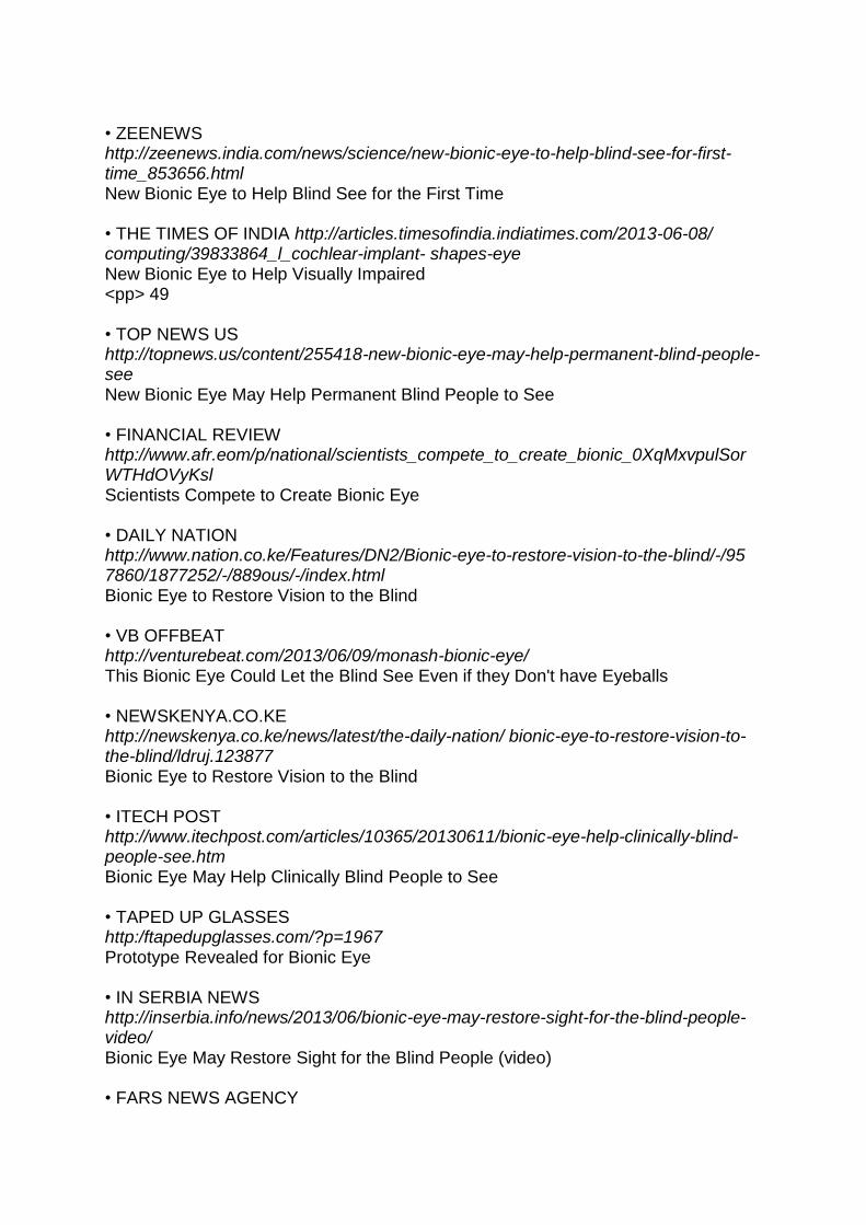
• ZEENEWS http://zeenews.india.com/news/science/new-bionic-eye-to-help-blind-see-for-first-time_853656.html New Bionic Eye to Help Blind See for the First Time • THE TIMES OF INDIA http://articles.timesofindia.indiatimes.com/2013-06-08/ computing/39833864_l_cochlear-implant- shapes-eye New Bionic Eye to Help Visually Impaired <pp> 49 • TOP NEWS US http://topnews.us/content/255418-new-bionic-eye-may-help-permanent-blind-people-see New Bionic Eye May Help Permanent Blind People to See • FINANCIAL REVIEW http://www.afr.eom/p/national/scientists_compete_to_create_bionic_0XqMxvpulSorWTHdOVyKsl Scientists Compete to Create Bionic Eye • DAILY NATION http://www.nation.co.ke/Features/DN2/Bionic-eye-to-restore-vision-to-the-blind/-/95 7860/1877252/-/889ous/-/index.html Bionic Eye to Restore Vision to the Blind • VB OFFBEAT http://venturebeat.com/2013/06/09/monash-bionic-eye/ This Bionic Eye Could Let the Blind See Even if they Don't have Eyeballs • NEWSKENYA.CO.KE http://newskenya.co.ke/news/latest/the-daily-nation/ bionic-eye-to-restore-vision-to-the-blind/ldruj.123877 Bionic Eye to Restore Vision to the Blind • ITECH POST http://www.itechpost.com/articles/10365/20130611/bionic-eye-help-clinically-blind-people-see.htm Bionic Eye May Help Clinically Blind People to See • TAPED UP GLASSES http:/ftapedupglasses.com/?p=1967 Prototype Revealed for Bionic Eye • IN SERBIA NEWS http://inserbia.info/news/2013/06/bionic-eye-may-restore-sight-for-the-blind-people-video/ Bionic Eye May Restore Sight for the Blind People (video) • FARS NEWS AGENCY

http: //english.farsnews.com /newstext.php?nn= 9203181132 Wireless Bionic Eye Being Developed at University • OVERCLOCKERS AUSTRALIA FORUMS http://forums.overclockers.com.au/showthread.php?p=l 5395022 Bionic Eye Prototype Unveiled by Victorian Scientists and Designers • OVERCLOCKERSCLUB http://www.overclockersclub.com/news/34250/ Bionic Eye Built • PC TECH MAGAZINE http://pctechmag.com/2013/06/the-blind-now-have-hope-to-see-bionic-eye/ Blind Now Have Hope to See - Bionic Eye • TG DAILY http://www.tgdaily.com/general-science-brief/72063-wirelss-bionic-eye-being-developed-at-university Wireless Bionic Eye Being Developed at University • 101.5FM RADIO ADELAIDE https://radio.adelaide.edu.au/bionic-eye-brings-new-outlook/ Bionic Eye Brings New Outlook • HERALD SUN LEADER COMMUNITY NEWSPAPERS http://www.heraldsun.com.au/leader/central/monash-university-team-design-wearable-bionic-eye/story-fngnvlpt-122666584195 Monash University Team Design "Wearable" Bionic Eye <pp> 50 • BEHIND THE NEWS - ABC http://www.abc.net.au/btn/story/s3779423.htm Bionic Eye • ABC CLASSIC FM MIDDAY http://www.abc.net.au/classic/content/2013/06/18/3783590.htm Podcast Audio • PSFK http://www.psfk.com/2013/06/bionic-eye-monash-university.html Bionic Eye Could Give Vision to the Blind • BLUESKY DESIGN GROUP http://www.blueskydesigngroup.com.au/news/view/bionic-eye/ Bionic Eye
July

• ENGLISH NEWS http://news.xinhuanet.com/english/sci/2013-07/09/c_132525148.htm Australian Gov't to Add Funds for Research of Bionic Eye • THE AUSTRALIAN, NATIONAL AFFAIRS http://www.theaustralian.com.au/national-affairs/health/rudd-adds-10m-muscle-to-2020-summit-pet-bionic-eye-projects/story-fn59nokw-1226676731858 Rudd Adds $10M Muscle to 2020 Summit Pet Bionic Eye Projects • AUSTRALIAN GOVERNMENT, SENATOR THE HON KIM CARR, MEDIA RELEASE http://minister.innovation.gov.au/honkimcarr/mediareleases/pages/$10mforbionicvisionresearch.aspx $10M for Bionic Vision Research • SHANGHAI DAILY http://www.shanghaidaily.com/article/article_xinhua.asp?id=152355 Australian Gov't to Add Funds for Research of Bionic Eye • 4-TRADERS http://www.4-traders.com/news/Department-of-Industry-Innovation-Climate-Change-$10m-for-Bionic-Vision-research--17076523/ Department of Industry, Innovation, Climate Change: $10M for Bionic Vision Research • MONASH UNIVERSITY http://monash.edu/news/releases/show/bionic-eye-teams-welcome-funding-announcement Bionic Eye Teams Welcome Funding Announcement • BIONICS INSTITUTE http://www.bionicsinstitute.org/news/Pages/Bionic-eye-teams-welcome-funding-announcement.aspx Bionic Eye Teams Welcome Funding Announcement • THE AUSTRALIAN HIGHER EDUCATION http://www.theaustralian.com.au/higher-education/funding-fillip-for-bionic-eye-projects/story-e6frgcjx-1226677234885 Funding Fillip for Bionic Eye Projects • BIONIC VISION AUSTRALIA http://bionicvision.org.au/media/archive/bionic_eye_teams_welcome_funding_ announcement Bionic Eye Teams Welcome Funding Announcement • INVEST VICTORIA http://blog.invest.vic.gov.au/2013/07/09/wearable-bionic-eye-helps-users-avoid-obstructions/ Wearable Bionic Eye Helps Users Avoid Obstructions

• AUSTRALIAN MANUFACTURING, THE LATEST NEWS & INFORMATION http://www.australianmanufacturing.com.au/8619/australian-bionic-vision-researchers-receive-10m-funding-from-rudd-labor-government Australian Bionic Vision Researchers Receive $10M Funding from Rudd Labor Government <pp> 51 • AUSTRALIAN LIFE SCIENTIST http://lifescientist.com.au/content/life-sciences/news/laureate-fellowships-and-bionic-eye-funding-325663497 L aureate Fellowships and Bionic Eye Funding • ENGINEERS AUSTRALIA http://www.engineersaustralia.org.au/news/bionic-vision-research-gets-further-funding Bionic Vision Research Gets Further Funding • THE SYDNEY MORNING HERALD TECHNOLOGY http://www.smh.com.au/technology/sci-tech/lunch-with-mark-armstrong-20130711-2pr18.html Lunch with Mark Armstrong • MONASH UNIVERSITY ENGINEERING http://www.eng.monash.edu.au/news/shownews.php?nid=41&year=2013 Bionic Eye Gains More Funding • NEWS.COM.AU http://www.news.com.au/national-news/queensland/monash-industrial-designers-prepare-for-human-trials-on-australian-directtobrain-bionic-eye/story-fnii5v6w-1226677576197 Monash Industrial Designers Prepare for Human Trials on Australian Direct-To-Brain Bionic Eye • PS NEWS http://www.psnews.com.au/Page_psn3702.html $10M Boost to Bionic Eye • AUSTRALIAN MEDICAL ASSOCIATION https://ama.com.au/ausmed/bionic-vision-within-sight Bionic Vision Within Sight • CENTRE FOR EYE RESEARCH AUSTRALIA http://www.cera.org.au/news-events/overview/news-item?newsItemId=173 Funding Boost for Bionic Eye Research
October • MONASH MAGAZINE

http://www.monash.edu.au/monashmag/articles/ october2013 /bionic-sight-needs-an-eye-catching-look.html#.UtdqjPQW2nY Bionic Sight Needs an Eye-Catching Look
December • MONASH UNIVERSITY FACULTY OF ENGINEERING http://www.eng.monash.edu.au/news/shownews. php?nid=77&year=2013 Bionic Sight Needs an Eye-Catching Look "This project has helped me to obtain skills that I otherwise would not have had the chance to gain in Australia. I consider myself extremely lucky to be part of such a potentially world changing project." Doctor Damien Browne <pp> 52
Journal Publications and Submissions
Elsevier Microelectronics Journal (April 2013), 44(4): 277-282. "A reduced data bandwidth integrated electrode driver for visual intracortical neural stimulation in 0.35μm high voltage CMOS" (Redoute, Browne, Fitrio, Lowery, Kleeman) Materials Research Bull (2013), 5026-5032 "Elastic modulus of rat whiskers - a key biomaterial feature in the rat whisker sensory system" (Kan, Rajan, Fu, Kang, Yan) PLoS ONE (May 2013), 8(5): e63454. "Cortical hypoexcitation defines neuronal responses in the immediate aftermath of traumatic brain injury" (Johnstone, Yan, Alwis, Rajan) PLoS ONE (June 2013), 8(6): e68276. "Relationship between size summation properties, contrast sensitivity and response latency in the dorsomedial and middle temporal areas of the primate extrastriate cortex" (Lui, Bourne, Rosa) Neuroscience (2013), 248:17-29. "Characterising effects of impact velocity and animal biometrics on brain and behaviour in a model of diffuse traumatic axonal injury" (Yan, Johnstone, Alwis, Morganti-Kossmann, Rajan) Brain Behav Evol. (Epub 14 June 2013), 81(4): 203-5. doi: 10.1159/000350241 "Panoptic neuroanatomy: digital microscopy of whole brains and brain-wide circuit mapping" (Mitra, Rosa, Karten)

J. Biomechanics (June 2013), 46: 1987 - 1995 "A truncated conical beam model for analysis of the vibration of rat whiskers" (Yan, Kan, Kergrene, Kang, Feng, Rajan) J. Neuroscience (July 2013), 33(30):12479-89. " Visually-evoked responses in extrastriate area MT following lesions of striate cortex in early life" (Yu, Chaplin, Egan, Reser, Worthy, Rosa) The Lancet Neurology (July 2013), 12(9): 882-893 "Blast-related traumatic brain injury" (Rosenfeld, McFarlane, Bragge, Armonda, Grimes, Ling) Disability and Rehabilitation: Assistive Technology (submitted 29 July 2013). "Cortical visual prostheses and vision impaired persons: Exploring expectations, motivations, concerns and decision-making" (Anderson, Lee, Ackland, Rajan, Warren) J. Neural Eng. (August 2013), 10(4):046010. "Characteristics of electrode impedance and stimulation efficacy of a chronic cortical implant using novel annulus electrodes in rat motor cortex" (Wang, Brunton, Haghgooie, Cassells, Lowery, Rajan) J. Neuroscience (September 2013), 33(88): 15120-15125 "A Conserved Pattern of Differential Expansion of Cortical Areas in Simian Primates" (Chaplin, Yu, Soares, Gattass, Rosa) Visual Neuroscience (Accepted September 2013), Manuscript ID VNS-2013-0015.R1 "Uniformity and diversity of response properties of neurones in the primary visual cortex: selectivity for orientation direction of motion and stimulus size from centre to far periphery" (Yu, Rosa) Health Policy and Technology (submitted 22 September 2013). "Criteria employed by potential recipients considering adopting emerging visual technologies: the case of visual prostheses" (Anderson, Warren, Lee) "It's heartbreaking to read stories from people who have lost their sight - so often they just want to see the ones they love and not feel so isolated. Knowing we are endeavouring to benefit people who are blind strengthens my commitment." Ms Collette Mann <pp> 54
Conference Proceedings
Australian Neuroscience Society 33rd Annual Meeting

(Melbourne, 3-6 February 2013). "Impedance variation and stimulation efficacy of chronic cortical implants in rat motor cortex" (Wang, Brunton, Haghgooie, Cassells, Rajan). DSTO Bionics Workshop - Nanotechnology: Applications and Enabling Technologies (Canberra, 21 February 2013). Oral presentation "Bionic applications in medicine, The Monash Bionic Vision Program and restoration of vision in the blind" (Rosenfeld). Rotary District 9820 Conference (Geelong, 24 February 2013). Keynote speaker "Helping blind people to see again: The Monash Bionic Vision Project" (Rosenfeld). The Technical Cooperation Program TP 12-Combat Casualty Care Meeting International Defence Meeting (Royal Brisbane and Women's Hospital, Brisbane, 14-17 May 2013). Invited speaker "Bionic Vision Research" (Rosenfeld). Defence Advanced Research Projects Agency Neural Interface Symposium (Melbourne, 21 May 2013). Invited speaker "The Monash Vision Group. Developing a Cortical Stimulation Device for Blindness" (Rosenfeld). The 16th Quadrennial Meeting of The World Society for Stereotactic & Functional Neurosurgery (Tokyo, 27-30 May 2013). Platform presentation "Progress in Bionic Vision Devices" (Rosenfeld). 2013 ASM: Visual Neuroscience: Modern Challenges and Australian Pioneers (Sydney, 27-28 June). Oral presentation "Simultaneous mapping of receptive fields of large neuronal populations in extrastriate cortex" (Haghgooie). Australian Medical Association (WA) 7th Annual Clinical Conference (Moscow & St Petersburg, 2-14 August 2013). Oral presentation "Seeing with Eyes Ancient and Modern: A Visit to Some Great Destinations Along the Electromagnetic Spectrum" (Beazley). Chinese Military Neurosurgical Society Meeting (Beijing, 30 August - 2 September 2013). Oral presentation "The development of a bionic vision device at Monash University" (Rosenfeld). The Alfred Longgang Central Hospital Neurosurgery Symposium (Shenzhen, 6-7 September 2013). Oral presentation "The development of a bionic vision device at Monash University" (Rosenfeld). International Symposium in Non-human Primate Cognition, Behaviour and Evolution (Brazil, 8-10 September 2013). Oral presentation "Multielectrode analysis of the topographic organisation and response properties of neurons in the dorsomedial extrastriate cortex" (Rosa). <pp> 55 IEEE International Conference on Image Processing (Melbourne, 15-18 September 2013).

Invited oral presentations "Psychophysics testing of bionic vision image processing algorithms using an FPGA Hatpack" (Josh) "Going beyond vision to improve vision" (Li) "Introducing the Monash Vision Group's Cortical Prosthesis" (Lowery) 6th IGAKUKEN International Symposium on Marmoset Neuroscience (Tokyo, Sept-Oct 2013). Oral presentation "Using multielectrode arrays for simultaneous mapping of receptive fields and response properties of neuronal populations in visual cortex" (Rosa) The Inaugural Frank MacFarlane-Burnet Annual Address - The Gippsland Regional Group of the Order of Australia Association (Victorian Branch) (Traralgon, 30 October 2013). Oral presentation "With an eye to the future - developing a bionic vision device for vision restoration in the blind" (Rosenfeld) Australasian Military Medicine Association (A M M A) - Repat Foundation Inc 2013 Joint Conference Innovations from the Battlefield (1-3 November 2013). Oral presentation "Emerging bionic vision solutions for blinded veterans" (Rosenfeld) IEEE/EMBS Conference on Neural Engineering (San Diego, 5-8 November 2013). Poster presentation "Optimising electrode surface area to minimise power consumption of a penetrating cortical prosthesis" (Brunton, Rajan, Lowery) Neuroscience 2013 (San Diego, 9-13 November 2013) Poster presentations "Body centred, mixed but not hand-centred coding of visual targets in the medial posterior parietal cortex of macaque monkeys during reaches in 3D space" (Hadjidimitrakis, Bertozzi, Breveglieri, Rosa, Galletti, Fattori) "Combined encoding of target and hand position in the medial posterior parietal cortex of macaque monkeys during reaches in 3D space" (Bertozzi, Hadjidimitrakis, Breveglieri, Dal Bo, Bosco, Galletti, Fattori) Medical Bionics - Engineering Solutions for Neural Disorders (Melbourne, 17-20 November 2013) Invited oral presentation "Monash Vision Group's Cortical Visual Prosthesis - The First Three Years" (Lowery) Oral presentation "Implantation of wireless multiple electrode arrays in brain for bionic eye prosthesis" (Yan, Rosenfeld, Cassells, Haghgooie, Rajan, Clarke) Poster presentations
"A single transmitter coil, multiple receiver coil, wireless power and data transmission system for multiple stimulating electrode arrays" (Browne, Mohan, Stephens, Yuce, Redoute, Lowery) "Use of titanium nitride in electrodes in a cortical visual prosthesis" (Brunton, Winther-Jensen, Lowery, Rajan) "Simultaneous mapping of receptive fields and response properties of large neuronal populations in extrastriate cortex" (Haghgooie, Yu, Davies, Price, Rosa) "Psychophysics testing of image processing algorithms for the Monash bionic eye" (Josh, Mann, Kleeman) "A fast and flexible image processing system designed for spatially irregular phosphene patterns" (Li) "Improving the depth perception of visual prostheses users by using depth sensing and spatial audio" (Tang, Li) The 4th Australasian Cognitive Neuroscience Conference (Melbourne, 28 November - 1 December 2013) Poster presentation "The impact of luminance threshold modes and phosphene dropout rates in psychophysics testing for the Monash Vision Group's cortical vision prosthesis Gennaris" (Mann, Josh, Kleeman, Li) IEEE International Conference in Computer Vision (Sydney, 3-6 December 2013). Oral presentation "A wearable computer vision system for a cortical visual prosthesis" (Li) <pp> 56
Financial Statement
Revenue, Expenditure and Contributions: 1 January - 31 December, 2013
Funds Received
A R C funds $2,124,475
Monash University funds $600,000
Total Revenue $2,724,475
A R C carry forwards from 2012 $407,037
Monash University carry forwards from 2012 $0
Total funds available in 2013 $3,131,512

Expenditure
A R C funds $2,030,939
Monash University funds $600,000
Total Expenditure $2,630,939
Balance Remaining $500,573
A R C funds to be carried forward to 2014 $500,573
In-Kind Contributions
Monash University $2,188,244
Alfred Health $19,130
Grey Innovation $171,413
MiniFAB $182,241
Total In-Kind Contributions $2,561,028
<pp> 57
Key Performance Indicators
Key Result Area Performance Measure 2013 Target Result
Governance Breadth/experience of Advisory Board Frequency/Effectiveness of Meetings Quality of strategic plan (judged by Advisory Board)
Reviewed 1
2 2
Reviewed 1
Adequacy of KPIs (judged by Advisory Board and A R C)
Reviewed 1
Recruitment of new staff as a result of project
Research and Development Staff 0 0
Technical Support Staff 0 0
Administrative Staff 0 0
Skills Development
Undergraduate Projects 8 7
Postgraduate students recruited 2 2
Postdoctoral researchers recruited 0 0
Industry secondment (to and from industry) 2 2
Visit to international facilities 2 2
Research Outputs
Publications in journals that are ranked in the top 25% of their field
12 12
International conferences (peer reviewed) 8 12

Invited review papers and invited conference presentations
4 15
Patent applications 4 3
International visitors staying more than two weeks
2 0
Outreach and Communication
Webpage developed N/A N/A
Press releases 4 3
Press articles 8 59
Media appearances 4 9
Expressions of interest in future human trials from sight-impaired people
10 >100
Internal task/issue tracking/communication Report annually
1
External stakeholder communication Report annually
1
Meetings with related research teams 2 4
Annual report 1 1
Lectures to students 8 15
Collaboration Meetings with Bionic Vision Australia group 2 14
Commercial Annual in-kind and cash contributions met 1 1
Commercialisation plan 0 1
Funding strategy 0 1
Funds raised 1 1
Autonomous business entity 1 1
<pp> 58
Acknowledgements
MVG gratefully acknowledges the support of the following organisations and individuals: • Alfred Health and The Alfred Hospital • Australian Research Council • Bionic Vision Australia and Professor David Penington • Blind Citizens Australia • Cochlear Ltd • Glaucoma Australia - Mr Geoff Pollard • Grey Innovation • Guide Dogs Victoria - Ms Karen Hayes and Mr Sandro Cirianni • MiniFAB • Monash University - Central Administration and Monash Research Office

- Industry Engagement and Commercialisation - Faculty of Engineering and Department of Electrical and Computer Systems Engineering - Faculty of Health, Medicine and Nursing Sciences - Faculty of Science - Monash Art, Design and Architecture • Mr Gerard Menses • Ms Vicki Tutungi • Professor David de Kretser A C • Professor Iain Clarke • Professor James Patrick • Professor Lyn Beazley • Professor Peter Seligmann • Retina Australia - Mr Leighton Boyd and Mrs Rosemary Boyd • Vision Australia - Doctor Kevin Murfitt, Mr David Speyer and Mr Jim Luscombe "The MVG project is truly making extraordinary progress towards meeting our goals. The outcome will be a landmark achievement and substantially improve the quality of life for people with vision impairment." Doctor Jerome Maller. Published by the Monash Vision Group and the Australian Research Council. This e-text version was produced by Vision Australia. Ph: 1300 3 6 7 0 5 5 Email: [email protected] Although every effort was made during the transcription process to ensure an accurate representation of the original document, Vision Australia does not accept responsibility for any errors or omissions, nor for the results of specific action taken on the basis of this transcription. The original document should always remain the point of reference for content accuracy.