Molecularlydeterminedtotaltumourloadinlymphnodesofstage I ... · touch-prep was performed to...
10
ORIGINAL ARTICLE Molecularly determined total tumour load in lymph nodes of stage I–II colon cancer patients correlates with high-risk factors. A multicentre prospective study Iban Aldecoa 1 & Begoña Atares 2 & Jordi Tarragona 3 & Laia Bernet 4 & Jose Domingo Sardon 5 & Teresa Pereda 6 & Carlos Villar 7 & M. Carmen Mendez 8 & Elvira Gonzalez-Obeso 8 & Kepa Elorriaga 9 & Guadalupe Lopez Alonso 10 & Javier Zamora 11 & Nuria Planell 12 & Jose Palacios 13 & Antoni Castells 14 & Xavier Matias-Guiu 3 & Miriam Cuatrecasas 1,15 Received: 6 April 2016 /Revised: 17 June 2016 /Accepted: 7 July 2016 /Published online: 22 July 2016 # The Author(s) 2016. This article is published with open access at Springerlink.com Abstract Stage I–II (pN0) colorectal cancer patients are sur- gically treated although up to 25 % will eventually die from disease recurrence. Lymph node (LN) status is an independent prognostic factor in colorectal cancer (CRC), and molecular tumour detection in LN of early-stage CRC patients is associ- ated with an increased risk of disease recurrence and poor survival. This prospective multicentre study aimed to deter- mine the relationship between LN molecular tumour burden and conventional high-risk factors in stage I–II colon cancer patients. A total of 1940 LN from 149 pathologically assessed pN0 colon cancer patients were analysed for the amount of tumour cytokeratin 19 (CK19) messenger RNA (mRNA) with the quantitative reverse transcription loop-mediated isother- mal amplification molecular assay One-Step Nucleic Acid Amplification. Patient’ s total tumour load (TTL) resulted from the sum of all CK19 mRNA tumour copies/μL of each posi- tive LN from the colectomy specimen. A median of 15 LN were procured per case (IQR 12;20). Molecular positivity cor- related with high-grade (p < 0.01), mucinous/signet ring type (p = 0.017), male gender (p = 0.02), number of collected LN (p = 0.012) and total LN weight per case (p < 0.01). The TTL was related to pT stage (p = 0.01) and tumour size (p < 0.01) in low-grade tumours. Multivariate logistic regression showed independent correlation of molecular positivity with gender, Previous presentations This work presented in part as an oral platform at the USCAP (United States and Canadian Association of Pathologists) 103rd Annual Meeting, San Diego, CA. March 1–7, 2014. Electronic supplementary material The online version of this article (doi:10.1007/s00428-016-1990-1) contains supplementary material, which is available to authorized users. * Miriam Cuatrecasas [email protected] 1 Pathology Department, Centre de Diagnòstic Biomèdic (CDB), Hospital Clínic, University of Barcelona, Escala 3, Planta 5. Villarroel 170, Barcelona 08036, Spain 2 Pathology Department, Alava University Hospital, Vitoria-Gasteiz, Spain 3 Pathology Department, Hospital Arnau de Vilanova, Lleida, Spain 4 Pathology Department, Hospital L. Alcanyis, Xativa, Spain 5 Surgery Department, Alava University Hospital, Txagorritxu, Spain 6 Pathology Department, Hospital Costa del Sol, Marbella, Spain 7 Pathology Department, Hospital Reina Sofia, Cordoba, Spain 8 Pathology Department, Hospital Severo Ochoa, Leganes, Madrid, Spain 9 Pathology Department, Hospital Onkologikoa, San Sebastian, Spain 10 Pathology Department, Hospital 12 Octubre, Madrid, Spain 11 Biostatistic Unit, Hospital Ramon y Cajal, Madrid, Spain 12 Gastroenterology Department and Bioinformatics Unit, CIBERehd, IDIBAPS, Hospital Clinic, University of Barcelona, Barcelona, Spain 13 Pathology Department, Hospital Ramon y Cajal, Madrid, Spain 14 Gastroenterology Department, Hospital Clinic, University of Barcelona, IDIBAPS, CIBERehd, Barcelona, Spain 15 CIBERehd, and Banc de Tumors-Biobanc Clinic-IDIBAPS-XBTC, Hospital Clinic, Barcelona, Spain Virchows Arch (2016) 469:385–394 DOI 10.1007/s00428-016-1990-1
Transcript of Molecularlydeterminedtotaltumourloadinlymphnodesofstage I ... · touch-prep was performed to...

ORIGINAL ARTICLE
Molecularly determined total tumour load in lymph nodes of stageI–II colon cancer patients correlates with high-riskfactors. A multicentre prospective study
Iban Aldecoa1 & Begoña Atares2 & Jordi Tarragona3 & Laia Bernet4 &
Jose Domingo Sardon5& Teresa Pereda6 & Carlos Villar7 & M. Carmen Mendez8 &
Elvira Gonzalez-Obeso8 & Kepa Elorriaga9 & Guadalupe Lopez Alonso10 &
Javier Zamora11 & Nuria Planell12 & Jose Palacios13 & Antoni Castells14 &
Xavier Matias-Guiu3& Miriam Cuatrecasas1,15
Received: 6 April 2016 /Revised: 17 June 2016 /Accepted: 7 July 2016 /Published online: 22 July 2016# The Author(s) 2016. This article is published with open access at Springerlink.com
Abstract Stage I–II (pN0) colorectal cancer patients are sur-gically treated although up to 25 % will eventually die fromdisease recurrence. Lymph node (LN) status is an independentprognostic factor in colorectal cancer (CRC), and moleculartumour detection in LN of early-stage CRC patients is associ-ated with an increased risk of disease recurrence and poorsurvival. This prospective multicentre study aimed to deter-mine the relationship between LN molecular tumour burdenand conventional high-risk factors in stage I–II colon cancerpatients. A total of 1940 LN from 149 pathologically assessedpN0 colon cancer patients were analysed for the amount oftumour cytokeratin 19 (CK19) messenger RNA (mRNA) with
the quantitative reverse transcription loop-mediated isother-mal amplification molecular assay One-Step Nucleic AcidAmplification. Patient’s total tumour load (TTL) resulted fromthe sum of all CK19 mRNA tumour copies/μL of each posi-tive LN from the colectomy specimen. A median of 15 LNwere procured per case (IQR 12;20). Molecular positivity cor-related with high-grade (p < 0.01), mucinous/signet ring type(p = 0.017), male gender (p = 0.02), number of collected LN(p = 0.012) and total LN weight per case (p < 0.01). The TTLwas related to pTstage (p = 0.01) and tumour size (p < 0.01) inlow-grade tumours. Multivariate logistic regression showedindependent correlation of molecular positivity with gender,
Previous presentations This work presented in part as an oral platformat the USCAP (United States and Canadian Association of Pathologists)103rd Annual Meeting, San Diego, CA. March 1–7, 2014.
Electronic supplementary material The online version of this article(doi:10.1007/s00428-016-1990-1) contains supplementary material,which is available to authorized users.
* Miriam [email protected]
1 Pathology Department, Centre de Diagnòstic Biomèdic (CDB),Hospital Clínic, University of Barcelona, Escala 3, Planta 5.Villarroel 170, Barcelona 08036, Spain
2 Pathology Department, Alava University Hospital,Vitoria-Gasteiz, Spain
3 Pathology Department, Hospital Arnau de Vilanova, Lleida, Spain4 Pathology Department, Hospital L. Alcanyis, Xativa, Spain5 Surgery Department, Alava University Hospital, Txagorritxu, Spain6 Pathology Department, Hospital Costa del Sol, Marbella, Spain7 Pathology Department, Hospital Reina Sofia, Cordoba, Spain
8 Pathology Department, Hospital Severo Ochoa, Leganes,Madrid, Spain
9 Pathology Department, Hospital Onkologikoa, San Sebastian, Spain10 Pathology Department, Hospital 12 Octubre, Madrid, Spain11 Biostatistic Unit, Hospital Ramon y Cajal, Madrid, Spain12 Gastroenterology Department and Bioinformatics Unit, CIBERehd,
IDIBAPS, Hospital Clinic, University of Barcelona,Barcelona, Spain
13 Pathology Department, Hospital Ramon y Cajal, Madrid, Spain14 Gastroenterology Department, Hospital Clinic, University of
Barcelona, IDIBAPS, CIBERehd, Barcelona, Spain15 CIBERehd, and Banc de Tumors-Biobanc Clinic-IDIBAPS-XBTC,
Hospital Clinic, Barcelona, Spain
Virchows Arch (2016) 469:385–394DOI 10.1007/s00428-016-1990-1

tumour grade and number of fresh LN [AUC = 0.71 (95 %CI = 0.62–0.79)]. Our results show that lymph node CK19mRNA detection correlates with classical high-risk factors instage I–II colon cancer patients. Total tumour load is a quan-titative and objective measure that may help to better stageearly colon cancer patients.
Keywords Colorectal neoplasms . Neoplasm staging .
Molecular pathology . Lymph nodes . Cytokeratin 19
Introduction
Surgical resection with no adjuvant therapy is recommendedfor most stage I–II colorectal cancer (CRC) patients, exceptfor selected high-risk stage II patients given the significantimpact of chemotherapy on stage III disease [1, 2]. Althoughthere is evidence that pathological nodal staging is far frombeing optimal, current NCCN guidelines are based onhaematoxylin and eosin (HE) lymph node (LN) staging,[3–7]. Its major weakness is the limited scope of histologicalLN analysis, based on a small sample provided by 2–5 μmLNsections, which comprise less than 0.5 % of the entire LN, andit may lead to false negative diagnoses [3, 7–9]. This maypartly explain why up to 25 % of CRC patients with histolog-ically negative LN die from recurrent disease after a potential-ly curative surgical resection. Some of these patients may havehad undetected LN metastases [3, 10, 11].
The use of additional techniques, i.e. immunohistochemis-try (IHC) or reverse transcriptase polymerase chain reaction,makes it possible to find LN tumour burden not detected withconventional HE analysis in 25 to 50 % of CRC patients, dueto both increased sensitivity and the more extensive study thanusually permitted by histological sections [3, 6, 7, 10–17].
Although the prognostic value of LN molecular tumourdetection in early-stage CRC is controversial [14–18], thereare enough data to support the use of more sensitive (i.e.molecular) methods of LN staging. As stated in three meta-analyses, the molecular detection of tumour cells in regionalLN of stage I–II CRC patients is associated with an increasedrisk of disease recurrence and poor survival [3, 10, 11].
Most studies have focused in dichotomic (positive-negative) or semi-quantitative scales (isolated tumour cells(ITC), micro- and macrometastases) to assess molecular re-sults [8, 10, 11, 14, 15, 17, 19, 20]. The molecular assay One-Step Nucleic Acid Amplification (OSNA; SysmexCorporation, Kobe, Japan) is a quantitative method whichanalyses the entire LN. It amplifies cytokeratin 19 (CK19)mRNA from LN tissue lysates using the reverse transcriptionloop-mediated isothermal amplification (RT-LAMP) method[21]. CK19 mRNA was selected among other CRC markersshowing the highest diagnostic performance and reproducibil-ity, with 94.9–95.2 % sensitivity and 97.7–97.9 % specificity
[22]. The system has been validated for breast and colon can-cer LN assessment, providing results comparable to extensivehistological and IHC LN analysis [8, 9, 21, 23–28]. Theamount of CK19 mRNA detected correlates with the size ofthe metastatic foci [8, 9, 21, 27, 29]. It alsomakes it possible tocalculate the total tumour load (TTL) present in a given pa-tient, by adding all CK19 mRNA copies from each positiveLN of a colectomy specimen [27, 28].
In this multicentre prospective study, we tried to correlatethe TTL, as determined by OSNA, with classical clinical andpathologic high-risk factors, in an effort to determine whetherthe TTL could be used as an additional factor to better selectstage I–II patients at risk of recurrence. Such an approach isnow widely used in the treatment of breast cancer [27, 30, 31].
Materials and methods
Study sample
This is a prospective observational study including 10 institu-tions. Inclusion criteria were patients over 18 years old, withprimary histologically confirmed colon cancer, cN0 preoper-ative diagnosis and positive CK19 IHC of the primary tumour.Exclusion criteria included rectal tumours, non-invasive pTisand pT0 tumours, positive LN on HE, synchronous tumoursor other malignancies, cN1, gross adipose tissue involvementby the tumour, metastatic cancer, neo-adjuvant chemotherapy,familial adenomatous polyposis, carcinomas on inflammatorybowel disease and presence of stent-type intraluminal devices.
Study procedure
Sample processing and fresh lymph node procuring
Fresh LN procurement from the mesocolon fat was performedwithin 50 min after surgical excision. When immediate LNdissection was not possible, the surgical specimen was kept upto 3 h in the refrigerator at 4 °C until LN dissection was done.During the LN harvesting process, the dissection area waskept cold by putting a thick layer of chopped ice under anelevated metallic surface and covered with a clean filter paperfor LN dissection (Figure available at Online Resource 1a).Microcentrifuge tubes were also kept cold by punching themin chopped ice (Figure 2 available at Online Resource 1b). Wefirst detached the mesocolon fat from the colon wall with asurgical blade. pT4 tumours corresponded to antimesentericserous tumour infiltration, and pT3 tumours corresponded on-ly to specimens with minimal tumour infiltration of themesocolon. We then dissected one by one all LN from themesocolon fat using different clean areas of the surgical bladefor small LN, or changing it after each LN dissection. When aLN was grossly suspicious of being positive, a cytology
386 Virchows Arch (2016) 469:385–394

touch-prep was performed to confirm or discard metastasis.Positive cases were discarded. All freshly dissected LN wereanalysed by both methods, HE and OSNA, using a modifiedprotocol from previous studies [8, 9, 21]. Each LN was num-bered and cut along the long axis. A central 1-mm slice wassubmitted for conventional formalin-fixation paraffin-embed-ding (FFPE) and HE analysis. The rest of the LNwas stored at−80 °C in microcentrifuge tubes for 1 to 7 days until deferredOSNA analysis was performed. Lymph nodes with weight≤0.07 g (average 5.5 mm) were defined as small.
After fresh LN harvesting, the specimen was fixed over-night in 10 % neutral buffered formalin. Then, the mesocolonfat was re-examined for remaining LNs, which were submit-ted only for conventional histopathology analysis.
CK19 immunohistochemistry
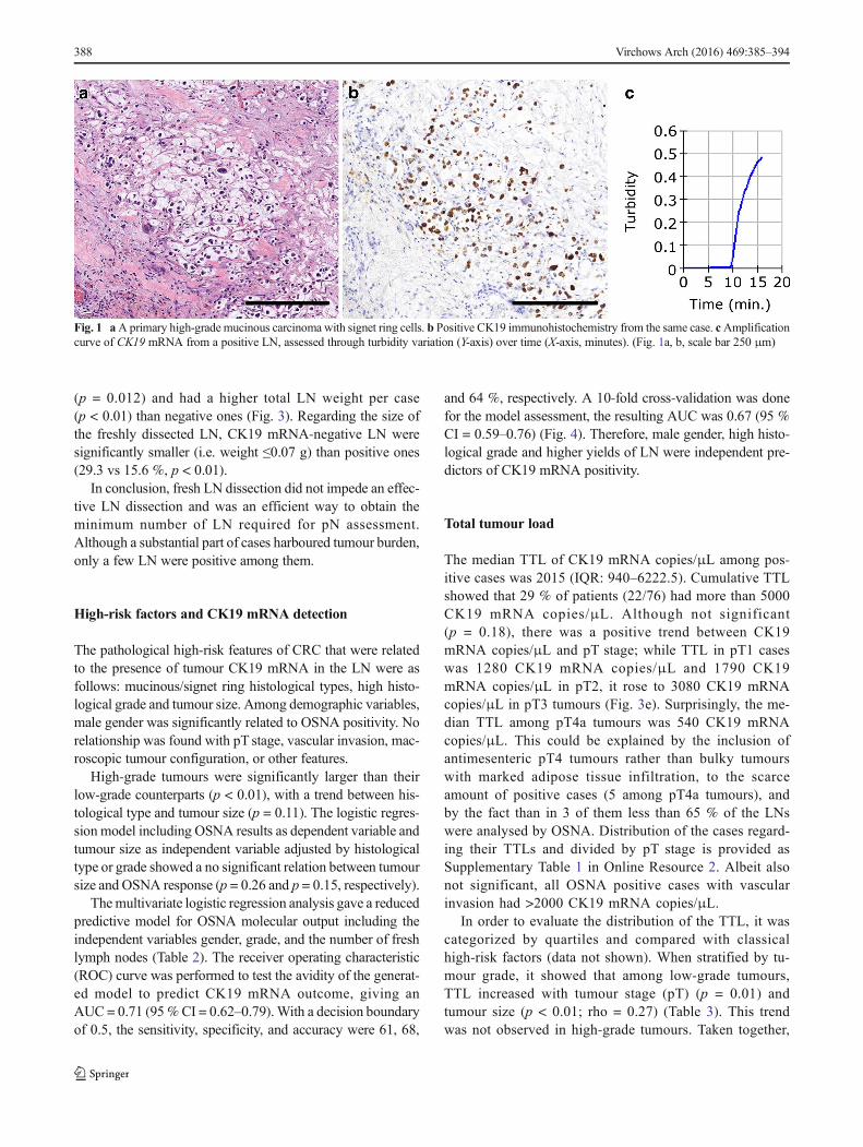
CK19 IHCwas performed on representative sections of all theprimary colon carcinomas to ensure reliable negative molec-ular CK19 mRNA results. A 2-μm section of each primarytumour was mounted on FLEX IHC microscope slides andpre-treated in PT-LINK (Dako, Glostrup, Denmark).Incubation for 20 min with the primary CK19 antibody(CK19 mouse monoclonal, clone RCK108; IR615 pre-dilut-ed. Dako) was performed in the AutostainerLink 48 (Dako).Membranous staining with or without cytoplasm staining of≥10 % of the tumour cells was defined as positive IHC incolon carcinomas (Fig. 1a, b).
Pathology report and LN staging
LN staging and pathology report were performed from theanalysis of HE stains according to the AJCC/UICC TNM,7th edition [32, 33]. Tumours ≥4 cm were defined aslarge. Pathologists and clinicians were both blinded tothe OSNA results.
OSNA procedure
The OSNA method was performed at each institution follow-ing the manufacturer’s instructions, using the protocol de-scribed by Tsujimoto et al. [21]. Briefly, LNs were weighed,homogenized with the lysis buffer Lynorhag (Sysmex) formRNA stabilization and genomic DNA precipitation. Aftercentrifugation for 1 min at 10,000 × g at room temperature;a 2-μL sample of the intermediate phase was mixed with thereagent LynoampBC (Sysmex). Analysis was performedusing the RT-LAMP isothermal amplification method withthe RD-100i automated gene-amplification system(Sysmex). The amount of CK19mRNA amplified was detect-ed by a change in turbidity upon precipitation of magnesiumpyrophosphate (Fig. 1c). The result was correlated to CK19mRNA copy number/μL of the original lysate through
calibrated standard curves containing different CK19 mRNAconcentrations. In every assay, a standard positive controlsample containing 5 × 10³ copies/μL of CK19 mRNA, anda negative control sample without CK19mRNAwere used forvalidation. The results were based on the number of CK19mRNA copies/μL obtained for each LN. The cutoff valuewas 250 copies/μL, based on a previous study [9].
Statistical analysis
The Fisher exact test and Pearson’s correlation coefficientwere used for testing the association between categorical ornumerical variables, respectively. The Mann-Whitney-Wilcoxon and Kruskal-Wallis test were applied to comparegroups’ distributions. A p < 0.05 was considered statisticallysignificant.
Logistic regression analysis was used to predict the OSNAoutcome. The backward stepwise algorithm was used to de-termine the best-fitting model. The classification avidity of themodel was assessed by the ROC curve and a 10-fold cross-validation technique was applied for themodel validation. TheCohen’s kappa was used to assess the degree of agreementbetween OSNA outcome and model prediction. All analyseswere performed using R statistical environment (V.3.1.0) [34].
Results
Sample size and characteristics
We analysed 3512 LN from 211 colon cancer patients recruit-ed in 10 hospitals between June 2012 and December 2013.Weexcluded 27 non-invasive tumours (9 pT0 and 18 pTis), 34cases with HE-positive LN, and one patient with synchronoustumours. Finally, 149 stage I–II colon cancer patients met thestudy selection criteria. All primary tumours showed positiv-ity for CK19 IHC. Demographic, clinical, pathological, andlymph node characteristics of the study sample and correlationwith CK19 mRNA results are summarized in Table 1 and thestudy flow diagram (Fig. 2).
Lymph node features and CK19 mRNA results
Among the 2483 LN procured from 149 cases, slightly over78 % of them were freshly isolated and submitted for OSNAanalysis (1940 fresh LN vs 543 FFPE LN). Hence, we obtain-ed a median of 15 LN per case, of which 12 LN were analysedby OSNA and HE.
CK19 mRNA positivity was observed in 76/149 patients(51 %). Most of those positive cases; i.e. 80%, had only 1 to 3positive LN. Thus, among all OSNA LN analysed, 9,8 %(190/1940) were positive. Association analysis showed thatOSNA-positive cases harboured more freshly procured LN
Virchows Arch (2016) 469:385–394 387
(p = 0.012) and had a higher total LN weight per case(p < 0.01) than negative ones (Fig. 3). Regarding the size ofthe freshly dissected LN, CK19 mRNA-negative LN weresignificantly smaller (i.e. weight ≤0.07 g) than positive ones(29.3 vs 15.6 %, p < 0.01).
In conclusion, fresh LN dissection did not impede an effec-tive LN dissection and was an efficient way to obtain theminimum number of LN required for pN assessment.Although a substantial part of cases harboured tumour burden,only a few LN were positive among them.
High-risk factors and CK19 mRNA detection
The pathological high-risk features of CRC that were relatedto the presence of tumour CK19 mRNA in the LN were asfollows: mucinous/signet ring histological types, high histo-logical grade and tumour size. Among demographic variables,male gender was significantly related to OSNA positivity. Norelationship was found with pT stage, vascular invasion, mac-roscopic tumour configuration, or other features.
High-grade tumours were significantly larger than theirlow-grade counterparts (p < 0.01), with a trend between his-tological type and tumour size (p = 0.11). The logistic regres-sion model including OSNA results as dependent variable andtumour size as independent variable adjusted by histologicaltype or grade showed a no significant relation between tumoursize and OSNA response (p = 0.26 and p = 0.15, respectively).
Themultivariate logistic regression analysis gave a reducedpredictive model for OSNA molecular output including theindependent variables gender, grade, and the number of freshlymph nodes (Table 2). The receiver operating characteristic(ROC) curve was performed to test the avidity of the generat-ed model to predict CK19 mRNA outcome, giving anAUC = 0.71 (95%CI = 0.62–0.79).With a decision boundaryof 0.5, the sensitivity, specificity, and accuracy were 61, 68,
and 64 %, respectively. A 10-fold cross-validation was donefor the model assessment, the resulting AUC was 0.67 (95 %CI = 0.59–0.76) (Fig. 4). Therefore, male gender, high histo-logical grade and higher yields of LN were independent pre-dictors of CK19 mRNA positivity.
Total tumour load
The median TTL of CK19 mRNA copies/μL among pos-itive cases was 2015 (IQR: 940–6222.5). Cumulative TTLshowed that 29 % of patients (22/76) had more than 5000CK19 mRNA copies/μL. Although not significant(p = 0.18), there was a positive trend between CK19mRNA copies/μL and pT stage; while TTL in pT1 caseswas 1280 CK19 mRNA copies/μL and 1790 CK19mRNA copies/μL in pT2, it rose to 3080 CK19 mRNAcopies/μL in pT3 tumours (Fig. 3e). Surprisingly, the me-dian TTL among pT4a tumours was 540 CK19 mRNAcopies/μL. This could be explained by the inclusion ofantimesenteric pT4 tumours rather than bulky tumourswith marked adipose tissue infiltration, to the scarceamount of positive cases (5 among pT4a tumours), andby the fact than in 3 of them less than 65 % of the LNswere analysed by OSNA. Distribution of the cases regard-ing their TTLs and divided by pT stage is provided asSupplementary Table 1 in Online Resource 2. Albeit alsonot significant, all OSNA positive cases with vascularinvasion had >2000 CK19 mRNA copies/μL.
In order to evaluate the distribution of the TTL, it wascategorized by quartiles and compared with classicalhigh-risk factors (data not shown). When stratified by tu-mour grade, it showed that among low-grade tumours,TTL increased with tumour stage (pT) (p = 0.01) andtumour size (p < 0.01; rho = 0.27) (Table 3). This trendwas not observed in high-grade tumours. Taken together,
388 Virchows Arch (2016) 469:385–394
Fig. 1 aA primary high-grade mucinous carcinoma with signet ring cells. b Positive CK19 immunohistochemistry from the same case. cAmplificationcurve of CK19 mRNA from a positive LN, assessed through turbidity variation (Y-axis) over time (X-axis, minutes). (Fig. 1a, b, scale bar 250 μm)

our results show that most patients had low TTLs. Thesevalues increased with tumour stage and followed differentdistributions among low- and high-grade tumours.
Patient’s follow-up
At the time of writing of this manuscript, all patients hada follow-up of 2 years (median 33 months, IQR 25–32.5);8.7 % (13/149) patients recurred (3 died and 10 were alivewith metastatic disease). Four of the cases were OSNA
positive, with a median TTL of 4375 CK19 mRNAcopies/μL (range 360–47,600); 2 of the 3 dead patientswere OSNA positive.
Discussion
This study quantifies the amount of total tumour load withinthe lymph nodes of stage I–II colon cancer patients, and sug-gests that LN molecular detection of CK19 mRNA could
Table 1 Patient’s demographics and specimen characteristics and correlation with CK19 mRNA results
Clinical parameter n (%) CK19 mRNAnegativen (%)
CK19 mRNApositiven (%)
p value
Cases 149 (100) 73 (49.0) 76 (51.0) –
Gender 0.02
Male 97 (65.1) 41 (42.3) 56 (57.7)
Female 52 (34.9) 32 (61.5) 20 (38.5)
Age (years)—median (IQR) 67 (61–75) 68 (61–74) 66 (61–75) 0.89
Surgical specimen characteristics
Specimen size (cm)—median (IQR) 20 (15–25) 20 (15–25) 19.5 (15–25) 0.81
Tumour size (cm)a—median (IQR) 3 (2–5) 3 (2–4) 4 (2–5.5) 0.045
Large tumours (>4 cm) 39 (26.7) 14 (38.9) 25 (64.1) 0.09
Small tumours (≤4 cm) 107 (73.3) 57 (53.3) 50 (46.7)
Tumour location 0.33
Right colon and caecum (incl. hepatic flexure) 67 (45) 37 (55.2) 30 (44.8)
Transverse colon 14 (9.4) 5 (35.7) 9 (64.3)
Left colon and sigmoid colon (incl. splenic flexure) 68 (45.6) 31 (45.6) 37 (54.4)
Macroscopic tumour configuration 0.57
Annular 8 (5.4) 5 (62.5) 3 (37.5)
Ulcerated 65 (43.6) 33 (50.8) 32 (49.2)
Polypoid 75 (50.3) 34 (45.3) 41 (54.7)
Other 1 (0.7) 1 (100) 0 (0)
Vascular invasion 0.45
No 137 (91.9) 65 (47.4) 72 (52.6)
Yes 12 (8.1) 8 (66.7) 4 (33.3)
Histological type 0.017
Adenocarcinoma 136 (91.3) 71 (52.2) 65 (47.8)
Mucinous/signet ring cell AC 13 (8.7) 2 (15.4) 11 (84.6)
Gradeb <0.01
High grade 22 (15.0) 4 (18.2) 18 (81.8)
Low grade 125 (85.0) 69 (55.2) 56 (44.8)
pT 0.19
pT1 40 (26.8) 17 (42.5) 23 (57.5)
pT2 31 (20.8) 20 (64.5) 11 (35.5)
pT3 66 (44.3) 29 (43.9) 37 (56.1)
pT4a 12 (8.1) 7 (58.3) 5 (41.7)
IQR interquartile range, AC adenocarcinomaa In three patients, tumour size could not be assessedb In two patients, tumour grade could not be assessed
Virchows Arch (2016) 469:385–394 389

become an objective indicator of risk in such individuals [11,20, 28, 35]. Stage I–II CRC is amenable to complete surgicalresection, but patients at risk of tumour recurrence are
clinically and pathologically difficult to identify [36, 37]. Inaddition, cancer dissemination to LNmay occur at early stagesof tumour development [6, 38], potentially implying a poor
Fig. 3 OSNA results and correlation with LN retrieval and pT stage,regarding number of LN retrieved (a), time spent on fresh LN search inminutes (b) and total weight of fresh LN per case (gr) (c). OSNA positivecases held significantly more LN due to a higher fresh LN yields. d
Shows that most cases in every pT stage held up to 3 OSNA positiveLN. e Although not significant, a trend was observed between pT stageand TTL
Fig. 2 Study flow diagram
390 Virchows Arch (2016) 469:385–394

prognostic impact [3, 7, 11, 15]. Thus, the assessment of thenodal staging arises as a key factor for CRC therapeutic man-agement [3, 10, 11].
Our findings confirm the presence of undetected nodal tu-mour burden in early-stage colon cancer using the OSNAmethod. Compared to previous studies, we have the highestpercentage of cases with CK19 mRNA detection in LN (i.e.51%) [23–25, 28]. Discrepancies may be due to differences inthe LN collection and analysis, such as the fact that weanalysed a larger amount of LN, including all evaluable LNby OSNA regardless of its size. Current guidelines recom-mend evaluating at least 12 LN to achieve a reliable histolog-ical staging [16, 32, 33, 39–42]. In compliance with them, weobtained a median of 15 LN per patient, of which 12 werefreshly obtained and analysed by OSNA.
CRC lymph nodes are usually small, especially in earlystages of the disease, but may still contain tumour burden, assize is not a good preoperative indicator of LN staging, or apredictor of the presence of tumour [43–45]. Although weidentified a significant association between CK19 mRNA de-tection and larger LN size, we found CK19 mRNA in 15.6 %of small LN. Furthermore, the multivariate analysis of OSNAresults showed that the number of collected LN, the genderand the histologic grade were independent predictors ofOSNA results. Our data highlights the importance of procur-ing any identifiable LN irrespective of its size [40–42, 45–47].
Our results show that the presence of CK19 mRNA inregional LN was associated with other classical high-risk fac-tors such as mucinous/signet ring types, histologic high-grade,tumour size and male gender. Moreover, it is noteworthy thetrend observed between TTL and pT stage, as pT3 had thehighest TTL (3080 CK19 mRNA copies/μL) and rate ofCK19 mRNA detection (37/66).
We also observed a different behaviour between low andhigh-grade tumours, with a significant orderly increase ofTTL in regional LN with the increase of pT stage and tumoursize in low-grade tumours. This phenomenon may beaccounted for by the fact that low-grade tumours tend to fol-low an orderly sequence of accumulation of molecular alter-ations as they grow and acquire infiltrative proprieties [48]. Incontrast, no association was found with any assessed variablesin high-grade tumours. This may be explained by the differentmolecular pathways involved in high-grade tumours and theiraggressive behaviour from the beginning [48]. Reinforcingthe importance of tumour grade in neoplastic LN spread, tu-mour size did not correlate with CK19mRNA positivity whenstratified by grade or histological type.
The biologic significance of LN molecular tumour detec-tion is of leading importance. Although the sole presence ofsmall amounts of tumour cells in regional LN does not neces-sarily imply poor prognosis, it may indicate a yet undefinedrisk of disease recurrence. This would explain why, with
Table 2 Logistic regression of clinical and histological variables related to CK19 mRNA positivity
Variables Univariate model Multivariate model
OR (95 % CI) Wald test p OR (95 % CI) Wald test p
Gender (male vs female) 2.2 (1.1–4.4) 0.026 3.1 (1.4–7.0) 0.006
Age (years) 0.99 (0.96–1.03) 0.74 – –
Tumour size 1.2 (1.0–1.4) 0.06 – –
Tumour location 0.31 – –
Transverse colon vs right colon and caecum (including hepatic flexure) 2.2 (0.7–7.9)
Left colon and sigmoid colon (including splenic flexure) vs Right colonand caecum (including hepatic flexure)
1.4 (0.7–2.9)
Macroscopic tumour configuration 0.59 – –
Ulcerated vs annular 1.6 (0.4–8.4)
Polypoid vs annular 2.0 (0.5–10.4)
Histological type (mucinous / signet ring cell AC vs adenocarcinoma) 0.02 – –
6.0 (1.5–39.8)
Vascular invasion (yes vs no) 0.5 (0.1–1.5) 0.21 – –
Grade (high vs low) 5.4 (1.9–19.5) <0.01 4.8 (1.5–18.9) 0.013
pT 0.20 – –
pT2 vs pT1 0.4 (0.2–1.1)
pT3 vs pT1 0.9 (0.4–2.1)
pT4a vs pT1 0.5 (0.1–1.9)
Fresh LN per case weight (gr) 1.1 (1.0–1.1) <0.01 1.1 (1.0–1.2) 0.017
AC adenocarcinoma, gr grammes
Virchows Arch (2016) 469:385–394 391

historical recurrence records of 2.5 % in pT1 N0 tumours, wehave found that almost 60 % had some CK19 mRNA tumourburden in their LN [11, 49]. In this context, low TTLs mayhave no further biologic consequences, but large amounts oftumour burden within the LN may imply higher odds of re-currence [7, 27]. A study using RT-qPCR detection ofGuanylyl cyclase C (GUCY2C) by RT-qPCR found 87.5 %patients with positive LN, although only 20.9 % developedrecurrent disease [7].
Quantifying the amount of tumour burden, not just thepresence or absence of tumour, in regional lymph nodes istherefore important in prognosticating from molecular results,as has been stated in CRC [3, 7, 11, 20, 35] and breast cancer[27, 31]. It should be highlighted that whereas the medianTTL of our cohort was 2015 CK19 mRNA copies/μL, valuesup to 15,000 CK19 mRNA copies/μL have been recently setin breast cancer sentinel LN as clinically relevant to predictadditional axillary LN metastases [27]. Some authors havealso demonstrated the relationship between the amount ofmetastatic tumour and prognosis, stressing the importance ofdistinguishing ITC from micrometastases [7, 11, 23, 24, 50].Two novel Japanese multicentre studies stress the need tostratify molecular outcomes in LN assessment. The first oneaddresses that TTL significantly increases with pN stage [28]and suggests it as a potential staging technique. The secondone has shown the correlation of LN micrometastasis volume,measured by qRT-PCR for CEA mRNA, as a predictor of
Table 3 Total tumour loaddistribution in high and low-gradetumours
pT CK19 mRNA copies/μL per case n (%) Total n (%) p value
<1000 <2000 <6000 ≥6000
All cases 0.246
pT1 8 (34.8)a 6 (26.1) 6 (26.1) 3 (13.0) 23 (100)
pT2 1 (9.1) 5 (45.4) 3 (27.3) 2 (18.2) 11 (100)
pT3 9 (24.3) 6 (16.2) 8 (21.6)a 14 (37.8) 37 (100)
pT4a 2 (40.0) 1 (20.0) 2 (40.0) 0 (0.0) 5 (100)
Total 20 (26.3) 18 (23.7) 19 (25) 19 (25) 76 (100)
High grade 0.61
pT1 1 (20.0) 1 (20.0) 2 (40.0) 1 (20.0) 5 (100)
pT2 1 (50.0) 0 (0.0) 0 (0.0) 1 (50.0) 2 (100)
pT3 1 (12.5) 4 (50.0) 1 (12.5) 2 (25.0) 8 (100)
pT4a 1 (33.3) 0 (0.0) 2 (66.7) 0 (0.0) 3 (100)
Total 4 (22.2) 5 (27.8) 5 (27.8) 4 (22.2) 18 (100)
Low grade 0.01
pT1 6 (35.3) 5 (29.4) 4 (23.5) 2 (11.8) 17 (100)
pT2 0 (0.0) 5 (55.6) 3 (33.3) 1 (11.1) 9 (100)
pT3 8 (28.6) 2 (7.1) 6 (21.4) 12 (42.9) 28 (100)
pT4a 1 (50.0) 1 (50.0) 0 (0.0) 0 (0.0) 2 (100)
Total 15 (26.8) 13 (23.2) 13 (23.2) 15 (26.8) 56 (100)
a In two patients, tumour grade could not be assessed
Fig. 4 ROC curve of the multivariate logistic regression model forOSNA results including gender, grade and the number of fresh lymphnodes as variables predicting molecular positivity. Continuous line: Alldata (0.71 (95 % CI = 0.62–0.79);Dashed line: 10-fold CV (AUC = 0.67[95 % CI = 0.59–0.76])
392 Virchows Arch (2016) 469:385–394

recurrence in stage II patients [51]. In addition to these find-ings, we have found that TTL unveils distinct progressionpatterns in low- and high-grade neoplasms.
Our study has some drawbacks; firstly, a follow-up period of2 years is a limited time to establish the amount of CK19mRNA copies that would have clinical significance; thus, theOSNA results could not be correlated with prognosis.Nevertheless, this study was not designed to determine the roleof the molecular results as a prognostic factor, but to find outwhether the OSNA results correlated with other classical CRChigh-risk factors. Secondly, in order to evaluate the histologicalhigh-risk factors, we focused on histologically pN0 specimens.To assess the potential predictive value of the TTL, colectomyspecimens should be studied regardless of their LN status, thuscomparing histological and molecular evaluation methods andtheir ability to predict disease recurrence.
In conclusion, this study identifies the presence of unde-tected tumour burden in LN of early colon cancer patients.TTL correlates with other CRC classical high-risk factorsand may be placed among them. We thereby support CK19mRNATTL as a fast and reliable approach to help better stageearly colon cancer. Long-term follow-up and validation stud-ies are needed to obtain a predictive prognostic scale for stageI–II colon cancer patients based on the patient’s TTL.
AC, adenocarcinoma; AUC, area under the curve; CK19,cytokeratin 19; CRC, colorectal cancer; FFPE, formalin-fixation paraffin-embedding; HE, Haematoxylin and Eosin;IHC, immunohistochemistry; IQR, interquartile range; ITC,isolated tumour cells; LN, lymph node; OSNA, one-stepnucleic acid amplification; pT, tumour stage; ROC, receiver-operator characteristic; RT-LAMP, reverse transcription loop-mediated isothermal amplification; TTL, total tumour load
Acknowledgments We thank Mireia Aragay for her help with the lan-guage editing, Jordi Camps and Dr. Peter C. Burger for their help inrevising the manuscript; Carla Montironi for contribution to collectionof data and to Eva Fernandez, Mireia Garcia, Olga Luna and Judit Velafor their technical support.
Compliance with ethical standards The study protocol was approvedby the Ethics and Scientific committee of each participating institution.All patients signed a written informed consent document for participationin the study. The study was performed in compliance with The Code ofEthics of the World Medical Association (Declaration of Helsinki).
Funding Work supported by the Banc de Tumors-Biobanc HospitalClinic-IDIBAPS and Xarxa de Bancs de Tumors de Catalunya (XBTC),and by grants from the Fundación Científica de la Asociación EspañolaContra el Cáncer (GCB13131592CAST), Ministerio de Economía yCompetitividad (SAF2014–54,453-R), Agència de Gestió d’AjutsUniversitaris i de Recerca (2014SGR135), and by Sysmex Coorp Spain(Sant Just Desvern, Spain). CIBERehd is funded by the Instituto de SaludCarlos III.
Conflict of interest The authors declare that they have no competinginterests.
Open Access This article is distributed under the terms of the CreativeCommons At t r ibut ion 4 .0 In te rna t ional License (h t tp : / /creativecommons.org/licenses/by/4.0/), which permits unrestricted use,distribution, and reproduction in any medium, provided you giveappropriate credit to the original author(s) and the source, provide a linkto the Creative Commons license, and indicate if changes were made.
References
1. Benson AB, Venook AP, Bekaii-Saab T, et al. (2014) Colon cancer,version 3.2014. J Natl Compr Cancer Netw 12:1028–1059
2. Van Cutsem E, Oliveira J (2009) Primary colon cancer: ESMOclinical recommendations for diagnosis, adjuvant treatment and fol-low-up. Ann Oncol 20(Suppl 4):49–50. doi:10.1093/annonc/mdp126
3. Rahbari NN, BorkU,Motschall E, et al. (2012)Molecular detectionof tumor cells in regional lymph nodes is associated with diseaserecurrence and poor survival in node-negative colorectal cancer: asystematic review and meta-analysis. J Clin Oncol 30:60–70.doi:10.1200/JCO.2011.36.9504
4. Compton CC (2007) Optimal pathologic staging: defining stage IIdisease. Clin Cancer Res 13:6862s–70s. doi:10.1158/1078-0432.CCR-07-1398
5. Weitz J, Koch M, Debus J, et al. (2005) Colorectal cancer. Lancet365:153–165. doi:10.1016/S0140-6736(05)17706-X
6. Weitz J, Kienle P, Magener A, et al. (1999) Detection of dissemi-nated colorectal cancer cells in lymph nodes, blood and bone mar-row. Clin Cancer Res 5:1830–1836
7. Hyslop T, Waldman SA (2013) Molecular staging of node negativepatients with colorectal cancer. J Cancer 4:193–199. doi:10.7150/jca.5830
8. Visser M, Jiwa M, Horstman A, et al. (2008) Intra-operative rapiddiagnostic method based on CK19 mRNA expression for the de-tection of lymph node metastases in breast cancer. Int J Cancer 122:2562–2567. doi:10.1002/ijc.23451
9. Yamamoto H, Sekimoto M, Oya M, et al. (2011) OSNA-basednovel molecular testing for lymph node metastases in colorectalcancer patients: results from a multicenter clinical performancestudy in Japan. Ann Surg Oncol 18:1891–1898. doi:10.1245/s10434-010-1539-5
10. Iddings D, Ahmad A, Elashoff D, Bilchik A (2006) The prognosticeffect of micrometastases in previously staged lymph node negative(N0) colorectal carcinoma: a meta-analysis. Ann Surg Oncol 13:1386–1392. doi:10.1245/s10434-006-9120-y
11. Sloothaak DAM, Sahami S, van der Zaag-Loonen HJ, et al. (2014)The prognostic value of micrometastases and isolated tumour cellsin histologically negative lymph nodes of patients with colorectalcancer: a systematic review andmeta-analysis. Eur J Surg Oncol 40:263–269. doi:10.1016/j.ejso.2013.12.002
12. Merkel S, Wein A, Günther K, et al. (2001) High-risk groups ofpatients with stage II colon carcinoma. Cancer 92:1435–1443
13. Sasaki M, Watanabe H, Jass JR, et al. (1997) Occult lymph nodemetastases detected by cytokeratin immunohistochemistry predictrecurrence in Bnode-negative^ colorectal cancer. J Gastroenterol 32:758–764
14. Davies M, Arumugam PJ, Shah VI, et al. (2008) The clinical sig-nificance of lymph node micrometastasis in stage I and stage IIcolorectal cancer. Clin Transl Oncol 10:175–179
15. Nicastri DG, Doucette JT, Godfrey TE, Hughes SJ (2007) Is occultlymph node disease in colorectal cancer patients clinically signifi-cant? A review of the relevant literature. J Mol Diagn 9:563–571.doi:10.2353/jmoldx.2007.070032
16. Law CHL, Wright FC, Rapanos T, et al. (2003) Impact of lymphnode retrieval and pathological ultra-staging on the prognosis of
Virchows Arch (2016) 469:385–394 393

stage II colon cancer. J Surg Oncol 84:120–126. doi:10.1002/jso.10309
17. Lips DJ, Koebrugge B, Liefers GJ, et al. (2011) The influence ofmicrometastases on prognosis and survival in stage I–II colon can-cer patients: the Enroute⊕ Study. BMC Surg 11:11. doi:10.1186/1471-2482-11-11
18. Rosenberg R, Hoos A, Mueller J, et al. (2002) Prognostic signifi-cance of cytokeratin-20 reverse transcriptase polymerase chain re-action in lymph nodes of node-negative colorectal cancer patients. JClin Oncol 20:1049–1055
19. Bilchik AJ, Hoon DSB, Saha S, et al. (2007) Prognostic impact ofmicrometastases in colon cancer: interim results of a prospectivemulticenter trial. Ann Surg 246:568–575 . doi:10.1097/SLA.0b013e318155a9c7discussion 575–7
20. Sirop S, Kanaan M, Korant A, et al. (2011) Detection and prognos-tic impact of micrometastasis in colorectal cancer. J Surg Oncol103:534–537. doi:10.1002/jso.21793
21. TsujimotoM, Nakabayashi K, Yoshidome K, et al. (2007) One-stepnucleic acid amplification for intraoperative detection of lymphnode metastasis in breast cancer patients. Clin Cancer Res 13:4807–4816. doi:10.1158/1078-0432.CCR-06-2512
22. Yamamoto N, DaitoM, Hiyama K, et al. (2013) An optimal mRNAmarker for OSNA (one-step nucleic acid amplification) basedlymph node metastasis detection in colorectal cancer patients. JpnJ Clin Oncol 43:264–270. doi:10.1093/jjco/hys227
23. Croner RS, Geppert C-I, Bader FG, et al. (2014) Molecular stagingof lymph node-negative colon carcinomas by one-step nucleic acidamplification (OSNA) results in upstaging of a quarter of patients ina prospective, European, multicentre study. Br J Cancer 110:2544–2550. doi:10.1038/bjc.2014.170
24. Vogelaar FJ, Reimers MS, van der Linden RLA, et al. (2014) Thediagnostic value of one-step nucleic acid amplification (OSNA) forsentinel lymph nodes in colon cancer patients. Ann Surg Oncol 21:3924–3930. doi:10.1245/s10434-014-3820-5
25. Güller U, Zettl A,WorniM, et al. (2012) Molecular investigation oflymph nodes in colon cancer patients using one-step nucleic acidamplification (OSNA): a new road to better staging? Cancer 118:6039–6045. doi:10.1002/cncr.27667
26. Tamaki Y, Akiyama F, Iwase T, et al. (2009) Molecular detection oflymph node metastases in breast cancer patients: results of a multi-center trial using the one-step nucleic acid amplification assay. ClinCancer Res 15:2879–2884. doi:10.1158/1078-0432.CCR-08-1881
27. Peg V, Espinosa-Bravo M, Vieites B, et al. (2013) Intraoperativemolecular analysis of total tumor load in sentinel lymph node: anew predictor of axillary status in early breast cancer patients.Breast Cancer Res Treat 139:87–93. doi:10.1007/s10549-013-2524-z
28. Yamamoto H, Tomita N, Inomata M, et al. (2015) OSNA-assistedmolecular staging in colorectal cancer: a prospective multicentertrial in Japan. Ann Surg Oncol:1–6. doi:10.1245/s10434-015-4880-x
29. Cserni G (2012) Intraoperative analysis of sentinel lymph nodes inbreast cancer by one-step nucleic acid amplification. J Clin Pathol65:193–199. doi:10.1136/jclinpath-2011-200301
30. Rubio IT, Espinosa-Bravo M, Rodrigo M, et al. (2014) Nomogramincluding the total tumoral load in the sentinel nodes assessed byone-step nucleic acid amplification as a new factor for predictingnonsentinel lymph node metastasis in breast cancer patients. BreastCancer Res Treat 147:371–380. doi:10.1007/s10549-014-3108-2
31. Espinosa-Bravo M, Sansano I, Pérez-Hoyos S, et al. (2013)Prediction of non-sentinel lymph node metastasis in early breastcancer by assessing total tumoral load in the sentinel lymph nodeby molecular assay. Eur J Surg Oncol 39:766–773. doi:10.1016/j.ejso.2013.03.011
32. Sobin LH, Gospodarowicz MK, Wittekind C (2009) UICC: TNMclassification of malignant tumours, 7th edn. Wiley-Blackwell,New York
33. Washington MK, Berlin J, Branton P, et al. (2009) Protocol for theexamination of specimens from patients with primary carcinoma ofthe colon and rectum. Arch Pathol Lab Med 133:1539–1551.doi:10.1043/1543-2165-133.10.1539
34. R_Core_Team (2014) R: A language and environment for statisticalcomputing. Found. Stat. Comput. Vienna, Austria
35. Wanebo HJ, LeGolvan M, Paty PB, et al. (2012) Meeting the bio-logic challenge of colorectal metastases. Clin Exp Metastasis 29:821–839. doi:10.1007/s10585-012-9517-x
36. O’Connor ES, Greenblatt DY, LoConte NK, et al. (2011) Adjuvantchemotherapy for stage II colon cancer with poor prognostic fea-tures. J Clin Oncol 29:3381–3388. doi:10.1200/JCO.2010.34.3426
37. Quah H-M, Chou JF, Gonen M, et al. (2008) Identification of pa-tients with high-risk stage II colon cancer for adjuvant therapy. DisColon Rectum 51:503–507. doi:10.1007/s10350-008-9246-z
38. Klein CA (2009) Parallel progression of primary tumours and me-tastases. Nat Rev Cancer 9:302–312. doi:10.1038/nrc2627
39. Compton CC, Fielding LP, Burgart LJ, et al. (2000) Prognosticfactors in colorectal cancer. College of American PathologistsConsensus Statement 1999. Arch Pathol Lab Med 124:979–994.doi:10.1043/0003-9985(2000)124<0979:PFICC>2.0.CO;2
40. Goldstein NS (2002) Lymph node recoveries from 2427 pT3 colo-rectal resection specimens spanning 45 years: recommendations fora minimum number of recovered lymph nodes based on predictiveprobabilities. Am J Surg Pathol 26:179–189
41. Chang GJ, Rodriguez-Bigas MA, Skibber JM, Moyer VA (2007)Lymph node evaluation and survival after curative resection of co-lon cancer: systematic review. J Natl Cancer Inst 99:433–441.doi:10.1093/jnci/djk092
42. Swanson RS, Compton CC, Stewart AK, Bland KI (2003) Theprognosis of T3N0 colon cancer is dependent on the number oflymph nodes examined. Ann Surg Oncol 10:65–71
43. Dhar DK, Yoshimura H, Kinukawa N, et al. (2005) Metastaticlymph node size and colorectal cancer prognosis. J Am Coll Surg200:20–28. doi:10.1016/j.jamcollsurg.2004.09.037
44. Märkl B, Rößle J, Arnholdt HM, et al. (2012) The clinical signifi-cance of lymph node size in colon cancer. Mod Pathol 25:1413–1422. doi:10.1038/modpathol.2012.92
45. Hyslop T, Weinberg DS, Schulz S, et al. (2012) Analytic lymphnode number establishes staging accuracy by occult tumor burdenin colorectal cancer. J Surg Oncol 106:24–30. doi:10.1002/jso.23051
46. Soni M, Wiese D, Korant A, et al. (2011) Comparison of nodalpositivity between SLNM vs conventional surgery in colon cancerpatients with <12 and ≥12 lymph nodes harvested. Am J Surg 202:207–213. doi:10.1016/j.amjsurg.2010.06.028
47. Cahill RA, Lindsey I, Cunningham C (2010) Sentinel node map-ping by colonic tattooing. Surg Endosc 24:2365–2366. doi:10.1007/s00464-010-0941-1
48. Hanahan D, Weinberg RA (2011) Hallmarks of cancer: the nextgeneration. Cell 144:646–674. doi:10.1016/j.cell.2011.02.013
49. Gunderson LL, Jessup JM, Sargent DJ, et al. (2010) Revised TNcategorization for colon cancer based on national survival outcomesdata. J Clin Oncol 28:264–271. doi:10.1200/JCO.2009.24.0952
50. Jeffers MD, O’DowdGM,Mulcahy H, et al. (1994) The prognosticsignificance of immunohistochemically detected lymph nodemicrometastases in colorectal carcinoma. J Pathol 172:183–187.doi:10.1002/path.1711720205
51. Yamamoto H, Murata K, Fukunaga M, et al . (2016)Micrometastasis volume in lymph nodes determines disease recur-rence rate of stage II colorectal cancer: a prospective multicentertrial. Clin Cancer Res. doi:10.1158/1078-0432.CCR-15-2199
394 Virchows Arch (2016) 469:385–394