Phylogeny, Morphology, Distribution, and Pathogenicity of ...

RESEARCH Open Access
Molecular phylogeny and pathogenicity ofindigenous Beauveria bassiana against thetomato leafminer, Tuta absoluta Meyrick1917 (Lepidoptera: Gelechiidae), in EthiopiaBirhan Aynalem1* , Diriba Muleta1, Juan Venegas2 and Fassil Assefa3
Abstract
Background: Tuta absoluta Meyrick 1917 (Lepidoptera: Gelechiidae) is an invasive, pesticide resistant, and a majortreat of tomato production in the world. It needs effective management options that naturally infect the insectwithout causing any identified side effects. Entomopathogenic fungi (EPF) are the most important options.However, geographic origin and climatic condition apparently creates genetic variation among EPF strains thatinfluence on their pathogenicity. Thus, screening of effective EPF strains from the local source is vital to developenvironmental friendly pest control tactic for T. absoluta.
Results: In this study, 27 indigenous Beauveria were isolated from the various types of soil and 12 of the isolateswere screened based on their biological efficiency index (BEI). These isolates scored 65.7–95.7% and 68.3–95% ofmortality against second and third instar larvae of T. absoluta at concentration of 1 × 10⁷spores·ml-1 in 7 days postinoculation, respectively. Out of these, five (18.5%) isolates scored above 90% mortality on both instar larvae withLT50 value of 3.33 to 5.33 days at the lowest (104 spores·ml-1) and 1.93 to 3.17 days at highest (108 spores·ml-1)spore concentrations and has LC50 value of 1.5 × 10³ to 1.1× 10⁵ spores·ml-1. Moreover, isolates exhibited thepromising mortality better (1.5 × 10⁶ to 3.5 × 10⁷ spores·ml-1), sporulated over the larval cadavers, well grown atoptimal temperature, and produced chitinolytic enzymes. Molecular analysis showed that isolates have nearlymonophyletic characters and grouped under species of Beauveria bassiana.
Conclusion: Different types of soil in Ethiopia are an important source of B. bassiana, and these isolates showedpromising pathogenicity against T. absoluta, which is crucial for ecofriendly biopesticide development. Althoughisolates were nearly monophyletic in phylogenetic study, five of them were highly effective in the laboratorybioassays against T. absoluta; however, further field evaluation is required for mass production.
Keywords: Biocontrol, Chitinolytic enzyme, Entomopathogenic fungi, Mycoinsecticides
BackgroundInsect pests are the major threats of vegetable produc-tion. The emergence of new invasive pests by global cli-mate change is aggravating the problem by facilitatingfast reproduction, easy adaptation, and rapid dissemin-ation of insects [1]. Thus, invasive pests are becoming
main challenges of food security against the continu-ously rising human population in the world.Tuta absoluta Meyrick 1917 (Lepidoptera: Gelechii-
dae) is a native tomato (Solanum lycopersicum L.) leaf-miner pest in South America for more than 50 years [2].Later on, it has become invasive with its notoriousspread starting from Spain in 2006 [3], and is currentlyrecognized as an economically important pest of solan-aceous plants worldwide [4, 5]. It is estimated that T.
© The Author(s). 2021 Open Access This article is licensed under a Creative Commons Attribution 4.0 International License,which permits use, sharing, adaptation, distribution and reproduction in any medium or format, as long as you giveappropriate credit to the original author(s) and the source, provide a link to the Creative Commons licence, and indicate ifchanges were made. The images or other third party material in this article are included in the article's Creative Commonslicence, unless indicated otherwise in a credit line to the material. If material is not included in the article's Creative Commonslicence and your intended use is not permitted by statutory regulation or exceeds the permitted use, you will need to obtainpermission directly from the copyright holder. To view a copy of this licence, visit http://creativecommons.org/licenses/by/4.0/.
* Correspondence: [email protected] of Biotechnology, Addis Ababa University, Addis Ababa, EthiopiaFull list of author information is available at the end of the article
Journal of Genetic Engineeringand Biotechnology
Aynalem et al. Journal of Genetic Engineering and Biotechnology (2021) 19:127 https://doi.org/10.1186/s43141-021-00227-x

absoluta has infested 41 out of 54 African countries [6].In Ethiopia, the pest has been detected since 2012 [7]and is causing more than 78% loss of tomato production[8]. Although tomato is the main host for feeding andoviposition of T. absoluta, several cultivated and wild so-lanaceous plants are serving as alternative hosts [9]. T.absoluta can cause up to 100% of damage on the tomatoproduction if it is not managed timely [10].The control difficulty of the insect emanates from the
enormous amount of eggs produced by the female thatlays 250 up to 300 on the plant leaves, hatched to feed-ing stage larvae, which feed on leaf mesophyll, bore intofruits and stem, as well as expose plants for secondaryinfection [11]. The mean time controlling option of theinsect is mainly dependent upon the application of syn-thetic insecticides as primary solution [12]. However, theinsect is fast developing resistance to individual chemi-cals [13] that forces farmers to try a combination of sev-eral types of inappropriate chemicals to improve thecontrol. Continuous application and flooding of hugedoses of inappropriate chemicals on the cropland hasbeen aggravating environmental pollution, food contam-ination, and human health problems [14]. Therefore, thismultiple problem is calling for environmentally friendlypest management alternatives.These days, the biocontrol method is given attention
as a promising technology to control insect pests as partof the integrated pest management (IPM) strategy [15].Entomopathogenic fungi (EPF) are the recognized partof mycoinsecticidal IPM agents against several insectpests [16]. The United States of Environmental Protec-tion Agency (USEPA) classified genus Beauveria as abiopesticide [17]. This insect pathogen is isolated fromthe soil [18, 19], insect cadavers [17], as well as plant tis-sues [20], and checked for their pathogenicity againstseveral agriculturally important pests [21]. Out of these,Beauveria bassiana is highly pathogenic to many insectpests through the mechanism of spore germination, cu-ticle penetration, and mycelial dissemination mode ofaction inside the body [22], and is formulated and avail-able in the market as biopesticides for many years.Although this fungal species is commercialized as
mycoinsecticides for more than decades and used forarthropod pest management [23], locally isolated strainsshowed better performance than formulated products.For instance, Wang and Zhang [24] identified virulent B.bassiana with 93% mortality against Frankliniella occi-dentalis Pergande from the local environment elsewhereindicating that the virulent strain can still be isolatedfrom given environmental conditions with better adapta-tion. The geographic distance of origin apparently cre-ates genetic variation among strains of B. bassiana [25].This could possibly infer that distinct strains of B. bassi-ana might be found in different ecological zones of
Ethiopia, which is imperative to explore entomopatho-genically effective ones from the various locations to usefor vegetable production by smallholder farmers with af-fordable economic return. Therefore, the objective ofthis study was isolation, molecular identification, andpathogenicity evaluation of local strains of B. bassianaagainst second and third instar larvae of T. absoluta, tovalidate and recommend for future biocontrol tactics.
MethodsSample collectionThe cropland and grassland soils were collected fromthe central rift valley area of Ethiopia. This area is poten-tial for tomato production using irrigation system andknown for T. absoluta infestation. The forest soil wascollected from “Menagesha National Forest” that coversan altitude between 2574 and 2948 m above sea level(masl). Soil samples were taken from the tomato farmand undisturbed places of grassland and forest; eachsoil-sampling site was replicated three times; soils werepooled together, composited, and collected into 2-kg–capacity ethanol-sterilized (70%) polyethylene bags. Atotal of 43 soil samples were collected and brought intoApplied Microbiology Laboratory, Addis Ababa Univer-sity (AAU) for further work. For rearing the insect pest,T. absoluta–infected tomato leaves and fruits harboringlarvae and pupae of the inset were collected from thesame tomato-grown rift valley area.
Rearing of Tuta absolutaThe T. absoluta–infected tomato leaves and fruits har-boring larvae and pupae collected from the central riftvalley area were transferred into 5-week-old pot-grown“Awash” cultivar tomato plants, kept under zipped cagesconstructed from wooden poles and meshed cottoncloth. Pot-growing tomato plants supplemented in rear-ing cages once per 2 weeks to make insect egg-layingprocess is continuous and emerging larval feed. Infectedtomato leaves in the rearing cages were periodicallyinspected for larval development until the suitable instarlarvae were obtained and the third generation (F3) of T.absoluta was used for EPF pathogenicity bioassay.
Isolation of Beauveria speciesThe great wax moth (Galleria mellonella) reared inAmbo Plant Protection Research Centre (APPRC) ac-cording to the methods of Meyling [26] was used forEPF baiting. Beauveria were isolated from the soil usingthe G. mellonella baiting method [26]. Briefly, the thirdinstar larvae of great wax moth were shocked for 10 s inhot (65 °C) water bath to reduce their fast movementwithin the soil and transferred into 1 1/2 L capacity ofscrew-caped glass jars filled with 1 kg of moisturizedsoil. The jars were inoculated with ten heat-shocked
Aynalem et al. Journal of Genetic Engineering and Biotechnology (2021) 19:127 Page 2 of 15

great wax moth larvae incubated at 30 °C for 10 daysunder complete dark condition.The death of larvae in the soil was inspected every 3
days, and the moisture content of the soil was adjustedby gentle moistening with sterile water each time follow-ing the death inspection. Cadavers of dead larvae werecarefully removed from the soil, surface sterilized byusing sodium hypochlorite (3%) followed by ethanol(70%) for 3 min each, rinsed five times with sterile water,placed on the sterile plastic plates lined with UV serial-ized and moistened tissue paper, and incubated at roomtemperature under dark condition until fungal myceliaand spores outgrow. Then the spore was scraped usinginoculating wire loop, transferred onto potato dextroseagar (PDA) medium supplemented with 0.03 g·l-1 ofchloramphenicol and incubated at 28 °C for 20 days.Subculturing of isolates onto fresh PDA medium wasused for purification, and pure cultures were maintainedon agar slants at 4 °C for further work.
Morphological identificationMorphology of the isolated fungi was characterized byusing methods of Rehner et al. [27]. The cultural charac-teristics such as colony size, mycelial color, colony re-verse, and color of conidial mass were examined fromthe PDA culture plates. Spore morphology such as shapeand size were inspected using light microscope (FishOlympus phase contrast microscope). For microscopicspore characterization, slide culture technique and phe-nol staining were used.
Molecular identificationDNA extractionThe genomic DNA of the isolates was extracted fromfour days of old mycelial culture grown on PDA follow-ing a quick and safe fungal DNA extraction method [28]in the laboratory of molecular biology, University ofChile, Santiago. Approximately 400 mg of mycelia grownon the PDA were transferred to a 1.5-ml Eppendorf tubecontaining 0.5 ml of DNA extraction buffer (1 M KCl;100 mM Tris-HCl; 10 mM EDTA) using sterile tooth-pick. Soon after mycelia transfer, mycelial tissue was pul-verized by using sterile plastic pestle fitted with aninstrument Black and Decker portable electronic drill(American manufacturer of power tools, Stanley Black &Decker, Inc.) for 2 to 3 s. The lysates were centrifuged at12,000 g for 10 min in order to separate cell debris andcontaminants from the supernatant. The DNA-containing supernatant was carefully transferred toanother 1.5-ml Eppendorf tube containing 0.3 ml of 2-propanol and mixed through tube inverting andcentrifuged at 13,000 g for 10 min. After discardingsupernatant, pellet in the Eppendorf tube was gentlywashed with 0.7 ml of ethanol (70%) and allowed for
ethanol evaporation at room temperature for 15 min.The pellet of DNA was dissolved by 100 μl of 20 mMTris solution through gentle tapping. The amount ofDNA in the suspension was quantified by transferring 2μl aliquots on to nanodrop microplate in duplicate usingan instrument, BioTek Synergy2
TM Multi-mode Micro-plate Reader, controlled by Gen5TM Data analysis soft-ware, USA. Furthermore, the purity of DNA waschecked by running the PCR products under agarose gelelectrophoresis and stored at − 20 °C for furtheractivities.
PCR amplificationThe polymerase chain reaction (PCR) of DNA was per-formed by using ITS1 and ITS4 primers. ITS1 (TCCGTAGGTGAACCTGCGG forward) and ITS4 (TCCTCCGCTTATTGATATGC reverse) were used to amplifythe target regions [29]. The master mix was preparedfrom the components namely 6.8 μl of water, 4 μl of buf-fer, 1 μl (2.5 mM·μl-1) MgCl2, 1 μl (0.5 mM·μl-1) ofdNTP, 0.2 μl (1 U·μl-1) of GoTaq polymerase, 2.5 μl (2.5μM·μl-1) of each ITS1 (forward) and ITS4 (reverse)primers, and 1 μl (30 μg μl-1) of genomic DNA. ThePCR reaction was conducted in a total volume of 20 μl.The thermo cycler settings were adjusted as 4-min initialdenaturation at 94 °C followed by 35 cycles of 1-min de-naturation at 94 °C, 1-min annealing at 56 °C, and 1-minextension at 72 °C with final extension for 5 min at 72°C and storage temperature of 4 °C. The product wasassayed by electrophoresis on a 2.5% agarose gel withTBE buffer (Tris; Borate; EDTA) at 100 V for 55 min.Then the gel was stained by shaking within 200 ml ofTBE buffer supplemented with 10 μl (v/v) of noncarci-nogenic dye “SafeViewTM Plus” for 50 min and photo-graphed under UV light using eight-mega pixels canonpc1201 digital camera. The PCR products amplified atthe volume of 50 μl was purified by using NucleoSpin®Gel and PCR Clean-up kits (Germany), checked forDNA purity using 2.5% of agarose gel electrophoresisand sent to Macrogen Inc. Seoul, South Korea, forsequencing.Sequences were aligned using CLUSTALx program,
edited by using Bioedit software, and the relationship ofthe isolates with other relatives were checked by se-quence using the BLAST search method from NCBIdatabase. The phylogenetic tree was constructed usingMEGA4 program, the neighbor-joining method, themaximum composite likelihood model and 1000 boot-strap runs.
Screening of isolates
Spore germination potential of isolates The conidialviability of the isolates were checked through
Aynalem et al. Journal of Genetic Engineering and Biotechnology (2021) 19:127 Page 3 of 15

germination test using procedures described by Habte-gebriel et al. [30]. Spores were collected from the 3weeks of old culture and transferred into a Falcon tubecontaining 10 ml of sterile distilled water supplementedwith Tween 80 (0.1% v/v). A 100-μl spore suspensionthat adjusted to 1 × 10⁶ conidia·ml-1 using an improvedNeubauer hemocytometer was overspread on the freshPDA and incubated at 25 °C for 24 h. Over germinationof the spores on the medium was halted by dispensing70% of ethanol. From which, 100 spores (both germi-nated and non-germinated ones) were counted using40× magnifying light microscope and the experimentwere repeated three times.
Spore production potential of isolates The sporulationrates of the isolates on the medium were tested throughthe plate culture method by incubating at 28 °C undercomplete dark condition. The plate cultures grown onPDA were checked daily for sporulation initiationsince 4 days after initial incubation. Sporulation initi-ation of each isolate was recorded for 20 consecutivedays (day 20 is considered as experimental lastingdate), and the experiment was undertaken in tripli-cates. To select fast-sporulating isolates, the relativesporulation rate (RSR) was calculated using the for-
mula: RSR ¼ Experimental lasting datePCS date of isolate ; where PCS (plate cul-
ture sporulation) date is the first date that theisolates were started to produce the spore on thePDA medium. Experimental lasting date is the finaldate of the experiment schedule, which is just at the20th day of first inoculation.
Pathogenicity screening of isolates using Galleriamellonella Pathogenic isolates were screened usingthird instar larvae of G. mellonella [30]. Spores from 3weeks grown culture were collected and adjusted to co-nidial concentration of 1 × 10⁷ ml-1 and suspended intoFalcon tubes containing 10 ml of distilled water plusTween 80 (0.1% v/v). Twenty of third instar larvae weredipped into spore suspensions for 15 s, air dried for 10min under laminar airflow hood, and transferred intosterile small jars filled with a mixture of wheat bran (25g), honey (40 g), and glycerol (90 ml), separately.Inoculated jars with larvae were incubated at room
temperature for 10 days under dark condition, fromwhich dead larvae were collected every 3 days, surfacesterilized, and transferred into sterilized plates lined withmoistened tissue paper. Moisture content of the tissuepaper was adjusted using sterile water spray to enhancemycelial outgrowth over the larval cadavers. Othertwenty larvae were also dipped in sterilized water plusTween 80 (0.1% v/v) and incubated at the same condi-tion as a control, and the experiments were done in
triplicates. Based on these screening procedures, the cu-mulative biological efficacy index (BEI) was computedby using formula stated by Sain et al. [31] with somemodification; BEI (%) = 37 (SG) + 13 (RSR) + 50 ( LM),where SG means spore germination, RSR means relativesporulation rate, and LM means larval mortality of G.mellonella in 10 days post inoculation of spores.
Pathogencity bioassay of isolates against Tuta absolutaTwelve of isolates, which were screened based on BEIvalues (isolates indicated by * in Table 1), were used forpathogenicity evaluation against second and third instarlarvae of T. absoluta [32]. These isolates were grown onthe PDA for 20 days, and their spore suspension wereadjusted to 1 × 10⁷ conidia·ml-1 consecration using ster-ile water plus Tween 80 (0.1% v/v) as before. The tomatoleaves were surface sterilized using ethanol (70%) for 3min and rinsed three times with sterile distilled water.The tomato leaf petioles were tied using UV-sterilizedcotton wool to retain water and prevent leaf drying. Theleaves were kept in sterile plastic plates (12 cm in diam-eter) and sprayed with 3 ml of the 1 × 10⁷ spores·ml-1
concentration and air dried under laminar airflow hoodfor 3 min. Then twenty of each second and third instarlarvae of T. absoluta were released over the spore-sprayed leaves, and the same number of larvae were re-leased over surface-sterilized leaves that sprayed withTween 80 (0.1% v/v) plus water as a control. All plateswere incubated at room temperature for 7 days underdark condition. Mortality of larvae was checked daily,and dead larvae were surface-sterilized and transferredinto other sterile plastic plates containing moistened tis-sue paper to see mycosis for 20 days and experimentwas repeated three times. The data for larval mortalitywere calculated by using the Abbettos formula [33].
Spore production potential of isolates over larval cadaversof Tuta absolutaPlates with larval cadaver of third instar from the formerexperiment were incubated at room temperature underdark condition to determine spore concentration after20 days of incubation. The spore concentration waschecked by stirring sporulated larval cadaver within 1 mlof sterile water and Tween 80 (0.1% v/v) mixture fromfive folds of dilution under 40× magnifying microscopeusing a hemocytomere, and total spore was determinedper volume of initial suspension concentration [34].
Dose response of isolates against Tuta absolutaThe most pathogenic eight isolates of Beauveria, whichwere selected based on their performance of pathogen-icity bioassay (isolates indicated by # in Table 2), wereevaluated for concentration (LC50 and LC90) mortalityresponse and time taken to kill 50% (LT50) [35]. The
Aynalem et al. Journal of Genetic Engineering and Biotechnology (2021) 19:127 Page 4 of 15

stock spore suspension of each isolate was prepared andvortex mixed in sterile distilled water of Tween 80 (0.1%v/v) and adjusted to concentrations of 1 × 10⁴, 1 × 105,1 × 106, 1 × 10⁷, and 1 × 10⁸ spores·ml-1.This test was undertaken on third instar larvae of T.
absoluta following the same procedure as before. Me-dian lethal time (LT50) calculation was performed onlyfor three intermediate concentrations (1 × 10⁴, 1 × 10⁶,and 1 × 10⁸ spores·ml-1) to see the effects of low,medium, and high spore concentrations on times re-quired to kill 50% of larvae, and all treatments were rep-licated three times. Median lethal concentration (LC50)was analyzed by using the probit analysis software ofSPSS version 25, and the concentration responses ofeach replicates were checked for estimation of lethaltime to kill 50% (LT50) of exposed larvae.
Chitinolytic enzyme production of isolatesOut of isolates evaluated for dose response, five(AAUB03, AAUB19, AAUB28, AAUB78, and AAUB90)isolates that scored relatively short LT50 and lowest LC50
values were tested for cuticle-degrading enzyme produc-tion on plate culture and determined by examination ofclear zone formation. The 1% of olive oil for lipase, 0.5%of casein for protease, and 0.5% of colloidal chitin forchitinase productions were added into autoclaved andtempered PDA medium, separately. After trough mixing,the medium was poured into 12-cm diameter of Petridish and allowed for solidification.The spores from 20 days of old culture were collected
by sterile spatula scraping and suspended into 10 ml ofsterile distilled water supplemented with Tween 80(0.1% v/v) and mixed through vortexing in a Falcon
Table 1 Percentage of spore germination, culture sporulation in 20 days, larval mortality of Galleria mellonella at 1 × 10⁷ spores·ml-1
concentration within 10 days, and biological efficacy index of the isolates
Isolates Genera Soil SG (%) ± SE DPCS ± SE RSR LM (%) ± SE BEI (%) ± SE
AAUB76* Beauveria Cropland 94.0 ± 2.60 a 10.7 ± 2.44a 1.876 100 ± 0.00a 85.0 ± 1.25a
AAUB28* Beauveria Cropland 94.0 ± 1.87ab 11.0 ± 1.60a 1.818 100 ± 0.00a 85.0 ± 0.75a
AAUB90* Beauveria Cropland 90.7 ± 3.17b 11.2 ± 2.02ab 1.792 100 ± 0.00a 83.8 ± 1.98ab
AAUB19* Beauveria Cropland 93.3 ± 1.43ab 11.5 ± 0.39a 1.739 100 ± 0.00a 84.8 ± 1.67a
AAUB39* Beauveria Cropland 92.0 ± 3.23b 12.7 ± 2.32ab 1.580 100 ± 0.00a 84.3 ± 2.12a
AAUB45 Beauveria Cropland 81.3 ± 4.12ef 14.0 ± 0.86c 1.429 90.0 ± 3.23b 75.3 ± 3.41bc
AAUB46 Beauveria Cropland 90.7 ± 2.28b 11.3 ± 2.43ab 1.765 90.0 ± 3..32b 78.7 ± 2.87b
AAUB22 Beauveria Cropland 86.3 ± 2.45c 15.0 ± 0.82d 1.333 91.3 ± 1.67b 77.8 ± 3.21b
AAUB49 Beauveria Cropland 92.0 ± 4.00b 15.0 ± 1.09d 1.333 86.7 ± 1.67dc 77.5 ± 2.45b
AAUB25 Beauveria Cropland 89.0 ± 2.09bc 12.2 ± 3.00ab 1.645 86.7 ± 1.78dc 76.5 ± 2.06b
AAUB70 Beauveria Cropland 87.3 ± 3.18c 11.2 ± 2.08ab 1.792 89.0 ± 4.02bc 77.1 ± 3.56b
AAUB08 Beauveria Cropland 91.0 ± 2.06b 14.8 ± 1.45bc 1.349 83.3 ± 1.07d 75.5 ± 1.12bc
AAUB60 Beauveria Cropland 84.3 ± 1.65d 12.7 ± 1.12ab 1.580 86.7 ± 1.67de 74.7 ± 1.89bc
AAUB85 Beauveria Cropland 85.7 ± 4.23cd 13.0 ± 2.09b 1.538 80.0 ± 2.00de 71.9 ± 3.80c
AAUB18 Beauveria Cropland 85.7 ± 2.68cd 13.0 ± 0.34b 1.538 76.7 ± 2.78fg 70.2 ± 4.56c
AAUB05* Beauveria Grassland 94.0 ± 2.09a 13.6 ± 0.64ab 1.467 100 ± 0.00a 85.0 ± 2.34a
AAUB03* Beauveria Grassland 96.7 ± 3.06a 10.8 ± 1.44a 1.847 100 ± 0.00a 86.0 ± 1.92a
AAUB06* Beauveria Grassland 91.3 ± 1.10b 14.3 ± 2.00c 1.396 100 ± 0.00a 84.0 ± 2.34ab
AAUB07 Beauveria Grassland 43.3 ± 3.19gh 14.0 ± 1.53c 1.429 92.2 ± 4.08b 62.3 ± 5.03d
AAUB59* Beauveria Forest 92.3 ± 1.45ab 11.2 ± 1.15ab 1.792 100 ± 0.00a 84.4 ± 2.57a
AAUB24* Beauveria Forest 97.0 ± 4.20a 12.0 ± 1.10ab 1.667 100 ± 0.00a 86.1 ± 1.56a
AAUB23* Beauveria Forest 88.3 ± 2.45bc 14.3 ± 1.27c 1.397 100 ± 0.00a 82.9 ± 3.67ab
AAUB29* Beauveria Forest 91.7 ± 1.89b 12.3 ± 0.72ab 1.622 96.7 ± 3.67ab 82.5 ± 2.56ab
AAUB26 Beauveria Forest 91.0 ± 4.29b 11.0 ± 2.10ab 1.818 90.0 ± 2.70b 78.9 ± 3.78b
AAUB69 Beauveria Forest 90.7 ± 2.54b 16.0 ± 1.78e 1.250 86.7 ± 3.78cd 77.0 ± 3.86b
AAUB09 Beauveria Forest 90.7 ± 3.00b 15.0 ± 1.60d 1.333 86.7 ± 3.67cd 77.1 ± 5.12b
AAUB20 Beauveria Forest 94.7 ± 0.89ab 14.7 ± 2.02cd 1.364 83.3 ± 3.67cd 76.9 ± 2.67bc
Legends: DPCS, date of plate culture sporulation; SG, spore germination; LM, larval mortality; SE, standard error; * selected isolates; RSR, relative sporulation rating;BE, biological efficacy. The same letters in the columns (a, b, c, d, e, f, g, and h) showed the mean values without significant difference at p ≤ 0.05
Aynalem et al. Journal of Genetic Engineering and Biotechnology (2021) 19:127 Page 5 of 15
tube. The spore concentrations in the suspensions wereadjusted to 1 × 10⁷ conidia·ml-1 using improved Neu-bauer hemocytometer, and 1 ml of each suspension wastransferred to Eppindorf tubes containing potato dex-trose broth and incubated at 28 °C for 24 h to initiatespore germination. Growth-initiated spores of each iso-lates were spotted on PDA medium by impregnatingUV-sterilized cotton buds (fohnsoni®). Inoculated platessealed with laboratorial film (Parafilm®) and incubated at28 °C in complete dark condition. The clear zone forma-tion on the medium was checked, and if clear zone isformed, the isolate is considered as an enzyme producer,and the size of the clear zone directly correlates with theamount of enzyme produced.
Effect of temperature on the biology of the isolatesThe effect of temperature on spore germination of theseabovementioned five effective isolates were further eval-uated as before by incubating at 15, 20, 28, 35, and 40 °Cin complete dark conditions, whereas radial growth ofthe fungi was determined by using the mycelium diskplating method. The cork borer (6 mm in diameter)–ex-cised mycelium from 72 h of old plate culture wereplaced on the center of fresh PDA medium, and incu-bated at 15, 20, 28, 35, and 40 °C under complete darkcondition. The diameter of the growing colony was mea-sured at the 12th day after initial inoculation using aruler, and plates incubated at 28 °C were used as control.The sporulation rate of isolates on different temperaturewas determined from the culture plates used for radialgrowth. The starting date of sporulation on the platewas checked side by side with radial growth, and it is ex-tended up to 25 days of first incubation. The date of the
first start of sporulation was taken as sporulation begin-ning date for that specific isolate.
Statistical data analysisSpore germination, sporulation date, screening test, andpathogenicity test results were analyzed by using oneway analysis of variance (ANOVA) and SPSS softwareversion 25 statistical programs. Mean separations werecalculated using Tukey’s honestly significant difference(HSD) test when the values were significant at ≤ 0.05.
ResultsIsolation and identification of Beauveria isolatesIn this study, 43 soil samples were tested for the pres-ence of the entomopathogenic Beauveria species, ofwhich 27 (63%) were positive for the same (Table 1).Most of them (15 isolates) were isolated from croplandsoil, followed by forest (8 isolates) and grassland (4 iso-lates) soils. All of these fungal isolates produced whiteconidial masses on the plate culture and had smoothplate reverses showing the distinct morphological fea-tures of Beauveria species. Microscopic spore morph-ology examination demonstrated that these isolates havecircular and small-sized spores (data not shown). Al-though the cultural and morphological distinctiveness ofBeauveria clearly gives inference for genus level identifi-cation, structural similarity and lack of typical pheno-typic attributes at species level required geneticallysupported identification.Therefore, the ribosomal internal transcribed spacer
(ITS1-8.5S-ITS2) rDNA of all isolates were subjected toconventional PCR amplification, and out of these, 21 iso-lates showed clear and informative band formation with
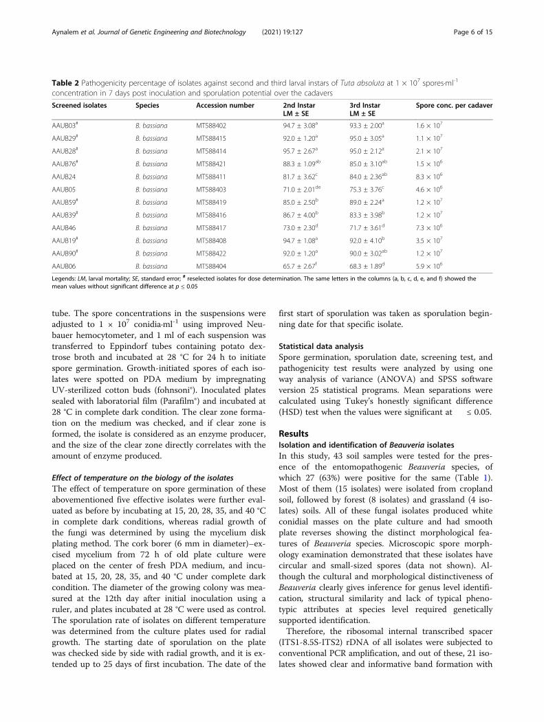
Table 2 Pathogenicity percentage of isolates against second and third larval instars of Tuta absoluta at 1 × 10⁷ spores·ml-1
concentration in 7 days post inoculation and sporulation potential over the cadavers
Screened isolates Species Accession number 2nd InstarLM ± SE
3rd InstarLM ± SE
Spore conc. per cadaver
AAUB03# B. bassiana MT588402 94.7 ± 3.08a 93.3 ± 2.00a 1.6 × 10⁷
AAUB29# B. bassiana MT588415 92.0 ± 1.20a 95.0 ± 3.05a 1.1 × 10⁷
AAUB28# B. bassiana MT588414 95.7 ± 2.67a 95.0 ± 2.12a 2.1 × 10⁷
AAUB76# B. bassiana MT588421 88.3 ± 1.09ab 85.0 ± 3.10ab 1.5 × 10⁶
AAUB24 B. bassiana MT588411 81.7 ± 3.62c 84.0 ± 2.36ab 8.3 × 10⁶
AAUB05 B. bassiana MT588403 71.0 ± 2.01de 75.3 ± 3.76c 4.6 × 10⁶
AAUB59# B. bassiana MT588419 85.0 ± 2.50b 89.0 ± 2.24a 1.2 × 10⁷
AAUB39# B. bassiana MT588416 86.7 ± 4.00b 83.3 ± 3.98b 1.2 × 10⁷
AAUB46 B. bassiana MT588417 73.0 ± 2.30d 71.7 ± 3.61d 7.3 × 10⁶
AAUB19# B. bassiana MT588408 94.7 ± 1.08a 92.0 ± 4.10b 3.5 × 10⁷
AAUB90# B. bassiana MT588422 92.0 ± 1.20a 90.0 ± 3.02ab 1.2 × 10⁷
AAUB06 B. bassiana MT588404 65.7 ± 2.67f 68.3 ± 1.89d 5.9 × 10⁶
Legends: LM, larval mortality; SE, standard error; # reselected isolates for dose determination. The same letters in the columns (a, b, c, d, e, and f) showed themean values without significant difference at p ≤ 0.05
Aynalem et al. Journal of Genetic Engineering and Biotechnology (2021) 19:127 Page 6 of 15

554 bp (data not shown). The amplified genes of these21 isolates were sequenced in both direction, andBLAST search results showed all the isolates had 99–100% similarity with previously documented B. bassiana.These sequences with other 20 (8 sequences obtainedfrom Rehner and Buckley [36], 8 directly from Genbank,and 4 from Belay et al. [18]) ITS sequences of isolatesretrieved from the database showed concordant topology(Fig. 1). The constructed phylogenetic tree showed theisolate category, and our isolates were grouped intothree categories. Out of 21 isolates, twelve, three, and sixisolates showed 56, 83, and 63% bootstrap support witheach other, respectively. Two strains under the secondcategory (AAUB05 and AAUB19) showed very distantevolutionary origin as compared with others with strong(96%) support with each other although they were
isolated from different (grassland and cropland) sourcesof soil. Moreover, almost none of the isolates showed re-lation with previously identified strains, rather, one iso-late from Brazil (B. bassiana-AY531974) matched ourisolates with bootstrap support of 56% (Fig. 1), andtherefore, this shows that our isolates were nearly mono-phyletic with each other and distinctive from the others.
Screening of potential isolatesMorphologically and molecularly identified isolates of B.bassiana were first screened based on their spore viabil-ity, sporulation rate, and pathogenicity against their hostinsect (G. mellonella) through the computation of thebiological efficiency index (BEI). All isolates displayedmore than 80% of spore germination by showing the sig-nificant difference with one another at p ≤ 0.05, except
Fig. 1 Molecular phylogenic analysis of ITS region of Beauveria isolated from Ethiopian soil and other related sequences retrieved from GenBank.Maximum likelihood phylogenetic tree (MEGA4.1) based on ITS sequences from Beauveria species using the neighbor-joining model to constructthe tree and the Jukes–Cantor sequence evolution algorithm, with 1000 bootstrap replications. Bootstrap values above 50% are shown. Thesequences of our isolates were deposited to NCBI database with accession numbers ranging between MT588402 and MT588422, whereasaccession numbers of other previously identified strains were presented on the tree followed by countries in parenthesis
Aynalem et al. Journal of Genetic Engineering and Biotechnology (2021) 19:127 Page 7 of 15

isolate AAUB07 with a low spore germination of 43%(Table 1). They attained sporulation (DPCS) within 10to12 days with relative sporulation ratings (RSR) of 1.2to 1.8 on the culture plate and scored 76.7–100% ofmortality on larvae of G. mellonella. The computed bio-logical efficiency index (BEI) of isolates was reneged be-tween 62 and 86%, defined on cumulative effect of allthe above-working parameters combined (Table 1).Out of the isolates explored in this study, 12 (44%) iso-
lates scored BEI values of above 80% and screened for fur-ther work. These isolates were effective in larval mortality(LM) of 96.7 to 100% against G. mellonella in 10 days ofpost inoculation. From these, five (AAUB28, AAUB03,AAUB24, AAUB05, and AAUB76) isolates scored highest(85 to 86.1%) BEI values, and other seven isolates scoredbetween 82 and 84.8% BEI values as indicated by asterisk(*) in Table 1. These twelve isolates were used for furtherpathogenicity bioassay against T. absoluta.
Pathogenicity bioassay against Tuta absolutaIt is interesting to note that most of the prescreened iso-lates were pathogenically effective against T. absoluta at1 × 107spores·ml-1concentration in 7 days post inocula-tion (Table 2). Almost all of these isolates rated moder-ate (65.7%) to high (95.7%) mortality against bothsecond and third instar larvae of T. absoluta. Five of theisolates (AAUB03, AAUB28, AAUB29, AAUB19, and
AAUB90) were highly effective and scored 95, 95, 93.3,92, and 94.7% of mortality against either of the instarlarvae, respectively. The best isolate, AAUB28, wasequally pathogenic against both second and third instarlarvae, and scored 95.7 and 95% of mortality, respect-ively. However, the mean pathogenicity among isolatesshowed marked statistical difference with F (26, 52) =8.97, p < 0.001 for second instar and F (14, 25) = 8.123,p < 0.001 for third instar larvae. The sporulation poten-tial of these isolates was considerable and produced 4.6× 10⁶ to 3.5 × 10⁷ spores·ml-1 over the larval cadavers(Table 2).Furthermore, all of these selected isolates of B. bassi-
ana were polyphyletic in source, biological efficiency,and pathogenicity (Fig. 2). For instance, AAUB19 andAAUB90 strains isolated from the cropland soil scoredhighly effective (HE) mortality against third instar larvaeof T. absoluta, whereas AAUB05 isolated from the grass-land soil scored moderate (ME) lethality, and all are cat-egorized under G2 of BEI. Most G1 strains obtainedfrom three soil types were scored moderate (ME) to high(HE) lethality against T. absoluta and distributed in dif-ferent categories. The remaining five strains were iso-lated from different types of soil; have BEI of G1, G2,and G3; scored moderate (ME) lethality; and grouped inone category (III-a). In general, none of the isolatesshowed congruence with respect to sources, BEI, and
Fig. 2 This cladogram showed the characteristics of twelve Ethiopian potent isolates. First sources of isolates, from grassland soil (▲), croplandsoil (♦), and forest soil (●) followed by codes for each isolate and biological efficacy index showing that if BEI > 85 = G1, BEI: 83–85 = G2, BEI: 80–83 = G3. Finally, percentage of mortality against Tuta absoluta was indicated, such as if value is < 70% (less effective, LE), 70–80% (moderatelyeffective, ME), 80–90% (highly effective, HE)
Aynalem et al. Journal of Genetic Engineering and Biotechnology (2021) 19:127 Page 8 of 15

pathogenicity, rather showed intermixed distribution inthe tree (Fig. 2).
Virulence of isolates against Tuta absolutaIn this study, eight of the most pathogenic strains of B.bassiana were evaluated to determine the effect of thespore concentration against third instar larvae of T.absoluta. These strains scored median lethal time (LT50)values ranging between 3.3 and 5.67 days at 104 spor-es·ml-1 and 1.93 to 3.17 days at 108 spores·ml-1 concen-trations (Table 3). Three strains, AAUB03, AAUB19,and AAUB28 attained the LT50 values at the same 3.33days, whereas strain AAUB76 scored relatively extended(5.76 days) LT50 values. Similarly, corresponding LC50
values for all selected isolates ranged between 1.5 × 10³
and 1.1 × 10⁵ spores·ml-1and LC90 value of 2.8 × 10⁵ and1.8 × 107 spores·ml-1 (Table 3). The isolates AAUB03,AAUB28, AAUB19, and AAUB90 scored LC50 values of5.3, 1.3, 1.8, and 2.6 × 10³, and AAUB29, AAUB76, andAAUB39 scored 6.5, 9.3, and 1.8 × 10⁴ spores·ml-1, re-spectively. The least LC50 value (1.1 × 10⁵ spores·ml-1)was scored by isolate AAUB59. In general, concurrentlarval mortality increase was recorded in all evaluatedisolates as spore concentration and exposure timeincreased.
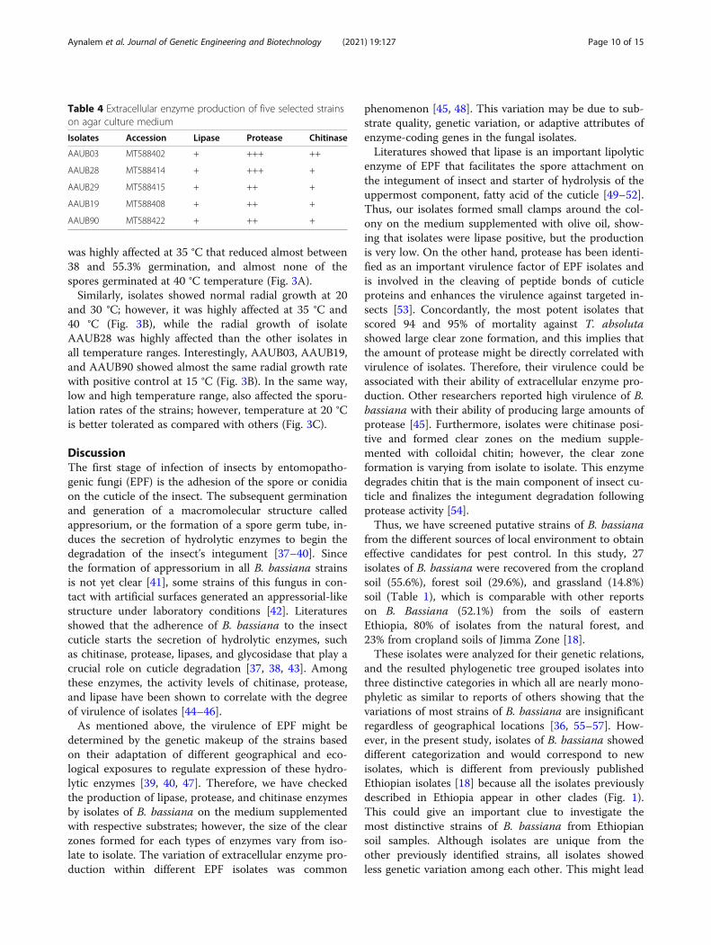
Chitinolytic enzyme production of effective isolatesOut of eight potent isolates, five isolates that scoredshort LT50 and lowest LC50 values were evaluated abouttheir pathogenicity-enhancing chitinolytic enzyme pro-duction potential using the clear zone determinationmethod. The strains of B. bassiana which are capable toproduce lipase, protease, and chitinase enzymes werehighly pathogenic against their target pests. Therefore,five of our isolates were lipase, protease, and chitinase
positive and showed clear zone formation on respectivemedium for each enzyme. However, the clear zone for-mation for each enzyme type varied among isolates(Table 4). Two isolates (AAUB03 and AAUB28) showedlarge (+++) clear zones on the PDA medium supple-mented with casein as compared with other three iso-lates (Table 4). The large clear zone formationphenomenon may correlate with the amount of proteasesecreted by the fungal isolates. The remaining three(AAUB29, AAUB19, and AAUB90) isolates that scored95, 92, and 90% of mortality against third instar larvae ofT. absoluta formed intermediate (++) clear zones byprotease (Table 4). The isolate AAUB03 continued pro-duction of better (++) clear zones on the medium sup-plemented with colloidal chitin than the others, thoseformed small (+) clear zones. Furthermore, isolatesshowed very small (+) clear zone formation on themedium supplemented with olive oil that correlates withless production of lipase enzyme (Table 4).
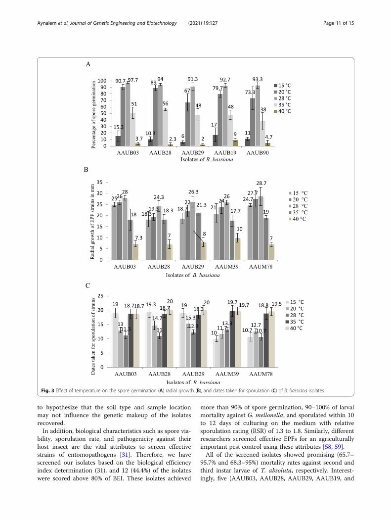
Effect of temperature on the biology of isolatesTemperature is the most important abiotic factor thatinfluences the biology of EPF strains. Isolates that adapttemperatures ranging out of the optimum may be con-sidered to be used in adverse conditions for pest control.Therefore, we have evaluated five of effective isolates fortheir spore germination, radial growth, and sporulationpotential at different temperature classes (Fig. 3). Conse-quently, all tested isolates showed an effect on conidialgerminations between 6 and 17%, and two of them(AAUB03 and AAUB90) showed better (15.3 and 17%)germination at 15 °C (Fig. 3A). All of the isolates showedalmost better conidial germination ranging between 67and 90.7% at 20 °C and above 90% of germination at 28°C (Fig. 3A). The conidial germination of all the strains
Table 3 The LT50 determination and summary of probit analysis to determine LC50 and LC90 concentrations of Beauveria bassianastrains against the third instar of Tuta absoluta larvae in 10 days post incubation
LT50 (mean ± SE)
AAUB03 AAUB29 AAUB28 AAUB76 AAUB59 AAUB39 AAUB19 AAUB90
Spores·ml-1
104 3.33± 0.58a 5.33 ± 0.58c 3.33± 0.58a 5.67 ± 0.58cd 5.00 ± 1.00c 3.67 ± 0.58ab 3.33 ± 0.58a 5.00 ± 1.00c
106 2.33± 0.58a 2.57 ± 0.51a 2.50± 0.00a 2.83 ± 0.29ab 2.93 ± 0.12ab 2.17 ± 0.29a 2.33 ± 0.29a 4.00 ± 0.00c
108 1.93± 0.12a 2.00 ± 0.00a 2.20± 0.35a 2.33 ± 0.29a 2.50 ± 0.50ab 2.00 ± 0.35a 2.00 ± 0.62a 3.17 ± 0.29c
Summary of probit analysis
LC50 5.3 × 10³ 6.5 × 10⁴ 1.5 × 10³ 9.3 × 10⁴ 1.1 × 10⁵ 1.8 × 10⁴ 1.8 × 10³ 2.6 × 10³
95% FL 1.3 × 10³ –7 × 10⁴ 2.2 × 10⁴–1.5 × 10⁵ 8.6 × 10²–4.9 × 10³ 9.9 × 10³–1.3 × 10⁴ 2 × 10⁴–3.3 × 10⁵ 3.9 × 10³–6 × 10⁴ 1 × 10³–6.1 × 10⁴ 1.2 × 10³–3.1 × 10⁴
LC90 2.8 × 10⁵ 1.7 × 10⁶ 2.9 × 10⁵ 1.6 × 10⁶ 1.8 × 10⁷ 1.6 × 10⁷ 7.6 × 10⁵ 1.2 × 10⁷
95% FL 2.1 × 10⁵ –6.3 × 10⁶ 6.3 × 10⁵–1.1 × 10⁷ 1.9 × 10⁴–2.9 × 10⁶ 1.8 × 10⁵–2.7 × 10⁷ 4.2 × 10⁶–2.8 × 10⁸ 2.2 × 10⁶–3.1 × 10⁸ 7.1 × 10⁴–2.1 × 10⁷ 1.5 × 10⁶–7.3 × 10⁸
Int ± SE 1.32 ± 0.91 4.33 ± 0.89 0.85 ± 0.83 1.16 ± 0.72 2.91 ± 0.66 1.54 ± 0.64 0.8 ± 0.75 1.19 ± 0.63
S ± SE 0.49 ± 0.18 0.9 ± 0.17 0.39 ± 0.16 0.39 ± 0.13 0.58 ± 0.11 0.39 ± 0.11 0.36 ± 0.14 0.35 ± 0.11
p value < 0.001 < 0.001 < 0.012 < 0.003 < 0.001 < 0.001 < 0.001 < 0.001
Legends: FL, fiducial limit; SE, standard error; LT, lethal time taken to kill fifty of experimental organisms; LC, lethal concentration; Int, intercept; S, slope. The same letters in thecolumns (a, b, c, d, e, and f) showed the mean values without significant difference at p ≤ 0.05
Aynalem et al. Journal of Genetic Engineering and Biotechnology (2021) 19:127 Page 9 of 15
was highly affected at 35 °C that reduced almost between38 and 55.3% germination, and almost none of thespores germinated at 40 °C temperature (Fig. 3A).Similarly, isolates showed normal radial growth at 20
and 30 °C; however, it was highly affected at 35 °C and40 °C (Fig. 3B), while the radial growth of isolateAAUB28 was highly affected than the other isolates inall temperature ranges. Interestingly, AAUB03, AAUB19,and AAUB90 showed almost the same radial growth ratewith positive control at 15 °C (Fig. 3B). In the same way,low and high temperature range, also affected the sporu-lation rates of the strains; however, temperature at 20 °Cis better tolerated as compared with others (Fig. 3C).
DiscussionThe first stage of infection of insects by entomopatho-genic fungi (EPF) is the adhesion of the spore or conidiaon the cuticle of the insect. The subsequent germinationand generation of a macromolecular structure calledappresorium, or the formation of a spore germ tube, in-duces the secretion of hydrolytic enzymes to begin thedegradation of the insect’s integument [37–40]. Sincethe formation of appressorium in all B. bassiana strainsis not yet clear [41], some strains of this fungus in con-tact with artificial surfaces generated an appressorial-likestructure under laboratory conditions [42]. Literaturesshowed that the adherence of B. bassiana to the insectcuticle starts the secretion of hydrolytic enzymes, suchas chitinase, protease, lipases, and glycosidase that play acrucial role on cuticle degradation [37, 38, 43]. Amongthese enzymes, the activity levels of chitinase, protease,and lipase have been shown to correlate with the degreeof virulence of isolates [44–46].As mentioned above, the virulence of EPF might be
determined by the genetic makeup of the strains basedon their adaptation of different geographical and eco-logical exposures to regulate expression of these hydro-lytic enzymes [39, 40, 47]. Therefore, we have checkedthe production of lipase, protease, and chitinase enzymesby isolates of B. bassiana on the medium supplementedwith respective substrates; however, the size of the clearzones formed for each types of enzymes vary from iso-late to isolate. The variation of extracellular enzyme pro-duction within different EPF isolates was common
phenomenon [45, 48]. This variation may be due to sub-strate quality, genetic variation, or adaptive attributes ofenzyme-coding genes in the fungal isolates.Literatures showed that lipase is an important lipolytic
enzyme of EPF that facilitates the spore attachment onthe integument of insect and starter of hydrolysis of theuppermost component, fatty acid of the cuticle [49–52].Thus, our isolates formed small clamps around the col-ony on the medium supplemented with olive oil, show-ing that isolates were lipase positive, but the productionis very low. On the other hand, protease has been identi-fied as an important virulence factor of EPF isolates andis involved in the cleaving of peptide bonds of cuticleproteins and enhances the virulence against targeted in-sects [53]. Concordantly, the most potent isolates thatscored 94 and 95% of mortality against T. absolutashowed large clear zone formation, and this implies thatthe amount of protease might be directly correlated withvirulence of isolates. Therefore, their virulence could beassociated with their ability of extracellular enzyme pro-duction. Other researchers reported high virulence of B.bassiana with their ability of producing large amounts ofprotease [45]. Furthermore, isolates were chitinase posi-tive and formed clear zones on the medium supple-mented with colloidal chitin; however, the clear zoneformation is varying from isolate to isolate. This enzymedegrades chitin that is the main component of insect cu-ticle and finalizes the integument degradation followingprotease activity [54].Thus, we have screened putative strains of B. bassiana
from the different sources of local environment to obtaineffective candidates for pest control. In this study, 27isolates of B. bassiana were recovered from the croplandsoil (55.6%), forest soil (29.6%), and grassland (14.8%)soil (Table 1), which is comparable with other reportson B. Bassiana (52.1%) from the soils of easternEthiopia, 80% of isolates from the natural forest, and23% from cropland soils of Jimma Zone [18].These isolates were analyzed for their genetic relations,
and the resulted phylogenetic tree grouped isolates intothree distinctive categories in which all are nearly mono-phyletic as similar to reports of others showing that thevariations of most strains of B. bassiana are insignificantregardless of geographical locations [36, 55–57]. How-ever, in the present study, isolates of B. bassiana showeddifferent categorization and would correspond to newisolates, which is different from previously publishedEthiopian isolates [18] because all the isolates previouslydescribed in Ethiopia appear in other clades (Fig. 1).This could give an important clue to investigate themost distinctive strains of B. bassiana from Ethiopiansoil samples. Although isolates are unique from theother previously identified strains, all isolates showedless genetic variation among each other. This might lead
Table 4 Extracellular enzyme production of five selected strainson agar culture medium
Isolates Accession Lipase Protease Chitinase
AAUB03 MT588402 + +++ ++
AAUB28 MT588414 + +++ +
AAUB29 MT588415 + ++ +
AAUB19 MT588408 + ++ +
AAUB90 MT588422 + ++ +
Aynalem et al. Journal of Genetic Engineering and Biotechnology (2021) 19:127 Page 10 of 15
to hypothesize that the soil type and sample locationmay not influence the genetic makeup of the isolatesrecovered.In addition, biological characteristics such as spore via-
bility, sporulation rate, and pathogenicity against theirhost insect are the vital attributes to screen effectivestrains of entomopathogens [31]. Therefore, we havescreened our isolates based on the biological efficiencyindex determination (31), and 12 (44.4%) of the isolateswere scored above 80% of BEI. These isolates achieved
more than 90% of spore germination, 90–100% of larvalmortality against G. mellonella, and sporulated within 10to 12 days of culturing on the medium with relativesporulation rating (RSR) of 1.3 to 1.8. Similarly, differentresearchers screened effective EPFs for an agriculturallyimportant pest control using these attributes [58, 59].All of the screened isolates showed promising (65.7–
95.7% and 68.3–95%) mortality rates against second andthird instar larvae of T. absoluta, respectively. Interest-ingly, five (AAUB03, AAUB28, AAUB29, AAUB19, and
Fig. 3 Effect of temperature on the spore germination (A) radial growth (B), and dates taken for sporulation (C) of B. bassiana isolates
Aynalem et al. Journal of Genetic Engineering and Biotechnology (2021) 19:127 Page 11 of 15
AAUB90) of the isolates were scored 90–95.7% of mor-tality at 1 × 10⁷ spores·ml-1 concentration. This is con-current with other reports on B. bassiana that scored90% of mortality against T. absoluta in Egypt [60]. Inaddition, recently explored Ethiopian isolate of B. bassi-ana displayed 95.8% of mortality against third instar lar-vae of T. absoluta at 2.5 × 10⁹ spores·ml-1 [61]. In allcases, Ethiopian isolates of B. bassiana performed betterthan the commercially formulated products (Beauvitech®WP) that scored 60.8% of larval mortality against T.absoluta at 108 spores·ml-1 in Rwanda [62]. This couldinfer that locally screened isolates might be pathogeni-cally effective than commercialized ones, and geographicorigin of isolates may determine their pathogenicity.Furthermore, these pathogenically valued isolates of B.
bassiana showed almost equal efficacy against both sec-ond and third instar larvae of T. absoluta by scoring95.7% and 95% of mortality, respectively, which is con-tradicting to reports of Tsownara and Port [63] whichshowed that third instar larvae of T. absoluta is moresusceptible to B. bassiana than the second instar larvae.In addition, these isolates produced considerable (4.6 ×10⁶ to 3.5 × 10⁷ spores·ml-1) amount of spores over thedead larval cadavers. This makes that fungal spores areeasily disseminated between insects through the processof auto-dissemination. Thus, high sporulation potentialof EPF over the cadavers enhanced horizontal pest infec-tion through the self-spore contamination process [64].Besides, our isolates scored shortest (1.93 to 3.17 days)median lethal time (LT50) at 10
8 spores·ml-1, which weredifferent from the two commercially formulated Beau-veria (Beauvitech® WP and Botanigard® ES) that scoredsomewhat extended LT50 values of 5.2 and 6.6 days atthe same spore concentration against T. absoluta, re-spectively [62]. This is at least twice the LT50 value ofthe Ethiopian isolates presented here. However, a formerEthiopian isolate of B. bassiana scored a LT50 value of5.2 days at the spore concentration of 2.5 × 10⁹ spor-es·ml-1 [61], which takes considerably longer time to kill50% of third instar larvae of T. absoluta as comparedwith their spore concentration.It is interesting to mention that effective isolates of B.
bassiana in this study scored promising LC50 and LC90
(1.5 × 10³ and 2.8 × 10⁵ spores·ml-1) values against thirdinstar larvae of T. absoluta, respectively. Similarly, otherisolates of B. bassiana scored LC50 values of 4.5 × 10⁵
and 3.6 × 10⁵ spores·ml-1against second and third instarlarvae of T. absoluta, respectively [65]. Usually, when thespore concentration and exposure time increases con-currently, larval mortality also increases in all evaluatedstrains of B. bassiana, which is comparable with otherfindings [61, 63, 65].Although locally isolated EPF strains showed better
performance on biocontrol activity, different abiotic
factors might influence survival of the bioagents.Temperature is one of the abiotic factors that could in-fluence spore germination, mycelial growth, and sporeproduction of the fungal isolates [66]. We have checkedboth lower and upper temperature ranges against sporegermination, radial growth, and sporulation rate of thesome selected potent isolates and temperatures at 20 to30 °C showed nil effect on spore germination, mycelialgrowth, and sporulation. Similarly, others reported thattemperature ranging at 24–30 °C was optimum for sporegermination, mycelial growth, and speculation [54].However, temperature lower than 20 °C and above 30 °Cwas highly adverse for spore germination of B. bassiana[67], which is concurrent to our study.
ConclusionThis study explains the presence of more indigenousstrains of B. bassiana in different soil types of Ethiopiawith better entomopathogenic characteristics and that iscrucial to develop ecofriendly biopesticides for sustain-able agriculture. Molecular analysis of these isolatesshowed monophyletic attributes about genetic relation-ships and polyphyletic characters about sources, bio-logical efficacy, and pathogenicity against the host insect.Among the isolates of B. bassiana, two (AAUB03 andAAUB28) isolates showed strong efficacy against T.absoluta with short LT50 and low LC50 values. Theseisolates were chitinolytic enzyme producers and bettergrowers at optimum temperatures. Therefore, these indi-genous B. bassiana were effective against T. absolutaunder laboratory conditions; however, further investiga-tion at the actual field is required for mass production.
AbbreviationsAAU: Addis Ababa University; ANOVA: Analysis of variance; APPRC: AmboPlant Protection Research Center; BEI: Biological Efficiency Index; BLAST: BasicLocal Alignment Search Tool; DPCS: Date of plate culture sporulation;EDTA: Ethylenediaminetetraacetic acid; EPF: Entomopathogenic fungi;HE: Highly effective; IPM: Integrated pest management; ITS: Internaltranscribed spacer; JC: Jukes–Cantor; LC: Lethal concentration; LE: Lesseffective; LM: Larval mortality; LT: Lethal time; NCBI: National Center forBiotechnology Information; NJ: Neighbor-joining; ME: Moderately effective;MEGA: Molecular Evolutionary Genetic Analysis; PCS: Plate culturesporulation; PCR: Polymerase chain reaction; PDA: Potato dextrose agar;RSR: Relative sporulation rate; SE: Standard error; SG: Spore germination;SPSS: Statistical Package for the Social Sciences; TBE: Tris-borate-EDTA;USEPA: United States of Environmental Protection Agency
AcknowledgmentsWe would like to acknowledge the financial support of “The HealthySeedling Project” granted by both the Ethiopian Biotechnology Institute(EBtI) and The Regional Project Supported by the Australian DevelopmentAgency (ADA).
Authors’ contributionsBA proposed the study, conducted experimental work, analyzed the data,and wrote manuscript draft. JV performed molecular work, processed DNAsequencing, and performed molecular data analysis. FA and DM read andedited the manuscript. The authors read and approved the final manuscript.
Aynalem et al. Journal of Genetic Engineering and Biotechnology (2021) 19:127 Page 12 of 15
FundingThis work was supported by the Healthy Seedling and the AustralianDevelopment Agency projects given to improve tomato production inEthiopia and Fellowship program of the University of Chile.
Availability of data and materialsThe datasets used for this work are available with the corresponding authorwhen legal request is presented.
Declarations
Ethics approval and consent to participateNot applicable
Consent for publicationNot applicable
Competing interestsThe authors declare that they have no competing interests.
Author details1Institute of Biotechnology, Addis Ababa University, Addis Ababa, Ethiopia.2Cellular and Molecular Biology Program, Faculty of Medicine, Institute ofBiomedical Sciences, University of Chile, Santiago, Chile. 3Department ofMicrobial, Cellular and Molecular Biology, College of Natural andComputational Sciences, Addis Ababa University, Addis Ababa, Ethiopia.
Received: 23 February 2021 Accepted: 10 August 2021
References1. Taylor JAR, Herms AD, Cardina J, Moore HR (2018) Climate change and pest
management: unanticipated consequences of trophic dislocation.Agronomy 8(7):1–23. https://doi.org/10.3390/agronomy8010007
2. Biondi A, Wan F, Guedes CNR, Desneux N (2018) Ecology, worldwidespread, and management of the invasive South American tomato pinworm,Tuta absoluta: past, present and future. Annu Rev Entomol 63:239–258.https://doi.org/10.1146/annurev-ento-031616-034933
3. Urbaneja A, Vercher R, Navarro V, Garci´a F, Porcuna L (2007) La polilla deltomate, Tuta absoluta. Phytoma Espan˜a 194:16–23 http://hdl.handle.net/20.500.11939/4087
4. Tonnang ZEH, Mohamed FS, Khamis F, Ekesi S (2015) Identification and riskassessment for worldwide invasion and spread of Tuta absoluta with afocus on Sub Saharan Africa: implications for phytosanitary measures andmanagement. PLoS One 10(9):e0138319. https://doi.org/10.1371/journal.pone.0138319
5. Santana AP, Kumar L, Da Silva SR, Picanço CM (2019) Global geographicdistribution of Tuta absoluta as affected by climate change. J Pest Sci 92:1373–1385. https://doi.org/10.1007/s10340-018-1057-y
6. Rwomushana I, Beale T, Chipabika G, Day R, Gonzalez-Moreno P,Lamontagne-Godwin J, Makale F, Pratt C, Tambo J (2019) Evidencenote: tomato leafminer (Tuta absoluta): impacts and coping strategiesfor Africa. CABI Working Paper:12–56. https://doi.org/10.1079/CABICOMM-62-8100
7. Retta NA, Berhe HD (2015) Tomato leafminer, Tuta absoluta (Meyrick),a devastating pest of tomatoes in the highlands of Northern Ethiopia:a call for attention and action. Rev Res J Agric Environ Manage 4(6):264–269
8. Shiberu T, Getu E (2018) Determination of the economic threshold level oftomato leaf miner, Tuta absoluta Meyrick (Lepidoptera: Gelechiidae) ontomato plant under glasshouse conditions. J Horticulture Forestry 10(2):9–16. https://doi.org/10.5897/JHF2018.0522
9. Arnó J, Gabarra R, Molina P, Godfrey EK, Zalom GF (2019) Tuta absoluta(Lepidoptera: Gelechiidae) success on common solanaceous species fromCalifornia tomato production. Environ Entomol 48(6):1394–1400. https://doi.org/10.1093/ee/nvz109
10. Desneux N, Wajnberg E, Wyckhuys GAK, Burgio G, Arpaia S, Narvaez-Vasquez AC, Gonzalez-Cabrera J, Ruescas CD, Tabone E, Frandon J, Pizzol J,Poncet C, Cabello T, Urbaneja A (2010) Biological invasion of Europeantomato crops by Tuta absoluta: ecology, geographic expansion and
prospects for biological control. J Pest Sci 83(3):197–215. https://doi.org/10.1007/s10340-010-0321-6
11. Cherif A, Mansour R, Grissa-Lebdi K (2013) Biological aspects of tomatoleafminerTuta absoluta (Lepidoptera: Gelechiidae) in conditions ofNortheastern Tunisia: possible implications for pest management. EnvironExp Biol 11:179–184
12. De Smedt C, Van Damme V, De Clercq P, Spanoghe P (2016)Insecticide effect of zeolites on the tomato leafminer Tuta absoluta(Lepidoptera: Gelechiidae). Insects 7(4):72–85. https://doi.org/10.3390/insects7040072
13. Roditakis E, Grispou M, Nauen R, Vasakis E, Stavrakaki M, Gravouil M, Bassi A(2015) First report of Tuta absoluta resistance to diamide insecticides. J PestSci 88(1):9–16. https://doi.org/10.1007/s10340-015-0643-5
14. Negatu B, Kromhout H, Mekonnen Y, Vermeulen R (2016) Use ofchemical pesticides in Ethiopia: a cross-sectional comparative study onknowledge attitude and practice of farmers and farm workers in threefarming systems. Ann Occup Hyg 60(5):551–566. https://doi.org/10.1093/annhyg/mew004
15. Naranjo ES, Ellsworth CP, Frisvold BG (2015) Economic value of biologicalcontrol in integrated pest management of managed plant systems. AnnuRev Entomol 60(1):621–645. https://doi.org/10.1146/annurev-ento-010814-021005
16. Inglis DG, Goettel SM, Butt MT, Strasser H (2001) Use of hyphomycetousfungi for managing insect pests. Fungi as Biocontrol Agents(Butt TM,Jackson C, Magan N, eds):23–70
17. Jaber S, Mercier A, Knio K, Brun S, Kambris Z (2016) Isolation of fungifrom dead arthropods and identification of a new mosquito naturalpathogen. Parasit Vectors 9(491):1–10. https://doi.org/10.1186/s13071-016-1763-3
18. Belay CY, Meressa HB, Alemu T, Hallmann J (2017) Molecular detection ofthe entomopathogenic fungus Beauveria bassiana from soils of coffeegrowing areas in Ethiopia using rDNA-ITS. J Appl Biosci 119:11943–11953
19. NiuX XW, Zhang J, Hu Q (2019) Biodiversity of entomopathogenic fungi inthe soils of South China. Microorganisms 7(311):1–14. https://doi.org/10.3390/microorganisms7090311
20. Allegrucci N, Velazquez SM, Russo LM, Perez E, Scorsetti CA (2017)Endophytic colonization of tomato by the entomopathogenic fungusBeauveria bassiana: the use of different inoculation techniques andtheir effects on the tomato leafminer Tuta absoluta (Lepidoptera:Gelechiidae). J Plant Protection Res 57(4):331–337. https://doi.org/10.1515/jppr-2017-0045
21. Rohrlich C, Merle I, Hassani I, Verger M, Zuin M, Besse S (2018) Variation inphysiological host range in three strains of two species of theentomopathogenic fungus Beauveria. PLoS One 13(7):e0199199. https://doi.org/10.1371/journal.pone.0199199
22. Mora EAM, Castilho CMA, Fraga EM (2017) Classification and infectionmechanism of entomopathogenic fungi (Review). Agric Microbiol ReviewArticle 84(0):1–10. https://doi.org/10.1590/1808-1657000552015
23. Faria MR, Wraight SP (2007) Mycoinsecticides and mycoacaricides: acomprehensive list with worldwide coverage and international classificationof formulation types. Biol Control 43(3):237–256. https://doi.org/10.1016/j.biocontrol.2007.08.001
24. Wang J, Zheng C (2012) Characterization of a newly discovered Beauveriabassiana isolate to Franklimiella occidentalis Perganda, a non-native invasivespecies in China. Microbiol Res 167(2):116–120. https://doi.org/10.1016/j.micres.2011.05.002
25. Fernandes ÉKK, Moraes ÁML, Pacheco RS, Rangel DEN, Miller MP, BittencourtVREP, Roberts DW (2009) Genetic diversity among Brazilian isolates ofBeauveria bassiana: comparisons with non-Brazilian isolates and otherBeauveria species. J Appl Microbiol 107(3):760–774. https://doi.org/10.1111/j.1365-2672.2009.04258.x
26. Meyling VN (2007) Methods for isolation of entomopathogenic fungi fromthe soil environment. Thorval dsensvej 40, DK-1871 Frederiksberg C,Denmark, pp1-19.
27. Rehner SA, Minnis AM, Sung GH, Luangsa-ard JJ, Devotto L, Humber RA(2011) Phylogeny and systematics of the anamorphic,entomopathogenic genus Beauveria. Mycologia 103(5):1055–1073.https://doi.org/10.3852/10-302
28. Chi M, Park S, Lee Y (2009) A quick and safe method for fungal DNAextraction. Plant Pathol J 25(1):108–111. https://doi.org/10.5423/PPJ.2009.25.1.108
Aynalem et al. Journal of Genetic Engineering and Biotechnology (2021) 19:127 Page 13 of 15

29. White TJ, Bruns T, Lee S, Taylor JW (1990) Amplification and directsequencing of fungal ribosomal RNA genes for phylogenetics. In:Innis MA, Gelfand DH, Sninsky JJ, White TJ (eds) PCR Protocols: aguide to methods and applications. Academic Press Inc, New Yorkpp, pp 315–322
30. Habtegebriel B, Getu E, Dawd M, Seyoum E, Atnafu G, Khamis F, Hilbur Y,Ekesi S, Larsson CM (2016) Molecular characterization and evaluation ofindigenous entomopathogenic fungal isolates against Sorghum Chafer,Pachnoda interrupta (Olivier) in Ethiopia. J Entomol Nematol 8(5):34–45.https://doi.org/10.5897/JEN2016.0159
31. Sain KS, Monga D, Kumar R, Nagrale TD, Hiremani SN, Kranthi S (2019)Compatibility of entomopathogenic fungi with insecticides and theirefficacy for IPM of Bemisia tabaci in cotton. J Pestic Sci 44(2):97–105. https://doi.org/10.1584/jpestics.D18-067
32. Sabbour MM, Singer SM (2014) Evaluations of two Metarhizium varietiesagainst Tuta absoluta (Meyrick) (Lepidoptera: Gelechiidae) in Egypt. IJSR3:1983–1987
33. Abbott WS (1925) A method of computing the effectiveness of aninsecticide. J Econ Entomol 18(2):265–267. https://doi.org/10.1093/jee/18.2.265a
34. Tefera T, Pringle LK (2003) Effect of exposure method to Beauveria bassianaand conidia concentration on mortality, mycosis, and sporulation incadavers of Chilo partell (Lepidoptera: Pyralidae). J Invertebr Pathol 84(2):90–95. https://doi.org/10.1016/j.jip.2003.08.001
35. Tefera T, Pringle LK (2004) Evaluation of Beauveria bassiana andMetarhizium anisopliae for controlling Chilo partellus (Lepidoptera:Crambidae) in Maize. Biocontrol Sci Tech 14(8):849–853. https://doi.org/10.1080/0958315041000172707
36. Rehner AS, Buckley E (2005) A Beauveria phylogeny inferred from nuclearITS and EF1-a sequences: evidence for cryptic diversification and links toCordyceps teleomorphs. Mycologia 97(1):84–98. https://doi.org/10.1080/15572536.2006.11832842
37. Binod P, Sukumaran RK, Shirke SV, Rajput JC, Pandey A (2007) Evaluation offungal culture fíltrate containing chitinase as a biocontrol agent againstHelicoverpa armigera. J Appl Microbiol 103(5):1845–1852. https://doi.org/10.1111/j.1365-2672.2007.03428.x
38. Esparza MA, Conteiro CAM, Fraga ME (2017) Classification and infectionmechanism of entomopathogenic fungi. Arq Inst Biol 84:1–10
39. Pinnamaneni R, Kalidas p, Rao S (2010) Cloning and expression of Bbchit1gene of Beauveria bassiana. Open Entomol J 4(1):30–35. https://doi.org/10.2174/1874407901004010030
40. Mondal S, Baksi S, Koris A, Vatai G (2016) Journey of enzymes inentomopathogenic fungi. Pacific Sci Rev A Nat Sci Eng 18(2):85–99. https://doi.org/10.1016/j.psra.2016.10.001
41. Guerri-Agulloo B, Gomez-Vidal S, Asensio L, Barranco P, Lopez-Llorca LV(2010) Infection of the red palm weevil (Rhynchophorus ferrugineus) by theentomopathogenic fungus Beauveria bassiana: a SEM study. Microsc ResTech 73:714–725. https://doi.org/10.1002/jemt.20812
42. Zhang S, Xia XY, Kim B, Keyhani ON (2010) Two hydrophobins are involvedin fungal spore coat rodlet layer assembly and each play distinct roles insurface interactions, development and pathogenesis in theentomopathogenic fungus, Beauveria bassiana. Mol Microbiol 80(3):811–826.https://doi.org/10.1111/j.1365-2958.2011.07613.x
43. Holder DJ, Kirkland BH, Lewis MW, Keyhani NO (2007) Surfacecharacteristics of the entomopathogenic fungus Beauveria bassiana.Microbiology 153(10):3448–3457. https://doi.org/10.1099/mic.0.2007/008524-0
44. Cho EM, Boucias D, Keyhani NO (2006) EST analysis of cDNA libraries fromthe entomopathogenic fungus Beauveria bassiana II. Fungal cellssporulating on chitin and producing oosporein. Microbiology.152:2855–2864.
45. Dhawan M, Joshi N (2017) Enzymatic comparison and mortality of Beauveriabassiana against cabbage caterpillar Pieris brassicae. Braz J Microbiol 48(3):522–529. https://doi.org/10.1016/j.bjm.2016.08.004
46. Khosravi R, Sendi JJ, Zibaee A, Shokrgozar MA (2015) Virulence of fourBeauveria bassiana (Balsamo) (Asc., Hypocreales) isolates on rose sawfly,Argerosae under laboratory condition. J. King Saud. Univ Sci 27:49–53
47. Arruda W, Lubeck I, Schrank A, Vainstein HM (2005) Morphologicalalterations of Metarhizium anisopliae during penetration of Boophilusmicroplus ticks. Exp Appl Acarol 37(4):231–244. https://doi.org/10.1007/s10493-005-3818-6
48. Mustafa U, Kaur G (2009) Extracellular enzyme production in Metarhiziumanisopliae isolates. Folia Microbiol 54(6):499–504. https://doi.org/10.1007/s12223-009-0071-0
49. Keyhani ON (2017) Lipid biology in fungal stress and virulence:entomopathogenic fungi. Review Fungal Biology XXX 122(6):1–12. https://doi.org/10.1016/j.funbio.2017.07.003
50. Charnley KA (2003) Fungal pathogens of insects: cuticle degrading enzymesand toxins. Adv Bot Res 40:241–321. https://doi.org/10.1016/S0065-2296(05)40006-3
51. Fernandes EG, Valerio HM, Feltrin T, Van der Sand ST (2012) Variability in theproduction of extracellular enzymes by entomopathogenic fungi grown ondifferent substrates. Braz J Microbiol 43(2):827–833. https://doi.org/10.1590/S1517-83822012000200049
52. Rodrigues CJBC, Perinotto WMD, da Silva WOB, Santi L, Berger M, MarcianoAF, de Sa FA, Nogueira MRD, QuinelatoS,Bittencourt VREP (2016) Virulence,proteolytic and lipolytic activities of Brazilian Beauveria bassiana s.l. isolates(Hypocreales: Clavicipitaceae) to Rhipicephalus microplus ticks (Acari:Ixodidae). Biocontrol Sci Tech 26(2):239–249. https://doi.org/10.1080/09583157.2015.1091876
53. St Leger R (2011) The role of cuticle-degrading proteases in fungalpathogenesis of insects. Can J Bot 73(1):1119–1125. https://doi.org/10.1139/b95-367
54. St Leger JR, Cooper MR, Charnley KA (1986) Cuticle-degrading enzymesof entomopathogenic fungi: regulation of production of chitinolyticenzymes. J Gen Microbiol 132(6):1509–1517. https://doi.org/10.1099/00221287-132-6-1509
55. Muro MA, Elliott S, Moore D, Parker BL, Skinner M, Reid W, El BouhssiniM (2005) Molecular characterization of Beauveria bassiana isolatesobtained from overwintering sites of Sunn pests (Eurygaster and Aeliaspecies). Mycol Res 109(3):294–306. https://doi.org/10.1017/S0953756204001832
56. Sayed MS, Ali FE, El-Arnaouty AS, Mahmoud FS, Amer AS (2018) Isolation,identification, and molecular diversity of indigenous isolates of Beauveriabassiana from Taif region, Saudi Arabia. Egyptian J Biological Pest Control28:1–6
57. Garrido-Jurado I, Márquez M, Ortiz-Urquiza A, Santiago-Álvarez C, IturriagaAE, Quesada-Moraga E, Enrique Monte E, Hermosa R (2011) Genetic analysesplace most Spanish isolates of Beauveria bassiana in a molecular group withword-wide distribution. BMC Microbiol 11(1):1–12. https://doi.org/10.1186/1471-2180-11-84
58. Mar TT, Lumyong S, Suwannarach N (2012) Isolation of entomopathogenicfungi from Northern Thailand and their production in cereal grains. World JMicrobiol Biotechnol 28(12):3281–3291. https://doi.org/10.1007/s11274-012-1139-6
59. Saleh MME, Abdel-Raheem AM, Ebadah MI, Elbehery HH (2016) Naturalabundance of entomopathogenic fungi in fruit orchards and their virulenceagainst Galleria mellonella larvae. Egyptian J Biological Pest Control 26(2):203–207
60. Youssef AN (2015) Efficacy of the entomopathogenic nematodes and fungifor controlling the tomato leaf miner, Tuta absoluta (Meyrick) (Lepidoptera:Gelechiidae). Arab Univ J Agri Sci 23(2):591–598
61. Tadele S, Emana G (2017) Entomopathogenic effect of Beauveriabassiana(Bals.) and Metarrhizium anisopliae (Metschn.) on Tuta absoluta (Meyrick)(Lepidoptera: Gelechiidae) larvae under laboratory and glasshouseconditions in Ethiopia. J Plant Pathol Microbiol 8(5):411–414. https://doi.org/10.4172/2157-7471.1000411
62. Ndereyimana A, Nyalala S, Murerwa P, Gaidashova S (2019) Pathogenicity ofsome commercial formulations of entomopathogenic fungi on the tomatoleaf miner, Tuta absoluta (Meyrick) (Lepidoptera: Gelechiidae). Egyptian JBiological Pest Control 29(70):1–5. https://doi.org/10.1186/s41938-019-0184-y
63. Tsowlnara D, Port G (2016) Efficacy of Beauveria bassiana strain, Bacillusthuringiensis and their combination against the tomato leafminer Tutaabsoluta. Entomologia Hellenica 25(2):23–30. https://doi.org/10.12681/eh.11548
64. Conceschi RM, Moral AR, D’Alessandr PC, Demetri BGC, Junior DI (2016)Transmission potential of the entomopathogenic fungi Isaria fumosoroseaand Beauveria bassiana from sporulated cadavers of Diaphorin acitri andToxopter acitricida to uninfected D. citri adults. BioControl 61(5):567–577.https://doi.org/10.1007/s10526-016-9733-4
65. Shalaby HH, Faragalla FH, El-Saadany HM, Ibrahim AA (2013) Efficacy ofthree entomopathogenic agents for control the tomato borer, Tuta absoluta(Meyrick) (Lepidoptera: Gelechiidae). Nat Sci 11:63–72
Aynalem et al. Journal of Genetic Engineering and Biotechnology (2021) 19:127 Page 14 of 15
66. Pelizza SA, Medina H, Ferreri NA, Elíades LA, Pocco ME, Stenglein SA,Lange CE (2020) Virulence and enzymatic activity of three newisolates of Beauveria bassiana (Ascomycota: Hypocreales) from theSouth American locust Schistocerca cancellata (Orthoptera: Acrididae).J King Saud Univ Sci 32(1):44–47. https://doi.org/10.1016/j.jksus.2017.11.006
67. Kiewnick S (2006) Effect of temperature on growth, germination,germ-tube extension and survival of Paecilomy ceslilacinus strain 251.Biocontrol Sci Tech 16(5):535–546. https://doi.org/10.1080/09583150500532766
Publisher’s NoteSpringer Nature remains neutral with regard to jurisdictional claims inpublished maps and institutional affiliations.
Aynalem et al. Journal of Genetic Engineering and Biotechnology (2021) 19:127 Page 15 of 15