Molecular Pectin Methylesterase Expression Rutgers ... · PDF fileRoots were washed to remove...
8
Plant Physiol. (1991) 97, 80-87 0032-0889/91 /97/0080/08/$01 .00/0 Received for publication January 28, 1991 Accepted May 5, 1991 Molecular Cloning of Tomato Pectin Methylesterase Gene and its Expression in Rutgers, Ripening Inhibitor, Nonripening, and Never Ripe Tomato Fruits1 Robert W. Harriman2, Denise M. Tieman, and Avtar K. Handa* Department of Horticulture, Purdue University, 1165 Horticulture Building, West Lafayette, Indiana 47907-1165 ABSTRACT We have purified pectin methylesterase (PME; EC 3.1.11) from mature green (MG) tomato (Lycopersicon esculentum Mill. cv Rutgers) pericarp to an apparent homogeneity, raised antibodies to the purified protein, and isolated a PME cDNA clone from a Xgtil expression library constructed from MG pericarp poly(A)+ RNA. Based on DNA sequencing, the PME cDNA clone isolated in the present study is different from that cloned earlier from cv Ailsa Craig (J Ray et al. [1989] Eur J Biochem 174:119-124). PME antibodies and the cDNA clone are used to determine changes in PME gene expression in developing fruits from normally ripening cv Rutgers and ripening-impaired mutants ripening inhibitor (rin), nonripening (nor), and never ripe (Nr). In Rutgers, PME mRNA is first detected in 15-day-old fruit, reaches a steady-state maxi- mum between 30-day-old fruit and MG stage, and declines there- after. PME activity is first detectable at day 10 and gradually increases until the turning stage. The increase in PME activity parallels an increase in PME protein; however, the levels of PME protein continue to increase beyond the tuming stage while PME activity begins to decline. Patterns of PME gene expression in nor and Nr fruits are similar to the normally ripening cv Rutgers. However, the rin mutation has a considerable effect on PME gene expression in tomato fruits. PME RNA is not detectable in rin fruits older than 45 days and PME activity and protein begin showing a decline at the same time. Even though PME activity levels comparable to 25-day-old fruit were found in root tissue of normal plants, PME protein and mRNA are not detected in vege- tative tissues using PME antibodies and cDNA as probes. Our data suggest that PME expression in tomato pericarp is highly regulated during fruit development and that mRNA synthesis and stability, protein stability, and delayed protein synthesis influence the level of PME activity in developing fruits. PME3 (EC 3.1.11) is a cell wall-associated protein that demethoxylates pectin to form a carboxylated pectin while releasing methanol and a proton. PME activity and Ca2" have 'Research supported by the Purdue University Agricultural Ex- perimental Station and a grant from Binational Agricultural Research and Development Fund to A.K.H. Journal paper No. 12,745 of the Purdue University Agricultural Experiment Station. 2 Present address: Rutgers University, Cook College, Department of Food Science, New Brunswick, NJ 08903. 'Abbreviations: PME, pectin methylesterase; rin, ripening inhibi- tor; nor, nonripening; Nr, never ripe; PG, polygalacturonase; kb, kilobase(s). long been implicated in causing the formation of a gel-like structure in the pectin of the middle lamella of plant cell walls. PME activity has been observed in all higher plants examined as well as in a number of plant pathogenic fungi and bacteria (5, 7, 15, 17, 28). Demethoxylation activity is associated with fruit ripening, cell wall maturation, abscission, and infection by plant pathogens (7, 19, 31). PME activity has been reported to increase during the development of avocado (1), apple (18), banana (4), and papaya (23) fruits. It is known that PME activity is present in immature tomato fruit and increases two- to threefold during ripening ( 12, 30, 34). Recently, Koch and Nevins (19) reported that the total uronic acid content of tomato fruit remains constant through- out ripening, whereas the degree of esterification decreases from 90% at mature green and breaker stages to 35% at the pink and ripe stages. Levels of PME activity in the ripening- impaired mutant Nr have been reported to be similar to those observed in normally ripening cultivars (13), whereas lower levels of PME activity have been observed in rin fruit (6, 14). The exact role of PME in tomato fruit development and ripening is yet to be determined. However, it has been hy- pothesized that deesterification of pectin by PME and depo- lymerization by polygalacturonase are involved in fruit soft- ening. This hypothesis is based on the observation that de- methoxylation of pectin by PME causes a severalfold increase in cell wall solubilization by polygalacturonase (26, 32). To gain insight into PME and its role in tomato fruit develop- ment, we have purified PME from mature green fruits, raised antibodies to the purified enzyme, and isolated a PME cDNA clone. In this paper, we describe the characterization of PME expression during tomato fruit development at the enzyme activity, and protein and mRNA levels in normally ripening cv Rutgers and in the ripening mutants, rin, nor, and Nr. Results show that PME mRNA, protein, and activity are first detected between 10 to 20 d and continue to accumulate in the later stages of fruit development before showing a decline. Our data indicate that the rin mutation strongly affects expres- sion of the PME gene during the later stages of fruit matura- tion, whereas the nor and Nr mutations have little effect on overall PME gene expression during fruit maturation. MATERIALS AND METHODS Plant Material Tomato plants (Lycopersicon esculentum Mill.) of the nor- mally ripening cv Rutgers and ripening-impaired mutants rin, 80 www.plantphysiol.org on May 14, 2018 - Published by Downloaded from Copyright © 1991 American Society of Plant Biologists. All rights reserved.
Transcript of Molecular Pectin Methylesterase Expression Rutgers ... · PDF fileRoots were washed to remove...

Plant Physiol. (1991) 97, 80-870032-0889/91 /97/0080/08/$01 .00/0
Received for publication January 28, 1991Accepted May 5, 1991
Molecular Cloning of Tomato Pectin MethylesteraseGene and its Expression in Rutgers, Ripening
Inhibitor, Nonripening, and Never Ripe Tomato Fruits1
Robert W. Harriman2, Denise M. Tieman, and Avtar K. Handa*Department of Horticulture, Purdue University, 1165 Horticulture Building, West Lafayette, Indiana 47907-1165
ABSTRACT
We have purified pectin methylesterase (PME; EC 3.1.11) frommature green (MG) tomato (Lycopersicon esculentum Mill. cvRutgers) pericarp to an apparent homogeneity, raised antibodiesto the purified protein, and isolated a PME cDNA clone from aXgtil expression library constructed from MG pericarp poly(A)+RNA. Based on DNA sequencing, the PME cDNA clone isolated inthe present study is different from that cloned earlier from cvAilsa Craig (J Ray et al. [1989] Eur J Biochem 174:119-124). PMEantibodies and the cDNA clone are used to determine changes inPME gene expression in developing fruits from normally ripeningcv Rutgers and ripening-impaired mutants ripening inhibitor (rin),nonripening (nor), and never ripe (Nr). In Rutgers, PME mRNA isfirst detected in 15-day-old fruit, reaches a steady-state maxi-mum between 30-day-old fruit and MG stage, and declines there-after. PME activity is first detectable at day 10 and graduallyincreases until the turning stage. The increase in PME activityparallels an increase in PME protein; however, the levels of PMEprotein continue to increase beyond the tuming stage while PMEactivity begins to decline. Patterns of PME gene expression innor and Nr fruits are similar to the normally ripening cv Rutgers.However, the rin mutation has a considerable effect on PME geneexpression in tomato fruits. PME RNA is not detectable in rinfruits older than 45 days and PME activity and protein beginshowing a decline at the same time. Even though PME activitylevels comparable to 25-day-old fruit were found in root tissue ofnormal plants, PME protein and mRNA are not detected in vege-tative tissues using PME antibodies and cDNA as probes. Ourdata suggest that PME expression in tomato pericarp is highlyregulated during fruit development and that mRNA synthesis andstability, protein stability, and delayed protein synthesis influencethe level of PME activity in developing fruits.
PME3 (EC 3.1.11) is a cell wall-associated protein thatdemethoxylates pectin to form a carboxylated pectin whilereleasing methanol and a proton. PME activity and Ca2" have
'Research supported by the Purdue University Agricultural Ex-perimental Station and a grant from Binational Agricultural Researchand Development Fund to A.K.H. Journal paper No. 12,745 of thePurdue University Agricultural Experiment Station.
2 Present address: Rutgers University, Cook College, Departmentof Food Science, New Brunswick, NJ 08903.
'Abbreviations: PME, pectin methylesterase; rin, ripening inhibi-tor; nor, nonripening; Nr, never ripe; PG, polygalacturonase; kb,kilobase(s).
long been implicated in causing the formation of a gel-likestructure in the pectin of the middle lamella of plant cellwalls. PME activity has been observed in all higher plantsexamined as well as in a number of plant pathogenic fungiand bacteria (5, 7, 15, 17, 28). Demethoxylation activity isassociated with fruit ripening, cell wall maturation, abscission,and infection by plant pathogens (7, 19, 31). PME activityhas been reported to increase during the development ofavocado (1), apple (18), banana (4), and papaya (23) fruits. Itis known that PME activity is present in immature tomatofruit and increases two- to threefold during ripening ( 12, 30,34). Recently, Koch and Nevins (19) reported that the totaluronic acid content oftomato fruit remains constant through-out ripening, whereas the degree of esterification decreasesfrom 90% at mature green and breaker stages to 35% at thepink and ripe stages. Levels of PME activity in the ripening-impaired mutant Nr have been reported to be similar to thoseobserved in normally ripening cultivars (13), whereas lowerlevels ofPME activity have been observed in rin fruit (6, 14).The exact role of PME in tomato fruit development and
ripening is yet to be determined. However, it has been hy-pothesized that deesterification of pectin by PME and depo-lymerization by polygalacturonase are involved in fruit soft-ening. This hypothesis is based on the observation that de-methoxylation of pectin by PME causes a severalfold increasein cell wall solubilization by polygalacturonase (26, 32). Togain insight into PME and its role in tomato fruit develop-ment, we have purified PME from mature green fruits, raisedantibodies to the purified enzyme, and isolated a PME cDNAclone. In this paper, we describe the characterization ofPMEexpression during tomato fruit development at the enzymeactivity, and protein and mRNA levels in normally ripeningcv Rutgers and in the ripening mutants, rin, nor, and Nr.Results show that PME mRNA, protein, and activity are firstdetected between 10 to 20 d and continue to accumulate inthe later stages of fruit development before showing a decline.Our data indicate that the rin mutation strongly affects expres-sion of the PME gene during the later stages of fruit matura-tion, whereas the nor and Nr mutations have little effect onoverall PME gene expression during fruit maturation.
MATERIALS AND METHODS
Plant Material
Tomato plants (Lycopersicon esculentum Mill.) of the nor-mally ripening cv Rutgers and ripening-impaired mutants rin,
80 www.plantphysiol.orgon May 14, 2018 - Published by Downloaded from Copyright © 1991 American Society of Plant Biologists. All rights reserved.
TOMATO PECTIN METHYLESTERASE GENE EXPRESSION
tior, and Nr nearly isogenic to Rutgers (eight, four, and sixbackcrosses to Rutgers, respectively) were grown in the green-house and fruits harvested as described earlier (2). The innercontents of the fruit were excised, and the pericarp tissuefrozen immediately by immersion in liquid N2. Stem and leaftissues were harvested from 5-week-old plants grown in green-house as described (2) and immediately frozen in liquid N2.Root tissue was harvested from 5-week-old plants grown insand and watered with a half-strength Hoagland solution.Roots were washed to remove sand and frozen in liquid N,immediately. All tissues were stored at -80°C until use.
PME Extraction and Activity Assay
Tissue samples were ground in liquid N,, and proteins wereextracted in an equal volume (w/v) of 2 M NaCl, pH 6, bystirring for 2 h at 4°C. The homogenate was centrifuged in atabletop centrifuge to remove cell debris. Total protein presentin the supernatants was determined by the method of Hartree(1 1) using BSA as a standard. The PME activity was deter-mined using a Horizon 5997 automated pH titrator in a 25mL reaction mixture (25°C) containing 0.5% citrus pectin(Sigma) in 150 mm NaCl, pH 7.5, and tissue extract. A 25mM NaOH solution was used for titration. One unit of PMEis the amount of enzyme that releases 1 Mmol of carboxylgroup min-' under these conditions.
Purification of PME from Tomato Fruit
Mature green tomato pericarp was homogenized in an equalvolume (w/v) of H20 with a Waring blender at 40C andcentrifuged at 1 0,000g for 10 min. The pellet was resuspendedin one volume of H,O and homogenized with a TeckmarTissuemizer, followed by centrifugation at 10,000g for 10min. The pellet was extracted with one volume of 1 M NaClat pH 6 for 2 h at 4°C and centrifuged at l0,000g for 10 min.The supernatant was fractionated by adding ammonium sul-fate, and protein precipitated between 35 to 85% saturationwas collected by centrifugation at 10,000g for 20 min at 4°C.The protein pellet was dissolved in ice-cold H20 and dialyzedagainst 10 mM Mes, pH 6.5, and 150 mm NaCl overnightwith two changes of the same buffer. The dialyzed proteinsample was chromatographed on a CM-Sephadex C25 col-umn (15 x 4 cm) and PME was eluted with a gradient of 0.15to 1 M NaCl in 10 mm Mes, pH 6.5. Fractions were analyzedfor PME activity and by SDS-PAGE. PME was first eluted atapproximately 400 mM NaCl and continued to be eluted fromthe column as the salt concentration increased. Fractions withPME activity were pooled and concentrated by adding am-monium sulfate to 85% saturation (see Fig. 1B, lane 1). Thepellet was dissolved in 5 mm sodium phosphate, pH 7.0,containing 50 mM NaCl and dialyzed overnight in the samebuffer with a single change. CM-Sephadex-purified PME wasdiluted fivefold in 20 mm sodium acetate buffer, pH 4.3, andchromatographed over a SynChropak S300 HPLC column, astrong cation exchanger (SynChrom Inc., Linden, IN), usinga Varian 5000 liquid chromatograph. Using a 0 to 500 mMNaCl gradient in 20 mm NaAc, pH 4.3, two peaks of activitywere collected (Fig. 1A) and dialyzed against 5 mm sodiumphosphate, pH 7.0, and 50 mm NaCl. SDS-polyacrylamide
electrophoresis and Coomassie brilliant blue R-250 or silverstaining were performed (3) to determine the purity of thePME sample (see Fig. 1B).
Preparation of PME Antibodies in Chickens
A chicken hen was immunized by injecting 50 jg ofpurifiedPME obtained from peak 2 of the HPLC S300 column (Fig.1A) with Freund's complete adjuvant and boosted twice withFreund's incomplete adjuvant at 2-week intervals. Egg yolksfrom approximately 1 week prior to antigen injection and 2weeks after the third antigen injection were used for prepara-tion of immunoglobulin Y antibodies (33). Briefly, an equalvolume of PS buffer (10 mm sodium phosphate, pH 7.5, and0.1 M NaCl, containing 0.01% NaN3) was added to the yolksand stirred. A 10.5% (w/v) solution of PEG-8000 (Sigma) inPS buffer was added to the yolk to yield a final concentrationof 3.5% (w/v) PEG. The mixture was stirred for 30 min atroom temperature and centrifuged at 12,000g for 20 min.The supernatant was filtered through two layers of cheeseclothinto a graduated cylinder. A 42% (w/v) solution of PEG-8000in PS buffer was added to make a final concentration of 12%(w/v) PEG. The mixture was stirred for 10 min and centri-fuged at 1 2,000g for 20 min. The pellet was dissolved in theoriginal yolk volume of PS buffer to which an equal volumeof 4 M ammonium sulfate was added. The mixture was stirredfor 30 min at 4°C and centrifuged for 20 min at 12,000g. Thepellet was dissolved in P buffer (10 mm phosphate, pH 7.5,0.01% NaN3) and dialyzed overnight against the same buffer.Aliquots of the dialysate were stored at -80°C until used.
Quantification of PME Using Western Blot Analysis
Ten micrograms of total proteins extracted from root, stem,leaf, and pericarp tissues, as described above, were electropho-resed on 12% polyacrylamide gels containing SDS (2) andtransferred to a nitrocellulose filter by semi-dry electro-blotting. On the same gel, increasing amounts (0-500 ng) ofpurified PME protein were electrophoresed to obtain a stand-ard curve to quantify the PME present in various samples.The blot was washed (22) for 15 min in TBST (10 mm Tris-HC1, pH 8.0, 150 mm NaCl, and 0.5% Tween 20) andremaining sites were blocked with TBST containing 0.1%casein (TBST+c). The blot was incubated with PME-specificantibodies in TBST+c for 1 h at room temperature, washedthree times for 10 min each in TBST+c, followed by incuba-tion with alkaline phosphatase-conjugated immunoglobulinY antibodies (Jackson Immuno Research Labs, Inc.) for 30min. After washing three times in TBST+c, the blot wasdeveloped by an alkaline phosphatase-mediated reaction asdescribed by Promega Biotech and scanned with a BeckmanDU-8 spectrophotometer. The detection limit for immuno-blotting was 10 ng of purified PME protein and a linearrelationship was observed between 0 and 250 ng of purifiedPME protein and relative densitometric intensities. For eachsample, relative densitometric intensities under each peakwere determined and compared to a standard curve ofpurifiedPME to quantify the PME present in the sample.
81
www.plantphysiol.orgon May 14, 2018 - Published by Downloaded from Copyright © 1991 American Society of Plant Biologists. All rights reserved.

Plant Physiol. Vol. 97, 1991
Preparation of a Xgtil cDNA Library from Mature GreenFruit Poly(A)+ RNA and Isolation of a PME cDNA Clone
Poly(A)+ RNA was extracted from mature green pericarptissue according to Biggs and Handa (2). The cDNA librarywas constructed in Xgtll as described (3, 16) and produced 3x 10⁶ recombinants from 6 Ag mature green poly(A)+ RNAstarting material. The mature green cDNA library (30,000recombinant plaques) was screened with anti-PME antibodies( 16). The antibody binding and washing conditions were thesame as used for Western blotting with the exception ofremoving antihost antibodies by incubating the antibody so-
lution with lysed host extract containing nonrecombinantphage for at least 30 min at room temperature. The lysed hostsolution was generated by creating small-scale plate lysateswith nonrecombinant Xgtll as described by Maniatis et al.(20). Clones were purified through several screenings until allplaques produced a positive signal. A single, isolated plaquewas then chosen for plate amplification (20). The putativecDNA clone was then multiplied by large-scale liquid ampli-fication and purified by centrifugation through a 40% glycerolpad as described (20).
Characterization of a PME cDNA Clone Using DNASequencing
First attempts at isolating an EcoRI insert from the putativePME Xgtll cDNA clone were unsuccessful due to the absenceof an EcoRI site on the 5' end of the cDNA clone. Tocircumvent this problem, the putative PME cDNA insert plusa fragment of the vector's ,B-galactosidase gene was excisedwith EcoRI and Sacl digestion and cloned into the EcoRI-SacI sites in pTZ19U, a multipurpose plasmid produced byUnited States Biochemical. The EcoRI-SacI fragment fromthis clone was purified on low melting agarose (FMC) anddigested with Sau3A. The Sau3A-EcoRI fragment was clonedinto BamHI-EcoRI sites ofpTZ 19U using routine techniques(20). This clone was designated as PET 1. DNA sequencingwas performed using Sanger's dideoxy termination method(29) using the Sequenase Kit from United States Biochemicalas described by the manufacturers. The DNA sequence was
compared with the sequence of PME cDNA from tomatocultivar Ailsa Craig (27) using the Seqaid program.
PME mRNA Analysis
Total RNAs from root, stem, leaf, and pericarp tissues wereextracted as described (2). Twenty-five micrograms of totalRNA from each sample was denatured with formaldehyde,separated by electrophoresis on a 1.2% agarose gel underdenaturing conditions, blotted to nitrocellulose, and analyzedas described (2). The PME cDNA insert from PET1 was 3plabeled with the random primers kit (BRL) and used as probe.The autoradiogram was scanned using a Beckman DU-8spectrophotometer (2) to determine relative intensities ofeachband.
RESULTS
Protein Purification
PME and PG are both very abundant proteins in the ripepericarp tissue of tomatoes, representing about 4.5 and 6%,
respectively, of the proteins extracted in 1 M NaCl, pH 6 (3,this study). To avoid possible contamination with PG protein,we chose to isolate PME from mature green tomato pericarpthat contains about 1.2% 1 M NaCl-extractable proteins asPME but no PG protein. Because PME remains bound to thecell wall under low salt, water-soluble proteins were removedby homogenizing the pericarp tissue in an equal volume ofwater before extracting with 1 M NaCl. NaCl-extractableproteins were fractionated using ammonium sulfate (between35 and 85% saturation) and PME was further purified usinga CM-Sephdex column. The CM-Sephadex-purified PMEcontained a contaminating protein of 37 kD in addition toseveral low mol wt proteins (Fig. 1 B, lane 2). Repeated effortsto separate the 37 kD protein from 34 kD PME protein usinggel filtration (P- 150), DEAE-cellulose chromatography, andcationic native gel electrophoresis were unsuccessful (9). Wewere able to separate the PME from other proteins on HPLCusing a SynChrom S300 column, a strong cation exchanger.Two major peaks of PME activity eluted from the S300column (Fig. IA). The smaller, first peak contained both thePME (34 kD) and 37 kD proteins (Fig. 1B, lane 4), whereasthe prominent second peak contained only PME protein asdetermined by SDS-PAGE (Fig. 1B, lane 3). PME obtainedfrom the second peak was used to raise PME antibodies inchickens (33).
Isolation of a Tomato PME cDNA Clone
The PME chicken antibodies were used to screen a cDNAlibrary constructed in the expression vector Xgtll usingpoly(A)+ RNA from mature green tomato pericarp. Of120,000 recombinants immunoscreened with PME antibod-ies, one recombinant produced a strong signal. This immu-nopositive plaque was purified and insert cDNA was sub-cloned as described in "Materials and Methods." The identityofthe cloned insert cDNA was confirmed by DNA sequencingand comparing the determined sequence with a publishedPME cDNA sequence (Fig. 2A) (27). This PME cDNA clone,which contained 674 base pairs, was designated as PET 1. TheDNA sequence of the PME cDNA, PET1, is 90.6% similarto the published PME cDNA sequence (27). The deducedamino acid sequence of PET1 shows about 94% similaritywith the rearranged primary amino acid sequence for thePME protein reported by Markovic and Jornell (21) (Fig. 2B,Table I).
Changes in PME Activity, Protein, and mRNA duringDevelopment of Tomato Fruit
PME activity was present in all stages of fruit developmenttested (Fig. 3A). The lowest level ofPME activity was detectedin 10-d-old fruit. The level of PME activity increased until amaximum was reached at the turning stage of fruit develop-ment (Fig. 3A). During early stages of fruit development, thePME activity parallels the increase in PME protein detectedby immunoblotting except that PME protein was not detectedin the pericarp of 10- and 15-d-old fruits (Fig. 3A, B). PMEprotein was first detected in 20-d-old fruit and levels ofPMEprotein increased gradually until the ripe stage (Fig. 3). Be-tween the mature green and breaker stages, there was an
82 HARRIMAN ET AL.
www.plantphysiol.orgon May 14, 2018 - Published by Downloaded from Copyright © 1991 American Society of Plant Biologists. All rights reserved.

TOMATO PECTIN METHYLESTERASE GENE EXPRESSION
M
I-kD
rN
Ir
-1
kD1 2 3 4
94.0 -
67.0 -
43.0 -
30.0 -
20.1 -
14.4 -
approximately twofold increase in PME protein (Fig. 3A, B).The highest levels ofPME protein were present in the pericarpof ripe fruit, and based on a standard curve for purified PMErepresented about 4.5% of the total protein extractable in 1 MNaCl, pH 6, from the pericarp of the ripe fruit.An RNA species of about 1.95 kb present in the total RNA
from tomato pericarp hybridized to the radiolabeled insertfrom PET1 and was absent in root, stem, and leaf (Fig. 3C).PME mRNA was first detected in 15-d-old fruit and reacheda maximum steady-state level in fruit that was 30 d old (Fig.3A, C). PME mRNA levels remained elevated through themature green stage (mature green fruit is approximately 40 d
APET1 GGAAGCTGATGGAGAGTTCGGGTAAGGACATTATAGCGAATGCAGTGGTGGCA^AAGATGENG-PME --------------------------------GG--------------------------
PET1 GAACAGGGAATTATCAAACACTTGCTGAAGCAGTTGCTGCAGCACCAGATAAGAGTAAGAENG-PME ----------A----G---------------T----------------------------
PET1 CGCGTTATGTAATTTATGTAAAGAGGGGAACTTATAAAGAGAATGTTGAGGTGGCTAGCAENG-PME -----------------------------------------------------AG-----
PET1 ATAAAATGAACTTGATGATTGTTGGTGATGGAATGTATGCTACGACCATTACTGGTAGCCENG-PME GG--------T---------A----------C-----------C-T---------G----
PET1 TTAATGTTGTCGAAGGATCAACAACCTTCCGCTCTGCCACTCTTGCTGCAGTCGGCCAAGENG-PME -------------T -- - A ----------------------A---
PET1 GATTTATACTACAGGACATATGTATACAGAACACAGCAGGGCCAGCGAGACCAGCAGENG-PME ----------------------------------------A----- T---C------- T-
PET1 TGGCACTTCGAGTTGGAGCTGATATGTCTGTCATAAATCGTTGTCGTATCGATGCTTATCENG-PME -T----------------------A-----------------------------------
PET1 AAGACACCCTTTATGCACATTCTCAAAGGCAATTCTATCGAGACTCCTACGTGACAGGGAENG-PME --------------------------------------- AGAG-----------------
PET1 CTGTTGATTTCATATTTGGTAATGCAGCAGTTGTATTCCAGAAATGCCAGCTCGTAGCTAENG-PME --A -------------C-------------------------------------------
PET1 GAAAACCGGGTAAATACCAGCAAAACATGGTGATCGCACAAGGCAGGACGGACCCAAATCENG-PME --------------------------------- CT------------------------
PET1 AGGCCACGGGGACATCAATTCAGTTCTGTAACATAATAGCAAGTTCGGACCTAGAACCAGENG-PME -------------------------T---G-T----------- C-T ------A------
PET1 TCCTGAACGAATTCENG-PME --G----A------
BPET1 KLMESSGKDIIANAVVAKDGTGNYRTLAEAVAAAPDKSKTRYVIYVKRGTYKENVEVASNM-PME -------- Q----D-Q ----------------------------------ENG-PME ----------G-----------R----------------------------------S-R
PET1 KMNLMIVGDGMYATTITGSLNVVEGSTTFRSATLAAVGQGFILQDICIQNTAGPAKDQAVM-PME -------------------------------------TL-------------R----ENG-PME ------I------- I -------- D----- H--------K --------------H---
PET1 ALRVGADMSVINDCRIDAYQDTLYAHSQRQFYRDSYVTGTVDFIFGNAAVVFQKCQLVARM-PME ------------R -------------------- D-----------G----KN------ENG-PME - K------R-------------------QS------I -------------------
PET1 KPGKYQQNMVIAQGRTDPNQATGTSIQFCNIIASSDLE. PVLNEFM-PME ---------- T----------------------- G---R--.. --
ENG-PME ----------T------------------ D----P--K. --VK--
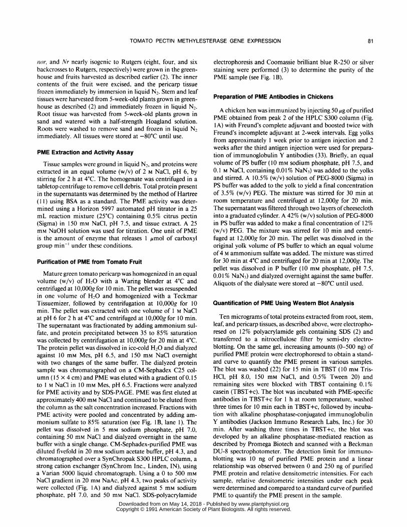
Figure 1. A, Elution profile of PME from SynChropak S300 strongcation exchange column. The HPLC SynChropak S300 column was
equilibrated with 20 mm Na acetate, pH 4.3, and CM-Sephadex-purified PME was chromatographed as described in "Materials andMethods." Elution peaks 1 and 2 represent absorbance at 280 nm.
Numbers indicate time (min) of elution for each peak from the Syn-Chropak S300 column. B, SDS-PAGE analysis of various fractionsduring PME purification. Gel was stained with Coomassie brilliantblue. Lanes: (1) 35 to 85% ammonium sulfate fraction; (2) CM-Sephadex C25-purified PME; (3) peak 2 from HPLC S300; (4) peak1 from HPLC S300.
Figure 2. A, DNA sequence of a partial PME cDNA clone (PET1) andits comparison with a PME cDNA clone (ENG-PME, nucleotide 316to 989) isolated by Ray et al. (27). Dashes represent homologousnucleotides and mismatched nucleotides are shown. B, Comparisonof deduced amino acid sequences from PET1 with the rearrangedprimary amino acid sequence of tomato PME determined by Markovicand Jornell (21) (M-PME) and deduced amino acid sequence fromENG-PME (nucleotide 318 to 989) isolated by Ray et al. (27). Theamino acid sequence of M-PME begins with the published mature N-terminal of tomato PME (21). The continuity of the amino acids in thepublished PME polypeptide chain (21) was rearranged to align theprimary amino acid sequence to the deduced amino acid sequences
of PET1 and ENG-PME (27). See text for details. Mismatched aminoacids are shown. Dashes represent homologous amino acids anddots represent absence of amino acids.
A
B
83
......
ANSM".
www.plantphysiol.orgon May 14, 2018 - Published by Downloaded from Copyright © 1991 American Society of Plant Biologists. All rights reserved.

Plant Physiol. Vol. 97, 1991
Table I. Comparison of PME Amino Acid Similarity Deduced fromTwo PME cDNA Clones and a Determined PME Primary AminoAcid Sequence
PET1 is the PME cDNA isolated in the present study; ENG-PMErepresents the PME cDNA isolated by Ray et al. (27); and M-PME isthe rearranged (Fig. 2) primary amino acid sequence of tomato PMEreported by Markovic and Jornell (21).
Amino Acid Conserved Total MismatchedI
Sequences Identity
ENG-PME/PET1 203 224 21 90.6ENG-PME/M-PME 184 210 26 87.6PET1 /M-PME 197 210 13 93.8
old) and declined as the fruit ripened (Fig. 3A, C). Interest-ingly, the levels ofPME mRNA declined just before a twofoldincrease in PME activity and protein during the ripeningphase.
Levels of PME Activity, Protein, and mRNA in TomatoRoot, Stem, and Leaf Tissues
Stem and leaf tissue contained PME activity similar to 10-and 15-d-old fruit, whereas root showed PME activity levelscomparable to 25-d-old fruit (Fig. 3A). However, no detecta-ble levels of PME protein and mRNA were found in root,stem, or leaf tissues using antibodies raised against fruit PMEand the PME cDNA cloned using fruit mRNA as probes,respectively (Fig. 3B, C). These results suggest that differentisozyme(s) of PME are present in these tissues that are notrecognized by fruit-specific PME antibodies and cDNAprobes.
PME Gene Expression during Mutant PericarpDevelopment
Pericarp from homozygous rin, nor, and Nr ripening mu-tants were similarly examined for PME gene expression dur-ing fruit development (Fig. 4). However, fruits from thesetomato mutants do not undergo normal ripening. This makesit difficult to compare between the expression of PME in thelate stages of mutant fruit development and the various stagesof wild-type fruit ripening. Thus, in the present investigationwe have characterized the expression ofPME in mutant fruitsbased on fruit age (up to 75 DAF). In general, fruits fromRutgers reach a fully ripe stage by 50 ± 5 DAF.PME activity profiles during the early stages of fruit devel-
opment (up to 45 DAF) in rin, nor, and Nr pericarp weresimilar to that observed in the normally ripening cultivarRutgers, but were different in the later stages of fruit devel-opment (Fig. 4). Pericarp from rin showed a sharp decline inPME activity in the later stages of fruit development (after 45DAF). The profile ofPME activity in nor pericarp was similarto the Rutgers pericarp, with a significant increase in PMEactivity between the 55 and 65 DAF stages. Developingpericarp from Nr fruits showed a pattern similar to nor fruitsbut contained lower levels ofPME activity with compared tonor fruits. As compared with Rutgers fruits, which showmaximum PME activity at the turning stage of fruit ripening(which occurs about 45 DAF), the maximum measured PME
activities in Nr and nor fruits were observed between 65 and75 DAF (Fig. 4).Changes in PME protein reflected observed changes in PME
activity during the development of pericarp in mutant fruits(Fig. 4). Detectable levels ofPME protein were present in 15-d-old rin pericarp. This could be due to the grouping of 10 to20 DAF pericarp to obtain the 15 DAF sample used in thisand previous studies (2). Levels ofPME protein in rin pericarpincreased up to 45 DAF and then showed a steady decline.Pericarp from Nr and nor did not show this decline duringfruit development.
-.~~..e
20 2530 35MG rTuFRR r t if
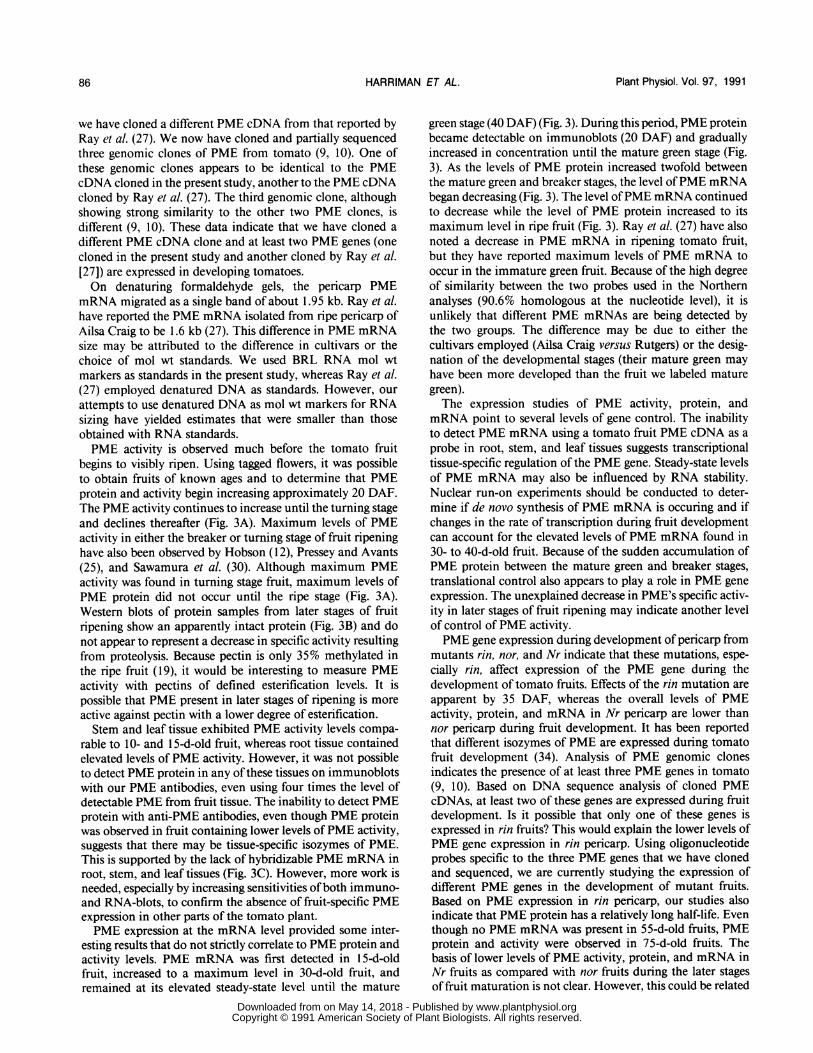
Figure 3. Quantification of the changes in the level of PME activity,protein, and mRNA during tomato fruit development and in root,stem, and leaf tissues. Protein was extracted from various tissues ofnormally ripening Rutgers in 1 M NaCI, pH 6, and assayed for PMEactivity using an automated pH titrator. The immunoblot, shown in B,was scanned with a Beckman DU-8 spectrophotometer to quantifyPME protein in samples. For immunoblots, 10 ,ug of 1 M NaCI-extractable protein for each sample was electrophoresed on 12%SDS-PAGE gel and PME protein was detected using PME antibodiesas described in "Materials and Methods." Purified PME protein (0-500 ng) was used to create a standard curve. The Northern blotautoradiogram, shown in C, was scanned with a Beckman DU-8spectrophotometer and the relative intensities were determined (ar-bitrary units). Twenty-five micrograms of total RNA from each samplewas used for Northern blot analyses. Numbered samples indicateage (DAF) of tomato fruits, and ripening stages are depicted byabbreviations: MG, mature green; Br, breaker; Tu, turning; Ri, ripe;RR, overripe. rt, st, and If represent root, shoot, and leaf tissue,respectively.
84 HARRIMAN ET AL.
www.plantphysiol.orgon May 14, 2018 - Published by Downloaded from Copyright © 1991 American Society of Plant Biologists. All rights reserved.

TOMATO PECTIN METHYLESTERASE GENE EXPRESSION
Nr Nr nor nor
15 25 35 45 55 65 75
15 25 35 45 55 65 75 15 25 35 45 55 65 75 15 25 35 45 55 65 75
Figure 4. Changes in PME activity, protein, andmRNA in developing tomato fruit of the ripeningmutants rin, nor, and Nr. Flowers were taggedat pollination and fruits were harvested at variousDAF. Fruits harvested throughout 10 d intervalsof age were grouped and midrange age wasused to indicate age. For example, the 25 DAFsample represents pericarp from fruits harvestedbetween 20 and 29 DAF. Other details are thesame as in Figure 3. Numbers indicate age (DAF)of fruits.
15 25 35 45 55 65 75 15 25 35 45 55 65 75X'IW-_
15 25 35 45 55 65 75
Days After Flowering
PME mRNA levels in rin pericarp increased during the first35 d of fruit development, a pattern similar to that observedin Rutgers pericarp. However, thereafter the PME mRNAlevels decreased rapidly and were barely detectable in 55, 65,and 75 DAF pericarp. This pattern ofPME mRNA in devel-oping rin pericarp was different from that of PME activityand protein (Fig. 4). Although both PME activity and proteinshowed decreases during the later stages of rin pericarp devel-opment, they were present until 75 DAF. This result suggeststhat PME protein has a relatively long half-life in rin tomatofruits. However, other more elaborate mechanisms, such as
translational efficiency of mRNA during fruit development,should also be considered. Pericarp from Nr and nor genotypesexhibited changes in PME mRNA levels similar to thoseobserved in pericarp from normally ripening Rutgers (Figs.3C, 4). The PME mRNA levels remained elevated until 45DAF and then showed a steady decline as fruits from Nr andnor matured.
DISCUSSION
PME activity in ripening tomato fruit has been studied bya number of research groups (6, 12, 25, 30, 34). The presenceof multiple forms of PME in tomato fruit has been noted byseveral investigators. Pressey and Avants (24) examined peri-carp protein extracts from four different cultivars of tomatoes(Green Marion, Homestead, Ripe Marion, and Pixie) bypassing the protein samples through a DEAE-Sephadex A50column and assaying for PME activity. Four peaks of PMEactivity were resolved and labeled PMEI through PMEIVbased on elution profiles. PMEIV was the most abundantform in all cultivars with the exception of Pixie. Tucker et al.(34) also used DEAE-Sephadex A50 separation to study "iso-zymes" of PME from the cultivar Ailsa Craig and reportedtwo forms of PME (PME1 and PME2) in both mature greenand ripe fruits. They showed that PME 1 decreases slightly as
the fruit ripens, whereas PME2 increases roughly twofoldfrom the mature green to the ripe stage (34). Delincee was
able to detect up to eight molecular forms of tomato PME
when thin layer isoelectric focusing was combined with activ-ity staining (8). By passing the CM-Sephadex-purified PMEthrough the HPLC S300 column, we are able to obtain twopeaks of PME activity. The first peak comprises two proteins(the 34 kD PME and 37 kD proteins), and the second peakcontains only the 34 kD PME protein (Fig. 1). At present, wedo not know if the PME present in the first and second peaksare different isozymes or the result of a molecular associationbetween PME and the 37 kD protein that influences theelution from the S300 column, giving rise to two peaks ofactivity. Because of the use of different ion exchange columns(DEAE-Sephedex A50 by Tucker et al. [34] and HPLC S300in the present study), it is not possible to say that the PME 1
and PME2 proteins separated by the HPLC S300 column are
the same as the PME 1 and PME2 proteins reported by Tuckeret al. (34). A biochemical explanation for the presence ofmultiforms of PME has not yet been established. However,based on analysis of genomic DNA clones, we have shownthe presence ofat least three PME genes in the tomato genome(9, 10).The deduced amino acid sequence from PET1 could be
aligned to the published primary amino acid sequence oftomato PME (21) only after rearranging the peptide fragmentsofthe PME polypeptide chain, a result similar to that observedby Ray et al. (27). As discussed by Ray et al. (27), thisdiscrepancy is likely due to misinterpretation of peptide over-
lap in the direct primary amino acid sequence analysis ratherthan posttranslational rearrangement of the PME precursor
polypeptide. The deduced amino acid sequence of PET 1 ismost similar (93.8%) to the rearranged PME amino acidsequence reported by Markovic and Jornell (21) (M-PME inFig. 2 and Table I) and is somewhat less similar (90.6%) tothe deduced amino acid sequence ofPME reported by Ray etal. (27) (ENG-PME in Fig. 2 and Table I). However, thePET1 sequence shows greater similarity to both M-PME andENG-PME than M-PME and ENG-PME show to each other(Table I). The comparison of our PME cDNA and the PMEcDNA isolated by Ray et al. (27) with the published PMEprotein sequence of Markovic and Jornell (21) indicates that
rin rin
CL.5
EN.~
C._
0L.CL
15 25 35 45 55 65 75
E
85
www.plantphysiol.orgon May 14, 2018 - Published by Downloaded from Copyright © 1991 American Society of Plant Biologists. All rights reserved.
Plant Physiol. Vol. 97, 1991
we have cloned a different PME cDNA from that reported byRay et al. (27). We now have cloned and partially sequencedthree genomic clones of PME from tomato (9, 10). One ofthese genomic clones appears to be identical to the PMEcDNA cloned in the present study, another to the PME cDNAcloned by Ray et al. (27). The third genomic clone, althoughshowing strong similarity to the other two PME clones, isdifferent (9, 10). These data indicate that we have cloned a
different PME cDNA clone and at least two PME genes (onecloned in the present study and another cloned by Ray et al.[27]) are expressed in developing tomatoes.On denaturing formaldehyde gels, the pericarp PME
mRNA migrated as a single band of about 1.95 kb. Ray et al.have reported the PME mRNA isolated from ripe pericarp ofAilsa Craig to be 1.6 kb (27). This difference in PME mRNAsize may be attributed to the difference in cultivars or thechoice of mol wt standards. We used BRL RNA mol wtmarkers as standards in the present study, whereas Ray et al.(27) employed denatured DNA as standards. However, our
attempts to use denatured DNA as mol wt markers for RNAsizing have yielded estimates that were smaller than thoseobtained with RNA standards.PME activity is observed much before the tomato fruit
begins to visibly ripen. Using tagged flowers, it was possibleto obtain fruits of known ages and to determine that PMEprotein and activity begin increasing approximately 20 DAF.The PME activity continues to increase until the turning stageand declines thereafter (Fig. 3A). Maximum levels of PMEactivity in either the breaker or turning stage of fruit ripeninghave also been observed by Hobson (12), Pressey and Avants(25), and Sawamura et al. (30). Although maximum PMEactivity was found in turning stage fruit, maximum levels ofPME protein did not occur until the ripe stage (Fig. 3A).Western blots of protein samples from later stages of fruitripening show an apparently intact protein (Fig. 3B) and donot appear to represent a decrease in specific activity resultingfrom proteolysis. Because pectin is only 35% methylated inthe ripe fruit (19), it would be interesting to measure PMEactivity with pectins of defined esterification levels. It ispossible that PME present in later stages of ripening is moreactive against pectin with a lower degree of esterification.Stem and leaf tissue exhibited PME activity levels compa-
rable to 10- and 15-d-old fruit, whereas root tissue containedelevated levels of PME activity. However, it was not possibleto detect PME protein in any ofthese tissues on immunoblotswith our PME antibodies, even using four times the level ofdetectable PME from fruit tissue. The inability to detect PMEprotein with anti-PME antibodies, even though PME proteinwas observed in fruit containing lower levels ofPME activity,suggests that there may be tissue-specific isozymes of PME.This is supported by the lack of hybridizable PME mRNA inroot, stem, and leaf tissues (Fig. 3C). However, more work isneeded, especially by increasing sensitivities ofboth immuno-and RNA-blots, to confirm the absence of fruit-specific PMEexpression in other parts of the tomato plant.PME expression at the mRNA level provided some inter-
esting results that do not strictly correlate to PME protein andactivity levels. PME mRNA was first detected in 15-d-oldfruit, increased to a maximum level in 30-d-old fruit, andremained at its elevated steady-state level until the mature
green stage (40 DAF) (Fig. 3). During this period, PME proteinbecame detectable on immunoblots (20 DAF) and graduallyincreased in concentration until the mature green stage (Fig.3). As the levels of PME protein increased twofold betweenthe mature green and breaker stages, the level ofPME mRNAbegan decreasing (Fig. 3). The level ofPME mRNA continuedto decrease while the level of PME protein increased to itsmaximum level in ripe fruit (Fig. 3). Ray et al. (27) have alsonoted a decrease in PME mRNA in ripening tomato fruit,but they have reported maximum levels of PME mRNA tooccur in the immature green fruit. Because of the high degreeof similarity between the two probes used in the Northernanalyses (90.6% homologous at the nucleotide level), it isunlikely that different PME mRNAs are being detected bythe two groups. The difference may be due to either thecultivars employed (Ailsa Craig versus Rutgers) or the desig-nation of the developmental stages (their mature green mayhave been more developed than the fruit we labeled maturegreen).The expression studies of PME activity, protein, and
mRNA point to several levels of gene control. The inabilityto detect PME mRNA using a tomato fruit PME cDNA as aprobe in root, stem, and leaf tissues suggests transcriptionaltissue-specific regulation of the PME gene. Steady-state levelsof PME mRNA may also be influenced by RNA stability.Nuclear run-on experiments should be conducted to deter-mine if de novo synthesis of PME mRNA is occuring and ifchanges in the rate of transcription during fruit developmentcan account for the elevated levels of PME mRNA found in30- to 40-d-old fruit. Because of the sudden accumulation ofPME protein between the mature green and breaker stages,translational control also appears to play a role in PME geneexpression. The unexplained decrease in PME's specific activ-ity in later stages of fruit ripening may indicate another levelof control of PME activity.PME gene expression during development of pericarp from
mutants rin, nor, and Nr indicate that these mutations, espe-cially rin, affect expression of the PME gene during thedevelopment of tomato fruits. Effects of the rin mutation areapparent by 35 DAF, whereas the overall levels of PMEactivity, protein, and mRNA in Nr pericarp are lower thannor pericarp during fruit development. It has been reportedthat different isozymes of PME are expressed during tomatofruit development (34). Analysis of PME genomic clonesindicates the presence of at least three PME genes in tomato(9, 10). Based on DNA sequence analysis of cloned PMEcDNAs, at least two of these genes are expressed during fruitdevelopment. Is it possible that only one of these genes isexpressed in rin fruits? This would explain the lower levels ofPME gene expression in rin pericarp. Using oligonucleotideprobes specific to the three PME genes that we have clonedand sequenced, we are currently studying the expression ofdifferent PME genes in the development of mutant fruits.Based on PME expression in rin pericarp, our studies alsoindicate that PME protein has a relatively long half-life. Eventhough no PME mRNA was present in 55-d-old fruits, PMEprotein and activity were observed in 75-d-old fruits. Thebasis of lower levels of PME activity, protein, and mRNA inNr fruits as compared with nor fruits during the later stagesof fruit maturation is not clear. However, this could be related
86 HARRIMAN ET AL.
www.plantphysiol.orgon May 14, 2018 - Published by Downloaded from Copyright © 1991 American Society of Plant Biologists. All rights reserved.

TOMATO PECTIN METHYLESTERASE GENE EXPRESSION
to the effects of these mutations on the overall developmentand ripening of mutant fruits.Comparison of the effects of rin, nor, and Nr mutations on
the expression of PME and PG genes during tomato fruitdevelopment provides further insight into the nature of thesemutations. The rin mutation shows the greatest effect on theexpression of PME, virtually eliminating PME mRNA by 55DAF. The effect of the rin mutation on PME gene expressionis similar to the effect observed on PG gene expression (2). Inan earlier study (2), we showed that PG mRNA was detectableat a very reduced level in 45 DAF rin fruit, but not in thelater stages of rin fruit development. Our data suggest that therin mutation has an effect on the expression of several genesin developing fruits and begins to manifest itself by 35 DAF.Although the nor mutation also greatly depressed PG geneexpression in tomato fruit (2), it does not impair the expres-sion of the PME gene. The effect of the Nr mutation on
temporal regulation of PME and PG gene expression at themRNA levels is similar. Collectively, the results indicate thatthese mutations have different effects on the expression ofgenes involved in tomato fruit development.
ACKNOWLEDGMENTS
We thank Paul Parker for excellent technical assistance, Drs. P.B.Goldsbrough, R.W. Woodson, and V. Zur for critical review of themanuscript, and Becky Fagan for typing the manuscript.
LITERATURE CITED
1. Awad M, Young RE (1979) Postharvest variation in cellulase,polygalacturonase, and pectin methylesterase in avocado (Per-sea americana Mill, cv Fuerte) fruit in relation to respirationand ethylene production. Plant Physiol 64: 306-308
2. Biggs MS, Handa AK (1989) Temporal regulation of polygalac-turonase gene expression in fruits from normal mutant andheterozygous tomato genotypes. Plant Physiol 89: 117-125
3. Biggs MS, Harriman RW, Handa AK (1986) Changes in geneexpression during tomato fruit ripening. Plant Physiol 81:395-403
4. Brady CJ (1976) The pectinesterase of pulp banana fruit. Aust JPlant Physiol 3: 163-172
5. Brady CJ (1987) Fruit ripening. Annu Rev Plant Physiol 38:179-204
6. Buescher RW, Tigchelaar EC (1975) Pectinesterase, polygalac-turonase, cx-cellulase activities and softening of the rin tomatomutant. HortScience 10: 624-625
7. Collmer A, Keen NT (1986) The role of pectic enzymes in plantpathogenesis. Annu Rev Phytopathol 24: 383-409
8. Delincee H (1976) Thin-layer isoelectric focusing of multipleforms of tomato pectinesterase. Phytochemistry 15: 903-906
9. Harriman RW (1990) Molecular cloning and regulation ofexpression of pectin methylesterase in ripening tomato. PhDthesis. Purdue University, West Lafayette, IN
10. Harriman RW, Handa AK (1990) Identification and character-ization of three pectin methylesterase genes in tomato (abstractNo. 249). Plant Physiol 93: S-44
11. Hartree EF (1972) Determination of protein: a modification ofthe Lowry method that gives a linear photometric response.Anal Biochem 48: 422-427
12. Hobson GE (1963) Pectinesterase in normal and abnormal to-mato fruit. Biochem J 86: 358-365
13. Hobson GE (1967) The effects of alleles at the "never ripe" locuson the ripening tomato fruit. Phytochemistry 6: 1337-1341
14. Hobson GE (1980) Effect of the introduction of non-ripeningmutant genes on the composition and enzyme content oftomato fruit. J Sci Food Agric 34: 578-584
15. Huber DJ (1983) The role of cell wall hydrolases in fruit soften-ing. Hortic Rev 5: 169-219
16. Huynh TV, Young RA, Davis RW (1984) Constructing andscreening cDNA libraries in Xgtll. In DM Glover, ed, DNACloning. Vol 1 1: A Practical Approach. IRL Press, Washing-ton. DC pp 49-78
17. Kertesz ZI, McColloch RJ (1950) Enzymes acting on pecticsubstances. In CS Hudson, SM Cantor, eds, Advances inCarbohydrate Chemistry and Biochemistry, Vol 5. AcademicPress, New York, pp 79-102
18. Knee M (1978) Metabolism of polymethylgalacturonate in applefruit cortical tissue during ripening. Phytochemistry 17: 126 1-1264
19. Koch JL, Nevins DM (1989) Tomato fruit cell wall: use ofpurified tomato polygalacturonase and pectinmethylesterase toidentify developmental changes in pectins. Plant Physiol 91:816-822
20. Maniatis T, Fritsch EF, Sambrook J (1982) Molecular Cloning:A Laboratory Manual. Cold Spring Harbor Laboratory Press,Cold Spring Harbor, NY
21. Markovic 0, Jornell H (1986) Pectinesterase: the primary struc-ture of the tomato enzyme. Eur J Biochem 158: 455-462
22. Marlow SJ, Handa AK (1987) Immuno slot-blot assay using amembrane which covalently binds protein. J Immunol Meth-ods 101: 133-139
23. Paull RE, Chen NJ (1983) Postharvest variation in cell wall-degrading enzymes of papaya (Carica papava L.) during fruitripening. Plant Physiol 72: 382-385
24. Pressey R, Avants JK (1972) Multiple forms of pectin esterasein tomatoes. Phytochemistry 11: 3139-3142
25. Pressey R, Avants JK (1982) Pectic enzymes in "Long Keeper'tomatoes. HortScience 17: 398-400
26. Pressey R, Avants JK (1982) Solubilization of cell walls bytomato polygalacturonases: effects of pectinesterases. J FoodBiochem 6: 57-74
27. Ray J, Knapp J, Grierson D, Bird C, Schuch W (1988) Identifi-cation and sequence determination of a cDNA clone for to-mato pectin esterase. Eur J Biochem 174: 119-124
28. Rexova-Benkova L, Markovic 0 (1976) Pectic enzymes. In RSTipson, D Horton, eds, Advances in Carbohydrate Chemistryand Biochemistry, Vol 33. Academic Press, New York pp 323-385
29. Sanger F, Nicklen S, Coulsen AR (1977) DNA sequencing withchain-terminating inhibitors. Proc Natl Acad Sci USA 74:5463-5467
30. Sawamura M, Knegt E, Bruinsma J (1978) Levels of endogenousethylene, carbon dioxide, and soluble pectin, and activities ofpectin methylesterase and polygalacturonase in ripening to-mato fruits. Plant Cell Physiol 19: 1061-1069
31. Sexton R, Roberts JA (1982) Cell biology of abscission. AnnuRev Plant Physiol 33: 133-162
32. Seymour GB, Lasslett Y, Tucker GA (1987) Differential effectsof pectolytic enzymes on tomato polyuronides in vivo and invitro. Phytochemistry 26: 3137-3139
33. Song C-H, Yu J-H, Bai DH, Hester PY, Kim K-H (1985)Antibodies to the a-subunit of insulin receptor from eggs ofimmunized hens. J Immunol 135: 3354-3359
34. Tucker GA, Robertson NG, Grierson D (1982) Purification andchanges in activities of tomato pectinesterase isoenzymes. J SciFood Agric 33: 396-400
87
www.plantphysiol.orgon May 14, 2018 - Published by Downloaded from Copyright © 1991 American Society of Plant Biologists. All rights reserved.