Molecular mechanisms of bacterial resistance to...
13
Molecular mechanisms of bacterial resistance to antimicrobial agents Sanath Kumar 1 and Manuel F. Varela 2 1 QC Laboratory, Harvest and Post Harvest Technology Division, Central Institute of Fisheries Education (CIFE), Seven Bungalows, Versova, Andheri (W), Mumbai 400061 India 2 Biology Department, Eastern New Mexico University, Portales, NM USA 88130 Infectious diseases caused by bacterial pathogens represent a serious public health concern. Antimicrobial agents such as anti-bacterial drugs are often indicated for chemotherapy of bacterial infections in clinical medicine. Thus, it is important to study the biological mechanisms that confer bacterial pathogenesis and virulence. Under selective evolutionary pressure when in the presence of antimicrobial agents, bacterial variants evolve mechanisms to survive in the presence of these inhibitory agents. Drug resistant bacteria that are selected with a single drug are also frequently multi-drug resistant against multiple structurally different drugs, thus confounding the chemotherapeutic efficacy of infectious disease caused by such pathogenic variants. There are several major classes of mechanisms for bacterial resistance to antimicrobial agents: (a) enzymatic inactivation of the drug results from the metabolic degradation of the drug into a form that is rendered ineffective in inhibiting bacterial growth; (b) alteration of the drug target results in the inability of the drug to bind to its biological target, thus rendering the drug unable to kill the bacteria. Bacterial cellular drug targets may include the protein synthesis apparatus, nucleic acid synthesis enzymes, cell wall synthesis machinery, and metabolite pathway enzymes; (c) drug permeability reduction mechanisms prevent cellular entry of drug into the inside of the bacterial cell; and (d) active efflux of drugs from bacteria results in the intracellular dilution of drugs, making the extruded drugs unavailable for their inhibitory action. Unfortunately, these drug and multi-drug resistance mechanisms are poorly understood at the molecular level, impeding our advances towards identifying new targets for possible inhibition of clinical multi-drug resistances; this prevents chemotherapeutic usefulness. Understanding how these bacterial resistance mechanisms work from the standpoint of molecular physiology and biochemistry will identify new targets for potential inhibition of multi-drug resistance and thus restore clinical utility of chemotherapy of infectious disease caused by serious bacterial pathogens. Keywords antimicrobial agent; drug; antibacterial drug; bacteria; antibiotic resistance; efflux; multidrug efflux 1. Introduction Bacteria that are causative agents of infectious disease represent a serious public health concern globally. Antimicrobial agents are indicated for the treatment of bacterial infections. Bacteria may be intrinsically resistant to antibacterial agents or acquire resistance by mutation or acquisition of resistance determinants. Use of antimicrobial agents selects for bacterial variants within a population that are less susceptible, or resistant, to the antimicrobial agent used, leading to a situation where the resistant variant predominates under such selective pressure [1]. Furthermore, selection of resistance to a single antimicrobial agent often results in bacterial variants that harbor transferable multidrug resistance determinants [2]. These selective pressure phenomena are thought to occur in areas where antimicrobial agents are extensively used, such as in human clinical medicine [3, 4], agriculture [5-7], and in natural soil and aquatic environments [8-12]. Therefore, antimicrobial use fosters bacterial drug resistance and dissemination of drug resistance determinants within populations. Multidrug resistant bacteria may be recalcitrant to clinically relevant chemotherapeutic agents, resulting in treatment failures of infectious diseases [13]. Study of these antimicrobial resistance mechanisms in infectious disease causing microorganisms is, therefore, necessary in order to find ways to circumvent conditions that foster such recalcitrant pathogens. Molecular, biochemical, physiological and structural analyses of bacterial multiple drug resistance mechanisms will foster their putative modulation and make possible the restoration of the efficacy of infectious disease chemotherapy [14-18]. Microbial pathogens and strategies for combating them: science, technology and education (A. Méndez-Vilas, Ed.) © FORMATEX 2013 ____________________________________________________________________________________________ 522
Transcript of Molecular mechanisms of bacterial resistance to...

Molecular mechanisms of bacterial resistance to antimicrobial agents
Sanath Kumar1 and Manuel F. Varela2 1QC Laboratory, Harvest and Post Harvest Technology Division, Central Institute of Fisheries Education (CIFE), Seven
Bungalows, Versova, Andheri (W), Mumbai 400061 India 2Biology Department, Eastern New Mexico University, Portales, NM USA 88130
Infectious diseases caused by bacterial pathogens represent a serious public health concern. Antimicrobial agents such as anti-bacterial drugs are often indicated for chemotherapy of bacterial infections in clinical medicine. Thus, it is important to study the biological mechanisms that confer bacterial pathogenesis and virulence. Under selective evolutionary pressure when in the presence of antimicrobial agents, bacterial variants evolve mechanisms to survive in the presence of these inhibitory agents. Drug resistant bacteria that are selected with a single drug are also frequently multi-drug resistant against multiple structurally different drugs, thus confounding the chemotherapeutic efficacy of infectious disease caused by such pathogenic variants. There are several major classes of mechanisms for bacterial resistance to antimicrobial agents: (a) enzymatic inactivation of the drug results from the metabolic degradation of the drug into a form that is rendered ineffective in inhibiting bacterial growth; (b) alteration of the drug target results in the inability of the drug to bind to its biological target, thus rendering the drug unable to kill the bacteria. Bacterial cellular drug targets may include the protein synthesis apparatus, nucleic acid synthesis enzymes, cell wall synthesis machinery, and metabolite pathway enzymes; (c) drug permeability reduction mechanisms prevent cellular entry of drug into the inside of the bacterial cell; and (d) active efflux of drugs from bacteria results in the intracellular dilution of drugs, making the extruded drugs unavailable for their inhibitory action. Unfortunately, these drug and multi-drug resistance mechanisms are poorly understood at the molecular level, impeding our advances towards identifying new targets for possible inhibition of clinical multi-drug resistances; this prevents chemotherapeutic usefulness. Understanding how these bacterial resistance mechanisms work from the standpoint of molecular physiology and biochemistry will identify new targets for potential inhibition of multi-drug resistance and thus restore clinical utility of chemotherapy of infectious disease caused by serious bacterial pathogens.
Keywords antimicrobial agent; drug; antibacterial drug; bacteria; antibiotic resistance; efflux; multidrug efflux
1. Introduction
Bacteria that are causative agents of infectious disease represent a serious public health concern globally. Antimicrobial agents are indicated for the treatment of bacterial infections. Bacteria may be intrinsically resistant to antibacterial agents or acquire resistance by mutation or acquisition of resistance determinants. Use of antimicrobial agents selects for bacterial variants within a population that are less susceptible, or resistant, to the antimicrobial agent used, leading to a situation where the resistant variant predominates under such selective pressure [1]. Furthermore, selection of resistance to a single antimicrobial agent often results in bacterial variants that harbor transferable multidrug resistance determinants [2]. These selective pressure phenomena are thought to occur in areas where antimicrobial agents are extensively used, such as in human clinical medicine [3, 4], agriculture [5-7], and in natural soil and aquatic environments [8-12]. Therefore, antimicrobial use fosters bacterial drug resistance and dissemination of drug resistance determinants within populations. Multidrug resistant bacteria may be recalcitrant to clinically relevant chemotherapeutic agents, resulting in treatment failures of infectious diseases [13]. Study of these antimicrobial resistance mechanisms in infectious disease causing microorganisms is, therefore, necessary in order to find ways to circumvent conditions that foster such recalcitrant pathogens. Molecular, biochemical, physiological and structural analyses of bacterial multiple drug resistance mechanisms will foster their putative modulation and make possible the restoration of the efficacy of infectious disease chemotherapy [14-18].
Microbial pathogens and strategies for combating them: science, technology and education (A. Méndez-Vilas, Ed.)
© FORMATEX 2013
____________________________________________________________________________________________
522
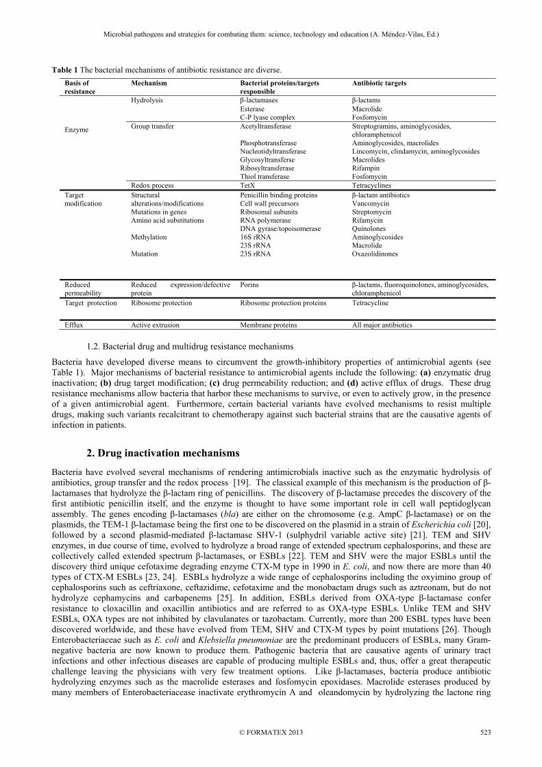
Table 1 The bacterial mechanisms of antibiotic resistance are diverse.
Basis of resistance
Mechanism Bacterial proteins/targets responsible
Antibiotic targets
Enzyme
Hydrolysis β-lactamases β-lactams Esterase Macrolide C-P lyase complex Fosfomycin
Group transfer Acetyltransferase Streptogramins, aminoglycosides, chloramphenicol
Phosphotransferase Aminoglycosides, macrolides Nucleotidyltransferase Lincomycin, clindamycin, aminoglycosides Glycosyltransferse Macrolides Ribosyltransferase Rifampin Thiol transferase Fosfomycin
Redox process TetX Tetracyclines Target modification
Structural alterations/modifications
Penicillin binding proteins β-lactam antibiotics Cell wall precursors Vancomycin
Mutations in genes Ribosomal subunits Streptomycin Amino acid substitutions RNA polymerase Rifamycin
DNA gyrase/topoisomerase Quinolones Methylation 16S rRNA Aminoglycosides
23S rRNA Macrolide Mutation 23S rRNA Oxazolidinones
Reduced permeability
Reduced expression/defective protein
Porins β-lactams, fluoroquinolones, aminoglycosides, chloramphenicol
Target protection Ribosome protection Ribosome protection proteins Tetracycline
Efflux Active extrusion Membrane proteins All major antibiotics
1.2. Bacterial drug and multidrug resistance mechanisms
Bacteria have developed diverse means to circumvent the growth-inhibitory properties of antimicrobial agents (see Table 1). Major mechanisms of bacterial resistance to antimicrobial agents include the following: (a) enzymatic drug inactivation; (b) drug target modification; (c) drug permeability reduction; and (d) active efflux of drugs. These drug resistance mechanisms allow bacteria that harbor these mechanisms to survive, or even to actively grow, in the presence of a given antimicrobial agent. Furthermore, certain bacterial variants have evolved mechanisms to resist multiple drugs, making such variants recalcitrant to chemotherapy against such bacterial strains that are the causative agents of infection in patients.
2. Drug inactivation mechanisms
Bacteria have evolved several mechanisms of rendering antimicrobials inactive such as the enzymatic hydrolysis of antibiotics, group transfer and the redox process [19]. The classical example of this mechanism is the production of β-lactamases that hydrolyze the β-lactam ring of penicillins. The discovery of β-lactamase precedes the discovery of the first antibiotic penicillin itself, and the enzyme is thought to have some important role in cell wall peptidoglycan assembly. The genes encoding β-lactamases (bla) are either on the chromosome (e.g. AmpC β-lactamase) or on the plasmids, the TEM-1 β-lactamase being the first one to be discovered on the plasmid in a strain of Escherichia coli [20], followed by a second plasmid-mediated β-lactamase SHV-1 (sulphydril variable active site) [21]. TEM and SHV enzymes, in due course of time, evolved to hydrolyze a broad range of extended spectrum cephalosporins, and these are collectively called extended spectrum β-lactamases, or ESBLs [22]. TEM and SHV were the major ESBLs until the discovery third unique cefotaxime degrading enzyme CTX-M type in 1990 in E. coli, and now there are more than 40 types of CTX-M ESBLs [23, 24]. ESBLs hydrolyze a wide range of cephalosporins including the oxyimino group of cephalosporins such as ceftriaxone, ceftazidime, cefotaxime and the monobactam drugs such as aztreonam, but do not hydrolyze cephamycins and carbapenems [25]. In addition, ESBLs derived from OXA-type β-lactamase confer resistance to cloxacillin and oxacillin antibiotics and are referred to as OXA-type ESBLs. Unlike TEM and SHV ESBLs, OXA types are not inhibited by clavulanates or tazobactam. Currently, more than 200 ESBL types have been discovered worldwide, and these have evolved from TEM, SHV and CTX-M types by point mutations [26]. Though Enterobacteriaceae such as E. coli and Klebsiella pneumoniae are the predominant producers of ESBLs, many Gram-negative bacteria are now known to produce them. Pathogenic bacteria that are causative agents of urinary tract infections and other infectious diseases are capable of producing multiple ESBLs and, thus, offer a great therapeutic challenge leaving the physicians with very few treatment options. Like β-lactamases, bacteria produce antibiotic hydrolyzing enzymes such as the macrolide esterases and fosfomycin epoxidases. Macrolide esterases produced by many members of Enterobacteriacease inactivate erythromycin A and oleandomycin by hydrolyzing the lactone ring
Microbial pathogens and strategies for combating them: science, technology and education (A. Méndez-Vilas, Ed.)
© FORMATEX 2013
____________________________________________________________________________________________
523
[27]. The other less studied mechanism of enzymatic degradation is the hydrolysis of the carbon-phosphorus bond in the epoxide antibiotic fosfomycin. This may be enzymatically achieved by a C-P lyase enzyme complex in many Gram-negative soil bacteria [28]. The second mechanism of antibiotic inactivation involves enzyme mediated structural alteration of the drug via transfer of a functional group such as an acyl, ribosyl, phosphoryl or thiol group [29]. The reaction is irreversible and the modified antibiotic is unable to bind to the target due to the resultant change in the structure. The antibiotics susceptible to this bacterial mechanism include aminoglycosides, fosfomycin, macrolides, lincomycin and chloramphenicol [19]. For instance, bacteria have evolved acetyl transferases which inactivate chloramphenicol [30], tetracycline-metabolizing enzymes that are largely uncharacterized [31, 32], and beta-lactamases that inactivate beta-lactams such as penicillin [33]. Highly active variants of these enzyme inactivation mechanisms for drugs are ubiquitous in the environment and have yet to be found within clinically-relevant bacterial pathogens [29, 34]. The enzymatic O-acetylation of chloramphenicol by chloramphenicol acetyltransferase (CATs) is responsible for the inactivation of this drug [30]. Similarity, the modification of aminoglycoside antibiotics into their inactive forms leading to bacterial resistance is achieved by aminoglycoside acetyltransferases or AACs [29]. The enzymes of this group vary in their choice of groups (hydroxyl or amino) as well as their positions on aminoglycoside antibiotics for acetyl group transfer, but their actions invariably lead to drastically reduced affinity of the antibiotics to their ribosomal targets [35]. The other enzyme-mediated inactivation of antibiotics include acetylation of streptogramins by streptogramin acetyl transferases (VATs, for virginiamycin acetyl transferases), aminoglycoside modification by aminoglycoside phosphotransferases (APHs), phosphorylation of macrolides by macrolide kinases (MPHs), glutathione induced fosfomycin inactivation by FosA (or FosB), ADP-ribosylation of rifampin by ADP-ribosyltransferases (ARRs), nucleotidylation of aminoglycosides and lincomycin by nucleotidyl transferases (ANTs and Lin), glycosylation of macrolide antibiotics by glycosyltransferases [29]. A less common mechanism is the inactivation of an antibiotic by redox process which involves flavin-dependent monoxygenase enzyme TetX. This enzyme transfers a single hydroxyl group to tetracycline at position 11a resulting in a structure that is less able to sequester Mg+ ions which are critical for binding of tetracycline to its bacterial target [36, 37]. TetX is present on a transposon, and this mechanism has been recently found to be responsible for bacterial resistance to a third generation tetracycline, tigecycline [32].
3. Ribosome protection
Certain bacteria have developed resistance mechanisms that protect the antimicrobial target. For example, in the case of bacterial protein synthesis inhibitors, such as tetracycline, the bacteria have the ability to produce ribosome protection proteins that bind to the ribosomal target thus preventing the binding of tetracycline to the ribosome [38]. Such ribosome protected bacteria will be able to grow in the presence of tetracycline as protein synthesis will be possible. Disease-causing bacteria harboring such ribosome protection mechanisms have been demonstrated to be clinically important, and these resistance determinants have been discussed extensively elsewhere [39, 40].
4. Biofilm formation
Biofilm production occurs in many loci, including teeth plaque, water environments, medical catheters, trauma wounds, etc. [41-43]. As such, microorganisms that are found in biofilms are protected from the entry of multiple antimicrobial agents [44]. Thus, biofilms are increasingly becoming a challenge in the human clinical medicine arena when considering potential chemotherapies with antibacterial agents; and this recently recognized new mode of resistance has been reviewed previously [45-47].
5. Target modification
Bacteria have found ways to alter the molecular targets of antimicrobial agents. Altered targets may include, for example, DNA gyrase, a target of quinolone antimicrobials [48], RNA polymerase, a target of rifampin [49, 50], the prokaryotic ribosome, a target of tetracycline and other protein synthesis inhibitors [51-53], and targets of antimetabolite drugs, such as the sulfonamides and related drugs [54]. One classical example of drug target modification is the staphylococcal mechanism of variously altering the penicillin binding protein (PBP) which is the target of β-lactam antibiotics. Staphylococcus aureus, the causative agent of serious infectious disease, becomes resistant to these antibiotics by any one of the several mechanisms such as mutation in PBP or acquisition of new PBP with reduced affinity to penicillins, over expression of PBP, etc [55]. Another example of an altered target mechanism includes substitution of amino acids in the quinolone-resistance determining region (QRDR) of DNA gyrase and topoisomerase IV resulting in less efficient binding of quinolone antibiotics [56]. This mechanism has been responsible for widespread quinolone resistance among the Enterobacteriaceae. Methylation of drug binding targets on 16S rRNA by rRNA methyl transferases is responsible for aminoglycoside resistance in several bacterial species [35]. On the other hand, mutations in genes (rrs) encoding ribosomal subunits
Microbial pathogens and strategies for combating them: science, technology and education (A. Méndez-Vilas, Ed.)
© FORMATEX 2013
____________________________________________________________________________________________
524

lead to altered ribosomal protein targets which resist aminoglycoside binding, a mechanism responsible for streptomycin resistance in Mycobacterium tuberculosis, the causative agent of tuberculosis and other infections [57]. Methylation of the 23S rRNA component of 50S ribosomal subunit by adenine-specific N-methyltransferases is a common mechanism of macrolide resistance in many Gram-positive and –negative bacteria [58]. Also, mutations around the methylated sites have also been responsible for additional macrolide resistance. Modification of the drug target site which involves a G to A substitution at position 2,032 in the peptidyl transferase center of 23S rRNA results in reduced affinity of linezolid to the 50S subunit [59]. The vancomycin resistant enterococci (VRE) have evolved a unique mechanism of synthesizing peptidoglycan using alternate pathway thereby producing the peptidoglycan precursors ending with acyl-D-Ala4-D-Lac5 instead of the vancomycin target acyl D-Ala4-D-Ala5 [60].
6. Reduced permeability
A drug resistant phenotype of a bacterium may be due to the inability of the antimicrobial agent to gain entry into the cell where the drug targets are located [61]. One mechanism that results in reduced drug permeability in bacteria is the cell wall’s lipopolysaccharide (LPS), which consists of lipid A, a core consisting of polysaccharide and O-antigen [62-65]. Bacteria that harbor LPS moieties show resistance to erythromycin, roxithromycin, clarithromycin and azithromycin in Gram-negative bacteria such as strains of Pseudomonas aeruginosa, V. cholerae and S. enterica, all of which are serious pathogens, especially in immune-compromised patients [66-68]. Another mechanism that confers reduced permeability involves the porin channels that reside in the outer membrane and allow small molecular weight molecules, such as antimicrobial agents, to gain cellular entry [62, 69-71]. Drug resistant bacteria alter the expression of these outer membrane proteins such that they fail to integrate into the outer membrane or are functionally defective, thus preventing the entrance of growth-inhibitory molecules [61, 69, 72, 73]. Clinically important bacterial pathogens like Serratia marcescens, E. cloacae, S. enterica, E. aerogenes, Klebsiella pneumoniae, and P. aeruginosa, have utilized this reduced drug uptake system to resist important antimicrobial agents, such as the beta-lactams, fluoroquinolones, aminoglycosides, as well as chloramphenicol [62, 74].
7. Active drug efflux
One of the most common drug resistance mechanisms is active efflux of drugs from the inside of bacterial cells [75]. Such drug resistant bacteria harbor energy-driven drug efflux pumps which extrude antimicrobial agents thus reducing their intracellular concentrations to sub- or non-inhibitory levels. There are two main types of active efflux pumps. The first type, called primary active transport, uses the hydrolysis of ATP to actively efflux drugs from cells, while the second type, called secondary active transport, uses an ion gradient for active drug efflux from cells [76-79]. The ATP-driven transporters are also known as ABC (for ATP-binding cassette) or P-glycoprotein transporters [80, 81]. Both active transport systems are used by bacteria to resist the inhibitory effects of antimicrobial agents and are often referred to as efflux pumps [16, 82, 83]. In addition to single-drug efflux pump systems [83-86], bacteria may also express efflux pumps that are able to extrude multiple structurally-different antimicrobial agents and are referred to as multidrug efflux pumps [16, 84, 87-93]. These efflux pumps function by using the energy of the cation gradient generated by cellular respiration to catalyze the “uphill” transport of solute (e.g., drug substrate) across the membrane by translocation of the cation (e.g., H+ or Na+) down its concentration graduate in a process called antiport, where cation moves in one direction across the membrane and drug (substrate) moves in the opposite direction [77, 94, 95]. Since the secondary active drug and multidrug efflux pumps are considered to be predominant virulence factors in bacterial pathogens, we focus our discussion on these types of resistance determinants [57].
7.1. The tetracycline efflux pumps
The first antimicrobial efflux pump was discovered by Stuart Levy and co-workers in which the bacterium E. coli harbored an integral membrane protein specific for the efflux of the tetracyclines [96-98]. The tetracycline efflux pump is a secondary active transporter as it is energized by a membrane proton gradient [99]. The tetracycline efflux pumps are referred to as TetA and fall into several classes, such as TetA(A), TetA(B), TetA(C), TetA(D), etc., sometimes referred to simply as Tet(A), Tet(B), Tet(C) and Tet(D), respectively [85, 100-103]. Both Gram-negative and -positive bacteria harbor the TetA pumps, which are encoded on their genomes or on extra-chromosomal molecules such as transposons or on plasmid molecules [84, 104]. The deduced amino acid sequences of the TetA pumps are highly related, share predicted protein secondary structures in the biological membrane, and posses a common evolutionary origin with seemingly unrelated transporters that have diverse substrates, such as structurally-unrelated drugs, sugars, amino acids, and Kreb’s cycle intermediates [105-109]. These similarities predict that these transporters share a common molecular mechanism for the transport of structurally dissimilar substrates across the membrane [76, 85, 86, 106]. These transporters constitute individual members of a very large superfamily of homologous and related transport
Microbial pathogens and strategies for combating them: science, technology and education (A. Méndez-Vilas, Ed.)
© FORMATEX 2013
____________________________________________________________________________________________
525

proteins, called the Major Facilitator Superfamily (MFS) which are cleverly organized into a Transporter Classification (TC) Database http://www.tcdb.org/ [110-113]. The class B tetracycline efflux pump, TetA(B) from transposon Tn10, is the most well-studied antimicrobial efflux pump of the MFS [114]. TetB was studied by cysteine-scanning mutagenesis in which all amino acids were systematically replaced by cystiene and analyzed for their accessibilities to N-ethyl maleimide (NEM), which binds sulfhydryl-containing Cys residues [114]. Over 40 NEM-assessable TetA(B) residues were found and thought to line an aqueous-filled channel through which the tetracycline molecules are thought to be transported across the membrane [114]. These residues lie in six of the 12 helices that constitute the TetA(B) pump. The majority of the MFS transporters share a highly conserved amino acid motif, G-62 x x x D-66 R x G R-70 R, also known as Motif A, which lies in the loop structure between helices 2 and 3 [115]. The functional roles of these residues were examined in Tn10 TetA(B) and in the model sugar transporter lactose permease, LacY, of E. coli [116-118], which showed that residues corresponding to Gly-62, Asp-66, and Arg-70 of TetA(B) are required for function [119-122]. Another study found that Asp-120 and Arg-70 form a salt-bridge [123, 124]. Taken together, these findings suggest that the loop between helices 2 and 3 acts as a gate during tetracycline transport [114]. The functional role of a highly conserved arginine in TetB was established in another study, further suggesting that residues of motif A play a gating role during antimicrobial efflux [125]. The tetracycline efflux pumps represent a well-studied and important model system for analysis of drug resistance [126-128].
7.2. Secondary active multidrug efflux pumps from bacteria
To date, several major groups of secondary active multidrug efflux pumps have been discovered in prokaryotes and eukaryotes [57, 129, 130]. One group is the Multidrug and Toxic Compound Extrusion (MATE) efflux pump family, which has recently been elegantly reviewed [131]. Another efflux pump system is comprised within the Resistance-Nodulation-Division (RND) superfamily [132, 133]. The last group is the very large MFS that was mentioned above and will be discussed below [91, 108, 134].
7.2.1. Multidrug efflux pumps and the major facilitator superfamily
The MFS was discovered by Prof. Peter Henderson and colleagues [105, 107, 135-137]. They noticed that members of the MFS had structurally diverse substrates, similar deduced amino acid sequences, similar predicted secondary membrane structures, and shared a common evolutionary origin [106, 111, 112]. Taken together these similarities suggest that these seemingly diverse transporters share a common transport mechanism. A model MFS transporter is the lactose permease of E. coli, a component of the well known lac operon, in which mutations with altered sugar-binding specificities, energy-coupling, expression, salt-bridging between charged amino acids and loss of the proton translocation have been discovered [138-141]. Elucidation of LacY crystal structures and molecular simulation dynamics have confirmed previously discovered biochemical, physiological and mechanistic properties of solute transport across the membrane [142-163]. Therefore, LacY is a good model system for comparative studies with newer MFS transporters. Using the well established LacY sugar-cation symport mechanism formulated by key work from many laboratories, some seminal studies of which originate back to the 1950s [78, 105, 138-140, 153, 164-170], a drug/cation antiport mechanism was elucidated [76, 91, 114, 171] in which the proton motive force drives the proton transport inwardly across the membrane down its concentration gradient to drive drug efflux outwardly against its drug concentration gradient [77]. The proton motive force is produced by cellular respiration resulting in the outside proton concentration being greater than that inside, producing a proton gradient across the membrane that can be used for biological work such as solute transport [77, 172]. In the initial state, the drug efflux pump is empty of substrate and cation; and the substrate binding site faces inward while the proton binding site faces outward. The proposed drug efflux transport mechanism [129, 171] is as follows: (a) the H+ binds the outside of empty pump (b) the drug binding affinity inside increases (c) the drug binds the inside of the pump (d) a conformational change occurs where drug and proton binding sites switch orientations so that the bound drug faces outside, and the bound H+ faces inside (e) the drug is released outwardly (f) the H+ is released inwardly, and (g) the efflux pump then reorients drug binding site back to the inside and the H+ binding site back to the outside. The empty efflux pump is thus ready to start another drug transport cycle.
7.2.2. Key bacterial MFS multidrug efflux pump systems
The efflux pump EmrB (also called Emr) from the Gram-negative bacterium E. coli confers resistance to structurally-distinct antimicrobials [173] and shares similarity with QacA from S. aureus, a well-known member of the MFS [174, 175]. EmrD transports detergents and uncouplers of oxidative phosphorylation [176, 177]. EmrD shares homology with the multidrug efflux pumps NorA of S. aureus, LmrP of L. lactis, Flor of S. enterica, Bmr of B. subtilis, and MdfA and Bcr of E. coli [91]. Thus, EmrD is a member of the MFS [91]. The crystal structure of EmrD was elucidated and represents the first MFS efflux pump for which a detailed molecular structure was determined to high resolution [178]. Key structural features include 12 transmembrane α-helices, a largely hydrophobic interior that accommodates its
Microbial pathogens and strategies for combating them: science, technology and education (A. Méndez-Vilas, Ed.)
© FORMATEX 2013
____________________________________________________________________________________________
526
diverse substrates, and two long intra-helical loops that protrude into the membrane’s inner leaflet [178], the latter structure of which predicts that substrates are taken up from the membrane and transported to the cell’s exterior. The MdfA transporter of E. coli [179] was originally called CmlA [180-182] and Cmr [183], all of which confer resistance to chloramphenicol [184]. The MdfA multidrug efflux pump has been intensively studied and represents a good model system for antimicrobial efflux [185-188]. QepA, a plasmid-encoded pump was isolated from a clinical E. coli strain [189]. The QepA determinant was predicted to contain 14 transmembrane domains and found to transport norfloxacin, a fluoroquinolone antimicrobial agent [189]. Our laboratory cloned the gene encoding the EmrD-3 multidrug efflux pump from the genome of a toxigenic strain of the Gram-negative bacterium Vibrio cholerae, the causative agent of cholera [217, 218]. We found that EmrD-3 conferred resistance to a variety of structurally distinct antimicrobials and catalyzed drug/H+ efflux activity [57, 217, 218]. LmrP, from the Gram-positive bacterium Lactococcus lactis, confers resistance to ethidium bromide, daunomycin, and tetraphenylphosphonium ion [190]. This transporter apparently binds lipophilic drugs that line the inner leaflets (cytoplasmic side) of the membrane and transports them outside, similar to that proposed for LmrA of L. lactis [191, 192]. The gene encoding Mdt(A) was cloned from a milk-isolate of Lactococcus lactis and shown to confer resistance to macrolides, lincosamides, streptogramins, and tetracyclines [193]. A milk-isolated pathogen, L. garvieae, susceptible to erythromycin and tetracycline, contained a variant of Mdt(A) where amino acids of Motif C, also known as the antiporter motif, Val-154 and Ile-296, were altered to Phe and Val, respectively [85, 86, 194]. MdtA is important because of its origins in agriculture. The plasmid encoded QacA determinant from S. aureus confers resistance to quaternary ammonium compounds [174, 195]. The qacA gene was cloned [196] and demonstrated to be homologous to TetA [175]. QacA-harboring pathogens are widely distributed in clinical patients and in the community [197-202]. Surprisingly, QacA has 14 transmembrane spanning domains and is a well-studied transporter [203]. In our laboratory, we cloned the lmrS gene from the genome of a methicillin-resistant S. aureus (MRSA) clinical isolate [204]. The LmrS efflux pump confers resistance to linezolid, trimethoprim, florfenicol, chloramphenicol, erythromycin, streptomycin, fusidic acid, and kanamycin [204], has14 transmembrane domains, is a member of the MFS, and harbors elements of the highly conserved amino acid sequence Motif C [86, 175, 204]. The gene encoding the MdeA efflux pump from the S. aureus genome was cloned and the pump activity characterized [205, 206]. MdeA is predicted to have 12 transmembrane domains [205, 206]. NorA from S. aureus confers resistance to fluoroquinolones, such as norfloxacin, enoxacin and sparfloxacin [207, 208]. NorA was later demonstrated to be a homologue of Bmr, a multidrug efflux pumps from B. subtilis [209]. Thus, NorA was suspected to also be a multidrug efflux pump and later shown to transport non-fluoroquinolone drugs [210]. NorA is relevant because of its ties to S. aureus pathogens and because of the discoveries of efflux pump inhibitors, representing a promising avenue for the restoration chemotherapeutic efficacy against MRSA [211]. Several pathogenic strains of S. aureus bacteria harbor the plasmid-encoded tetracycline efflux pump Tet(K) [212]. Tet(K) has 14 predicted transmembrane domains [213] and is closely related to Tet38 from S. aureus [214] and Tet(L) from B. subtilis [215]. Tet(K) and Tet(L) catalyze transport of Na+ and K+ in an Na+/K+ antiport mechanism with H+, plus transport of tetracycline and H+, demonstrating that they function in other physiological processes in bacteria that are independent of resistance to antimicrobials [216]. In summary, investigations of these and other bacterial multidrug efflux pumps from the MFS will enhance our insights into their molecular mechanisms for drug resistance and transport across the membrane. Such knowledge can be exploited in order to modulate the transport activities of these drug and multidrug resistances for the ultimate purpose of restoring the efficacy of clinically important antimicrobial agents and reducing conditions that foster infectious disease.
8. Future directions
Prudent use of antimicrobial agents is highly recommended for clinicians, veterinarians, ranchers, and farmers [217-219]. Appropriate sanitation and hand-washing practices are extremely helpful for reducing the conditions that foster transfer of bacterial resistance determinants within populations, especially in the clinical settings [220]. There will always be, however, a tremendous need for the development of new antimicrobials, especially those drugs with novel molecular and cellular targets. Until new drugs with novel targets become realized, one promising avenue lies in the study of extant multidrug efflux pump systems for the purpose of developing inhibitors [17, 211, 221-225]. Phage therapy for the treatment of infectious disease is making a long awaited comeback [226]. Genomic analysis will help identify new targets for antimicrobial agents [227]. Another area where a tremendous amount of effort is being expended is the chemical modification of extant antimicrobials to develop semi-synthetic antimicrobial agents [225]. Inhibitors of enzymatic inactivation systems have shown clinical utility [228, 229]. In short, investigators have much work to accomplish if multidrug resistant bacterial pathogens are to be effectively controlled and eradicated.
Acknowledgements This publication was supported by a grant from the National Institute of General Medical Sciences (P20GM103451) of the National Institutes of Health and by Eastern New Mexico University administration.
Microbial pathogens and strategies for combating them: science, technology and education (A. Méndez-Vilas, Ed.)
© FORMATEX 2013
____________________________________________________________________________________________
527
References [1] Levy SB. The challenge of antibiotic resistance. Scientific American 1998;278:46-53. [2] Summers AO. Generally overlooked fundamentals of bacterial genetics and ecology. Clin Infect Dis 2002;34 Suppl 3:S85-92. [3] Speer BS, Shoemaker NB, Salyers AA. Bacterial resistance to tetracycline: mechanisms, transfer, and clinical significance.
Clinical microbiology reviews 1992;5:387-99. [4] Beovic B. The issue of antimicrobial resistance in human medicine. International journal of food microbiology 2006;112:280-7. [5] Silbergeld EK, Graham J, Price LB. Industrial food animal production, antimicrobial resistance, and human health. Annual
review of public health 2008;29:151-69. [6] Peng Y, Hernandez RL, Crow RR, Jones SE, Mathews SA, Arnold AM, et al. Pasteurized whole milk confers reduced
susceptibilities to the antimicrobial agents trimethoprim, gatifloxacin, cefotaxime and tetracycline via the marRAB locus in Escherichia coli. The Journal of dairy research 2008;75:491-6.
[7] Burgos JM, Ellington BA, Varela MF. Presence of multidrug-resistant enteric bacteria in dairy farm topsoil. Journal of dairy science 2005;88:1391-8.
[8] Aminov RI. Horizontal gene exchange in environmental microbiota. Frontiers in microbiology 2011;2:158. [9] Aminov RI. The role of antibiotics and antibiotic resistance in nature. Environmental microbiology 2009;11:2970-88. [10] Baquero F, Martinez JL, Canton R. Antibiotics and antibiotic resistance in water environments. Current opinion in
biotechnology 2008. [11] D'Costa VM, Griffiths E, Wright GD. Expanding the soil antibiotic resistome: exploring environmental diversity. Current
opinion in microbiology 2007;10:481-9. [12] Forsberg KJ, Reyes A, Wang B, Selleck EM, Sommer MO, Dantas G. The shared antibiotic resistome of soil bacteria and
human pathogens. Science (New York, NY) 2012;337:1107-11. [13] Neu HC. The crisis in antibiotic resistance. Science (New York, NY) 1992;257:1064-73. [14] Verhoeven VJ, Hysi PG, Wojciechowski R, Fan Q, Guggenheim JA, Hohn R, et al. Genome-wide meta-analyses of
multiancestry cohorts identify multiple new susceptibility loci for refractive error and myopia. Nature genetics 2013;45:314-8. [15] Schweizer HP. Understanding efflux in Gram-negative bacteria: opportunities for drug discovery. Expert opinion on drug
discovery 2012;7:633-42. [16] Kumar S, Varela MF. Biochemistry of bacterial multidrug efflux pumps. Int J Mol Sci 2012;13:4484-95. [17] Bhardwaj AK, Mohanty P. Bacterial Efflux Pumps Involved in Multidrug Resistance and their Inhibitors: Rejuvinating the
Antimicrobial Chemotherapy. Recent Pat Antiinfect Drug Discov 2012. [18] Pages JM, Sandrine AF, Mahamoud A, Bolla JM, Davin-Regli A, Chevalier J, et al. Efflux pumps of gram-negative bacteria, a
new target for new molecules. Current topics in medicinal chemistry 2010;10:1848-57. [19] Davies J. Inactivation of antibiotics and the dissemination of resistance genes. Science (New York, NY) 1994;264:375-82. [20] Datta N, Kontomichalou P. Penicillinase synthesis controlled by infectious R factors in Enterobacteriaceae. Nature
1965;208:239-41. [21] Livermore DM. beta-Lactamases in laboratory and clinical resistance. Clinical microbiology reviews 1995;8:557-84. [22] Philippon A, Labia R, Jacoby G. Extended-spectrum beta-lactamases. Antimicrobial agents and chemotherapy 1989;33:1131-6. [23] Bauernfeind A, Grimm H, Schweighart S. A new plasmidic cefotaximase in a clinical isolate of Escherichia coli. Infection
1990;18:294-8. [24] Bonnet R. Growing group of extended-spectrum beta-lactamases: the CTX-M enzymes. Antimicrobial agents and chemotherapy
2004;48:1-14. [25] Bradford PA. Extended-spectrum beta-lactamases in the 21st century: characterization, epidemiology, and detection of this
important resistance threat. Clinical microbiology reviews 2001;14:933-51, table of contents. [26] Gniadkowski M. Evolution and epidemiology of extended-spectrum beta-lactamases (ESBLs) and ESBL-producing
microorganisms. Clin Microbiol Infect 2001;7:597-608. [27] Barthelemy P, Autissier D, Gerbaud G, Courvalin P. Enzymic hydrolysis of erythromycin by a strain of Escherichia coli. A new
mechanism of resistance. The Journal of antibiotics 1984;37:1692-6. [28] McGrath JW, Hammerschmidt F, Quinn JP. Biodegradation of phosphonomycin by Rhizobium huakuii PMY1. Applied and
environmental microbiology 1998;64:356-8. [29] Wright GD. Bacterial resistance to antibiotics: enzymatic degradation and modification. Advanced drug delivery reviews
2005;57:1451-70. [30] Schwarz S, Kehrenberg C, Doublet B, Cloeckaert A. Molecular basis of bacterial resistance to chloramphenicol and florfenicol.
FEMS microbiology reviews 2004;28:519-42. [31] Roberts MC. Acquired tetracycline and/or macrolide-lincosamides-streptogramin resistance in anaerobes. Anaerobe 2003;9:63-
9. [32] Moore IF, Hughes DW, Wright GD. Tigecycline is modified by the flavin-dependent monooxygenase TetX. Biochemistry
2005;44:11829-35. [33] Patterson JE. Extended-spectrum beta-lactamases. An overview. Postgraduate medicine 2001;109:32-8. [34] D'Costa VM, McGrann KM, Hughes DW, Wright GD. Sampling the antibiotic resistome. Science (New York, NY
2006;311:374-7. [35] Jana S, Deb JK. Molecular understanding of aminoglycoside action and resistance. Applied microbiology and biotechnology
2006;70:140-50. [36] Park BH, Levy SB. The cryptic tetracycline resistance determinant on Tn4400 mediates tetracycline degradation as well as
tetracycline efflux. Antimicrobial agents and chemotherapy 1988;32:1797-800. [37] Yang W, Moore IF, Koteva KP, Bareich DC, Hughes DW, Wright GD. TetX is a flavin-dependent monooxygenase conferring
resistance to tetracycline antibiotics. The Journal of biological chemistry 2004;279:52346-52. [38] Roberts MC. Update on acquired tetracycline resistance genes. FEMS microbiology letters 2005;245:195-203.
Microbial pathogens and strategies for combating them: science, technology and education (A. Méndez-Vilas, Ed.)
© FORMATEX 2013
____________________________________________________________________________________________
528

[39] Roberts MC. Tetracycline therapy: update. Clin Infect Dis 2003;36:462-7. [40] Roberts MC. Antibiotic resistance in oral/respiratory bacteria. Critical reviews in oral biology and medicine : an official
publication of the American Association of Oral Biologists 1998;9:522-40. [41] Roberts AP, Mullany P. Oral biofilms: a reservoir of transferable, bacterial, antimicrobial resistance. Expert review of anti-
infective therapy 2010;8:1441-50. [42] Szczotka-Flynn LB, Pearlman E, Ghannoum M. Microbial contamination of contact lenses, lens care solutions, and their
accessories: a literature review. Eye & contact lens 2010;36:116-29. [43] del Pozo JL. Role of antibiotic lock therapy for the treatment of catheter-related bloodstream infections. The International
journal of artificial organs 2009;32:678-88. [44] Hoiby N, Bjarnsholt T, Givskov M, Molin S, Ciofu O. Antibiotic resistance of bacterial biofilms. International journal of
antimicrobial agents 2010;35:322-32. [45] Ammons MC. Anti-biofilm strategies and the need for innovations in wound care. Recent patents on anti-infective drug
discovery 2010;5:10-7. [46] Wimpenny J. Microbial metropolis. Advances in microbial physiology 2009;56:29-84. [47] Donlan RM. Biofilms on central venous catheters: is eradication possible? Current topics in microbiology and immunology
2008;322:133-61. [48] Fabrega A, Madurga S, Giralt E, Vila J. Mechanism of action of and resistance to quinolones. Microbial biotechnology
2009;2:40-61. [49] Spratt BG. Resistance to antibiotics mediated by target alterations. Science (New York, NY 1994;264:388-93. [50] Drapeau CM, Grilli E, Petrosillo N. Rifampicin combined regimens for gram-negative infections: data from the literature.
International journal of antimicrobial agents 2010;35:39-44. [51] Widdowson CA, Klugman KP. The molecular mechanisms of tetracycline resistance in the pneumococcus. Microbial drug
resistance 1998;4:79-84. [52] Roberts MC. Tetracycline resistance determinants: mechanisms of action, regulation of expression, genetic mobility, and
distribution. FEMS microbiology reviews 1996;19:1-24. [53] Schnappinger D, Hillen W. Tetracyclines: antibiotic action, uptake, and resistance mechanisms. Archives of microbiology
1996;165:359-69. [54] Paladino JA, Crass RE. Amphotericin B and flucytosine in the treatment of candidal cystitis. Clinical pharmacy 1982;1:349-52. [55] Fisher JF, Meroueh SO, Mobashery S. Bacterial resistance to beta-lactam antibiotics: compelling opportunism, compelling
opportunity. Chemical reviews 2005;105:395-424. [56] Robicsek A, Jacoby GA, Hooper DC. The worldwide emergence of plasmid-mediated quinolone resistance. The Lancet
infectious diseases 2006;6:629-40. [57] Kumar S, Varela MF. Biochemistry of Bacterial Multidrug Efflux Pumps. International Journal of Molecular Sciences
2012;13:4484-95. [58] Lambert PA. Bacterial resistance to antibiotics: modified target sites. Advanced drug delivery reviews 2005;57:1471-85. [59] Xiong L, Kloss P, Douthwaite S, Andersen NM, Swaney S, Shinabarger DL, et al. Oxazolidinone resistance mutations in 23S
rRNA of Escherichia coli reveal the central region of domain V as the primary site of drug action. Journal of bacteriology 2000;182:5325-31.
[60] Reynolds PE, Courvalin P. Vancomycin resistance in enterococci due to synthesis of precursors terminating in D-alanyl-D-serine. Antimicrobial agents and chemotherapy 2005;49:21-5.
[61] Kumar A, Schweizer HP. Bacterial resistance to antibiotics: active efflux and reduced uptake. Advanced drug delivery reviews 2005;57:1486-513.
[62] Delcour AH. Outer membrane permeability and antibiotic resistance. Biochimica et biophysica acta 2009;1794:808-16. [63] Gunn JS. The Salmonella PmrAB regulon: lipopolysaccharide modifications, antimicrobial peptide resistance and more. Trends
in microbiology 2008;16:284-90. [64] Skurnik M, Bengoechea JA. The biosynthesis and biological role of lipopolysaccharide O-antigens of pathogenic Yersiniae.
Carbohydrate research 2003;338:2521-9. [65] Wiese A, Brandenburg K, Ulmer AJ, Seydel U, Muller-Loennies S. The dual role of lipopolysaccharide as effector and target
molecule. Biological chemistry 1999;380:767-84. [66] Monack DM. Salmonella persistence and transmission strategies. Current opinion in microbiology 2012;15:100-7. [67] Strateva T, Yordanov D. Pseudomonas aeruginosa - a phenomenon of bacterial resistance. Journal of medical microbiology
2009;58:1133-48. [68] Kitaoka M, Miyata ST, Unterweger D, Pukatzki S. Antibiotic resistance mechanisms of Vibrio cholerae. Journal of medical
microbiology 2011;60:397-407. [69] Pages JM, James CE, Winterhalter M. The porin and the permeating antibiotic: a selective diffusion barrier in Gram-negative
bacteria. Nature reviews Microbiology 2008;6:893-903. [70] Torres JA, Villegas MV, Quinn JP. Current concepts in antibiotic-resistant gram-negative bacteria. Expert review of anti-
infective therapy 2007;5:833-43. [71] Livermore DM. Antibiotic uptake and transport by bacteria. Scandinavian journal of infectious diseases 1990;74:15-22. [72] Lin J, Huang S, Zhang Q. Outer membrane proteins: key players for bacterial adaptation in host niches. Microbes and infection /
Institut Pasteur 2002;4:325-31. [73] Zgurskaya HI, Nikaido H. Multidrug resistance mechanisms: drug efflux across two membranes. Molecular microbiology
2000;37:219-25. [74] Page MG. The role of the outer membrane of Gram-negative bacteria in antibiotic resistance: Ajax' shield or Achilles' heel?
Handbook of experimental pharmacology 2012:67-86. [75] Webber MA, Piddock LJ. The importance of efflux pumps in bacterial antibiotic resistance. The Journal of antimicrobial
chemotherapy 2003;51:9-11.
Microbial pathogens and strategies for combating them: science, technology and education (A. Méndez-Vilas, Ed.)
© FORMATEX 2013
____________________________________________________________________________________________
529
[76] Henderson PJ. Studies of translocation catalysis. Bioscience reports 1991;11:477-53; discussion 534-8. [77] Mitchell P. Vectorial chemiosmotic processes. Annual review of biochemistry 1977;46:996-1005. [78] Mitchell P. Osmochemistry of solute translocation. Research in microbiology 1990;141:286-9. [79] Mitchell P. Foundations of vectorial metabolism and osmochemistry. Bioscience reports 1991;11:297-344; discussion 5-6. [80] Higgins CF. ABC transporters: from microorganisms to man. Annual review of cell biology 1992;8:67-113. [81] Davidson AL, Maloney PC. ABC transporters: how small machines do a big job. Trends in microbiology 2007;15:448-55. [82] Lewis K. Multidrug resistance pumps in bacteria: variations on a theme. Trends in biochemical sciences 1994;19:119-23. [83] Levy SB. Active efflux, a common mechanism for biocide and antibiotic resistance. Journal of applied microbiology 2002;92
Suppl:65S-71S. [84] Alekshun MN, Levy SB. Molecular mechanisms of antibacterial multidrug resistance. Cell 2007;128:1037-50. [85] Varela MF, Griffith JK. Nucleotide and deduced protein sequences of the class D tetracycline resistance determinant:
relationship to other antimicrobial transport proteins. Antimicrobial agents and chemotherapy 1993;37:1253-8. [86] Varela MF, Sansom CE, Griffith JK. Mutational analysis and molecular modelling of an amino acid sequence motif conserved
in antiporters but not symporters in a transporter superfamily. Molecular membrane biology 1995;12:313-9. [87] Bhardwaj AK, Mohanty P. Bacterial efflux pumps involved in multidrug resistance and their inhibitors: rejuvinating the
antimicrobial chemotherapy. Recent patents on anti-infective drug discovery 2012;7:73-89. [88] Kerr ID, Jones PM, George AM. Multidrug efflux pumps: the structures of prokaryotic ATP-binding cassette transporter efflux
pumps and implications for our understanding of eukaryotic P-glycoproteins and homologues. The FEBS journal 2010;277:550-63.
[89] Li XZ, Nikaido H. Efflux-mediated drug resistance in bacteria: an update. Drugs 2009;69:1555-623. [90] Nikaido H. Multidrug resistance in bacteria. Annual review of biochemistry 2009;78:119-46. [91] Paulsen IT, Brown MH, Skurray RA. Proton-dependent multidrug efflux systems. Microbiological reviews 1996;60:575-608. [92] Nikaido H. Multidrug efflux pumps of gram-negative bacteria. Journal of bacteriology 1996;178:5853-9. [93] George AM. Multidrug resistance in enteric and other gram-negative bacteria. FEMS microbiology letters 1996;139:1-10. [94] Mitchell P. Translocations through natural membranes. Advances in enzymology and related areas of molecular biology
1967;29:33-87. [95] West IC, Mitchell P. Proton/sodium ion antiport in Escherichia coli. The Biochemical journal 1974;144:87-90. [96] Nelson ML, Levy SB. The history of the tetracyclines. Annals of the New York Academy of Sciences 2011;1241:17-32. [97] McMurry L, Petrucci RE, Jr., Levy SB. Active efflux of tetracycline encoded by four genetically different tetracycline resistance
determinants in Escherichia coli. Proceedings of the National Academy of Sciences of the United States of America 1980;77:3974-7.
[98] McMurry L, Levy SB. Two transport systems for tetracycline in sensitive Escherichia coli: critical role for an initial rapid uptake system insensitive to energy inhibitors. Antimicrobial agents and chemotherapy 1978;14:201-9.
[99] McMurry LM, Cullinane JC, Petrucci RE, Jr., Levy SB. Active uptake of tetracycline by membrane vesicles from susceptible Escherichia coli. Antimicrobial agents and chemotherapy 1981;20:307-13.
[100] Mendez B, Tachibana C, Levy SB. Heterogeneity of tetracycline resistance determinants. Plasmid 1980;3:99-108. [101] Curiale MS, Levy SB. Two complementation groups mediate tetracycline resistance determined by Tn10. Journal of
bacteriology 1982;151:209-15. [102] Hickman RK, Levy SB. Evidence that TET protein functions as a multimer in the inner membrane of Escherichia coli. Journal
of bacteriology 1988;170:1715-20. [103] Levy SB. Evolution and spread of tetracycline resistance determinants. The Journal of antimicrobial chemotherapy 1989;24:1-
3. [104] Levy SB. Active efflux, a common mechanism for biocide and antibiotic resistance. Symposium series 2002:65S-71S. [105] Henderson PJ. Proton-linked sugar transport systems in bacteria. Journal of bioenergetics and biomembranes 1990;22:525-69. [106] Griffith JK, Baker ME, Rouch DA, Page MG, Skurray RA, Paulsen IT, et al. Membrane transport proteins: implications of
sequence comparisons. Current opinion in cell biology 1992;4:684-95. [107] Henderson PJ, Roberts PE, Martin GE, Seamon KB, Walmsley AR, Rutherford NG, et al. Homologous sugar-transport
proteins in microbes and man. Biochemical Society transactions 1993;21:1002-6. [108] Saidijam M, Benedetti G, Ren Q, Xu Z, Hoyle CJ, Palmer SL, et al. Microbial drug efflux proteins of the major facilitator
superfamily. Current drug targets 2006;7:793-811. [109] Henderson PJ, Maiden MC. Homologous sugar transport proteins in Escherichia coli and their relatives in both prokaryotes and
eukaryotes. Philosophical transactions of the Royal Society of London 1990;326:391-410. [110] Saier MH, Jr., Paulsen IT, Sliwinski MK, Pao SS, Skurray RA, Nikaido H. Evolutionary origins of multidrug and drug-specific
efflux pumps in bacteria. Faseb J 1998;12:265-74. [111] Pao SS, Paulsen IT, Saier MH, Jr. Major facilitator superfamily. Microbiol Mol Biol Rev 1998;62:1-34. [112] Saier MH, Jr., Beatty JT, Goffeau A, Harley KT, Heijne WH, Huang SC, et al. The major facilitator superfamily. Journal of
molecular microbiology and biotechnology 1999;1:257-79. [113] Saier MH, Jr., Yen MR, Noto K, Tamang DG, Elkan C. The Transporter Classification Database: recent advances. Nucleic
acids research 2009;37:D274-8. [114] Tamura N, Konishi S, Yamaguchi A. Mechanisms of drug/H+ antiport: complete cysteine-scanning mutagenesis and the
protein engineering approach. Current opinion in chemical biology 2003;7:570-9. [115] Maiden MC, Davis EO, Baldwin SA, Moore DC, Henderson PJ. Mammalian and bacterial sugar transport proteins are
homologous. Nature 1987;325:641-3. [116] Yamaguchi A, Someya Y, Sawai T. Metal-tetracycline/H+ antiporter of Escherichia coli encoded by transposon Tn10. The role
of a conserved sequence motif, GXXXXRXGRR, in a putative cytoplasmic loop between helices 2 and 3. The Journal of biological chemistry 1992;267:19155-62.
Microbial pathogens and strategies for combating them: science, technology and education (A. Méndez-Vilas, Ed.)
© FORMATEX 2013
____________________________________________________________________________________________
530
[117] Pazdernik NJ, Matzke EA, Jessen-Marshall AE, Brooker RJ. Roles of charged residues in the conserved motif, G-X-X-X-D/E-R/K-X-G-[X]-R/K-R/K, of the lactose permease of Escherichia coli. The Journal of membrane biology 2000;174:31-40.
[118] Jessen-Marshall AE, Paul NJ, Brooker RJ. The conserved motif, GXXX(D/E)(R/K)XG[X](R/K)(R/K), in hydrophilic loop 2/3 of the lactose permease. The Journal of biological chemistry 1995;270:16251-7.
[119] Yamaguchi A, Kimura T, Sawai T. Hot spots for sulfhydryl inactivation of Cys mutants in the widely conserved sequence motifs of the metal-tetracycline/H+ antiporter of Escherichia coli. Journal of biochemistry 1994;115:958-64.
[120] Yamaguchi A, Nakatani M, Sawai T. Aspartic acid-66 is the only essential negatively charged residue in the putative hydrophilic loop region of the metal-tetracycline/H+ antiporter encoded by transposon Tn10 of Escherichia coli. Biochemistry 1992;31:8344-8.
[121] Yamaguchi A, Akasaka T, Ono N, Someya Y, Nakatani M, Sawai T. Metal-tetracycline/H+ antiporter of Escherichia coli encoded by transposon Tn10. Roles of the aspartyl residues located in the putative transmembrane helices. The Journal of biological chemistry 1992;267:7490-8.
[122] Yamaguchi A, Ono N, Akasaka T, Noumi T, Sawai T. Metal-tetracycline/H+ antiporter of Escherichia coli encoded by a transposon, Tn10. The role of the conserved dipeptide, Ser65-Asp66, in tetracycline transport. The Journal of biological chemistry 1990;265:15525-30.
[123] Someya Y, Kimura-Someya T, Yamaguchi A. Role of the charge interaction between Arg(70) and Asp(120) in the Tn10-encoded metal-tetracycline/H(+) antiporter of Escherichia coli. The Journal of biological chemistry 2000;275:210-4.
[124] Someya Y, Yamaguchi A. Mercaptide formed between the residue Cys70 and Hg2+ or Co2+ behaves as a functional positively charged side chain operative in the Arg70-->Cys mutant of the metal-tetracycline/H+ antiporter of Escherichia coli. Biochemistry 1996;35:9385-91.
[125] Kimura T, Nakatani M, Kawabe T, Yamaguchi A. Roles of conserved arginine residues in the metal-tetracycline/H+ antiporter of Escherichia coli. Biochemistry 1998;37:5475-80.
[126] Nelson ML, Levy SB. The history of the tetracyclines. Annals of the New York Academy of Sciences 2011;1241:17-32. [127] Alekshun MN, Levy SB. Commensals upon us. Biochemical pharmacology 2006;71:893-900. [128] Levy SB. Antibiotic resistance-the problem intensifies. Advanced drug delivery reviews 2005;57:1446-50. [129] Kramer R. Functional principles of solute transport systems: concepts and perspectives. Biochimica et biophysica acta
1994;1185:1-34. [130] Poolman B, Konings WN. Secondary solute transport in bacteria. Biochimica et biophysica acta 1993;1183:5-39. [131] Kuroda T, Tsuchiya T. Multidrug efflux transporters in the MATE family. Biochimica et biophysica acta 2009;1794:763-8. [132] Nikaido H. Structure and mechanism of RND-type multidrug efflux pumps. Advances in enzymology and related areas of
molecular biology 2011;77:1-60. [133] Nikaido H, Takatsuka Y. Mechanisms of RND multidrug efflux pumps. Biochimica et biophysica acta 2009;1794:769-81. [134] Marger MD, Saier MH, Jr. A major superfamily of transmembrane facilitators that catalyse uniport, symport and antiport.
Trends in biochemical sciences 1993;18:13-20. [135] Baldwin SA, Henderson PJ. Homologies between sugar transporters from eukaryotes and prokaryotes. Annual review of
physiology 1989;51:459-71. [136] Henderson PJ, Baldwin SA, Cairns MT, Charalambous BM, Dent HC, Gunn F, et al. Sugar-cation symport systems in bacteria.
International review of cytology 1992;137:149-208. [137] Henderson PJ. The 12-transmembrane helix transporters. Current opinion in cell biology 1993;5:708-21. [138] Varela MF, Wilson TH. Molecular biology of the lactose carrier of Escherichia coli. Biochimica et biophysica acta
1996;1276:21-34. [139] Buttin G, Cohen GN, Monod J, Rickenberg HV. [Galactoside-permease of Escherichia coli]. Ann Inst Pasteur (Paris)
1956;91:829-57. [140] Rickenberg HV. The site of galactoside-permease activity in Escherichia coli. Biochimica et biophysica acta 1957;25:206-7. [141] Jacob F, Monod J. [Genes of structure and genes of regulation in the biosynthesis of proteins]. C R Hebd Seances Acad Sci
1959;249:1282-4. [142] Varela MF, Wilson TH, Rodon-Rivera V, Shepherd S, Dehne TA, Rector AC. Mutants of the lactose carrier of Escherichia coli
which show altered sugar recognition plus a severe defect in sugar accumulation. The Journal of membrane biology 2000;174:199-205.
[143] Varela MF, Brooker RJ, Wilson TH. Lactose carrier mutants of Escherichia coli with changes in sugar recognition (lactose versus melibiose). Journal of bacteriology 1997;179:5570-3.
[144] Lee JI, Varela MF, Wilson TH. Physiological evidence for an interaction between Glu-325 and His-322 in the lactose carrier of Escherichia coli. Biochimica et biophysica acta 1996;1278:111-8.
[145] Matos ME, Wilson TH. Characterization and sequencing of an uncoupled lactose carrier mutant of Escherichia coli. Biochemical and biophysical research communications 1994;200:268-74.
[146] Lee JI, Okazaki N, Tsuchiya T, Wilson TH. Cloning and sequencing of the gene for the lactose carrier of Citrobacter freundii. Biochemical and biophysical research communications 1994;203:1882-8.
[147] Lee JI, Hwang PP, Wilson TH. Lysine 319 interacts with both glutamic acid 269 and aspartic acid 240 in the lactose carrier of Escherichia coli. The Journal of biological chemistry 1993;268:20007-15.
[148] Hama H, Wilson TH. Cation-coupling in chimeric melibiose carriers derived from Escherichia coli and Klebsiella pneumoniae. The amino-terminal portion is crucial for Na+ recognition in melibiose transport. The Journal of biological chemistry 1993;268:10060-5.
[149] Lee JI, Hwang PP, Hansen C, Wilson TH. Possible salt bridges between transmembrane alpha-helices of the lactose carrier of Escherichia coli. The Journal of biological chemistry 1992;267:20758-64.
[150] King SC, Hansen CL, Wilson TH. The interaction between aspartic acid 237 and lysine 358 in the lactose carrier of Escherichia coli. Biochimica et biophysica acta 1991;1062:177-86.
Microbial pathogens and strategies for combating them: science, technology and education (A. Méndez-Vilas, Ed.)
© FORMATEX 2013
____________________________________________________________________________________________
531
[151] King SC, Wilson TH. Characterization of Escherichia coli lactose carrier mutants that transport protons without a cosubstrate. Probes for the energy barrier to uncoupled transport. The Journal of biological chemistry 1990;265:9645-51.
[152] King SC, Wilson TH. Identification of valine 177 as a mutation altering specificity for transport of sugars by the Escherichia coli lactose carrier. Enhanced specificity for sucrose and maltose. The Journal of biological chemistry 1990;265:9638-44.
[153] King SC, Wilson TH. Towards an understanding of the structural basis of 'forbidden' transport pathways in the Escherichia coli lactose carrier: mutations probing the energy barriers to uncoupled transport. Molecular microbiology 1990;4:1433-8.
[154] King SC, Wilson TH. Galactoside-dependent proton transport by mutants of the Escherichia coli lactose carrier. Replacement of histidine 322 by tyrosine or phenylalanine. The Journal of biological chemistry 1989;264:7390-4.
[155] King SC, Wilson TH. Galactoside-dependent proton transport by mutants of the Escherichia coli lactose carrier: substitution of tyrosine for histidine-322 and of leucine for serine-306. Biochimica et biophysica acta 1989;982:253-64.
[156] Brooker RJ, Wilson TH. Site-specific alteration of cysteine 176 and cysteine 234 in the lactose carrier of Escherichia coli. The Journal of biological chemistry 1986;261:11765-9.
[157] Brooker RJ, Wilson TH. Isolation, characterization, and nucleotide sequences of lactose permease mutants that have acquired the ability to transport maltose. Annals of the New York Academy of Sciences 1985;456:350.
[158] Brooker RJ, Wilson TH. Isolation and nucleotide sequencing of lactose carrier mutants that transport maltose. Proceedings of the National Academy of Sciences of the United States of America 1985;82:3959-63.
[159] Brooker RJ, Fiebig K, Wilson TH. Characterization of lactose carrier mutants which transport maltose. The Journal of biological chemistry 1985;260:16181-6.
[160] Wilson TH, Seto-Young D, Bedu S. Reconstitution of the lactose carrier from mutant and parent cells of Escherichia coli. Biochemical Society transactions 1984;12:148-50.
[161] Guan L, Kaback HR. Lessons from lactose permease. Annual review of biophysics and biomolecular structure 2006;35:67-91. [162] Pendse PY, Brooks BR, Klauda JB. Probing the periplasmic-open state of lactose permease in response to sugar binding and
proton translocation. Journal of molecular biology 2010;404:506-21. [163] Klauda JB, Brooks BR. Sugar binding in lactose permease: anomeric state of a disaccharide influences binding structure.
Journal of molecular biology 2007;367:1523-34. [164] Kennedy EP, Fox CF, Carter JR. Membrane structure and function. The Journal of general physiology 1966;49:347-54. [165] Brooker RJ. The lactose permease of Escherichia coli. Research in microbiology 1990;141:309-15. [166] Wright JK, Dornmair K, Mitaku S, Moroy T, Neuhaus JM, Seckler R, et al. Lactose: H+ carrier of Escherichia coli: kinetic
mechanism, purification, and structure. Annals of the New York Academy of Sciences 1985;456:326-41. [167] Kaback HR, Sahin-Toth M, Weinglass AB. The kamikaze approach to membrane transport. Nature reviews Molecular cell
biology 2001;2:610-20. [168] Lin EC. The genetics of bacterial transport systems. Annual review of genetics 1970;4:225-62. [169] Mueller-Hill B, Rickenberg HV, Wallenfels K. Specificity of the Induction of the Enzymes of the Lac Operon in Escherichia
Coli. Journal of molecular biology 1964;10:303-18. [170] West IC, Mitchell P. Stoicheiometry of lactose-H+ symport across the plasma membrane of Escherichia coli. The Biochemical
journal 1973;132:587-92. [171] Yamato I. Ordered binding model as a general mechanistic mechanism for secondary active transport systems. FEBS letters
1992;298:1-5. [172] Mitchell P, Moyle J. Respiratory-chain protonmotive stoicheiometry. Biochemical Society transactions 1979;7:887-94. [173] Lomovskaya O, Lewis K. Emr, an Escherichia coli locus for multidrug resistance. Proceedings of the National Academy of
Sciences of the United States of America 1992;89:8938-42. [174] Tennent JM, Lyon BR, Midgley M, Jones IG, Purewal AS, Skurray RA. Physical and biochemical characterization of the qacA
gene encoding antiseptic and disinfectant resistance in Staphylococcus aureus. Journal of general microbiology 1989;135:1-10. [175] Rouch DA, Cram DS, DiBerardino D, Littlejohn TG, Skurray RA. Efflux-mediated antiseptic resistance gene qacA from
Staphylococcus aureus: common ancestry with tetracycline- and sugar-transport proteins. Molecular microbiology 1990;4:2051-62.
[176] Naroditskaya V, Schlosser MJ, Fang NY, Lewis K. An E. coli gene emrD is involved in adaptation to low energy shock. Biochemical and biophysical research communications 1993;196:803-9.
[177] Nishino K, Yamaguchi A. Analysis of a complete library of putative drug transporter genes in Escherichia coli. Journal of bacteriology 2001;183:5803-12.
[178] Yin Y, He X, Szewczyk P, Nguyen T, Chang G. Structure of the multidrug transporter EmrD from Escherichia coli. Science (New York, NY 2006;312:741-4.
[179] Edgar R, Bibi E. MdfA, an Escherichia coli multidrug resistance protein with an extraordinarily broad spectrum of drug recognition. Journal of bacteriology 1997;179:2274-80.
[180] Bissonnette L, Champetier S, Buisson JP, Roy PH. Characterization of the nonenzymatic chloramphenicol resistance (cmlA) gene of the In4 integron of Tn1696: similarity of the product to transmembrane transport proteins. Journal of bacteriology 1991;173:4493-502.
[181] Reeve EC. Characteristics of some single-step mutants to chloramphenicol resistance in Escherichia coli K12 and their interactions with R-factor genes. Genetical research 1966;7:281-6.
[182] Reeve EC, Suttie DR. Chromosomal location of a mutation causing chloramphenicol resistance in Escherichia coli K 12. Genetical research 1968;11:97-104.
[183] Nilsen IW, Bakke I, Vader A, Olsvik O, El-Gewely MR. Isolation of cmr, a novel Escherichia coli chloramphenicol resistance gene encoding a putative efflux pump. Journal of bacteriology 1996;178:3188-93.
[184] Bohn C, Bouloc P. The Escherichia coli cmlA gene encodes the multidrug efflux pump Cmr/MdfA and is responsible for isopropyl-beta-D-thiogalactopyranoside exclusion and spectinomycin sensitivity. Journal of bacteriology 1998;180:6072-5.
[185] Sigal N, Cohen-Karni D, Siemion S, Bibi E. MdfA from Escherichia coli, a model protein for studying secondary multidrug transport. Journal of molecular microbiology and biotechnology 2006;11:308-17.
Microbial pathogens and strategies for combating them: science, technology and education (A. Méndez-Vilas, Ed.)
© FORMATEX 2013
____________________________________________________________________________________________
532

[186] Krulwich TA, Lewinson O, Padan E, Bibi E. Do physiological roles foster persistence of drug/multidrug-efflux transporters? A case study. Nature reviews 2005;3:566-72.
[187] Bibi E, Adler J, Lewinson O, Edgar R. MdfA, an interesting model protein for studying multidrug transport. Journal of molecular microbiology and biotechnology 2001;3:171-7.
[188] Zheleznova EE, Markham P, Edgar R, Bibi E, Neyfakh AA, Brennan RG. A structure-based mechanism for drug binding by multidrug transporters. Trends in biochemical sciences 2000;25:39-43.
[189] Yamane K, Wachino J, Suzuki S, Kimura K, Shibata N, Kato H, et al. New plasmid-mediated fluoroquinolone efflux pump, QepA, found in an Escherichia coli clinical isolate. Antimicrobial agents and chemotherapy 2007;51:3354-60.
[190] Bolhuis H, Poelarends G, van Veen HW, Poolman B, Driessen AJ, Konings WN. The Lactococcal lmrP gene encodes a proton motive force-dependent drug transporter. The Journal of biological chemistry 1995;270:26092-8.
[191] Bolhuis H, van Veen HW, Brands JR, Putman M, Poolman B, Driessen AJ, et al. Energetics and mechanism of drug transport mediated by the lactococcal multidrug transporter LmrP. The Journal of biological chemistry 1996;271:24123-8.
[192] Bolhuis H, Molenaar D, Poelarends G, van Veen HW, Poolman B, Driessen AJ, et al. Proton motive force-driven and ATP-dependent drug extrusion systems in multidrug-resistant Lactococcus lactis. Journal of bacteriology 1994;176:6957-64.
[193] Perreten V, Schwarz FV, Teuber M, Levy SB. Mdt(A), a new efflux protein conferring multiple antibiotic resistance in Lactococcus lactis and Escherichia coli. Antimicrobial agents and chemotherapy 2001;45:1109-14.
[194] Walther C, Rossano A, Thomann A, Perreten V. Antibiotic resistance in Lactococcus species from bovine milk: presence of a mutated multidrug transporter mdt(A) gene in susceptible Lactococcus garvieae strains. Veterinary microbiology 2008;131:348-57.
[195] Brown MH, Skurray RA. Staphylococcal multidrug efflux protein QacA. Journal of molecular microbiology and biotechnology 2001;3:163-70.
[196] Tennent JM, Lyon BR, Gillespie MT, May JW, Skurray RA. Cloning and expression of Staphylococcus aureus plasmid-mediated quaternary ammonium resistance in Escherichia coli. Antimicrobial agents and chemotherapy 1985;27:79-83.
[197] Heir E, Sundheim G, Holck AL. Resistance to quaternary ammonium compounds in Staphylococcus spp. isolated from the food industry and nucleotide sequence of the resistance plasmid pST827. The Journal of applied bacteriology 1995;79:149-56.
[198] Mayer S, Boos M, Beyer A, Fluit AC, Schmitz FJ. Distribution of the antiseptic resistance genes qacA, qacB and qacC in 497 methicillin-resistant and -susceptible European isolates of Staphylococcus aureus. The Journal of antimicrobial chemotherapy 2001;47:896-7.
[199] Anthonisen IL, Sunde M, Steinum TM, Sidhu MS, Sorum H. Organization of the antiseptic resistance gene qacA and Tn552-related beta-lactamase genes in multidrug- resistant Staphylococcus haemolyticus strains of animal and human origins. Antimicrobial agents and chemotherapy 2002;46:3606-12.
[200] Alam MM, Kobayashi N, Uehara N, Watanabe N. Analysis on distribution and genomic diversity of high-level antiseptic resistance genes qacA and qacB in human clinical isolates of Staphylococcus aureus. Microbial drug resistance (Larchmont, NY 2003;9:109-21.
[201] Bjorland J, Steinum T, Kvitle B, Waage S, Sunde M, Heir E. Widespread distribution of disinfectant resistance genes among staphylococci of bovine and caprine origin in Norway. Journal of clinical microbiology 2005;43:4363-8.
[202] Noguchi N, Nakaminami H, Nishijima S, Kurokawa I, So H, Sasatsu M. Antimicrobial agent of susceptibilities and antiseptic resistance gene distribution among methicillin-resistant Staphylococcus aureus isolates from patients with impetigo and staphylococcal scalded skin syndrome. Journal of clinical microbiology 2006;44:2119-25.
[203] Paulsen IT, Brown MH, Littlejohn TG, Mitchell BA, Skurray RA. Multidrug resistance proteins QacA and QacB from Staphylococcus aureus: membrane topology and identification of residues involved in substrate specificity. Proceedings of the National Academy of Sciences of the United States of America 1996;93:3630-5.
[204] Floyd JL, Smith KP, Kumar SH, Floyd JT, Varela MF. LmrS is a multidrug efflux pump of the major facilitator superfamily from Staphylococcus aureus. Antimicrobial agents and chemotherapy 2010;54:5406-12.
[205] Huang J, O'Toole PW, Shen W, Amrine-Madsen H, Jiang X, Lobo N, et al. Novel chromosomally encoded multidrug efflux transporter MdeA in Staphylococcus aureus. Antimicrobial agents and chemotherapy 2004;48:909-17.
[206] Yamada Y, Shiota S, Mizushima T, Kuroda T, Tsuchiya T. Functional gene cloning and characterization of MdeA, a multidrug efflux pump from Staphylococcus aureus. Biological & pharmaceutical bulletin 2006;29:801-4.
[207] Ubukata K, Itoh-Yamashita N, Konno M. Cloning and expression of the norA gene for fluoroquinolone resistance in Staphylococcus aureus. Antimicrobial agents and chemotherapy 1989;33:1535-9.
[208] Yoshida H, Bogaki M, Nakamura S, Ubukata K, Konno M. Nucleotide sequence and characterization of the Staphylococcus aureus norA gene, which confers resistance to quinolones. Journal of bacteriology 1990;172:6942-9.
[209] Neyfakh AA. The multidrug efflux transporter of Bacillus subtilis is a structural and functional homolog of the Staphylococcus NorA protein. Antimicrobial agents and chemotherapy 1992;36:484-5.
[210] Neyfakh AA, Borsch CM, Kaatz GW. Fluoroquinolone resistance protein NorA of Staphylococcus aureus is a multidrug efflux transporter. Antimicrobial agents and chemotherapy 1993;37:128-9.
[211] Kalia NP, Mahajan P, Mehra R, Nargotra A, Sharma JP, Koul S, et al. Capsaicin, a novel inhibitor of the NorA efflux pump, reduces the intracellular invasion of Staphylococcus aureus. The Journal of antimicrobial chemotherapy 2012;67:2401-8.
[212] Guay GG, Rothstein DM. Expression of the tetK gene from Staphylococcus aureus in Escherichia coli: comparison of substrate specificities of TetA(B), TetA(C), and TetK efflux proteins. Antimicrobial agents and chemotherapy 1993;37:191-8.
[213] Guay GG, Khan SA, Rothstein DM. The tet(K) gene of plasmid pT181 of Staphylococcus aureus encodes an efflux protein that contains 14 transmembrane helices. Plasmid 1993;30:163-6.
[214] Truong-Bolduc QC, Dunman PM, Strahilevitz J, Projan SJ, Hooper DC. MgrA is a multiple regulator of two new efflux pumps in Staphylococcus aureus. Journal of bacteriology 2005;187:2395-405.
[215] Hoshino T, Ikeda T, Tomizuka N, Furukawa K. Nucleotide sequence of the tetracycline resistance gene of pTHT15, a thermophilic Bacillus plasmid: comparison with staphylococcal TcR controls. Gene 1985;37:131-8.
Microbial pathogens and strategies for combating them: science, technology and education (A. Méndez-Vilas, Ed.)
© FORMATEX 2013
____________________________________________________________________________________________
533
[216] Krulwich TA, Jin J, Guffanti AA, Bechhofer H. Functions of tetracycline efflux proteins that do not involve tetracycline. Journal of molecular microbiology and biotechnology 2001;3:237-46.
[217] Smith KP, Kumar S, Varela MF. Identification, cloning, and functional characterization of EmrD-3, a putative multidrug efflux pump of the major facilitator superfamily from Vibrio cholerae O395. Archives of Microbiology 2009; 191:903–911.
[218] Floyd, JT, Kumar S, Mukherjee MM, He GX, Varela MF. A review of the molecular mechanisms of drug efflux in pathogenic bacteria: A structure-function perspective, p. 15-66. In P. Shankar (ed.), Recent Research Developments in Membrane Biology, 2013; vol. 3. Research Signpost, Kerala, India.
[219] Niederman MS. Principles of appropriate antibiotic use. International journal of antimicrobial agents 2005;26 Suppl 3:S170-5. [220] Wang L, Barrett JF. Control and prevention of MRSA infections. Methods in molecular biology (Clifton, NJ 2007;391:209-25. [221] Schindler BD, Patel D, Seo SM, Kaatz GW. Mutagenesis and modeling to predict structural and functional characteristics of
the Staphylococcus aureus MepA multidrug efflux pump. Journal of bacteriology 2013;195:523-33. [222] Brincat JP, Carosati E, Sabatini S, Manfroni G, Fravolini A, Raygada JL, et al. Discovery of novel inhibitors of the NorA
multidrug transporter of Staphylococcus aureus. Journal of medicinal chemistry 2011;54:354-65. [223] Zechini B, Versace I. Inhibitors of multidrug resistant efflux systems in bacteria. Recent Pat Antiinfect Drug Discov
2009;4:37-50. [224] Lewis K. In search of natural substrates and inhibitors of MDR pumps. Journal of molecular microbiology and biotechnology
2001;3:247-54. [225] Slover CM, Rodvold KA, Danziger LH. Tigecycline: a novel broad-spectrum antimicrobial. The Annals of pharmacotherapy
2007;41:965-72. [226] Carlton RM, Noordman WH, Biswas B, de Meester ED, Loessner MJ. Bacteriophage P100 for control of Listeria
monocytogenes in foods: genome sequence, bioinformatic analyses, oral toxicity study, and application. Regulatory toxicology and pharmacology : RTP 2005;43:301-12.
[227] Kumar S, Lindquist IE, Sundararajan A, Rajanna C, Floyd JT, Smith KP, Andersen JL, He G, Ayers RM, Johnson JA, Werdann JJ, Sandoval AA, Mojica NM, Schilkey FD, Mudge J, Varela MF. Genome sequence of non-O1 Vibrio cholerae PS15. Genome Announcements 2013; 1:e00227-12.
[228] Rolinson GN. The history and background of Augmentin. South African medical journal = Suid-Afrikaanse tydskrif vir geneeskunde 1982;62:3A-4A.
[229] Bush K. Beta-lactamase inhibitors from laboratory to clinic. Clinical microbiology reviews 1988;1:109-23.
Microbial pathogens and strategies for combating them: science, technology and education (A. Méndez-Vilas, Ed.)
© FORMATEX 2013
____________________________________________________________________________________________
534