Molecular Imaging with Optical, Magnetic Resonance and Radioisotope Techniques Jonathan S. Maltz...
62
Molecular Imaging with Optical, Magnetic Resonance and Radioisotope Techniques Jonathan S. Maltz Thomas F. Budinger Department of Nuclear Medicine and Functional Imaging Berkeley Lab University of California, Berkeley
-
Upload
katherine-barton -
Category
Documents
-
view
217 -
download
0
Transcript of Molecular Imaging with Optical, Magnetic Resonance and Radioisotope Techniques Jonathan S. Maltz...
Molecular Imaging with Optical, Magnetic Resonanceand Radioisotope Techniques
Jonathan S. MaltzThomas F. Budinger
Department of Nuclear Medicine and Functional ImagingBerkeley Lab
University of California, Berkeley

BiologyAnatomyMetabolismReceptor bindingGene expressionCell traffickingCell death
ModalityX-rayCTSPECTPETMRIBioluminescenceFluorescence
Major diseasesCardiovascular diseaseCancerDepressionArthritisAlzheimer’s DiseaseParkinson’s disease
Overview
1. Review of Contrast Mechanisms2. Frontiers in Molecular Imaging
1. Hardware2. Reconstruction algorithms3. Imaging gene expression4. Imaging cell trafficking

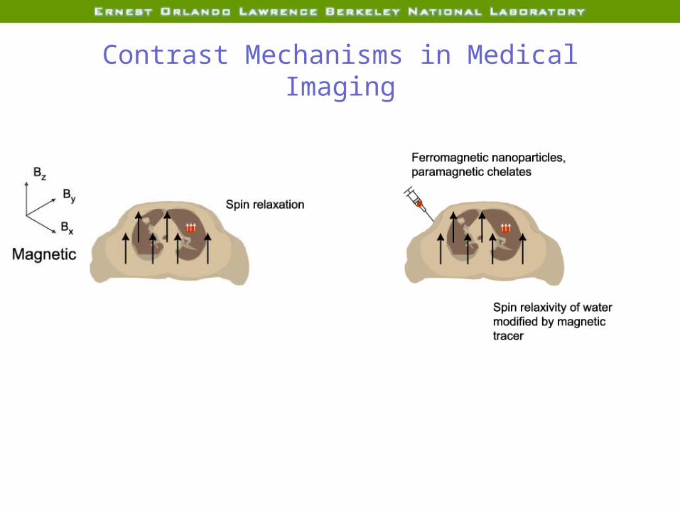
Contrast Mechanisms in Medical Imaging
Planar X-ray, X-ray CT
PET,SPECT
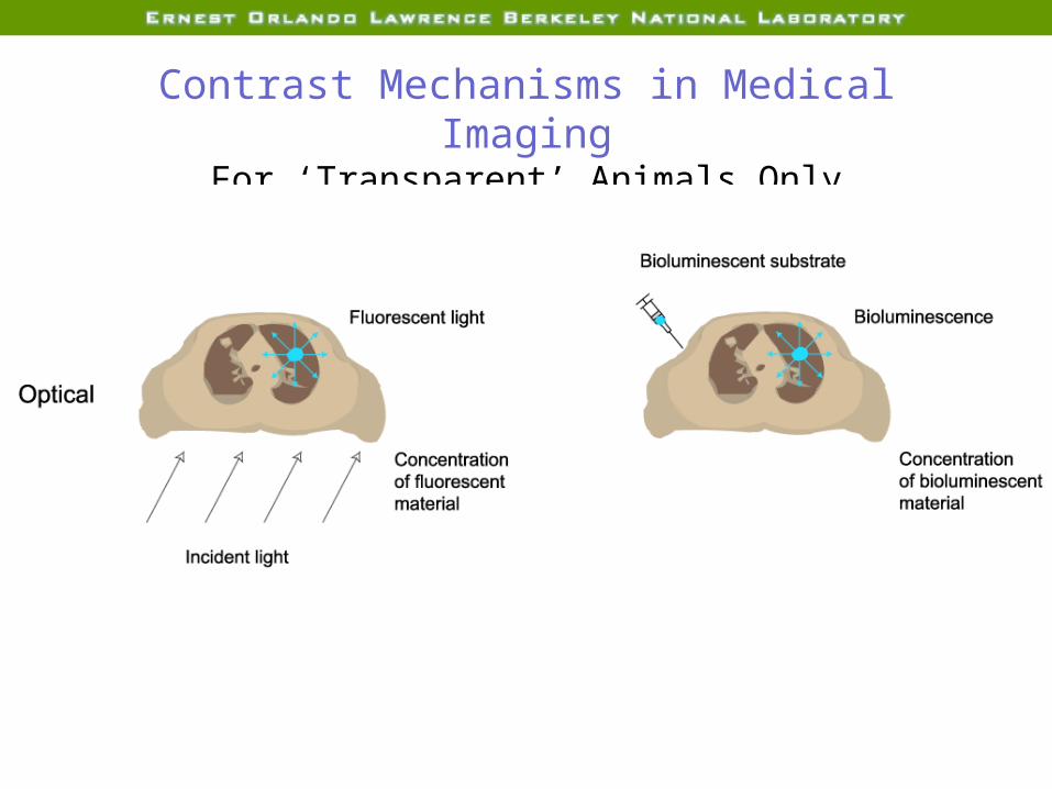
Contrast Mechanisms in Medical ImagingFor ‘Transparent’ Animals Only
Contrast Mechanisms in Medical Imaging

Overview
1. Review of Contrast Mechanisms2. Frontiers in Molecular Imaging
1. Hardware2. Reconstruction algorithms3. Imaging gene expression4. Imaging cell trafficking
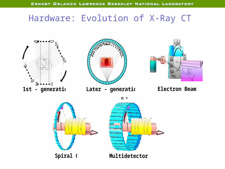
Evolution of X-ray CT
1st - generation CT Later - generation CT Electron Beam CT
Spiral CT Multidetector CT
n > 8
Hardware: Evolution of X-Ray CT
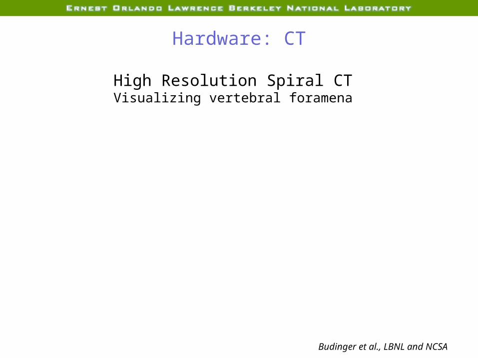
Hardware: CT
High Resolution Spiral CTVisualizing vertebral foramena
Budinger et al., LBNL and NCSA
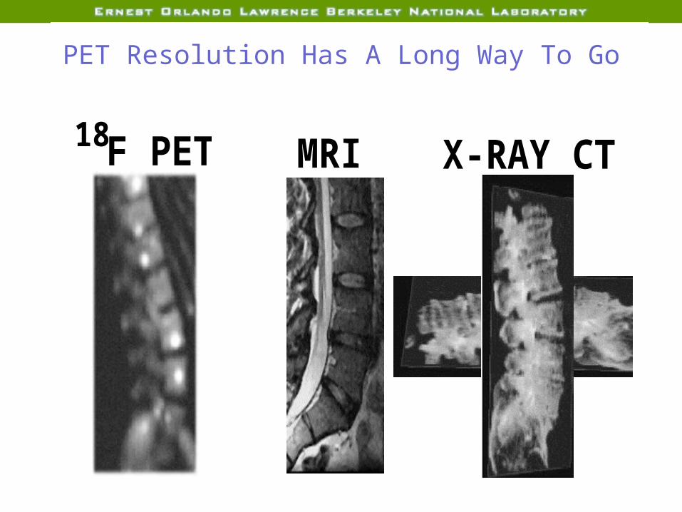
Vertebrae & Spinal Cord
MRI X-RAY CTF PET18
PET Resolution Has A Long Way To Go
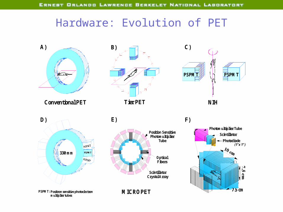
Conventional PET
Position SensitivePhotomultiplier
Tube
Optical Fibers
Scintillator Crystal Array
MICROPETPSPMT:
330 mm PSPMT
PSPMT
PSPMT
Positron sensitive photoelectron multiplier tubes
A)
D) E) F)Photomultiplier Tube
Photodiode
Scintillator
(1"x 1")
10 cm
7.5 cm
7.5 cm
PSPMT
NIH
C)
PSPMT
Tier PET
B)
Hardware: Evolution of PET
6 mm8 mm
8 mm
4 mm
6 mm4 mm4 mm
5 mm
5 mm
6 mm
8 mm
8 mm
2 mm
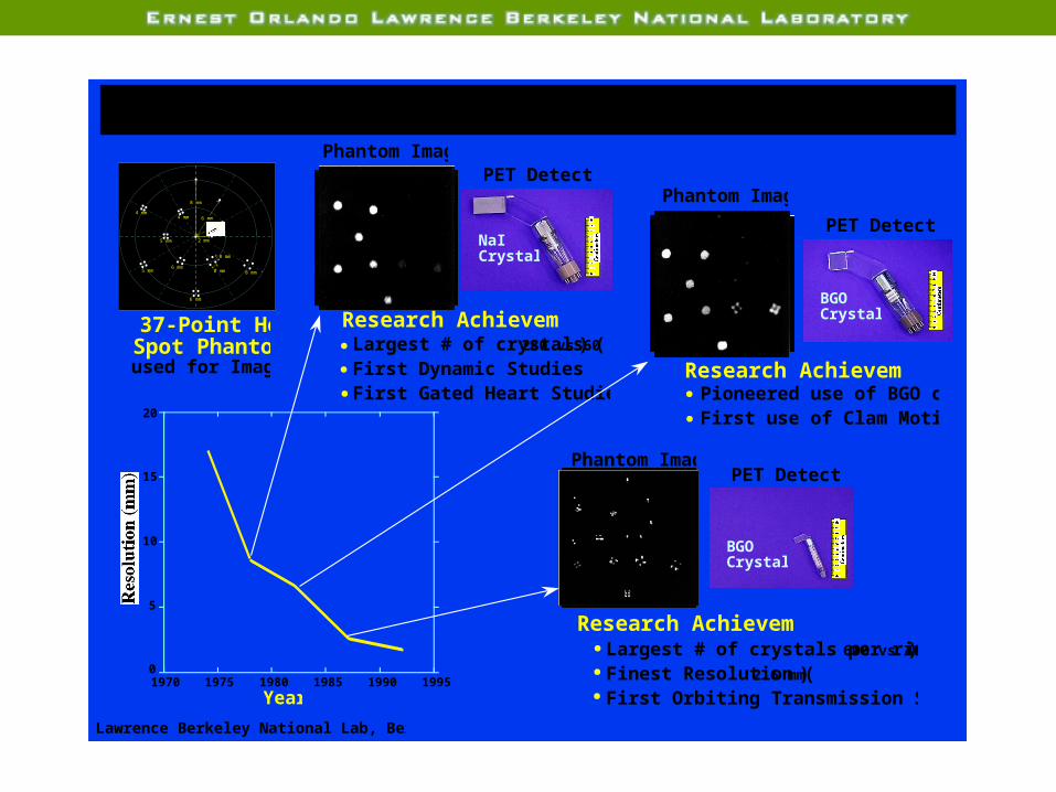
Positron Emission Tomography Resolution Improvements
1970 1975 1980 1985 1990 19950
5
10
15
20
Year
Resolution (mm)
Phantom ImagePET Detector
37-Point Hot Spot Phantom used for Imaging
Research AchievementsLargest # of crystals (280 vs 60)First Dynamic StudiesFirst Gated Heart Studies
NaI Crystal
0
1
2
3
4
5
6
7
8
9
10
Centimeters
0
1
2
3
4
5
6
7
8
9
10
Centimeters
Research AchievementsPioneered use of BGO crystalsFirst use of Clam Motion
Phantom Image
PET Detector
BGOCrystal
Research AchievementsLargest # of crystals per ring (600 vs 400)Finest Resolution (2.6 mm)First Orbiting Transmission Source
0
1
2
3
4
5
6
7
8
9
10
Centimeters
Phantom ImagePET Detector
BGOCrystal
Lawrence Berkeley National Lab, Berkeley, CA
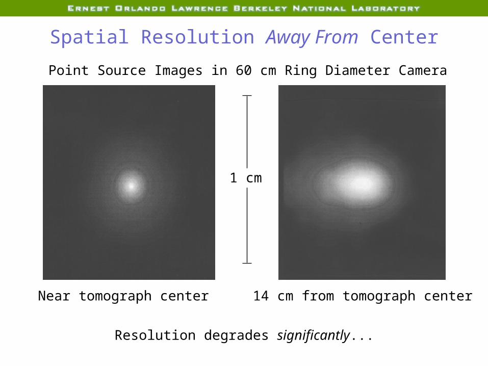
Spatial Resolution Away From Center
1 cm
Near tomograph center 14 cm from tomograph center
Point Source Images in 60 cm Ring Diameter Camera
Resolution degrades significantly...
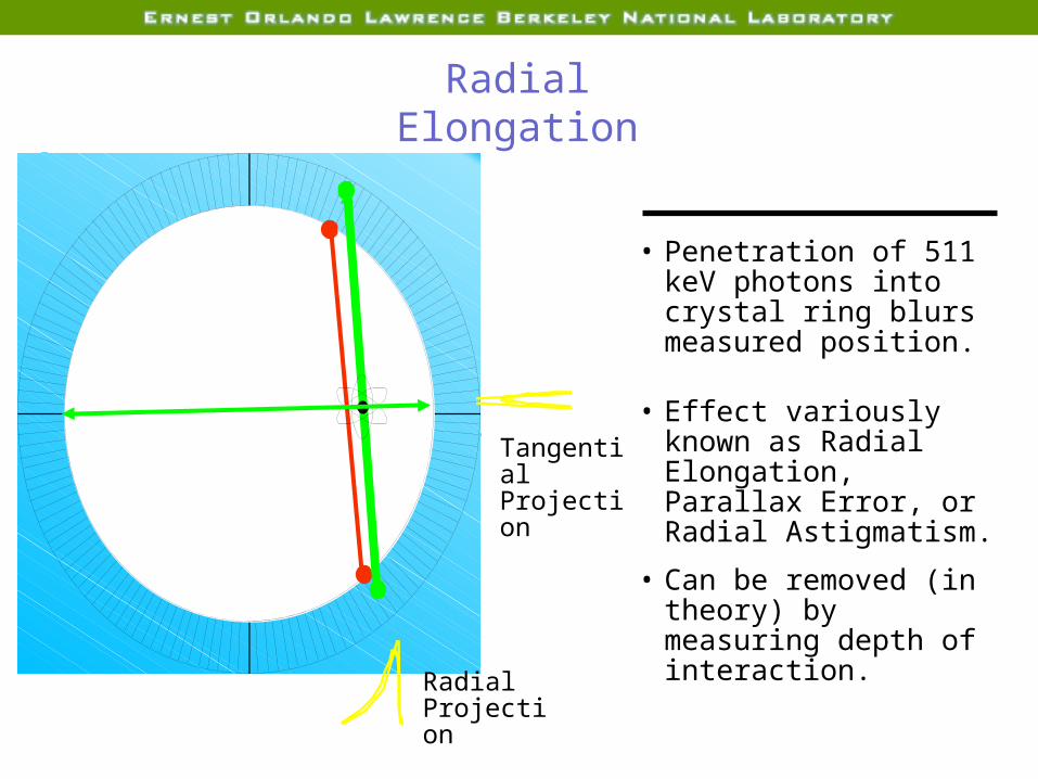
Radial Elongation
• Penetration of 511 keV photons into crystal ring blurs measured position.
• Effect variously known as Radial Elongation, Parallax Error, or Radial Astigmatism.
• Can be removed (in theory) by measuring depth of interaction.
Radial Projection
Tangential Projection

LBNL Detector Module Design
25 mm
25 mm
30 mm
Photomultiplier Tube
64 Photodiodes
64 LSO Crystals(3x3x30 mm)
•PMT Provides Timing Pulse and Energy Discrimination•PD Array Identifies Crystal of Interaction
•PD+PMT Measures Energy Deposit•PD / (PD+PMT) Measures Depth of Interaction
Custom IC(64
Channels)
Moses et al., LBNL
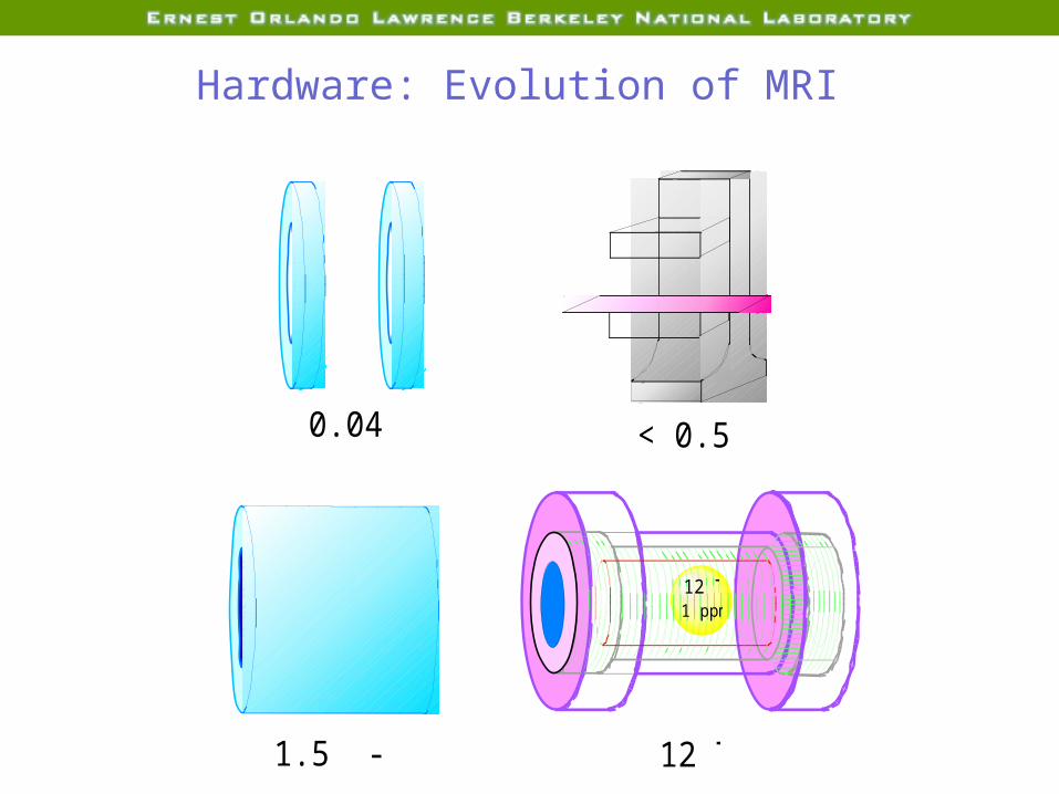
< 0.5 T
1.5 - 8 T
12 T1 ppm
12 T
0.04 T
Magnetic Resonance DevelopmentsHardware: Evolution of MRI

MRI Imaging of Anatomy
High Resolution MRI
Markers of Alzheimer’s Disease:Hippocampal Atrophy
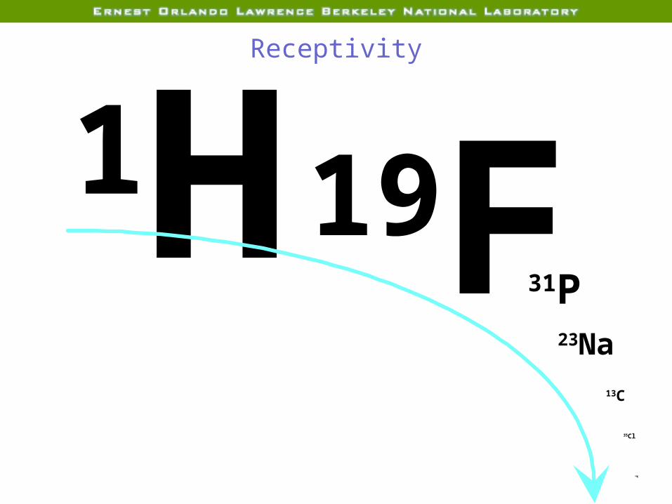
Receptivity
39K
1H19F31P23Na
13C
35Cl

2. Spectral dispersion increases with field increase.
Other Reasons for High Field
1. SNR increases with field increase
If the field goes from 1.5 T to 12T the time required to achieve a given SNR would be reduced from 5 hours to 5 minutes.

Human Noninvasive Studies Enabled by 12 TExamples
Cellular and extracellular concentrations of Na, K, Cl with relevance to mental disorders, hypertension and cancer
Role of the pentose monophosphate shunt in cancer, congestive heart failure and ischemic heart disease
Glutamate / Glutamine metabolism in the normal CNS and in stroke and trauma
Amyloid plaque concentration changes on the CNS in neurodegenerative disorders
Carbohydrate and fatty acid metabolism in obesity and nutrition

80
100
120
140
160
180
-20 0 20 40 60
Time (min)
Reperfusion20 min
Reperfusion40 min
Globalischemia
23
Na NMR TQF signal (%)
- 20 min Ischemia
- 40 min Ischemia
23Na NMR TQF Signal in Isolated Rat Heart Clamp Global Ischemia at 37° C
(n = 6)
(n = 6)

Enabling Technology: Nb3Sn Superconductors


Overview
1. Review of Contrast Mechanisms2. Frontiers in Molecular Imaging
1. Hardware2. Reconstruction algorithms3. Imaging gene expression4. Imaging cell trafficking

Reconstruction Algorithms for Dynamic Imaging

Algorithms: Making Movies with SPECT 99m Tc-teboroxime Reconstruction
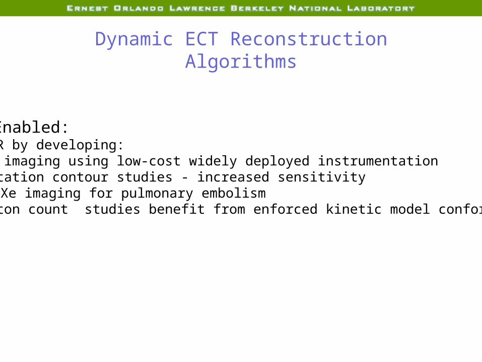
Dynamic ECT Reconstruction Algorithms
Studies Enabled:Improve SNR by developing:1. Dynamic imaging using low-cost widely deployed instrumentation2. Slow rotation contour studies - increased sensitivity
e.g. 123 Xe imaging for pulmonary embolism3. Low photon count studies benefit from enforced kinetic model conformity

head
foot
chest back
Extent of Contractile MotionBase moves 9-14 mm towards apexWalls thicken from 10 mm to 16 mm
Transverse Coronal Sagittal
chest
back
right left
head
foot
right left
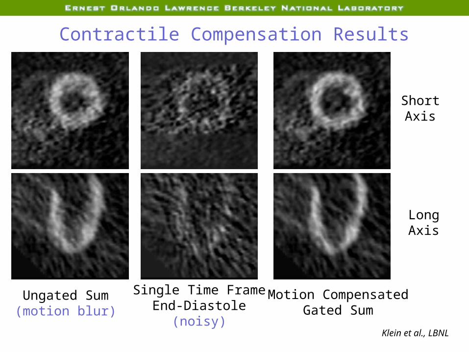
Contractile Compensation Results
Single Time FrameEnd-Diastole
(noisy)
Motion CompensatedGated Sum
Ungated Sum(motion blur)
ShortAxis
LongAxis
Klein et al., LBNL

Overview
1. Review of Contrast Mechanisms2. Frontiers in Molecular Imaging
1. Hardware2. Reconstruction algorithms3. Imaging gene expression4. Imaging cell trafficking
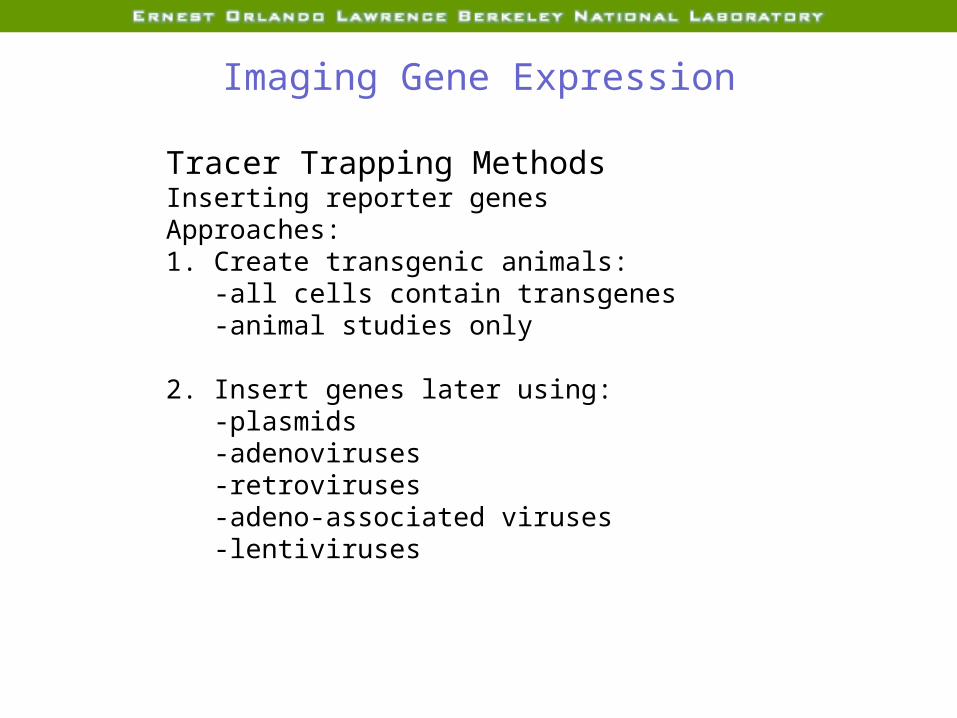
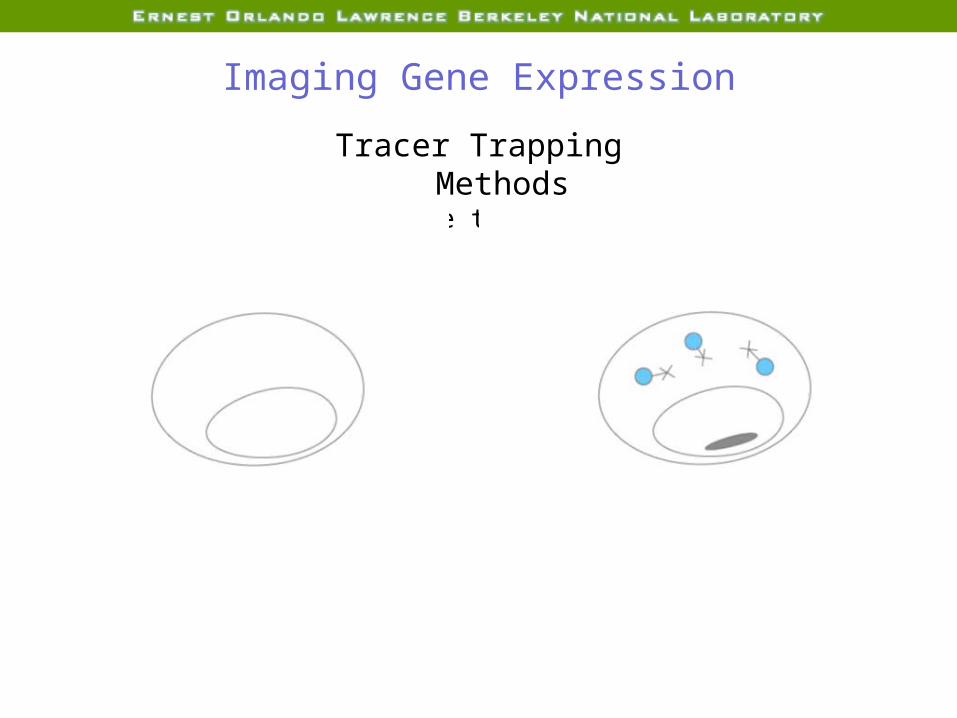
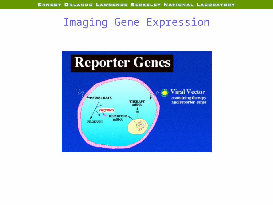
Imaging Gene Expression
Tracer Trapping MethodsInserting reporter genesApproaches:1. Create transgenic animals:
-all cells contain transgenes-animal studies only
2. Insert genes later using:-plasmids-adenoviruses-retroviruses-adeno-associated viruses-lentiviruses

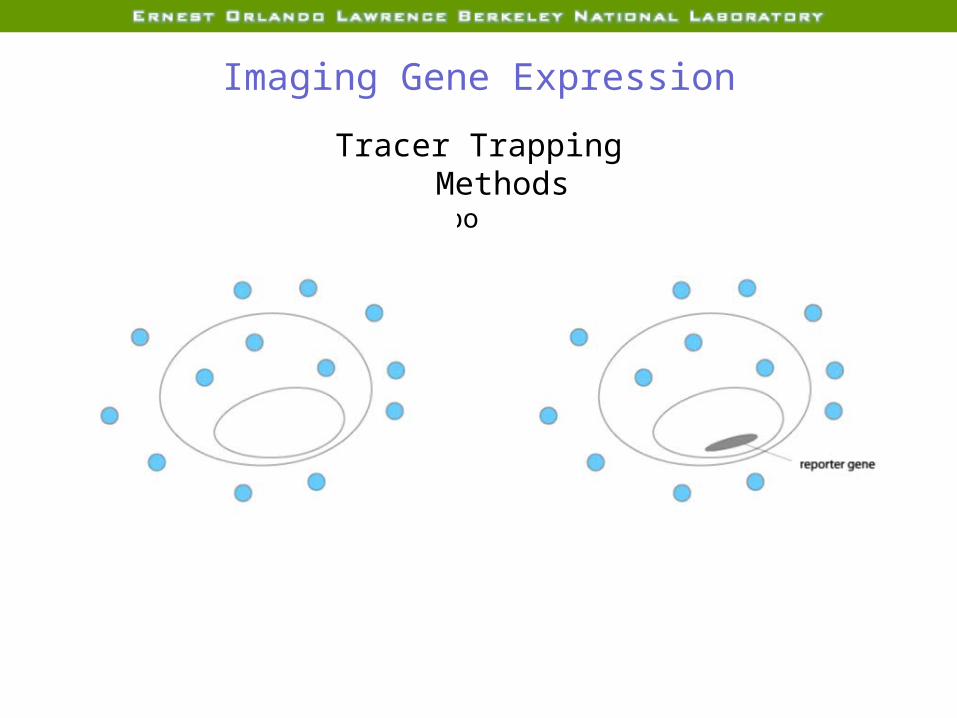
Imaging Gene Expression
Tracer Trapping MethodsEnzyme trapping

Imaging Gene Expression
Tracer Trapping MethodsEnzyme trapping
Imaging Gene Expression
Tracer Trapping MethodsEnzyme trapping

Trapping Mechanisms (PET, SPECT)
1. PhosphorylationHSV1-TK traps radiolabeled acyclovir(nucleotide-5’-monophosphates cannot cross plasma membrane)
2. DeaminationCytosine deaminate traps radiolabeled 5-fluorocytosine
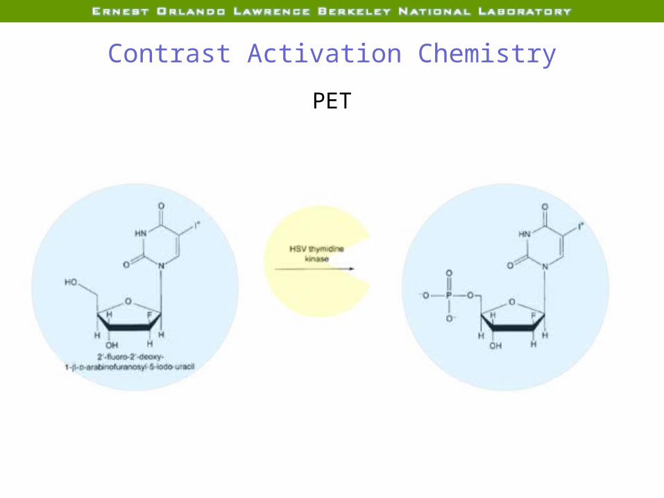
Contrast Activation Chemistry
PET
Imaging Gene Expression
Tracer Trapping MethodsLigand reporter probes
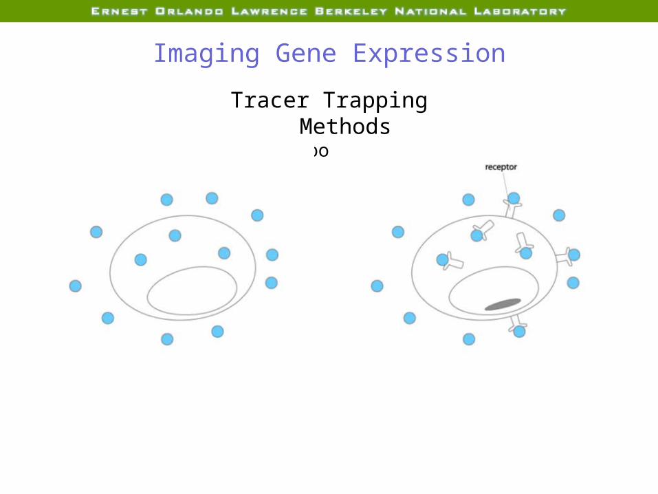
Imaging Gene Expression
Tracer Trapping MethodsLigand reporter probes

Imaging Gene Expression
Tracer Trapping MethodsLigand reporter probes

Trapping Mechanisms
-Reporter gene expression leads to dopamine D2 receptor production. Traps dopamine antagonist spiperone.
-Expression of transferrin receptors traps ion-oxide nanoparticlesbonded to transferrin.
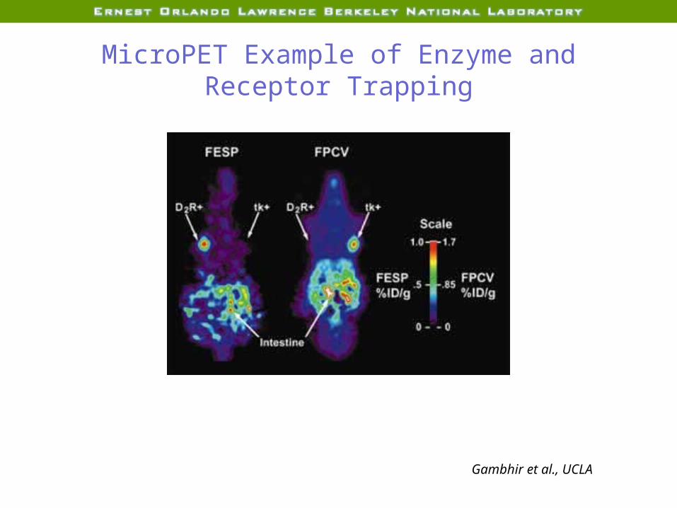
MicroPET Example of Enzyme and Receptor Trapping
Gambhir et al., UCLA

MRIContrast Activation Chemistry
Galactopyronosecap
-galactosidase
Meade et al., Caltech
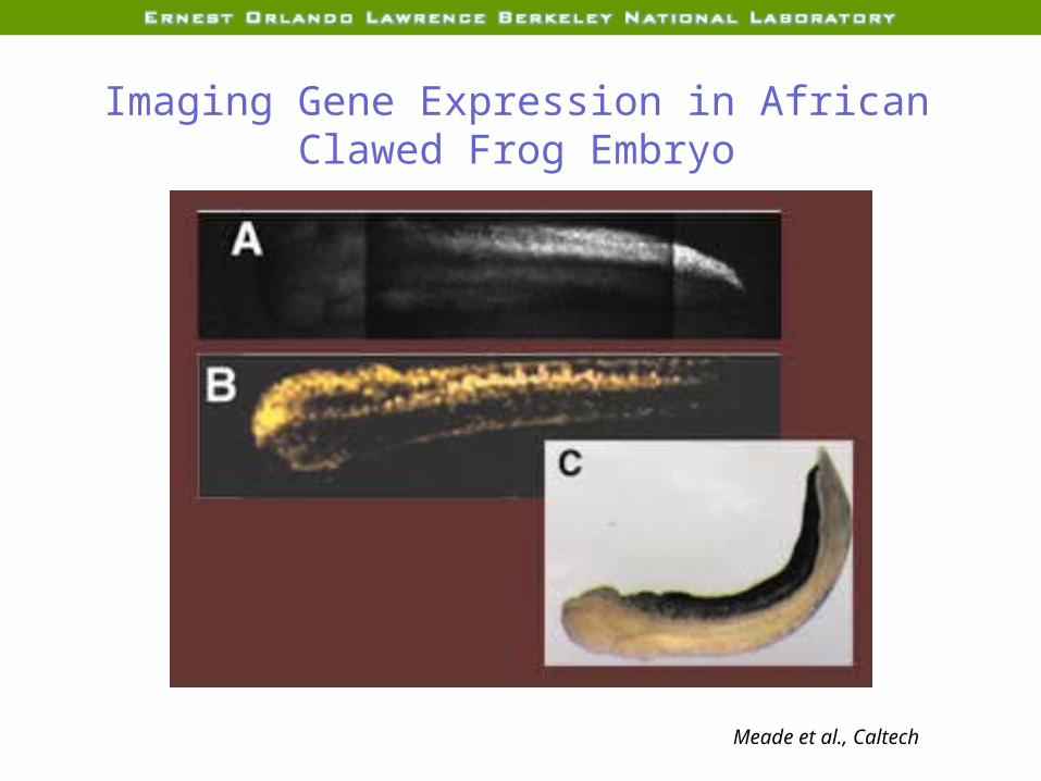
Imaging Gene Expression in African Clawed Frog Embryo
Meade et al., Caltech
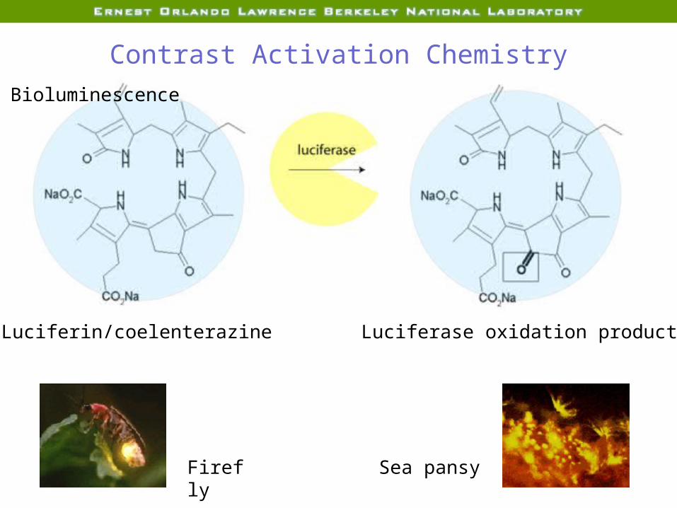
Contrast Activation Chemistry
Firefly Sea pansy
Bioluminescence
Luciferin/coelenterazine Luciferase oxidation product
Overview
1. Review of Contrast Mechanisms2. Frontiers in Molecular Imaging
1. Hardware2. Reconstruction algorithms3. Imaging gene expression4. Imaging cell trafficking

*CD15 receptor
* * * ***
*****
*
Neutrophil Lymphocyte*
**** * *
***
****CD4
receptor glycoproteins
Cell Trafficking: Targeting Surface Proteins
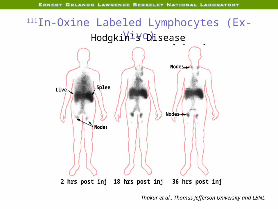
2 hrs post injection 18 hrs post injection 36 hrs post injection
111In - Oxine Labelled Lymphocytes (ex-vivo)
Liver Spleen
Nodes
Nodes
Nodes
111In-Oxine Labeled Lymphocytes (Ex-Vivo)Hodgkin’s Disease
Thakur et al., Thomas Jefferson University and LBNL
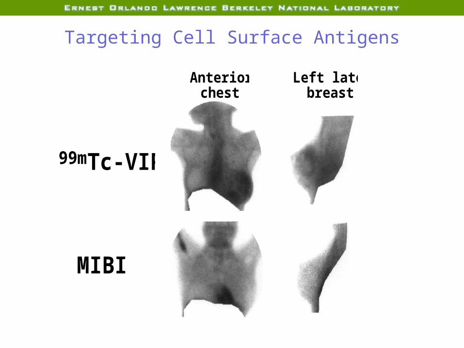
Targeting Cell Surface Antigens
99mTc-VIP
Left lateral breast
Anterior chest
MIBI
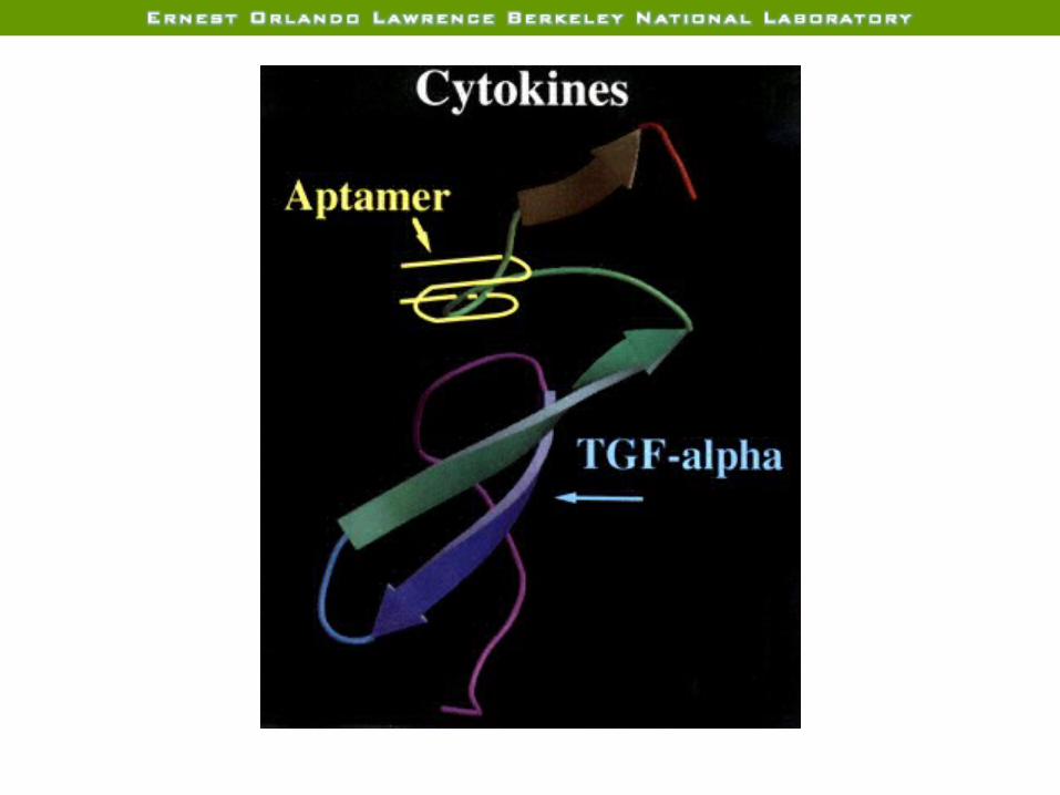
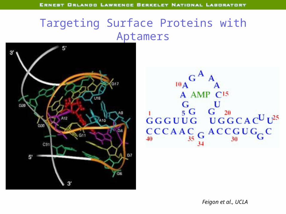
Targeting Surface Proteins with Aptamers
Feigon et al., UCLA

Acknowledgements
Katie BrennanMatthew DarmalingumJamie Eberling Greg KleinBill MosesScott Taylor
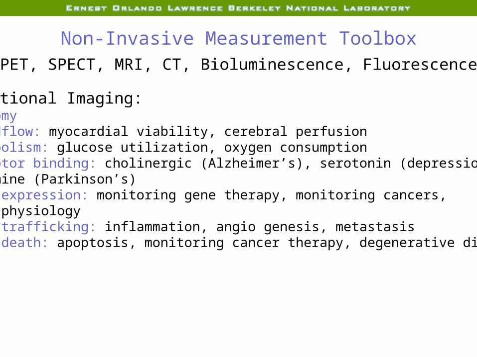
Non-Invasive Measurement ToolboxPET, SPECT, MRI, CT, Bioluminescence, Fluorescence
Functional Imaging:AnatomyBloodflow: myocardial viability, cerebral perfusionMetabolism: glucose utilization, oxygen consumptionReceptor binding: cholinergic (Alzheimer’s), serotonin (depression), dopamine (Parkinson’s)Gene expression: monitoring gene therapy, monitoring cancers, cell physiologyCell trafficking: inflammation, angio genesis, metastasisCell death: apoptosis, monitoring cancer therapy, degenerative disease

BloodflowMyocardial ViabilitySPECT evaluation of myocardial viability is the most common clinical study in Nuclear Medicine
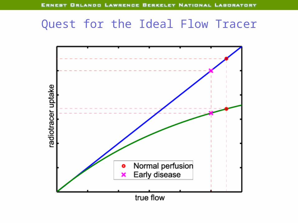
Quest for the Ideal Flow Tracer
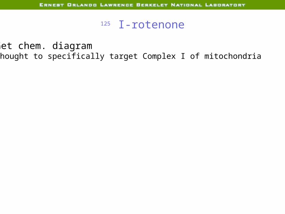
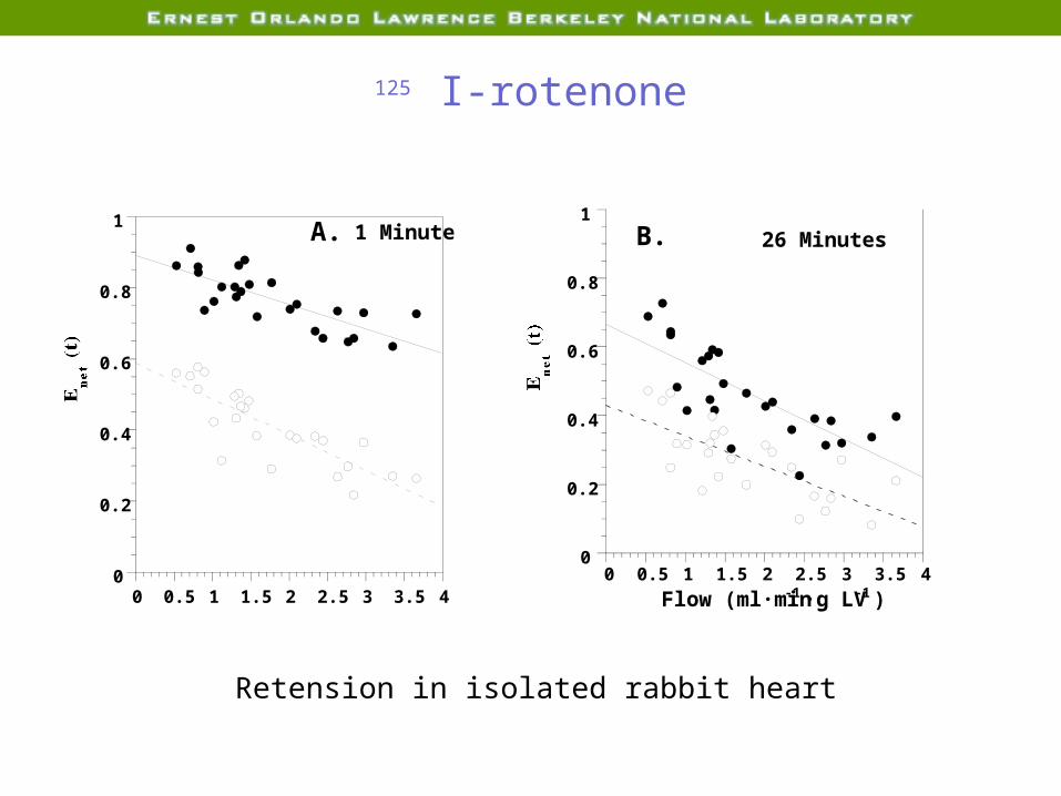
125 I-rotenone
Get chem. diagramThought to specifically target Complex I of mitochondria
125 I-rotenone
0
0.2
0.4
0.6
0.8
11 Minute
0 0.5 1 1.5 2 2.5 3 3.5 4
Enet
(t)
A.
.
.
0
0.2
0.4
0.6
0.8
1
0 0.5 1 1.5 2 2.5 3 3.5 4
26 Minutes
Enet
(t)
Flow (ml·min-1 ·g LV-1 )
B.
Retension in isolated rabbit heart
125 I-rotenone
Tracer extraction
Get graph
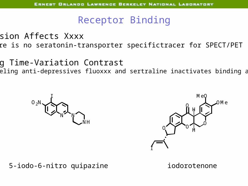
Receptor Binding
Depression Affects XxxxYet there is no seratonin-transporter specifictracer for SPECT/PET
Imaging Time-Variation ContrastRadiolabeling anti-depressives fluoxxx and sertraline inactivates binding ability
N NNH
O2NI
5-iodo-6-nitro quipazine
O
I
OOO
H
H
MeOOMe
iodorotenone
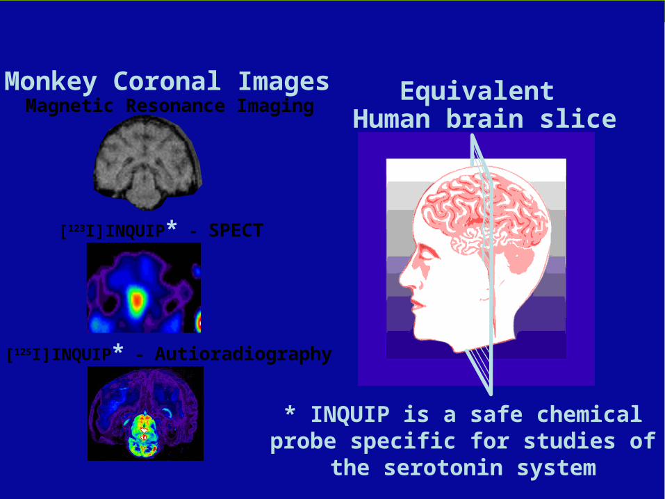
KT
[123I]INQUIP* - SPECT
[125I]INQUIP* - Autioradiography
Magnetic Resonance ImagingMonkey Coronal Images Equivalent
Human brain slice
* INQUIP is a safe chemical probe specific for studies of the serotonin
system
Imaging Gene Expression

Three Frontiers in Medical Imaging
Improving ContrastImprove SNR by developing:1. More sensitive detectors2. More specific tracers3. Improved tracer kinetics
Imaging Time-Variation Contrast1. Better temporal resolution2. Algorithms for dynamic image reconstruction
New Contrast Mechanisms1. Expression of specific gene2. Concentration of specific enzyme3. Presence of a specific receptor inside/on a cell4. Rate of specific metabolic process5. Rate of fluid flow

12 Tesla Whole Body Magnet
NbTi outer coils
13.2 Tpeak
Nb3Sn center coils
12 T1 ppm