Innovative Magnetic Nanoparticles for PET/MRI Bimodal Imaging

2484-15
ICTP-IAEA Joint Workshop on Nuclear Data for Science and Technology: Medical Applications
H. Herzog
30 September - 4 October, 2013
Institute of Neuroscience and Medicine - 4 Forschungszentrum Juelich Germany
Molecular Imaging Part IIIa: PET-MR

Forschungszentrum Jülich
Hans Herzog
Institute of Neuroscience and Medicine - 4
Forschungszentrum Jülich
Molecular Imaging Part IIIa: PET-MR
Titelbild

Forschungszentrum Jülich
MR Compared to PET
Parameter MR PET
Anatomical Detail Excellent Poor
Spatial Resolution Excellent Gets better Clinical Penetration Excellent Improving
Sensitivity Poor Excellent Molecular Imaging Limited Excellent
MR-PET >> MR + PET

Forschungszentrum Jülich
Today‘s Commonly Combined Use of PET and MRI
T1-MRI: morphology
56
40 32 24
8 fmol/ml
16
48
11C-Flumazenil-PET: benzodiazepine-receptors

Forschungszentrum Jülich
Combing Anatomy and Function with PET/CT
D.Townsend 1995
Metastasis of a malignent melanoma

Forschungszentrum Jülich
MR instead of CT in PET/CT ?
M.Schwaiger, S.Ziegler, et al.,
2005
No radiation dose
Better soft tissue contrast
Also functional information

Forschungszentrum Jülich
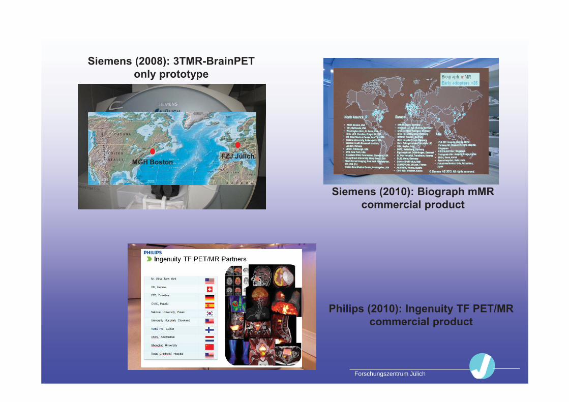
Siemens (2008): 3TMR-BrainPET only prototype
Siemens (2010): Biograph mMR commercial product
Philips (2010): Ingenuity TF PET/MR commercial product
Forschungszentrum Jülich
Siemens (2008): 3TMR-BrainPET only prototype
Siemens (2010): Biograph mMR commercial product
Philips (2010): Ingenuity TF PET/MR commercial product
MGH Boston FZJ Jülich

Forschungszentrum Jülich
3TMR-BrainPET

Forschungszentrum Jülich
Avalanche Photo Diodes (APD)
vs. Photo Multiplier Tubes (PMT)
PMT APD
~5 ns ~1 ns Risetime Up to 200 Up to 106 Gain 5x5 mm 10-52 mm dia. Size
insensitive
Magnetically sensitive
APD PMT

Forschungszentrum Jülich
Future : SiPMT / GM-APD ?
PMT APD
Otte et al., 2005
Gain: similar to PMT
Journal of Nuclear Medicine, April 2011 Sun Il Kwon et al: Development of Small-Animal PET Prototype Using
Silicon Photomultiplier (SiPM): Initial Results of Phantom and Animal Imaging Studies

Forschungszentrum Jülich 11
3TMR-BrainPET:
APD-based PET Detector Cassette
Consists of: • Six 12 x 12 arrays of 2.5 x 2.5 x 20 mm3 LSO
crystals read out by 9 APDs (Hamamatsu)• Preamplifiers & driver electronics • Temperature stability with compressed air
Forschungszentrum Jülich 12
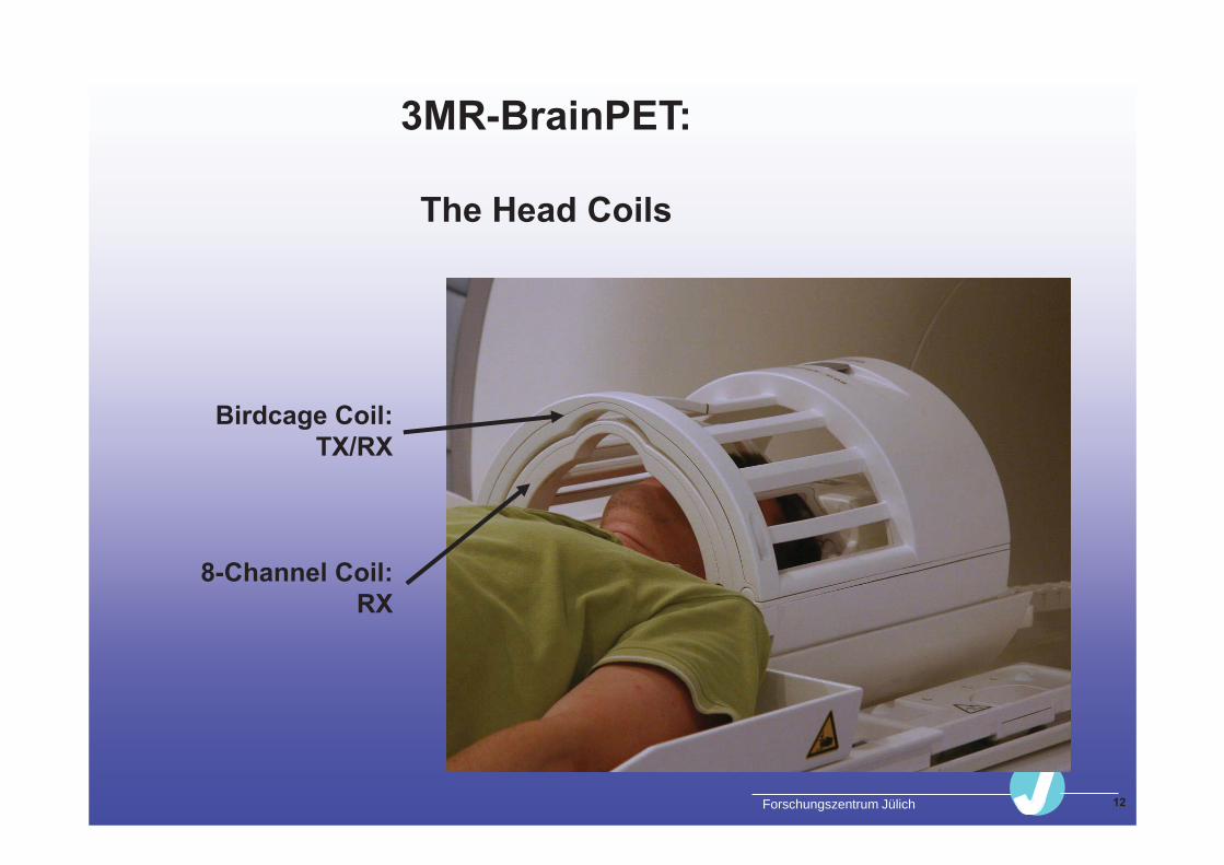
3MR-BrainPET:
The Head Coils
Birdcage Coil: TX/RX
8-Channel Coil: RX

Forschungszentrum Jülich
Technical Challenges by Possible Interferences between PET and MR
Minimal susceptibility (< 0.25 ppm local distortion) to avoid homogeneity distortion
No generation of time varying fields Tolerate vibration due to current changes in the
gradient coil in the range of 1-10 m in the kHz range Temperature changes due to average current changes
in the gradient coil from 20-70 oC There should be no loss in sensitivity

Forschungszentrum Jülich 14
Some Technical Parameters
Scatter fraction: 27%
Point source sensitivity: 6%
Resolution (FWHM, mm) :
r = 0 cm 2.5 cm 5 cm 7.5 cm 10 cm Tangential: 3.0 3.0 3.1 3.0 3.8 Radial 2.9 3.0 3.9 4.5 4.9
Z-Direction 3.0 3.6
§ reconstructed with 3DFBP (STIR)

Forschungszentrum Jülich
Simultaneous MR-PET in a Brain Tumor
Brain Tumor studied with [18F]-fluoro-ethyl-tyrosine (FET)
HR+: 40 - 50 min p.i.
BrainPET: 55 - 85 min p.i.
Trio: MPRAGE
Simultaneous MRI

Forschungszentrum Jülich
A Prerequisite for Quantitation: Attenuation Correction
the detector measures:
Emissionsmessung Transmissionsmessung
Rotating line source with positron emitter Ge68
PE = A(x,y) dl * exp( - μ (x,y) dl') AF = exp( - μ (x,y) dl')
PEKorr = PE / AF = A(x,y) dl
the detector measures:

Forschungszentrum Jülich
Attenuation Correction No Longer Based on Transmission Measurement !!
CT
MRI
Different Bone signal
Rota Kops et al., IEEE 2006
PET-Tx MRI-T1 MRI-T1 segmented

Forschungszentrum Jülich
18
Determination of Attenuation Map
Individual Attenuation Map
?
MR-Template
Patient FT4_609
PET-Tx-Template
E. Rota Kops, IEEE MIC 2008
SPM-Deformation
Template of Head
Coils
+

Forschungszentrum Jülich
19
MR-Based Attenuation Correction Using UTE Sequences
V. Keereman, JNM 2010
C. Catana, JNM 2010
A. Ribeiro, NIMA 2012
CT UTE-based

Forschungszentrum Jülich 20
FET- Dynamics Recorded in the 3TMR-BrainPET
Time Activity Curves
0
10
20
30
40
50
0 10 20 30 40 50 60 70 80 90
Time (min)
Activ
ity (k
Bq/c
c)
Tumour
Cortex
Carotis
HR+
Quantitation achieved !
BrainPET
The time-activity curves of the
BrainPET and HR+ match !

Forschungszentrum Jülich
MR-FDG-BrainPET
20-50 min p.i. 18FDG-PET
Simultan T1 MPRAGE
Fusion HR+

Forschungszentrum Jülich
Clinical Applications
Forschungszentrum Jülich 23
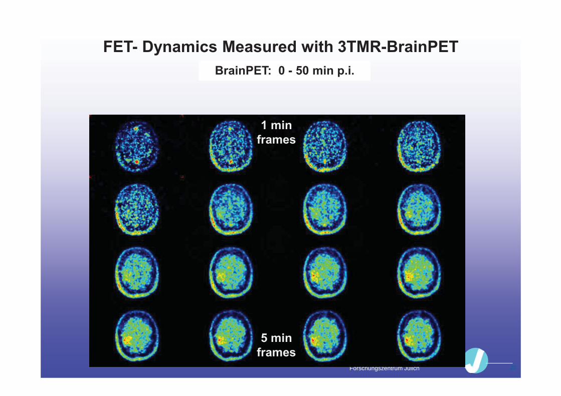
FET- Dynamics Measured with 3TMR-BrainPET BrainPET: 0 - 50 min p.i.
1 min frames
5 min frames

Forschungszentrum Jülich
Mismatch Match
T1 T2
FDG FET
Cerebral Gliomas: PET with O-(2-[18F]fluoroethyl)-L-tyrosine (FET)
Completes MRI Based Diagnosis
Preoperative determination of tumor
extent in FET-PET and MRI
MRI: Sensitivity: 96 % Specificity: 53 %
MRI+FET: 93 % 94 %
Pauleit et al. Brain 2005

Forschungszentrum Jülich
Hybrid MR-PET imaging
Dynamic FET PET 0-50
T1 / T2 PWI T1-weighted + contrast fMRI MRSI DTI

Forschungszentrum Jülich 26
Neuroactivation by Finger Tapping EPI-Study for 12 min
Dummy
right
left
bi-manual
R

Forschungszentrum Jülich
One-stop-shop Examination: Identifying the Cortical Motor Area
Affected by the Brain Tumor
MPRAGE: Structural MR Imaging
fMRI: Right Hand Activation
fMRI: Left Hand Activation
FET-PET: Tumor-
Delineation

Forschungszentrum Jülich
MR-PET in a Patient with Epilepsy
PET after injection of
the GABAnergic
receptor ligand
11C-flumazenil:
Looking for the
epileptical focus
HR+: 10 - 25 min p.i.
BrainPET: 30-50 min p.i. and simultaneous:
T1- MPRAGE

Forschungszentrum Jülich
MR-PET in a Patient with Parkinson’s Disease
PET after injection of
the dopaminergic
transporter ligand
18F-FP-CIT T1- MPRAGE
BrainPET: 120 - 150 min p.i. and simultaneous:
HR+: 60 - 100 min p.i.

Forschungszentrum Jülich
Dynamic MR-FDGPET Aim: to measure cerebral glucose consumption
30-50 min p.i. FDG-PET reconstructed with Siemens OSEM3D
15 – 75 s after injection: Arterial phase
Arterial phase together with T1-MPRAGE
Forschungszentrum Jülich
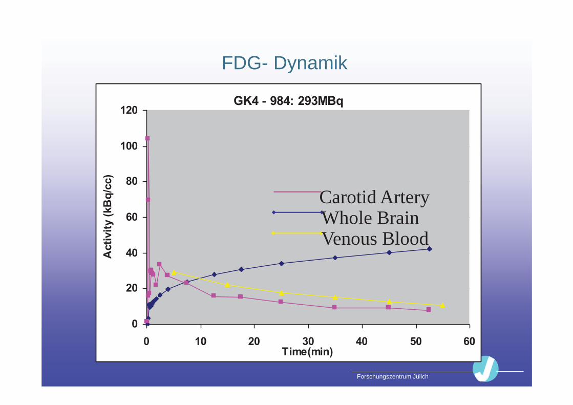
GK4 - 984: 293MBq
0
20
40
60
80
100
120
0 10 20 30 40 50 60Time(min)
Act
ivity
(kB
q/cc
)
FDG- Dynamik
Carotid Artery Whole Brain Venous Blood

Forschungszentrum Jülich
0 μmol/100g/min 67
Measurement of Cerebral Glucose Consumption
from Act. to LCRMglc
Blood Data
0 kBq/ml 645
0 0
250
60
Plasma Hirngewebe
B l u t
H i r n
Schranke
K* 1
K* 1
K* 2
[C]-Deoxyglucose (C*)
11
p
[C]-Deoxyglucose (C*)
11
c
Glucose (C) p
Glucose (C) p
Vorstufe
K* 2 K* 4
K* 3
K* 3
K* 4
[C]-Deoxyglucose-6-Phosphat
11
(C*) M
Glucose-6-Phosphat (C) M
CO HO 22
[C]-Deoxyglucose-1-Phosphat 11
Glycoprotein Glycolipide
Desoxyglycogen
Metabolite
Activity Image
Cross-Calibration
Deoxyglucose Model
(by Sokoloff, 1977) Glucose Consumption
T Tkk
PTkk
P
Tkk
PTkk
TP
dtetcedttcLC
dtetceKTccLCMRGlc
0 0
)()(
0
)()(1
3232
3232
)(*)(*
)(*)(
Forschungszentrum Jülich
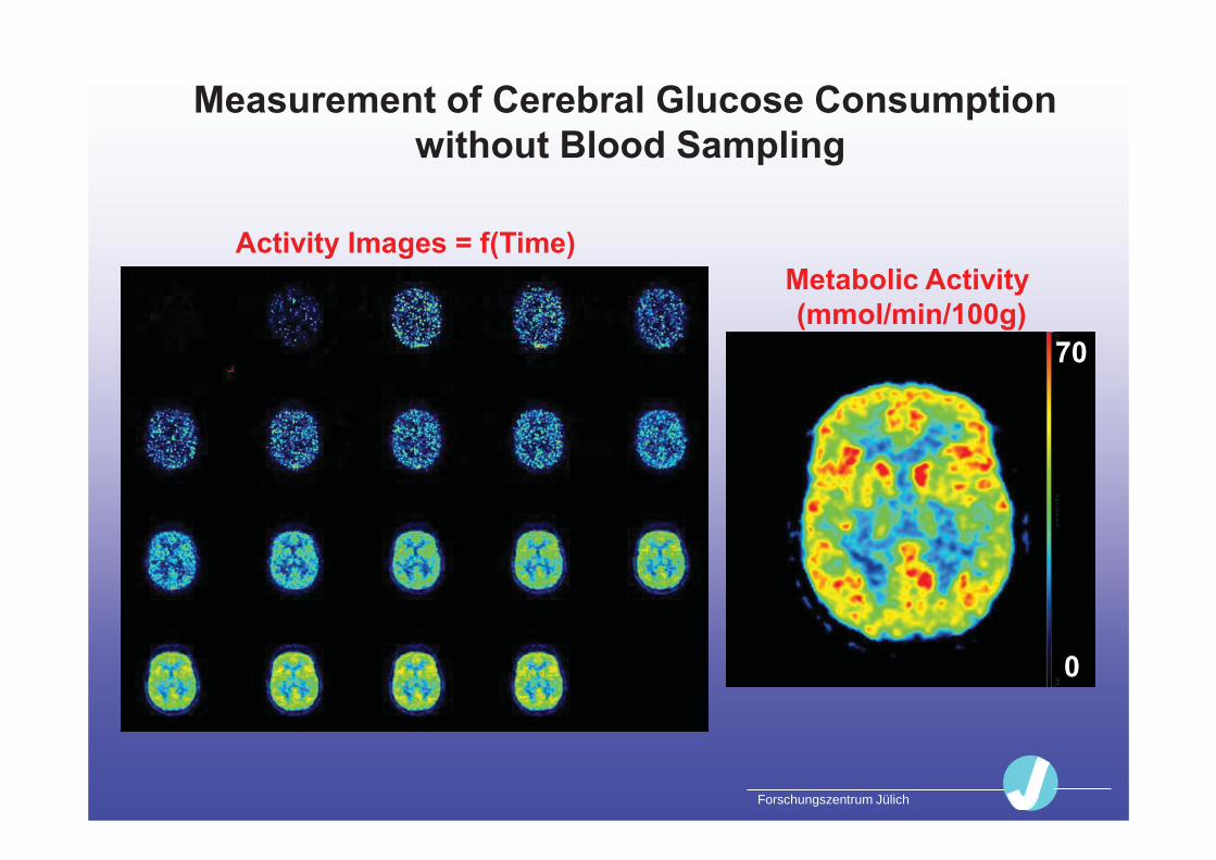
Measurement of Cerebral Glucose Consumption without Blood Sampling
Activity Images = f(Time)
70
0
Metabolic Activity (mmol/min/100g)

Forschungszentrum Jülich
Research Applications

Forschungszentrum Jülich 35
Centers of cerebral data processing
Domain of fMRI
The Two Parts of Cerebral Communication
Chemical interface at the synapses modulated by internal neurotransmitters or drugs
Domain of PET

Forschungszentrum Jülich 36
Centers of increased alertness
after 1mg nicotine
fMRI and Receptor-PET with a Pharmaco-Challenge
Distribution volume of [18F]-2-A-85830:
Smokers
minus nosmokers
PET
Thiel et al., 2005 Herzog et al., 2006
fMRI
PET bolus-Infusion
t = 0 t = 100min fMRI-1 fMRI-2
pharmaco-challenge
Now such studies can be combined with PET/MRI !!

Forschungszentrum Jülich 37
Timing of a Combined PET-MRI-fMRI Study
PET Listmode Acquisition t t + 100
MRI Acquisitions
2
Unit: min
5 6 20
localizer
Bolus 11C-Flumazenil
ASL EPI
2 x 5 40
MRS MPRAGE
6
ASL EPI
Infusion 11C-Flumazenil
Challenge

Forschungszentrum Jülich 38
Timing of a Combined PET-MRI-fMRI Study
PET Listmode Acquisition t t + 100
MRI Acquisitions
2
Unit: min
5 6 20
localizer CBF fMRI
2 x 5 40
MRS Anatomy
6
CBF fMRI
Gabanergic Neuroreceptors
Challenge
Forschungszentrum Jülich
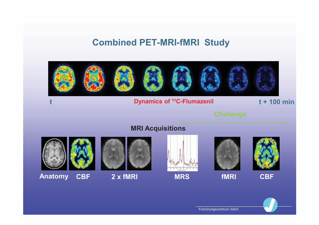
Combined PET-MRI-fMRI Study
MRI Acquisitions
CBF 2 x fMRI MRS Anatomy CBF fMRI
t t + 100 min Dynamics of 11C-Flumazenil
Challenge
Forschungszentrum Jülich
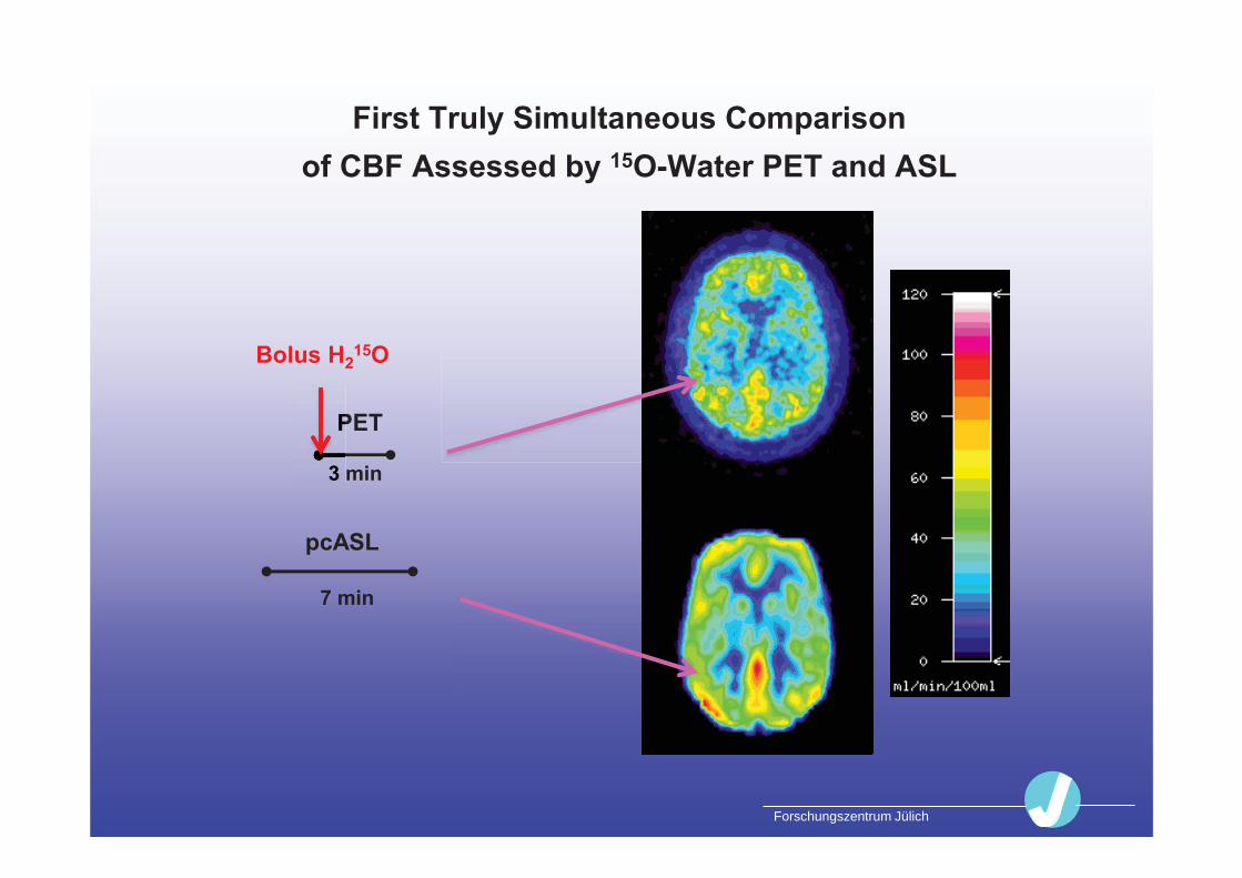
First Truly Simultaneous Comparison of CBF Assessed by 15O-Water PET and ASL
PET
3 min
Bolus H215O
pcASL
P
3
7 min
Forschungszentrum Jülich
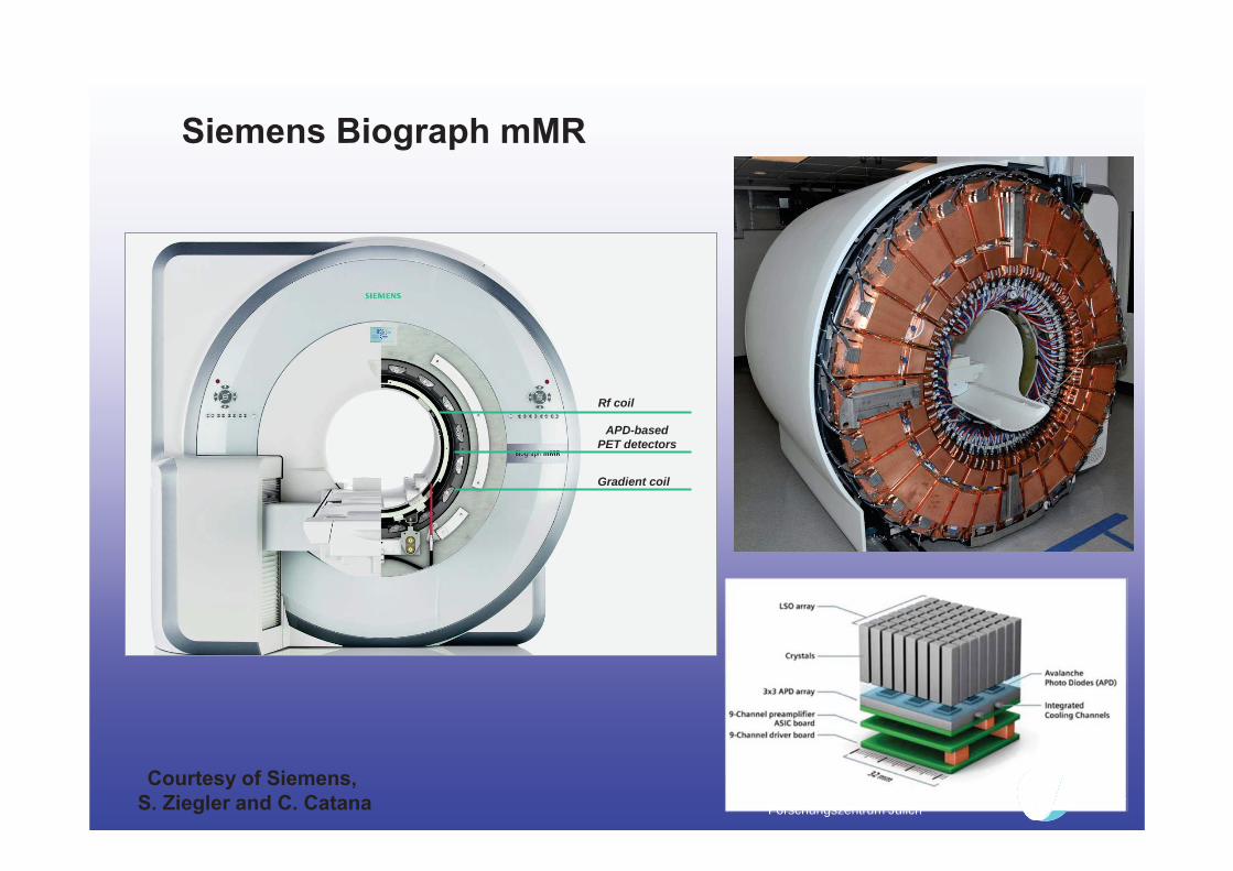
APD-based PET detectors
Rf coil
Gradient coil
Forschungszentrum Jülich
Courtesy of Siemens, S. Ziegler and C. Catana
Siemens Biograph mMR

Forschungszentrum Jülich 42
Some Technical Parameters
Scatter fraction: 36.7%
Resolution (FWHM, mm) : r = 1 cm 10 cm Tangential: 4.3 4.8 Radial 4.3 5.2 Z-Direction 4.3 66
§ reconstructed with 3D-FRP
Detector ring diameter 65.6 cm Bore diameter 60 cm Axial FOV 25.8 cm Crystal 4 x 4 x 20 mm3 LSO Concidence window 6 ns Energy window 430 – 610 keV
Courtesy of G. Delso and S. Ziegler

Forschungszentrum Jülich 43
Avg ∆SUV lesions: 2.3% Segmented dual-echo Dixon MRI vs. CT
CT-based AC
MRI-based AC
MR MR
Seg CT
Martinez-Möller A et al., JNM 2009
The method based on segmentation is fast and reliable. Some bias (5-13%) for osseous lesions due to neglecting bone.
MR-Based AC: Dixon Imaging for Fat/Water Separation
Forschungszentrum Jülich
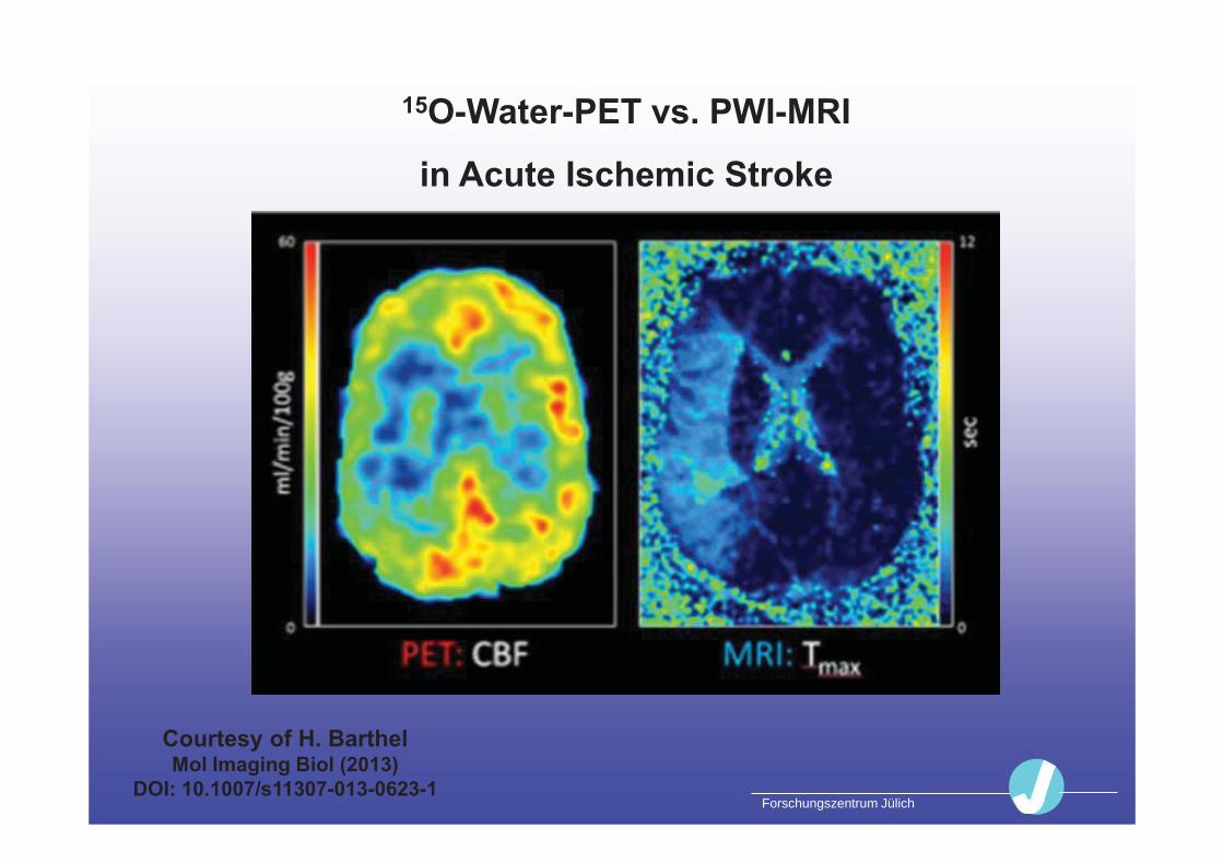
Courtesy of H. Barthel Mol Imaging Biol (2013)
DOI: 10.1007/s11307-013-0623-1
15O-Water-PET vs. PWI-MRI
in Acute Ischemic Stroke

Forschungszentrum Jülich 45
Ingenuity TF PET/MRI

Forschungszentrum Jülich
46
Some Technical Parameters
Scatter fraction: 26% - 35%
Resolution (FWHM, mm) : r = 1 cm 10 cm Tangential: 4.7 5.3 Radial 4.7 5.0 Z-Direction 4.6 5.0
§ reconstructed with 3D-FRP
Detector ring diameter 90.3 cm Bore diameter 60 cm (PET 70.7 cm) Axial FOV 18.0 cm Crystal 4 x 4 x 22 mm3 LYSO Concidence window 6 ns Energy window 460 – 665 keV TOF capability
Zaidi H, Phys Med Biol, 2011

Forschungszentrum Jülich Courtesy of O. Ratib, 2nd Juelich MR-PET Workshop 2010
CT MR PET/MR PET
CT and PET/MR in a Patient
with Ewing Sarkoma

Forschungszentrum Jülich
Patient with Head/Neck Cancer
I. Platzek et al., EJNMMI 2013
T1-weightedTSE STIR TSE
PET max. Intensity Fusion
Forschungszentrum Jülich
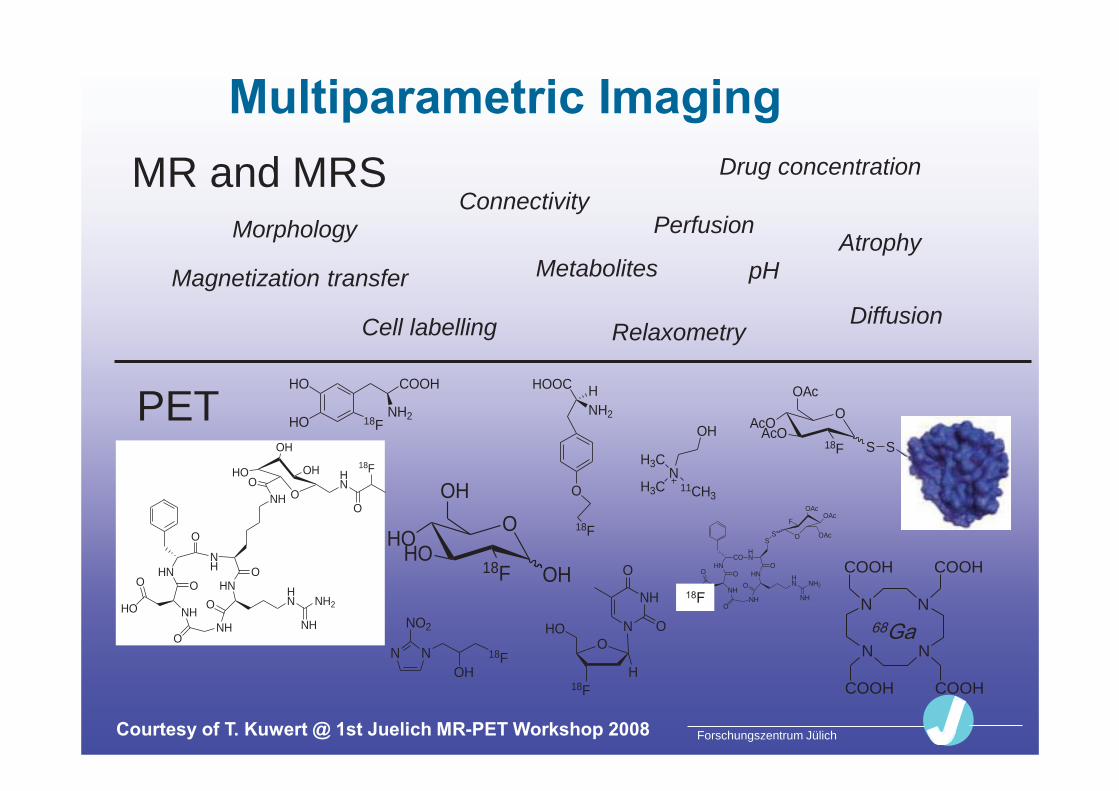
Multiparametric Imaging
OHO
HOOH18F
OH
NN
NO2
18FOH
O
NH2
HHOOC
18F
O
18F
HO N
NH
O
O
H
HO
HO
COOH
18FNH2
NH3C
H3C
OH
11CH3
HNNH
O
NHHO
O
ONH
OHN H
N NH2
NH
O
O
NH O
HO
OH
OHO H
N
O
18F
COHN
HN
O
NHHO
O
ONH
OHN H
N NH2
NH
O
O
OAcOAc
F
OAcS
S
N
N N
N
COOHCOOH
COOH COOH
S
OAcO
AcO18F
OAc
S
PET
MR and MRS Morphology Perfusion
pH
Cell labelling
Metabolites
Drug concentration
Diffusion Relaxometry
Connectivity
Magnetization transfer Atrophy
Courtesy of T. Kuwert @ 1st Juelich MR-PET Workshop 2008
Forschungszentrum Jülich 50
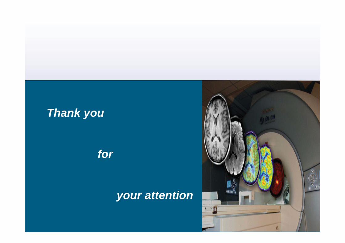
Thank you
for
your attention