Molecular Identification of Bacteria Isolated from Dairy...
8
Kafkas Univ Vet Fak Derg 16 (6): 1025-1032, 2010 RESEARCH ARTICLE Summary The purpose of the present study was to identify environmental and contagious aerobic pathogen agents causing bovine clinical and subclinical mastitis using sequencing. A total of 244 cows were studied for the presence of mastitis using California Mastitis Test (CMT), clinical observations and microbiological isolations. Milk samples were obtained from 226 quarters of 123 cows which were diagnosed having clinical (11.4%) and subclinical (88.6%) mastitis. From these milk samples, 38 (16.8%) had no bacterial growth and from remaining 188 samples (83.2%) microorganisms were isolated. A total of 42 species were identified by sequencing amplified 16S rRNA fragments. The most common species were Staphylococcus aureus (22.9%) followed by Escherichia coli (10.1%), S. chromogenes (8.5%), S. haemoliticus (7.5%) and Hafnia alvei (6.4%). Of 188 isolates 43 were found contagious (22.9%), and 145 (77.1%) environmental agents. Use of sequencing offer identification of aetiological agent species level useful and practical that can help to chose a suitable therapeutic agent. Keywords: Mastitis, Sequencing, Microorganism, Identification Mastitisli Sığır Sütlerinden İzole Edilen Bakterilerin Moleküler İdentifikasyonu Özet Bu çalışmanın amacı, klinik ve subklinik mastitislere neden olan bulaşıcı ve çevresel aerobik patojen bakteriyel etkenlerin sekans analizi kullanılarak identifikasyonlarının yapılmasıdır. Toplam 244 sığır Kalifornia Mastitis Test (CMT), klinik gözlemler ve mikrobiyolojik identifikasyonlar yapılarak mastitis yönünden incelenmiştir. Yüzyirmiüç sığırın 226 meme lobundan alınan sütlere klinik (%11.4) ve subklinik (%88.6) mastitis teşhisi konulmuştur. Bunların 38 (%16.8)’inde herhangi bir bakteri üremez iken; 188 (%83.2) sütten izolasyon yapılmıştır. Kırk iki tür 16S rRNA fragmentleri çoğaltılarak sekans analizi ile identifiye edilmiştir. Staphylococcus aureus (%22.9) en çok izole edilen tür olmakla birlikte bunu Escherichia coli (%10.1), S. chromogenes (%8.5), S. haemoliticus (%7.5) ve Hafnia alvei (%6.4)’nin izlediği belirlenmiştir. Yüzseksensekiz izolatın 43 (%22.9)’ü bulaşıcı ve 145 (%77.1)’i çevresel etkenler olduğu tespit edilmiştir. İzolatların tür düzeyinde doğru bir şekilde identifikasyonlarının yapılmasında sekans analizinin kullanılmasının faydalı ve pratik olduğu; ayrıca uygun antibiyotik seçimine katkı sağlayacağı düşünülmektedir. Anahtar sözcükler: Mastitis, Sekans analizi, Mikroorganizma, İdentifikasyon Molecular Identification of Bacteria Isolated from Dairy Herds with Mastitis Süheyla TÜRKYILMAZ (*) (**) Ömer YILDIZ ** Erman ORYAŞIN ** Seyhan KAYNARCA *** Bülent BOZDOĞAN (**) (****) * ** *** **** Adnan Menderes Üniversitesi, Veteriner Fakültesi, Mikrobiyoloji Anabilim Dalı, TR-09010 Aydın - TÜRKİYE Adnan Menderes Üniversitesi, ADU BILTEM Epidemiyoloji Birimi, TR-09010 Aydın - TÜRKİYE Adnan Menderes Üniversitesi, Sağlık Bilimleri Enstitüsü, TR-09010 Aydın - TÜRKİYE Adnan Menderes Üniversitesi, Tıp Fakültesi, Tıbbi Mikrobiyoloji Anabilim Dalı, TR-09000 Aydın - TÜRKİYE Makale Kodu (Article Code): KVFD-2010-2300 Bovine mass is a frequent cause of economic loss in dairy herds in Turkey 1-3 as well as throughout the world 4-6 . The economic losses caused by bovine mass in Turkey reach to about 28 million dollars 7 . The aeology of bovine mass is characterized by the inflammaon of the mammary gland which is mostly INTRODUCTION İleşim (Correspondence) +90 256 2470700 [email protected]
Transcript of Molecular Identification of Bacteria Isolated from Dairy...
Kafkas Univ Vet Fak Derg16 (6): 1025-1032, 2010 RESEARCH ARTICLE
SummaryThe purpose of the present study was to identify environmental and contagious aerobic pathogen agents causing bovine
clinical and subclinical mastitis using sequencing. A total of 244 cows were studied for the presence of mastitis using California
Mastitis Test (CMT), clinical observations and microbiological isolations. Milk samples were obtained from 226 quarters of 123
cows which were diagnosed having clinical (11.4%) and subclinical (88.6%) mastitis. From these milk samples, 38 (16.8%) had no
bacterial growth and from remaining 188 samples (83.2%) microorganisms were isolated. A total of 42 species were identified
by sequencing amplified 16S rRNA fragments. The most common species were Staphylococcus aureus (22.9%) followed by
Escherichia coli (10.1%), S. chromogenes (8.5%), S. haemoliticus (7.5%) and Hafnia alvei (6.4%). Of 188 isolates 43 were found
contagious (22.9%), and 145 (77.1%) environmental agents. Use of sequencing offer identification of aetiological agent species
level useful and practical that can help to chose a suitable therapeutic agent.
Keywords: Mastitis, Sequencing, Microorganism, Identification
Mastitisli Sığır Sütlerinden İzole Edilen BakterilerinMoleküler İdentifikasyonu
ÖzetBu çalışmanın amacı, klinik ve subklinik mastitislere neden olan bulaşıcı ve çevresel aerobik patojen bakteriyel etkenlerin
sekans analizi kullanılarak identifikasyonlarının yapılmasıdır. Toplam 244 sığır Kalifornia Mastitis Test (CMT), klinik gözlemler ve
mikrobiyolojik identifikasyonlar yapılarak mastitis yönünden incelenmiştir. Yüzyirmiüç sığırın 226 meme lobundan alınan sütlere
klinik (%11.4) ve subklinik (%88.6) mastitis teşhisi konulmuştur. Bunların 38 (%16.8)’inde herhangi bir bakteri üremez iken;
188 (%83.2) sütten izolasyon yapılmıştır. Kırk iki tür 16S rRNA fragmentleri çoğaltılarak sekans analizi ile identifiye edilmiştir.
Staphylococcus aureus (%22.9) en çok izole edilen tür olmakla birlikte bunu Escherichia coli (%10.1), S. chromogenes (%8.5),
S. haemoliticus (%7.5) ve Hafnia alvei (%6.4)’nin izlediği belirlenmiştir. Yüzseksensekiz izolatın 43 (%22.9)’ü bulaşıcı ve 145
(%77.1)’i çevresel etkenler olduğu tespit edilmiştir. İzolatların tür düzeyinde doğru bir şekilde identifikasyonlarının yapılmasında
sekans analizinin kullanılmasının faydalı ve pratik olduğu; ayrıca uygun antibiyotik seçimine katkı sağlayacağı düşünülmektedir.
Anahtar sözcükler: Mastitis, Sekans analizi, Mikroorganizma, İdentifikasyon
Molecular Identification of Bacteria Isolated from Dairy Herdswith Mastitis
Süheyla TÜRKYILMAZ (*) (**) Ömer YILDIZ ** Erman ORYAŞIN **Seyhan KAYNARCA *** Bülent BOZDOĞAN (**) (****)
***
*******
Adnan Menderes Üniversitesi, Veteriner Fakültesi, Mikrobiyoloji Anabilim Dalı, TR-09010 Aydın - TÜRKİYEAdnan Menderes Üniversitesi, ADU BILTEM Epidemiyoloji Birimi, TR-09010 Aydın - TÜRKİYEAdnan Menderes Üniversitesi, Sağlık Bilimleri Enstitüsü, TR-09010 Aydın - TÜRKİYEAdnan Menderes Üniversitesi, Tıp Fakültesi, Tıbbi Mikrobiyoloji Anabilim Dalı, TR-09000 Aydın - TÜRKİYE
Makale Kodu (Article Code): KVFD-2010-2300
Bovine masti ti s is a frequent cause of economic loss in dairy herds in Turkey 1-3 as well as throughout the world 4-6. The economic losses caused by bovine
masti ti s in Turkey reach to about 28 million dollars 7. The aeti ology of bovine masti ti s is characterized by the infl ammati on of the mammary gland which is mostly
INTRODUCTION
İleti şim (Correspondence)+90 256 [email protected]
ELFİDA
Typewritten Text
DOI:10.9775/kvfd.2010.2300

1026Molecular Identifi cation of Bacteria ...
caused by infecti ous agents. Among infecti ous agents that cause masti ti s, bacteria, yeasts and algae can be cited 8,9. Many microorganisms were determined as causati ve agents of masti ti s and the most common cause of masti ti s in Turkey were reported as Staphylococcus aureus, Staphylococcus epidermidis, Streptococcus agalactiae, Streptococcus dysgalactiae, Streptococcus uberis, Actinomyces pyogenes, Escherichia coli, Corynebacterium bovis, Pasteurella multocida, Bacillus subti lis, Bacillus cereus, and Micrococcus spp. 1-3,10-12.
Bacteria involved in bovine masti ti s are classifi ed as either contagious or environmental pathogens based on their epidemiological associati on with the disease. A major group of masti ti s pathogens which include S. aureus, S. agalacti ae and Mycoplasma spp. are classifi ed as contagious. Milking procedure helps the spread of these bacteria from one cow to another. A second group of masti ti s agents are called environmental pathogens. These are the opportunisti c microorganisms that can be found in the vicinity of where cows live, including in soil, water, manure, as well as bacteria from its own fl ora. The major pathogens that can cause environmental masti ti s are S. uberis, S. dysgalacti ae, Enterococcus spp., E. coli, Klebsiella spp., Enterobacter aerogenes, Pseudomonas aeruginosa, Bacillus cereus, Arcanobacterium pyogenes, Serrati a spp., Nocardia spp. 8,13.
Masti ti s either occurs with clinical symptoms (clinical masti ti s) or without them (subclinical masti ti s). The reducti on in milk producti on att ributed to subclinical masti ti s may account for 70%-80% of the total losses 14. Somati c cell counts (SCC) in milk may be used to identi fy the presence of subclinical masti ti s. California Masti ti s Test (CMT), a qualitati ve measurement of the SCC in milk, is a screening test for masti ti s that can be used easily. Although CMT and SCC are used for the determinati on of masti ti s, the defi niti ve test for the diagnosis of masti ti s is bacteriological isolati on and identi fi cati on 15,16.
Approximately 150 agents were reported to cause masti ti s 8,9. The correct identi fi cati on of pathogen that causes infecti on facilitates the choice of anti bioti cs for therapy. The use of bacterial identi fi cati on based on 16S rRNA sequencing is also helpful in veterinary microbiology especially for identi fi cati on of coagulase negati ve staphylococci (CNS) 17,18. For molecular identi fi cati on a pure culture, DNA extracti on and 16S rRNA amplifi cati on is necessary 19. The purpose of the present study was to identi fy environmental and contagious aerobic pathogen agents causing bovine clinical and subclinical masti ti s using sequencing.
MATERIAL and METHODS
Materials
A total of 244 dairy catt le at 13 dairy farms were investi gated from January 2008 to December 2008. Among these catt le 123 were with clinical or subclinical masti ti s and 266 milk samples were obtained from 123 dairy catt le with (15-20 samples per herd). Milk samples were taken by a veterinary practi ti oner in all these herds
Diagnosis of Masti ti s
Clinical Masti ti s: Clinical fi ndings like abnormaliti es of secreti ons, abnormaliti es of size, consistency and temperature of mammary gland were examined by visual inspecti on and palpati on. Pain reacti on upon palpati on, changes in the milk (blood ti nged milk, watery secreti ons, clots, pus), and changes in consistency of udder were considered as indicati ons of the presence of clinical masti ti s.
Subclinical Masti ti s: Cows, which did not have clinical masti ti s, were subjected to further investi gati on for subclinical masti ti s by using California Masti ti s Test (CMT). The procedures and interpretati ons were performed according to Quinn et al.20.
Sampling
Milk samples were collected asepti cally with the following procedure: Before sampling, teat ends were disinfected with cott on swabs soaked in 70% alcohol and allowed to dry and the fi rst streams of milk were discarded. Sterile tubes were fi lled with samples about 5 ml 21 by the veterinarian and transported in icebox to the Laboratory of Microbiology, Veterinary Faculty of Adnan Menderes University for further studies.
Methods
Isolati on and Identi fi cati on of Microorganisms
All positi ve samples (clinic and subclinic) were analyzed microbiologically as described previously 20. For this, 0.01 ml milk was plated onto 7% sheep blood agar, as well as on Mac Conkey agar. The plates were incubated at 37ºC for 72 h under aerobic conditi ons. The classical characteristics (colony morphology,haemolysis, Gram stain, catalase, coagulase, potassium hydroxide (KOH 3%) and oxidase test) were incesti gated of isolated microorganisms.
DNA Extracti on
Following isolati on, DNA extracti on was performed isolated strains.

1027
TÜRKYILMAZ, YILDIZ, ORYAŞINKAYNARCA, BOZDOĞAN
From Gram Negati ve Bacteria: Frozen bacteria were subcultered on blood agar and DNA was extracted from colonies by simple boiling method. Shortly, few colonies were removed and suspended in 100 μl of sterile disti lled water in 0.2 ml tube and boiled 15 min at 94ºC in thermalcycler (Eppendorf AG, Hamburg, Germany). Aft er centrifugati on at 16.000 rpm for 5 min, 2 μl of supernatant was used for the PCR.
From Gram Positi ve Bacteria: For DNA extracti on, a single bacterial colony was obtained from a fresh culture and suspended in 30 μl of lysis soluti on (250 U/ml lysozyme and 25 U/ml lysostaphin 10 mM Tris-HCl, 5 mM EDTA). The suspension was incubated at 30 min at 37ºC followed by 10 min at 95ºC. Aft er phenol-chloroform extracti on and ethanol precipitati on, DNA was re-suspended in 50 μl disti lled water and 2 μl of bacterial DNA was used as a template for PCR amplifi cati on (htt p://saureus.mlst.net/misc/info.asp). Allextracted DNA was stored at -20ºC unti l use. For all experiments, quality control strains, E. coli strain (ATCC 25922) and S. aureus (ATCC 29213) were used.
Polymerase Chain Reacti on (PCR): 16S rRNA genes were amplifi ed by PCR using universal 16S primers. To amplify 16S rRNA gene universal primers S16S20 5’ AGA GTT TGA TCC TGG CTC AG 3’ and 16S1390 5’ GAC GGG CGG TGT GTA CAA 3’ were used 22,23. PCR experiments were carried out the following selected conditi ons: 2.5 U Taq polymerase (Fermentas), 10X Taqbuff er (100 mM Tris-HCl, pH 8.3, 500 mM KCl), 2 mM MgCl2, 0.4 pmol primers, and 0.2 mM dNTP, 2 μl of template sample DNA in a fi nal volume of 30 μl. Amplifi cati on was obtained with an initi al denaturati on step at 94ºC for 10 min followed by 35 cycles at 94ºC for 30 s, and 50°C for 30 s and 72°C for 1 min, followed by a fi nal extension step at 72ºC for 5 min. Five μl of
PCR products were separated on a 1% agarose gel and stained in 2 μl/ml ethidium bromide. The DNA fragments were visualized by UV. Samples with expected size (1371 bp) amplicons were further analyzed by sequencing.
Sequence
The amplicons with expected size were sent to Macrogen Korea in 96 well plates for sequence analysis (Macrogen Inc., 1001 World Meridian Venture Center, #60-24, Gasan-dong, Geumchun-gu, Seoul, 153-781, Korea). Sequence analysis was done aft er purifi cati on using ABI Primse sequencing system. Sequences obtained were compared to gene bank using Nucleoti de-Nucleoti de BLAST program at Nati onal Centre of Biotechnology Informati on web page (htt p://www.ncbi.nlm.nih.gov) (Fig. 1).
RESULTS
Isolati on
A total of 244 cows were investi gated from 13 dairy farms surveyed for masti ti s cases and clinical or sub-clinical masti ti s were diagnosed in 123 (54.9%) of these cows.
Of these 123 cows 14 (11.4%) had clinical and 109 (88.6%) had subclinical masti ti s. From 123 cows with masti ti s 226 milk samples were taken whereas 22 of them had masti ti s in only one quarter, 99 had in two quarters and 2 had in three quarters. Among 226 samples 38 (16.8%) had no bacterial growth and from remaining 188 (83.2%) milk samples microorganisms were isolated. The total number of all cows, cows with masti ti s, milk samples taken, and no growth and isolati on status were given in Table 1. Among these 188 masti ti s
Fig 1. Molecular identifi cation of bacteria based on 16S rRNA sequencing
Şekil 1. Bakterilerin 16S rRNA sekansına dayalı moleküler identifi kasyonu

1028Molecular Identifi cation of Bacteria ...
agents 136 were Gram positi ve (72.3%) and 52 were Gram negati ve (27.7%) bacteria (Table 2).
PCR
PCR was done for all strains to amplify from 16S rRNA gene. A 1371 bp long band was obtained with PCR by using universal 16S primers (Fig. 2).
Sequence
All amplicons obtained by 16S rRNA amplifi cati on
were sequenced. The results of bovine masti ti s aeti ology presented in Table 2 and in Fig. 3.
A total of 42 microorganism species were isolated from milk samples. Especially staphylococci are the most frequently isolated masti ti s agent.
Contagious and Environmental Microorganisms
Of 188 isolates 43 (22.9%) were contagious (S. aureus), and 145 (77.1%) environmental. The most common
Gram Positives Gram Negatives
Microorganism Number of Isolated (%) Microorganism Number of
Isolated (%)
Contagious (22.9) Contagious (0.0)
S. aureus 43 (22.9) ………………………… 0 (0.0)Environmental (49.8) Environmental (27.3)
S. chromogenes 16 (8.5) E. coli 20 (10.1)S. haemolyticus 14 (7.5) H. alvei 12 (6.4)B. licheniformis 9 (4.7) E. agglomerans 5 (2.7)E. faecalis 7 (3.6) E. cloacae 4 (2.1)L. garvieae 5 (2.7) E. hormaechei 3 (1.6)S. simulans, S. pseudointermedius 4 (2.0) C. freundii, P. stutzeri 2 (1.0)S. epidermidis, S. cohnii, B. cereus 3 (1.6) ** 1 (0.6)S. pasteuri, S. uberis, L. lactis, B. subtilis, B. pumilus 2 (1.0)
* 1 (06)
Table 2. Contagious and environmental agents of bovine mastitis isolated from milk samples Tablo 2. Mastitisli sığır sütlerinden izole edilen bulaşıcı ve çevresel etkenler
* S. sciuri, S. vitulis, S. equorum, S. xylosus, S. warneri, S. parauberis, S. dysgalactiae, E. durans, A. viridans, A. aneurinilyticus, K. gibsoni, C. mucifaciensis, C. fl avescens, M. luteus, A. gandavensis** E. amnigenus, S. somnei, K. planticola, Y. enterocolytica
FarmNumber of
Total Bovine Bovine with Mastitis Milk Sample Taken No Growth Isolates
Herd 1 25 7 15 0 15Herd 2 26 8 16 1 15Herd 3 14 9 18 2 16Herd 4 16 10 20 1 19Herd 5 16 8 15 3 12Herd 6 13 10 16 1 15Herd 7 14 10 19 5 14Herd 8 16 9 19 8 11Herd 9 15 10 20 5 14Herd 10 13 8 16 5 11Herd 11 15 12 19 5 14Herd 12 35 12 18 2 16Herd 13 26 10 15 0 15
TOTAL 244 123 226 38 188
Table 1. The total number of all cows, cows with mastitis, milk samples taken, no growth and isolation status Tablo 1. Tüm sığır, mastitisli sığır, alınan süt örneği, üreme olmayan örnek ve izolat sayıları

1029
TÜRKYILMAZ, YILDIZ, ORYAŞINKAYNARCA, BOZDOĞAN
species were S. aureus (22.9%) followed by E. coli (10.1 % of all isolates), S. chromogenes (8.5), S. haemoliti cus (7.5) and H. alvei (6.4). Total number of CNS reached to 51 isolates (27.1%) of 12 species. Among 52 Gram negati ve isolates (27.7%) 20 were (10.1) Escherichia coli, 12 (6.4%) Hafnia alvei, 5 (2.7%) Enterobacter agglomerans, 4 (2.1%) E. cloacae, 3 (1.6%) E. hormaechei, 2 (1.0%) Citrobacter freundii and Pseudomonas stutzeri, 1 (0.6%) E. amnigenus, Shigella somnei, Klebsiella planti cola, Yersinia enterocolyti ca, and Agrobacterium tumefaciens. Among 136 Gram positi ves isolates 43 were S. aureus (22.9%), 16 (8.5%) S. chromogenes, 14 (7.5%) S. haemolyti cus, 9 (4.7%) Bacillus licheniformis, 7 (3.6%) Enterococcus faecalis, 5 (2.7%) Lactococcus garvieae, 4 (2.0%) S. pseudointermedius and S. simulans, 3 (1.6%) S. epidermidis, S. cohnii and B. cereus, 2 (1.0%) S. pasteuri, S. uberis, L. lacti s, B. subti lis and B. pumilus, 1 (0.6%) S. dysgalacti ae, S. sciuri, S. vitulis, S. equorum, S. xylosus, S. warneri, S. parauberis, E. durans, Aerococcus viridans, Aneurinibacillus aneurinilyti cus, Kurthia gibsoni, Corynebacterium mucifaciensis, C. fl avescens, and Micrococcus luteus (Table 3).
Clinical and Subclinical Masti ti s Agents
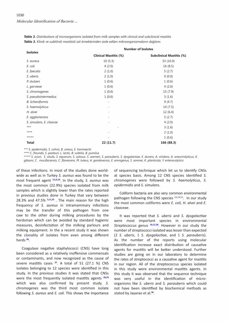
Of 123 cows 14 (11.4%) had clinical masti ti s. Twenty
two microorganisms were isolated from these cases. The most frequent pathogens in clinical cases were 10 (5.3%) S. aureus, 4 (2.0%) E. coli, 2 (1.0%) E. faecalis, 2 (1.0%) S. uberis, and 1 (0.6%) S. pseudointermedius, L. garvieae, S. chromogenes, S. chromogenes, S. pseudointermedius while 2 samples had no bacterial growth. Of 123 cows 109 (88.6%) had subclinical masti ti s. From these 166 microorganisms were isolated. The most frequently isolated pathogens were 33 (16.9%) S. aureus, followed by 16 (8.5%) E. coli, 15 (7.9%) S.chromogenes, 14 (7.5%) S. haemolyti cus, 12 (6.4%) H. alvei, 5 (2.7%) E. faecalis and E. agglomerans, 4 (2.0%) L. garvieae, 3 (1.6%) S. pseudointermedius, S. epidermidis, S. cohnii and B. cereus, 2 (1.0%) C. freundii, S. pasteuri, L. lacti s, B. subti lis and B. pumilus, 1 (0.6%) P. stutzeri and others (E. amnigenus, S. somnei, K. planti cola, Y. enterocolyti ca, S. sciuri, S. vitulis, S. equorum, S. xylosus, S. warneri, S. parauberis, S.dysgalacti ae, E. durans, A. viridans, A. aneurinilyti cus, K. gibsoni, C. mucifaciensis, C. fl avescens, M. luteus, A. gandavensis) (Table 3).
DISCUSSION
Determinati on of aeti ological agents of masti ti s by conti nuous survey studies would be helpful for treatment
Fig 2. PCR performed by using 16S rRNA universal primers. M: Marker (Lambda phage DNA restricted with PstI enzyme) 1-16: PCR performed by using isolated microorganism’s DNA. 17: Negative control (without DNA master mix) 18: Positive control (S. aureus ATCC 29213)
Şekil 2. 16S universal primerleri kullanılarak gerçekleştirilen M: Marker (PstI enzimi ile kesilmiş lambda faj DNA’sı). PZR. 1-16: İzole edilen mikroorganizmaların DNA’ları ile kullanılarak yapılan PZR, 17: Negatif Kontrol (DNA’sız master miks), 18: Pozitif Kontrol (S. aureus ATCC 29213)
Fig 3. Microorganisms and their numbers isolated from milk samples obtained from cows with mastitis. A total of 42 different species were isolated from 188 isolates and from 38 samples there were no bacterial growth
Şekil 3. Mastitisli sığır süt örneklerinden izole edilen mikroorganizmalar ve sayıları. Otuzsekiz örnekten bakteriyolojik izolasyon yapılamaz iken, 188 izolattan 42 farklı tür mikroorganizma izolasyonu yapıldı
1030Molecular Identifi cation of Bacteria ...
of these infecti ons. In most of the studies done world-wide as well as in Turkey S. aureus was found to be the most frequent agent 3,5,6,24. In the study, S. aureus was the most common (22.9%) species isolated from milk samples which is slightly lower than the rates reported in previous studies done in Turkey that vary between 28.3% and 47.5% 3,11,25 . The main reason for the high frequency of S. aureus in intramammary infecti ons may be the transfer of this pathogen from one caw to the other during milking procedures by the herdsman which can be avoided by standard hygienic measures, desinfectati on of the milking parlours and milking equipment. In the a recent study it was shown the clonality of isolates from even among diff erent herds 26.
Coagulase negati ve staphylococci (CNS) have long been considered as a relati vely inoff ensive commensals or contaminants, and now recognized as the cause of severe masti ti s cases 27. A total of 51 (27.1 %) CNS isolates belonging to 12 species were identi fi ed in this study. In the previous studies it was stated that CNSs were the most frequently isolated masti ti s agents 28,29
which was also confi rmed by present study. S. chromogenes was the third most common isolate following S. aureus and E. coli. This shows the importance
of sequencing technique which let us to identi fy CNSs at species basis. Among 12 CNS species identi fi ed S. chromogenes were followed by S. haemolyti cus, S. epidermidis and S. simulans.
Coliform bacteria are also very common environmental pathogen following the CNS species 3,6,10,11. In our study the most common coliforms were E. coli, H. alvei and E. cloaceae.
It was reported that S. uberis and S. dysgalacti ae were most important species in environmental Streptecoccus genus 10,11,28. However in our study the number of streptococci isolated was lesser than expected (2 S. uberis, 1 S. dysgalacti ae, and 1 S. parauberis). As the number of the reports using molecular identi fi cati on increase exact distributi on of causati ve agents for masti ti s will be bett er understood. Further studies are going on in our laboratory to determine the rates of streptococci as a causati ve agent for masti ti s in our region. All of the streptococcus species isolated in this study were environmental masti ti s agents. In this study it was observed that the sequence technique was very useful in the identi fi cati on of micro-organisms like S. uberis and S. parauberis which could not have been identi fi ed by biochemical methods as stated by Jayarao et al.30.
IsolatesNumber of Isolates
Clinical Mastitis (%) Subclinical Mastitis (%)
S. aureus 10 (5.3) 33 (16.9)E. coli 4 (2.0) 16 (8.5)E. faecalis 2 (1.0) 5 (2.7)S. uberis 2 (1.0) 0 (0.0)P. stutzeri 1 (0.6) 1 (0.6)L. garvieae 1 (0.6) 4 (2.0)S. chromogenes 1 (0.6) 15 (7.9)S. pseudointermedius 1 (0.6) 3 (1.6)B. licheniformis - 9 (4.7)S. haemolyticus - 14 (7.5)H. alvei - 12 (6.4)E. agglomerans - 5 (2.7)S. simulans, E. cloacae - 4 (2.0)*** - 3 (1.6)**** - 2 (1.0) ***** - 1 (0.6) Total 22 (11.7) 166 (88.3)
Table 3. Distributions of microorganisms isolated from milk samples with clinical and subclinical mastitis Tablo 3. Klinik ve subklinik mastitisli süt örneklerinden izole edilen mikroorganizmaların dağılımı
*** S. epidermidis, S. cohnii, B. cereus, E. hormaechi**** C. freundii, S. pasteuri, L. lactis, B. subtilis, B. pumilus ***** S. sciuri, S. vitulis, S. equorum, S. xylosus, S. warneri, S. parauberis, S. dysgalactiae, E. durans, A. viridans, A. aneurinilyticus, K. gibsoni, C. mucifaciensis, C. fl avescens, M. luteus, A. gandavensis, E. amnigenus, S. somnei, K. planticola, Y. enterocolytica

1031
TÜRKYILMAZ, YILDIZ, ORYAŞINKAYNARCA, BOZDOĞAN
The pathogens which cause clinic or subclinic masti ti s are not always the same. The diversity of causati ve agents for subclinical masti ti s is very much higher than clinical masti ti s. In both clinical and subclinical masti ti s S. aureus and E. coli are the most important agents however their rates are diff erent; S. aureus are the 45% of all clinical masti ti s but only 19% of subclinical masti ti s. E. coli rates are twice higher for clinical masti ti s than subclinical masti ti s (18% versus 9.6%). The rates of CNSs are 29.5% among subclinical masti ti s but only 9% among clinical masti ti s.
In some of the previous studies CNSs were the most frequently isolated bacteria from subclinical cases 1,24,31-33. However, some authors reported high percentage of clinical cases evoked by CNS 34. Bayar 1 have reported that all Staphylococcus spp. (S. haemolyti cus, S. simulans S. auricularis, S. hominis, S. warneri, S. capiti s, S. cohnii, S. xylosus, S. epidermidis and S. sciuri) isolated from 221 subclinical masti ti s suspected milk samples were CNS. Gianneechini et al.24 have also reported that 37 (7.4%) and 3 (7.5%) CNS isolati ons were performed from subclinical and clinical masti ti s, respecti vely. In this study 51 CNS isolati ons were obtained from which 49 were from subclinical cases. Under the considerati on that staphylococci are found in the normal fl ora of the teat and teat skin, this high rati o (26.2%) explains that masti ti s control measurements were insuffi cient.
As menti oned above, in our survey 16.8% of the bacteriological cultures were negati ve for all obtained samples. This rati o was varied between 38.0% and 7.3% in other studies 3,11,24,35,36. In the authors’ opinion the possible reasons for this may be the suspected microorganism may be an anaerobic microorganism, a virus, mycoplasma or other microorganisms requiring special mediums for growth.
A successful control program for masti ti s can be established with an eff ecti ve monitoring system for all dairy herds and correct identi fi cati on of pathogen that cause masti ti s. Molecular methods may help for correct identi fi cati on of agents. Sequencing technique used in identi fying bacteria is becoming a frequent method. High cost and insuffi cient number of specialists were the limiti ng factors in the expanding use of molecular identi fi cati on methods in routi ne veterinary laboratories. But nowadays sequence technique may have greater advantages over traditi onal phenotypic identi fi cati on methods by means of reduced cost of sequence and increasing knowledge of the technical personnel.
In conclusion, contagious and environmental agents were isolated at a rate of 22.9% and 77.1% from masti ti s
suspected milk samples, respecti vely. Isolati on of forty-two species indicates that the choice of anti bacterial agents needs a bett er identi fi cati on of causati ve agents to be more eff ecti ve in curing a wide range of intra-mammary infecti ons.
REFERENCES
1. Bayar S: Isolation and identification of Staphylococcus and Streptococcus species from milk and determination of their antibiotic susceptibilities. Msc Thesis. Kahramanmaraş Sütçü İmam University Institute of Natural and Applied Sciences Department of Biology, 2007.
2. Gülcü HB, Ertaş HB: Bacteriological investigation of udder lobes of cows with mastitis slaughtered in the Elazığ region. Tr Vet Anim Sci, 28, 91-94, 2004.
3. Tel OY, Keskin O, Zonturlu KA, Kaya NBA: Subclinical mastitis prevalence and determination of the antibiotics susceptibility in Şanlıurfa region. Fırat Univ Sağ Bil Vet Derg, 23, 101-106, 2009.
4. Gooraninejad S, Ghorbanpoor M, Salati AM: Antibiotic susceptibility of staphylococci isolated from bovine sub-clinical mastitis. Pak J Bio Sci, 10, 2781-2783, 2007.
5. Lakew M, Tolosa T, Tigre W: Prevalence and major bacterial causes of bovine mastitis in Asella, South Eastern Ethiopia. Trop Anim Health Prod, 41, 1525-1530, 2009.
6. Sghir A, Antonopoulos D, Mackie RI: Prevalence of bovine mastitis amongst small holder dairy herds in Kenya. Israel J Vet Med, 59, 1-6, 2004.
7. Tekeli T: Mastitis. AB Sürecinde Kaliteli Süt Üretimi ve Somatik Hücre Sayısı. Güzeliş Matbaası, Konya. pp. 19-35, 2005 (in Turkish).
8. Bradley AJ: Bovine mastitis: an evolving disease. Vet J, 164, 116-128, 2002.
9. Costa EO, Ribeiro AR, Watanabe ET, Melville P: Infectious bovine mastitis caused by environmental organism. Zbl Vet Med (B), 45, 65-71. 1998.
10. Ak S, Horoz H, Ilgaz A: Bacterial agents cause contagious and environmental bovine mastitis in Trakya district and their susceptibilty to antibiotics. Istanbul Univ Vet Fak Derg, 26, 353-365, 2000.
11. Kuyucuoglu Y, Ucar M: Determination of the subclinical and clinical mastitis rates in dairy cows, of Afyon region and effective antibiotics. Vet Hek Mikrobiyol Derg, 1, 19-24, 2001.
12. Risvanli A, Kalkan C: The effect of age and breed on somatic cell count and microbiological isolation rates in milk of dairy cows with subclinical mastitis. YYÜ Vet Fak Derg, 13, 84-87, 2002.
13. Garcia A: Contagious vs. environmental mastitis. http://agbiopubs.sdstate.edu/articles/ExEx4028.pdf, Accessed: 12. 11.2009.
14. Philpot WN, Nickerson SC: Mastitis attack. Surge International - Babson Bros Co Naperville, Illinois, USA, pp. 10-28, 1991.
15. Leslie KE, Jansen JT, Lim GH: Opportunities and implications for improved on farm cowside diagnostics. Proc DeLaval Hygiene Sym, pp. 147-160, 2002.
1032Molecular Identifi cation of Bacteria ...
16. Anonymus: National Mastitis Council: Laboratory Hand-book on Bovine Mastitis. Revised ed., pp. 1-30, Madison, Wisconsin - Natl Mastitis Counc, 1999.
17. Chai H, Archambault M, Prescott JF: 16S Ribosomal RNA sequence-based identification of veterinary clinical bacteria. J Vet Diagn Invest, 15, 465-469, 2003.
18. Drancourt M, Bollet C, Carlioz A, Martelin R, Gayral JP, Raoult D: 16S ribosomal DNA sequence analysis of a large collection of environmental and clinical unidentifiable bacterial isolates. J Clin Microbiol, 38, 3623-3630, 2000.
19. Ludwig W, Schleifer KH: Bacterial phylogeny based on 16S, 23 sRNA sequence analysis. FEMS Microbiol Rev, 15, 155-173, 1992.
20. Quinn PJ, Carter ME, Markey BK, Carter GR: Clinical Veterinary Microbiology. pp. 40-190, Mosby-Year Book Europe Limited, Lynton House, London, England 1994.
21. Baştan A: İneklerde Meme Hastalıkları. Hatipoğlu Basım ve Yayım San. Tic. Ltd. Şti., Ankara, pp: 25–78. 2002.
22. Sghir A, Antonopoulos D, Mackie RI: Design and evaluation of a lactobacillus group-specific ribosomal RNA-targeted hybridization probe and its application to the study of intestinal microecology in pigs. Syst Appl Microbiol, 21, 291-296, 1998.
23. Suau A, Bonnet R, Sutren M, Godon JJ, Gibson G, Collins Dore´J MD: Direct rDNA community analysis reveals a myriad of novel bacterial lineages within the human gut. Appl Environ Microbiol, 65, 4799-4807, 1999.
24. Gianneechini R, Concha C, Rivero R, Delucci I, López MJ: Occurrence of clinical and sub-clinical mastitis in dairy herds in the west littoral region in Uruguay. Acta Vet Scand, 43, 221-230, 2002.
25. Bademkıran S, Yeşilmen S, Gürbulak K: The effect of daily milking frequency on clinical mastitis and milk yield of dairy cows. YYÜ Vet Fak Derg, 16, 17-21, 2005.
26. Türkyılmaz S, Tekbıyık S, Oryaşın E, Bozdogan B: Molecular epidemiology and antimicrobial resistance mechanisms of methicillin-resistant Staphylococcus aureus isolated from bovine milk. Zoonoses Public Hlth, 57, 197-203, 2010.
27. Taponen S, Simojoki H, Haveri M, Larsen HD, Pyorala S: Clinical characteristics and persistence of bovine mastitis caused by different species of coagulase negative staphylococci identified with API or AFLP. Vet Microbiol, 15, 199-207, 2006.
28. Pitkala A, Haveri M, Pyorala S, Myllysi V, Honkanen-Buzaski T: Bovine mastitis in Finland 2001 prevalence, distribution of bacteria, and antimicrobial resistance. J Dairy Sci, 87, 2433-2441, 2004.
29. Sampimon O, Barkema HW, Berends I, Sol J, Lam T: Prevalance of intramammary infection in Dutch herds. J Dairy Res, 76, 129-136, 2009.
30. Jayarao BM, Doré JJ, Baumbach GA, Matthews KR, Oliver SP: Differentiation of Streptococcus uberis from Streptococcus parauberis by polymerase chain reaction and restriction fragment length polymorphism analysis of 16S ribosomal DNA. J Clin Microbiol, 29, 2774-2778, 1991.
31. Myllys V, Aspund K, Brofeldt E, Hirvela-Koski V, Hankanen-Buzalske T, Kulkas L, Myllykangas O, Niskanen M, Saloniemi H, Sandholm M, Saranpaa T: Bovine mastitis in Finland in 1988 and 1995. Changes and prevalence and antimicrobial resistance. Acta Vet Scand, 39, 119-126, 1998.
32. Kaynarca S, Türkyılmaz S: Methicillin resistance and slime positivity of staphylococci isolated from bovine mastitis. Kafkas Univ Vet Fak Derg, 16, 567-572, 2010.
33. Ünal N, Yıldırım M: Antibiotic resistance profiles of staphylococci species isolated from milks, teat skins and noses mucous of cows. Kafkas Univ Vet Fak Derg, 16, 389-396, 2010.
34. Jarp J: Classification of coagulase negative staphylococci isolated from bovine clinical and subclinical mastitis. Vet Microbiol, 27, 151-158, 1991.
35. Bartlett PC, Miller GY, Lance SE, Heider LE: Managerial determinants of intramammary coliform and environmental streptococci infections in Ohio dairy herds. J Dairy Sci, 75, 1241-1252, 1992.
36. Giovannini G, Piccinini R, Zecconi A: Epidemiology of clinical mastitis in Italy. 39th Annual Meeting, National Mastitis Council, Inc. Madison, WI 53704, USA, pp. 176-178, 2000.