Molecular Docking Studies of (1 E,3E,5E)-1,6-Bis ...
11
January 2013 55 Biol. Pharm. Bull. 36(1) 55–65 (2013) © 2013 The Pharmaceutical Society of Japan Regular Article Molecular Docking Studies of (1E,3E,5E)-1,6-Bis(substituted phenyl)- hexa-1,3,5-triene and 1,4-Bis(substituted trans-styryl)benzene Analogs as Novel Tyrosinase Inhibitors Young Mi Ha, a Hye Jin Lee, b Daeui Park, a Hyoung Oh Jeong, a Ji Young Park, b Yun Jung Park, b Kyung Jin Lee, b Ji Yeon Lee, b Hyung Ryong Moon,* ,b and Hae Young Chung* ,a a Molecular Inflammation Research Center for Aging Intervention (MRCA), College of Pharmacy, Pusan National University; and b Laboratory of Medicinal Chemistry, College of Pharmacy, Pusan National University; Kumjeong-gu, Busan 609–735, Republic of Korea. Received July 10, 2012; accepted September 29, 2012 We simulated the docking of the tertiary structure of mushroom tyrosinase with our compounds. From the structure-tyrosinase inhibitory activity relationship, it is notable that compounds 4, 8 and 11 showed similar or better activity rates than kojic acid which was used as a positive control. Compounds 17, 21, and 23 among benzene analogs that possess the same substituent showed significantly lower tyrosinase inhibitory effects. Therefore, we have confirmed that among the compounds showing better tyrosinase inhibitory effects than kojic acid, the compounds with triene analogs have better tyrosinase inhibitory effect than the com- pounds with benzene analogs. Docking simulation suggested the mechanism of compounds by several key res- idues which had possible hydrogen bonding interactions. The pharmacophore model underlined the features of active compounds, 4,4′-((1E,3E,5E)-hexa-1,3,5-triene-1,6-diyl)diphenol, 5,5′-((1E,3E,5E)-hexa-1,3,5-triene- 1,6-diyl)bis(2-methoxy-phenol), and 5,5′-((1E,3E,5E)-hexa-1,3,5-triene-1,6-diyl)dibenzene-1,3-diol among triene derivatives which had several hydrogen bond groups on both terminal rings. The soundness of the docking results and the agreement with the pharmacophores suggest that it can be conveniently exploited to design inhibitors with an improved affinity for tyrosinase. Key words docking simulation; tyrosinase inhibitory activity; shared pharmacophore Tyrosinase is involved not only in melanin synthesis in peripheral tissues but also in the substantia nigra of mice and humans. 1,2) Recently, it has also been reported that secretion of neuronal tyrosinase plays an important role in antiapoptosis. 3) Hence, over-expression of tyrosinase in brain might either be toxic, because of increased melanin precursors such as 3,4-dihydroxyphenylalanine (DOPA) and dopaquinone, 4) or neuroprotective, because of the formation of neuromelanin. 5) Tyrosinase has been also linked to Parkinson’s and other neurodegenerative diseases. 6–8) Therefore, identification of ty- rosinase inhibitors has promising potential in the treatment of such diseases. 9) In an effort to identify and explore new, potent, and safer tyrosinase inhibitors, we reported hydroxyl-substituted phe- nyl naphthalenes analogs of resveratrol, 10) hydroxy-substi- tuted phenyl-benzo[ d]thiazole analogs, 11) and 5-(substituted benzylidene)hydantoin analogs in our previous studies. 12) We also found several potent tyrosinase inhibitors in natural materials 10,13,14) that have an even greater potency than kojic acid, a well-known tyrosinase inhibitor. 10,15) Accumulation of β-amyloid plaques in brain parenchyma and neurofibril- lary tangles in the neuron 16,17) has been linked to Alzheimer’s disease, 18) which is a common neurodegenerative disorder. 19) Many studies have reported that biphenyl trienes, 20) phe- nolic bis-trans-styrylbenzenes 21) and polyfluorinated bis- styrylbenzene 22) displayed high binding affinities to β-amyloid plaques. However, the association of these compounds with tyrosinase activity has not yet been reported. We designed novel (1E,3E,5E)-1,6-bis(substituted phenyl)- hexa-1,3,5-triene and 1,4-bis(substituted trans-styryl)benzene analogs as potential tyrosinase inhibitors. We found that some of the compounds are more potent inhibitors than kojic acid, the standard tyrosinase inhibitor. 23) Our kinetic and struc- ture–activity relationship studies revealed novel clues to the identification, active site-binding and mechanism of action of the pharmacophores present in these inhibitors. Molecular docking was performed using AutoDock4.2 to examine the binding of the active compounds to the active site of the ty- rosinase enzyme. MATERIALS AND METHODS Synthesis of Compound 1 A solution of trans-1,4- dibromobutene (2.0 g, 9.35 mmol) in triethylphosphite (4 mL) was refluxed for 5 h. After cooling, volatiles were evaporated. The residue was purified by silica gel column chromatography using hexane and ethyl acetate (EtOAc) (1 : 3) to give diphos- phonate 1 (3.12 g, 99%). Synthesis of Compounds 3a–g tert-Butyldimethylsilyl chloride (1.25 eq×the number of hydroxyl groups) was added to a stirred solution of hydroxyl-substituted benzaldehydes (1.0 eq) and imidazole (2.5 eq×the number of hydroxyl groups) in methylene chloride and/or dimethylformamide (DMF) and the reaction mixture was stirred at room temperature and par- titioned between methylene chloride and water. The organic layers were dried, filtered, and evaporated. The residue was purified by silica gel column chromatography using hexane and EtOAc (50 : 1) to give compounds 3a–g. Synthesis of Compounds 4–8, 11, and 14 A solution of compound 1 (0.6 eq) in tetrahydrofuran (THF) and a solu- tion of tert-butyldimethylsilyl (TBS)-protected benzaldehydes (1.0 eq, compounds 3a–g) in THF were added to a stirred sus- pension of NaH (1.5 eq) in THF and the reaction mixture was stirred at room temperature for 2–5 h. The reaction mixture * To whom correspondence should be addressed. e-mail: [email protected]; [email protected] The authors declare no conflict of interest.
Transcript of Molecular Docking Studies of (1 E,3E,5E)-1,6-Bis ...

January 2013 55Biol. Pharm. Bull. 36(1) 55–65 (2013)
© 2013 The Pharmaceutical Society of Japan
Regular Article
Molecular Docking Studies of (1E,3E,5E)-1,6-Bis(substituted phenyl)-hexa-1,3,5-triene and 1,4-Bis(substituted trans-styryl)benzene Analogs as Novel Tyrosinase InhibitorsYoung Mi Ha,a Hye Jin Lee,b Daeui Park,a Hyoung Oh Jeong,a Ji Young Park,b Yun Jung Park,b Kyung Jin Lee,b Ji Yeon Lee,b Hyung Ryong Moon,*,b and Hae Young Chung*,a
a Molecular Inflammation Research Center for Aging Intervention (MRCA), College of Pharmacy, Pusan National University; and b Laboratory of Medicinal Chemistry, College of Pharmacy, Pusan National University; Kumjeong-gu, Busan 609–735, Republic of Korea. Received July 10, 2012; accepted September 29, 2012
We simulated the docking of the tertiary structure of mushroom tyrosinase with our compounds. From the structure-tyrosinase inhibitory activity relationship, it is notable that compounds 4, 8 and 11 showed similar or better activity rates than kojic acid which was used as a positive control. Compounds 17, 21, and 23 among benzene analogs that possess the same substituent showed significantly lower tyrosinase inhibitory effects. Therefore, we have confirmed that among the compounds showing better tyrosinase inhibitory effects than kojic acid, the compounds with triene analogs have better tyrosinase inhibitory effect than the com-pounds with benzene analogs. Docking simulation suggested the mechanism of compounds by several key res-idues which had possible hydrogen bonding interactions. The pharmacophore model underlined the features of active compounds, 4,4′-((1E,3E,5E)-hexa-1,3,5-triene-1,6-diyl)diphenol, 5,5′-((1E,3E,5E)-hexa-1,3,5-triene- 1,6-diyl)bis(2-methoxy-phenol), and 5,5′-((1E,3E,5E)-hexa-1,3,5-triene-1,6-diyl)dibenzene-1,3-diol among triene derivatives which had several hydrogen bond groups on both terminal rings. The soundness of the docking results and the agreement with the pharmacophores suggest that it can be conveniently exploited to design inhibitors with an improved affinity for tyrosinase.
Key words docking simulation; tyrosinase inhibitory activity; shared pharmacophore
Tyrosinase is involved not only in melanin synthesis in peripheral tissues but also in the substantia nigra of mice and humans.1,2) Recently, it has also been reported that secretion of neuronal tyrosinase plays an important role in anti apoptosis.3) Hence, over-expression of tyrosinase in brain might either be toxic, because of increased melanin precursors such as 3,4-dihydroxyphenylalanine (DOPA) and dopaquinone,4) or neuroprotective, because of the formation of neuromelanin.5) Tyrosinase has been also linked to Parkinson’s and other neurodegenerative diseases.6–8) Therefore, identification of ty-rosinase inhibitors has promising potential in the treatment of such diseases.9)
In an effort to identify and explore new, potent, and safer tyrosinase inhibitors, we reported hydroxyl-substituted phe-nyl naphthalenes analogs of resveratrol,10) hydroxy-substi-tuted phenyl-benzo[d] thiazole analogs,11) and 5-(substituted benzylidene) hydantoin analogs in our previous studies.12) We also found several potent tyrosinase inhibitors in natural materials10,13,14) that have an even greater potency than kojic acid, a well-known tyrosinase inhibitor.10,15) Accumulation of β-amyloid plaques in brain parenchyma and neurofibril-lary tangles in the neuron16,17) has been linked to Alzheimer’s disease,18) which is a common neurodegenerative disorder.19) Many studies have reported that biphenyl trienes,20) phe-nolic bis-trans-styrylbenzenes21) and polyfluorinated bis-styrylbenzene22) displayed high binding affinities to β-amyloid plaques. However, the association of these compounds with tyrosinase activity has not yet been reported.
We designed novel (1E,3E,5E)-1,6-bis(substituted phenyl)-hexa-1,3,5-triene and 1,4-bis(substituted trans-styryl) benzene analogs as potential tyrosinase inhibitors. We found that some
of the compounds are more potent inhibitors than kojic acid, the standard tyrosinase inhibitor.23) Our kinetic and struc-ture–activity relationship studies revealed novel clues to the identification, active site-binding and mechanism of action of the pharmacophores present in these inhibitors. Molecular docking was performed using AutoDock4.2 to examine the binding of the active compounds to the active site of the ty-rosinase enzyme.
MATeRIALS AnD MeTHODS
Synthesis of Compound 1 A solution of trans-1,4-dibromobutene (2.0 g, 9.35 mmol) in triethylphosphite (4 mL) was refluxed for 5 h. After cooling, volatiles were evaporated. The residue was purified by silica gel column chromatography using hexane and ethyl acetate (etOAc) (1 : 3) to give diphos-phonate 1 (3.12 g, 99%).
Synthesis of Compounds 3a–g tert-Butyldimethylsilyl chloride (1.25 eq×the number of hydroxyl groups) was added to a stirred solution of hydroxyl-substituted benzaldehydes (1.0 eq) and imidazole (2.5 eq×the number of hydroxyl groups) in methylene chloride and/or dimethylformamide (DMF) and the reaction mixture was stirred at room temperature and par-titioned between methylene chloride and water. The organic layers were dried, filtered, and evaporated. The residue was purified by silica gel column chromatography using hexane and etOAc (50 : 1) to give compounds 3a–g.
Synthesis of Compounds 4–8, 11, and 14 A solution of compound 1 (0.6 eq) in tetrahydrofuran (THF) and a solu-tion of tert-butyldimethylsilyl (TBS)-protected benzaldehydes (1.0 eq, compounds 3a–g) in THF were added to a stirred sus-pension of naH (1.5 eq) in THF and the reaction mixture was stirred at room temperature for 2–5 h. The reaction mixture
* To whom correspondence should be addressed. e-mail: [email protected]; [email protected]
The authors declare no conflict of interest.

56 Vol. 36, no. 1
was partitioned between methylene chloride and aqueous nH4Cl solution and the organic layers were dried, filtered, and evaporated. The residue was purified by silica gel column chromatography using hexane and etOAc (2 : 1) to give crude trienes. To a solution of crude trienes in THF and methanol (4 : 1) was added 12 n HCl and the reaction mixture was stirred at room temperature overnight. The reaction mixture was par-titioned between methylene chloride or etOAc and water and the organic layers were washed with aqueous naHCO3 solu-tion. The organic layers were dried, filtered, and evaporated and the residue was recrystallized or purified by silica gel column chromatography to give compounds 4–8, 11, and 14.
Synthesis of Compounds 9, 10, 12, and 13 A solution of compound 1 (0.6 eq) in THF and a solution of methoxy-substi-tuted benzaldehydes (1.0 eq) in THF were added to a stirred suspension of naH (1.5 eq) in THF and the reaction mixture was stirred at room temperature. The reaction mixture was partitioned between methylene chloride and aqueous nH4Cl solution and the organic layers were dried, filtered, and evapo-rated. The residue was purified by silica gel column chroma-tography using hexane and etOAc (8 : 1) to give compounds 9, 10, 12, and 13.
(E)-Tetraethyl but-2-ene-1,4-diyldiphosphonate (1): Oil; reaction time, 5 h; yield, 99%; 1H-nMR (400 MHz, CDCl3) δ: 5.52 (m, 2H, 2×vinylic H), 4.01 (m, 8H, 4×OCH2), 2.52 (dd, 4H, J=4.0, 18.0 Hz, 1-CH2, 4-CH2), 1.23 (td, 12H, J=1.2, 6.8 Hz, 4×CH3); 13C-nMR (100 MHz, CDCl3) δ: 124.5 (d, J=1.5 Hz, 2×vinylic C), 62.1 (d, J=6.1 Hz, 4×OCH2), 30.7 (dd, J=4.6, 141.1 Hz, C1, C4), 16.6 (d, J=5.3 Hz, 4×CH3).
4-(tert-Butyldimethylsilyloxy) benzaldehyde (3a): Oil; reac-tion time, overnight; yield, 75%; 1H-nMR (400 MHz, CDCl3) δ: 9.84 (s, 1H, CHO), 7.75 (d, 2H, J=8.4 Hz, 2-H, 6-H), 6.90 (d, 2H, J=8.4 Hz, 3-H, 5-H), 0.95 (s, 9H, t-Bu), 0.21 (s, 6H, 2×SiCH3); 13C-nMR (100 MHz, CDCl3) δ: 191.0 (CHO), 161.7 (C4), 132.1 (C2, C6), 130.6 (C1), 120.7 (C3, C5), 25.7 (3×CH3), 18.4 (quaternary C), −4.2 (2×SiCH3).
3,4-Bis(tert-butyldimethylsilyloxy) benzaldehyde (3b): Solid; reaction time, 6 h; yield, 74%; 1H-nMR (500 MHz, CDCl3) δ: 9.80 (s, 1H, CHO), 7.36 (d, 1H, J=7.5 Hz, 6-H), 7.35 (s, 1H, 2-H), 6.94 (d, 1H, J=7.5 Hz, 5-H), 0.99 (s, 18H, 2×t-Bu), 0.25 (s, 6H, 2×SiCH3), 0.23 (s, 6H, 2×SiCH3).
4-(tert-Butyldimethylsilyloxy)-3-methoxybenzaldehyde (3c): Oil; reaction time, 2 h; yield, 100%; 1H-nMR (400 MHz, CDCl3) δ: 9.82 (s, 1H, CHO), 7.38 (d, 1H, J=2.0 Hz, 2-H), 7.34 (dd, 1H, J=2.0, 8.0 Hz, 6-H), 6.94 (d, 1H, J=8.0 Hz, 5-H), 3.85 (s, 3H, 3-OCH3), 0.98 (s, 9H, t-Bu), 0.17 (s, 6H, 2×SiCH3);13C-nMR (100 MHz, CDCl3) δ: 191.2 (CHO), 151.8 (C3), 151.6 (C4), 131.1 (C1), 126.4 (C6), 120.9 (C5), 110.3 (C2), 55.7 (3-OCH3), 25.8 (3×CH3), 18.7 (quaternary C), −4.4 (2×SiCH3).
4-(tert-Butyldimethylsilyloxy)-3-ethoxybenzaldehyde (3d): Oil; reaction time, 4 h; yield, 95%; 1H-nMR (400 MHz, CDCl3) δ: 9.81 (s, 1H, CHO), 7.35 (d, 1H, J=2.0 Hz, 2-H), 7.32 (dd, 1H, J=2.0, 8.0 Hz, 6-H), 6.93 (d, 1H, J=8.0 Hz, 5-H), 4.07 (q, 2H, J=7.2 Hz, OCH2), 1.44 (t, 3H, J=7.2 Hz, OCH2CH3), 0.98 (s, 9H, t-Bu), 0.18 (s, 6H, 2×SiCH3); 13C-nMR (100 MHz, CDCl3) δ: 191.3 (CHO), 151.5 (C3), 151.3 (C4), 131.2 (C1), 126.2 (C6), 121.0 (C5), 111.1 (C2), 64.2 (OCH2), 25.8 (3×CH3), 18.7 (quaternary C), 14.9 (OCH2CH3), −4.3 (2×SiCH3).
3-(tert-Butyldimethylsilyloxy)-4-methoxybenzaldehyde (3e): Oil; reaction time, overnight; yield, 99%; 1H-nMR (500 MHz,
CDCl3) δ: 9.81 (s, 1H, CHO), 7.47 (dd, 1H, J=2.0, 8.5 Hz, 6-H), 7.37 (d, 1H, J=2.0 Hz, 2-H), 6.95 (d, 1H, J=8.5 Hz, 5-H), 3.89 (s, 3H, OCH3), 1.00 (s, 9H, t-Bu), 0.17 (s, 6H, 2×SiCH3); 13C-nMR (100 MHz, CDCl3) δ: 191.1 (CHO), 156.8 (C4), 145.8 (C3), 130.4 (C1), 126.5 (C6), 120.3 (C2), 111.4 (C5), 55.8 (OCH3), 25.8 (3×CH3), 18.6 (quaternary C), −4.4 (2×SiCH3).
3,5-Bis(tert-butyldimethylsilyloxy) benzaldehyde (3f): Solid; reaction time, 5 h; yield, 76%; 1H-nMR (500 MHz, CDCl3) δ: 9.85 (s, 1H, CHO), 6.95 (s, 2H, 2-H, 6-H), 6.58 (s, 1H, 4-H), 0.98 (s, 18H, 2×t-Bu), 0.21 (s, 12H, 4×SiCH3).
4-(tert-Butyldimethylsilyloxy)-3,5-dimethoxybenzaldehyde (3g): Solid; reaction time, overnight; yield, 95%; melting point, 71.9–72.0°C; 1H-nMR (500 MHz, CDCl3) δ: 9.82 (s, 1H, CHO), 7.10 (s, 2H, 2-H, 6-H), 3.87 (s, 6H, OCH3), 1.01 (s, 9H, t-Bu), 0.16 (s, 6H, 2×SiCH3); 13C-nMR (100 MHz, CDCl3) δ: 191.3 (CHO), 152.2 (C3, C5), 140.8 (C4), 129.5 (C1), 106.9 (C2, C6), 56.0 (OCH3), 25.9 (3×CH3), 19.1 (quaternary C), −4.3 (2×SiCH3).
4,4′-((1E,3E,5E)-Hexa-1,3,5-triene-1,6-diyl) diphenol (4): Dark brown solid; reaction time, 2 h/overnight; yield, 2% (two-step yield); melting point, 255.9–258.6°C; 1H-nMR (400 MHz, DMSO-d6) δ: 9.55 (s, 2H, 2×OH), 7.23 (d, 4H, J= 8.8 Hz, 3-H, 5-H, 3′-H, 5′-H), 6.73 (ddd, 2H, J=3.2, 7.2, 15.6 Hz, 2″-H, 5″-H), 6.69 (d, 4H, J=8.4 Hz, 2-H, 6-H, 2′-H, 6′-H), 6.46 (d, 2H, J=15.6 Hz, 1″-H, 6″-H), 6.41 (2H, J=3.2, 7.2 Hz, 2″-H, 3″-H, 4″-H); 13C-nMR (100 MHz, DMSO-d6) δ: 157.8 (C1, C1′), 133.2 (C1″, C6″), 132.4 (C3″, C4″), 129.0 (C4, C4′), 128.3 (C3, C5, C3′, C5′), 127.0 (C2″, C5″), 116.3 (C2, C6, C2′, C6′); Anal. Calcd for C18H16O2: C, 81.79; H, 6.10. Found: C, 81.91; H, 5.97.
4,4′-((1E,3E,5E)-Hexa-1,3,5-triene-1,6-diyl) dibenzene-1,2-diol (5): Bluish gray solid; reaction time, 3.5 h/overnight; yield, 5% (two-step yield); melting point, 235.9–238.6°C; 1H-nMR (500 MHz, CD3OD) δ: 6.89 (d, 2 H, J=1.5 Hz, 3-H, 3′-H), 6.75 (dd, 2H, J=1.5, 8.0 Hz, 5-H, 5′-H), 6.70 (d, 2H, J= 8.0 Hz, 6-H, 6′-H), 6.68 (ddd, 2H, J=2.5, 7.0, 15.0 Hz, 2″-H, 5″-H), 6.41 (d, 2H, J=15.0 Hz, 1″-H, 6″-H), 6.41 (dd, 2H, J=3.0, 7.5 Hz, 3″-H, 4″-H); 13C-nMR (100 MHz, CD3OD) δ: 145.2 (C1, C1′), 145.2 (C2, C2′), 132.5 (C1″, C6″), 132.0 (C3″, C4″), 130.3 (C4, C4′), 126.7 (C2″, C5″), 118.9 (C5, C5′), 115.3 (C6, C6′), 112.5 (C3, C3′); Anal. Calcd for C18H16O4: C, 72.96; H, 5.44. Found: C, 73.06; H, 5.41.
4,4′-((1E,3E,5E)-Hexa-1,3,5-triene-1,6-diyl) bis(2-methoxy-phenol) (6): Orange-colored solid; reaction time, 3.5 h/overnight; yield, 10% (two-step yield); melting point, 214.3–215.7°C; 1H-nMR (400 MHz, DMSO-d6) δ: 9.11 (s, 2H, 2×OH), 7.04 (d, 2H, J=1.6 Hz, 3-H, 3′-H), 6.83 (dd, 2H, J= 1.6, 8.0 Hz, 5-H, 5′-H), 6.81 (ddd, 2H, J=3.2, 7.2, 16.0 Hz, 2″-H, 5″-H), 6.69 (d, 2H, J=8.0 Hz, 6-H, 6′-H), 6.47 (d, 2H, J= 16.0 Hz, 1″-H, 6″-H), 6.42 (dd, 2H, J=3.2, 7.2 Hz, 3″-H, 4″-H), 3.77 (s, 6 H, 2×OCH3); 13C-nMR (100 MHz, DMSO-d6) δ: 148.5 (C2, C2′), 147.2 (C1, C1′), 133.2 (C1″, C6″), 132.7 (C3″, C4″), 129.6 (C4, C4′), 127.3 (C2″, C5″), 120.6 (C5, C5′), 116.3 (C6, C6′), 110.2 (C3, C3′), 56.2 (2×OCH3); Anal. Calcd for C20H20O4: C, 74.06; H, 6.21. Found: C, 73.88; H, 6.04.
4,4′-((1E,3E,5E)-Hexa-1,3,5-triene-1,6-diyl) bis(2-ethoxy-phenol) (7): Beige solid; reaction time, 3.5 h/overnight; yield, 8% (two-step yield); melting point, 223.1–225.5°C; 1H-nMR (400 MHz, DMSO-d6) δ: 9.02 (s, 2H, 2×OH), 7.01 (s, 2H, 3-H, 3′-H), 6.83 (d, 2H, J=8.0 Hz, 5-H, 5′-H), 6.78 (ddd, 2H, J=2.4,
January 2013 57
6.8, 15.6 Hz, 2″-H, 5″-H), 6.70 (d, 2H, J=8.0 Hz, 6-H, 6′-H), 6.44 (d, 2H, J=15.6 Hz, 1″-H, 6″-H), 6.41 (dd, 2 H, J=2.4, 6.8 Hz, 3″-H, 4″-H), 4.01 (q, 4H, J=6.8 Hz, 2×OCH2), 1.30 (t, 6H, J=6.8 Hz, 2×OCH2CH3); 13C-nMR (100 MHz, DMSO-d6) δ: 147.6 (C2, C2′), 147.5 (C1, C1′), 133.2 (C1″, C6″), 132.7 (C3″, C4″), 129.6 (C4, C4′), 127.3 (C2″, C5″), 120.6 (C5, C5′), 116.3 (C6, C6′), 111.6 (C3, C3′), 64.5 (2×OCH2), 15.4 (2×CH2CH3); Anal. Calcd for C22H24O4: C, 74.98; H, 6.86. Found: C, 75.11; H, 6.93.
5,5′-((1E,3E,5E)-Hexa-1,3,5-triene-1,6-diyl) bis(2-methoxy-phenol) (8): Dark yellow solid; reaction time, 5 h/overnight; yield, 8% (two-step yield); melting point, 248.5–251.6°C; 1H-nMR (500 MHz, DMSO-d6) δ: 8.98 (s, 2H, 2×OH), 6.90 (s, 2H, 6-H, 6′-H), 6.86 (d, 2H, J=8.5 Hz, 4-H, 4′-H), 6.84 (d, 2H, J=8.5 Hz, 3-H, 3′-H), 6.74 (ddd, 2H, J=3.0, 7.0, 15.5 Hz, 2″-H, 5″-H), 6.46 (d, 2H, J=15.5 Hz, 1″-H, 6″-H), 6.45 (dd, 2H, J=3.0, 7.0 Hz, 3″-H, 4″-H), 3.75 (s, 6H, 2×CH3); 13C-nMR (100 MHz, DMSO-d6) δ: 148.3 (C2, C2′), 147.3 (C1, C1′), 133.4 (C1″, C6″), 132.5 (C3″, C4″), 131.0 (C5, C5′), 127.8 (C2″, C5″), 118.9 (C4, C4′), 113.3 (C6, C6′), 112.9 (C3, C3′), 56.3 (2×OCH3); Anal. Calcd for C20H20O4: C, 74.06; H, 6.21. Found: C, 74.20; H, 6.38.
(1E,3E,5E)-1,6-Bis(4-methoxyphenyl) hexa-1,3,5-triene (9): Light yellow solid; reaction time, 12 h; yield, 11%; melt-ing point, 247.5–248.9°C; 1H-nMR (500 MHz, CDCl3) δ: 7.37 (d, 4H, J=8.5 Hz, 2′-H, 6′-H, 2″-H, 6″-H), 6.88 (d, 4H, J=8.5 Hz, 3′-H, 5′-H, 3″-H, 5″-H), 6.77 (ddd, 2H, J=2.5, 7.0, 15.5 Hz, 2-H, 5-H), 6.54 (d, 2H, J=15.5 Hz, 1-H, 6-H), 6.47 (dd, 2H, J=2.5, 7.0 Hz, 3-H, 4-H), 3.84 (s, 6H, 4′-OCH3, 4″-OCH3); 13C-nMR (100 MHz, CDCl3) δ: 159.4 (C4′, C4″), 133.0 (C1, C6), 131.9 (C3, C4), 130.6 (C1′, C1″), 127.7 (C2′, C6′, C2″, C6″), 127.5 (C2, C5), 114.3 (C3′, C5′, C3″, C5″), 55.5 (2×OCH3); Anal. Calcd for C20H20O2: C, 82.16; H, 6.89. Found: C, 82.31; H, 6.83.
(1E,3E,5E)-1,6-Bis(3,4-dimethoxyphenyl) hexa-1,3,5-triene (10): Light yellow solid; reaction time, 7 h; yield, 12%; melting point, 238.4–240.9°C; 1H-nMR (400 MHz, CDCl3) δ: 6.95 (s, 2H, 2′-H, 2″-H), 6.94 (d, 2H, J=8.0 Hz, 6′-H, 6″-H), 6.80 (d, 2H, J=8.0 Hz, 5′-H, 5″-H), 6.74 (ddd, 2H, J=3.2, 7.2, 15.6 Hz, 2-H, 5-H), 6.50 (d, 2H, J=15.6 Hz, 1-H, 6-H), 6.46 (dd, 2H, J= 3.2, 7.2 Hz, 3-H, 4-H), 3.90 (s, 6H, 2×OCH3), 3.87 (s, 6H, 2×OCH3); 13C-nMR (100 MHz, CDCl3) δ: 149.3 (C3′, C3″), 149.1 (C4′, C4″), 133.1 (C1, C6), 132.2 (C3, C4), 130.9 (C1′, C1″), 127.7 (C2, C5), 120.0 (C6′, C6″), 111.5 (C5′, C5″), 108.8 (C2′, C2″), 56.2 (2×OCH3), 56.1 (2×OCH3); Anal. Calcd for C22H24O4: C, 74.98; H, 6.86. Found: C, 74.75; H, 6.66.
5,5′-((1E,3E,5E)-Hexa-1,3,5-triene-1,6-diyl) dibenzene-1,3-diol (11): Yellow solid; reaction time, 3.5 h/overnight; yield, 1% (two-step yield); 1H-nMR (500 MHz, CD3OD) δ: 6.81 (ddd, 2H, J=3.0, 7.5, 15.5 Hz, 2″-H, 5″-H), 6.49 (dd, 2H, J=3.0, 7.5 Hz, 3″-H, 4″-H), 6.44 (d, 2H, J=15.5 Hz, 1″-H, 6″-H), 6.39 (d, 4H, J=2.0 Hz, 4-H, 6-H, 4′-H, 6′-H), 6.17 (t, 2H, J=2.0 Hz, 2-H, 2′-H); 13C-nMR (100 MHz, CD3OD) δ: 158.4 (C1, C3, C1′, C3′), 139.7 (C5, C5′), 133.5 (C1″, C6″), 132.8 (C3″, C4″), 129.0 (C2″, C5″), 104.8 (C4, C6, C4′, C6′), 102.0 (C2, C2′); Anal. Calcd for C18H16O4: C, 72.96; H, 5.44. Found: C, 72.95; H, 5.35.
(1E,3E,5E)-1,6-Bis(2,4-dimethoxyphenyl) hexa-1,3,5-triene (12): Chrome yellow solid; reaction time, 2 d; yield, 15%; melting point, 192.1–194.3°C; 1H-nMR (500 MHz, CDCl3) δ: 7.42 (d, 2H, J=8.5 Hz, 6′-H, 6″-H), 6.83 (m, 4 H, 2-H, 5-H,
5′-H, 5″-H), 6.45 (m, 6H, 1-H, 6-H, 3-H, 4-H, 3′-H, 3″-H), 3.86 (s, 6H, 2×OCH3), 3.84 (s, 6H, 2×OCH3); 13C-nMR (100 MHz, CDCl3) δ: 160.5 (C4′, C4″), 158.0 (C2′, C2″), 133.5 (C6′, C6″), 128.3, 127.3, 126.5, 120.0 (C1′, C1″), 105.2 (C5′, C5″), 98.7 (C3′, C3″), 55.7 (2′-OCH3, 2″-OCH3), 55.6 (4′-OCH3, 4″-OCH3); Anal. Calcd for C22H24O4: C, 74.98; H, 6.86. Found: C, 75.03; H, 6.70.
(1E,3E,5E)-1,6-Bis(3,4,5-trimethoxyphenyl) hexa-1,3,5-triene (13): Light yellow solid; reaction time, 9.5 h; yield, 27%; melting point, 203.5–207.8°C; 1H-nMR (500 MHz, CDCl3) δ: 6.81 (ddd, 2H, J=2.5, 7.0, 15.5 Hz, 2-H, 5-H), 6.65 (s, 4H, 2′-H, 6′-H, 2″-H, 6″-H), 6.53 (d, 2H, J =15.5 Hz, 1-H, 6-H), 6.52 (dd, 2H, J=2.5, 7.0 Hz, 3-H, 4-H), 3.91 (s, 12H, 3′-OCH3, 5′-OCH3, 3″-OCH3, 5″-OCH3), 3.87 (s, 6H, 4′-OCH3, 4″-OCH3); 13C-nMR (100 MHz, CDCl3) δ: 153.6 (C3′, C5′, C3″, C5″), 138.2 (C4′, C4″), 133.5 (C1, C6), 133.3 (C1′, C1″), 132.7 (C3, C4), 128.9 (C2, C5), 103.7 (C2′, C6′, C2″, C6″), 61.2 (4′-OCH3, 4″-OCH3), 56.3 (3′-OCH3, 5′-OCH3, 3″-OCH3, 5″-OCH3); Anal. Calcd for C24H28O6: C, 69.88; H, 6.84. Found: C, 69.94; H, 7.01.
4,4′-((1E,3E,5E)-Hexa-1,3,5-triene-1,6-diyl) bis(2,6-di-methoxy phenol) (14): Dark brown solid; reaction time, 5 h/overnight; yield, 7% (two-step yield); melting point, 217.5–220.6°C; 1H-nMR (500 MHz, CDCl3) δ: 6.75 (ddd, 2H, J= 2.5, 7.0, 15.0 Hz, 2″-, 5″-H), 6.66 (s, 4H, 3-H, 5-H, 3′-H, 5′-H), 6.49 (d, 2H, J=15.0 Hz, 1″-H, 6″-H) 6.48 (dd, 2H, J=2.5, 7.0 Hz, 3″-H, 4″-H), 5.58 (s, 2H, 2×OH), 3.92 (s, 12H, 2-OCH3, 6-OCH3, 2′-OCH3, 6′-OCH3); 13C-nMR (100 MHz, CDCl3) δ: 147.4 (C2, C6, C2′, C6′), 135.0 (C1, C1′), 133.0 (C1″, C6″), 132.6 (C3″, C4″), 129.3 (C4, C4′), 127.7 (C2″, C4″), 103.3 (C3, C5, C3′, C5′), 56.5 (2-OCH3, 6-OCH3, 2′-OCH3, 6′-OCH3); Anal. Calcd for C22H24O6: C, 68.74; H, 6.29. Found: C, 68.71; H, 6.17.
Synthesis of Compound 15 A solution of α,α′-dichloro-p-xylene (4.0 g, 22.85 mmol) in triethylphosphite (29 mL) was refluxed for 24 h. After cooling, volatiles were evaporated. The residue was purified by silica gel column chromatogra-phy using hexane and etOAc (1 : 4) to give diphosphonate 15 (8.56 g, 99%).
Synthesis of Compounds 17–21, 23, and 26 A solu-tion of 15 (0.6 eq) in THF and a solution of TBS-protected benzaldehydes (1.0 eq, 3a–g) in THF were added to a stirred suspension of naH (1.5 eq) in THF and the reaction mixture was stirred at room temperature for 4–8.5 h. The reaction mix-ture was partitioned between methylene chloride and aqueous nH4Cl solution and the organic layers were dried, filtered and evaporated to give crude 1,4-bis-trans-styrylbenzenes. To a solution of crude 1,4-bis-trans-styrylbenzenes in THF and methanol (4 : 1) was added 12 n HCl and the reaction mixture was stirred at room temperature overnight. After evapora-tion of volatiles, the precipitates generated were filtered and washed with water and/or methylene chloride and/or hexane to give compounds 17–21, 23, and 26.
Synthesis of Compounds 22, 24, and 25 A solution of 15 (0.6 eq) in THF and a solution of methoxy-substituted benz-aldehydes (1.0 eq) in THF were added to a stirred suspension of naH (1.5 eq) in THF and the reaction mixture was stirred at room temperature. After evaporation of volatiles, the pre-cipitates generated were filtered using water and washed with water and/or methylene chloride and/or hexane to give com-pounds 22, 24, and 25.

58 Vol. 36, no. 1
Synthesis of Compound 27 A solution of MeMCl (0.06 mL, 0.53 mmol) in methylene chloride (1 mL) was added to a stirred solution of 3-bromo-4-hydroxybenzaldehyde (80 mg, 0.40 mmol) and N,N′-diisopropylethylamine (0.11 mL, 0.65 mmol) in methylene chloride (2 mL) and the reaction mix-ture was stirred at room temperature for 12 h and partitioned between methylene chloride and water. The organic layers were dried, filtered, and evaporated. The residue was puri-fied by silica gel column chromatography using hexane and etOAc (3 : 1) as the eluent to give MeM-protected benzalde-hyde 16 (114.1 mg, 99%). A solution of compound 15 (89.6 mg, 0.24 mmol) in THF (2 mL) and a solution of compound 16 (114.1 mg, 0.39 mmol) in THF (2 mL) were added to a stirred suspension of naH (23.7 mg, 0.59 mmol) in THF (2 mL) and the reaction mixture was stirred at room temperature over-night. The reaction mixture was partitioned between methyl-ene chloride and aqueous nH4Cl solution and the organic lay-ers were dried, filtered and evaporated to give crude 1,4-bis-trans-styrylbenzenes. Hydrogen chloride (12 n) was added to a solution of crude 1,4-bis-trans-styrylbenzenes in THF (2 mL) and methanol (1 mL) and the reaction mixture was stirred at room temperature for 2 d. The precipitates generated were filtered and washed with water and methylene chloride and hexane (2 : 1) to give compound 27.
Tetraethyl 1,4-Phenylenebis(methylene) diphosphonate (15): White solid; reaction time, 24 h; yield, 99%; melting point, 72.5–72.8°C; 1H-nMR (400 MHz, CDCl3) δ: 7.19 (s, 4H, Ar-H), 3.95 (m, 8H, 4×OCH2), 3.07 (d, 4H, J=20.4 Hz, 2×PCH2), 1.18 (t, 12H, J=7.2 Hz, 4×OCH2CH3); 13C-nMR (100 MHz, CDCl3) δ: 131.1 (d, J=5.3 Hz, C1, C4), 130.1 (d, J=1.5 Hz, C2, C3, C5, C6), 62.2 (d, J=6.8 Hz, OCH2), 33.6 (dd, J=1.5, 138.1 Hz, PCH2), 16.5 (d, J=6.1 Hz, OCH2CH3).
3-Bromo-4-((2-methoxyethoxy) methoxy) benzaldehyde (16): Oil; reaction time, 12 h; yield, 99%; 1H-nMR (500 MHz, CDCl3) δ: 9.86 (s, 1H, CHO), 8.09 (d, 1H, J=2.0 Hz, 2-H), 7.79 (dd, 1H, J=2.0, 8.5 Hz, 6-H), 7.32 (d, 1H, J=8.5 Hz, 5-H), 5.44 (s, 2H, OCH2O), 3.88 (m, 2H, OCH2), 3.56 (m, 2H, OCH2), 3.36 (s, 3H, OCH3).
4,4″-(1E,1′E)-2,2′-(1,4-Phenylene) bis(ethene-2,1-diyl) di-phenol (17): Pale greenish solid; reaction time, 4.5 h/overnight; yield, 38.8% (two-step yield); melting point, 271.3–274.3°C; 1H-nMR (500 MHz, DMSO-d6) δ: 9.55 (s, 2H, 1-OH, 1′-OH), 7.50 (s, 4H, Ar″-H), 7.41 (d, 4H, J=8.5 Hz, 3-H, 5-H, 3′-H, 5′-H), 7.14 (d, 2H, J=16.5 Hz, 2×vinylic H), 6.99 (d, 2H, J=16.5 Hz, 2×vinylic H), 6.76 (d, 4H, J=8.5 Hz, 2-H, 6-H, 2′-H, 6′-H); 13C-nMR (100 MHz, DMSO-d6) δ: 157.9 (C1, C1′), 136.9 (C1″, C4″), 128.9 (C4, C4′), 128.7 (2×vinylic C), 128.5 (C3, C5, C3′, C5′), 127.0 (C2″, C3″, C5″, C6″), 125.5 (2×vi-nylic C), 116.2 (C2, C6, C2′, C6′); Anal. Calcd for C22H18O2: C, 84.05; H, 5.77. Found: C, 83.99; H, 5.94.
4,4′-(1E,1′E)-2,2′-(1,4-Phenylene) bis(ethene-2,1-diyl) di-benzene-1,2-diol (18): Greenish brown solid; reaction time, 5.5 h/overnight; yield, 14% (two-step yield); melting point, 235.1–235.9°C; 1H-nMR (400 MHz, DMSO-d6) δ: 8.98 (br s, 4H, 1-OH, 2-OH, 1′-OH, 2′-OH), 7.47 (s, 4H, Ar″-H), 7.04 (d, 2H, J=16.0 Hz, 2×vinylic H), 6.97 (s, 2H, 3-H, 3′-H), 6.91 (d, 2H, J=16.0 Hz, 2×vinylic H), 6.84 (d, 2H, J=8.4 Hz, 5-H, 5′-H), 6.70 (d, 2H, J=8.4 Hz, 6-H, 6′-H); 13C-nMR (100 MHz, DMSO-d6) δ: 146.3 (C1, C1′), 146.1 (C2, C2′), 136.9 (C1″, C4″), 129.4 (C4, C4′), 129.1 (2×vinylic C), 127.0 (C2″, C3″, C5″, C6″), 125.4 (2×vinylic C), 119.3 (C5, C5′), 116.4 (C6,
C6′), 114.0 (C3, C3′); Anal. Calcd for C22H18O4: C, 76.29; H, 5.24. Found: C, 76.04; H, 5.33.
4,4′-(1E,1′E)-2,2′-(1,4-Phenylene) Bis(ethene-2,1-diyl) bis(2-methoxyphenol) (19): Yellowish solid; reaction time, 7 h/overnight; yield, 32% (two-step yield); melting point, 273.1–274.5°C; 1H-nMR (400 MHz, DMSO-d6) δ: 9.13 (s, 2H, 1-OH, 1′-OH), 7.50 (s, 4H, Ar″-H), 7.17 (d, 2H, J=1.6 Hz, 3-H, 3′-H), 7.12 (d, 2H, J=16.4 Hz, 2×vinylic H), 7.03 (d, 2H, J=16.4 Hz, 2×vinylic H), 6.96 (dd, 2H, J=1.6, 8.0 Hz, 5-H, 5′-H), 6.74 (d, 2H, J=8.0 Hz, 6-H, 6′-H), 3.80 (s, 6H, 2×OCH3); 13C-nMR (100 MHz, DMSO-d6) δ: 148.5 (C2, C2′), 147.4 (C1, C1′), 137.0 (C1″, C4″), 129.4 (C4, C4′), 129.0 (2×vi-nylic C), 127.0 (C2″, C3″, C5″, C6″), 125.7 (2×vinylic C), 120.8 (C5, C5′), 116.3 (C6, C6′), 110.5 (C3, C3′), 56.3 (2-OCH3, 2′-OCH3); Anal. Calcd for C24H22O4: C, 76.99; H, 5.92. Found: C, 76.92; H, 6.15.
4,4′-(1E,1′E)-2,2′-(1,4-Phenylene) bis(ethene-2,1-diyl) bis(2-ethoxyphenol) (20): Yellow solid; reaction time, 4 h/overnight; yield, 21% (two-step yield); melting point, 187.5–190.5°C; 1H-nMR (400 MHz, DMSO-d6) δ: 9.04 (s, 2H, 1-OH, 1′-OH), 7.48 (s, 4H, Ar″-H), 7.15 (d, 2H, J=1.6 Hz, 3-H, 3′-H), 7.10 (d, 2H, J=16.0 Hz, 2×vinylic H), 6.99 (d, 2H, J=16.0 Hz, 2×vinylic H), 6.95 (dd, 2H, J=1.6, 8.0 Hz, 5-H, 5′-H), 6.74 (d, 2H, J=8.0 Hz, 6-H, 6′-H), 4.05 (q, 4H, J=6.8 Hz, 2-OCH2, 2′-OCH2), 1.32 (t, 6H, J=6.8 Hz, 2×CH3); 13C-nMR (100 MHz, DMSO-d6) δ: 147.6 (C2, C2′), 147.6 (C1, C1′), 137.0 (C1″, C4″), 129.4 (C4, C4′), 129.0 (2×vinylic C), 127.0 (C2″, C3″, C5″, C6″), 125.7 (2×vinylic C), 120.8 (C5, C5′), 116.3 (C6, C6′), 111.9 (C3, C3′), 64.5 (2-OCH2, 2′-OCH2), 15.5 (2×CH3); Anal. Calcd for C26H26O4: C, 77.59; H, 6.51. Found: C, 77.38; H, 6.45.
5,5′-(1E,1″E)-2,2′-(1,4-Phenylene) bis(ethene-2,1-diyl) bis(2-methoxyphenol) (21): Pale greenish solid; reaction time, 6 h/overnight; yield, 17% (two-step yield); melting point, >300°C; 1H-nMR (400 MHz, DMSO-d6) δ: 8.98 (br s, 2H, 1-OH, 1′-OH), 7.49 (s, 4H, Ar″-H), 7.08 (d, 2H, J=16.0 Hz, 2×vinylic H), 7.02 (s, 2H, 6-H, 6′-H), 6.95 (d, 2H, J=8.4 Hz, 4-H, 4′-H), 6.94 (d, 2H, J=16.0 Hz, 2×vinylic H), 6.87 (d, 2H, J=8.4 Hz, 3-H, 3′-H), 3.75 (s, 6H, 2-OCH3, 2′-OCH3); 13C-nMR (100 MHz, DMSO-d6) δ: 148.4 (C2, C2′), 147.3 (C1, C1′), 136.9 (C1″, C4″), 130.9 (C5, C5′), 128.8 (2×vinylic C), 127.2 (C2″, C3″, C5″, C6″), 126.3 (2×vinylic C), 119.1 (C4, C4′), 113.6 (C6, C6′), 112.8 (C3, C3′), 56.3 (2-OCH3, 2′-OCH3); Anal. Calcd for C24H22O4: C, 76.99; H, 5.92. Found: C, 77.14; H, 6.11.
1,4-Bis(3,4-dimethoxystyryl) benzene (22): Green solid; reaction time, 13 h; yield, 44%; melting point, 265.8–266.1°C; 1H-nMR (400 MHz, CDCl3) δ: 7.47 (s, 4H, Ar-H), 7.07–7.03 (m, 6H, 2′-H, 2″-H, 6′-H, 6″-H, 2×vinylic H), 6.95 (d, 2H, J=16.4 Hz, 2×vinylic H), 6.85 (d, 2H, J=8.4 Hz, 5′-H, 5″-H), 3.94 (s, 6H, 2×OCH3), 3.89 (s, 6H, 2×OCH3); 13C-nMR (100 MHz, CDCl3) δ: 149.4 (C3′, C3″), 149.2 (C4′, C4″), 136.8 (C1, C4), 130.7 (C1′, C1″), 128.4 (2×vinylic C), 126.8 (C2, C3, C5, C6), 126.7 (2×vinylic C), 120.1 (C6′, C6″), 111.4 (C5′, C5″), 108.9 (C2′, C2″), 56.2 (2×OCH3), 56.1 (2×OCH3); Anal. Calcd for C26H26O4: C, 77.59; H, 6.51. Found: C, 77.88; H, 6.59.
5,5′-(1E,1′E)-2,2′-(1,4-Phenylene) bis(ethene-2,1-diyl) di-benzene-1,3-diol (23): Brown solid; reaction time, 4 h/overnight; yield, 26% (two-step yield); 1H-nMR (400 MHz, CD3OD) δ: 7.48 (s, 4H, Ar″-H), 7.02 (d, 2H, J=16.4 Hz, 2×vi-nylic H), 6.98 (d, 2H, J=16.4 Hz, 2×vinylic H), 6.47 (d, 4H,

January 2013 59
J= 2.0 Hz, 4-H, 6-H, 4′-H, 6′-H), 6.17 (t, 2H, J=2.0 Hz, 2-H, 2′-H); 13C-nMR (100 MHz, CD3OD) δ: 158.6 (C1, C3, C1′, C3′), 139.6 (C5, C5′), 136.9 (C1″, C4″), 128.7 (2×vinylic C), 127.9 (2×vinylic C), 126.6 (C2″, C3″, C5″, C6″), 104.9 (C4, C6, C4′, C6′), 102.0 (C2, C2′); Anal. Calcd for C22H18O4: C, 76.29; H, 5.24. Found: C, 76.18; H, 5.50.
1,4-Bis(2,4-dimethoxystyryl) benzene (24): Green solid; reaction time, 2 d; yield, 35%; melting point, 181.3–182.6°C; 1H-nMR (400 MHz, CDCl3) δ: 7.50 (d, 2H, J=8.4 Hz, 6′-H, 6″-H), 7.46 (s, 4H, Ar-H), 7.38 (d, 2H, J=16.4 Hz, 2×vinylic H), 6.99 (d, 2H, J=16.4 Hz, 2×vinylic H), 6.50 (dd, 2H, J=2.4, 8.4 Hz, 5′-H, 5″-H), 6.46 (d, 2H, J=2.4 Hz, 3′-H, 3″-H), 3.86 (s, 6H, 2×OCH3), 3.82 (s, 6H, 2×OCH3); 13C-nMR (100 MHz, CDCl3) δ: 160.7 (C4′, C4″), 158.2 (C2′, C2″), 137.3 (C1, C4), 127.4 (2×vinylic C), 127.0 (2×vinylic C), 126.7 (C2, C3, C5, C6), 122.9 (C6′, C6″), 119.9 (C1′, C1″), 105.2 (C5′, C5″), 98.7 (C3′, C3″), 55.7 (2′-OCH3, 2″-OCH3), 55.6 (4′-OCH3, 4″-OCH3); Anal. Calcd for C26H26O4: C, 77.59; H, 6.51. Found: C, 77.56; H, 6.62.
1,4-Bis(3,4,5-trimethoxystyryl) benzene (25): Greenish yel-low solid; reaction time, 12 h; yield, 34%; melting point, 201.5–204.0°C; 1H-nMR (400 MHz, CDCl3) δ: 7.48 (s, 4H, Ar-H), 7.05 (d, 2H, J=16.0 Hz, 2×vinylic H), 6.98 (d, 2H, J=16.0 Hz, 2×vinylic H), 6.73 (s, 4H, 2′-H, 6′-H, 2″-H, 6″-H), 3.90 (s, 12H, 2×OCH3), 3.86 (s, 6H, 2×OCH3); 13C-nMR (100 MHz, CDCl3) δ: 153.6 (C3′, C5′, C3″, C5″), 138.2 (C4′, C4″), 136.8 (C1, C4), 133.3 (C1′, C1″), 128.8 (2×vinylic C), 127.9 (2×vinylic C), 127.0 (C2, C3, C5, C6), 103.8 (C2′, C6′, C2″, C6″), 61.2 (4′-OCH3, 4″-OCH3), 56.3 (3′-OCH3, 5′-OCH3, 3″-OCH3, 5″-OCH3); Anal. Calcd for C28H30O6: C, 72.71; H, 6.54. Found: C, 72.96; H, 6.37.
4,4′-(1E,1′E)-2,2′-(1,4-Phenylene) bis(ethene-2,1-diyl) bis(2,6-dimethoxyphenol) (26): Brownish yellow solid; reaction time, 8.5 h/overnight; yield, 23% (two-step yield); melting point, 241.5–243.1°C; 1H-nMR (400 MHz, DMSO-d6) δ: 8.50 (s, 2H, 1-OH, 1′-OH), 7.51 (s, 4H, Ar″-H), 7.13 (d, 2H, J=16.0 Hz, 2×vinylic H), 7.06 (d, 2H, J=16.0 Hz, 2×vinylic H), 6.86 (s, 4H, 3-H, 5-H, 3′-H, 5′-H), 3.78 (s, 12H, 2-OCH3, 6-OCH3, 2′-OCH3, 6′-OCH3,); 13C-nMR (100 MHz, DMSO-d6) δ: 148.8 (C2, C6, C2′, C6′), 137.0 (C1″, C4″), 136.4 (C1, C1′), 129.4 (2×vinylic C), 128.3 (C4, C4′), 127.1 (C2″, C3″, C5″, C6″), 126.1 (2×vinylic C), 104.9 (C3, C5, C3′, C5′), 56.7 (2-OCH3, 6-OCH3, 2′-OCH3, 6′-OCH3); Anal. Calcd for C26H26O6: C, 71.87; H, 6.03. Found: C, 71.79; H, 5.94.
4,4′-(1E,1′E)-2,2′-(1,4-Phenylene) bis(ethene-2,1-diyl) bis(2-bromophenol) (27): Pale green solid; reaction time, over-night/2 d; yield, 20%; melting point, 120.7–121.2°C; 1H-nMR (500 MHz, DMSO-d6) δ: 7.86 (s, 2H, 3-H, 3′-H), 7.57 (s, 4H, Ar″-H), 7.55 (d, 2H, J=9.0 Hz, 5-H, 5′-H), 7.23 (d, 2H, J=9.0 Hz, 6-H, 6′-H), 7.20 (s, 4H, 4×vinylic H); Anal. Calcd for C22H16Br2O2: C, 55.96; H, 3.42. Found: C, 56.10; H, 3.19.
Materials Mushroom tyrosinase, l-tyrosine [3-(4- hydroxy phenyl)-l-alanine, (S)-2-amino-3-(4-hydroxy phenyl)-pro pio nic acid], and kojic acid [5-hydroxy-2-(hydroxy-methyl)-4H-pyran-4-one] were purchased from Sigma Chemi-cal Co. (St. Louis, MO, U.S.A.).
Assay to Measure Inhibitory Effects on Mushroom Ty-rosinase Mushroom tyrosinase was used to measure tyrosi-nase activity for the entire study. Tyrosinase activity was de-termined as described previously with minor modification.24) Briefly, 20 µL of aqueous mushroom tyrosinase solution (1000
units) was added to a 96-well microplate (nunc, Denmark) in a 200 µL assay mixture containing 1 mm l-tyrosine solu-tion and 50 mm phosphate buffer (pH 6.5). The assay mixture was incubated at 25°C for 30 min. Following incubation, the amount of dopachrome produced was determined spectro-photometrically at 492 nm (OD492) using a microplate reader (Hewlett Packard, Palo Alto, CA, U.S.A.). IC50, or inhibitory concentration 50, is the concentration of a compound that in-hibits the maximal enzyme velocity by half. IC50 is derived from the X-axis on an inhibitor concentration versus product formation curve and is determined from the alignment of a dose–response curve on the dependent Y-axis. In the present study, dose-dependent inhibition experiments were performed in triplicate to determine the IC50 of compounds. According to the inhibition percentage of 3 doses in each experiment, the log-linear curves and their equations were determined. Indi-vidual IC50 values were then calculated as the concentration at which the Y-axis equaled 50% inhibition. The results from 3 experiments are shown.
Kinetic Analysis of Tyrosinase Inhibition Varying l-tyrosine concentrations (0.25 to 2 mm) as a substrate, 20 µL of aqueous mushroom tyrosinase solution (1000 units), and 50 mm potassium phosphate buffer (pH 6.5) with or without test samples at various concentrations, were added to a 96-well plate to a total volume of 200 µL. Using a microplate reader, the initial rate of dopachrome formation was determined by the increase in absorbance at a wavelength of 492 nm per min-ute (ΔOD492·min–1). The Michaelis constant (Km) and maximal velocity (Vmax) of tyrosinase activity were determined using a Lineweaver–Burk plot at varying l-tyrosine concentration. Re-action kinetics required modification of the Michaelis–Menten equation due to competitive inhibition by the compounds to-gether with substrate inhibition by l-tyrosine. The results of the 3 experiments are shown.
In Silico Docking between Tyrosinase and Target Com-pounds For docking simulations, we used the Dock6.3 and AutoDock4.2 program. Among the many tools available for in silico protein-ligand docking, AutoDock4.2 is the most com-monly used because of its automated docking capability.25) The 3D structure of tyrosinase was used the crystal structure of Agaricus bisporus (PDB ID: 2Y9X) without homology modeling. To define the docking pocket, we used a predefined active site of 2Y9X in complex with kojic acid which is used the inhibitor of tyrosinase. We conducted simulations of the docking of tyrosinase to novel compounds and kojic acid. To prepare compounds for the docking simulation, we performed the following steps: (1) conversion of 2D structures into 3D structures, (2) calculation of charges, and (3) addition of hy-drogen atoms using the ChemOffice program (http://www.cambridgeoft.com).
Pharmacophore Prediction of Inhibitor Compounds Pharmacophore is an ensemble of ligand features that is nec-essary for interaction with a specific receptor for biological response.26) Because the three active inhibitors were similar with respect to their chemical structures and kinetic responses for tyrosinase inhibition, we searched the pharmacophores of inhibitor compounds. The pharmacophore model was gener-ated by the LigandScout 3.0 program.27) On the basis of the atom types, the chemical features of inhibitor compounds were defined as various pharmacophore elements, such as a hydrogen bond acceptor, a hydrogen bond donor, a positive

60 Vol. 36, no. 1
ionizable area, a negative ionizable area, hydrophobic interac-tions, and an aromatic ring. Specifically, we searched shared pharmacophore by aligning predicted pharmacophore of in-hibitor compounds.
Statistical Analysis Inhibition of tyrosinase activity is expressed as a percentage of inhibition based on 100−[(A× 100)/B], where A=OD492 with a test sample and B=OD492
without a test sample. Data collected have a mean, standard error (n=3). Statistical significance of the differences among groups was determined by one-factor analysis of variance (ANOVA) followed by the Fisher’s protected least-significant difference post hoc test. Values of p<0.05 were considered statistically significant.
(a) (etO)3P, reflux; (b) TBSCl, imidazole, CH2Cl2 or DMF, rt; (c) 3a–g, naH, THF, rt, then 12 n HCl, THF–MeOH (4 : 1), rt for 4–8, 11, and 14; methoxy-substituted benzaldehydes, naH, THF, rt for 9, 10, 12, and 13.
Chart 1. Synthesis of Target Compounds 4–14
(a) (etO)3P, reflux; (b) MEMCl, DIPEA, CH2Cl2, rt; (c) 3a–g, naH, THF, rt then 12 n HCl, THF–MeOH (4 : 1), rt for 17–21, 23, and 26; methoxy-substituted benzalde-hydes, naH, THF, rt for 22, 24, and 25; 16, naH, THF, rt then 12 n HCl, THF–MeOH (2 : 1), rt for 27.
Chart 2. Synthesis of Target Compounds 17–27
January 2013 61
ReSULTS AnD DISCUSSIOn
The general strategy for the syntheses of the target compounds, (1E,3E,5E)-1,6-bis(substituted phenyl) hexa-1,3,5-triene derivatives 4–14, and 1,4-bis(substituted trans-styryl)-benzene derivatives 17–27 was based on the Horner–emmons olefination reaction between diphosphonate 1 or 15 and an ap-propriate benzaldehyde, as outlined in Charts 1 and 2. Diphos-phonates 1 and 15 were prepared by refluxing trans-1,4-dibro-mobutene or α,α′-dichloro-p-xylene in triethylphosphite. Benz-aldehydes bearing a phenolic hydroxyl group were protected with tert-butyldimethylsilylchloride (TBSCl) or 2-methoxy-ethoxymethyl chloride (MeMCl) for the Horner–emmons olefination reaction. Olefination reaction of diphosphonates 1 and 15 with benzaldehydes afforded only (E)-olefin products,
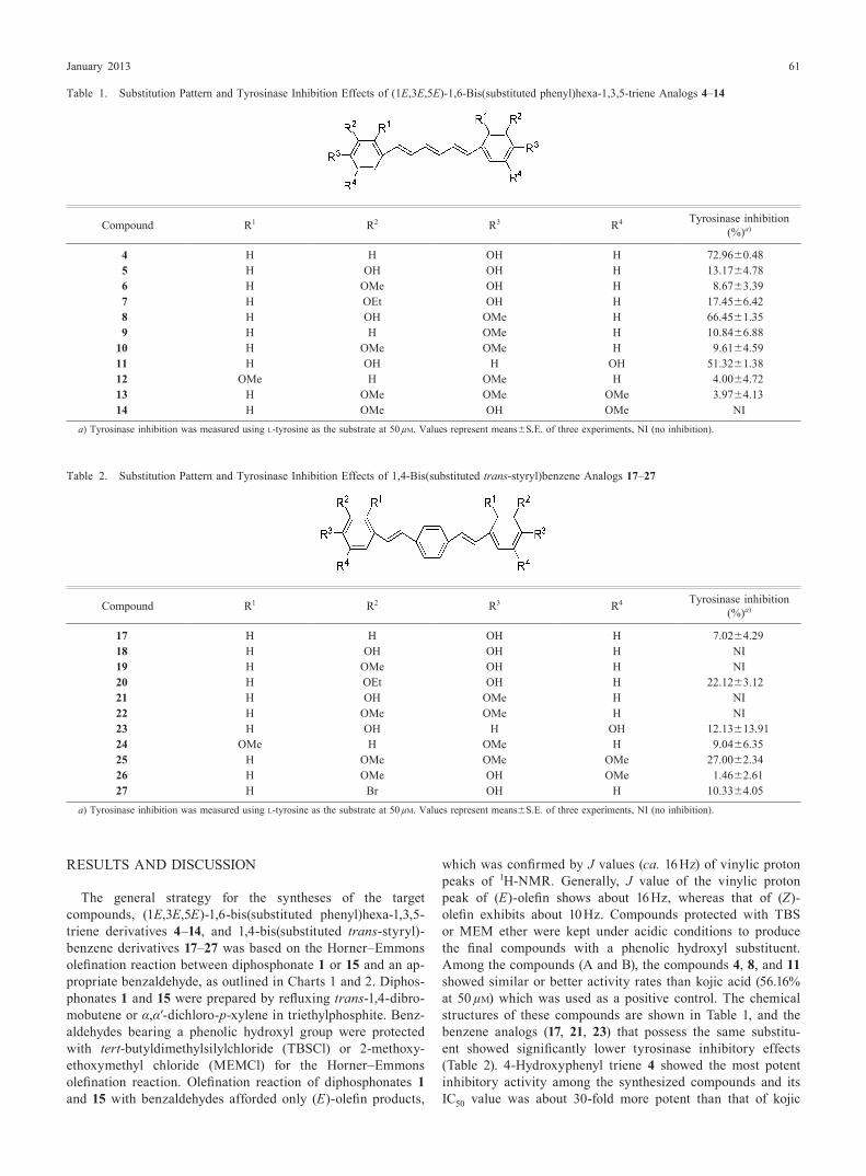
which was confirmed by J values (ca. 16 Hz) of vinylic proton peaks of 1H-nMR. Generally, J value of the vinylic proton peak of (E)-olefin shows about 16 Hz, whereas that of (Z)-olefin exhibits about 10 Hz. Compounds protected with TBS or MeM ether were kept under acidic conditions to produce the final compounds with a phenolic hydroxyl substituent. Among the compounds (A and B), the compounds 4, 8, and 11 showed similar or better activity rates than kojic acid (56.16% at 50 µm) which was used as a positive control. The chemical structures of these compounds are shown in Table 1, and the benzene analogs (17, 21, 23) that possess the same substitu-ent showed significantly lower tyrosinase inhibitory effects (Table 2). 4-Hydroxyphenyl triene 4 showed the most potent inhibitory activity among the synthesized compounds and its IC50 value was about 30-fold more potent than that of kojic
Table 1. Substitution Pattern and Tyrosinase Inhibition effects of (1E,3E,5E)-1,6-Bis(substituted phenyl)hexa-1,3,5-triene Analogs 4–14
Compound R1 R2 R3 R4 Tyrosinase inhibition (%)a)
4 H H OH H 72.96±0.485 H OH OH H 13.17±4.786 H OMe OH H 8.67±3.397 H Oet OH H 17.45±6.428 H OH OMe H 66.45±1.359 H H OMe H 10.84±6.88
10 H OMe OMe H 9.61±4.5911 H OH H OH 51.32±1.3812 OMe H OMe H 4.00±4.7213 H OMe OMe OMe 3.97±4.1314 H OMe OH OMe nI
a) Tyrosinase inhibition was measured using l-tyrosine as the substrate at 50 µm. Values represent means±S.e. of three experiments, nI (no inhibition).
Table 2. Substitution Pattern and Tyrosinase Inhibition effects of 1,4-Bis(substituted trans-styryl)benzene Analogs 17–27
Compound R1 R2 R3 R4 Tyrosinase inhibition (%)a)
17 H H OH H 7.02±4.2918 H OH OH H nI19 H OMe OH H nI20 H Oet OH H 22.12±3.1221 H OH OMe H nI22 H OMe OMe H nI23 H OH H OH 12.13±13.9124 OMe H OMe H 9.04±6.3525 H OMe OMe OMe 27.00±2.3426 H OMe OH OMe 1.46±2.6127 H Br OH H 10.33±4.05
a) Tyrosinase inhibition was measured using l-tyrosine as the substrate at 50 µm. Values represent means±S.e. of three experiments, nI (no inhibition).

62 Vol. 36, no. 1
acid, which was used as a positive control (Table 3). As shown in Table 1, introduction of an additional hydroxyl, methoxy or ethoxy substituent (e.g., 5, 6, 7) at the R2 position on the phenyl ring of compound 4 decreased the inhibitory activity, and replacement of the 4-hydroxy group of compound 4 by a methoxy group also reduced the inhibitory activity (e.g., 9) dramatically. However, in case of replacement of the hydroxyl group of compound 4 by a methoxy group, introduction of an additional hydroxyl group at the R2 position (e.g., 8) recovered the tyrosinase inhibitory effect. Triene derivatives bearing 3,4-dimethoxy (e.g., 10), 2,4-dimethoxy (e.g., 12), 3,4,5-tri-methoxy (e.g., 13) or 3,5-dimethoxy-4-hydroxy (e.g., 14)
substituent showed low inhibitory activity or no activity. A compound with a hydroxyl group at both the R2 and R4 posi-tion (e.g., 11) also exhibited potent inhibitory activity.
We characterized the inhibition kinetics of compounds 4, 8, and 11, which showed a significant tyrosinase inhibitory effect. To explore the mechanism of active inhibitors, we con-ducted a study of the kinetic behavior of tyrosinase activity in the presence of inhibitors. We measured the reaction rates in the presence of active inhibitors at various concentrations of l-tyrosine as a substrate. As the concentrations of active in-hibitors 4, 8, and 11 increased, Km values gradually increased, but Vmax values did not change, thereby indicating the inhibi-tors all act as competitive inhibitors of mushroom tyrosinase (Fig. 1). The general order of inhibition potency was found to be: compound 4>compound 8>compound 11>kojic acid (Fig. 1, Table 3). A comparison of the Km and Ki values of the compounds with that of kojic acid revealed that they possess much higher affinity to tyrosinase than kojic acid. It is likely that this high potency renders them more selective toward ty-rosinase than kojic acid.
Through simulating of docking, we were able to detail the binding configuration of tertiary structure of mushroom tyrosinase with active compounds, supporting the hypothesis that active compounds interact with residues in the active site of tyrosinase (Fig. 2). We found that kojic acid and the novel inhibitor compounds bound the inner region of tyrosine active
Table 3. Tyrosinase Inhibition effects Kinetic Analysis of Active Com-pounds
Compound IC50a) (µm) Type of
inhibitionb) Kic) (µm)
4 1.63±0.05 Competitive 1.318 22.91±1.06 Competitive 2.61
11 62.67±1.80 Competitive 5.24Kojic acid 49.08±0.36 Competitive 5.96
a) 50% inhibitory concentration (IC50). b) Lineweaver–Burk plot of mushroom tyrosinase: Data are presented as mean values of 1/V, which is the inverse of the increase in absorbance at a wavelength 492 nm/min (ΔA492/min), for three independent tests with different concentrations of l-tyrosine as the substrate. c) Values was mea-sured at 5 µm of active compounds and Ki (inhibitor constant).
Fig. 1. Lineweaver–Burk Plot of Mushroom TyrosinaseData were obtained as mean values of 1/V, the inverse of the absorbance increase at a wavelength of 492 nm per min (ΔA492·min−1), of three independent tests with dif-
ferent concentrations of l-tyrosine as a substrate. enzyme inhibitors are indicated as follows: 1.25 µm (circle), 5 µm (square), 25.0 µm (asterisk), 50 µm (diamond), and no. of compounds 4, 8, or 11 (triangle).

January 2013 63
site which contains copper ion. As shown in Fig. 2, the dock-ing simulation was successful, with significant scores. The binding energies of compounds were −6.59 kcal/mol (com-pound 4), −6.05 kcal/mol (compound 8), and −5.56 kcal/mol (compound 11). Furthermore, the binding energy of kojic acid was −4.20 kcal/mol. The docking score between the ligand
and the receptor is represented by various energy terms such as electrostatic energy, van der Waals energy terms and the solvation energy. The docking simulation supported the hy-pothesis that the binding affinity of the compounds 4, 8, and 11 were higher than kojic acid, which was used as a control compound. Also, the binding affinities of docking simulation
Fig. 2. The Docking Simulation between Tyrosinase and Kojic Acid or Inhibitor CompoundsCopper ions are shown as gold balls. Kojic acid, which was used as a control compound, is shown in magenta. Compound 4 is shown in red, compound 8 is shown in
cyan, and compound 11 is shown in blue. The binding energies of compounds were −6.59 kcal/mol (compound 4), −6.05 kcal/mol (compound 8), −5.56 kcal/mol (com-pound 11), and −4.20 kcal/mol (kojic acid). (Color images were converted into gray scale.)
Fig. 3. Possible Hydrogen Bonding Interactions between Tyrosinase Residues and Inhibitor Compounds or Kojic AcidWe searched for hydrogen binding interactions between tyrosinase and inhibitor compounds or kojic acid in the simulated docked structures. The pharmacophore model
was generated using the LigandScout 3.0 program. The red arrow is a hydrogen bond acceptor, the green arrow is a hydrogen bond donor, and the yellow rings are hydro-phobic regions. Only Met280 residue of the tyrosinase was responsible for the hydrogen bonding interactions with kojic acid. However, His296 (compounds 4, 11) residue of the tyrosinase was predicted to exert hydrogen bonding interactions with target compounds. (Color images were converted into gray scale.)
64 Vol. 36, no. 1
had similar a tendency with the inhibition potency by kinetics results. Further, we found differences in the docking position of kojic acid compared to the novel compounds.
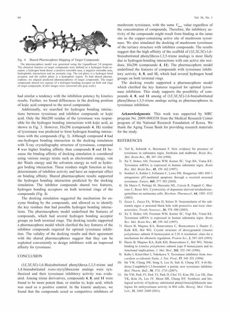
Additionally, we searched for hydrogen bonding interac-tions between tyrosinase and inhibitor compounds or kojic acid. Only the Met280 residue of the tyrosinase was respon-sible for the hydrogen bonding interactions with kojic acid, as shown in Fig. 3. However, His296 (compounds 4, 11) residue of tyrosinase was predicted to form hydrogen bonding interac-tions with the compounds (Fig. 3). Although compound 4 had one-hydrogen bonding interaction in the docking simulation with X-ray crystallography structure of tyrosinase, compound 4 was higher binding affinity than compounds 8 and 11 be-cause the binding affinity of docking simulation is considered using various energy terms such as electrostatic energy, van der Waals energy and the solvation energy as well as hydro-gen boding interaction. These residues might function as key determinants of inhibitor activity and have an important effect on binding affinity. Shared pharmacophore results supported the hydrogen bonding interaction predicted by the docking simulation. The inhibitor compounds shared two features, hydrogen bonding acceptors on both terminal rings of the compounds (Fig. 4).
The docking simulation suggested the mechanism for en-zyme binding by the compounds, and allowed us to identify the key residues that had possible hydrogen bonding interac-tions. The pharmacophore model underlined the features of compounds, which had several hydrogen bonding acceptor groups on both terminal rings. The docking results supported a pharmacophore model which clarified the key features of the inhibitor compounds required for optimal tyrosinase inhibi-tion. The validity of the docking results and their agreement with the shared pharmacophores suggest that they can be exploited conveniently to design inhibitors with an improved affinity for tyrosinase.
COnCLUSIOn
(1E,3E,5E)-1,6-Bis(substituted phenyl) hexa-1,3,5-triene and 1,4-bis(substituted trans-styryl) benzene analogs were syn-thesized and their tyrosinase inhibitory activity was evalu-ated. Among triene derivatives, compounds 4, 8, and 11 were found to be more potent than, or similar to, kojic acid, which was used as a positive control. In the kinetic analyses, we found that the compounds acted as competitive inhibitors of
mushroom tyrosinase, with the same Vmax value regardless of the concentration of compounds. Therefore, the inhibitory ac-tivity of the compounds might result from binding at the same site as the copper-containing active site of mushroom tyrosi-nase. We also simulated the docking of mushroom tyrosinase of the tertiary structure with inhibitor compounds. The results suggest that the high affinity of the scaffold of (1E,3E,5E)-1,6-bis(substituted phenyl) hexa-1,3,5-triene analogs is most likely due to hydrogen-bonding interactions with one active site resi-dues, His296 (compounds 4, 11). The pharmacophore model underlined the features of compounds with tyrosinase inhibi-tory activity, 4, 8, and 11, which had several hydrogen bond groups on both terminal rings.
The docking results supported a pharmacophore model which clarified the key features required for optimal tyrosi-nase inhibition. This study supports the possibility of com-pounds 4, 8, and 11 among of (1E,3E,5E)-1,6-bis(substituted phenyl) hexa-1,3,5-triene analogs acting as pharmacophores in tyrosinase inhibition.
Acknowledgments This work was supported by MRC program no. 2009-0083538 from the Medical Research Center program of the national Research Foundation of Korea. We thank the Aging Tissue Bank for providing research materials for the study.
ReFeRenCeS
1) Tief K, Schmidt A, Beermann F. new evidence for presence of tyrosinase in substantia nigra, forebrain and midbrain. Brain Res. Mol. Brain Res., 53, 307–310 (1998).
2) Xu Y, Stokes AH, Freeman WM, Kumer SC, Vogt BA, Vrana Ke. Tyrosinase mRnA is expressed in human substantia nigra. Brain Res. Mol. Brain Res., 45, 159–162 (1997).
3) Sendoel A, Kohler I, Fellmann C, Lowe SW, Hengartner MO. HIF-1 antagonizes p53-mediated apoptosis through a secreted neuronal tyrosinase. Nature, 465, 577–583 (2010).
4) De Marco F, Perluigi M, Marcante ML, Coccia R, Foppoli C, Blar-zino C, Rosei MA. Cytotoxicity of dopamine-derived tetrahydroiso-quinolines on melanoma cells. Biochem. Pharmacol., 64, 1503–1512 (2002).
5) Zecca L, Zucca FA, Wilms H, Sulzer D. neuromelanin of the sub-stantia nigra: a neuronal black hole with protective and toxic char-acteristics. Trends Neurosci., 26, 578–580 (2003).
6) Xu Y, Stokes AH, Freeman WM, Kumer SC, Vogt BA, Vrana Ke. Tyrosinase mRnA is expressed in human substantia nigra. Brain Res. Mol. Brain Res., 45, 159–162 (1997).
7) Hazes B, Magnus KA, Bonaventura C, Bonaventura J, Dauter Z, Kalk KH, Hol WG. Crystal structure of deoxygenated Limulus polyphemus subunit II hemocyanin at 2.18 A resolution: clues for a mechanism for allosteric regulation. Protein Sci., 2, 597–619 (1993).
8) Hazes B, Magnus KA, Kalk KH, Bonaventura C, Hol WG. nitrate binding to Limulus polyphemus subunit type II hemocyanin and its functional implications. J. Mol. Biol., 262, 532–541 (1996).
9) Kubo I, Kinst-Hori I, Yokokawa Y. Tyrosinase inhibitors from Ana-cardium occidentale fruits. J. Nat. Prod., 57, 545–551 (1994).
10) Ha YM, Chung SW, Song S, Lee H, Suh H, Chung HY. 4-(6-Hy-droxy-2-naphthyl)-1,3-bezendiol: a potent, new tyrosinase inhibitor. Biol. Pharm. Bull., 30, 1711–1715 (2007).
11) Ha YM, Park JY, Park YJ, Park D, Choi YJ, Kim JM, Lee eK, Han YK, Kim JA, Lee JY, Moon HR, Chung HY. Synthesis and bio-logical activity of hydroxy substituted phenyl-benzo[d]thiazole ana-logues for antityrosinase activity in B16 cells. Bioorg. Med. Chem. Lett., 21, 2445–2449 (2011).
Fig. 4. Shared Pharmcophore Mapping of Target CompoundsThe pharmacophore model was generated using the LigandScout 3.0 program.
The chemical features of target compounds were defined as a hydrogen bond ac-ceptor, a hydrogen bond donor, a positive ionizable area, a negative ionizable area, hydrophobic interactions and an aromatic ring. The red phore is a hydrogen bond acceptor, and the yellow phore is a hydrophoic region. To find shared pharma-cophore, we aligned predicted pharmacophores of target compounds. The target compounds shared two regions of a hydrogen bonding acceptor on both end rings of target compounds. (Color images were converted into gray scale.)

January 2013 65
12) Ha YM, Kim JA, Park YJ, Park D, Kim JM, Chung KW, Lee eK, Park JY, Lee JY, Lee HJ, Yoon JH, Moon HR, Chung HY. Analogs of 5-(substituted benzylidene)hydantoin as inhibitors of tyrosinase and melanin formation. Biochim. Biophys. Acta, 1810, 612–619 (2011).
13) Kim YJ, No JK, Lee JH, Chung HY. 4,4′-Dihydroxybiphenyl as a new potent tyrosinase inhibitor. Biol. Pharm. Bull., 28, 323–327 (2005).
14) no JK, Kim MS, Kim YJ, Bae SJ, Choi JS, Chung HY. Inhibition of tyrosinase by protocatechuic aldehyde. Am. J. Chin. Med., 32, 97–103 (2004).
15) Lim JT, Frcpi, Fams. Treatment of melasma using kojic acid in a gel containing hydroquinone and glycolic acid. Dermatol. Surg., 25, 282–284 (1999).
16) Alzheimer A. Uber eine eigenartige erkrankung der Hirnrinde. Allg. Z. Psychiatr., 64, 146–148 (1907).
17) Selkoe DJ. Alzheimer’s disease: genotypes, phenotypes, and treat-ments. Science, 275, 630–631 (1997).
18) Goedert M, Spillantini MG. A century of Alzheimer’s disease. Sci-ence, 314, 777–781 (2006).
19) Hebert Le, Scherr PA, Bienias JL, Bennett DA, evans DA. Alz-heimer disease in the US population: prevalence estimates using the 2000 census. Arch. Neurol., 60, 1119–1122 (2003).
20) Zhuang ZP, Kung MP, Kung HF. Synthesis of biphenyltrienes as probes for beta-amyloid plaques. J. Med. Chem., 49, 2841–2844 (2006).
21) Flaherty DP, Kiyota T, Dong Y, Ikezu T, Vennerstrom JL. Phenolic bis-styrylbenzenes as β-amyloid binding ligands and free radical scavengers. J. Med. Chem., 53, 7992–7999 (2010).
22) Flaherty DP, Walsh SM, Kiyota T, Dong Y, Ikezu T, Vennerstrom JL. Polyfluorinated bis-styrylbenzene beta-amyloid plaque binding ligands. J. Med. Chem., 50, 4986–4992 (2007).
23) Chen JS, Wei C, Marshall MR. Inhibition mechanism of kojic acid on polyphenol oxidase. J. Agric. Food Chem., 39, 1897–1901 (1991).
24) no JK, Soung DY, Kim YJ, Shim KH, Jun YS, Rhee SH, Yokozawa T, Chung HY. Inhibition of tyrosinase by green tea components. Life Sci., 65, PL241–PL246 (1999).
25) Arnold K, Bordoli L, Kopp J, Schwede T. The SWISS-MODeL workspace: a web-based environment for protein structure homol-ogy modelling. Bioinformatics, 22, 195–201 (2006).
26) Wermuth CG, Ganellin CR, Lindberg P, Mitscher LA. Glossary of terms used in medicinal chemistry (IUPAC Recommendations 1998). Pure Appl. Chem., 70, 1129–1143 (1998).
27) Wolber G, Langer T. LigandScout: 3-D pharmacophores derived from protein-bound ligands and their use as virtual screening fil-ters. J. Chem. Inf. Model., 45, 160–169 (2005).


![5 1,6 1,4 1,6 1 arXiv:1811.02473v1 [cond-mat.str-el] 6 Nov ...](https://static.fdocuments.us/doc/165x107/618116d1de1d3906bf1a5c99/5-16-14-16-1-arxiv181102473v1-cond-matstr-el-6-nov-.jpg)