MOLECULAR CHARACTERIZATION OF HYDROLYTIC ENZYMES …
144
MOLECULAR CHARACTERIZATION OF HYDROLYTIC ENZYMES FROM HYPERTHERMOPHILIC ARCHAEA Wilfried G.B. Voorhorst Bacterial Genetics Group Laboratory of Microbiology Department ofBiomolecular Science Wageningen Agricultural University CENTRALE LANDBOUWCATALOGUS 0000 0872 0951
Transcript of MOLECULAR CHARACTERIZATION OF HYDROLYTIC ENZYMES …

MOLECULAR CHARACTERIZATION
OF
HYDROLYTIC ENZYMES
FROM
HYPERTHERMOPHILIC ARCHAEA
Wilfried G.B. Voorhorst
Bacterial Genetics Group
Laboratory of Microbiology
Department of Biomolecular Science
Wageningen Agricultural University
CENTRALE LANDBOUWCATALOGUS
0000 0872 0951

Promotor: dr. W.M. de Vos
hoogleraar in de microbiologie

W.G.B. Voorhorst
Molecular characterization of hydrolytic enzymes from hyperthermophilic archaea
Proefschrift
ter verkrijging van de graad van doctor
op gezag van de rector magnificus
van de Landbouwuniversiteit Wageningen,
dr. C.M. Karssen,
in het openbaar te verdedigen
op vrijdag 24 april 1998
des namiddags te vier uur in de Aula

ISBN 90-5485-796-X
Cover: 3-Dimensional modeis of the catalytic domain core of the hypermostable serine protease pyrolysin showing aromatic-aromatic interactions (front) and ionic-ionic interactions (back). Drawn by Jack Leunissen (CAOS/CAMM Centre, Nijmegen, the Netherlands) using MOLSCRIPT.
BIBLIOTHEEK LANDBOUWi;NIVr!<SnTi'"
WAGENIMCEN
-\2'L
Stellingen
1. De conclusie dat P.furiosus vijf intracellulaire proteases produceert kan pas hard
gemaakt worden als aangetoond is dat soniceren van een celpellet niet resulteert in het
vrijkomen van eiwitten die geassocieerd zijn met of gebonden zijn aan de membraan.
Blumentals, I.I., Robinson, A.S., and Kelly, R.M. (1990) Characterization of sodium dodecyl sulfate-resistant proteolytic activity in the hyperthermophilic archaebacterium Pyrococcus furiosus. Appl. Environm. Microbiol. 56, 1992-1998.
2. Het dirigeren van eiwitten naar het periplasma van Escherichia coli kan niet worden
gedefinieerd als periplasmatische expressie.
Makrides, S.C. (1996) Strategies of achieving high-level expression of genes in Escherichia coli. Microbiol. Rev. 60, 512-538.
3. Op een sodium dodecyl sulfaat polyacryl amide gel kan geen moleculaire massa
bepaald worden van een eiwit dat nog actief is.
Klingeberg, M. Galunsky, B., Sjoholm, Kasche, V., and Antranikian, G., (1995). Purification and properties of a highly thermostable, sodium dodecyl sulfate-resistant and stereospecific proteinase from the extremely thermophilic archaeon Thermococcus steuert Appl. Environm. Microbiol. 61, 3098-3104.
4. Met de opmerking dat bijna alle niet-cytoplasmatische eiwitten geglycosyleerd zijn
wordt zomogelijk het grootste deel van het leven op aarde over het hoofd gezien daar
er meer leven op aarde is dan alleen de Eucaryoten.
Imberty, A., and Pérez, S. (1995). Stereochemistry of the A'-glycosylation sites in glycoproteins. Prot. Engng. 8, 699-709.
5. Voor de identificatie van plaatsen in een eiwit die betrokken zijn bij thermostabiliteit
is meer nodig dan de sequentie van één mogelijk gen.
Völkl, P., Markiewiz, P., Stetter, K.O., and Miller, J.H. (1994) The sequence of a subtilisin-type protease (aerolysin) from the hyperthermophilic archaeum Pyrobaculum aerophilum reveals sites important to thermostability. Prot. Science 3, 1329-1340.
6. Thermofiele enzymen bestaan niet.

7. De omschrijving van een promotie onderwerp is sterk afhankelijk van de
onderzoekschool waar het deel van moet uitmaken.
8. De fabrikanten van printers zijn het aan hun stand verplicht de standaardinstellingen
van verschillende printers te standaardiseren.
9. Een deel van de in de literatuur beschreven heterogeniteit van enzymatische activiteit
voor proteases van hyperthermofielen vindt zijn oorsprong in de gevolgde procedure.
10. Het verwijderen van uitvoegstroken op provinciale wegen heeft een nadelig effect op
het milieu en de verkeersveiligheid.
11. In het weekend en op de avonden lijkt in sommige gevallen de instelling van een
huisarts het meest weg te hebben van een ik blijf liever thuis arts.
12. Reclameborden langs de snelweg moeten snel weg.
Stellingen behorende bij het proefschrift "Molecular characterization of hydrolytic enzymes from hyperthermophilic archaea". Wilfried G.B. Voorhorst Wageningen, 24 april 1998

Voorwoord
Door middel van dit voorwoord wil ik iedereen bedanken die er toe hebben bijgedragen dat dit proefschrift tot stand is gekomen. De echte waarde van hun bijdrage is niet te beschrijven. Een aantal wil ik hier met name noemen.
Te beginnen bij mijn promoter Prof. dr. Willem M. de Vos. Willem, ik wil je bedanken voor de enthousiaste begeleiding van het onderzoek, je altijd kritische analyses en het ongelofelijke tempo waarmee je de zoveelste versie van een manuscript door wist te worstelen. Het perfectionisme waarmee je te werk gaat heeft zeker zijn sporen achter gelaten. Rik Eggen, jou wil ik bedanken voor het gestelde vertrouwen en je onmiskenbare aanwezigheid als persoon. De waardevolle wetenschappelijke discussies met jou over de eerste resultaten hebben gedragen tot het ontrafelen van de protease puzzel. Ans Geerling als grote steun en toeverlaat voor heel BacGen, je was de sleutelfiguur die de boel draaiende wist te houden en zorgde voor een prima sfeer. Onmiskenbaar hierbij waren de werkgroep etentjes en de snoeppot, zodat menig student en (gast)medewerker met veel plezier terug kijkt naar de BacGen tijd. Joyce Lebbink, je was herkenbaar aanwezig met je uitbundige karakter, enthousiasme. Tevens wil ik je bedanken voor de hulp bij het bepalen van het goede substraat (blauwe epje). Hauke Smidt, je zorgde voor een rustige basis op de kamer. John van der Oost, de discussies met jou over het hoe en wat binnen het celB locus waren soms eindeloos, moge de tijd komen dat alle functies duidelijk zijn. Roland Siezen ben ik zeer erkentelijk voor de samenwerking rond pyrolysine en stetterlysine. Yannick Gueguen, your presence in the lab and your work on the endo-glucanase has been a great pleasure. Gerti Schut voor de uitwisseling van resultaten rond de inductie experimenten met Pyrococcus. Vele studenten hebben tijdens de promotie een essentiële bijdrage geleverd aan de voortgang van het onderzoek: Peter Steenbakker, Frank Wagener, Annechien Hengeveld, Mariken Gijsen, Han Zendman, Joost Kolkman, Marcel Dijkgraaf, Angela Warner, Fiona Kaper, Wilfred IJkel, Theo Smits, Bas Lemmens, Esther Poelwijk en Vincent Wittenhorst. Alle mede promovendi, AIO's en OIO's en de ander collega's van de vakgroep Microbiologie wil ik hartelijk bedanken voor de alle hulp die ze mij gegeven hebben en voor de onvergetelijke tijd op de vakgroep.
Mijn ouders, broers en zus wil ik bedanken voor alles wat ze door de jaren heen voor mij gedaan hebben. Jeanet, hoewel woorden niet kunnen beschrijven wat je voor me betekent wil ik je bedanken voor je geduld en steun. Lindsay en Danyon jullie hebben een nieuwe dimensie aan mijn leven toegevoegd, één die ik niet had willen missen.
Pa, we hadden je er ontzettend graag bij willen hebben. t //['*./>/

Contents
Chapter 1 General introduction 1
Chapter 2 Characterization of the celB gene coding for ß-glucosidase 27
from the hyperthermophilic archaeon Pyrococcus furiosus and
its expression and site-directed mutation in Escherichia coli
Chapter 3 Genetic and biochemical characterization of a short-chain and 37
an iron-containing alcohol dehydrogenase from the
hyperthermophilic archeaon Pyrococcus furiosus
Chapter 4 Cloning and sequencing of the lamA gene from the 53
hyperthermophilic archaeon Pyrococcus furiosus coding for an
endo-ß-l,3-ghicanase and its expression and site-directed
mutation in Escherichia coli
Chapter 5 Transcriptional regulation in the hyperthermophilic archaeon 71
Pyrococcus furiosus: Coordinated expression of divergently
transcribed genes in response to ß-linked glucose polymers
Chapter 6 Isolation and characterization of the hyperthermostable serine 91
protease, pyrolysin, and its gene from the hyperthermophilic
archaeon Pyrococcus furiosus
Chapter 7 Homology modelling of two subtilisin-like serine proteases 99
from the hyperthermophilic archaea Pyrococcus furiosus and
Thermococcus stetteri
Chapter 8 Summary and concluding remarks 111
Chapter 9 Samenvatting 121
Curriculum vitae 13 3
List of publications 13 5

General introduction
Chapter 1
General Introduction

Chapter 1
In the last few decades microorganisms have been isolated from rather uncommon and
hostile locations, such as those with high salt concentrations, an extreme pH, or low or
high temperatures. Microorganisms isolated from these environments are referred to as
extremophiles (Horikoshi, 1997). The most extensively studied group of these
extremophiles are the hyperthermophiles, microorganisms that have an optimum
temperature for growth above 80°C (Stetter, 1990). Hyperthermophilic microorganisms
appear to be widely spread and have been isolated from hot spots located all around the
globe, such as hydrothermal vents, black or white smokers, solfataric fields, hot springs
and oil wells. With the isolation of Pyrolobus fumarii, the upper temperature of life has
been set on 113°C (Blöchl etat, 1997).
Except for two bacterial genera, the Thermotogales and Aquifex, all
hyperthermophiles isolated to date belong to the domain of the Archaea (Fig. 1 and
Table 1). The Archaea, formerly archaebacteria, compose together with the Bacteria
and Eucarya the three domains of life (Fig. 1). Their discovery caused a splitting of the
prokaryotic kingdom, recognized in the late 70's based on analysis of 16S rRNA
sequences (Woese etal, 1990) (Fig. 1).
Eucarya
Bacteria Archaea Green
non sulfur bacteria
gram positives proteobacteria .
cyanobacteria\ 1 ftavobactetia ^ \ l
Arttttêav ^^^
1 1 1
Desulfurococcus
i \ ! Y
Pyrodictium 1 Thermotoga
Sulfolobus
f Thermofilum M ^ Thermoproteus
f^^^^mPyrobaculum ^ Pyrocoeeus
JU Thermococcus
jMethano J thefmuy
S ^ ^ M Methanopyrus 1 \ ^ ^ ^ ^ ^ ^ m Methanococcus v**
y Methanobacterium
Arehaeogfobus
^*"***» Halobacterium
^**%, Methanosplrillum
\ Methanosarcina
Figure 1. Phylogenese tree of life based on 16S rRNA sequence analysis. Hyperthermophilic genera are depicted as thick lines (Modified from Woese et al., 1990 and Stetter 1993).

General introduction
The Archaea represent the shortest branches in the phylogenetic tree with
hyperthermophiles being the most slowely evolving representatives (Woese et al,
1990). Therefore, it has been suggested that the last common ancestor, the so called
'progenote', could have been a hyperthermophile (Stetter, 1994).
With a few exceptions, all hyperthermophiles grow under strict anaerobic
conditions and the most of them depend upon the reduction of elemental sulphur to H2S
for optimal growth (Schönheit and Schäfer, 1995). Except for a limited number of
autotrophs, all hyperthermophiles are able to utilize proteins and peptides and several
are also able to utilize complex polysaccharides (Table 1) (Kelly and Adams, 1994;
Schönheit and Schäfer, 1995). Below an overview is given of (i) hydrolytic enzymes of
hyperthermophiles involved in the polymer degradation and the fate of the degradation
products in the main metabolic pathways, (ii) the stabilization mechanisms of the
enzymes from hyperthermophiles, so-called thermozymes, and (iii) the unique aspects
of the molecular biology of Archaea. In the discussion of these aspects specific attention
will be given to hyperthermophilic archaeon Pyrococcus fitriosus, since most of the
work with hyperthermophiles has been focussed on this versatile microorganism that
can grow rapidly, with a doubling time of 37 min, at the normal boiling temperature of
water (Fiala and Stetter, 1986). Moreover, P.fitriosus is able to grow without elemental
sulfur and utilize a large range of substrates, including proteins, polysaccharides and
pyruvate. Disaccharides, such as cellobiose and maltose, can yield relatively high cell
densities, but the monosaccharide glucose can not support growth of P.fitriosus.
Degradation of polymers in hyperthermophiles
The degradation of polymeric substrates, such as polysaccharides and proteins, is
accomplished by the action of various hydrolytic enzymes. Two types of hydrolytic
enzymes can be distinguished: (i) hydrolases that act on polymers and cleave internally,
so-called endo-acting hydrolases, and (ii) hydrolases that cleaves the terminal residues
from either an oligomer or larger polymer, designated exo-acting hydrolases. Below an
overview is given of hydrolytic enzymes of hyperthermophiles involved in the
degradation of polysaccharides and proteins followed by the .main features of their
catabolism.

Chapter 1
Table 1. Classification and characteristics of hyperthermophilic microorgansims.
Order
ARCHAEA
Sulfolobales
Thermoproteales
Desulfurococcales
'Pyrolobalus 'Pyrodictiales '
Themococcales
'Archeoglobales ' Methanobacteriales Methanococcales 'Methanopyrales '
BACTERIA
Thermotogales
'Aquificales'
Genus
Sulfolobus Metallosphaera Acidianus Stygiolobus Thermoproteus Pyrobaculum Thermofilum Desulfiirococcus Staphylothermus Pyrolobus Pyrodictium Hyperthermus Thermodiscus Thermococcus Pyrococcus Archaeoglobus Methanothermus Methanococcus Methanopyrus
Thermotoga Thermosipho Fervidobacterium Aquifex
Tmax(°C)
85-87 80
75-95 89 97 103 95
95-97 98 113 110 108 98
93-98 103 92 97
70-91 110
84-90 77 80 95
Polymeric substrates* peptides
+ + +
+ + + + +
+ + + + + +
+ + +
polysaccharides
+
+
+
+ + +
+
+
' The capacity to grow on polymeric substrates is listed; 'peptides' indicate complex compounds, e.g. yeast extract, peptone, tryptone, casamino acids and caséine and 'polysaccharides indicate glucose multimers of 2 or more subunits.
Degradation of polysaccharides
The naturally occurring polysaccharides can be divided into two groups depending on
the type of linkage between carbohydrate moieties that can be either a-glycosidic or a ß-
glycosidic. Enzymes able to hydrolyse these glycosidic bonds are classified as glycosyl
hydrolases. Currently, over 60 different families of glycosyl hydrolases have been
identified based on amino acid sequence homologies (Henrissat, 1997; Henrissat and
Bairoch, 1996). The endo-acting glycosyl hydrolases cleave a polysaccharide chain of

General introduction
more than 4 subunits, while the exo-acting glycosyl hydrolases remove the terminal
glycosidic linkage from usually smaller polysaccharides (less than 5 subunits) (Bauer et
al, 1996). In general, the degradation of polysaccharides is initiated by endo-acting
glycosyl hydrolases that depolymerize the large substrate resulting in oligosaccharides
and monosaccharides. Exo-acting glycosyl hydrolases cleave the oligosaccharides into
subunits that subsequently are metabolized. Recently, several reviews have appeared
that summarize the properties of glycosyl hydrolases from microorganisms growing
above 70°C (Leuschner and Antranikian, 1995; Bauer et al, 1996; Sunna et al, 1997).
To introduce the work presented in this thesis the ß-glycosyl hydrolases of
hyperthermophiles will be described in more detail. A relatively large number of ß-
glycosyl hydrolases have been characterized from hyperthermophiles. Many bacterial
endo-acting and exo-acting glycosyl hydrolases have been identified in Thermotogales
spp. and these have been reviewed recently (Leuschner and Antranikian, 1995). In
contrast, only a few endo-acting ß-glycosyl hydrolases, both endoglucanases and
endoxylanases, have been characterized from hyperthermophilic archaea (Table 2). One
of them is the endo-ß-l,3-glucanase from P.furiosus that is optimally active between
100 and 105°C (See chapter 4; Gueguen et al, 1997). A number of exo-acting ß-
glycosyl hydrolases have been described in hyperthermophilic archaea, including one
xylosidase and various glycosidases, that all belong to the family 1 of glycosyl
hydrolases (Table 2). Notably, the extensively studied P.furiosus appears to be well
equiped for growth on a range of complex polysaccharides as judged by the presence of
various ß-glycosyl hydrolases (Table 2).
Table 2. ß-Glycosyl hydrolases that have been characterized in hyperthermophilic archaea.
Type* endo-acting
exo-acting
Enzyme endo-ß-1,3-glucanase endoxylanase ß-glucosidase
ß-glycosidase ß-mannosidase
ß-glycosidase ß-glycosidase ß-xylosidase
Gene lamA
celB
bglX bmn
lacS
Organism Pyrococcus furiosus
Pyrodictium abysii Pyrococcus furiosus
Pyrococcus furiosus Pyrococcus furiosus
Sulfolobus solfataricus Sulfolobus solfataricus Pyrodictium abysii
MT4 P2
References Gueguen et al., (1997)
Andrade era/., (1996) Kengen et al., (1993) Voorhorst et al, (1995) Verhees et al., (1997) Kengen etal, (1996) Bauer era/., (1996) Nucci era/., (1993) Grogan (1991) Andrade era/., (1996)
* Type of glycosyl hydrolase depending on the site of cleavage: endo-acting, cleavage in a polymer of more than 4 subunits; exo-acting cleavage from the terminal residues, usually of 4 or less subunits.

Chapter 1
The pyrococcal ß-glucosidase, CelB, has been characterized in considerable detail and
with a half-life value of 85 h at 100°C it is the most thermostable glycosyl hydrolase
known to date (Kengen et al, 1993; Kengen and Stams, 1994). The celB gene encoding
this ß-glucosidase was isolated and analysis of the deduced amino acid sequence
showed that it belonged to the family 1 of glycosyl hydrolases (Chapter 2; Voorhorst et
al, 1995). Functional expression of the celB gene in E.coli resulted in an enzyme with
the same kinetic and stability properties as that purified from P.furiosus, which allowed
for mutational analysis of the ß-glucosidase to gain insight in structure-function
relations (Chapter 2; Voorhorst et al, 1995). Recently, a ß-mannosidase has been
characterized from P.furiosus (Bauer et al, 1996; Kengen et al, 1996). The bmn gene
coding for this ß-mannosidase was cloned and the deduced amino acid sequence
showed this enzyme also is a member of the glycosyl hydrolase family 1, with 46.5 %
identity to the ß-glucosidase of P.furiosus (Bauer et al, 1996).
Like the pyrococcal ß-glucosidase CelB, the homolog from Sulfolobus
solfataricus, the ß-glycosidase LacS has been extensively studied. The lacS gene has
been expressed in different hosts, including mammalian cells, and yeast, and used as
reporter gene (Cannio et al, 1994; Moracci et al, 1992). The substrate specificity of
the ß-glycosidase as well as effects of temperature and SDS have been studied (Nucci et
al, 1993; Nucci, et al, 1995). Mutational analysis showed that two glutamate residues
are essential for catalysis (Moracci et al, 1996). The ß-glycosidase from S.solfataricus
has been used in transglycosylation, however, it was only able to accept secondary
alcohols as non-sugar aglycons in the synthesis reaction, whereas the pyrococcal ß-
glucosidase was also able to use tertiary alcohols (Fisher et al, 1996; Trincone et al,
1994; Trincone and Pagnotta, 1995). Recently, the crystal structure of the S.solfataricus
LacS has been elucidated and showed a large number of ion-pairs that are involved in
networks and a relatively large number of solvent molecules that are buried in
hydrophilic cavities (Aguilar et al, 1997).
The metabolism of glucose, an endproduct of glucose polymer hydrolysis, has
been studied in hyperthermophiles belonging to the bacterial genus Thermotoga and in
different genera of Archaea such as Pyrococcus, Sulfolobus, Desulfurococcus and
Thermoproteus. The hyperthermophilic bacterium Thermotoga maritima appeared to
use the classical Embden-Meyerhof pathway (Schönheit and Schäfer, 1995; Selig et al,
1997). In contrast, in the hyperthermophilic archaea modifications have been identified
in the Embden-Meyerhof pathway and in the Entner-Douderoff pathway [for reviews
see Schönheitand Schäfer, 1995; Kengen et al, 1996; Selig et al, 1997].

General introduction
Cell membrane
Dihydroxyacetone-P
NADPH * H
Figure 2 Proposed pathway of glucose fermentation in P.fitriosus. Glucose polymer, consisting of more than four glucose subunits; glucose oligomer, consisting of 2 to 4 glucose subunits.l, endo-acting glycosyl hydrolase; 2, unknown transport system; 3, endo-acting glycosyl hydrolase; 4, ADP-dependent glucokinase; 5, phosphoglucoisomerase; 6, ADP-dependent phosphofructokinase; 7, fructose-1,6-biphosphate aldolase; 8, triosephosphate isomerase; 9, glyceraldehyde-3-phosphate:ferredoxin oxidoreductase; 10, phosphoglycerate mutase; 11, enolase; 12, pyruvate kinase; 13, pyruvate:ferredoxin oxidoreductase; 14, acetyl-CoAsynthetase (ADP-forming); 15, glutamate dehydrogenase; 16, alanine aminotransferase; Fd, ferredoxin. (Modified from Kengen et ai, 1996).

Chapter 1
Modifications observed in the Embden-Meyerhof pathway used by P.fiiriosus and
Thermococcus spp. include the presence of two ADP-dependent kinases, glucokinase
and phosphofructokinase (Fig. 2) (Kengen et al, 1994; Kengen et al, 1996).
Additionally, a glyceraldehyde 3-phosphate ferredoxin oxidoreductase has been
identified in P.fiiriosus which converts glyceraldehyde-3-phosphate to 3-phospho-
glycerate, without substrate phosphorylation (Fig. 2) (Mukund and Adams, 1995; van
der Oost et al, 1997). This contrasts with the classical glycolysis that uses
glyceraldehyde-3-phosphate dehydrogenase. Low levels of this glyceraldehyde-3-
phosphate dehydrogenase were also identified in P.fiiriosus, but the enzyme is thought
to function in gluconeogenesis (Schäfer and Schönheit, 1993; van der Oost et al, 1997).
A partially non-phosphorylated Entner-Douderoff pathway has been established in
Sulfolobus spp. (De Rosa et al, 1984) and in Thermoplasma acidophilum (Budgen and
Danson, 1986; Danson, 1988; Danson, 1989). Thermoproteus tenax, one of the deep
branches of the Archaea, was found to contain the Embden-Meyerhof pathway with an
ATP-dependent hexokinase and a PPrdependent phosphofructokinase (Siebers and
Hensel, 1993). However, intermediates of the non-phosphorylated Entner-Douderoff
pathway have been also been identified this archaeon (Hensel et al, 1987). In all
Archaea and hyperthermophilic bacteria, analyzed so far, the conversion from pyruvate
to acetate involves an oxidative decarboxylation reaction by pyruvate:ferredoxin
oxidoreductase to yield acetyl-CoA (Schönheit and Schäfer, 1995). The conversion of
acetyl-CoA to acetate appears to be a catalyzed by an ADP-forming acetyl-CoA
synthetase in all acetate-forming archaea (Schäfer et al, 1993).
Degradation of proteins and peptides
Proteases are the key enzymes in the conversion of proteinaceous substrates to peptides
and amino acids. They have been classified based on their active center and the
following classes can be distinguished: serine, cysteine, aspartic, or métallo proteases
(Bairoch, 1997). During catalysis serine and cysteine proteases form a covalent acyl-
enzyme intermediate, while métallo and aspartic proteases activate a water molecule
that subsequently attacks the substrate. Serine proteases have an active centre composed
of a catalytic triade of three residues, aspartate, histidine and a serine, that form a charge
relay system, and an asparagine that stabilizes the oxyanion generated in the transition
state (Carter and Wells, 1990). Proteases from this class are sensitive to inhibition by

General introduction
phenylmethylsulphonyl fluoride and diisopropylfluorophosphate. The serine proteases
can be divided into many subgroups, with the two major groups being the subtilisin-like
and trypsin-like serine proteases (Siezen et al, 1991). The subtilisin-like serine
proteases have been extensively studied in many other organisms with respect to
structure-function and structure-stability relations [for review (Siezen et al, 1991)].
Cysteine proteases contain a cysteine and a histidine residue within the active site.
Aspartic proteases (also referred to as acid proteases) contain two active-site aspartic
acid residues in close proximity, one of which is ionized at low pH. Métallo proteases
contain divalent cations, mainly zinc, that are chelated to two or more histidine residues,
while a glutamate acts as a catalytic base. Thermostable proteases have recently been
reviewed (Daniel et al, 1995).
Various proteases have been identified in hyperfhermophilic genera and many of
those were classified as serine proteases (Table 3). The first archaeal serine proteases
was isolated from cell-free supernatant of the hyperthermophile Desulfurococcus strain
Tok]2S„ and designated archaelysin (Cowan et al, 1987). More recently, from the
related strain, Desulfurococcus strain SY, a cell-associated serine protease was
characterized that showed a half-life of 4.3 hours at 95°C (Hanzawa et al, 1996).
Various proteases have been characterized from the acidophilic Sulfolobus solfataricus
strains, including two serine proteases (Fusi etal, 1991). The properties of extracellular
proteases from a number of Thermococcus species have been analyzed and found to be
serine proteases composed of multiple activity bands (Klingeberg et al, 1991). From
one of these, T.stetteri, the extracellularly located serine protease has been
characterized. This 68 kDa protease was highly stable and resistant to chemical
denaturation as illustrated by a half-life of 2.5 h at 100°C and retention of 70% of its
activity in the presence of 1% SDS (Klingeberg et al, 1995).
A globular serine protease has found to be associated with the stalk of a filiform
glycoprotein complex, termed tetrabrachion, at the surface of the Staphylothermus
marinus (Mayr et al, 1996). This serine protease, designated STABLE, was found to be
extremely thermostable especially if bound to the tetrabrachion where residual activity
could still be detected after 10 min incubation at 135°C (Mayr et al, 1996). The gene
encoding STABLE has been isolated and the deduced sequence showed highest
homology with subtilisin-like serine proteases, although a large insert was found within
the catalytic domain and a relatively large C-terminal extension was observed (Mayr et
al, 1996). Another gene encoding a subtilisin-like serine protease, designated aerolysin,
has been cloned from Pyrobaculum aerophilum and its deduced sequence was modelled

ei
S a o o *
. S n C/i
(*- o •S h
o
en h* o
-C o o > o os --
QÎ
c CU 00
w
s o Os
~
-2
.s
- S3
T3 O
ffi ffi
so OS
"a
m & .§
u.
^ T3
£
3 O . S 3
en h-O
•e o o >
Os Os
- ; a
3 o >
r--Os Os
"o eu C
X>
u-
a s Os
>—' r i Os Os
„
Os t ^ Os Os ~ - OS
"B -{ ** Q 1) -K,
*-" "Ü
•si 00 -g c o -3 o
w >
Os OS
—-(N Os Os
„
Os Os
' "a eu 00
>-< eu -O eu 00 .S
5
Os Os
~ <N Os Os
„
Os Os
•^i 0
eu 00 t-CU
-O <u oo .g 5
Os Os
~— (N Os Os
„
Os Os
" ' "3 cu 00
fc-eu X eu oo G
5
rsl Os Os
"a eu
'e
3
m
„ -«-I 0
'S M tu CA 3
fe O Os Os
H *o § 00
.5
Os Os
^ (N Os Os
„
Os OS
"" "o 00
fc-CD -O CU 00 c
SO Os Os
-J "3 i—
>> • ~ es
5 S
Os Os
" r
"3 eu 00 u CÜ
XI eu 00
.g 2
,—, (N Os OS s—'
"a 00
fc-eu X eu 00 c
CÖ
C3 T3 "O a> si CA
3 3 a. c 3
O
-H o - 3 O W >
c eu dû «J
e o
i t !
a>
> tu
+
" Ö CD
n) o
.s §
o os C-ïï c e
o S oo -
O
o 1-1
.S .S CA CA
3 3 u
(U
a + « eu 'S + e/ï
ÉL-S-S S 3 S . c -e o.
o o
+ + + + +
3
tu & O. cu tu
•C £ T3 .2 'C 'C <u 2 c js « tu
H CA
C
CA
«î CA
C
CA
C
CA
tu
.s CA
(U
.3 m
O. • o U
CD
tu
.S <1>
.s
E e w
_ ^ s T J .S so c en s o eu > sCO -
° ¥~ £<& s o.a. o.
.S w
< H
» C/3
D O 'S
< ? < . ej
se o U o
3£
C -S •M î».
•Ï3si> S s s c e-i e^ eu > Jï -S K K
ej
<i P § S 3
!Ü -Î5. E? ra
3 "S -g o
isf at ta H U < -,
-es S
II
s %
o
"+3 o-
« .9
10

General introduction
on known structures of subtilisin-type proteases (Völkl et al, 1995).
Multiple proteolytic activities have been observed in P.fiiriosus, consisting of 5
to 13 activity bands with different molecular weights on SDS-PAGE (Blumentals et al,
1990; Eggen et al, 1990; Connaris et al, 1991). Two of these, S66 and S102, were
found to be SDS-resistant, but showed no immunological cross reaction (Blumentals et
al, 1990). More recently, it was found that the S66 protease was a homomultimer
consisting of 18.8-kDa subunits, designated Pfpl (Halio et al, 1996). Only the tetramer
and some higher aggregation forms showed proteolytic activity, but not the monomeric
subunits The pfpl gene was cloned and functionally expressed in E.coli, although the
specific activity of the recombinant enzyme was lower than observed with the enzyme
purified from P.fiiriosus. The deduced amino sequence of Pfpl showed no conserved
protease motifs nor significant homology with any other known protease (Halio et al,
1996).
The cell-envelope associated protease activity of P.furiosus was designated
pyrolysin and was found to be a highly stable serine protease activity with a half-life of
20 min at 105°C (Eggen et al, 1990). The multiple proteolytic bands appeared to be
processed during autoincubation from high molecular weight bands to give a final 65
kDa activity band (Eggen et al, 1990). Recently, pyrolysin was purified from
membrane fractions of P.furious resulting in a high and a low molecular weight fraction
pyrolysin, the latter form being a processing product of the high molecular weight
fraction (Chapter 6; Voorhorst et al, 1996). Both forms of pyrolysin were glycosylated
and active towards a broad range of substrates. The pis gene, encoding pyrolysin, was
cloned via reversed genetics and its deduced sequence analysed. It was shown that
pyrolysin is a subtilisin-like serine protease, based the presence of the high homology
with representatives from this class of proteases (Chapter 6; Voorhorst et al, 1996).
Other types of proteases identified from hyperthermophiles are thermopsin from
S.acidocaldarius, a thiolprotease from Pyrococcus sp. KOD1 (Morikawa et al, 1994;
Fujiwara et al, 1996) and a prolylpeptidase (PEPase) from P.furiosus (Robinson et al,
1995; Harwood et al, 1997). Thermopsin, a new type of acidic protease, has been
extensively characterized and showed optimal activity at 75°C and pH 2 (Fusek et al,
1990). The gene encoding thermopsin was cloned and its deduced sequence predicted
the presence of a large number of 7V-glycosylation sites (Lin and Tang, 1990). From
hyperthermophilic bacteria only a few proteases have been identified, including an
extracellular serine protease from Thermobacteroidesproteolytics (Klingeberg et al,
11

Chapter I
HU
©
f PEPTIDES
©
©
H3 Cell membrane
1 ^ - Fd(ox) +
X ^ Fd(red)
^ y * . Aldehyde Acy lCoA» CO,
NADP NADPH Fd(ox) ] \ ^ P ' — ^ A T P *
2-Ketoglutarate
Fd(ox) + CoASH
Figure 3. Proposed pathway for peptide fermentation by S°-dependent hyperthermophiles. 1, extracellular protease; 2, unknown peptide transport system; 3, amino and other peptidases; 4, transaminase; 5, 2-keto acid ferredoxin oxidoreductases (POR, IOR, KGOR, VOR); 6 acetyl-CoA synthetase; 7, aldehyde ferredoxin oxidoreductase; 8 alcohol dehydrogenase; 9 glutamte dehydrogenase CoASH, coenzyme A; Fd, Ferredoxin; (Modified from Mai and Adams, 1996).
1991) and a serine protease that was active toward kératine and other insoluble proteins
from Fervidobacteriumpennavorans (Klingeberg et al, 1992) (Table 3).
Oligopeptidases and dipeptidases, identified in numberous bacteria, have not
been characterized in hyperthermophiles, although their presence has been proposed
based on sequence homologies (Fitz-Gibbon et al, 1997; Voorhorst, unpublished data).
Only little is known about the intracellular conversion of proteinaceous
substrates in hyperthermophiles. Most information has been derived from P.furiosus
and Thermococcus litoralis and a metabolic pathway for amino acids has been proposed
in these archaea with the first step being an aminotransferase reaction to yield 2-keto
acids (Fig. 3) (Kelly and Adams, 1994). Alternatively, a decarboxylation reaction may
occur resulting in an aldehyde (Adams and Kletzin, 1996). Subsequently, the 2-keto
12

General introduction
acids are converted by specific ferredoxin oxidoreductases in a CoASH-dependent
reaction to form the corresponding activated keto-acid. As primary substrates they may
use indole pyruvate (IOR; Mai and Adams, 1994), 2-ketoisovalerate (VOR; Heider et
al, 1996) or 2-ketoglutarate (KGOR; Mai and Adams, 1996). Two reversible and ADP-
dependent acetyl Coenzyme A synthetase have been proposed to function in P.fiiriosus
in the conversion of these keto-acids to yield the corresponding acids (Fig. 3) (Mai and
Adams, 1996).
Thermostabilization of proteins from hyperthermophiles
A major reason for the increasing interest in hyperthermophiles is the extreme stability
of their proteins and enzymes. Because of their potential in a variety of applications, the
latter have also been referred to as thermozymes (Adams, 1993; Vieille and Zeikus,
1996). In addition to their extreme thermostability, thermozymes showed an increased
resistance to chemical denaturation and proteolytic degradation compared to their less
stable counterparts (Fontana, 1991; Vieille and Zeikus, 1996). The primary stability of
proteins is encoded by their amino acid sequence and involves a large number of
attacking and repulsing forces. In addition, this intrinsic stability may be further
improved by external factors, such as salt or metal-ion-binding [reviewed by Gupta,
1991 and Gray, 1993] and the compatible solutes such as mannosylglycerate and di-
myo-inositol-phosphate present in P.fiiriosus (Martins and Santos, 1995). Different
approaches have been used to identify factors that may explain the relatively high
intrinsic stability of proteins from hyperthermophilic origin. The most simple approach
is comparison on the amino acid sequences and the overall amino acid compositions of
enzymes with different thermostability. These comparisons have indicated a decrease in
the chain flexibility and content of chemically labile residues (Asn, Cys) with increasing
temperature (Zwickl et al, 1990; Eggen et al, 1994; Vieille and Zeikus, 1996).
Additionally, an increased average hydrophobicity and frequency of aromatic residues
were suggested to be correlated with higher thermostability (Zwickl et al, 1990; Hess,
1995: for recent review see Vieille and Zeikus, 1996). Sequence alignments of
homologous proteins with different thermostability has led to the identification of
structural and functional features, such as insertions, deletions, that may contribute to
stability, and conservation of (active-site) amino acid residues among homologous
enzymes (Bouyoub etal, 1996).
13

Chapter 1
For a more detailed identification of structural features related to the
thermostability of enzymes, a comparative structural approach has been used, that
include the comparison of three-dimensional structures (for recent reviews see Querol et
al, 1996; Vieille and Zeikus, 1996). In the cases that these structures were not available,
molecular modelling served as alternative to obtain insight in the structural features
related to protein stabilization. This approach resulted in identification a variety of
mechanisms by which proteins from hyperthermophiles could be stabilized (Table 4).
These mechanisms resemble those identified for mesophilic model proteins (Fersht and
Serrano, 1993) and it seems that each thermostable protein is stabilized by a unique
combination of different mechanisms resulting in a more rigid structure (Table 4)
(Querol et al, 1996; Vieille and Zeikus, 1996). Analysis of the structures identified so
far for proteins from hyperthermophiles, suggests that an increased number of ion-pairs,
that may be arranged in extensive networks, may be an important factor contributing to
the observed extreme stability. However, prospective, protein-engineering based studies
are still scarce (Tomschy etal, 1994; Lebbink et al, 1995).
14

C * ^ ~^
'CC ^
Os O — Os
II I Q
£31
œ o f-
c o
wi tS J * CO
ice ar
ea
d bur
ied a
tom
s le
twor
ks ne
two:
es
by io
nic i
nte
11 of •s-S s-5 o § '« § C L - - CL _o
lven
t-ex
m
ber o
f un
it ion
: in
trasu
l
s §•§ s
smal
l la
rge
intr
as
decr
e
I e CL c .2
•o a £> c
j : . -<+-< u o s 4> ,_ O
ü o o
S S *" >•
3 •§ o > § • £ £ s S ffl O 3 CL CL C L . Q c c £ s •2 .2 g | y y M fl 'c 'c c .S 3 3 O „ , x> xi -a & 3 3 ö S çg çn « « i j «j u 5
M .I .£ -s
•M
o u
.S > o
> "éö CL C o
1 3
C S
1 +2
CL
g • o X
J= c
3 -D
u
S.s ô S w O
•s s.
= w C £ 4) +3
a 1 CL
S O
u
"03 . c
è
•s, «,
2 .1
I I
s
s
vi <N
U O
•fi. *, «.
1
£• U
5 'ï 1 1 3 £•
o.
l-g
u 2 2 H £ < X H G
15
Chapter 1
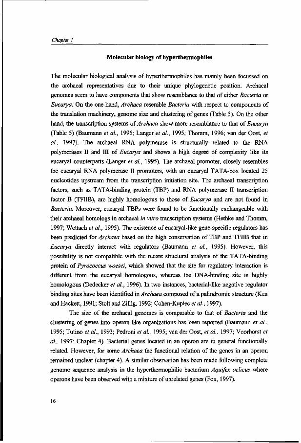
Molecular biology of hyperthermophiles
The molecular biological analysis of hyperthermophiles has mainly been focussed on
the archaeal representatives due to their unique phylogenetic position. Archaeal
genomes seem to have components that show resemblance to that of either Bacteria or
Eucarya. On the one hand, Archaea resemble Bacteria with respect to components of
the translation machinery, genome size and clustering of genes (Table 5). On the other
hand, the transcription systems of Archaea show more resemblance to that of Eucarya
(Table 5) (Baumann et al, 1995; Langer et al, 1995; Thomm, 1996; van der Oost, et
al, 1997). The archaeal RNA polymerase is structurally related to the RNA
polymerases II and III of Eucarya and shows a high degree of complexity like its
eucaryal counterparts (Langer et al, 1995). The archaeal promoter, closely resembles
the eucaryal RNA polymerase II promoters, with an eucaryal TATA-box located 25
nucleotides upstream from the transcription initiation site. The archaeal transcription
factors, such as TATA-binding protein (TBP) and RNA polymerase II transcription
factor B (TFIIB), are highly homologous to those of Eucarya and are not found in
Bacteria. Moreover, eucaryal TBPs were found to be functionally exchangeable with
their archaeal homologs in archaeal in vitro transcription systems (Hethke and Thomm,
1997; Wettach et al, 1995). The existence of eucaryal-like gene-specific regulators has
been predicted for Archaea based on the high conservation of TBP and TFIIB that in
Eucarya directly interact with regulators (Baumann et al, 1995). However, this
possibility is not compatible with the recent structural analysis of the TATA-binding
protein of Pyrococcus woesei, which showed that the site for regulatory interaction is
different from the eucaryal homologous, whereas the DNA-binding site is highly
homologous (Dedecker et al, 1996). In two instances, bacterial-like negative regulator
binding sites have been identified in Archaea composed of a palindromic structure (Ken
andHackett, 1991; Stolt and Zillig, 1992; Cohen-Kupiec etal, 1997).
The size of the archaeal genomes is comparable to that of Bacteria and the
clustering of genes into operon-like organizations has been reported (Baumann et al.,
1995; Tutino et al, 1993; Pedroni et al, 1995; van der Oost, et al, 1997; Voorhorst et
al, 1997: Chapter 4). Bacterial genes located in an operon are in general functionally
related. However, for some Archaea the functional relation of the genes in an operon
remained unclear (chapter 4). A similar observation has been made following complete
genome sequence analysis in the hyperthermophilic bacterium Aquifex aelicus where
opérons have been observed with a mixture of unrelated genes (Fox, 1997).
16

General introduction
Table 5. Some discriminating molecular features of the three domains of life.
Genome
Plasmids polycistronic transcripts RNA polymerase
promoter signatures Transcription factors
Translation initiation
Bacteria single, circular, haploid + +
one class (ßß'a2)
-35/-10 c
SD + AUG
Archaea single, circular, haploid + +
one class (8-10 subunits) TATA-box (-25) TBP TFIIB SD + AUG
Eucarya
multiple, lineair, diploid
--
three classes, complex composition TATA-box (-25) TBP TFIIB AUG
SD, Shine Dalgarno sequence; a, sigma factor
It has been postulated that control of archaeal genes resembles that found in
Bacteria (Zillig et al., 1993). Bacterial-like transcriptional regulators have been
identified on the sequence level such as Lad homolog in P.aerophilum (Fitz-Gibbon et
al, 1997) and homologs of the E.coli general activator Lrp have been identified in
Pyrococcus, Sulfolobus and Methanococcus (Kyrpides and Ouzounis, 1995: Lebbink et
al., 1997; Sensen et al, 1996; Bult et al, 1996), but their functionality remains to be
proven.
17
Chapter 1
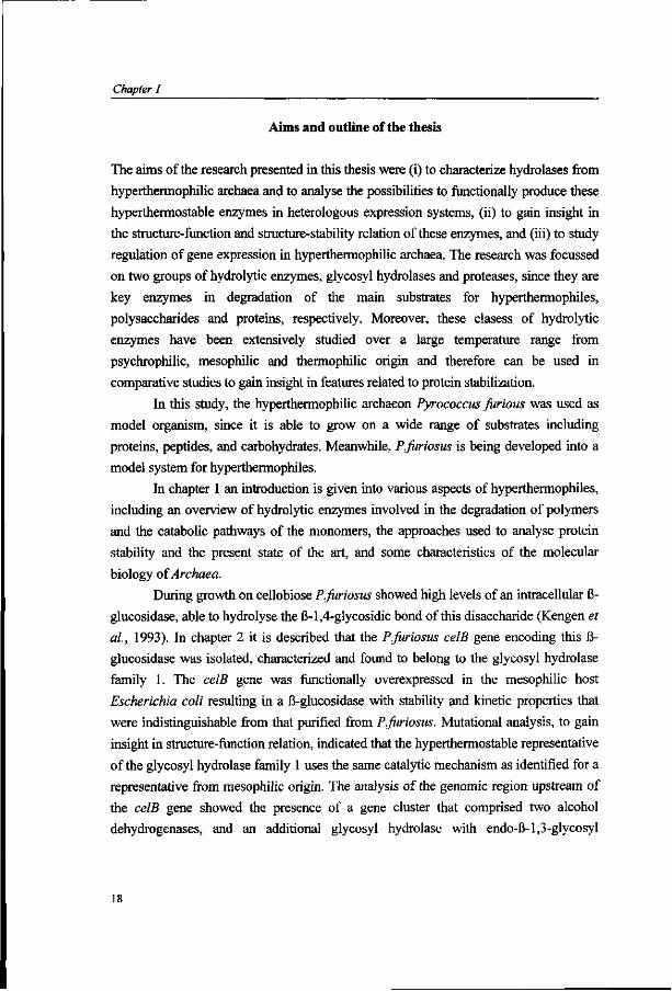
Aims and outline of the thesis
The aims of the research presented in this thesis were (i) to characterize hydrolases from
hyperthermophilic archaea and to analyse the possibilities to functionally produce these
hyperthermostable enzymes in heterologous expression systems, (ii) to gain insight in
the structure-function and structure-stability relation of these enzymes, and (iii) to study
regulation of gene expression in hyperthermophilic archaea. The research was focussed
on two groups of hydrolytic enzymes, glycosyl hydrolases and proteases, since they are
key enzymes in degradation of the main substrates for hyperthermophiles,
polysaccharides and proteins, respectively. Moreover, these clasess of hydrolytic
enzymes have been extensively studied over a large temperature range from
psychrophilic, mesophilic and thermophilic origin and therefore can be used in
comparative studies to gain insight in features related to protein stabilization.
In this study, the hyperthermophilic archaeon Pyrococcus furious was used as
model organism, since it is able to grow on a wide range of substrates including
proteins, peptides, and carbohydrates. Meanwhile, P.furiosus is being developed into a
model system for hyperthermophiles.
In chapter 1 an introduction is given into various aspects of hyperthermophiles,
including an overview of hydrolytic enzymes involved in the degradation of polymers
and the catabolic pathways of the monomers, the approaches used to analyse protein
stability and the present state of the art, and some characteristics of the molecular
biology of Archaea.
During growth on cellobiose P.furiosus showed high levels of an intracellular ß-
glucosidase, able to hydrolyse the ß-l,4-glycosidic bond of this disaccharide (Kengen et
al, 1993). In chapter 2 it is described that the P.furiosus celB gene encoding this ß-
glucosidase was isolated, characterized and found to belong to the glycosyl hydrolase
family 1. The celB gene was functionally overexpressed in the mesophilic host
Escherichia coli resulting in a ß-glucosidase with stability and kinetic properties that
were indistinguishable from that purified from P.furiosus. Mutational analysis, to gain
insight in structure-function relation, indicated that the hyperthermostable representative
of the glycosyl hydrolase family 1 uses the same catalytic mechanism as identified for a
representative from mesophilic origin. The analysis of the genomic region upstream of
the celB gene showed the presence of a gene cluster that comprised two alcohol
dehydrogenases, and an additional glycosyl hydrolase with endo-ß-l,3-glycosyl

General introduction
hydrolase activity. The organization, expression and function of this gene cluster is
studied in the following chapters (Chapter 3-5).
Chapter 3 describes the overproduction and characterization of the two alcohol
dehydrogenases, a short-chain and an iron-containing alcohol. Chapter 4 deals with the
expression in E.coli of the endo-ß-l,4-glucanase encoding gene, lamA, of the ce/5-locus
and characterization of the produced hydrolase. The endo-ß-l,3-glucanase hydrolyses ß-
1,3 and 1,4-glycosyl bonds, generating oligosaccharides that subsequently may be
converted to monosaccharides by the ß-glucosidase CelB. Mutational analysis was
performed to analyse the substrate specificity of the enzyme. The gene cluster
containing the adhA-adhB-lamA genes was found to be a operon, that was designated
the lamA operon. The lamA transcript was found to be co-regulated with the divergently
orientated celB gene by ß-linked glucose polymers as is described in Chapter 5.
Chapters 6 and 7 are concerned with the second group of hydrolytic enzymes
studied, the extracellular subtilisin-like serine proteases. Chapter 6 describes the
purification and characterization of pyrolysin, an abundant extracellular protease in
P.furiosus when growing on proteinaceous substrates. Pyrolysin, one of the most
thermostable serine proteases known to date, was found to be N-glycosylated and
autocatalytically processed at its C-terminus. Via reversed genetics the gene was
isolated and its sequence showed to be a subtilisin-like serine protease (Chapter 6). A
pyrolysin-like encoding gene was also isolated from Thermococcus stetteri. The
deduced product, designated stetterlysin, showed high homology with pyrolysin and
with subtilases that have a known three-dimensional structure allowing for homology
modelling. Comparisons have been performed of the predicted three-dimensional
models of the catalytic domain of stetterlysin and pyrolysin with the crystal structure of
subtilases from mesophilic and thermophilic origin and a model structure for a subtilase
from psychrophilic origin. This resulted in the identification of features that could be
related to protein thermostabilization (Chapter 7). Modelling of substrates into the
predicted structure revealed the possible enzyme-substrate interactions and suggest that
pyrolysin is able to remove its own pro-peptide and thus is autocatalytically activated. A
summary and concluding remarks are given in Chapter 8.
19

Chapter 1
References
Adams, M.W.W. (1993) Enzymes and proteins from organisms that grow near and above 100°C. Ann. Rev. Microbiol. 47: 627-658.
Adams, M.W.W., and Kletzin, A. (1996) Oxidoreductase-type enzymes and redox proteins involved in fermentative metabolisms of hyperthermophilic Archaea. Adv. Prot. Chem. 48: 101-180.
Aguilar, CF., Sanderson, I., Moracci, M., Ciaramella, M., Nucci, R., Rossi, M., and Pearl, L.H. (1997) Crystal structure of the ß-glycosidase from the hyperthermophilic archaeon Sulfohbus solfataricus: Resilience as a key factor in thermostability. J. Mol. Microbiol. (In press).
Andrade, CM., Morana, A., de Rosa, M., and Antranikian, G. (1996) Production and characterization of amylolytic and xylanolytic enzymes form the hyperthermophilic archaeon Pyrodictium abyssi. First International Congres on Extremophiles, Estoril, Portugal, p 98.
Bauer, M.W., Bylina, E.J., Swanson, R.V., and Kelly, R.M. (1996) Comparison of a ß-glucosidase and a ß-mannosidase from the hyperthermophilic archaeon Pyrococcus furiosus. Purification, characterization, gene cloning, and sequence analysis. J. Biol. Chem. 271: 23749-23755.
Bauer, M.W., Halio, S.B., and Kelly, R.M. (1996) Proteases and glycosyl hydrolases from hyperthermophilic microorganisms. In Enzymes and Proteins from Hyperthermophilic Microorganisms. NY, Academic Press, pp. 271-310.
Baumann, P., Qureshi, S.A., and Jackson, S.P. (1995) Transcription: new insights from studies on Archaea. Trends Genet. 11: 279-283.
Blake, P.R., Park, J.-B., Zhou, Z.H., Hare, D.R., Adams, M.W.W., and Summers, M.F. (1992) Solution-state structure by NMR of zinc-substituted rubredoxin from the marine hyperthermophilic archaebacterium Pyrococcus furiosus. Protein Sei. 1: 1508-1521.
Bloch), E., Rachel, R., Burggraf, S., Hafenbradl, D., Jannasch, H.W., and Stetter, K.O. (1997) Pyrolobus fumarii, gen. and sp. nov., represents a novel group of archaea, extending the upper temperature limit for life to 113°C. Extremophiles 1: 14-21.
Blumentals, I.I., Robinson, A.S., and Kelly, R.M. (1990) Characterization of sodium dodecyl sulfate-resistant proteolyltic activity in the hyperthermophilic archaebacterium Pyrococcus furiosus. Appl. Environm. Microbiol. 56: 1992-1998.
Bouyoub, A., Barbier, G., Forterre, P., and Labedan, B. (1996) The adenylosuccinate synthetase from the hyperthermophilic archaeon Pyrococcus species displays unusual structural features. J. Mol. Biol. 261: 144-154.
Budgen, N., and Danson, M.J. (1986) Metabolism of glucose via a modified Entner-Douderoff pathway in the thermoacidophilic archaebacterium Thermoplasma acidophilum. FEBS Lett. 196: 206-210.
Burlini, N., Magnani, P., Villa, A., Macchi, F., Tortora, P., and Guerritore, A. (1992) A heat-stable serine proteinase from the extreme thermophilic archaebacterium Sulfohbus solfataricus. Biochim. Biophys. Acta 1122: 283-292.
Cannio, R., de Pascale, D., Rossi, M., and Bartolucci, S. (1994) Gene expression of a thermostable ß-galactosidase in mammalian cells and its application in assays of eukaryotic promoter activity. Biotechn. Appl. Biochem. 19: 233-244.
Carter, P., and Wells, J.A. (1988) Dissecting the catalytic triad of a serine protease. Nature 332: 564-568.
Cavagnero, S., Zhou, Z.H., Adams, M.W.W., and Chan, S.I. (1995) Response of ruberdoxin from Pyrococcus furiosus to environmental changes: Implications for the origin of hyperthermostability. Biochemistry 34: 9865-9873.
Chan, M.K, Mukund, S., Kletzin, A., Adams, M.W.W., and Rees, D.C. (1995) Structure of a hypethermophilic tungstopterin enzyme, aldehyde ferredoxin oxidoreductase. Science 267: 1463-1469.
Cohen-Kupiec, R., Blank, C , and Leigh, J.A. (1997) Transcriptional regulation in Archaea: in vivo demonstration of a repressor binding site in a methanogen. Proc. Natl. Acad. Sei. USA 94: 1316-1320.
20
General introduction
Connaris, H., Cowan, D.A., and Sharp, R.J. (1991) Heterogeneity of proteinases from the hyperthermophilic archaebacterium Pyrococcusfuriosus. J. Gen. Microbiol. 137: 1193-1199.
Cowan, D.A., Smolensk!, K.A., Daniel, R.M., and Morgan, H.W. (1987) An extremely thermostable extracellular proteinase from a strain of the archaebacterium Desulfwococcus growing at 88°C. Biochem. J. 247: 121-133.
Daniel, R.M., Toogood, H.S., and Bergquist, P.L. (1995) Thermostable proteases. In: Biotechnology and Genetic Engineering Reviews. Vol. 13. pp 51-100.
Danson, M.J. (1988) Archaebacteria: the comparative enzymology of their central metabolic pathways. Adv. Microbial Physiol. 29: 166-231.
Danson, M.J. (1989) Central metabolism of the archaebacteria: an overview. Can. J. Microbiol. 35: 58-64.
De Rosa, M., Gambacorta, A., Nicoleus, B., Giardina, P., Poerio, E., and Buonocore, V. (1984) Glucose metabolism in the extreme thermoacidophilic archaebacterium Sulfolobus solfataricus. Biochem. J. 224:407-414.
Dedecker, B.S., O'Brien, R., Fleming, P.L., Geiger, J.H., Jackson, S.P., and Sigler, P.B. (1996) The crystal structure of a hyperthermophilic archaeal TATA-box binding protein. J. Mol. Biol. 264: 1072-1084.
Eggen, H.I.L., Geerling, A., Watts, J., and de Vos, W.M. (1990) Characterization of pyrolysin, a hyperthermoactive serine protease from the archaebacterium Pyrococcus furiosus. FEMS Microbiol. Lett. 71: 17-20.
Eggen, R.I.L., Geerling, A.C.M., Voorhorst, W.G.B., Kort, R., and de Vos, W.M. (1994) Molecular and comparative analysis of the hyperthermostable Pyrococcus furiosus glutamate dehydrogenase and its gene. Biocatalysis 8: 131-141.
Fiala, G., and Stetter, K.O. (1986) Pyrococcus furiosus sp. nov. represents a novel genus of marine heterotrophic archaebacteria growing optimally at 100°C. Arch. Microbiol. 145: 56-61.
Fitz-Gibbon, S., Choi, A.J., Miller, J.H., Stetter, K.O., Simon, M.I., Swanson, R., and Kim, U. (1997) A fosmid-based genomic map and identification of 474 genes of the hyperthermophilic archaeon Pyrobaculum aerophilum. Extremophiles 1: 36-51.
Fontana, A. (1991) How nature engineers proteins thermostability. In Life under extreme conditions: Biochemical adaptation. Berlin, Springer. 89-113.
Fox, J.L (1997) Whole E.coW. Microbial sequences in log-phase growth. ASMNews,63; 187-192. Fersht, A.R., and Serrano, L. (1993) Principles of protein stability derived from protein engineering
experiments. Current Opinion in Structural Biology 3: 75-83. Fujiwara, S., Okuyama, S., and Imanaka, T. (1996) The world of archaea: genome analysis, evolution
and thermostable enzymes. Gene 179: 165-170. Fusek, M., Lin, X.L., and Tang, J. (1990) Enzymic properties of thermopsin. J. Biol. Chem. 265:
1496-1501. Gray, C.J. (1993) Stabilisation of enzymes with soluble additives.In: Thermostability of enzymes. Ed
Gupta, M.N. Springer Verlag, NY, pp 124-145. Grogan, D. (1991) Evidence that ß-galactosidase of Sulfolobus solfataricus is only one of the several
activities of a thermostable ß-D-glycosidase./4/Tp/. Environ. Microbiol. 57: 1644-1649. Gueguen, Y., Voorhorst, W.G.B., van der Oost, J., and de Vos, W.M. (1997) Molecular and
biochemical characterization of an endo-ß-l,3-glucanase of the hyperthermophilic archaeon Pyrococcus furiosus. J. Biol. Chem. (in press)
Gupta M.N. (1991) Thermostabilization of proteins. Biotechn. Appl. Biochem. 14:1-11. Halio, S.B., Blumentals, I., Short, S.A., Merrill, B.M., and Kelly, R. (1996) Sequence, expression in
Escherichia coli, and analysis of the gene encoding a novel intracellular protease (Pfpl) from the hyperthermophilic archaeon Pyrococcus furiosus. J. Bacteriol 178: 2605-2612.
Hanzawa, S., Hoaki, T., Jannasch, H.W., and Maruyama, T. (1996) An extremely thermostable serine protease from a hyperthermophilic archaeum, Desulfwococcus strain SY, isolated from a deep-sea hydrothermal vent. J. Mar. Biotechnol. 4: 121-126.
21

Chapter 1
Harwood, V.J., Denson, J.D., Robinson-Bidle, K.A., and Schreier, H.J. (1997) Overexpression and characterization of a prolyl endopeptidase from the hyperthermophilic archaeon Pyrocoocus fiiriosus.J. Bacteriol. 179: 3613-3618.
Haseltine, C , Rolfsmeier, M., and Blum, P. (1996) The glucose effect and regulation of a a-amylase synthesis in the hyperthermophilic archaeon Sulfolobus solfataricus. J. Bacteriol. 178: 945-950.
Heider, J., Mai, X., and Adams, M.W.W. (1996) Characterization of 2-ketoisovalerate ferredoxin oxidoreductase, a new and reversible coenzyme A-dependent enzyme involved in peptide fermentation by hyperthermophilic archaea. J. Bacteriol. 178: 780-787.
Hennig, M., Darimont, B., Sterner, R., Kirschner, K., and Jansonius, J.N. (1995) 2.0 Â structure of indole-3-glycerol phosphate synthase from the hyperthermophile Sulfolobus solfataricus: possible determinants of protein stability. Structure 3: 1295-1306.
Henrissat, B. (1997) http://expasy.hcuge.ch/cgi-bin/lists7glycosid.txt Henrissat B., and Bairoch A. (1996) Updating the sequence-based classification of glycosyl hydrolases.
Biochem. J. 316: 695-696. Hensel, R., Laumann, S., Lang, J., Heumann, H., and Lottspeich, F. (1987) Characterization of two
D-glyceraldehyde-3-phosphate dehydrogenases form the extremely thermophilic archaebacterium Thermoproteus tenax. Eur. J. Biochem. 170: 325-333.
Hess, D., Krüger, K., Knappik, A., Palm, P., and Hensel, R. (1995) Dimeric 3-phosphoglycerate kinases from hyperthermophilic archaea: Cloning, sequencing and expression of the 3-phosphoglycerate kinase gene of Pyrococcus furiosus in Escherichia coli and characterization of the protein. Structural and functional comparison with the 3-phosphoglycerate kinase of Methanothermusfervidus. Eur. J. Biochem. 233: 227-237.
Hethke, C , and Thomm, T. (1997) Unpublished data. Horikoshi, K. (1997) A new microbial world-Extremophiles. Extremophiles 1: Editorial. Kelly, R.M., and Adams, M.W.W. (1994) Metabolism in hyperthermophilic microorganisms. Antonie
Van Leeuwenhoek 66: 247-270. Ken, R., and Hackett, N.R. (1991) Halobacterium halobium strains lysogenic from phage <J>H contain a
protein resembling coliphage repressors. J. Bacteriol. 173: 955-960. Kengen, S.W.M., de Bok, F.A.M., van Loo, N.D., Dijkema, C , Stams, A.J.M., and de Vos, W.M.
(1994) Evidence for the operation of a novel Embden-Meyerhof pathway that involves ADP-dependent kinases during sugar fermentation by Pyrococcus furiosus. J. Biol. Chem. 269: 17537-17541.
Kengen, S.W.M., Luesink, E.J., Stams, A.J.M., and Zehnder, A.J.B. (1993) Purification and characterization of an extremely stable ß-glucosidase from the hyperthermophilic archaeon Pyrococcus furiosus. Eur. J. Biochem. 213: 305-312.
Kengen, S.W.M., and Stams, A.J.M. (1994) An extremely thermostable ß-glucosidase from the hyperthermophilic archaeon Pyrococcus furiosus. Biocatlysis. 11: 79-88.
Kengen, S.W.M., Stams, A.J.M., and de Vos, W.M. (1996) Sugar metabolism of hyperthermophiles. FEMS Microbiol. Rev. 18: 119-137.
Klingeberg, M., Friedrich, A., and Antranikian, G. (1992) Production of heat-stable proteases from thermophilic microorganisms and their application in the degradation of chicken feathers. DECHEMA Biotechnological Conferences 5: 173-176.
Klingeberg, M., Galunsky, B., Sjoholm, C , Kasche, V., and Antranikian, G. (1995) Purification and properties of highly thermostable, SDS resistant and stereospecific proteinase from the extreme thermophilic archaeon Thermococcus stetteri. Appl. Environ. Microbiol. 61: 3098-3104.
Klingeberg, M., Hashwa, F., and Antranikian, G. (1991) Properties of extremely thermostable proteases from anaerobic hyperthermophilic bacteria. Appl. Microbiol. Biotechnol 34: 715-719.
Knapp, S., de Vos, W.M., Rice, D., and Ladenstein, R. (1997) Crystal structure of glutamate dehydrogenase from the hyperthermophilic eubacterium Thermotoga maritima at 3.0 Â resolution. J. Mol. Biol. 267: 916-932.
Korndörfer, I., Steipe, B., Huber, R., Tomschy, A., and Jaenicke, R. (1995) The crystal structure of holo-glyceraldehyde-3-phosphate dehydrogenase from the hyperthermophilic bacterium Thermotoga maritima at 2.5 Â resolution. J. Mol. Biol. 246: 511 -521.
22

General introduction
Kyrpides, and Ouzounis (1995) The eubacterial transcriptional activator Lrp is present in the archaeon Pyrococcusfuriosus. Trends Biochem. Sei. 20: 140-141.
Langer, D., Hain, J., Thuriaux, P., and Zillig, W. (1995) Transcription in Archaea: Similarity to that of Eucarya. Proc. Natl. Acad. Sei. USA 92: 5768-5772.
Lebbink, J.H.G., Eggen, R.I.L., Geerling, A.C.M., Consalvi, V., Chiaraluce, R., Scandurra, R., and de Vos, W.M. (1995) Exchange of domains of glutamate dehydrogenase from the hyperthermophilic archaeon Pyrococcus fiiriosus and the mesophilic bacterium Clostridium difficile: effects on catalysis, thermoactivity and stability. Prot. Engng. 8: 1287-1294.
Lebbink, J.H.G., Tuininga, J.E., van der Oost, J., and de Vos, W.M. (1997) Unpublished results. Leuschner, C , and Antranikian, G. (1995) Heat-stable enzymes from extremely thermophilic and
hyperthermophilic microorganisms. World J. Microbiol. Biotechn. 11: 95-114. Lin, X.L., and Tang, J. (1990) Purification characterization and gene cloning of thermopsine a
thermostable acid protease from Sulfolobus acidocaldarius. J. Biol. Chem. 265: 1490-1495. Mai, X., and Adams, M.W.W. (1996) Purification and characterization of two reversible and
ADP-dependent acetyl Coenzyme A synthetases from the hyperthermophilic archaeon Pyrococcus fiiriosus. J. Bacterial. 178: 5897-5903.
Mai, X.H., and Adams, M.W.W. (1994) Indolepyruvate ferredoxin oxidoreductase from the hyperthermophilic archaeon Pyrococcus fiiriosus: A new enzyme involved in peptide fermentation../. Biol. Chem. 269: 16726-16732.
Martins, L.O., and Santos, H. (1995) Accumulation of mannosylglycerate and di-myo-inositol-phosphate by Pyrococcus fiiriosus in response to salinity and temperature. Appl. Environ, Microbiol. 61:3299-3303.
Mayr, J., Lupas, A., Kellermann, J., Eckerskorn, C , Baumeister, W., and Peters, J. (1996) A hyperthermostable protease of the subtilisin family bound to the surface layer of the Archaeon Staphylothermus marinus. Current Biology 6: 739-749.
Moracci, M., Capalbo, L., Ciaramella, M., and Rossi. M. (1996) Identification of two glutamic acid residues essential for catalysis in the ß-glycosidase from the thermoacidophilic archaeon Sulfolobus solfataricus. Protein Engng. 9: 1191-1195.
Moracci, M., La Volpe, A., Pulitzer, J.F., Rossi, M., and Ciaramella, M. (1992) Expression of the thermostable ß-galactosidase gene from the archaebacterium Sulfolobus solfataricus in Saccharomyces cerevisiae and characterization of a new inducible promoter for heterologous expression. J. Bacteriol. 174: 873-882.
Muir, J.M., Russell, R.J.M., Hough, D.W., and Danson, M.J. (1995) Citrate synthase from the hyperthermophilic archaeon Pyrococcusfuriosus. Protein Engng. 8: 583-592.
Mukund, S., and Adams, M.W.W. (1995) GlyceraIdehyde-3-phosphate ferredoxin oxidoreductase, a novel tungsten-containing enzyme with a potential glycolytic role in the hyperthermophilic archaeon Pyrococcus fiiriosus. J. Biol. Chem. 270: 8389-8392.
Nucci, R., D'Auria, S., Febbraio, F., Vaccaro, C , Morana, A., De Rosa, M., and Rossi, M. (1995) A thermostable ß-glycosidase from Sulfolobus solfararicus: temperature and SDS effects on its functional and structural properties. Biotechn. Appl. Biochem. 21: 265-274.
Nucci, R., Moracci, M., Vaccaro, C , Vespa, N., and Rossi, M. (1993) Exoglucosidase activity and substrate specificity of the ß-glycosidase isolated from the extreme thermophile Sulfolobus solfataricus. Biotechn. Appl. Biochem. 17: 239-250.
Pedroni, P., Delia Volpe, A., Galli, G., Mura, G.M., Pratesi, C , and Grandi, G. (1995) Characterization of the locus encoding the [Ni-Fe] sulfhydrogenase from the archaeon Pyrococcus furiosus: evidence for a relationship to bacterial sulfite reductases.Mi'craè/o/ogy 141:449-
Querol, E., Perex-Pons, J.A., and Mozo-Villarias, A. (1996) Analysis of protein conformation characterisitics related to thermostability. Protein Engng. 9: 265-271.
Rice, D.W., Engel,P.C, Ohshima, T., Robb, F.T., and Scandurra, R. (1996) Generic lessons on protein stability from studies on glutamate dehydrogenase. Thermophiles 96 Conference, p 54.
23

Chapter 1
Robinson, K.A., Bartley, D.A., Robb, F.T., and Schreier, H.J. (1995) A gene from the hyperthermophile Pyrococcus furiosus whose deduced product is homologous to members of the prolyl oligopeptidase family of proteases. Gene 152: 103-106.
Schäfer, T., and Schönheit, P. (1993) Gluconeogenesis from pyruvate in the hyperthermophilic archaeon Pyrococcus furiosus - involvement of reactions of the Embden-Meyerhof pathway. Arch. Microbiol. 159: 354-363.
Schäfer, T., Selig, M., and Schönheit, P. (1993) Acetyl-CoA synthetase (ADP-forming) in Archaea, a novel enzyme involved in acetate formation and ATP synthesis. Arch. Microbiol. 159: 72-83.
Schönheit, P., and Schäfer, T. (1995) Metabolism of hyperthermophiles. World J. Microbiol. Biotechn. 11:26-57.
Selig, M., Xavier, K., Santos, H., and Schönheit, P. (1997) Comparative analysis of Embden-Meyerhof and Entner-Douderoff glycolytic pathways in hyperthermophilic archaea and the bacterium Thermotoga. Arch Microbiol. 167: 217-232.
Sensen, C.W., Klenk, H.-P., Singh, R.K., Allard, G., Chan, C.-Y., Lui, Q.Y., Penny, SX., Young, F., Schenk, M.E., Gaasterland, T., Doolittle, W.F., Ragan, M.A., and Charlebois, R.L. (1996) Organizational characteristics and information content of an archaeal genome: 156 kb of sequence from Sulfolobus solfataricus P2. Mol. Micriobiol. 21
Siebers, B., and Hensel, R. (1993) Glucose catabolism of the hyperthermophilic archaeum Thermoproteus tenax. FEMSMicrobiol. Lett. I l l : 1-8.
Siezen, R.J., de Vos, W.M., Leunissen, J.A.M., and Dijkstra, B.W. (1991) Homology modelling and protein engineering strategy of subtilases, the family of subtilisin-like serine proteases. Protein Engng. 4: 719-737.
Siezen, R.J., and Leunissen (1997) Subtilases: the superfamily of subtilisin-like serine proteases. Protein Sei. 6:501-523.
Stetter, K.O., Fiala, G., Huber, G., Huber, R., and Segerer, A. (1990) Hyperthermophilic microorganisms. FEMS Microbiol. Rev. 75: 117-124.
Stetter, K.O. (1994) The lesson of Archaebacteria. In Nobel symposium. New York, Columbia University Press. 143-151.
Stolt, P., and Zillig, W. (1992) In vivo studies on the effects of immunity genes on early lytic transcription in the Halobacterium salinarium phage phi H. Mol. Gen. Genet. 235: 197-204.
Sunna, A., Moracci, M., Rossi, M., and Antranikian, G. (1997) Glycosyl hydrolases from hyperthermophiles. Extremophiles 1: 2-13.
Szilagyi, A., and Zavodsky, P. (1995) Structural basis for the extreme thermostability of D-glyceraldehyde-3-phosphate dehydrogenase from Thermotoga maritima: analysis based on homology modelling. Protein Engng. 8: 779-789.
Tomschy, A., Böhm, G., and Jaenicke, R. (1994) The effect of ion pairs on the thermal stability of D-glyceraldehyde-3-phosphate dehydrogenase from the hyperthermophilic bacterium Thermotoga maritima. Prot. Engngl: 1471-1478.
Tutino, M.L., Scarano, G., Marino, G., Sannia, G., and Cubellis, M.V. (1993) Tryptophan biosynthesis genes trpEGC in the thermoacidophilic archaebacterium Sulfolobus solfataricus. J. Bacteriol. 175: 299-
Teng, Q., Zhou, Z.H., Smith, W.T., Busse, S.C., Howard, J.B., Adams, M.W.W., and La Mar, G.N. (1994) Solution of 'H NMR determination of secondary structure for the three-iron form of ferredoxin from the hyperthermophilic archaeaon Pyrococcus furiosus. Biochemistry 33: 6316-6326.
Thomm, M. (1996) Archaeal transcription factors and their role in transcription initiation. FEMS Microbiol. Rev. 18: 159-171.
Van der Oost, J., Ciaramella, M., Moracci, M., Pisani, F.M., Rossi, M., de Vos, W.M. (1997) Molecular biology of hyperthermophilic Archaea Adv. Biochem Engng./Biotechnol. (in press)
Van der Oost, J., Schut, G., Kengen, S.W.M., Hagen, W.R., Thomm, M., De Vos, W.M. (1997) The ferredoxin-dependent conversion of glyceraldehyde-3-phosphate in the hyperthermophilic archaeon Pyrococcus furiosus represents a novel site of glycolytic regulation, (in preparation).
Verhees, C , van der Oost, J., and de Vos, W.M. (1997) (Unpublished results).
24

General introduction
Vieille, C , and Zeikus, J.G. (1996) Thermozymes: identifying molecular determinants of protein structural and functional stability. Trends Biotechn. 14: 183-190.
Völkl, P., Markiewicz, P., Stetter, K.O., and Miller, J.H. (1995) The sequence of a subtilisin-type protease (aerolysin) from the hyperthermophilic archaeum Pyrobaculum aerophilum reveals sites important to thermostability. Protein Science 3: 1329-1340.
Voorhorst, W.G.B., Eggen, R.I.L., Geerling, A.C.M., Platteeuw, C , Siezen, R.J., and de Vos, W.M. (1996) Isolation and characterization of the hyperthermostable serine protease, pyrolysin, and its gene from the hyperthermophilic archaeon Pyrococcus furiosus. J. Biol. Chem. 271: 20426-20431.
Voorhorst, W.G.B., Eggen, R.I.L., Luesink, E.J., and de Vos, W.M. (1995) Characterization of the celB gene coding for ß-glucosidase from the hyperthermophilic archaeon Pyrococcus fariosus and its expression and site-directed mutation in Escherichia coli. J. Bacterial. Ill: 7105-7111.
Voorhorst, W.G.B., Gueguen, Y., Schut, G., Dahlke, I., Thomm, M., van der Oost, J., and de Vos, W.M. (1997) Transcriptional regulation in the hyperthermophilic archaeon Pyrococcus furiosus: Coordinated expression of divergently transcribed genes in response to ß-linked glucose polymers. Submitted for publication.
Voorhorst, W.G.B., Warner, A., de Vos, W.M., and Siezen, R.J. (1997) Homology modelling of two subtilisin-like serine proteases form the hyperthermophilic archaea Pyrococcus furiosus and Thermococcus stetteri. Protein Engng. 10:
Wettach, J., Gohl, H.P., Tschochner, H., and Thomm, M. (1995) Functional interaction of yeast and human TATA-binding proteins with an archaeal RNA polymerase and promoter. Proc. Natl. Acad. Sei. USA 92: 472-476.
Woese, CR., Kandier, O., and Wheelis, M. (1990) Towards a natural system of organisms: proposal for the domains Archaea, Bacteria and Eucarya. Proc. Natl. Acad. Sei. USA 87: 4576-4579.
Yip, K.S.P., Stillman, T.J., Britton, K.L., Artymiuk, P.J., Baker, P.J., Sedelnikova, S.E., Engel, P.C., Pasquo, A., Chiaraluce, R., Consalvi, V. et at (1995) The structure of Pyrococcus furiosus glutamate dehydrogenase reveals a key role for ion-pair networks in maintaining enzyme stability at extreme temperatures. Structure 3: 1147-1158.
Zwickl, P., Fabry, S., Bogedain, C , Haas, A., and Hensel, R. (1990) Glyceraldehyde-3-phosphate dehydrogenase from the hyperthermophilic archaebacterium Pyrococcus woesei: characterization of the enzyme, cloning and sequencing of the gene, and expression. J. Bacteriol. 172: 4329-4338.
Zillig, W., Palm, P., Klenk, H-P., Langer, D., HUdepohl, U., Hain, J., Lanzendörfer, M., and Holz, I. (1993) Transcription in Archaea. In The biochemistry of Archaea (Archaebacteria) M. Kates, Kushner, D.J., and Matheson, A.T., Elsevier, NY, p367-391.
25

Chapter 1
26

ß-glucosidase
Chapter 2
Characterization of the celB Gene Coding for
ß-Glucosidase from the Hyperthermophilic Archaeon
Pyrococcus furiosus and Its Expression and Site-Directed
Mutation in Escherichia coli
Wilfried G.B. Voorhorst, Rik I.L. Eggen, Evert J. Leusink, and Willem M. de Vos
reprinted with permission from the Journal of Bacteriology, 1995, 7105-7111
27

Chapter 2
28

JOURNAL OF BACTERIOLOGY, Dec. 1995, p. 7105-7111
0021-9193/95/$04.00+0 Copyright © 1995, American Society for Microbiology
Vol. 177, No. 24
Characterization of the celB Gene Coding for ß-Glucosidase from the Hyperthermophilic Archaeon Pyrococcus furiosus and Its Expression
and Site-Directed Mutation in Escherichia coli WILFRIED G. B. VOORHORST, RIK I. L. EGGEN.t EVERT J. LUESINK,* AND WILLEM M. DE VOS*
Bacterial Genetics Group, Department of Microbiology, Wageningen Agricultural University, 6703 CT Wageningen, The Netherlands
Received 14 July 1995/Accepted 12 October 1995
The celB gene encoding the cellobiose-hydrolyzing enzyme ß-glucosidase from the hyperthermophilic archaeon Pyrococcus furiosus has been identified, cloned, and sequenced. The transcription and translation initiation sites of the celB gene have been determined, and archaeal control sequences were identified. The celB gene was overexpressed in Escherichia coli, resulting in high-level (up to 20% of total protein) production of ß-glucosidase that could be purified by a two-step purification procedure. The ß-glucosidase produced by E. coli had kinetic and stability properties similar to those of the ß-glucosidase purified from P. furiosus. The deduced amino acid sequence of CelB showed high similarity with those of ß-glycosidases that belong to glycosyl hydrolase family 1, implicating a conserved structure. Replacement of the conserved glutamate 372 in the P. furiosus ß-glucosidase by an aspartate or a glutamine led to a high reduction in specific activity (200- or 1,000-fold, respectively), indicating that this residue is the active site nucleophile involved in catalysis above 100°C.
The most extensively studied representative of the hyperthermophilic organisms that have an optimum growth temperature above 85°C is Pyrococcus furiosus (12). P. furiosus is able to grow on a wide range of substrates, including complex polymers such as starch, glycogen, peptone, and casein or simple carbon compounds like cellobiose, maltose, and pyruvate (12, 20, 33). The main fermentation products are CÖ2 and H2 or alanine, the latter acting as an alternative electron sink (22). The disaccharides cellobiose and maltose are hydrolyzed by intracellular glucosidases (5, 20). The generated glucose was proposed to be further metabolized via a nonphosphorylated Entner-Doudoroff pathway (27, 33). However, recently it was discovered that sugars are fermented by P. furiosus via an Embden-Meyerhof pathway that involves two ADP-dependent kinases (19).
Characterization of proteins from hyperthermophiles revealed that they are extremely thermostable and may have an optimum temperature of catalysis that exceeds the maximum growth temperature of their host (1,18). In addition to their remarkable thermostability, proteins from hyperthermophiles are often found to be highly resistant to chemical denaturation and to degradation by proteases (13). One of the most thermostable enzymes identified up to now is the ß-glucosidase from P. furiosus, with a half-life of 85 h at 100°C (20). During growth on cellobiose, ß-glucosidase can make up to 5% of the total cell protein of P. furiosus and is involved in the hydrolysis of the ß-1,4-glycosidic bond between the two glucose moieties of the disaccharide (20). In addition, ß-glucosidases constitute a group of well-studied enzymes among members of all three domains of life, Eucarya, Bacteria, and Archaea (16, 17, 35). Therefore, the pyrococcal ß-glucosidase is a suitable model
* Corresponding author. Mailing address: Department of Microbiology, Wageningen Agricultural University, Hesselink van Suchtelen-weg4, 6703 CT Wageningen, The Netherlands. Phone: 31 3174 83100. Fax: 31 3174 83829. Electronic mail address: WILLEM.DEVOS® ALGEMEEN.MICR.WAU.NL.
t Present address: EAWAG, Dübendorf, Switzerland. + Present address: NIZO, Ede, The Netherlands.
enzyme for the molecular characterization of structure-function relations of hyperthermostable enzymes. To study these relations by protein engineering it is required to express the gene and produce a functional enzyme in an accessible heterologous host, since for hyperthermophilic members of the Archaea no genetic systems are presently available.
Here we report the isolation, cloning, and sequencing of the ß-glucosidase (celB) gene from P. furiosus. Control regions involved in celB transcription initiation were identified, and the celB gene was functionally overexpressed in Escherichia coli. Subsequently, the CelB was purified and compared to the original enzyme and the active site nucleophile was identified by protein engineering.
MATERIALS AND METHODS
Organisms, media, and plasmids. P. furiosus (DSM 3638) was cultured at 98°C in synthetic seawater as previously described (20). E. coli TGI (15), MC1061 (3), and JM109(DE3) (34), obtained from Pharmacia (Uppsala, Sweden), were grown in L broth and handled as described previously (31). The phagemids pTZ18R and pTZ19R and the expression vector pTTQ19 were obtained from Pharmacia.
Purification of ß-glucosidase From P. furiosus. ß-Glucosidase was purified from P. furiosus cells grown on cellobiose as previously described (20), and its NH2-terminal amino acid sequence was determined by Edman degradation using an Applied Biosystems model 477A (gas-phase amino acid Sequenator) (Applied Biosystems, Foster City, Calif.) (courtesy of SON protein sequence facility, Leiden, The Netherlands).
Cloning of the ß-glucosidase gene. On the basis of the NH2-terminal amino acid sequence of the purified ß-glucosidase, two oppositely orientated PCR oligonucleotides were designed: 5'-AARTTYCCNAARAAYTTYATGTTYGG (OL92-9), based on amino acid residues 2 to 9, and 5'-CCCATYTCRAAYT GRAA (OL92-10), based on amino acid residues 16 to 21 (where N is A, C, G, or T; R is A or G; and Y is C or T). Subsequently, part of the celB gene was amplified by PCR on a DNA Thermal Cycler (Perkin Elmer Cetus, Norwalk, Conn.), using 175 ng of chromosomal DNA of P. furiosus isolated as described previously (10) and 100 ng of both OL92-9 and OL92-10 in a final volume of 100 u.1. Thirty cycles (1 min at 94°C, 2 min at 45°C, and 3 min at 72°C) were followed by a final incubation at 72°C for 7 min. To visualize the expected small product (59 bp) on a sequencing gel, [a-32P]dATP (110 TBq/mmol) was added during amplification. A product with the expected size was used as probe in a Southern hybridization with chromosomal DNA from P. furiosus. Hybridizing fragments were isolated from agarose gels using Gene Clean (BIO 101, La Jolla, Calif.) and cloned in E. colt by established methods (31).
Nucleotide sequence analysis. Phagemids pTZ18R and pTZ19R were used to
7105 29

7106 VOORHORST ET AL. J. BACTERIOL.
produce single-stranded DNA for sequence analysis as described by the supplier (Pharmacia). To obtain overlapping sequences, deletions were generated with exonuclease III (Life Technologies B.V., Breda, The Netherlands) (31). Sequencing was carried out on single-stranded DNA template using the dideoxy terminator method (32).
Computer analysis of nucleotide and protein sequences and alignments of these sequences were done with the PC/GENE program version 5.01 (IntelliGe-netics Inc., Mountain View, Calif.) and the GCG package version 7.0 (7) at the CAOS/CAMM Centre of the University of Nijmegen.
RNA isolation and primer extension. Total RNA of P. furiosus was isolated from cells grown on cellobiose using guanidinium isothiocyanate and ß-mercap-toethanol (4). For this purpose, cells were resuspended in denaturation buffer (4 M guanidinium isothiocyanate, 42 mM sodium citrate, 0.83% AMaury! sarcosine, 0.2 mM ß-mercaptoethanol) followed by the addition of 0.1 volume of 2 M ammonium acetate (pH 4) and 1 volume of phenol-chloroform-isoamyl alcohol (25:24:1). The mixture was incubated on ice for 15 min, and phase separation was obtained by centrifugation for 20 min (10,000 x g, 4°C). The aqueous phase was removed and supplemented with an equal volume of isopropanol, and RNA was precipitated at -20°C. The pelleted RNA was resuspended in denaturation buffer, precipitated again, washed with ice-cold 75% ethanol, dried, and resuspended in water. This purified RNA was used as a template in primer extension experiments with oligonucleotide BG72 (5-CCAAGAATATCCAAACATGA AG), as previously described (10).
Mutagenesis of the celB gene. Mutations were introduced in the celB gene using the CLONE AMP site-directed mutagenesis system (Life Technologies) (28). The mutagenic oligonucleotides (BG 95 [5'-CCAAUGAUAAUUACAN AVAACGGUAUG] and BG 79 [5'-GGCCAUACCGUUBUNUGUAA], where N is A, C, G, or U; V is G, C, or A; and B is G, C, or U) were each used in combination with a d\J-lacZ primer during a PCR amplification with 5 ng of 5ocI-linearized pLUW503 for 30 cycles in a total volume of 100 u l The PCR products were combined, and the pAMP cloning vector was added. After removal of the uracil residues by uracil DNA glycosylase, the DNA was introduced by transformation into the supplied E. coli DH5a and the resulting recombinant plasmids were analyzed by sequencing the region containing the mutation. From the plasmids containing a mutated celB gene, the 190-bp Apal-Aflll fragment coding for residues 357 to 418 was cloned into ^/wiI-^/ÀI-digested pLUW503 and transformed into E. coli JM109(DE3).
Characterization of ß-glncosidase produced in E. coli. E. coli TGI or E. coli JM109(DE3) harboring either the wild-type or mutated celB gene was grown overnight in 5 ml of L broth with 100 u,g of ampicillin per ml. If appropriate, 3 mM isopropyl-ß-D-thiogalactopyranoside was added. Cells were harvested by centrifugation, concentrated 15 x in citrate buffer (50 mM sodium citrate, pH 6.5), and disrupted by sonication. The cell debris was pelleted by centrifugation, and the resulting supernatant was used as cell extract. The ß-glucosi-dase activity was determined using ß-D-glucopyranoside-p-nitrophenyl (Boehr-inger GmbH, Mannheim, Germany) as a substrate (20). The kinetic parameters, temperature optimum, and thermostability of the purified ß-glucosidase were determined by using the same synthetic substrate and done as previously described (20). Routine measurements were performed on a Beekman DU 7500 spectrophotometer with supplied temperature controller at 90DC (Beck-man Instruments, Inc. Fullerton, Calif.). For the thermostability assay, ß-glucosidase was incubated at 100°C at a concentration of 0.5 mg/ml in sealed glass vials.
Electrophoretic analysis of protein samples was done by sodium dodecyl sul-fate-polyacrylamide gel electrophoresis (SDS-PAGE) by the method of Laemmli (24). Protein samples for SDS-PAGE were prepared by heating for 10 min at 110°C in an equal volume of sample buffer (0.1 M Tris-HCl, 5% SDS, 0.9% 2-mercaptoethanol, 20% glycerol, pH 6.8).
Overproduction of pyrococcal ß-glucosidase in E. coli and its purification. E. coli MC1061 harboring pLUW510 was grown in L broth with 300 fig of ampicillin per ml to an optical density at 600 nm of 0.5. The tac promoter was subsequently induced by the addition of 3 mM isopropyl-ß-D-thiogalactopyranoside, and the culture was incubated for another 10 h. Cells were harvested by centrifugation, resuspended in P buffer (50 mM sodium phosphate, pH 7.5), and broken by using a French press. RNase (10 u.g/ml), DNase (10 u,g/ml), and MgCl2 (10 mM) were added, and after a 30-min incubation at 37°C the mixture was transferred to 80°C and incubated for another 45 min. During the latter incubation the majority of the E. colt proteins were denatured and could be removed by centrifugation. The supernatant was subsequently dialyzed against P buffer, loaded onto a Q-Sepha-rose Fast Flow (Pharmacia) column (5.0 by 2.5 cm) preequilibrated with the same buffer and eluted by a linear gradient of NaCl (0 to 1 M in P buffer). Activity fractions that eluted around 0.6 M NaCl were pooled and dialyzed against P buffer. The NH2-termina! amino acid sequence of the CelB produced in E. colt was determined by Edman degradation using a gas-phase amino acid Sequenator.
Nucleotide sequence accession number. The nucleotide sequence reported has been submitted to the GenBank/EMBL Data Bank with the accession number U37557.
PLASMID ACTIVITY
BanMI I
EcoRI 1
celB JWiel EcoRI
1 1
Sspl Smal 1 1
pLUWSOI
pLUW502
pLUW503
PLUW504
pLUW510
FIG. 1. Physical map of plasmids containing celB. The orientation of the vector-located tacUV promoter is shown. In addition, thermoactive ß-glucosidase activity in cell extracts of E. coli harboring the respective plasmids is indicated: +, activity of approximately 4 to 20 U/mg protein; ~, no detectable activity.
RESULTS
Cloning and expression of the celB gene encoding ß-glucosidase from P. furiosus. The P. furiosus ß-glucosidase was purified to homogeneity from cells grown on cellobiose, and its NH2-terminal 23-amino-acid sequence showed the sequence NH2-[M/A]KFPKNFMFGYXXSGFQFEMGLP (X is non-identified residue). Based on amino acid residues 2 to 9 and 16 to 21, two degenerate oligonucleotides (OL92-9 and OL92-10) were designed and used as primers in a PCR reaction with P. furiosus DNA. An amplified fragment with the expected size of 59 bp was obtained and subsequently used as a probe in Southern hybridization with chromosomal DNA from P. furiosus digested with various restriction endonucleases. A BamHl-Pstl fragment of 4.8 kb hybridized strongly and was cloned into pTZ19R digested with BamUl and Pstl, resulting in pLUW500 (Fig. 1). Sequence analysis showed that this fragment contained an incomplete open reading frame comprising the 5' end of the ß-glucosidase gene, designated celB (see below).
To obtain a fragment containing the complete celB gene, the 4.8-kb BamHl-Pstl fragment was used as a probe in a Southern hybridization with DNA from P. furiosus. A hybridizing 7.7-kb SamHI-£coRI fragment was isolated and cloned into a BomHI-EcoRI-digested pTZ19R resulting in pLUW501 (Fig. 1). A cell extract of E. coli TGI harboring pLUW501 showed ß-glucosidase activity at 90°C, indicating that the P. furiosus gene for ß-glucosidase had been cloned and was functionally expressed (Fig. 1). The 7.7-kb insert of pLUW501 could be reduced in size by deleting a 4.2-kb Xbal (multiple cloning sile)-Nhel (insert) fragment, resulting in pLUW503, that in E. coli TGI was able to produce a functional ß-glucosidase (Fig.
I)-Cloning of the inserts of pLUW501 and pLUW503 in
pTZ18R resulted in plasmids pLUW502 and pLUW504, respectively, that in E. coli TGI showed similar ß-glucosidase activities (4 to 7 U/mg of protein; Fig. 1). This indicates that expression of the celB gene is not controlled by the vector-located lac promoter in these plasmids and that the region upstream of the celB gene is involved in transcription initiation in E. coli.
Nucleotide sequence analysis of the celB gene. The nucleotide sequence of the insert of pLUW503 was determined in both directions and showed the presence of an open reading frame of which the first ATG codon is preceded by a consensus, purine-rich ribosome binding site (Fig. 2). The sequence starting from this ATG initiation codon to the TAG stop codon includes 1,416 bp and could encode a 472-amino-acid protein
30

V O L . 177, 1995 EXPRESSION AND MUTATION OF THE P. FURIOSUS celB GENE 7107
sspl 1
-121 TTTTGAAGAGCCTTGAGCCTCCTCAAMGCAGTTAACATATATCTATCITCCCACATTTATAATTCCATGGGAAATAITATAAATCACAATATCAAAATATAAAGCIAGAGGTGGAAACT
1 ATGAAGTTCCCAAAAAACTTCATGTTTGGATATTCTTGGTCTGGTTTCCAGTTTGAGATGGGACTGCCAGGAAGTGAAGTGGAAAGCGACTGGTGGGTGTGGGTTCACGACAAGGAGAAC 1 M K . F P K N F H F G Y S U S G F O F E H G L P G S E V E S D U U V U V H D K E N
121 ATAGCATCAGGTCTAGTAAGTGGAGATCTACCAGAGAACGGCCCAGCATATTGGCACCTCTATAAGCAAGATCATGACATTGCAGAAAAGCTAGGAATGGATTGTATTAGAGGTGGCATT 4 1 I A S G L V S G D L P E N G P A Y U H L Y K 0 D H D I A E K L G M D C I R G G I
241 GAGTGGGCAAGAATTTTTCCAAAGCCAACATTTGACGTTAAAGTTGATGTGGAAAAGGATGAAGAAGGCAACATAATTTCCGTAGACGTTCCAGAGAGTACAATAAAAGAGCTAGAGAAA 8 1 E U A R I F P K P T F D V K V D V E K D E E G N I I S V D V P E S T I K E L E K
361 ATTGCCAACATGGAGGCCCTTGAACATTATGCGAAGATTTACTCAGACTGGAAGGAGAGGGGCAAAACCTTCATATTAAACCTCTACCACTGGCCTCTTCCATTATGGATTCATGACCCA 1 2 1 1 A N M E A I E H Y A R I Y S D U K E R G K T F I L N L Y H U P L P L U I H D P
Pst I 481 ATTGCAGTAAGGAAACTTGGCCCGGATAGGGCTCCTGCAGGATGGTTAGATGAGAAGACAGTGGTAGAGtTTGTGAAGTTTGCCGCCTTCGTTGCTTATCACCTTGATGACCTCGTTGAC 1 6 1 I A V R K L G P D R A P A G U L D E K T V V E F V K F A A F V A Y H I D D L V D
601 ATGTGGAGCACAATGAACGAACCAAACGTAGTCTACAATCAAGGTTACATTAATCTACGTTCAGGATTTCCACCAGGATATCTAAGCTTTGAAGCAGCAGAAAAGGCAAAATTCAACTTA 2 0 1 M W S T M N E P N V V Y N O G Y I N L R S G F P P G Y L S F E A A E I C A K F N L
721 ATTCAGGCTCACATCGGAGCATATGATGCCATAAAAGAGTATICAGAAAAATCCGTGGGAGTGATATACGCCTTTGCTTGGCACGATCCTCTAGCGGAGGAGTATAAGGATGAAGTAGAG 2 4 1 I 0 A H I G A Y D A I K E Y S E K S V G V I Y A F A U H D P L A E E Y K D E V E
841 GAAATCAGAAAGAAAGACTATGAGTTTGTAACAATTCTACACTCAAAAGGAAAGCTAGACTGGATCGGCGTAAACTACTACTCCAGGCTGGTATATGGAGCCAAAGATGGACACCTAGTT 2 8 1 E 1 R K K D Y E F V T 1 L H S K G K L D W I G V N Y Y S R L V Y G A K D G H L V
Aflll 961 CCTTTACCTGGATATGGATTTATGAGTGAGAGAGGAGGATTTGCAAAGTCAGGAAGACCTGCTAGTGACTTTGGATGGGAAAIGTACCCAGAGGGCCTTGAGAACCTICTTAAGTATTTA 3 2 1 P L P G Y G F M S E R G G F A K S G R P A S D F G W E M Y P E G L E N L L K Y L
1081 AACAATGCCTACGAGCTACCAATGATAATTACAGAGAACGGTATGGCCGATGCAGCAGATAGATACAGGCCACACTATCTCGTAAGCCATCTAAAGGCAGTTTACAATGCTATGAAAGAA 3 6 1 N N A Y E L P M I I T E N G M A D A A 0 R Y R P H Y L V S H L K A V Y N A M K E
* Apal 1201 GGTGCTGATGTTAGAGGGTATCTCCACTGGICTCTAACAGACAACTACGAATGGGCCCAAGGGTTCAGGATGAGATTTGGATTGGTTTACGTGGATTTCGAGACAAAGAAGAGAIATTTA
4 0 1 G A D V R G Y L H U S L T D N Y E U A Q G F R M R F G L V Y V D F E T K K R Y L
1321 AGGCCAAGCGCCCTGGTATTCAGAGAAATAGCCACTCAAAAAGAAATTCCAGAAGAATTAGCTCACCTCGCAGACCTCAAATTTGTTACAAGAAAGTAGCCATTTCATTTTTICTTTGTT 4 4 1 R P S A L V F R E I A T Q K E I P E E L A H L A D L K F V T R K *
1441 TTrÎAACTCATATÎTTTGGGAAAATAAGATCATAAAAGCTTAACAAGAGAAACCATGCCATAAAAATAGCAAAGATTGATAGCCCTCAATTICCATGGATTCIACATACATATGGATGAT
1561 CCGTCATAAAGTTTATAAATCCATAGCAAGGTGATTTTATTGGTAGTATCCC
FIG. 2. Nucleotide sequence of the P. furiosus ce!B gene and its flanking regions. The deduced amino acid sequence of the open reading frame corresponding to CelB is depicted and relevant restriction sites are indicated. The transcription initiation site is indicated by the arrow. Regulatory sequences are indicated as follows: the box A of celB and the following open reading frame (encoding tRNA1*1"1 ) (37) are underlined, the ribosome binding site is indicated by asterisks, and the NH2-terminal amino acid residues of the purified ß-glucosidase are underlined. In addition, T stretches downstream of the celB gene are overlined, and the inverted repeat is shown by the oppositely oriented arrows.
that is homologous to various ß-glucosidases (see below). Since the deduced amino acid sequence of residues 1 to 23 is identical to that of the NH2-terminal end of the purified ß-glucosidase (Fig. 2), the coding region represents the P. furiosus celB gene. The calculated molecular mass of the deduced primary sequence is 54,580 Da, which is in agreement with the apparent molecular mass of 58 kDa determined by SDS-PAGE for the ß-glucosidase purified from P. furiosus (20).
Transcriptional organization of the celB gene. Sequence analysis of the upstream region of the celB gene revealed a putative box A, the archaeal equivalent of the TATA box that directs transcription (Fig. 2) (38). To localize the celB transcription initiation site, primer extension was performed on total RNA isolated from P. furiosus cells grown on cellobiose. This resulted in an extension product with a size that corresponded with transcription initiation at an adenine residue located at position — 14 that was located 25 nucleotides downstream of box A (Fig. 2 and 3). Termination of transcription may occur downstream of the celB gene, where three T-rich stretches are present, including the termination consensus sequence of at least four successive T residues (Fig. 2) (6). The last two of these T stretches are included in the left part of an inverted repeat.
Primary sequence comparison. Comparison of the deduced primary sequence of celB with ß-glycosidases present in the SWISS PROT database (release 30) showed the highest similarities (69 to 44%) with representatives of glycosyl hydrolase family 1 and to a lower extent with members of family 3 (30%
or lower) as defined by Henrissat (16). The CelB sequence was aligned with representatives of the family 1 of glycosyl hydrolases, which includes glucosidases, galactosidases, and 6-phos-pho-glycosidases of different origins (Fig. 4). Among the large number of conserved residues are the glutamate 372 and glu-
T T C G A T C T
C c A
A C G
« •
^ *
•Wf
4b» 4k>
• *
T
« * •Nk
flfr
•
PE
«säfc
FIG. 3. Identification of the transcription initiation site of the celB gene by primer extension. Primer-extended product (PE) was electrophoresed in parallel with the sequence ladder generated with the same primer on the noncoding strand of celB.
31

7108 VOORHORST ET AL. J. BACTERIOL.
P. furhsua S.solfatarhus 1 S.solfatarhus 2
T.maritima C. saccharotyticum Agrobacterium sp.
L.lactis L. casai S.aureus
1 50 100 HKFPKHFNFGVSUSGFQFEHCLPGSE-WSDUUVUVKOICEHIASCLVSGDLPENGPAYUHLYKWHDIAEKLGHOCIRGGIEUMtlFPKPTFOVKVDVEICDEE-GNIISVDV
HÏSFPHSFRFGUSOAGF0SEMGTPGSEDPHTDUÏKWVHDPEHHAAGlVSG0LPENGf>GYWGNyiCTFH0HA0KMGLKIARLHVEWSmFP«PLPRro-**«F0ESK00VTEVEI HLSFPKGFKFGWSQSGF05EHGTPGSEDPHSDUHVWVHDRENIVS0WSGDLPENGPGYWGNYKRFHDEAEKIGLNAVRINVEUSRIFPRPLPKPEH0TGTDKEMSPV1SVDL
HNVKKFPEGFLUGVATASYQIEGSPLADGAGMSIUHTFSHTPGNVKNGDTG--HDHSFPKGFLUGAATA5YaiEGAWNEDGICGE5IUDRFTHQKRHILYGHNG--'
HTDPKTLAARFPGDfLFGVATASFQIEGSTKADGRKPSIUDAFCNHPGHVFGRHNG--
HTKTLPKDF1FGGATAAY0AEGATHTDGKGPVAUDKYLEDNYUYTA HSKOLPOOFVMGGATAAYaVEGATKEDGKGRVLUDOfLDKQGRFKP HTKTLPEDFIFGGATAAYQAEGATNTDGKGRVAUDTYLEEKYUYTA •
DVACDHYMRUKEDIEMEKLGVXAYRFSISUPRILPEGTGRVN-DVACDHYHRFEEOVSLMKELGLKAYRFSIAUTRIFPOGFGTVtl"
•DIAC0HYNRUEEDLDIIKEMGVEAYRFSLAWPRI1PDGFGPIH--
EPAS0FYHKYPVDLELAEEYGVNG1RISIAUSR1FPTGYGEVH--0PAAOFYHRYDEDLALAEKYGHQVIRVSIAUSRIFPDGAGEVE--EPASDFYNRYPVDLEISEKFGVHGIRISIAWSRIFPHGYGEVN--
P. furhsus S.soifataricus \ S.solfatarhus 2
T.maritima C.saccharotyticum Agrobacterium sp.
L.lactis L.casei S.aureus
PESTIKELEKIAKHEALEHYRKIYSDUKERGKTFILNLYHUPLPLWIHDP1AVRKLGPDRAPAGULDEKTWEFVKFAAFVAYHL0DLVDHWSTHNEPNWYNOGYINLRSGFPPGYL-S NEHELKRLDEYAMKDALHHYREIFKDLXSRGLYFILNMYHUPLPLULHDPIRVRR-GOFTGPSGULSTRTVYEFARFSAYIAUKFDOLVDEYSTHNEPNWGGLGWGVKSGFPPGYL-S NESKLREHONYANHEALSHYRHILEOLRHRGFHIVLHMYHUTLPIWLHDPIRVRR-GOFTGPTGWLKSRTVYEFARFSAYVAUKLDDLASEYATMNEPHWUGAGYAFPRAGFPPNYL-S
-OKGLOFYHRIIDTLLEKGITPFVTIYKUOLPFALOU---OKGLEFY0RLINKLVENGIEPWTLYHUDLPOKLO0I--
-EKGLDFYDRLVDGCKARGIKTYATLYHUDLPLTLNGO--
-GGUAHREIADWFAEYSRVLFEHFGDRVKNWITLHEPUWA1VGHI.Y--GVHAPGKR-D -GGUAHPEIVNYYFDYAHIVINRYKDKVKKW1TFNEPYCIAFLGYFH--GIHAPGIK-D •GGWASRSTAHAFaRYAKTVMARLGORLOAVATFNEPWCAVWLSKLY--GVHAPCER-H
-EKGVEFYHKLFAECHKRHVEPFVTLHHFDTPEALHSN GDFLKREHIEHfIDYAAFCFEEFPE-VHYWTTFMEIGPIGDGQYLV--GKFPPGIKYO -PRGVAFYHKLFADCAAHH1EPFVHHHFDTPERIHEA GDVLSMHLDDFVAYAKFCFEEF!SE-VKYWITIMEPTSHAVQQYTT"GÏFPPAESGR -PKGVEYYHKLFAECHKRHVEPFVTLHHFDTPEVIHKD GDFLNRKT1DYFV0YAEYCFKEFPE-VKYWTTFNEIGPIGDGQYLV--GKFPPGIKYD
P. furhsus S.soifataricus 1 S.soifataricus 2
T.maritima C. saccharotyticum Agrobactarhim sp.
L.lactis L.casei S.aureus
FEAAEKAKFNLIOAH1GAYDA1ICEY5EKS-VGVIYAFAWH0PLA EEYKDEVEEIRKICD--YEFVTUHS • FELSRRHKYNIIOAHARAYDGIKSVSKKP-VGIIYANSSFQPLT 0KDKEAVEMAEHDNRUWFFDAIIRGEITRGNEKIVR0OL-FRLSEIAKUNUOAHARAYDAIKSVSKKS-VGIIYAHTSYYPLR PODHEAVEIAERLNRUSFFDS1IKGE1TSEGQN-VREDL-
300 -KGKLDU -KGRLDW
•-RNRLDW
lYVAFRAVHHLLRAHARAVKVFRETVKDGKtGIVFHMGYFEPASEK EEOIRAVRFHHQFHNYPLFLNPIYRGDYPELVLEFAREYLPENYKDDHS EIOEKIDF FKVAH0WHSlNLSHFKVVKAVKEtlMI0VEVGITLHLTPVYLOTERLGYtCVS-EIEREHVSLSSQL0N-OLFLDPVLt(GSYPOKLLDYLVOroLLDSMCALSMOO€VK ENF1FP0F MEAALAAMHHIKLAHGFGVEASRHVAPKVPVGLVLHAHSAIPASOG EADLKAAERAF«FHN-GAFF0PVFKGEYPAEHHEALGORHPVVEAEOLG I1SQKLDU
LAKVFQSHHHMHVSHARAVKIYKDKGYKGEIGWHALPTKYPYDPEH PADVRAAELED11HN-tCFILDAYYIGHYSDKTHEGVNHIL-AENGGELOLR0EOFQA1.0AAKDLHOF FDXTFOAEHNQNVAHARIVNIYXSWILGGQIGIVHALOTVYPY-SDS AVOHHAAELQOALEN-RLYLDGTLAGEYHOETLALVKElLDANHQPHFaSTPQEHKAIDEAAHOLDF FEKVFOSHHNMHVAHARAVKLFKDGGYKGEIGWHALPYXYPFDPSH PEDVRAAELEDIIHN-KFILDATYLGKY5RETMEGVOH)l-SVNGGKLNtTDEDYAILDAAXDLNDF
P. furhsus S.soifataricus 1 S.soifataricus 2
T.maritima C.saccharotyticum Agrobacterium sp.
L.lactis L.casei S.aureus
1GVHYYSRLVYGAKDGHLVPLPGYGFHSERGGFAKSGR PASDFGWEHYPEGL-ENLLKYUHAYELP--HIIYENGMA-MA ORYRPHYLVSHLKAVYN IGVHYYTRTWKRTEKGYVSLGGYGHGCERNSVSLAGL PTSOFGUEFFPEGLYDVLIKYWRR-YHLY--MVVTE(tGIA-DOA 0YORPYYLVSHVYOVHR 1GVHYYTRTWTKAESGYLTLPGYGDRCÉRHSLSLANU PTS0FGWEFFPEGLYDV1.LKYUHR-YGLP--LYVHENGIA-ODA DYQRPYYLVSHIYQVHR
VGLMYYSGHLVKFDPOAPAKVSFVERDLPK YAMGUEIVPEGIYI4ILKKV-KEEYRPPE-VYITENGAAFD0WSE0GR VHOONRIOYLKAHIMAWK LGIHYYTRAVRIYDEHSSW1FP1RUEHPAGEY TEHGWEVFPOGlFDLLIUI-KESYPQIP-IYlTEtlGAAYWUVTEOGK VHOSKRIEYLKOHFEAARK UGLNYYTPHRVADOATPGVEFPAY»4PAPAVSDVK ÏD1GWVYAPALHTLVETL-YERYDIPE-CYITERGACY»-MGVÉNGE VHDQPR1.DYYAEHI.GIVAD
LGINYYHSOUHOAFDGEYEIlHHGKGEXGSSKYOlKGVGRRVAPDYVPRTOUDUIIYPEGLYOQimv-KHDYPHYKKlYITENGLGYKDEFVDMT VY0OGRI0YVKQKLEVLSO VGVHHYFSKULRAYHGKSETIHMGOGTKGSSVARLQGVGEEICLPDGIEYTDWDUSIYPRGHYDILHRI-HiroYPLWVTYVTENGIGLKESLPENATPOTVIEDPItRIOYVriCYLSAHAO LGINYYHSDUHRGYDGESElTHNATGDKGGSKYOLKGVG<mEFDVDVPRTDWDWHIYPOGLYDQINRV-Via)YPHYHK[YlTENGLGYia>EFIE5EKT"-VH00ARiDYVRaHLHVIA0
P. furhsus S.soifataricus 1 S.solfatarhus 2
T.maritima C.saccharotyticum Agrobacterium sp.
L.lactis L.casei S.aureus
AMKEGADVRGYLHUSLTDKYEUAOGFRHRFGLVYVDFEYKKRYLRPSALVFREIATQKEIPEEI.AHLADLKFVT--RK 472 AIMSGAOVRGYLHUSLA0NYEUASGF»iRFGLLKVDYNTKRLYURPSALVYREIATNGAIT0EIEHLNSVPPVKPLRH 486 ALMEGVDVRGYLHUSLADNYEUSSGFSHRFGLLKVDYLTKRLYURPSALVYREITRSNGIPEELEHLKRVPP1XPLRH 491
AIKGVPLKGYFVWSLLDHFEUAEGYSXRFGiVYVDYSTOKRIVKDSGYWYSNVVKMNGLED AIEMGVDlRGYFVWSLNDKFEUAHGYTKRFGnYVDYETOKRlKKOSFYFYOOYIXENS IIROCYPMRGYFAUSLWNFEUAEGYRHRFGLVHVOYETQVRTVKNSGKUYSALASGFPKGHHGVAKG
AlAOGANVKGVFIUSLMDVFSUSNGYEKRYGLFYVDFDTQERYPKKSAKUYKKLAETaVlE A1H0GAHVKGYFIUSLQDQFSUTHGYSKRYGLFFV0FPT0HRYIKQSAEWFKSVSEYHIIPD AHDGAKVKGYFIUSLHDVFSUSNGYEKRYGLFYVDFETOERYPKKSAYUYKELAETKEIK
446 455 459
468 474 470
FIG. 4. Alignment of P. furiosus CelB with other members of family 1 of glycosyl hydrolases. Sequences were deduced from the following accession numbers: Sulfolobus solfataricus 1 ß-galactosidase (P22498); S. solfataricus 2 ß-galactosidase (P14288); Thermotoga maritima ß-glucosidase; Caldocellum saccharotyticum ß-glucosidase (S03813); Agrobacterium sp. ß-glucosidase (A28673); Lactococcus lactis 6-phospho-ß-galactosidase (B37168); Lactobacillus casei 6-phosphogalactosidase (A29897); and Staphylococcus aureus 6-phospho-ß-galactosidase (A27233). The numbering of the P. furiosus CelB is indicated. Conserved residues are indicated by an asterisk below the alignment, while dots represent amino acids with the same characteristics. The arrows above the sequence indicate the conserved glutamate residues that have been identified as acid-base catalyst and active site nucleophile (39, 40). The highly charged region (residue 273 to 288) found within the CelB sequence is overlined.
tamate 207 in the CelB sequence (Fig. 4). Glutamate 372 corresponds with the catalytically active nucleophile of the glycosyl hydrolases from Agrobacterium sp. and Staphylococcus aureus, while glutamate 207 has been identified as the acid-base catalyst in the staphylococcal enzyme (39, 40).
Purification and characterization of the P. füriosus ß-glucosidase overproduced in E. coli. To allow for a biochemical characterization of the P. furiosus CelB produced in E. coli, it was of interest to purify it to homogeneity from a highly overproducing strain. In the case of the pTZ derivatives pLUW501, pLUW502, pLUW503, and pLUW504, the level of celB expression in E. coli was relatively low (4 to 20 U/mg of ß-glu
cosidase activity) and not controlled by the lac promoter of the vector (Fig. 1). To improve and control the expression level of celB in E. coli, a 1.9-kb Sspl-Smal fragment of pLUW503 was inserted into the Smal site of the expression vector pTTQl9, resulting in pLÜW510 with the celB gene under control of the vector-located tac promoter. Upon induction with isopropyl-ß-D-thiogalactopyranoside the level of CelB production in E. coli MC1061 harboring the pLUW510 could be increased up to 200 U/mg of total protein. Using this expression system, CelB could be produced up to 20% of the total cell protein. All ß-glucosidase was soluble and no indications for the formation of inclusion bodies were found. The heterologous CelB could
32

VOL. 177, 1995 EXPRESSION AND MUTATION OF THE P. FURJOSUS celB GENE 7109
easily be purified to homogeneity by a two-step purification procedure that involved a thermoincubation for 45 min at 80°C inducing denaturation of the majority of the host proteins, followed by an anion-exchange chromatography step (data not shown).
The kinetic and stability properties of the CelB produced by E. coli were compared with those from CelB purified from P. furiosus. This comparison showed that after a 30-h incubation at 100°C both enzymes retained 80 to 90% of the initial activity. In addition, the enzyme from both sources had the same temperature optimum (105°C) and showed identical Km using ß-D-glucopyranoside-p-nitrophenyl as substrate (0.39 and 0.41 mM for P. furiosus and E. coli, respectively) and a somewhat higher Kmax, which may be explained by the rapid purification method, for the E. coft'-produced enzyme (1,169 and 828 u,mol/ min/mg of protein for E. coli and P. furiosus, respectively). Moreover, the CelB produced in E. coli showed a linear Ar-rhenius plot irrespective of whether the extract had been heated (data not shown). An activation energy of 45 kj/mol could be calculated for the E. coft'-purified CelB similar to that reported previously for the pyrococcal CelB (20). Finally, analysis of the N-terminal residues of the CelB produced in E. coli showed that these were identical to that of the pyrococcal enzyme (data not shown). These results imply that CelB produced in E. coli is correctly folded and cannot be distinguished from that from P. furiosus, allowing protein engineering of this hyperthermostable enzyme.
Site-directed mutation of the P. furiosus ß-glucosidase. To investigate whether the conserved glutamate at position 372 in CelB is the catalytically active nucleophile, it was replaced by an aspartate or a glutamine residue analogous to the approach used for glycosyl hydrolases of mesophilic bacteria (39, 40). The resulting P. furiosus CelB mutants E372D and E372Q were produced in E. coli in slightly lower amounts than the wild-type CelB (data not shown). Comparison of the specific activities in the cell extract at 90°C of the mutants E372D and E372Q with that of the wild type showed decreased specific activities of 200- and more than 1,000-fold, respectively.
DISCUSSION
In order to develop a model system for the structure-function analysis of the hyperthermostable P. furiosus ß-glucosidase, the celB gene for this archaeal enzyme was characterized, overexpressed in E. coli, and subjected to site-directed mutagenesis. In addition, the transcription and translation initiation signals of the celB gene were identified in P. furiosus.
Recently, the regulatory regions of various genes from hy-perthermophilic members of the Archaea have been aligned and used to define consensus sequences (6). Upstream of these genes, the consensus hexanucleotide motif TTTAA/TA was found that resembles the box A positioned -25 to -27 of the transcription initiation site of other archaeal genes (2,38). This sequence was found to interact with the RNA polymerase of the close relative Pyrococcus woesei (30). Downstream of the genes for hyperthermophiles, a conserved stretch of at least four successive T residues was found to be conserved around the site of transcription termination (2, 6). The transcription of the celB gene is initiated at an adenine residue that is preceded by the hexanucleotide ATTATA at the conserved distance of 25 nucleotides. This box A sequence is somewhat different from the consensus found in hyperthermophilic members of the Archaea with respect to the first nucleotide, which is an adenine instead of a thymidine. Transcription termination sequences could be identified downstream of the stop codon of celB. Additionally, an inverted repeat is located downstream of
the gene, but its functionality in transcription termination remains to be determined.
Comparison of the NH2-terminal amino acid residues of the purified ß-glucosidase with that deduced from the celB gene sequence indicates that the initiation methionine is not removed from the mature enzyme in P. furiosus by posttransla-tional modification as found for the glutamate dehydrogenases from P. furiosus, P. woesei, and Pyrococcus endeavori (formerly ES4) (9, 11, 26). Analysis of the codon usage of the celB gene showed a preference for the codons without a CG dinucle-otide, which is in line with observations done for other pyrococcal genes (9, 11, 23). Remarkably, the bias towards the CG dinucleotide in the celB gene is observed only in the 5' to 3' direction and not in the opposite direction, suggesting that it affects translation.
The organization of the celB gene indicates that it is transcribed as a single unit, which is substantiated by the presence of transcription initiation signals of an additional open reading frame downstream of celB, encoding a tRNAphe that has its own transcription signals (37). The genetic organization of the P. furiosus celB gene is clearly different from that of the Sul-folobus solfataricus lacS gene encoding another archaeal glycosyl hydrolase. This latter lacS gene is preceded by a gene encoding a putative permease that is oriented in the same direction (29), while celB is preceded by a gene that is transcribed in the opposite orientation and does not show the characteristics of a hydrophobic permease (37).
The alignment of the deduced primary sequence of celB from P. furiosus with other members of glycosyl hydrolase family 1 shows a large number of amino acids that are conserved. Three subgroups can be distinguished within this family: one group consists of the archaeal glycosyl hydrolases with ß-glycosidase from P. furiosus and those from the extreme thermophilic archaeon Sulfolobus solfataricus, a second group includes the glycosidases from the Eucarya and Bacteria, and the third group comprises the 6-phosphoglycosidases from bacterial origin (Fig. 3). This subdivision is supported by differences in biochemical characteristics of the archaeal glycosidases and those of the other glycosidases. The archaeal glycosidases contain, unlike most other glycosidases, no essential -SH groups for catalysis and form tetramers instead of monomers (21). One other multimeric member of the family is the bacterial glucosidase from the hyperthermophile Thermotoga maritima, which is a dimeric enzyme (14). This multimerization in enzymes from extreme thermophiles may be a way to protect groups that are susceptible to modification at high temperatures or to prevent local unfolding and thereby increasing their stability. The increased thermostability of archaeal glycosidases in comparison to other glycosyl hydrolases of this family could be related to the presence of one or more insertions in the archaeal enzymes.
General trends in changes of amino acid composition related to increased stability in proteins from hyperthermophiles compared to their mesophilic homologs include a decreased number of cysteines, an increased overall hydrophobicity, and a decreased average chain flexibility (18). In addition to these general trends, some other differences in amino acid composition are noted when the CelB sequence is compared to that of other glycosyl hydrolases. These include a significantly increased number of acid residues, notably glutamate (42 in P. furiosus and on average 33 in other glycosidases). One region within the CelB amino acid sequence from residue 273 to residue 288 shows a high number of charged residues (mainly glutamate) and could be involved in the electrostatic interactions, possibly between different subunits. Whether the local or overall increased number of charged residues that could form
33
7110 VOORHORST ET AL. J. BACTERIOL.
salt bridges is indeed related to the extreme stability of CelB remains to be determined.
The expression of the P. furiosus celB gene in E. coli was significantly improved by its positioning under control of the tac promoter in the pTTQ derivative pLUW510. In this way we were able to control celB expression and reach a production level up to 20% of total E. coli protein. Successful expression of P. furiosus genes for glyceraldehyde-3-phosphate dehydrogenase, DNA polymerase, and a-amylase in E. coli has been demonstrated previously, but enzyme production levels were low (23, 36, 41). However, high expression of the P. furiosus gdh gene was recently obtained that resulted in functional glutamate dehydrogenase (8, 11, 25). A heat treatment was needed to obtain a fully active enzyme after expression of pyrococcal gdh gene in E. coli (8). In contrast, such a heat treatment was not required to activate CelB produced in E. coli.
Residues that are important for the catalytic activity of gly-cosidases have been identified by protein engineering (35). Comparison of the CelB produced in E. coli with the native enzyme purified from P. furiosus showed no significant differences in kinetic and stability properties. Moreover, the NH2-terminal residues of both enzymes are identical and not post-translationally modified. The observed identity between the E. coli- and P. furiosus- produced ß-glucosidases validates the use of the heterologous enzyme as model for protein engineering studies. This approach was used to identify the active site nucleophile of the P. furiosus ß-glucosidase involved in catalysis at high temperature. Alignment of glycosyl hydrolases of family 1 shows the conservation of glutamate 372 in the P. furiosus ß-glucosidase that in the corresponding proteins from the mesophites Agrobacterium sp. and S. aureus has been identified as the active site nucleophile at low temperatures (39, 40). Replacement of the glutamate 372 by an aspartate or glutamine residue in the P. furiosus ß-glucosidase reduced its activity more than 200-fold. The large effect of these relative small changes, i.e., size reduction of the side chain or elimination of the negative charge, indicates that glutamate 372 is essential for catalysis of the pyrococcal CelB. This strongly suggests that this residue is the active site nucleophile involved in catalysis above 100°C and shows the feasibility of protein engineering of this highly stable hydrolase.
ACKNOWLEDGMENTS
The assistance of Fiona Kaper, Peter Steenbakker, Frank Wagner, and Han Zendman is early phases of this work is greatly appreciated. We thank Serve Kengen for helpful discussion.
Part of this work was supported by contract BIOT-CT93-0274 of the European Union.
REFERENCES 1. Badr, H. R., K. A. Sims, and M. W. W. Adams. 1994. Purification and
characterization of sucrose a-glucohydrolase (invertase) from the hyperther-mophilic archaeon Pyrococcus furiosus. Syst. Appl. Microbiol. 17:1H5.
2. Brown, J. W., C. J. Daniels, and J. N. Reeve. 1989. Gene structure, organization, and expression in Archaebacteria. Crit. Rev. Microbiol. 16:287-338.
3. Casadaban, M. J., J. Chou, and S. N. Cohen. 1980. In vitro gene fusions that join an enzymatically active ß-galactosidase segment to amino-terminal fragments of exogenous proteins: Escherichia coli plasmid vectors for the detection and cloning of translational initiation signals. J. Bacteriol. 143:971-980.
4. Chirgwin, J. M., A E. Przybyla, R. J. MacDonald, and W. J. Rut ter. 1979. Isolation of biologically active ribonucleic acid from sources enriched in ribonuclease. Biochemistry 18:5294-5299.
5. Constantino, H. R., S. H. Brown, and R. M. Kelly. 1990. Purification and characterization of an a-glucosidase from a hyperthermophilic archaebacte-rium, Pyrococcus furiosus, exhibiting a temperature optimum of 105 to 115°C. J. Bacteriol. 172:3654-3660.
6. Dalgaard, J. Z., and R. A. Garrett. 1993. Archaeal hyperthermophile genes, p. 535-563. In M. Kates et al. (ed.), The biochemistry of Archaea (Archaebacteria). Elsevier, Amsterdam.
7. Devereux, J., P. Haeberli, and O. Smithies. 1990. A comprehensive set of
sequence analysis programs for the VAX. Nucleic Acids Res. 12:387-395. 8. Diruggiero, J., and F. T. Robb. 1995. Expression and in vitro assembly of
recombinant glutamate dehydrogenase from the hyperthermophilic archaeon Pyrococcus furiosus. Appl. Environ. Microbiol. 61:159-164.
9. Diruggiero, J., F. T. Robb, R. Jagus, H. H. Klump, K. M. Borges, M. Kessel, X. H. Mai, and M. W. W. Adams. 1993. Characterization, cloning, and in vitro expression of the extremely thermostable glutamate dehydrogenase from the hyperthermophilic archaeon, ES4. J. Biol. Chem. 268:17767-17774.
10. Eggen, R. I. L, A. C M. Geerling, M. S. M. Jetten, and W. M. de Vos. 1991. Cloning, expression and sequence analysis of the genes for carbon monoxide dehydrogenase of Methanothrix soehngenii. J. Biol. Chem. 266:6883-6887.
11. Eggen, R. I. L., A. C. M. Geerling, W. G. B. Voorhorst, R. Kort, and W. M. de Vos. 1994. Molecular and comparative analysis of the hyperthermostable Pyrococcus furiosus glutamate dehydrogenase and its gene. Biocatalysis 8:131-141.
12. Fiala, G., and K. O. Stetter. 1986. Pyrococcus furiosus sp. nov. represents a novel genus of marine heterotrophic archaebacteria growing optimally at 100°C. Arch. Microbiol. 145:56-61.
13. Fontana, A. 1991. How nature engineers proteins thermostability, p. 89-113. In G. de Presco (ed.), Life under extreme conditions: biochemical adaptation. Springer, Berlin.
14. Gabelsberger, J., W. Liebl, and K.-H. Schleifer. 1993. Purification and properties of recombinant ß-glucosidase of the hyperthermophilic bacterium Thermotoga maritima. Appl. Microbiol. Biotechnol. 40:44-52.
15. Gibson, T. J. 1984. Studies on the Epstein-Barr-Virus genome. Ph.D. thesis. Cambridge University, Cambridge.
16. Henrissat, B. 1991. A classification of glycosyl hydrolases based on amino acid sequence similarities. Biochem. J. 280:309-316.
17. Henrissat, B., and A Bairoch. 1993. New families in the classification of glycosyl hydrolases based on amino acid sequence similarities. Biochem. J. 193:781-788.
18. Hen sel, R. 1993. Proteins of extreme thermophiles, p. 209-221. In M. Kates et al. (ed.), The biochemistry of Archaea (Archaebacteria). Elsevier, Amsterdam.
19. Kengen, S. W. M.. F. A M. de Bok, N. D. van Loo, C. Dijkema, A. J. M. Stains, and W. M. de Vos. 1994. Evidence for the operation of a novel Embden-Meyerhof pathway that involves ADP-dependent kinases during sugar fermentation by Pyrococcus furiosus. J. Biol. Chem. 269:17537-17541.
20. Kengen, S. W. M.. E. J. Luesink, A. J. M. Stams, and A J. B. Zehnder. 1993. Purification and characterization of an extremely stable ß-glucosidase from the hyperthermophilic archaeon Pyrococcus furiosus. Eur. J. Biochem. 213: 305-312. Kengen, S. W. M., and A. J. M. Stams. 1994. An extremely thermostable ß-glucosidase from the hyperthermophilic archaeon Pyrococcus furiosus: a comparison with other glycosidases. Biocatalysis 11:79-88.
22. Kengen, S. W. M., arid A J. M. Stams. 1994. Formation of L-alanine as a reduced end product in carbohydrate fermentation by the hyperthermophilic archaeon Pyrococcus furiosus. Arch. Microbiol. 161:168-175. Laderman, K. A, K, Asada, T. Uemori, H. Mukai, Y. Taguchi, I. Kato, and C. B. Anflnsen. 1993. a-Amylase from the hyperthermophilic archaebacte-rium Pyrococcus furiosus—cloning and sequencing of the gene and expression in Escherichia coli. J. Biol. Chem. 268:24402-24407.
24. Laemmli, U. K. 1970. Cleavage of structural proteins during the assembly of the head of bacteriophage T4. Nature (London) 227:680-685.
25. Lebbink, J. H. G., R. I. L. Eggen, A. C. M. Geerling, V. Consalvi, R. Chi-araluce, R. Scandurra, and W. M. de Vos. Exchange of domains of glutamate dehydrogenase from the hyperthermophilic archaeon Pyrococcus furiosus and the mesophilic bacterium Clostridium difficile: effects on catalysis, ther-moactivity and stability. Prot. Eng., in press.
26. Maras, B-, S. Valiante, R. Chiaraluce, V. Consalvi, L. Politi, M. Derosa, F. Bossa, R. Scandurra, and D. Barra. 1994. The amino acid sequence of glutamate dehydrogenase from Pyrococcus furiosus, a hyperthermophilic ar-chaebacterium. J. Prot. Chem. 13:253-259.
27. Mukund, S., and M. W. W. Adams. 1991. The novel tungsten-iron-sulfur protein of the hyperthermophilic archaebacterium, Pyrococcus furiosus, is an aldehyde ferredoxin oxidoreductase—evidence for its participation in a unique glycolytic pathway. J. Biol. Chem. 266:14208-14216.
28. Owen, J. L-. C. Hay, D. M. Schuster, and A Rashtchian. 1994. A highly efficient method of site-directed mutagenesis using PCR and UDG cloning. Focus 16:39-44.
29. Prisco, A, M. Moracci, M. Rossi, and M. Ciaramella. 1995. A gene encoding a putative membrane protein homologous to the major facilitator superfam-ily of transporters maps upstream of the ß-glycosidase gene in the archaeon Sulfolobus solfataricus. J. Bacteriol. 177:1614-1619.
30. Rowlands, T., P. Baumann, and S. P. Jackson. 1994. The TATA-binding protein: a general transcription factor in eukaryotes and archaebacteria. Science 264:1326-1329.
31. Sambrook, J., E. F. Fritsch, and T. Maniatis. 1989. Molecular cloning: a laboratory manual, 2nd ed. Cold Spring Harbor Laboratory Press, Cold Spring Harbor, N.Y.
32. Sanger, F., S. Nicklen, and A. R. Coulson. 1977. DNA sequencing with chain-terminating inhibitors. Proc. Natl. Acad. Sei. USA 74:5463-5467.
21
23.
34
VOL. 177, 1995 EXPRESSION AND MUTATION OF THE P. FURIOSUS celB GENE 7111
33. Schäfer, T., and P. Schönheit. 1991. Pyruvate metabolism of the hyperther- interaction of yeast and human TATA-binding proteins with an archaeal mophilic archaebacterium Pyrococcus furiosus—acetate formation from RNA polymerase and promoter. Proc. Natl. Acad. Sei. USA 92:472-476. acetyl-CoA and ATP synthesis are catalyzed by an acetyl-CoA synthetase 39. Whiters, S. G., K. Rupitz, D. Trimbur, and R.A.J. Warren. 1992. Mecha-(ADP forming). Arch. Microbiol. 155:366-377. nistic consequences of mutation of the active site nucleophile Glu 358 in
34. Studier, F. W., B. A. Moffat, and J. Dunn. 1986. Use of bacteriophage T7 Agrobacterium ß-glucosidase. Biochemistry 31:9979-9985. RNA polymerase to direct high level expression of cloned genes. J. Mol. 40. Witt, E., R. Frank, and W. Hengstenberg. 1993. 6-Phospho-ß-galactodisases Biol. 189:113-130. of Gram-positive and 6-phospho-ß-galactosidase B of Gram-negative bacte-
35. Svensson, B., and M. Sogaard. 1993. Mutational analysis of glycosylase ria: comparison of structure and function by kinetic and immunological function. J. Biotechnol. 29:1-37. methods and mutagenesis of the lacG gene of Staphylococcus aureus. Prot.
36. Uemori, T., Y. Ishino, H. Toh, K. Asada, and I. Kato. 1993. Organization and Eng. 6:913-920. nucleotide sequence of the DNA polymerase gene from the archaeon Pyro- 41. Zwickl, P., S. Fabry, C. Bogedain, A Haas, and R. Hensel. 1990. Glyceral-coccus furiosus. Nucleic Acids Res. 21:259-265. dehyde-3-phosphate dehydrogenase from the hyperthermophilic archaebac-
37. Voorhorst, W. G. B., and W. M. de Vos. Unpublished results. terium Pyrococcus woesei: characterization of the enzyme, cloning and se-38. Wettach, J., H. P. Gohl, H. Tschochner, and M. Thomm. 1995. Functional quencing of the gene, and expression. J. Bacterid. 172:4329-4338.
35

Chapter 2
36

Alcohol dehydrogenases
Chapter 3
Genetic and Biochemical Characterization of
a Short-Chain and an Iron-Containing Alcohol
Dehydrogenase from the Hyperthermophilic Archaeon
Pyrococcus furiosus
Wilfried G.B. Voorhorst, Vincent Wittenhorst, Yannick Gueguen,
John van der Oost, and Willem M. de Vos
submitted for publication
37

Chapter 3
Abstract
Analysis of the genomic region preceding the celB gene of the hyperthermophilic
archaeon Pyrococcus furiosus, revealed the presence of two tandem genes, oppositely
orientated of celB. These have been designated adhA and adhB, since their predicted
products showed high homology to short-chain and iron-containing NADP(H)-
dependent alcohol dehydrogenases, respectively. Overexpression of the adhA and adhB
genes in Escherichia coli showed the presence of two prominent, heat-stable proteins
with an apparent subunit size of 27 kDa and 44 kDa, respectively, that corresponded
closely to that calculated from their gene sequence. AdhB showed a low, but
reproducible NADP(H)-dependent alcohol dehydrogenase activity with methanol as
substrate, that was rapidly lost, preventing further characterization. In contrast, AdhA
showed a stable NADP(H)-dependent alcohol dehydrogenase activity and was purified
to homogeneity from the overproducing E.coli cells. Activity measurements and
immunoblot analysis of cell-extracts showed that the adhA gene was expressed in
P.furiosus and resulted in an AdhA with identical subunit size as that found in the
overproducing E.coli. The purified AdhA showed an apparent molecular mass of 95
kDa indicating a homotetrameric structure and appeared to have an optimal enzymatic
activity at 90°C at which temperature it had a half-life of 22.5 h. AdhA reversibly
converted alcohols to the corresponding aldehydes, with a preference for the oxidation
of secondary over the primary alcohols. Highest conversion rates were found towards 2-
pentanol at pH 10.5 in the oxidation reaction and towards pyruvaldehyde in the
reduction reaction at neutral pH, suggesting that aldehyde reduction is the physiological
role of AdhA in P.furiosus.
Introduction
The alcohol dehydrogenases (ADHs) that are dependent on NAD or NADP for
catalysis, can be divided into three groups; (i) long-chain or zinc-containing ADH, (ii)
short-chain or insect-type ADH, and (iii) iron-containing ADH (17). Representatives of
all three groups have been extensively studied in Bacteria and Eucarya on the
biochemical and genetic level with specific attention for their substrate specificity,
metabolic functions and biotechnological potential (2,16,17).
38

Alcohol dehydrogenases
Within the Archaea, the third domain of life, so far only a limited number of
ADHs have been characterized. In some methanogens NAD(P)-dependent ADHs have
been found that are involved in utilization of alcohols, other than methanol, as substrate
(3). Instead of utilizing alcohols, several hyperthermophilic archaea that grow optimally
above 80°C are known to produce alcohols as fermentation product (20). Alcohol
dehydrogenases identified in these extreme microorganisms include a NADP-dependent
ADH in Hyperthermus butylicus, that preferably converts secondary alcohols (2), and
three iron-containing ADHs from Thermococcus sp. that have been purified and showed
to be dependent on NADP(H) for catalysis (13, 14, 15). For two of these thermococcal
enzymes a role has been suggested in the dispose of reduction equivalents through the
formation of alcohols (14,15). The genetic characterization of archaeal ADHs is limited
to the gene coding for an iron-containing ADH from Thermococcus strain AN1 (13) and
two closely related zinc-containing ADHs from two Sulfolobus spp. for which the genes
have recently been cloned and expressed in Escherichia coli (1). There is considerable
interest in ADHs from hyperthermophiles, since they are potential alternatives for the
less thermostally and chemically stable counterparts that are currently used as industrial
biocatalysts (2).
The most extensively studied representative of the hyperthermophilic archaea is
Pyrococcus fiiriosus that is able to utilize carbohydrates and polypeptides (5). The main
fermentation products include H2 or H2S, C02, acetate, and alanine (5, 8, 10). In
addition, the production of small amounts of ethanol has been documented (8). Here we
report the identification and characterization of two tandem genes, adhA and adhB,
encoding different types of ADHs from P.fiiriosus and their expression in E. coli. One
of these, AdhA, that showed high homology with short-chain ADHs has been purified
to homogeneity and characterized with respect to substrate specificity, stereoselectivity,
kinetics and stability.
Materials and methods
Strains, plasmids and cloning procedure. P.fiiriosus (DSM3638) was cultured
at 98°C in synthetic seawater as previously described using cellobiose (10 mM) as
growth substrate (9). All plasmids used are derivatives of pTZ19R (Pharmacia
Biotechnologies, Upsalla, Sweden) containing chromosomal DNA fragments of
P.fiiriosus (Fig. 1) and were constructed in E.coli TGI (6) using established methods
39

Chapter 3
(19). Plasmids pLUW500 and pLUW501 have been described previously (22). The
Plasmids pLUW521 and pLUW520 were constructed by reducing the upstream region
of the celB gene in pLUW500 by deletion of either the Smal-Smal fragment or the
EcoRV-Smal fragment, respectively (Fig.l), using the vector located Smal-site.
Nucleotide sequence analysis. Automated nucleotide sequence analysis of
plasmid DNA was carried out on an Applied Biosystems 373A sequencer (Applied
Biosystems Inc. Foster City, CA) using a Prism™ Ready Reaction DyeDeoxy™
Terminator cycle sequencing kit and a Li-Cor 4000L sequencer (Li-Cor Inc. Lincoln,
NE) using the Thermo Sequenase cycle sequencing kit with 7-deaza-dGTP (Amersham
International pic, UK) and IRD41-labelled oligonucleotides (MWG-Biotech, München,
Germany).
Computer analysis of nucleotide and deduced amino acid sequences were
carried out with the PC/GENE program version 5.01 (IntelliGenetics Inc.) and the GCG
package version 7.0 (4) at the CAOS/CAMM centre of the University of Nijmegen (The
Netherlands). Amino acid sequence were aligned using Clustal W (1.6) and from these
results phylogenetic trees were constructed by the neighbour-joining method (18) using
the TREECON program (21).
Analysis of expression products and purification of ADH. Cells of E.coli
TGI harboring pTZ19R-derivatives were grown overnight in 5 ml L-broth with 100 ug
of ampicillin per ml, harvested by centrifugation. Cells were resuspended in 0.2 ml Tris-
buffer (20 mM Tris-HCl, pH 7.8). For small scale experiments (up to 10 ml of culture),
cells were disrupted by sonication and the cell extracts were incubated for 15 min at
70°C. Cell debris and denatured E.coli proteins were removed by centrifugation and the
supernatants were subjected to further analysis. For large scale experiments (1 liter of
culture) aimed at the purification of ADH, cells were disrupted using a French press.
RNase (50 mg/ml), DNase (50 mg/ml) and MgCl2 (1 mM) were added, and the mixture
was incubated for 10 min at 37°C followed by the denaturation step at 70°C. After
centrifugation, the supernatant was two-fold diluted with Tris-buffer and loaded onto a
Q-Sepharose High Performance (Pharmacia) column (5.0 by 15.0 cm) that was
preequilibrated in the same Tris-buffer. Bound proteins were eluted by a linear gradient
of sodium chloride (0-1 M in Tris-buffer). Active fractions were pooled and
concentrated by ultrafiltration with a stirred cell using a membrane with a nominal cut
off of 10 kDa (Amicon, Inc., Beverly, MA).
40

Alcohol dehydrogenases
Polyacrylamide gel electrophoresis and Western blotting. Protein
composition was analyzed by sodium dodecyl sulphate-polyacrylamide (11%) gel
electrophoresis (SDS-PAGE) (11). Protein samples for SDS-PAGE were prepared by
heating for 5 min at 100°C in an equal volume of sample buffer (0.1 M Tris-HCl, 5%
SDS, 0.9% 2-mercaptoethanol, 20% glycerol, pH 6.8).
For Western blot analysis proteins were separated by SDS-PAGE and
transferred to a nitrocellulose membrane by electroblotting using a semi-dry transfer cell
(Bio-Rad Laboratories Hercules, CA). Purified AdhA was used to raise rabbit
antibodies, and crude serum was subsequently used in immunoblot experiments.
Detection of AdhA on nitrocellulose membranes was performed using a goat anti-rabbit
alkaline phosphatase conjugate as described in the Protoblot Immunoscreening system
protocol (Promega Corporation Madison, WI).
Activity assays. Alcohol oxidation and aldehyde reduction were determined at
70°C, unless stated otherwise, by following either the reduction of NADP or the
oxidation of NADPH spectrophotometrically at 340 nm using a Beekman DU7500
spectrophotometer with supplied temperature controller (Beekman Instruments, Inc.
Fullerton, CA). Each oxidation reaction contained 50 mM glycine (pH 10.5), 0.25 M of
alcohol and 0.28 mM NADP. The aldehyde reduction contained 0.1 M sodium
phosphate (pH 7.0), 0.34 M aldehyde and 0.28 mM NADPH. In addition these reactions
were performed with the non-phosphorylated cofactors NAD and NADH. One unit of
ADH was defined as the formation or removal of 1 mmol of NADPH per min. In all
assays the reaction was initiated by addition of enzyme. Protein concentration was
determined using Bradford reagents (Bio-Rad) with bovine serum albumin as a
standard.
Characterization of AdhA. The optimum pH for alcohol oxidation was
determined in the pH-range of 6.3 to 11.5 using a 50 mM glycine buffer, whereas the
optimum pH for aldehyde reduction was determined in the pH-range of 5.0 to 7.0 using
a buffer containing 100 mM citrate and 100 mM sodium phosphate. The kinetic
parameters K,„ and Vmax were calculated from multiple measurements using the program
Enzfitter (12). The thermostability of AdhA (0.18 mg/ml) was determined in Tris-buffer
by measuring the residual activity after various times of incubation at 80°, 90°C, or
100°C.
41

Chapter 3
Nucleotide sequence accession number. The nucleotide sequence reported has
been submitted to GenBank/EMBL Data Bank with the accession number AF013603
Results
Characterization of adhA and adhB genes and their deduced proteins.
Nucleotide sequence analysis of the 7.7-kb P.fitriosus DNA in plasmid pLUW501 (22),
revealed the presence of four clustered complete open reading frames (ORFs) in
opposite orientation of the previously described celB gene, encoding a ß-glucosidase
(Fig.l) (22). The last two ORFs will be discussed elsewhere and include the lamA gene
coding for a secreted ß-l,3-endoglucanase (7) and the birA gene probably coding for a
biotin ligase (23). The first ORF has been designated adhA (see below) and contains an
ATG start codon preceded by a purine-rich sequence, that is likely to function as a
ribosome-binding site. The stop codon TGA of adhA was immediately followed by
another purine-rich stretch of 10 nucleotides preceding an ATG start codon that initiates
a second gene, designated adhB (see below) (Fig.l).
celB adhA adhB lamA birA >o- AdhA AdhB [U] [mU]
EcoRI pLUW501 \-U—
PstlNhel EcoRV - I I I
BamHi
1
pLUW503Hl£-
pLUW500
PLUW521
pLUW520
nd
0
28
25
34
nd
0
nd
0.33
0.05
I 1 1M>
Figure 1. Schematic representation of the organization of the P.fitriosus celB locus. The location and direction of the genes deduced from the DNA sequence are shown as is a restriction map with relevant sites used for plasmid constructions. The lamA encodes a secreted ß-l,3-endoglucanase (7) and birA a biotin ligase-like protein (23). The right panel shows the specific activity of AdhA and AdhB in extracts of E.coli harboring the indicated plasmids, which were determined using the substrates 2-pentanol or methanol, respectively; nd: not determined.
42

î ! î l | : ,JJI lâ
" UT'yl •o - •
: III I 333 M ÊCSS ai ECS |§
3Ë3" SIS
m sss
îlll || S S 35 Se sssï ss 8 S S 6 §§ p o O o o o
Ig e r si SÜSS
îîî x i ;
£ £ £ uu
ill ïï ï ï ê il 555 s s
II i UU a t S 8 (S îî I BEI"* s; s S E S
I SIS
SSCI S? n m w a n H 3 2 ©
î ï ï Et E SEE
~11É 3 55
3sSs
Illl s I § § si M U M U) 2 A
gggg gg SBSS BS
-EÎB5 SE SS5 M M O O O (i C O U
?>ïj 3 3 5 2 5 S > 2 H *" a
s& Ï B « e > « « ; j i: *r * û. Ë i £
n s £ E S •>£ p. S. o. € 2 -s 2 3 2 * I Vi «J *- IM (>. t f l f l U U (I T J f l T )
o *n -a a> S «
•>> Ë •S a
M « M O
M a..
il S t>
"2 is
a, c
.2 w
SB
a < .s c s
o S3 S£ "S
g e
l 3
S ^3
i ? l i £ £ i Jj < l"5 ai t l
'S S S
I S o 1 1.2 a.s-£ w ta ^ S e s C o > O «3 >
=3 'S < o.
o. E § S i § a 8.M
,30 M B .S -S S m 3 * !
O (U
•S 5 0 o
ffl S2
1 £
'il o.
T3
.2 :5
g -5, o °i
a l l V u <u
•g -o •-C St «J
•9 S-S
,5 » e si * •ä ^ £
g»-i ë
D . - 0 K l » S 2 's S » ° £ K § » 9
•S ,w <J
s s -s 0o5 V <L> G &
•S , a
H O.
M J3
« • S o •a o o. " -a ° „ • u ä B 2? «
i * o. ooS g 73 > 8
43

Chapter 3
AdhB
AdhA
44 kDa
27kDa
Figure 3. SDS-PAGE of cell-extracts of plasmid-harboring E.coli TGI. Cell-extracts of E.coli harboring the indicated plasmids either prior (-) or after heat-treatment (+) were separated by SDS-PAGE and the proteins were stained with Coomassie Brilliant Blue.
If the assumed initiation codons are used, adhA and adhB genes could encode proteins
with a size of 233 and 389 amino acids and calculated molecular masses of 26 kDa and
43 kDa, respectively. The deduced amino acid sequence of AdhA from P.furiosus
showed high homologies with members of the group of short-chain ADHs (Fig. 2). The
highest homology was found with eucaryal estradiol 20-ß-hydroxysteroid
dehydrogenases (35 % identity). The deduced amino acid sequence of the pyrococcal
AdhB showed high homologies with members of the group of iron-containing ADHs
and up to 35% identity was observed with the two closely related NADH-dependent
butanol dehydrogenases from Clostridium acetobutylicum and 34% identity with the
only other archaeal member of this group, Thermococcus strain AN1 (13).
Cloning and overexpression of adhA and adhB genes in E.coli. In cell-
extracts of E.coli harboring pLUW501 two prominent heat-stable proteins could be
detected by SDS-PAGE with apparent molecular masses of 27 kDa and 44 kDa (Fig. 3).
Together, these proteins accounted for approximately 10% of the soluble protein
44
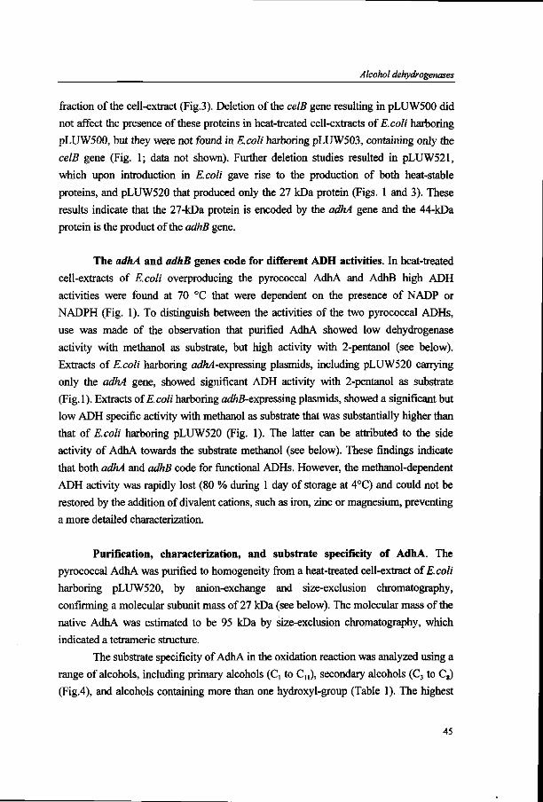
Alcohol dehydrogenases
fraction of the cell-extract (Fig.3). Deletion of the celB gene resulting in pLUW500 did
not affect the presence of these proteins in heat-treated cell-extracts of E.coli harboring
pLUW500, but they were not found in E.coli harboring pLUW503, containing only the
celB gene (Fig. 1; data not shown). Further deletion studies resulted in pLUW521,
which upon introduction in E.coli gave rise to the production of both heat-stable
proteins, and pLUW520 that produced only the 27 kDa protein (Figs. 1 and 3). These
results indicate that the 27-kDa protein is encoded by the adhA gene and the 44-kDa
protein is the product of the adhB gene.
The adhA and adhB genes code for different ADH activities. In heat-treated
cell-extracts of E.coli overproducing the pyrococcal AdhA and AdhB high ADH
activities were found at 70 °C that were dependent on the presence of NADP or
NADPH (Fig. 1). To distinguish between the activities of the two pyrococcal ADHs,
use was made of the observation that purified AdhA showed low dehydrogenase
activity with methanol as substrate, but high activity with 2-pentanol (see below).
Extracts of E.coli harboring ac#L4-expressing plasmids, including pLUW520 carrying
only the adhA gene, showed significant ADH activity with 2-pentanol as substrate
(Fig.l). Extracts of E.coli harboring a<#z#-expressing plasmids, showed a significant but
low ADH specific activity with methanol as substrate that was substantially higher than
that of E.coli harboring pLUW520 (Fig. 1). The latter can be attributed to the side
activity of AdhA towards the substrate methanol (see below). These findings indicate
that both adhA and adhB code for functional ADHs. However, the methanol-dependent
ADH activity was rapidly lost (80 % during 1 day of storage at 4°C) and could not be
restored by the addition of divalent cations, such as iron, zinc or magnesium, preventing
a more detailed characterization.
Purification, characterization, and substrate specificity of AdhA. The
pyrococcal AdhA was purified to homogeneity from a heat-treated cell-extract of E.coli
harboring pLUW520, by anion-exchange and size-exclusion chromatography,
confirming a molecular subunit mass of 27 kDa (see below). The molecular mass of the
native AdhA was estimated to be 95 kDa by size-exclusion chromatography, which
indicated a tetrameric structure.
The substrate specificity of AdhA in the oxidation reaction was analyzed using a
range of alcohols, including primary alcohols (C, to Cn), secondary alcohols (C3 to C8)
(Fig.4), and alcohols containing more than one hydroxyl-group (Table 1). The highest
45

Chapter 3
activity of AdhA (41.6 U/mg) was found when 2-pentanol was oxidized. A broad range
of primary alcohols could be oxidized by AdhA and include substrates with 2-10 carbon
atoms (Fig.4). Similarly, AdhA showed activity towards a broad range of secondary
alcohols that on average were faster oxidized than the primary alcohols (Fig.4).
Alcohols with a second or a third hydroxyl-group were less efficiently converted by
AdhA, most likely due to steric hindrance (Table 1).
The highest reduction rate of AdhA was observed with pyruvaldehyde. The
other aldehydes tested, showed only 10% or less of this activity (Table 2).
Secondary alcohols can have two stereoisomers that may show different
enzymatic conversion rates. The stereoselectivity of AdhA was tested using the two
enantiomers of 2-butanol and revealed only a slight preference for the S-2-butanol (21.8
U/mg and 32.3 U/mg with R- and S-2-butanol, respectively).
specific activity (U/mg)
50
•primary l i secondary alcohol | lalcohol
Figure 4. Specific activities of the purified P.fariosus AdhA towards primary and secondary alcohols
Optimum, kinetics and stability of AdhA. The oxidation reaction catalyzed by
AdhA showed a pH optimum between 10 and 11. The aldehyde reduction by AdhA
showed maximal activity in a broad pH range with an optimum at pH 7.5 and activities
that were only 20 % lower at pH 6.7 and pH 9.0. The optimum activity of AdhA was at
90°C, but due to instability of the cofactors at that temperature all other activity
measurements were performed at 70°C. Kinetic properties of AdhA were determined
for both the oxidation and the reduction reaction using the substrate that showed the
highest activity, 2-pentanol and pyruvaldehyde, respectively (Table 3). The results
indicate that AdhA has the highest affinity for pyruvaldehyde and is dependent on the
phosphorylated cofactor for catalysis.
46
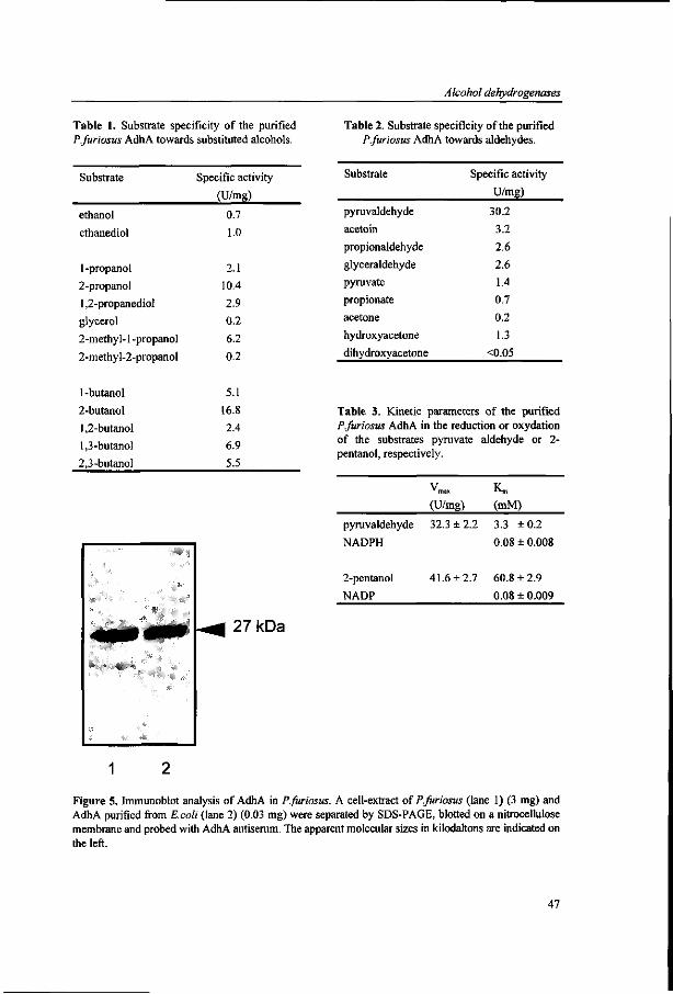
Alcohol dehydrogenases
Table 1. Substrate specificity of the purified P.furiosus AdhA towards substituted alcohols.
Table 2. Substrate specificity of the purified P.furiosus AdhA towards aldehydes.
Substrate Specific activity
(U/mg)
Substrate
ethanol
ethanediol
1 -propanol
2-propanol
1,2-propanediol
glycerol
2-methyl-1 -propanol
2-methyl-2-propanol
1-butanol
2-butanol
1,2-butanol
1,3-butanol
2,3-butanol
0.7
1.0
2.1
10.4
2.9
0.2
6.2
0.2
5.1
16.8
2.4
6.9
5.5
27kDa
Specific activity
U/mg)
pyruvaldehyde
acetoin
propionaldehyde
glyceraldehyde
pyruvate
propionate
acetone
hydroxyacetone
dihydroxyacetone
30.2
3.2
2.6
2.6
1.4
0.7
0.2
1.3
<0.05
Table 3. Kinetic parameters of the purified P.furiosus AdhA in the reduction or oxydation of the substrates pyruvate aldehyde or 2-pentanol, respectively.
(U/mg) (mM)
pyruvaldehyde
NADPH
2-pentanol
NADP
32.3 ±2.2
41.6 ±2.7
3.3 ±0.2
0.08 ± 0.008
60.8 ±2.9
0.08 ± 0.009
1 2
Figure 5. Immunoblot analysis of AdhA in P.furiosus. A cell-extract of P.furiosus (lane 1) (3 mg) and AdhA purified from E.coli (lane 2) (0.03 mg) were separated by SDS-PAGE, blotted on a nitrocellulose membrane and probed with AdhA antiserum. The apparent molecular sizes in kilodaltons are indicated on the left.
47

Chapter 3
Storage of AdhA for several months at 4°C did not affect the enzyme activity
significantly. The resistance to thermal inactivation of AdhA at higher temperatures was
analyzed and half-life values of 150 h, 22.5 h and 25 min were found at temperatures of
80°C, 90°C and 100°C, respectively.
The presence of ADH activity in P.furiosus. Activity measurements of AdhA
were performed in cell-extracts of P.furiosus using 2-pentanol as substrate and showed
a slow, but significant oxidation rate of this substrate (0.27 U/mg), which would
correspond with approximately 0.7 % of the total soluble protein fraction. To further
confirm the presence of AdhA in P.furiosus, immunoblot experiments were performed
using antibodies raised against the AdhA purified form E.coli (Fig. 5). The results show
that AdhA is produced in P.furiosus grown on cellobiose and confirm that its size on
SDS-PAGE is identical to that of the AdhA produced by E.coli.
Discussion
In P.furiosus two tandem ADH genes, designated adhA and adhB, have been identified
that were positioned adjacent and in opposite orientation of the celB gene. E.coli
harboring plasmids that contained these two genes overproduced (i) AdhB, an iron-
containing ADH, that rapidly lost its activity, and (ii) AdhA, a short-chain ADH, that
was characterized in detail. AdhA was found to be produced in an active form in
P.furiosus as could be concluded from both activity measurements and immunological
detection. Overexpression of the adhA and adhB genes in E.coli was not controlled by
the IPTG-inducable vector-located /ac-promoter indicating that the region upstream of
the genes is involved in transcription initiation, as was previously found for the celB
gene (22).
The deduced primary sequence of AdhA from P.furiosus showed significant
homology with a number of short-chain ADHs, but to the best of our knowledge it
represents the first described archaeal member of this ADH family. Like all other short-
chain ADHs characterized to date, the pyrococcal AdhA contains the predicted
coenzyme-binding site at the N-terminus surrounded by highly conserved residues (Fig.
2) (16, 17). Alignment of the deduced amino acid sequence of the pyrococcal AdhA
with other short-chain ADHs showed that all other known features of short-chain
ADHs, including Asp59, Lysl84, and Tyrl80 as ionizable group, are conserved in this
48

Alcohol dehydrogenases
archaeal AdhA (Fig. 2) (16, 17). Additionally, the conservation of a large number of
glycine residues (9 of 19 residues) was noted, indicating that the conformational
conservation found for short-chain ADHs from bacterial and eucaryal origin can be
extended to the domain of Archaea (16).
The deduced primary sequence of the second ADH from P.fiiriosus, AdhB,
shows high homologies with members of the group of iron-containing ADHs, including
that of Thermococcus AN1. This group is characterized by the size of its primary
product of approximately 385 residues, and by the presence of four conserved histidine
residues that were also found in AdhB (17). Three of these histidine residues are
predicted to be involved in the iron-binding, which appears to be involved in the
catalytic activity as has been demonstrated for the iron-containing ADH from
Zymomonas mobilis (17). Remarkably, both archaeal representatives of the iron-
containing ADH family showed an additional proline residue within the putative iron-
binding motif the function of which remains to be established. The instability of the
pyrococcal AdhB produced by E.coli prevented a detailed characterization and
comparison with the other archaeal ADHs.
The functional expression of the P.fiiriosus adhA gene in E.coli and the stability
of its product AdhA allowed for a simple two-step purification of this short-chain ADH
and a more detailed biochemical characterization. For the conversion of alcohols to the
corresponding aldehyde as well as for the reversed reaction, AdhA could only use the
phosphorylated cofactor NADP or NADPH. AdhA showed a broad substrate specificity
with primary and secondary alcohols and revealed an optimum for substrates with a
carbon chain of 5 residues. A preference was observed for the secondary alcohols that
are converted faster than the corresponding primary alcohol.
The pyrococcal AdhA has a half-life value of 150 h at 80°C and is the most
thermostable short-chain ADH known to date. In contrast, the stability of the iron-
containing AdhB produced by E.coli was found to be low as reflected by its rapid loss
of activity, which prevented its biochemical characterization. This instability may be
explained by the loss iron, as has been described for the other archaeal iron-containing
ADH (14, 15). However, attempts to restore the activity by addition of iron or other
divalent cations were not successful.
The physiological function of AdhA is most likely the reduction of the
aldehyde, since AdhA displayed a higher affinity for the aldehydes than for the alcohols
(Table 3). The high conversion rate found with pyruvaldehyde suggests that the natural
substrate of AdhA in P.fiiriosus may have a structure similar to this aldehyde. The
49

Chapter 3
conversion of the aldehyde to the corresponding alcohol may be a means to remove
reduction equivalents and to regenerate the cofactor. Alternatively, or combined with
this role as electron sink, AdhA and possibly AdhB may be involved in the metabolism
of ß-linked glucose polymers. This suggestion is based on the localization of the adhA
and adhB genes in the vicinity of the lamA and celB genes, which are coordinately
expressed and whose products are involved in carbohydrate metabolism of ß-linked
glucose polymers in P.fiiriosus (7, 22, 23). If so, this would contrast with Thermococcus
sp. where the iron-containing ADHs are induced during growth on proteins and peptides
without elemental sulphur (14). Under these conditions an excess of reduction
equivalents may be disposed in the form of alcohols by the reduction of the aldehydes
that are produced by different aldehyde ferredoxin oxidoreductases (14).
In conclusion, we have identified two P.furiosus genes that encode functional
NAD(P)-dependent alcohol dehydrogenases belonging to ADH superfamily and include
a short-chain and an iron-containing ADH. The observed conserved primary structure of
the pyrococcal AdhA, suggests the presence of a short-chain ADH in the last common
ancestor, i.e before the splitting of the phylogenetic tree.
Acknowledgements.
We thank Tony van Kampen and Ans Geerling for assistance with the sequence
analysis. Part of this work was supported by contracts BIOT-CT93-0274 and BIOT-
CT96-0488 of the European Union.
References
1. Cannio, R., G. Fiorentino, P. Carpinelli, M. Rossi and S. Bartolucci. 1996. Cloning and overexpression in Escherichia coli of the genes encoding NAD-dependent alcohol dehydrogenase from two Sulfolobus species. J. Bacteriol. 178:301-305.
2. Cowan, D. A. 1992. Biotechnology of the Archaea. Trends Biotech. Sei. 10:315-323. 3. Daniels, L. 1993. Biochemistry of metanogenesis, p. p. 41-112. In M. Kates et al. (ed.), The
biochemistry of Archaea (Archaeabacteria), Elsevier, Amsterdam. 4. Devereux, J., P. Haeberli and O. Smithies. 1990. A comprehensive set of sequence analysis
programs for the VAX. Nucl. Acids Res. 12:387-395. 5. Fiala, G. and K. O. Stetter. 1986. Pyrococcus fiiriosus sp. nov. represents a novel genus of marine
heterotrophic archaebacteria growing optimally at 100°C. Arch. Microbiol. 145:56-61. 6. Gibson, T. J. 1984. Studies on the Epstein-Barr-Virus genomeed. Cambridge, UK.
50

Alcohol dehydrogenases
7. Gueguen, Y., W. G. B. Voorhorst, J. van der Oost and W. M. de Vos. 1997. Molecular and biochemical characterization of an endo-ß-l,3-glucanase of the hyperthermophilic archaeon Pyrococcus furiosus. J. Biol. Chem. (in press).
8. Kengen, S. W. M., F. A. M. de Bok, N. D. van Loo, C. Dijkema, A. J. M. Stams and W. M. de Vos. 1994. Evidence for the operation of a novel Embden-Meyerhof pathway that involves ADP-dependent kinases during sugar fermentation by Pyrococcus furiosus. J. Biol. Chem. 269:17537-17541.
9. Kengen, S. W. M., E. J. Luesink, A. J. M. Stams and A. J. B. Zehnder. 1993. Purification and characterization of an extremely stable ß-glucosidase from the hyperthermophilic archaeon Pyrococcus furiosus. Eur. J. Biochem. 213:305-312.
10. Kengen, S. W. M., A. J. M. Stams and W. M. de Vos. 1996. Sugar metabolism of hyperthermophiles. FEMS Microbiol. Rev. 18:119-137.
11. Laemmli, U. K. 1970. Cleavage of structural proteins during the assembly of the head of bacteriophage T4. Nature 227:680-685.
12. Leatherbarrow, R. J. ( 1987). ENZFITTER. In Cambridge, UK: Elsevier-Biosoft. 13. Li, D. and K. J. Stevenson. 1997. Purification and sequence analysis of a novel
NADP(H)-dependent type III alcohol dehydrogenase from Thermococcus strain AN1. J.Bacteriol. 179:4433-4437.
14. Ma, K., H. Loessner, J. Heider, M. K. Johnson and M. W. W. Adams. 1995. Effects of elemental sulfur on the metabolism of the deep-sea hyperthermophilic archaeon Thermococcus strain ES-1: characterization of a sulfur-regulated, non-heme iron alcohol dehydrogenase. J. Bacteriol. 177:4748-4756.
15. Ma, K., F. T. Robb and M. W. W. Adams. 1994. Purification and characterization of NADP-specific alcohol dehydrogenase and glutamate dehydrogenase from the hyperthermophilic archaeon Thermococcus litoralis. Appl. Environ. Microbiol. 60:562-568.
16. Persson, B., M. Krook and H. Jornvall. 1991. Characteristics of short-chain alcohol dehydrogenases and related enzymes. Eur. J. Biochem. 200:537-543.
17. Reid, M. F. and C. A. Fewson. 1994. Molecular characterization of microbial alcohol dehydrogenases. Crit. Rev. Microbiol. 20:13-56.
18. Saitou, R. and R. Nei. 1987. A Neigbour-joining method: A new method for reconstructing phylogenetic trees. Mol. Biol. Evol. 44:406-425.
19. Sambrook, J., E. F. Fritsch and T. Maniatis. 1989. Molecular Cloning. A Laboratory Manual, 2nd ed. Cold Spring Harbor Laboratory Press, Cold Spring Harbor, NY.
20. Schönheit, P. and T. Schäfer. 1995. Metabolism of hyperthermophiles. World J. Microbiol. Biotechn. 11:26-57.
21. van de Peer, Y. and R. de Wachter. 1994. Treecon for Windows: A software package for teh construction and drawing of evolutionary trees for Microsoft Windows environment. Comput. Applic. Biosci. 10:569-570.
22. Voorhorst, W. G. B., R. I. L. Eggen, E. J. Luesink and W. M. de Vos. 1995. Characterization of the celB gene coding for ß-glucosidase from the hyperthermophilic archaeon Pyrococcus furiosus and its expression and site-directed mutation in Escherichia coli. J. Bacteriol. 177:7105-7111.
23. Voorhorst, W. G. B., Y. Guenguen, G. Schut, I. Dahlke, M. Thomm, J. van der Oost and W. M. de Vos. 1997. Transcriptional regulation in the hyperthermophilic archaeon Pyrococcus furiosus: Coordinated expression of divergently transcribed genes in response to ß-linked glucose polymers. Submitted for publication
51

Chapter 3
52

Endo-ß-1,3-gIucanase
Chapter 4
Cloning and Sequencing of the lamA Gene from the
Hyperthermophilic Archaeon Pyrococcus furiosus Coding
for an Endo-ß-l,3-Glucanase and Its Expression and
Site-directed Mutation in Escherichia coli
Yannick Gueguen, Wilfried G. B. Voorhorst, John van der Oost,
and Willem M. de Vos
Journal of Biological Chemistry, 1997, 272, 31258-31264
53

Chapter 4
Abstract
We report the molecular characterization of the first endo-ß-l,3-glucanase from an archaeon. Pyrococcus furiosus is a hyperthermophilic archaeon that is capable of saccharolytic growth. The isolated lamA gene encodes an extracellular enzyme that shares homology with both endo-ß-1,3- and endo-ß-l,3-l,4-glucanases of the glycosyl hydrolase family 16. After deletion of the N-terminal leader sequence, a lamA fragment encoding an active endo-ß-l,3-glucanase was overexpressed in E. coli using the T7-expression system. The P. furiosus endoglucanase was purified and showed highest activity on the ß-1,3 glucose polymer laminarin, but has also activity on the ß-1,3-1,4 glucose polymers lichenan and ß-glucan. The enzyme shows optimal activity between 100 and 105 °C and was demonstrated to be the most thermostable endo-ß-1,3-glucanase described up to now. Site-directed mutagenesis was performed to study the role of an additional methionine residue, in the predicted active site, found only in glycosyl hydrolases of the family 16 that show predominantly endo-ß-l,3-glucanase activity. The deletion of this methionine did not change the substrate specificity of the endoglucanase, but it did cause a severe reduction in its catalytic activity, suggesting a structural role of this residue in constituting the active site. HPLC analysis showed in vitro hydrolysis of laminarin by the endo-ß-l,3-glucanase proceeds more efficiently in combination with an exo-ß-glycosidase from Pyrococcus furiosus (CelB). This most probably reflects the physiological role of both enzymes: co-operation during growth ofPyrococcus furiosus on ß-glucans.
Introduction
ß-l,3-Glucanases are widely distributed among bacteria, fungi and higher
plants. They are classified as exo-ß-l,3-glucanases (ß-l,3-glucan glucanohydrolase
[EC 3.2.1.58]) and endo-ß-l,3-glucanases (ß-l,3-glucan glucanohydrolase [EC 3.2.1.6
or EC 3.2.1.39]). Distinct physiological roles of ß-1,3-glucanases have been proposed.
In plants, involvement in cell differentiation and defence against fungal pathogens has
been suggested (1). In fungi, ß-1,3-glucanases seem to have different functions in
morphogenetic processes, ß-glucan mobilization, and fungal pathogen-plant
interactions (2). Recently, the first metazoan ß-1,3-glucanase, that may be involved in
the early embryogenesis, has been described (3). In bacteria, a metabolic function has
been reported for endo-ß-l,3-glucanases and endo-ß-l,3-l,4-glucanases (4). Both
types of bacterial enzymes are polysaccharide endohydrolases with closely related
54

Endo-ß-1,3-glucanase
specificities (5,6). ß-l,3-glucanases hydrolyse 1,3-ß-glucosyl linkages, but usually
require a region of unsubstituted, contiguous 1,3-ß-linked glucosyl residues. In
contrast, ß-l,3-l,4-glucanases catalyse the hydrolysis of 1,4-ß-glucosyl linkages, only
when the glucosyl residue itself is linked at the (0)3 position (7).
Genes encoding bacterial ß-1,3 and ß-l,3-l,4-glucanases have been cloned and
sequenced from different Bacillus species (8-16), Fibrobacter succinogenes (17, 18),
Cellvibrio mixtus (19), Thermotoga neapolitana (20), Ruminococcus flavefaciens (21,
22), Oerskovia xanthineolytica (23), Clostridium thermocellum (24, 25, 26, 27), and
Rhodothermus marinus (28). All bacterial endo-ß-1,3-glucanases (laminarases) known
to date share sequence similarity with endo-ß-l,3-l,4-glucanases (lichenases) and
have been classified in the same family 16 of glycosyl hydrolases (29, 30, 31). On the
other hand, eukaryal endo-ß-l,3-l,4-glucanases and endo-ß-1,3-glucanases have been
classified in the family 17 of glycosyl hydrolases. However, the first metazoan ß-1,3-
glucanase, described from a sea urchin, shares homology with both ß-1,3 and ß-1,3-
1,4-glucanases of glycosyl hydrolases family 16 (3).
Presently, no ß-specific endoglucanases have been reported in the Archaea, the
third domain of life (32, 37). In this study, we report the characterization of an endo-ß
-1,3-glucanase from Pyrococcus furiosus, a heterotrophic hyperthermophilic archaeon
that is able to grow optimally at 100°C on a wide range of carbohydrate substrates
including starch, maltose, cellobiose (32, 33). P. furiosus was found to contain several
hydrolytic enzyme activities related to sugars degradation (34) and utilizes a modified
Embden-Meyerhof Pathway, which involves at least three unique enzymes: two ADP-
dependent kinases (35), and a glyceraldehyde-3-phosphate: ferredoxin oxidoreductase
(36). However, P. furiosus cannot grow on cellulose or carboxymethylcellulose and
nothing has been described concerning the growth on other ß-linked carbohydrates
substrates such as laminarin, ß-glucan and lichenan (32, 37).
The P. furiosus lamA gene was characterized and found to encode a secreted
endo-ß-l,3-glucanase belonging to the glycosyl hydrolyse family 16. Further
characterization of the overproduced catalytic domain of this enzyme revealed that it
was the most thermostable endo-ß-1,3-glucanase detected so far. A mutant form of the
catalytic domain was obtained after site directed mutagenesis of the lamA gene and
revealed that a conserved methionine residue is required for full activity. Based on the
specificity of the endo-ß-1,3-glucanase and the here described capacity of P. furiosus
to grow on laminarin, a role for this extracellular enzyme during the fermentation of
ß-l,3-linked glucose polymer is proposed.
55

Chapter 4
Experimental procedures
Organisms and growth conditions- P. furiosus (DSM 3638) was used in this
study (32). E. coli BL21 (DE3) harboring pLysE was used as host strain for the
recombinant plasmid of pGEF (38, 39). E. coli TGI was also used as host strain for
the cloning vector pLUW530 and pLUW531 (40). E. coli was grown in L broth
medium in a rotary shaker at 37°C. Ampicillin was added to L broth in a final
concentration of 100 u,g/ml. Isopropyl-ß-D-thiogalactopyranoside (IPTG) was added
in a final concentration of 0.4 mM, for the induction of gene expression.
Cloning of the endo-ß-l,3-glucanase gene- A 4.8 kb fragment containing the
lamA gene coding for P. furiosus endo-ß-l,3-glucanase was cloned into pTZ19R (41).
Based on its sequence, primers were designed to amplify the lamA gene by the
polymerase chain reaction on a DNA Thermal Cycler (Perkin Elmer Cetus, Norwalk,
Conn.) (Fig. 1). The two primers with Ncol and BamHl restriction sites (in bold),were
the following: BG 194 (5'-GCG CGC CAT GGT CCC TGA AGT GAT AGA AAT
AGAT, sense) and BG 195 (5'-CGC GCG GAT CCT CAA CCA CTA ACG AAT
GAG TA, antisense). In addition to the template and the primers, the 100 ul reaction
mixture contained 0.2 mM dNTPs, Taq DNA polymerase buffer, 5 mM MgCl2, 5 U
Taq DNA polymerase (Life Technologies Inc.) and was subjected to 30 cycles of
amplification (30 s at 94°C, 30 s at 50°C and 30 s at 72°C). The putative signal
peptide of the endoglucanase was excluded to produce the mature enzyme in the
cytoplasm of E. coli. A PCR product with the expected size was digested with Ncol
and BamHl and cloned in a pGEF vector (38), resulting in pLUW530, and
transformed into E. coli TGI and BL21 (DE3), pLysE using standard procedures (42).
Overexpression of the lamA gene and purification of the endo-ß-1,3-
glucanase- An overnight culture of E. coli BL21 (DE3), pLysE harboring pLUW530
was diluted 1:20 and grown until the AgQO reached 0.6. The culture was induced with
0.4 mM of IPTG for 4 h. Cells were harvested by centrifugation, resuspended in
lOOmM sodium citrate buffer (pH 5.5) and sonicated using a Branson sonifier. Cell
debris was removed by centrifugation (10,000 x g for 10 min). The resulting
supernatant was heated for 30 min at 70°C and precipitated proteins were removed by
another centrifugation. The supernatant was subsequently dialyzed against Tris-HCl
buffer (20 mM, pH 8.0), loaded on a Q-Sepharose Fast Flow (Pharmacia) column (5 x
56

Endo-ß-1,3-glucanase
25 cm) that was equilibrated with the same buffer. Bound proteins were eluted by a
linear gradient of NaCl (0 to 1 M in Tris-HCl buffer). Active fractions eluted around
0.5MNaCl.
Protein samples were analysed by sodium dodecyl sulphate Polyacrylamide gel
electrophoresis (SDS-PAGE) using the method of Laemmli (43). Proteins samples for
SDS-PAGE were prepared by heating for 10 min at 110°C in an equal volume of
sample buffer (0.1 M citrate-phosphate buffer, 5% SDS, 0.9% 2-mercaptoethanol,
20% glycerol, pH 6.8).
ß-Glucanase assay, protein determination and western blot- The standard
assay for ß-glucanase activity was carried out at 80°C, for 10 min, using 0.5% (w/v)
laminarin (Sigma Chemical Co.) as substrate in 0.1 M phosphate
(NaH2P04/K.2HP04) buffer (pH 6.5). The reducing sugars released were detected by
the dinitrosalicylic acid (DNS) method, with glucose as a standard (44). Enzyme and
substrate blanks were also included. 1 U enzyme activity is defined as the amount of
enzyme required to release 1 umol of reducing sugars/min. Protein concentrations
were determined by the method of Bradford, with BSA as the standard (45).
Antibodies were raised against LamA by injecting enzyme purified from E.
coli into rabbits. The proteins were separated by SDS-PAGE and blotted onto
nitrocellulose filters. These were incubated with serum containing antibodies
(Dilution 1/1000) raised against LamA. The reaction of proteins with the antibodies
was detected using the western blot Alkaline Phosphatase system (Promega).
Temperature and pH optima, thermal stability, kinetic parameters- The
temperature optimum was determined by running the standard assay at temperatures
from 40°C to 115°C. The pH optimum of the enzyme was determined by running the
standard assay at 80°C, using citrate-phosphate (citric acid/NaH2P04) buffer (0.1 M)
and phosphate (Na^PC^/K^HPC^) buffer (0.1M) for the pH range 4-7 and 6-8,
respectively. Thermostability was determined using diluted enzyme (0.14 mg/ml pure
enzyme was diluted 10 times in 200 itiM Tris-HCl buffer, pH 6.5), incubated at 80°C,
90°C and 100°C. Samples were taken at time intervals and residual activity was
determined by the standard assay at 80°C. Michaelis-Menten constants were
determined from Lineweaver-Burk representations of data obtained by determining
the initial rate of laminarin and lichenan hydrolysis under the assay conditions
described above and using a range of 0.25 to 10 mg of substrate per ml.
57
Chapter 4
Mutagenesis of the lamA gene- The plasmid pLUW500 carrying the lamA
gene was used as a template for mutagenesis following the splicing by overlap
extension PCR (46). The first step comprised the use of primers introducing the
mutation and the two flanking primers (BG 194 and BG195), described above. The
two mutagenesis primers were the following: BG 227 (5'-GCC AAG GAA TTC TAT
GTC TAT TTC TCC ACA ATT TGG C, antisense) and BG 226 (5'-GAA ATA GAC
ATA GAA TTC CTT GGC CAT GAG CAA, sense). The second step used the
overlapping products of the first two PCR reactions as a template, and the flanking
primers (BG 194, BG 195) to yield the lamA gene with the mutation. In order to
simplify detection of mutants, an £coRI site (in bold in the primers) was created. The
amplified DNA was cloned as HindllVBamHI fragment, into the lamA gene in
pLUW530. The mutation was checked by EcoKL digestion and DNA sequence
analysis. The resulted plasmid (pLUW531) was transformed to E. coli BL21 (DE3),
pLysE for overproduction of the protein.
DNA sequencing- Nucleotide sequencing was performed by the
dideoxynucleotide chain termination method (47) with a Li-Cor automatic sequencing
system (model 4000L). Computer analysis of sequencing data was performed by using
the PC/GENE program version 5.01 (IntelliGenetics Inc., Moutain view, Calif.) and
the GCG package version 7.0 at the CAOS/CAMM Centre of the University of
Nijmegen.
Nucleotide sequence accession number- The nucleotide sequence reported
has been submitted to the GenBank/EMBL Data Bank with the accession number
AF013169.
Results
Cloning and sequencing of the lamA gene encoding an endo-ß-1,3-
glucanase from P. furiosus- The nucleotide sequence of a part of the 4.8 kb DNA
insert from pLUW500 (41, 48) revealed the presence of an 894 bp open reading frame
(Fig. 1). The encoded 297-amino acid protein is homologous to a family bacterial ß-
1,3 and ß-l,3-l,4-endoglucanases (see below) and contains a putative signal peptide
of 20 residues (49). The molecular mass of the predicted mature protein is 31,550 Da
58

Endo-ß-1,3-glucanase
and the estimated isoelectric point 5.15. In order to overproduce this ß-glucanase, the
lamA gene was amplified by the PCR method and cloned into pGEF (38) under
control of the T7 promotor (39). The resulting plasmid, containing the catalytic
domain of the endo-ß-1,3-glucanase (starting at residue 35 and called LamA), was
named pLUW530. The plamid pLUW530 harboring lamA was checked by DNA
sequence analysis.
pLUW500
celB adhA adhB lamA
pLUW530
pLUWS31
BG19S
Figure 1. Genetic organisation of the lamA region. This DNA fragment was originally cloned as a fragment from chromosomal DNA of P. furiosus and inserted into the EcoRI / PstI sites of pTZ19R (41). Primers BG194 and BG195 used in the cloning of the lamA gene and primers BG226 and BG227 used in site-directed mutagenesis of LamA are indicated by arrows. Relevant restriction sites are indicated. H, Hindill; E, EcoRI; N , Ncol; B, BamHl; P, Pstl.
Primary structure comparison- Comparison of the deduced primary strucutre
of lamA with enzymes present in the GenBank database indicated highest similarity
with representatives of the family 16 of glycosyl hydrolases, which includes bacterial
endo-ß-1,3- and endo-ß-l,3-l,4-glucanases, Streptomyces coelicolor agarase and
Alteromonas carrageenovora K-carrageenase. Scores of 52.4 and 46.9 % identity were
found with the ß-glucanases of T. neapolitana and O. xanthineolytica, respectively.
Multiple alignments of the deduced amino acid sequence of P. fiiriosus lamA and
other ß-glucanases showed many conserved amino acids (Fig. 2) including glutamate
170 and glutamate 175 (P. furiosus amino acid numbering is used throughout the
paper). The results of structural analysis, mutagenesis (50, 51, 52, 53) combined with
inhibitor attachment (52, 54) studies of Bacillus ß-glucanases showed the direct
involvement of these two carboxylic acids in catalysis. Glutamate 175 corresponds to
the general acid and glutamate 170 to the catalytic nucleophile or plays a role in
59

Chapter 4
electrostatic stabilization of an oxocarbonium intermediate ion (55). Moreover, the
crystal structure of the hybrid Bacillus ß-l,3-l,4-ß-glucanase (52) showed the
presence of a calcium binding site on the surface of the enzyme. Experiments
demonstrated that calcium, bound to the hybrid Bacillus enzyme, stabilizes the three-
dimensional structure of the protein leading to increased thermal stability (56, 57).
The calcium atom is bound (52) on the convex face of the protein, to the backbone
carbonyl oxygens atoms of proline [9], glycine 78 [45] and aspartate 287 [207] and to
the carboxylate oxygen of aspartate 287 [207] (italic numbers correspond to Bacillus
hybrid ß-glucanase numbering (52)). This aspartate 287 residue is conserved among
most of the ß-glucanases of the family 16 (Fig. 2). The P. furiosus ß-l,3-glucanase
also does contain this aspartate residue, and indeed was found to be protected by
calcium against thermal inactivation (data not shown).
P.furiosus T.neapolitana O.xan thineolyeica C.thermocellum l R.marinus B.licheniformis B.subtilis C.thermocellum 2 B.brevis B.macerans
47
WRLIWHDEPB.
WQLVWSQEFD...DGVIDPNVWNFEIGNGHAKGIPGWGNAELEYYTD...KNAFVENGCLVIEARKEQVSD.EYGTYDYTSARITTEGKF
ESLAWSDEFDGAAGSAPNPDVWNHETGAG GWGHAELQNYTTSRVNSALDGQGNLVITALQESDGS YTSARLTTQGNV
HRLVWSDEPNG. . .SEINMANWSYDDPTN GRWNGEVQSYTQ. . .NNAYIKDGALVIEARKEDITEPSGETYHYTSSKLITKGKK
WELVWSDEFDY. .SGLPDPEKWDYDVGGH GWGNQELQYYTRARIENARVGGGVLIIEARHEPY EGREYTSARLVTRGKA
TGGSFYEPFNNYNTGL WQKAD GYSKGNMFNCTWRANNVSMTSLGEMRLSLTSPS YNKFDCGENRSV
TGGSFFDPFNGYNSGF WQKAD GYSNGNMFNCTWRANNVSMTSLGEMRLALTSPA YNKFDCGENRSV
VNTPFVAVFSNFDSSQ WEKAD WAMGSVFNCVWKPSQVTF.SNGKMILTLDREY GGSYPYKSGEYRTKS
MGTAFYESFDAFDDER WSKAG VWTHGQMFNATWYPEQVTA. . DGLMRLTIAKKTTSARN YKAGELRTNDFY
AGSVFWEPLSYFNRST WEKAD GYSMGGVFNCTWRANNVNFTNDGKLKLGLTS S A YNKFDCAEYRST
P. furiosus
T.neapolitana
O.xanthineolytica C.thermocellum 1 R.m nus
B.licheniformis B.subtilis C.thermocellum 2 B.brevis B.macerans
P.furiosus T.neapolitana O.xanthineolytica C.thermocellum 1 R.marinus B.1icheniformis B.subtilis C.thermocellum 2 B. brevi s B.mac
EIK.YGKIEIRAKLPKGKGIWPALWMLGNN. . IGEVGWPTCGEIDIMEMLGHDTRTVLRTAHGPGYSGGASIGV. .AYHLPEEVPDFSED
QPQ.FGRIEARIQIPRGQGIWSAFWMVGAN..LPDTPWPTSGEIDIMENVGNAPHEVHGTVHGPGYSG..DNGIMGTYQHPQGW.SFADD
SWK.YGKFEIRAKMPQGOGIWPAIWMMPEDEPFYGT.WPKCGEIDIHBLLGHEPDKIYGTIHF.GEPH....ESQGTYTLPEGQ.TFADD
SWT.YGRFEIRARLPSGRGTWPAIWMLPDRQTYGSAYHPDNGEIDIMEHVGFNPDWHGTVHTKAYNHL.LGTQRGGSIRVPTART.. . D
QTYGYGLYEVNMKPAKNVGIVSSFFTYTG..PTDGTPW.
QTYGYGLYEVRMKPAKNTGIVSSFFTYTG..PTDGTPW.
FF GYGYYEVRMKAAKNVGIVSSFFTYTG..PSDNNPW.
H...YGLFEVSMKPAKVEGTVSSFFTYTGEWDWDGDPW.
QTYGYGLYEVSHKPAKNTGIVSSFFTYTG..PAHGTQW.
DEIDI. EFLGKDTTKVQFNYYTNGVGN HEKIVNLGFDAANS
DEIDI. KFLGKDTTKVQFNYYTNGAGN HEKIVDLGFDAANA
DEIDI. EFLGKDTTKVQFNWYKNGVGG NEYLHNLGFDASQD
DEIDI. EFLGKDTTRIQFNYFTNGVGG NEFYYDLGFDASES
DEIDI.EFLGKDTTKVQFHYYTMGVGG HEKVISLGFDASKG
216 297
FHVFGIVWYPDKIKWYVDGTFYHEV...TKEQVEAMGYE.WV...FDKPFYIILNLAVGGYWPGNPD.ATTPFPAKMWDYVRVYSFVSG
FHVFSIEWDENEVEWYVDGQLYHVL...SKDELAELGLE.WV...FDHPFFLILNVAMGGYWPGYPD.ETTQFPQRMYIDYIRVYKDMNP
FHTFGIDWTPGEITWLVDGQEYHRV TTADVGANQWV. . . FDQPFFLILNVAIGGQWPGNPD. ATTPFPQQMKVDYVRVYDNATQ
FHVYSIEWEPGEIRWYIDGKLYHVANDWYSRDPYLADDYTYPA.PFDQNFFLILNISVGGGWPGYPD.ETTVFPQQMVVDYVRVYQKDKY
FHVYAIEWTPEEIRWFVDDSLYYRF. . . .PNERLTDPEADWRHWPFDQPFHLIMNIAVGGAWGQQQGVDPEAFPAQLWDYVRVYRWVE .
YHTYAFDWQPNSIKWYVDGQLKHTATTQIPQTPGKIHMNLWNGAGVDEWL GSYNG VTPLSRSLHWVRYTKR. . .
YHTYAFDWQPNSIKWYVDGQLKHTATNQIPTTLGKIMN1NLWNGTGVDEWL GSYNG VNPLYAHYDWVRYTKK . . .
FHTYGFEWRPDYIDFYVDGKKVYRGTRNIPVTPGKIMMNLWPGIGVDEWL GRYDGR TPLQAEYEYVKYYPHGVP
FNTYAFEWREDSITWYVNGEAVHTATENIPQTPQKIMMNLWPGVGVDGWT GVFDGD NTPVYSYYDWVRYTPLQNY
FHTYAFDWQPG YIKWYVDGVLKHTATANI PSTPGKIMMNLWHGTGtoDWll GSYNG AHPLYAEYDWVKYTSN. . .
Figure. 2. Alignment of P. furiosus LamA with other members of family 16 of glycosyl hydrolases. Sequences were deduced from the following accession numbers: T. neapolitana laminarase (Z47974); O. xanthineolytica ß-1,3-glucanase (U56935); C. the'rmocellum 1 ß-l,3(4)-glucanase, licA (X89732); R. marinus ß-glucanase (U04836); B. licheniformis ß-l,3-l,4-glucanase (X57279); B. subtilis ß-glucanase (X00754); B. macerans ß-1,3-1,4-glucanase (Z25874); C. thermocellum 2 ß-l,3-l,4-glucanase, ließ (X63355); B. brevis ß-l,3-I,4-glucanase (M84339). The numbering of the P. furiosus LamA is indicated. Conserved residues are indicated in bold. The asterisks above the sequences indicate the conserved glutamate residues that have been identified as acid-base catalyst and active site nucleophile. Underlined and boxed regions correspond to ß-strands and a-helix, respectively, according to B. licheniformis/B. macerans hybrid ß-l,3-M-glucanase secondary structure (50, 52).
60
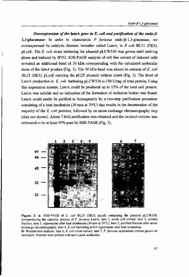
Endo-ß-1,3-glucanase
Overexpression of the lam A gene in E. coli and purification of the endo-ß-
1,3-glucanase- In order to characterize P furiosus endo-ß-1,3-glucanase, we
overexpressed its catalytic domain, hereafter called LamA, in E coli BL21 (DE3),
pLysE. The E. coli strain harboring the plasmid pLUW530 was grown until mid-log
phase and induced by IPTG. SDS-PAGE analysis of cell free extract of induced cells
revealed an additional band of 30 kDa corresponding with the calculated molecular
mass of the lamA product (Fig. 3). The 30 kDa band was absent in extracts of E. coli
BL21 (DE3), pLysE carrying the pGEF plasmid without insert (Fig. 3). The level of
LamA production in E. coli harboring pLUW530 is 150 U/mg of total protein. Using
this expression system, LamA could be produced up to 15% of the total cell protein.
LamA was soluble and no indication of the formation of inclusion bodies was found.
LamA could easily be purified to homogeneity by a two-step purification procedure
consisting of a heat incubation (30 min at 70°C) that results in the denaturation of the
majority of the E. coli proteins, followed by an anion exchange chromatography step
(data not shown). About 7-fold purification was obtained and the isolated enzyme was
estimated to be at least 95% pure by SDS-PAGE (Fig. 3).
6 i
*
«*
r •
f
«
7
l
' 1
' - ; < * i
^ ^ ^
• ^ i * •
Figure. 3. A- SDS-PAGE of E. coli BL21 (DE3), pLysE containing the plasmid pLUW530, overproducing the catalytic domain of P. furiosus LamA. lane 1, crude cell extract; lane 2, soluble fraction; lane 3, supernatant after heat incubation (30 min at 70°C); lane 5, purified fraction after anion exchange chromatography, lane 4, E.coli harboring pGEF supernatant after heat incubation. B- Western blot analysis, lane 6, E. coli crude extract; lane 7, P. furiosus supernatant culture grown on laminarin. Proteins were probed with anti-LamA antibodies.
61
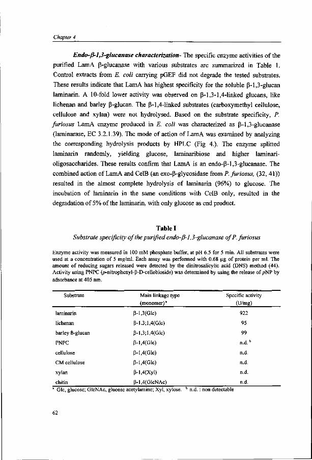
Chapter 4
Endo-ß-l,3-glucanase characterization- The specific enzyme activities of the
purified LamA ß-glucanase with various substrates are summarized in Table 1.
Control extracts from E. coli carrying pGEF did not degrade the tested substrates.
These results indicate that LamA has highest specificity for the soluble ß-l,3-glucan
laminarin. A 10-fold lower activity was observed on ß-l,3-l,4-linked glucans, like
lichenan and barley ß-glucan. The ß-l,4-linked substrates (carboxymethyl cellulose,
cellulose and xylan) were not hydrolysed. Based on the substrate specificity, P.
furiosus LamA enzyme produced in E. coli was characterized as ß-l,3-glucanase
(laminarase, EC 3.2.1.39). The mode of action of LamA was examined by analyzing
the corresponding hydrolysis products by HPLC (Fig 4.). The enzyme splitted
laminarin randomly, yielding glucose, laminaribiose and higher laminari-
oligosaccharides. These results confirm that LamA is an endo-ß-l,3-glucanase. The
combined action of LamA and CelB (an exo-ß-glycosidase from P. furiosus, (32, 41))
resulted in the almost complete hydrolysis of laminarin (96%) to glucose. The
incubation of laminarin in the same conditions with CelB only, resulted in the
degradation of 5% of the laminarin, with only glucose as end product.
Table I Substrate specificity of the purified endo-ß-1,3-glucanase of P. furiosus
Enzyme activity was measured in 100 mM phosphate buffer, at pH 6.5 for 5 min. All substrates were used at a concentration of 5 mg/ml. Each assay was performed with 0.68 |ig of protein per ml. The amount of reducing sugars released were detected by the dinirrosalicylic acid (DNS) method (44). Activity using PNPC (p-nitrophenyl-ß-D-cellobioside) was determined by using the release of pNP by adsorbance at 405 nm.
Substrate
laminarin
lichenan
barley ß-glucan
PNPC
cellulose
CM cellulose
xylan
chitin
Main linkage type
(monomer) *
ß-l,3(Glc)
ß-l,3;l,4(Glc)
ß-l,3;l,4(Glc)
ß-l,4(Glc)
ß-l,4(Glc)
ß-l,4(Glc)
ß-l,4(Xyl)
ß-l,4(GlcNAc)
Specific activity (U/mg)
922
95
99
n.d."
n.d.
n.d.
n.d.
n.d.
Glc, glucose; GlcNAc, glucose acetylamine; Xyl, xylose. b n.d. : non detectable
62

Endo-ß-1,3-glucanase
1
c
/ ~iw
Li l . 'A L
V ,.,......».(-..)
Figure 4. HPLC analysis of?, furiosus LamA and CelB action on laminarin (0.5% w/v). (A) laminarin control, incubation at 80°C for 4 hours; (B) incubation with LamA (5 ug/ml), at 80°C for 4 hours; (C) incubation with CelB (5 ug/ml), at 80°C for 4 hours; (D) incubation with LamA (5 ug/ml) and CelB(5 ug/ml), at 80°C for 4 hours. Samples were analysed on an Aminex HPX-87-H column (Biorad). Identified products are: 1-laminarin; 2-laminari-oligomers; 3-laminaribiose; 4-glucose.
30 40 50 60 70 80 90 100 110 120 Temperature (°C)
Figure 5. Influence of pH and temperature on the activity of the purified endo-ß-1,3-glucanase of P. furiosus. (A) Enzyme activity was measured, at 80°C for 5 min using laminarin as substrate, in 100 mM citrate-phosphate buffer ( • ) at pH 4 to 7 and in 100 mM phosphate buffer ( • ) at pH 6 to 8. The 100% activity corresponds to 935 U/mg of protein. (B) Enzyme activity was measured in 100 mM phosphate buffer, at pH 6.5 for 5 min, using laminarin as substrate. The 100% activity corresponds to 1842 U/mg of protein. Each assays were performed with 0.68 ug of protein per ml.
Figure 6. Thermal stability of the P. furiosus endo-ß-1,3-glucanase. The enzyme was pre-incubated at 80°C ( • ), 90°C ( • ) and 100°C ( • ), in 200 mM Tris-HCl buffer, pH 6.5. Residual activity was measured in 100 mM phosphate buffer, at pH 6.5 for 5 min, using laminarin as substrate. The 100% activity corresponds to 945 U/mg of protein.
63

Chapter 4
The laminarase enzyme activity was investigated at different pH values and
temperatures (Fig. 5). The pH optimum was found to be 6-6.5. The enzyme exhibited
at least 80% of its optimal activity over a rather broad pH range, from 5 to 7 (Fig. 5 A).
The temperature for maximum activity was 100-105°C. The enzyme was almost
inactive at 40°C and began to show significant activity above 60°C (Fig. 5B). Thermal
stability was investigated by incubating the pure enzyme up to 110 h at different
temperatures (Fig. 6). The enzyme retained 100% of its laminarase activity after 110 h
incubation at 80°C. At higher temperatures, thermostability decreased: at 90°C and
100°C, the enzyme showed a half life of 64 h and 19 h, respectively. At 110°C, the
enzyme was rapidly inactivated, with a half life of only 15 min (data not shown).
Site-directed mutagenesis of the P. furiosus endo-ß-l,3-glucanase- To
investigate whether the deletion of methionine at position 174 in LamA, would affect
the ß-1,3 substrate specificity of the enzyme, a mutated lamA gene was constructed in
plasmid pLUW530. Relative activities and kinetic characteristics of mutant and wild-
type enzymes were determined with laminarin and lichenan as substrates (Table 2).
The specific activities of the mutant LamA, on both laminarin and lichenan, are about
10-fold lower than respective wild-type values. Catalytic parameters were determined
at 80°C and pH 6.5 (optimal conditions for the wild-type enzyme). The methionine
deletion led to an enzyme with 13.1% of wild-type Fmax and a slightly higher Km , on
laminarin as substrate. With lichenan as substrate, the mutant LamA has 7.9% of the
wild-type Vmax and the same Km value.
Detection of the endo-ß-l,3-glucanase in P. furiosus- The unexpected
discovery of the endo-ß-l,3-glucanase in P. furiosus prompted us to study the
capacity of this hyperthermophilic archaeon to grow on ß-1,3-linked glucose
polymers. Remarkably, P. furiosus was found to grow on laminarin (0.3% w/v) and
lichenan (0.3% w/v) as substrate (data not shown) and endo-ß-l,3-glucanase activity
was detected in the culture medium. Western blot analysis with immune serum
directed against LamA gave an immunoreactive band with the culture supernatant of
P. furiosus, grown on laminarin as substrate (Fig. 3 ). The size of the product from P.
furiosus is slightly higher (around 1 kDa) than the one produced in E. coli, which is in
agreement with the fact that, we have deleted, during the overexpression step, a N-
terminal fragment of 34 amino acid which is larger than the predicted signal peptide
of 20 amino acid.
64

Endo-ß-1,3-glucanase
Table II
Enzyme activity and kinetic parameters of wild type and mutant LamA of P. furiosus.
Lineweaver-Burk plots were used to determine the kinetic constants. Activity on laminarin and lichenan were measured in 100 mM phosphate buffer, at pH 6.5 for 5 min. Substrates were used at different initial concentrations. Each assay was performed with 0.68 ug of protein per ml. The amount of reducing sugars released were detected by the dinitrosalicylic acid (DNS) method (44).
Substrate
laminarin
ß-U(Glc)
lichenan
P-l,3-l,4(Glc)
Enzyme
wt
mutant
wt
mutant
Specific
Activity2
(U/mg)
1073
111.6
85
7.1
Activity1"
100
10.4
100
8.3
(%) K-m
(mg/ml)
2.8
3.5
4.7
4.9
V b
' max
(%)
100
13.1
100
7.9 ' Enzyme activity was measured in 100 mM phosphate buffer pH 6.5, at 80°C, for 10 min. 5 Percentages of residual activity and Km„ are expressed relative to the wild type for each substrate.
Discussion
In this paper, we describe the overexpression of the lamA gene which encodes
a extremely thermostable endo-ß-l,3-glucanase from the hyperthermophilic archaeon
P. furiosus. The overproduction and purification of the endo-ß-1,3-glucanase LamA
was carried out by cloning a lamA gene fragment into the T7 expression system, after
deletion of the hydrophobic N-terminal sequence (34 amino acids). This is the first
description of an endo-ß-1,3-glucanase from an archaeon and it is the most
thermostable endo-ß-1,3-glucanase known up to now. The second most thermostable
endoglucanase of the family 16 of glycosyl hydrolases characterized, is that of the
thermophilic bacterium R. marinus, which has an optimum temperature of 85°C and
has 85% residual activity after 16 h incubation at 80°C. The optimum temperature of
the P. furiosus ß-glucanase is 100-105°C and the enzyme retained 100% and 50% of
its activity after incubation for 85 h at 80°C and 16 h at 100°C, respectively. The
optimum temperature of the enzyme is near the optimal growth temperature (100°C)
of the P. furiosus strain (35). The enzyme activity has a broad pH optimum around pH
6, which is comparable to other endo-ß-l,3-glucanases and endo-ß-1,3-1,4-
65

Chapter 4
glucanases, with the exception of the B. brevis enzyme, which has optimal activity at
pH 9.0. The P. furiosus ß-glucanase was able to hydrolyse both ß-l,3-glucan and ß-
1,3-1,4-glucan, but not the ß-l,4-linked substrates like cellulose. Bacterial laminarases
and lichenases often yield trisaccharides and tetrasaccharides as the main degradation
products (58). On the other hand, as other bacterial and fungal endo-ß-l,3-glucanases
(2, 26, 28), the endo-ß-l,3-glucanase of P. furiosus, hydrolyses ß-l,3-glucans with
glucose, laminaribiose, and laminaritriose as end products.
All the bacterial laminarases and lichenases sequenced so far have been
classified in the same family 16 of glycosyl hydrolases. The mechanism for ß-
glucanase action must take into account two general considerations (55, 59). The
hydrolysis of the glycosyl bond can proceed via either retention or inversion of the
configuration at the anomeric carbon atom. Since NMR analysis has indicated
retention of configuration in case of the B. licheniformis ß-glucanase (60), the same
stereochemical course is assumed for all other members of the family (61). This
mechanism requires two functional groups of the enzyme to be present in the
appropriate spatial setting. The first either acting as a nucleophile or by providing
electrostatic stabilization, and the second as a general acid. Several experiments with
ß-glucanases from different Bacilli (54, 55) demonstated the role of glutamate 170 as
the catalytic residue involved in the nucleophilic attack on the substrate. Glutamate
175 was demonstrated to be the most likely candidate to function as the general
catalyst (50, 51). As demonstrated by the crystal structure of the B. macerans enzyme
and a substrate analogue (50, 52, 62), the ß-glucanase substrate binds to a pronounced
channel on the molecular surface, in which four residues, glutamate 170, aspartate
172, glutamate 175, and glycine 178, are located on the same ß-strand (52). These
residues are completely conserved in all available sequences of ß-glucanases (Fig. 2).
Interestingly, the alignment suggests that amino acid sequences surrounding the active
site can be divided into two categories (Fig. 2). From glutamate 170 to phenylalanine
176, there is a strict alternation of polar and nonpolar side chains in the catalytic sites
of the endo-ß-l,3-l,4-glucanases. This organization is different in the endo-ß-1,3-
glucanases because of the insertion of an extra methionine residue between isoleucine
173 and glutamate 175. Assuming that both the active site structure and the catalytic
mechanism are similar in a same family, this residue has been proposed to form a ß-
buldge, introducing a kink in the ß-strand and thereby allowing the methionine side
chain to point towards the hydrophobic core and the glutamate 175 to participate in
catalysis (50, 63). This structural rearrangement of the active site most likely will
66
Endo-ß-1,3-glucanase
affect catalysis. Morever, substrate specificity analysis within the glycosyl hydrolyse
family 16 shows that these enzymes can be classified in two subfamilies : (i) a group
of ß-glucanases able to hydrolyse ß-l,3-glucan or both ß-1,3 and ß-l,3-l-4-glucan
(such in the case of P. furiosus and R. marinus) and (ii) a group of ß-1,3-1-4-
glucanases. The structural change brought about by the inserted methionine has been
correlated with the altered specificity, from ß-1,3-1,4 linked substrates to ß-1,3
linkages ones (28, 50). The role of this extra methionine in P. furiosus ß-glucanase
was investigated by site-directed mutagenesis. Removal of methionine 174 in the P.
furiosus ß-glucanase resulted in a decreased enzyme activity by a factor of 10 for both
laminarin and lichenan. However, we did not observe a shift of relative affinity of the
mutant enzyme from ß-l,3-glucan towards ß-l,3-l,4-glucan substrates. Hence,
deletion of methionine 174 affected mostly Kmax , but not Km The results suggest that
the methionine 174 residue may assist in catalysis as a neighbouring group to the
essential glutamate 175 and suggest a structural role of this residue in the constitution
of the active site. However, it does not significantly contribute to specific substrate
binding, as seen by the unchanged Km upon deletion. Moreover, studies of a mutant
B. macerans ß-glucanase in which a methionine has been inserted in the active site
region show it to be enzymatically inactive (50). These results suggest that further
modifications are required to extend the substrate specificity of ß-l,3-glucanases, for
example amino acids responsible for the binding of the different polyaccharide
substrates. Ferrer and co-workers (23) noted that conserved regions of the ß-
glucanases family are rich in aromatic residues, tryptophan in particular. This residue
was demonstrated to play a role in substrate binding of some cellulases (64).
Furthermore, a limited number of tryptophan residues is invariant either in ß-1,3-
glucanases or in ß-l,3-l,4-glucanases [in positions 46, 50, 89, 150, 154, 257, 270],
suggesting a possible function in ß-glucanase substrate specificity of these residues.
Moreover, Juncosa and co-workers (51) proposed, after site directed mutagenesis on
Bacillus licheniformis ß-l,3-l,4-glucanase, a possible role in substrate binding of two
glutamate residues (in position 122 and 203), which are conserved only among ß-1,3-
1,4-glucanases. A better understanding of the basis of the ß-1,3- and ß-1,3-1,4 ß-
glucanase substrate specificity awaits a detailed comparison of the three dimensional
structures of these two classes of enzymes and experimental verifications of the
derived conclusions by protein engineering.
Previously, it has been described that P. furiosus is capable to degrade De
linked glucose polymers like starch and glycogen by the concerted action of oc-
67

Chapter 4
amylase, pullulanase and oc-glucosidase (65). Here, we have demonstrated that, in
addition, P. furiosus is able to grow on ß-linked glucose polymers. Two enzymes are
involved in the hydrolysis of laminarin to glucose : the LamA encoded extracellular
endo-ß-l,3-glucanase and the ß-glucosidase (CelB). The physiological substrate of the
latter endo-ß-l,3-glucanase may be a ß-l,3-linked carbohydrate component of the cell
wall of a variety of marine organisms, such as eukaryotic algae (laminarin) or
methanogenic archaea (pseudopeptidoglycan) (66). As it has been demonstrated in
vitro, the LamA and CelB enzymes are anticipated to catalyse the hydrolysis of sugars
polymers to glucose in a cooperative manner. The extracellular endo-ß-l,3-glucanase
with high-level thermostability could function around the living area of P. furiosus
degrading the ß-1,3 -glucose polymer to smaller oligomers, that are imported into the
cell and hydrolysed to glucose by the exo-glycosidase (CelB). Thus, these two
enzymes could start the catabolic pathway of P. furiosus growing on ß-l,3-glucose
polymer, that could ressemble the natural substrate of the strain in in vivo conditions.
Morever, the latnA gene is located near the ß-glucosidase celB gene and two alcohol
dehydrogenases (Fig. 1) (48). Clustering of the lamA and celB genes may be of
functional significance. The regulation of the celB and lamA genes in P. furiosus is
currently under investigation (48).
Acknowledgements
The authors gratefully acknowledge Dr. J.P. Schanstra and Dr. D.B. Janssen
(Groningen) for providing the expression vector pGEF.
References
1. Grenier, J., Potvin, C , and Asselin, A. (1993). Plant Physiol. 103, 1277-1283 2. Cruz, de la J., Pintor-Toro, J. A., Benitez, T., Llobell, A., and Romero, L. C. (1995) J. Bacteriol.
177, 6937-6945 3. Batchman, E. S., and McClay, D.R. (1996) Proc. Natl. Acad Sei. USA. 93, 6808-6813 4. Watanabe, T., Kasahara, N., Aida, K., and Tanaka, H. (1992) J. Bacteriol. 174, 186-190 5. Chen, L., Fincher, G. B., and Hoj, P.B. (1993) J. Biol. Chem. 268, 13318-13326 6. Varghese, J. N., Garrett, T. P., Colman, P. M., Chen, L., Hoj, P. B., and Fincher, G. B. (1994) Proc.
Natl. Acad. Sei. U. S. A. 91, 2785-2789 7. Parrish, F. W., Perlin, A. S., and Reese, E.T. (1960) Can. J. Chem. 38, 2094-2104 8. Borriss, R., Buettner, K., and Maentsaelae, P. (1990) Mol. Gen. Genet. 222,278-283 9. Bueno, A., Vazquez de Aldana, C. R., Correa, J., Villa, T. G., and de Rey, F. (1990) J. Bacteriol.
172,2160-2167
68

Endo-ß-1,3-glucanase
10. Fiske, M. J., Tobey-Fincher, K. L., and Fuchs, R. L. (1990)7. Gen. Microbiol. 136, 2377-2383 11. Gosalbes, M. J., Perez-Gonzalez, J. A., Gonzalez, R., and Navarro, A. (1991) J. Bacteriol. 173,
7705-7710 12. Hofemeister, J., Kurtz, A., Borriss, R., and Knowles, J. (1986) Gene 49, 177-187 13. Lloberas, J., Perez-Pons, J. A., and Querol, E. (1991) Eur. J. Biochem. 197, 337-343 14. Louw, M. E., Reid, S. J., and Watson, T. G. ( 1 9 9 3 ) ^ / . Microbiol. Biotechnol. 38, 507-513 15. Murphy, N., McConnell, D. J., and Cantwell, B. A. (1984) Nucleic Acids Res. 12, 5355-5367 16. Yahata, N., Watanabe, T., Nakamura, Y., Yamamoto, Y., Kamimiya, S., and Tanaka, H. (1990)
Gene 86, 113-117 17. Erfle, J. D., Teather, R. M., Wood, P. J., and Irvin, J. E. (1988) Biochem. J. 255, 833-841 18. Irvin, J. E., and Teather, R. M. (1988) Appl. Environ. Microbiol. 54,2672-2676 19. Sakellaris, H., Pemberton, J. M., and Manners, J. M. (1993) FEMS Microbiol. Lett. 109, 269-272 20. Dakhova, O. N., Kurepina, N. E., Zverlov, V. V., Svetlichnyi, V. A., and Velikodvorskaya, G. A.
(\WS) Biochem. Biophys. Res. Commun. 194, 1359-1364 21. Flint, H. J., McPherson, C. A., and Bisset, J. (1989). Appl. Environ. Microbiol. 55, 1230-1233 22. Flint, H. J., Martin, J., McPherson, C. A., Daniel, A. S., and Zhang, J. X. (1993). J. Bacteriol. 175,
2943-2951 23. Ferrer, P., Halkier, T., Hedegaard, L., Savva, D., Diers, I., and Asenjo, J. A. (1996) J. Bacteriol.
178,4751-4757 24. Schwartz, W. H., Schimming, S., and Staudenbauer, W. L. (1988) Biotechnol. Lett. 101,225-230 25. Schimming, S., Schwarz, W. H., and Staudenbauer, W. L. (1991) Biochem. Biophys. Res.
Commun.lll, 447-452 26. Schimming, S., Schwarz, W. H., and Staudenbauer, W. L. (1992) Eur. J. Biochem. 204, 13-19 27. Zverlov, V. V., Laptev, D. A., Tishkov, V. L, and Velikodvorskaja, G. A. (1991) Biochem Biophys.
Res. Commun. 181, 507-512 28. Spilliaert, R., Hreggvidsson, G. O., Kristjansson, J. K., Eggertsson, G., and Palsdottir, A. (1994)
Eur. J. Biochem. 224, 923-930 29. Henrissat, B. (1991) Biochem. J. 280, 309-316 30. Henrissat, B., and Bairoch, A. (1993) Biochem. J. 293, 781-788 31. Henrissat, B., and Bairoch, A. (1996) Biochem. J. 316,695-696 32. Kengen, S. W., Luesink, E. J., Stams, A. J., and Zehnder, A. J. (1993) Eur. J. Biochem. 213, 305-
312 33. Fiala, G., and Stetter K. O. (1986) Arch Microbiol. 145, 56-61 34. Kengen, S. W. M., de Boek, F. A. M., van Loo, N. D., Dijkema, C , Stams, A. J. M., and de Vos,
W. M. (1994) J. Biol. Chem. 269, 17537-17541 35. Kengen, S. W. M., Stams, A. J. M., and de Vos, W. M. (1996) FEMS Microbiol. Rev. 18, 119-137 36. Ma, K., Zhou, Z. H., and Adams, M. W. W. (1994) FEMS Microbiol. Lett. 122,245-250 37. Bauer, M. W., Bylina, E. J., Swanson, R. V., and Kelly, R. M. (1996) J. Biol. Chem. 271, 23749-
23755 38. Schanstra, J. P., Rink, R., Pries, F., and Janssen, D. B. (1993) Protein Expression Purif. 4,479-489 39. Studier, F. W., Rosenberg, A. H., Dunn, J. J., and Dubendorff, J. W. (1990) Methods Enzymol. 185,
60-89 40. Gibson, T. J. (1984) Studies on the Epstein-Barr-Virus genome. Ph.D. thesis. Cambridge
University, Cambridge 41. Voorhorst, W. G. B., Eggen, R. 1. L., Luesink, E. J., and de Vos, W. M. (1995) J. Bacteriol.
177,7105-7111 42. Sambrook, J., Fritsch, E. F., and Maniatis, T. (1989) Molecular cloning: A laboratory manual, 2nd
Ed., Cold Spring Harbor Laboratory, Cold Spring Harbor, NY 43. Laemmli, U.K. (1970) Nature. 227, 680-685 44. Summer, J. B., and Somers, G. F. (1949) Dinitrosalisylic method for glucose, Laboratory
experiments in biological chemistry, Academic Press, NY 45. Bradford, M. M. (1976) Anal Biochem. 72, 248-254
69

Chapter 4
46. Ho, N. S., Pullen, J. K., Horton, R. M., Hunt H. D., and Pease,, L. R.. (1990) DNA Protein Eng. Tech. 2, 50-55
47. Sanger, F., Nicklen, S., and Coulson, A. R. (1977). Proc. Natl. Acad. Sei. USA 74, 5463-5467 48. Voorhorst, W. G. B. (1997). Article in preparation. 49. Gierasch, L. M. (1989) Biochemistry 28, 923-930 50. Hahn, M., Olsen, O., Politz, O., Borriss, R., and Heinemann, U. (1995). J. Biol. Chem. 270, 3081-
3088 51. Juncosa, M., Pons, J., Dot, T., Querol, E., and Planas, A. (1994) J. Biol. Chem. 269, 14530-14535 52. Keitel, T., Simon, O., Borriss, R., and Heinemann, U. (1993) Proc. Natl. Acad. Sei. U. S. A. 90,
5287-5291 53. Planas, A., Juncosa, M., Lloberas, J., and Querol, E. (1992) FEBSLett. 308, 141-145 54. Hoj, P. B., Condron, R., Traeger, J. C , McAuliffe, J. C , and Stone, B. A. (1992) J. Biol. Chem.
267, 25059-25066 55. Sinnot, M. L. (1990) Chem. Rev. 90, 1171-1202 56. Keitel, T., Meldegaard, M., and Heinemann, U. (1994) Eur. J. Biochem. 222, 203-214 57. Welfle, K., Misselwitz, R., Welfle, H., Politz, O., and Borriss, R. (1995) Eur. J. Biochem. 229, 726-
735 58. Bacon, J. C , Gordon, A. H., Jones, D., Taylor, I. F., and Webley, D. M. (1970) Biochem. J. 120,
67-71 59. Svensson, B., and Soggard, M. (1993)7. Biotechnol. 29, 1-37 60. Malet, C , Gimenez-barbero, J., Bernabe, M., Brosa, C , and Planas, A. (1993) Biochem. J. 296,
753-758 61. Gebier, J., Gilkes, N. R., Claeyssens, M., Wilson, D. B., Béguin, P., Wakarchuk, W. W„ Kilburn,
D. G., Miller Jr, R. C , Warren, R. A., and Withers, S. G. (1992). J. Biol. Chem. 267, 12559-12561
62. Hahn, M., Keitel, T., and Heinemann, U. (1995) Eur. J. Biochem. 232, 849-858 63. Richardson, J. S., Getzoff, E. D., and Richardson, D. C. (1978) Proc. Natl. Acad. Sei. USA. 75,
2574-2578 64. Din, N., Forsythe, 1. J., Burtnick, L. D., Gilkes, N. R., Miller, R. C , Warren, R. A. J., and Kilburn,
D. G. (1994) Mol. Microbiol. 11, 747-755 65. Sunna, A., Moracci, M., and Antranikian, G. (1997) Extremophiles 1, 2-13 66. König, H., Kandier, M., Jensen, M. and Rietschel, E. T. (1983) Hoppe-Seyler's Z. Physiol. Chem.
364, 627-636
70

Transcriptional regulation
Chapter 5
Transcriptional Regulation in the Hyperthermophilic
Archaeon Pyrococcus furiosus: Coordinated Expression of
Divergently Transcribed Genes in Response to ß-Linked
Glucose Polymers
Wilfried G.B. Voorhorst, Yannick Gueguen, Gerti Schut, Isabell Dahlke,
Michael Thomm, John van der Oost, and Willem M. de Vos
submitted for publication
71

Chapter 5
Abstract
The genetic organization, expression, and regulation of the celB locus of the
hyperthermophilic archaeon Pyrococcus furiosus was analyzed. This locus includes the
celB gene, coding for a ß-glucosidase, and a divergently orientated gene cluster with the
order of adhA-adhB-lamA that encodes two alcohol dehydrogenases and an extracellular
ß-l,3-endoglucanase. Northern blot analysis showed that this gene cluster forms an 2.8
kb operon, designated the lamA operon. The amounts of the ß-glucosidase and the ß-
1,3-endoglucanase were found to be largely dependent on the carbon source present and
were highest when P.furiosus was grown on the ß-linked glucose polymers cellobiose
or laminarin. Northern blot analysis showed that the expression of the monocistronic
celB gene was controlled at the transcriptional level and that induction occured within
ten minutes after addition of cellobiose. The transcription of the lamA operon was found
to be co-regulated with that of the divergent celB gene and was also induced by growth
on ß-linked glucose polymers. The transcription initiation sites of the celB gene and the
lamA operon were identified and their promoters showed a high degree of homology,
including identical TATA box sequences. The same transcription initiation sites were
also found using a P.furiosus cell-free transcription system. However, the relatively low
in vitro transcription efficiency suggests the contribution of a positive regulator in
P.furiosus. A putative regulator binding motif was identified in the intergenic region of
celB and lamA that showed a striking homology to a bacterial FNR box.
Introduction
One of the most striking differences that underlies the phylogenetic divergence of
Archaea, Bacteria and Eucarya is the process of gene expression. Whilst the genome
size and gene organization into opérons resembles the bacterial situation (Zillig et al,
1993; Baumann et al, 1995; Ciaramella et al, 1995), the archaeal transcription
initiation process shows a great deal of similarity with that of Eucarya (Baumann et al.,
1995; Langer et al, 1995; Hausner et al, 1996; Thomm, 1996). The archaeal RNA
polymerase is structurally related to the RNA polymerases II and III of Eucarya (Langer
et al, 1995). The archaeal promoter, closely resembles the eucaryal RNA polymerase II
promoters, with the archaeal TATA box equivalent to the eucaryal TATA-box located
approximately 25 nucleotides upstream of the transcription initiation site (Reiter et al,
72

Transcriptional regulation
1990; Hausner et al, 1991; Palmer and Daniels, 1995). Various archaeal cell-free
transcription systems have been established, which efficiently initiate transcription at
sites that are also used in vivo (Frey et al, 1990; Hüdepohl et al, 1990; Hethke et al,
1996), and require at least two transcription factors, the TAT A-binding protein (TBP)
and the transcription factor IIB (TFIIB), components homologous to the eucaryal
counterparts (Hausner et al, 1996; Thomm, 1996). Moreover, eucaryal TBPs were
found to be functionally exchangeable with their archaeal homologs in archaeal in vitro
transcription systems (Hethke and Thomm, 1997; Wettach et al, 1995).
In contrast to the growing understanding of the transcription initiation
machinery in Archaea, little is known about the molecular mechanisms controlling
regulation of archaeal transcription. The few studies that address regulation of
transcription have mainly focussed on describing modulations of gene expression in
Crenarchaeotes and two subgroups of the Euryarchaeota, the halophiles and
methanogens and their viruses (Zillig et al, 1993; Zillig et al, 1988; Reiter, et al,
1988). Recently, it was found in thermophilic methanogens that the expression of genes
involved in methane production is controlled at the transcriptional level in response to
growth-phase and substrate levels (Morgan et al, 1997; Nölling and Reeve, 1997).
Presently, no transcription regulators have been identified in Archaea (Zillig et
al, 1993; Ciaramella et al, 1995). Gene specific regulators that interact with TBP
and/or TFIIB, as in Eucarya, have been suggested for Archaea based on the regions of
homology among their TBPs (Baumann et al, 1995). However, recent structural
analysis of the TBP of Pyrococcus woesei has revealed that the site for regulatory
interaction differs from its eucaryal homolog, whereas the DNA-binding site is highly
conserved (Dedecker et al, 1996). It is therefore possible that bacterial regulators are
functional in Archaea, which is supported by the identification of a number of
homologs of bacterial regulators at the sequence level (Bult et al, 1996; Fitz-Gibbon et
al, 1997). Moreover, two palindromic repressor binding sites have found to be
functional in halophilic and methanogenic archaea, although the repressors have not yet
been identified (Ken and Hackett, 1991; Stolt and Zillig, 1992; Cohen-Kupiec et al,
1997).
In the present study, we extend the molecular analysis of archaeal transcription
regulation with the third subgroup of the Euryarchaeota, the hyperthermophiles. The
most extensively studied representative of this group is Pyrococcus furiosus (Fiala and
Stetter, 1986), growing optimally at 100°C on a wide range of substrates, such as
proteins, carbohydrates and pyruvate (Kengen et al, 1996). It was therefore anticipated
73

Chapter 5
that genes encoding enzymes involved in polymer utilization might be regulated.
Preliminary studies revealed the presence of two maltose-regulated genes in P.fiiriosus,
mir A and mlr-2, the function of which in maltose utilization remains to be elucidated
(Robinson et al, 1994; Robinson and Schreier, 1994; Harwoord et al, 1997). In this
study we have analyzed the regulation of transcription of the celB locus of P.fiiriosus,
that is involved in the growth on ß-linked glucose polymers. This locus is composed of
the celB gene for a ß-glucosidase and a divergently orientated gene cluster that includes
the lamA gene for an extracellular ß-l,3-endoglucanase (Gueguen et al, 1997 Voorhorst
et al, 1996; Voorhorst et al, 1997). Here we show that the expression the celB gene
and the lamA operon of which is controlled at the transcription initiation level by
growth on ß-linked glucose polymers.
Results
Genetic organization of the celB gene and the lamA operon
Upstream and in opposite orientation of the P.fiiriosus celB gene, coding for a ß-
glucosidase (Voorhorst et al, 1995), a cluster of three genes has been found that here
has been designated the lamA operon (Fig. 1; see below). The lamA operon starts with
two tandem alcohol dehydrogenase encoding genes, adhA and adhB, that is directly
followed by a gene coding for a ß-l,3-endoglucanase, lamA (Fig. 1) (Voorhorst et al,
1997; Gueguen et al, 1997).
Psü BstK Sa/l ßamHI
tRNA celB adhA adhB lamA birA pgk'
1 kb
Figure 1. Genetic organization of the celB locus. The location and orientation of the genes and transcripts of this locus are indicated with arrows. Relevant restriction sites used for cloning and in vitro transcription analysis are indicated.
74

Transcriptional regulation
Downstream of the lamA operon an unknown gene is located, designated birA based on
the high homology (54 % similarity) of its predicted translation product with the biotin
ligase domain of BirA-proteins (Howard et al, 1985; Wilson et al, 1992). The birA
gene is followed by an oppositely orientated open reading frame, which could encode a
product with high homology with the C-terminal end of the 2-phosphoglycerate kinases
from Methanothermus fervidus and Methanococcus jannaschii, respectively 63% and
65 % similarity in 157 amino acids (Lehmacher and Hensel, 1994; Bult et al, 1996)
(Fig. 1).
The celB gene and the lamA operon are separated by a 175-bp intergenic region
that is highly AT-rich (71%) in comparison with the average AT-content (52%)
determined for the pyrococcal genome (Fiala and Stetter, 1986) (Fig. 1; and see below).
The stop and start codon of the adhA-adhB and adhB-lamA genes are separated by only
10 and 12 purine-rich nucleotides, respectively, that may function as ribosome binding
sites. The lamA gene is followed by a pyrimidine-rich stretch of residues (12
pyrimidines out of 15 residues) with homology to archaeal termination sequences
(Brown et al, 1989; Dalgaard and Garrett, 1993; Thomm et al, 1994).While a 41-bp
intergenic region separates the lamA operon from the first initiation codon of the birA
gene, the end of the birA gene overlapped by 12 nucleotides thepgk gene that is located
in opposite orientation. In the coding strands, both genes are followed by stretches of
pyrimidines residues (mainly thymidines) with homology to archaeal termination
sequences.
Transcriptional organization of the celB gene and the lamA operon
The observed close spacing of the adhA-adhB-lamA genes prompted us to study their
transcriptional organization and compare this with that of divergent celB gene. Northern
blot analysis was performed with total RNA extracted from P.fiiriosus cells grown on
cellobiose. Hybridization with a '2P-labelled ce/S-specific probe resulted in a
hybridizing band with a size of approximately 1.5 kb, which corresponded to the
expected size of a monocistronic celB transcript (Fig. 2). With a specific probe for the
adhA gene a hybridizing band of approximatley 2.8 kb could be identified on the
Northern blot, which corresponded to the size of a polycistronic transcript that includes
adhA, adhB and lamA (Fig. 2, and see also below). In contrast, a specific probe for the
birA gene revealed a hybridizing band of approximatley 0.7 kb on the Northern blot
(data not shown). Hybridization of the Northern blots with probes specific for the adhB
75

Chapter 5
celB
4.2
1.35-i*.
1.5
P C
lamA
2.8
P C
Figure 2. Northern blot analysis of total RNA extracted from P.furiosus cells grown on various substrates. P.furiosus cells were grown on pyruvate (P)„ and cellobiose (C). Specific probes included 32P-labeled fragments of the celB and lamA genes as indicated.
and lamA genes showed the same hybridizing 2.8-kb transcript that was induced under
the same conditions (see below; Fig. 5). These results indicate that the lamA Operon of
P.furiosus is transcribed as a polycistronic mRNA, including the adhA, adhB, and lamA
genes, and is likely to terminate at the thymidine-rich region downstream of lamA.
Common induction of CelB, AdhA and LamA activity
Cells of P.furiosus grown on cellobiose as sole carbon and energy source are known to
contain high activity levels of the intracellular cellobiose-hydrolysing ß-glucosidase
CelB (see below; Kengen et al, 1993). In contrast, pyrococcal cells grown on pyruvate
showed only a low level of this activity. Since in these pyruvate-grown cells no CelB
protein could be identified by immunological analysis (data not shown) and no celB
transcript was found by Northern blot analysis (see below), it is likely that the obsered
low ß-glucosidase activity is due to other hydrolysing enzymes that are known to be
present in P.furiosus, such as a ß-mannosidase (Bauer et al, 1996; Kengen et al, 1996).
For the identification of compounds that could induce the CelB activity, cells of
P.furiosus were grown on pyruvate, maltose, cellobiose, laminarin or peptides and
analyzed the presence of CelB activity and protein. High activity levels of CelB (12-18
76

Transcriptional regulation
U/mg) were observed in cell-free extracts of cells grown on cellobiose or laminarin. In
contrast, background activity levels of CelB (1.8 ± 0.3 U/mg) were found in cells were
grown on pyruvate, maltose or peptides. Similar background levels were observed when
P.furiosus cells were grown on pyruvate supplemented with either glucose or isopropyl-
ß-D-thiogalactopyranoside.
To investigate the induction of the lamA operon enzymes, pyrococcal cells were
grown on different substrates and analyzed for the presence of the alcohol
dehydrogenase AdhA, and the extracellular ß-l,3-endoglucanase LamA. Specific
antisera were used for this analysis, since activity measurements were hampered by
background activities and residual substrates in cells and supernatant. This
immunological detection showed that AdhA and LamA were present in cell-free
extracts and culture supernatant, respectively, when pyrococcal cells were grown on
cellobiose or laminarin (Fig.3). In contrast, when cells of P.furiosus were grown on
maltose or pyruvate, neither AdhA nor LamA protein could be detected (Fig.3). These
results indicate that enzymes encoded by the lamA operon are induced in the same way
as the CelB activity by growth of P.furiosus on the ß-linked sugars cellobiose and
laminarin.
AdhA
P M C L
Figure 3. Western blot analysis of AdhA and LamA of P.furiosus when grown on different substrates. P.furiosus cells were grown on pyruvate (P), cellobiose (C), laminarin (L), and maltose (M). Cell-free extracts (A) and concentrated culture supernatants (B) of pyrococcal cells were separated by SDS-PAGE and AdhA and LamA were detected using polyclonal antiserum against the enzymes purified from E.coli.
LamA
'Éi.
P M C L
77

Chapter 5
Regulation and kinetics of induction of the celB gene expression
To analyze the control of the CelB activity in P.fiiriosus, Northern blot analyses were
performed with total RNA extracted from cells grown on different carbon and energy
sources. Hybridization with a ce/ß-specific probe showed a high signal intensity with
RNA extracted from cells that showed high CelB activity levels, i.e grown on cellobiose
or larninarin. No hybridizing bands were detected with RNA extracted from P.fiiriosus
cells grown on pyruvate, maltose or peptides (Fig. 2) (data not shown). These results
indicate that the celB transcription is strongly regulated by the available carbon source.
To study the effect of the ß-linked glucose polymers on the celB gene expression
in more detail, P.fiiriosus was grown on pyruvate and supplemented with cellobiose at
different stages during growth. Specific ß-glucosidase activity was found to increase
most rapidly when cellobiose was added to a pyruvate-grown culture in the mid-
exponential phase. After addition of cellobiose the specific ß-glucosidase activity
rapidly increased within lh from the background level up to 9 U/mg (Fig. 4A).
celB specific activity (U) 10
4.2 - 0 »
1.35 - ^ r IttWW 0 10 20 40 80
Time (min)
20 40 60 80 100 Time (min)
Figure 4. Kinetics of the induction of ß-glucosidase activity and celB transcription by cellobiose. A Specific ß-glucosidase activity in P.fiiriosus cells grown on pyruvate following the addition of cellobiose ( • - • ) or without (0—0). B. Northern blot analysis of total RNA extracted from P.fiiriosus cells grown on pyruvate following addition of cellobiose. Hybridization was performed with a 32P-labelled probe specific for celB.
To determine whether the observed CelB induction takes place at the
transcriptional level, Northern blot analysis was performed on samples from an
induction experiment in which pyruvate-grown cells were supplemented with cellobiose
at mid-exponential growth phase. A hybridizing band with the expected size of 1.5 kb
was obtained with a ce/5-specific probe when RNA was extracted from induced cells,
78

Transcriptional regulation
but not from non-induced, pyruvate-grown cells (Fig. 4B). In the induced cells, which
had a doubling time of approximately 50 min, the celB transcript was detectable within
10 min after induction with cellobiose and the intensity reached an maximum after 20
min (Fig. 4B).
Regulation of transcript of the lamA operon in P.furiosus The presence of celB, adhA and lamA gene expression products in pyrococcal cells
grown on different substrates suggested a co-regulation of the celB gene and the lamA
operon. To analyze whether the transcription of the lamA operon is indeed induced by
cellobiose, we performed Northern blot analysis with total RNA extracted from
P.furiosus cells grown on pyruvate, cellobiose or maltose. Northern blot hybridization
with a specific probe for the adhA transcript resulted in a band with the expected size
with RNA extracted from cells grown on cellobiose, but not with RNA from cells
grown on maltose or pyruvate (Fig. 5). Subsequent hybridization with specific probes
for adhB and lamA resulted in the same hybridization pattern as with the adhA specific
probe (Fig. 5). Moreover, the transcript that hybridized to all probes showed the same
size as expected from the operon structure (see also Fig. 1).
Transcription initiation of the lamA operon and celB gene and cell-free transcription
For the identification of the transcription initiation sites of the two divergent transcripts,
primer extension experiments were performed using total RNA extracted from
P.furiosus. The transcription initiation start site of the lamA transcript could be
identified 10 nucleotides upstream of the translation start site, using total RNA extracted
from P.furiosus cells grown on cellobiose (Fig. 6A). At a conserved distance of 25
nucleotides, the transcription start site was preceeded by a hexanucleotide sequence that
resembles the archaeal TATA box sequence (Fig. 7) (Brown et al, 1989; Dalgaard and
Garrett, 1993; Thomm, 1996).
79

Chapter 5
adhA
4.2
1.35
P C M
adhB
P C M
lamA
MM ••'"J.*
2.8
P C M
Figure 5. Detection of the transcript of the lamA Operon in RNA extracted from cells grown on pyruvate (P), cellobiose (C), and maltose (M). The Northern blots were hybridized with the indicated 32P-labeled probes.
A cell-free transcription system for Pyrococcus, that consists of TBP, TFIIB and
RNA polymerase (Hausner et al, 1996), efficiently initiates transcription from the
promoter of the P.fiiriosus gdh gene, coding for a glutamate dehydrogenase (Eggen et
al, 1993; Hethke et al, 1996). This system has been applied to analyze transcription
initiation of the divergently orientated lamA Operon (Fig. 6A) and celB gene (Fig. 6B).
Primer extension on the in vitro produced transcript of the celB gene and the lamA
operon showed that initiation in vitro occured at the same site as in vivo (Fig. 6B)
(Voorhorst et al, 1995). To compare the in vitro activity of these regulated promoters
with the strong constitutively expressed gdh promoter, RNA products synthesized at
linearized templates were analysed in run-off transcription experiments. When plasmid
pLUW504 (Voorhorst et al, 1995) was cleaved in the celB coding region with ÄrtXI or
in the adhA coding region with Sail (Fig. 1), run-off transcripts of 185 and 244
nucleotides should be synthesized. Transcripts of this size were observed as expected
(Fig. 6C, lanes 2 and 3). Quantitation of the run-off products revealed that the activity of
the celB promoter was only 1/90, and that of the adhA promoter only 1/600 ofthat of
the gdh promoter obtained with template pLUW479 (Hetke et al, 1996).
80

(S
4
& fc
t
i - C X D O - O i - O ü
t
O I—
< o
• i t • • » i • § • i
tl t i
i
•i »il i *
' <OUUHOUO\
S 3 -e s -5 A-
illIÏHf
g » g .£•£.§« 34.
& | o •p-a w o.y s ß v 5 » i g .S § u » «j w . „ G •s"Sa H « f e - s | - s
O 0 C 0 c JE u ra — S !"<§•§ •§ * 8 T3 .b -•=5 -P « ^ +
•S SB-ga l a ? l •£3 u •* Q :2 ^ ' C "O w
0 Ç a S a « 6 e ' jS .o j< 5.S-' o. & <u e ~; CS i2 ä * r ü « oui- ^ « >*-C M
: S S ^ S 2 c
<C ° T 3 g ° S3 U —
•~^-£ Cuglö g-T3~
•*>Z 8 5 . 1 ^ 3 »
g g-5« j'Ss äv-sg-
•c-g 1 -» b p g - ^
S O S .B-O § -a =5 b
•-; S < 'S -S y 8--C u
I»
Si S.
t. „ S C (N .5 r- flj-5 - t u s V -^2' % <L> S £3 —
, 0 < K ~ J3 0 1 a
81

Chapter 5
CATACTTTCCACCTCTAGCTTTATATTTTGATATTGTGATTTATAATATTTCCCATGCAATTATAAATGTGGGAAGATAGATATATGTTTA iGTAlTGAAAGGTGGAGATCGAAATATAAAACTATAACACTAalATATTATrAAAGGGTACGTTAATATTTACACCCTTCTATCTATATACAAAT cmXB <—' TATA box
- S3 +1 i TATA b o x I—» a d h A
AGO CTCAA1 rCTCTTCATAAATGTCCAAAA^TTATA|AAAAACATCAAGCTTATATTGCTGGGAGGGATAAA|ÄTG| AACTTGCTT TTTGAG
TTGAACGAAAAACTC:TCCjGAGTTA]AGAGAAGTATTTACAGGTTTTTAATATTTTTTGTAGTTCGAA.TATAACGACCCTCCCTATTTTAC
-BS i-< i -< -100 I O ! " 3
FNR-box
Figure 7. The intergenic region between the celB and the adhA genes. Transcription initiation sites are indicated by arrows (+1); conserved residues surrounding the TATA boxes (boxed) are indicated in bold face, the putative ribosome binding sites are underlined and initiation codons are boxed. For clarity reasons only inverted repeats (1-1 to 1-4) and the palindromic sequence (P) are shown.
Discussion
To provide insight in the way gene expression is controlled in hyperthermophilic
archaea, we studied the transcriptional control of the P.furiosus celB gene and the
divergently transcribed lamA operon. The transcription initiation of the celB gene and
the lamA operon of P.furiosus was found to be controlled by the ß-linked glucose
polymers, cellobiose and laminarin. The celB gene encodes a ß-glucosidase, and the
lamA operon includes three clustered genes, the last of which, lamA, encodes the second
glycosyl hydrolase of the locus, the extracellular ß-l,3-endoglucanase. The celB gene is
transcribed as monocistronic mRNA indicating the functionality of the typical archaeal
termination sequence downstream of its coding region (Fig. 1) (Voorhorst et al, 1995).
Northern blot analysis showed that the transcript of the lamA operon is a polycistronic
mRNA. Termination of this 2.8 kb transcript most likely occurs downstream of the
lamA gene at a stretch of pyrimidine-rich residues, since the following birA gene is
transcribed in a single mRNA that is probably inititated in the 41-bp intergenic region.
Only few reports exist to date on the control of archaeal gene expression. The
identification of the divergent transcripts of the P.furiosus celB locus allowed for a more
detailed analysis of transcriptional regulation in hyperthermophilic Archaea. Induction
studies and their kinetics showed a rapid increase of ß-glucosidase activity levels upon
82

Transcriptional regulation
addition of ß-linked glucose polymers to pyruvate growing cells. This was corrobated
by transcriptional analysis that revealed a tight control at transcriptional level of the
celB expression in P.furiosus and a rapid induction response within one fifth of the
generation time in this hyperthermophilic archaeon.
Several of the enzymes encoded by genes of the celB locus have been implied to
function cooperatively in substrate utilization of the large glucose polymer laminarin.
The extracellular ß-l,3-endoglucanase LamA is involved in the hydrolysis of this
polymer into glucose oligomers that, following transport, are intracellularly degraded by
the ß-glucosidase CelB to generate glucose (Gueguen et al, 1997). The resulting
glucose can be further metabolized via the modified Embden-Meyerhof-Parnas pathway
that operates in P.furiosus (Kengen et al, 1994). In agreement with the key role of the
two glycosyl hydrolases, high activities of the intracellular ß-glucosidase and large
amounts of the extracellular ß-l,3-endoglucanase were detected in cultures grown on
cellobiose or laminarin. No or only background levels of these enzymes were observed
in pyrococcal cells grown on pyruvate or maltose. In accordance with the celB and the
lamA transcripts were only found with total RNA extracted from cells grown on
cellobiose or laminarin, indicating co-regulation of the divergently orientated
transcripts.
Initiation of transcription of the celB gene and the lamA operon using the cell-
free transcription system for P.furiosus occured at the same sites as identified in vivo.
Nevertheless, the efficiency of transcription initiation of these transcripts in the cell-free
system was more than 90-fold lower than found with the gdh gene that is known to be
expressed efficiently in P.furiosus (Hethke et al, 1996). This may indicate that an
activator is required for efficient transcription initiation of these genes in P.furiosus.
The archaeal TATA box, equivalent to the eucaryal TATA-box, has been shown
to interact with the TBP and is involved in directing the transcription (Rowlands et al,
1994; Hausner et al, 1996). Remarkably, the promoters of the celB gene and the lamA
operon contain two identical TATA box sequences (ATT AT A) both spaced by 25
nucleotides from their transcription initiation site. Moreover, the sequences surrounding
the TATA box sequences of both genes showed high degree of conservation (Fig. 7)
and may play a role in determining the efficiencty of transcription intitiation. The
upstream region of the TATA box sequence is expected to interact with the initiation
complex, in which the TFIIB stabilizes the TBP-DNA binary comples (Hausner et al.,
1996). The highly A-T rich (71 %) intergenic region was further analyzed for features
that could be related to the control of the transcriptional co-regulation of the celB gene
83

Chapter 5
and the lamA operon and was found to consist of four inverted and direct repeats of
more than 5 residues, and a palindromic sequence (Fig. 7). An almost perfect inverted
repeat (labeled 1-4) is present with a striking similarity with the binding-sites for the
FNR/CRP family of bacterial transcription regulators, and may be involved in the
coordinated regulation of the divergently orientated celB gene and lamA operon. The
location of this putative binding site (-54 to -68 from the adhA and -86 to -100 from the
celB transcription initiation sites) is in agreement with that found for activators in
bacterial systems (Gralla and Collado-Vides, 1996). These results suggest the presence
of a bacterial-like activator system that should control gene expression in the
hyperthermophilic archaeon P.fiiriosus.
Experimental procedures
Sequence analysis of the celB locus o/P.furiosus
To complete the nucleotide sequence of the P.fiiriosus celB locus we sequenced the
pyrococcal DNA in pLUW500 (Voorhorst et al, 1995) covering the 3.5-kb Pstl-BamHl
fragment, including the adhA, adhB and lamA genes (Fig. 1). Nucleotide sequence was
carried out on automated DNA sequencers Applied Biosystems 3 73A using a Prism™
Ready Reaction DyeDeoxy™ Terminator cycle sequencing kit or the Li-Cor 4000L
with the Thermo Sequenase fluorescent labelled primer cycle sequencing kit with 7-
deaza-dGTP (Amersham) using IR-labelled oligonucleotides (MWG-Biotech).
Computer analysis of nucleotide and deduced amino acid sequences were
carried out with the PC/GENE program version 5.01 (IntelliGenetics Inc.) and the GCG
package version 7.0 (Devereux et al, 1990) at the CAOS/CAMM centre of the
University of Nijmegen (The Netherlands) and the multiple sequence program ClustalW
1.6 available on internet (BCM Search Launcher, Human Genome Center, Baylor
College of Medicine, Houston TX).
Growth and induction conditions
P.furiosus strain DSM3638 was cultured at 98°C in synthetic seawater as previously
described (Kengen et al, 1993) using pyruvate (40 mM), cellobiose (10 mM), maltose
(10 mM) or laminarin (2 g/1) as growth substrate. Cells were grown for several
generations on these substrate to allow adaptation before any analysis was performed.
Growth of P.furiosus was followed by spectrophotometrically analyzing the increase in
84

Transcriptional regulation
optical density and determining the hydrogen production using a Packard
gaschromatograph. Under these conditions late exponential growth phase corresponds
with an OD at 450 nm of 0.8-0.9.
For the induction with cellobiose, cells were grown on pyruvate to OD at 450
nm of 0.7-0.8 and supplemented with cellobiose (10 mM). Samples were taken before
and after induction and immediately cooled on ice prior to further processing.
Preparation of cell-free extracts and enzyme activities
Cells grown to late exponential growth phase were harvested by centrifugation, washed
with fresh medium, and subsequently resuspended in citrate buffer as described
previously (Voorhorst et al, 1995). Following sonication to disrupt the cells, the cell
debris was pelleted and the resulting supernatant was used as cell-free extract for
activity measurements and immunological analysis. The ß-glucosidase activity was
determined using by hydrolysis of ß-D-glucopyranoside-p-nitrophenyl (Boehringer,
Mannheim GmbH, FRG) (Kengen et al, 1993). Protein concentration was measured
using Bradford reagents (Bio-Rad Laboratories) with BSA as standard.
Polyacryl amide gel electrophoresis and Western-blotting
Electrophoretic analysis of protein samples was performed by an 11% sodium dodecyl
sulphate-polyacrylamide gel electrophoresis (SDS-PAGE) (Laemmli, 1970). Protein
samples for SDS-PAGE were prepared by heating for 5 min at 100°C in an equal
volume of sample buffer (0.1 M Tris-HCl, 5% SDS, 0.9% 2-mercaptoethanol, 20%
glycerol, pH 6.8). Proteins separated by SDS-PAGE were transferred to nitrocellulose
membrane by semi-dry blotting (Bio-Rad Laboratories). Immunological detection was
performed as previously described (Voorhorst et al, 1997) using antiserum raised
against the AdhA and LamA protein purified from E.coli (Gueguen et al, 1997;
Voorhorst et al, 1997).
Isolation of total mRNA, Northern analysis and primer extension
Cells were harvested by centrifugation and RNA was isolated using guanidinium
isothyiocyanate and ß-mercaptoethanol as previously described (Voorhorst et al, 1995).
For Northern blot analysis 15 ug RNA was separated on a formaldehyde-1% agarose
gel. Following gel electrophoresis the RNA was transferred by capillary blotting to a
Hybond N+-membrane (Sambrook et al, 1989). Gene-specific probes were obtained
after appropriate digestion of DNA from the plasmid pLUW500 or pLUW501
85
Chapter 5
(Voorhorst et al, 1995), purified by Gene Clean (Bio 101, La Jolla, CA) and labelled by
nick-translation (Sambrook et al, 1989). During the hybridizations Southern blots were
included to verify the specificity of the probes. Primer extension experiments on the
isolated or synthesized RNA templates were done as previously described (Hethke et
al, 1996). The following oligonucleotides were used for the indicated templates 5'-CCA
AGA ATA TCC AAA CAT GAA G-3' (celB) and 5'-GGC AAT CTT CTC TAA CCT
ATC AAC-3' (adhA)
Cell-free transcription system of P.furiosus.
Cell-free transcription reactions using partially purified transcription factors and highly
purified RNA polymerase (Superdex fraction) were essentially done as previously
described (Hethke et al, 1996). Cell-free transcription assays were performed at 70°C
with a potassium chloride concentration of 300 mM.
Nucleotide sequence accession number.
The nucleotide sequence reported has been submitted to GenBank/EMBL Data Bank
with the accesion number xxxxx
Acknowledgement We are grateful to Ans Geerling for assistance with determination of the nucleotide
sequences. Part of this work was supported by contracts BIOT-CT93-0274 and BIOT-
CT96-0488 of the European Union
References
Bauer, M.W., Bylina, E.J., Swanson, R.V., and Kelly, R.M. (1996) Comparison of a ß-glucosidase and a ß-mannosidase from the hyperthermophilic archaeon Pyrococcus furiosus. Purification, characterization, gene cloning, and sequence analysis. J. Biol. Chem. 271: 23749-23755.
Baumann, P., Qureshi, S.A., and Jackson, S.P. (1995) Transcription: new insights from studies on Archaea. Trends Genet. 11: 279-283.
Brown, J.W., Daniels, C.J., and Reeve, J.N. (1989) Gene structure, organization, and expression in archaebacteria. CRC Critic. Rev. Microbiol. 16: 287-338.
Bult, C.J., White, O., Olsen, G.J., Zhou, L., Fleischniann, R.D., Sutton, G.G., Blake, J.A., FitzGerald, L.M., Clayton, R.A., Gocayne, J.D., Kerlavage, A.R., et al (1996) Complete genome sequence of the methanogenic archaeaon, Methanococcus jannaschii. Science 273: 1058-1073.
Ciaramella, M., Cannio, R., Moracci, M., Pisani, F.M., and Rossi, M. (1995) Molecular biology of extremophiles. World J. Microbiol. Biotechnol. 11: 71-84.
86

Transcriptional regulation
Cohen-Kupiec, R., Blank, C , and Leigh, J.A. (1997) Transcriptional regulation in Archaea: in vivo demonstration of a repressor binding site in a methanogen. Proc. Natl. Acad. Sei. USA 94: 1316-1320.
Dalgaard, J.Z., and Garrett, R.A. (1993) Archaeal hyperthermophile genes. In The biochemistry of Archaea (Archaebacteria). M. Kates, Kusher, D.J., and Matheson, A.T. (eds). Amsterdam: Elsevier, pp. 535-563.
Dedecker, B.S., O'Brien, R., Fleming, P.L., Geiger, J.H., Jackson, S.P., and Sigler, P.B. (1996) The crystal structure of a hyperthermophilic archaeal TATA-box binding protein. J. Mol. Biol. 264: 1072-1084.
Eggen, R.I.L., Geerling, A.C.M., Waldkötter, K., Antranikian, G., and de Vos, W.M. (1993) The glutamate dehydrogenase-encoding gene of the hyperthermophilic archaeon Pyrococcus furiosus: sequence, transcription and analysis of the deduced amino acid sequence. Gene 132: 143-148.
Fiala, G., and Stetter, K.O. (1986) Pyrococcus furiosus sp. nov. represents a novel genus of marine heterotrophic archaebacteria growing optimally at 100°C. Arch. Microbiol. 145: 56-61.
Fitz-Gibbon, S., Choi, A.J., Miller, J.H., Stetter, K.O., Simon, M.I., Swanson, R, and Kim, U. (1997) A fosmid-based genomic map and identification of 474 genes of the hyperthermophilic archaeon Pyrobaculum aerophilum. Extremophiles 1: 36-51.
Frey, G., Thomm, M., Brudigam, B., Gohl, H.P., and Hausner, W. (1990) An archaebacterial cell-free transcription system. The expression of tRNA genes from Methanococcus vannielii is mediated by transcription factor. Nucleic Acids Res. 18: 1361-1367.
Gralla, J.D., and Collado-Vides, J. (1996) Organization and function of transcription regulatory elements. In Escherichia coli and Salmonella cellular and molecular biology. F. C. Neidhardt (Ed. in Chief). Washington: ASM Press, second edition, pp.1232-1262.
Gueguen, Y., Voorhorst, W.G.B., van der Oost, J., and de Vos, W.M. (1997) Molecular and biochemical characterization of an endo-ß-l,3-glucanase of the hyperthermophilic archaeon Pyrococcus furiosus. J. Biol. Chem. (in press)
Harwood, V.J. Denson, J.D., Robinson-Bidle, K.A., Schreier, H.J. (1997) Overexpression and characterization of a prolyl endopeptidase from the hyperthermophilic archaeon Pyrococcus furiosus. J. Bacteriol. 179: 3613-3618.
Hausner, W., Frey, G., and Thomm, M. (1991) Control regions of an archaeal gene. A TATA box and an initiator element promote cell-free transcription of the tRNA™ gne of Methanococcus vannielii. J. Mol. Biol. 222: 495-508.
Hausner, W., Wettach, J., Hethke, C , and Thomm, M. (1996) Two transcription factors related with the eucaryal transcription factors TATA-binding protein and transcription factore IIB direct promoter recognition by an archaeal RNA polymerase. J. Biol. Chem. 271: 30144-30148.
Hensel, R. (1993) Proteins of extreme thermophiles. In The biochemistry of Archaea (Archaebacteria). M. Kates, Kushner, D.J., and Matheson, A.T. (eds). Amsterdam: Elsevier, pp. 209-221.
Hethke, C , Geerling, A.C.M., Grondahl, B., de Vos, W.M., and Thomm, M. (1996) A cell-free transcription system for the hyperthermophilic archaeon Pyrococcus furiosus. Nucleic Acids Res. 24: 2369-2376.
Hethke, C , and Thomm, M. (1997) Unpublished data. Howard, P.K., Shaw, J., and Otsuka, A.J. (1985) Nucleotide sequence of the birA gene encoding the
biotin operon repressor and biotin holoenzyme synthetase functions of Escherichia coli. Gene 35:321-331.
HUdepohl, U., Reiter, W.D., and Zillig, W. (1990) In vitro transcription of two rRNA genes of the archaebacterium Sulfolobus sp. B 12 indicates a factor requirement for specific initiation. Proc. Natl. Acad. Sei. USA 87: 5851-5855.
Ken, R., and Hackett, N.R. (1991) Halobacterium halobium strains lysogenic from phage OH contain a protein resembling coliphage repressors J. Bacteriol. 173: 955-960.
87

Chapter 5
Kengen, S.W.M., de Bok, F.A.M., van Loo, N.D., Dijkema, C , Stams, A.J.M., and de Vos, W.M. (1994) Evidence for the operation of a novel Embden-Meyerhof pathway that involves ADP-dependent kinases during sugar fermentation by Pyrococcus furiosus. J. Biol. Chem. 269: 17537-17541.
Kengen, S.W.M., Luesink, E.J., Stams, A.J.M., and Zehnder, A.J.B. (1993) Purification and characterization of an extremely stable ß-glucosidase from the hyperthermophilic archaeon Pyrococcus furiosus. Eur. J. Biochem. 213: 305-312.
Kengen, S.W.M., Stams, A.J.M., and de Vos, W.M. (1996) Sugar metabolism of hyperthermophiles. FEMS Micrbiol. Rev. 18: 119-137.
Laemmli, U.K. (1970) Cleavage of structural proteins during the assembly of the head of bacteriophage T4. Nature 227: 680-685.
Langer, D., Hain, J., Thuriaux, P., and Zillig, W. (1995) Transcription in Archaea: Similarity to that of Eucarya. Proc. Natl. Acad. Sei. USA 92: 5768-5772.
Lehmacher, A., and Hensel, R. (1994) Cloning, sequencing and expression of the gene encoding 2-phosphoglycerate kinase from Methanothermus fervidus. Mol. Gen. Genet. 242: 163-168.
Morgan, R.M., Pihl, T.D., Nulling, J., and Reeve, J.N. (1997) Hydrogen regulation of growth, growth yields, and methane gene transcription in Methanobacterium thermoautotrophicum AH. J. Bacteriol. 179: 889-898.
Nulling, J., and Reeve, J.N. (1997) Growth- and substrate-dependent transcription of the fomate dehydrogenase (fdhCAB) operon in Methanobacterium thermoformicicum Z-245. J. Bacteriol. 179: 899-908.
Palmer, J.R., and Daniels, C.J. (1995) In vivo definition of an archaeal promoter. J. Bacteriol. 177: 1844-1849.
Reiter, W.D., Zillig, W., and Palm, P. (1988) In: Advances in Virus Research, Vol. 34 (Maramorosch, K., Murhy, F.A., and Shatkin, A.J., Eds.), Academic Press, Orlando, FL., pp. 143-188.
Reiter, W.D., Httdepohl, U., and Zillig, W. (1990) Mutational analysis of an archaebacterial promoter: Essential role of a TATA box transcription efficiency and start-site selection. Proc. Natl. Acad. Sei. USA 87: 9509-9513.
Robinson, K.A., Robb, F.T., and Schreier, H.J. (1994) Isolation of maltose-regulated genes from the hyperthermophilic archaeum, Pyrococcus furiosus, by subtractive hybridization. Gene 148: 137-141.
Robinson, K.A., and Schreier, H.J. (1994) Isolation, sequence and characterization of the maltose-regulated mir A gene from the hyperthermophilic archaeum Pyrococcus furiosus. Gene 151: 173-176.
Rowlands, T., Baumann, P., and Jackson, S.P. (1994) The TATA-binding protein: A general transcription factor in eukaryotes and archaebacteria. Science 264: 1326-1329.
Sambrook, J., Fritsch, E.F., and Maniatis, T. (1989). Molecular Cloning. A Laboratory Manual. Cold Spring Harbor, New York, Cold Spring Harbor Laboratory Press.
Stolt, P., and Zillig, W. (1992) In vivo studies on the effects of immunity genes on early lytic transcription in the Halobacterium salinarium phage O H. Mol. Gen. Genet. 235: 197-204.
Thomm, M. (1996) Archaeal transcription factors and their role in transcription initiation. FEMS Microbiol. Rev. 18: 159-171.
Thomm, M., Hausner, W., and Hethke, C. (1994) Transcription Factors and Termination of Transcription in Methanococcus. Syst. Appl. Microbiol. 16: 648-655.
Voorhorst, W.G.B., Eggen, R.I.L., Luesink, E.J., and de Vos, W.M. (1995) Characterization of the celB gene coding for ß-glucosidase from the hyperthermophilic archaeon Pyrococcus furiosus and its expression and site-directed mutation in Escherichia coli. J. Bacteriol. 177: 7105-7111.
Voorhorst, W.G.B., Wittenhorst, V., van der Oost, J., and de Vos, W.M. (1997) Genetic and biochemical characterization of a short-chain and an iron-containing alcohol dehydrogenase from the hyperthermophilic archaeon Pyrococcus furiosus. Submitted for publication.
Wettach, J., Gohl, H.P., Tschochner, H. and Thomm, M. (1995) Functional interaction of yeast and human TATA-binding proteins with an archaeal RNA polymerase and promoter. Proc. Natl. Acad. Sei. USA 92: 472-476.

Transcriptional regulation
Wilson, K.P., Shewchuk, L.M., Brennan, R.G., Otsuka, A.J., and Matthews, B.W. (1992) Escherichia coli biotin holoenzyme synthetase/bio repressor crystal structure delineates the biotin- and DNA-binding domains. Proc. Natl. Acad. Sei. USA 89: 9257-9261.
Zillig, W., Palm, P., Klenk, H.P., Langer, D., Hiidepohl, U., Hain, J., Lanzendorfer, M., and Holz, I. ( 1993) Transcription in Archaea. In The Biochemistry of Archaea (Archaebacteria). M. Kates, Kushner, DJ., and Matheson, A.T. (eds). Amsterdam: Elsevier, pp. 367-391.
Zillig, W., Reiter, W-D., Palm, P., Gropp, F., Neumann, H., and Rettenberger, M. (1999) Viruses in archaebacteria. In The Bacteriophages, (RXalender, Ed.), Vol. 1 of The Virusses (Fränkel-Conrat, H. and Wagner, R.R., series Eds.), New York. Plenum Press, pp. 517-558.
89

Chapter 5
90

Pyrolysin
Chapter 6
Isolation and Characterization of the
Hyperthermostable Serine Protease, Pyrolysin,
and Its Gene from the Hyperthermophilic
Archaeon Pyrococcus furiosus
Wilfried G.B. Voorhorst, Rik I.L. Eggen, Ans, C.M. Geerling, Christ Platteeuw,
Roland, J. Siezen, and Willem M. de Vos
reprinted with permission from the Journal of Biological Chemistry, 1996, 271 (34), 20426-20431
91

Chapter 6
92
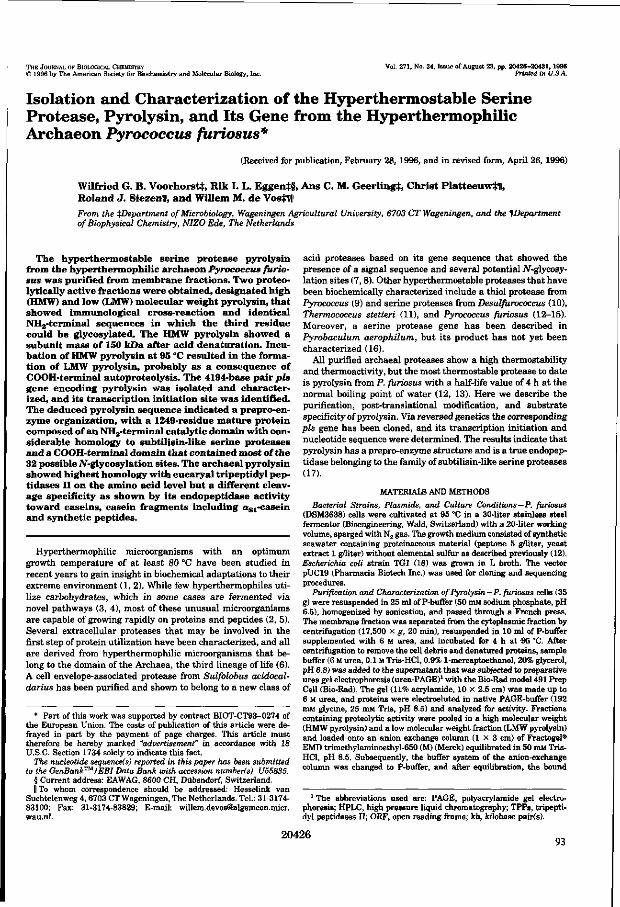
THE JOURNAL OF BIOLOGICAL CHEMISTRY © 1996 by The American Society for Biochemistry and Molecular Biology,.Inc.
Vol. 271, No. 34, Issue of August 23, pp. 20426-20431, 1996 Printed in U.SA.
Isolation and Characterization of the Hyperthermostable Serine Protease, Pyrolysin, and Its Gene from the Hyperthennophilic Archaeon Pyrococcus furiosus*
(Received for publication, February 28, 1996, and in revised form, April 26, 1996)
Wilfried G. B . Voorhorsfcj:, R ik I. L. Eggent§ , A n s C. M. Geer l ing i , Christ P latteeuwft l , Ro land J . SiezenH, a n d Wil lem M. d e Vostffll
From the department of Microbiology, Wageningen Agricultural University, 6703 CT Wageningen, and the ^Department of Biophysical Chemistry, NIZO Ede, The Netherlands
The hyperthermostable serine protease pyrolysin from the hyperthennophi l ic archaeon Pyrococcus furiosus was purified from membrane fractions. Two proteo-lytically active fractions were obtained, des ignated h igh (HMW) and low (LMW) molecular weight pyrolysin, that showed immunological cross-reaction and identical NH2-terminal sequences in wh ich the third residue could be glycosylated. The HMW pyrolysin showed a subunit mass of 150 kDa after acid denaturation. Incubation of HMW pyrolysin at 95 °C resulted in the formation of LMW pyrolysin, probably as a consequence of COOH-terminal autoproteolysis . The 4194-base pair pis gene encoding pyrolysin was isolated and characterized, and its transcription init iation s ite was identified. The deduced pyrolysin sequence indicated a prepro-en-zyme organization, wi th a 1249-residue mature protein composed of an NH2-terminal catalytic domain wi th considerable homology to subtilisin-like serine proteases and a COOH-terminal domain that contained most of the 32 possible iV-glycosylation s ites. The archaeal pyrolysin showed highest homology wi th eucaryal tripeptidyl peptidases II on the amino acid level but a different cleavage specificity as shown by i ts endopeptidase activity toward caseins, casein fragments including a s l - case in and synthetic peptides.
Hyperthennophilic microorganisms with an optimum growth temperature of a t least 80 °C have been studied in recent years to gain insight in biochemical adaptations to their extreme environment (1, 2). While few hyperthermophiles utilize carbohydrates, which in some cases are fermented via novel pathways (3, 4), most of these unusual microorganisms are capable of growing rapidly on proteins and peptides (2, 5). Several extracellular proteases tha t may be involved in the first step of protein utilization have been characterized, and all are derived from hyperthermophilic microorganisms that belong to the domain of the Archaea, the third lineage of life (6). A cell envelope-associated protease from Sulfolobus acidocal-darius has been purified and shown to belong to a new class of
* Part of this work was supported by contract BIOT-CT93-0274 of the European Union. The costs of publication of this article were defrayed in part by the payment of page charges. This article must therefore be hereby marked "advertisement" in accordance with 18 U.S.C. Section 1734 solely to indicate this fact.
The nucleotide sequence(s) reported in this paper has been submitted to the GenBank™/EBI Data Bank with accession Humberts) U55835.
§ Current address: EAWAG, 8600 CH, Dübendorf, Switzerland. II To whom correspondence should be addressed: Hesselink van
Suchtelenweg 4, 6703 CT Wageningen, The Netherlands. Tel.: 31-3174-83100; Fax: 31-3174-83829; E-mail: [email protected]. wau.nl.
acid proteases based on its gene sequence that showed the presence of a signal sequence and several potential Af-glycosy-lation sites (7,8). Other hyperthermostable proteases that have been biochemically characterized include a thiol protease from Pyrococcus (9) and serine proteases from Desulfurococcus (10), Tkermococcus stetteri (11), and Pyrococcus furiosus (12—15). Moreover, a serine protease gene has been described in Pyrobaculum aerophilum, but its product has not yet been characterized (16).
All purified archaeal proteases show a high thermostability and thermoactivity, but the most thermostable protease to date is pyrolysin from P. furiosus with a half-life value of 4 h a t the normal boiling point of water (12, 13). Here we describe the purification, post-translational modification, and substrate specificity of pyrolysin. Via reversed genetics the corresponding pis gene has been cloned, and its transcription initiation and nucleotide sequence were determined. The results indicate that pyrolysin has a prepro-enzyme structure and is a t rue endopeptidase belonging to the family of subtilisin-like serine proteases (17).
MATERIALS AND METHODS
Bacterial Strains, Plasmids, and Culture Conditions—P. furiosus (DSM3638) cells were cultivated at 95 °C in a 30-liter stainless steel fermentor (Bioengineering, Wald, Switzerland) with a 20-liter working volume, sparged with N2 gas. The growth medium consisted of synthetic seawater containing proteinaceous material (peptone 5 g/liter, yeast extract 1 g/liter) without elemental sulfur as described previously (12). Escherichia coli strain TGI (18) was grown in L broth. The vector pUC19 (Pharmacia Biotech Inc.) was used for cloning and sequencing procedures.
Purification and Characterization of Pyrolysin—P. furiosus cells (35 g) were resuspended in 25 ml of P-buffer (50 mM sodium phosphate, pH 6.5), homogenized by sonication, and passed through a French press. The membrane fraction was separated from the cytoplasmic fraction by centrifugation (17,500 x g, 20 min), resuspended in 10 ml of P-buffer supplemented with 6 M urea, and incubated for 4 h at 95 °C. After centrifugation to remove the cell debris and denatured proteins, sample buffer (6 M urea, 0.1 M Tris-HCl, 0.9% 1-mercaptoethanol, 20% glycerol, pH 6.8) was added to the supernatant that was subjected to preparative urea gel electrophoresis (urea-PAGE)1 with the Bio-Rad model 491 Prep Cell (Bio-Rad). The gel (11% acrylamide, 10 X 2.5 cm) was made up to 6 M urea, and proteins were electroeluted in native PAGE-buffer (192 mM glycine, 25 mM Tris, pH 8.5) and analyzed for activity. Fractions containing proteolytic activity were pooled in a high molecular weight (HMW pyrolysin) and a low molecular weight fraction (LMW pyrolysin) and loaded onto an anion exchange column ( 1 x 3 cm) of Fractogel® EMD trimethylaminoethyl-650 (M) (Merck) equilibrated in 50 mM Tris-HCl, pH 8.5. Subsequently, the buffer system of the anion-exchange column was changed to P-buffer, and after equilibration, the bound
1 The abbreviations used are: PAGE, Polyacrylamide gel electrophoresis; HPLC, high pressure liquid chromatography; TPPs, tripeptidyl peptidases II; ORF, open reading frame; kb, kilobase pair(s).
20426 93

Characterization of Pyrolysin and Its Gene from P. furiosus 20427
proteins were eluted by a linear gradient 0-1.5 M NaCl in P-buffer. Fractions containing proteolytic activity were pooled, dialyzed, and concentrated using a diaflow cell with a cut-off value of 30 kDa (Filtron Technology Corp.)- The NH3-terminal amino acid sequences of the purified proteases were determined, either from immobilized proteins that had been separated by SDS-PAGE or directly from a purified preparation, by Edman degradation using an Applied Biosystems 477A Protein Sequencer (Applied Biosystems). Purified protease fractions were used to raise antibodies in rabbits, and crude serum was used in immunoblot experiments as described previously (19). Glycoproteins were stained following urea-SDS-PAGE by the periodic acid-Schiff technique using fuchsin-sulfite stain (Sigma-Aldrich) (20).
Polyacrylamide Gel Electrophoresis — SDS-PAGE was carried out according to Laemmli (21) using 9.5% Polyacrylamide gels. Acid denatur-ation of pyrolysin prior to electrophoresis was achieved by incubation in 5 M formic acid for 45 min at room temperature. Equimolar amounts of potassium hydroxide were used for subsequent neutralization. Analytical urea-SDS-PAGE was essentially the same as SDS-PAGE with the addition of 6 M urea, and samples were prepared as described for the preparative urea-PAGE using sample buffer supplemented with 5% SDS. A method was developed for the in situ detection of proteolytic active proteins on urea-(SDS)-PAGE using an overlayer of Kodak X-OMAT AR röntgen film (Eastman Kodak Co.). Following a 10-min incubation at 95 °C the gelatin layer of the Kodak film was transferred to the gel and degraded on places where proteolytic activity was present. The nondegraded gelatin background was subsequently stained with Coomassie Brilliant Blue, resulting in a blue gel with cleared bands of activity.
Activity Assays and Substrate Specificity—The proteolytic activity of pyrolysin toward casein was assayed for 30 min at 95 °C as described previously (12). Total casein and purified asi-, ß-, and k-casein were incubated with pyrolysin, and degradation products were analyzed by SDS-PAGE (22). The enzyme activity toward synthetic substrates (1 mM final concentration) was assayed in 0.5 ml of 0.1 M potassium phosphate buffer, pH 7.5, at 95 °C and followed spectrophotometrically at 405 nm on a Beekman DU7500 spectrophotometer (Beekman Instruments Inc.). Synthetic substrates were obtained from Bachern Feinchemikalien (Bubendorf, Switzerland) or from Chromogenix AB (Mölndal, Sweden). The tripeptidyl-peptidase substrate Ala-Ala-Phe-p-nitroanilide and its inhibitor, Arg-Ala-SAla-Val-Ala (where SAla is SAla, dehydroalanine), were kindly provided by B. Tomkinson (University of Uppsala, Sweden) (23, 24). The cleavage specificity of pyrolysin in the peptide asl-casein-(l-23) was determined by HPLC analysis of initial cleavage products (22, 25) obtained by incubating 25 ml of a solution of asl-casein-(l-23) (10 mg/ml), 1 ml of the purified LMW pyrolysin (0.25 mg/ml), and 50 ml of 0.1 M potassium phosphate buffer, pH 7.5, at 95 °C. At various times, aliquots of 20 ml were cooled and supplemented with an equal volume of solvent B, composed of acetonitrile/water/trifluoro-acetic acid (900:100:0.7, v/v). As a reference the degradation pattern of asl-casein-(l-23) obtained by the Pill-type protease from Lactococcus lactis was used as described previously (22, 25).
Cloning of the pis Gene Encoding Pyrolysin—A degenerated oligonucleotide was designed against the conserved region around the histidine active site residue of subtilisin-like serine proteases, with the sequence GTICCIGCIACRTGIGTICCRTG-3' (I, inosine; R, A, or G) (primer 1, Fig. 2). Additionally, the NH2-terminal amino acid sequence of the purified pyrolysin was used to design a degenerated oligonucleotide against residues 5-11 with the sequence, ACITGGGTIATWAAY-GCIYT-3' (I, inosine; W, A or T; Y, C or T) (primer 2, Fig. 2). PCR reactions were performed using 100 ng of both oligonucleotides and 250 ng P. furiosus chromosomal DNA as template (19) in a final volume of 100 ml. After denaturation of the template (5 min 95 °C), 30 cycles (1 min 95 "C, 2 min 35 °C, and 3 min 72°) were performed, followed by a final extension of 7 min at 72 °C on a DNA Thermal Cycler (Perkin-Elmer). Southern hybridization of P. furiosus chromosomal DNA was performed with the obtained 0.6-kb PCR fragment. Hybridizing fragments were isolated from agarose gels using GeneClean (BIO 101), cloned into the vector pUC19, and transformed to E. coli TGI by established methods (26).
For the construction of a lambda library, pyrococcal DNA was digested with Sau3AI under conditions optimized for the generation of fragments with a size of 15-20 kb that were isolated using Qiaex (Qiagen Inc.), cloned in lambda EMBL3 SamHI-arms provided by Pro-mega and introduced in E. coli KW251 following in vitro packaging using the PackageneR Lambda DNA Packaging System (Promega Corp.).
RNA Isolation and Primer Extension—Total RNA was isolated from P. furiosus cells grown on peptone using guanidinium isothiocyanate as
94
1 2
HMW^ ^ 105 KDa
-4 80 kDa
Î150 kDa 130 kOa 105 kOa
FIG. 1. Urea-SDS-polyacrylamide gel electrophoresis of purified HMW and LMW pyrolysin. Purified HMW (lane 1) and LMW (lane 2) pyrolysin were subjected directly to urea-SDS-PAGE (A, B, and C) or after acid denaturation (D). A and D, proteins stained with Coomassie Brilliant Blue. B, activity staining using a Kodak film overlay; lane 1' represents HMW pyrolysin after 1-h incubation at 95 °C. C, detection of glycosylated proteins using periodic acid-Schiff staining.
described previously (27). The purified RNA was used as template in primer extension experiments as described previously (19) with oligonucleotide, GTTGGTTAGGGAGTATAGAAGTTG-3' (primer 3, Fig. 2).
Sequence Analysis—Nucleotide sequence analysis of PCR products was carried out by the AmpliTaqR cycle sequencing kit (Perkin-Elmer) and analyzed on a automated DNA sequencer (Applied Biosystems 373A). A Prism™ Ready Reaction DyeDeoxy™ Terminator cycle sequencing kit (Applied Biosystems) was used to sequence both strands of Plasmid DNA.
Computer analysis of nucleotide and deduced amino acid sequences were carried out with the PC/GENE program version 5.01 (IntelliGe-netics Inc.) and the GCG package version 7.0 (28) at the CAOS/CAMM Centre of the University of Nijmegen (The Netherlands).
Nucleotide Sequence Accession Number—The nucleotide sequence reported has been submitted to the GenBank/EMBL Data Bank with the accession number U55835.
RESULTS
Purification of Pyrolysin — Cells of P. furiosus grown on peptides are known to contain high levels of pyrolysin, the highly thermostable serine protease tha t is mainly associated with the cell envelope (12). The membrane fraction of pyrococcal cells grown on peptone was enriched for pyrolysin via a preincubation in 6 M urea at 95 °C, during which the proteolytic activity remained constant while the total protein concentration decreased approximately 100-fold, most likely due to the proteolytic activity of pyrolysin. The remaining proteins were separated by preparative urea-PAGE followed by a final purification step using anion-exchange chromatography. This resulted in HMW and an LMW pyrolysin fraction (Fig. LA) with apparent molecular masses on urea-SDS-PAGE of 105 and 80 kDa, respectively. Activity staining showed that HMW and LMW pyrolysin each contain one proteolytically active band (Fig. Iß ) .
Characterization of HMW and LMW Pyrolysin — The NH2-terminal amino acid sequences of both HMW (35 residues) and LMW (5 residues) pyrolysin were determined and found to be

20428 Characterization of Pyrolysin and Its Gene from P. furiosus
I S 3 H
PLUW701 ( -
1 E
-{ 1 Kb
pLUW 702
B
- 1 PLUW703
pis »U-
1 ATTACAACCAAAAGTAQAATGGTAGCGGQQCGAaQATTTGAACCTCGGACCTCCQQGTTATGAGCCCaOCGQCCACTCCTAGCTQCCCCACCCCGCTGCACQACCCATTACTAACTTTTA
121 TTGGTAGGCTTATAAAGTTTGCGCTCTCTGGTCCTTAATTAATTTAATTTTATCCTCCTTTATCCTCCTTQATTTAAATTATAAGATATAATCACTCCGAGTGATGAGTAAGATACATCA
LTG^TTATÄ^TTGGAACGCAGTGAATATACAAAATGAATATAACCTCQGAQGTGACTQTAGAATQAATAAGAAGGGACTTACTGTGCTATTTATAacOATAATGCTC H N K K G L T V L F T A r M T.
3 6 1 CTTTCAGTAGTTCCAGTGCACTTTGTGTCCGCAGGAACACCACCGGTTAGTTCAGAAAATTCAACAACTTCTATACTCCCTAACCAACAAGTTGTGACAAAAGAAGTTTCACAAGCGGCG - 1 3 4 L S V V P V H F V B A G T P P V S S E N S T T S I L P N Q Q . V V T E E V S Q A A
4 8 1 CTTAATGCTATAATGAAAOGACAACCCAACATGGTTCTTATAATCAAGACTAAGGAAGGCAAACTTGAAGAGCSCAAAAACCGAGCTTGAAAAGCTAGGTGCAGAGATTCTTGACGAAAAT
- 9 4 1 , H A 1 H E G Q P N H V 1 , 1 1 E T K B G K L S E A E T E L E E 1 ' G A S I L Q E H
6 0 1 AGAGTTC^TAACATGTTGCTAGTTAAGATTAAGCCTC^GAAAGTTAAAGAGCTa^CTATATCTCATCTCTTGJVAAAAGCCTGGCTTAACAGAGAAGTTAAGCTTTCCCCTCCAATTGTC - 5 4 B V L N H L L V S I S P S E V E E L H Y I S S L B S A W L N E B V E L S P P I V
7 2 1 GAAAAGGACGTCAAGACTAAGGAGCCCTCCCTAGAACCAAAAATGTATAACAGCACCTGGaTAATTAATGCTCTCCAGTTCATCCAGGAATTTGGATATGATGGTAGTGaTaTTGTTGTT - 1 4 B K D V E T K E P S L E P K « , , Y N S T W V I H A r . O g l O K P Q Y D f l a a V V V
8 4 1 GCAGTACTTGACACGGGAGTTGATCCGAACCATCCTTTCTTGAQCATAACTCCAGATGGÄCQCAGGAAAATTATAGAATGGAAGGATTTTACAQACGAGGGATTCGTGaATACATCATTC 27 K V T , T l - P f l V P P N H P F L S I T P D G R R K I I E W K D F T D E Q F V D T S F
4 2 0 1 CCAAGAGTCTCAATCTTGACACTTAACTrcCTTGGCTACTCATaGTACAGACTATATTCACAGAAGTTTGACGAATTGTACCAAAAGGCCCTTGAATTGGGAGTGGACAACGAOACATTA 1147 P R V S I L T L N F L G Y S W Y R L Y S Q K F D S L Y Q K A L E t G V D H H T I ,
4 3 2 1 GCTTTAGCCCTCAaCTACCATGAAAAAGCCAAAGAGTACTACGAAAAGGCCCTTGAGCTTAGCGAaGGTAACATAATCCAATACCTTGGAGACATAAaACTATTACCTCCATTAAGACAG
1 1 8 7 A L A I j S Y H B K A K E Y Y B K A L E l i S E G N I I Q Y L G S I R L I i P F I i R g
4 4 4 1 GCATACATCAATGAAATGy^GGCAGTTAAGATACTGGAAAAGGCCATAGAAGAATTAGAGGGTGAAGAGTAATCTCCAATTTTTCCCACTTTTTCTTTTATAACATTCCAAGCCTTTTCT 1 2 2 7 A Y I N B H B A V K l b S K A l E B L B G E E *
F I G . 2 . G e n e t i c o r g a n i z a t i o n a n d p a r t i a l n u c l e o t i d e a n d a m i n o a c i d s e q u e n c e o f t h e pis g e n e . A, p h y s i c a l a n d g e n e t i c m a p o f t h e pis
g e n e a n d f l a n k i n g r e g i o n s w i t h s u b c l o n e s a n d r e l e v a n t r e s t r i c t i o n s i t e s (S, Sspl; H, H i n d l l l ; P, Pst\; E, EcoRl). T h e arrowheads i n d i c a t e t h e l o c a t i o n a n d o r i e n t a t i o n of t h e p r i m e r s t h a t w e r e u s e d i n P C R ( p r i m e r s 1 a n d 2 ) o r p r i m e r e x t e n s i o n e x p e r i m e n t s ( p r i m e r 3 ) . B, n u c l e o t i d e a n d d e d u c e d a m i n o a c i d s e q u e n c e o f t h e 3 ' e n d o f t h e pis g e n e a n d i t s u p s t r e a m r e g i o n , i n c l u d i n g t h e t R N A M e t g e n e (overlined). T A T A b o x o f t h e pis g e n e i s i n d i c a t e d ; i t s t r a n s c r i p t i o n i n i t i a t i o n s i t e i s m a r k e d b y t h e arrow, a n d i t s r i b o s o m e b i n d i n g s i t e i s s h o w n b y t h e asterisks. T h e s i g n a l s e q u e n c e of t h e p y r o l y s i n p r e c u r s o r i s s h o w n i n italics a n d underlined, w h e r e a s t h e i d e n t i f i e d N H 2 - t e r m i n a l a m i n o a c i d r e s i d u e s of t h e m a t u r e p y r o l y s i n a r e underlined a n d shaded. C h a r g e d r e s i d u e s p r e s e n t i n t h e p r o p e p t i d e a r e underlined. C, n u c l e o t i d e a n d d e d u c e d a m i n o a c i d s e q u e n c e o f t h e 5 ' e n d of t h e pis g e n e w i t h t h e p r o p o s e d t e r m i n a t i o n s e q u e n c e c o m p o s e d of s t r e t c h e s of t h y m i d i n e r e s i d u e s (overlined). T h e p r e d o m i n a n t L e u a n d G l u r e s i d u e s a r e underlined.
i d e n t i c a l ( F i g . 2 ) . T h e r e l a t e d n e s s o f b o t h p y r o l y s i n f r a c t i o n s
w a s s u p p o r t e d b y t h e o b s e r v a t i o n t h a t t h e t h i r d r e s i d u e o f t h e i r
N H 2 t e r m i n i c o u l d n o t b e d e t e r m i n e d f o l l o w i n g E d m a n d e g r a
d a t i o n , e v e n a f t e r r e p e a t e d t r i a l s ( d a t a n o t s h o w n ) . I n s p e c t i o n
of the amino acid sequence deduced from the pis gene (Fig. 2, see below) revealed that the third position of the mature pyro-lysin is an asparagine that could be post-translationally modified, since it is the first residue of the well-known JV-glycosy-lation motif Asn-X-(Ser/Thr) (29). Therefore, periodic acid-Schiff staining was performed on HMW and LMW pyrolysin separated by urea-SDS-PAGE (Fig. 1C). The results indicate that both proteins are glycosylated to a similar extent and tha t LMW is not a deglycosylated form of HMW pyrolysin.
A mutual relation between the HMW and LMW pyrolysin could further be demonstrated by immunological cross-reaction using antisera raised against both purified pyrolysin fractions (data not shown). Moreover, a prolonged incubation of purified HMW pyrolysin a t 95 °C resulted in its decrease and the formation of LMW pyrolysin. This is illustrated by the semiquantitative in situ detection of a new proteolytic activity with the size of LMW that originated from HMW pyrolysin, indicating that LMW pyrolysin is a processing product of HMW pyro-lysin (Fig. IB). Finally, a multimer structure of HMW pyrolysin was further excluded by applying rigorous acid denaturation
conditions followed by separation by urea-SDS-PAGE (Fig. LD). HMW pyrolysin showed a single band with apparent molecular mass of 150 kDa. Two proteins were obtained after acid denaturation of the LMW pyrolysin fraction with apparent molecular masses of 130 and 105 kDa, the smallest of which is most likely a result of autoproteolytic processing of the 130-kDa protein.
Cloning and Characterization of the pis Gene Encoding Py-rolysin—A data base search revealed tha t the NH2-terminal amino acid sequence of pyrolysin showed significant similarity with subtilisin-like serine proteases (17). This includes a potential active site aspartate residue at position 30 (Fig. 2, see below), suggesting that pyrolysin is a member of this family of serine proteases. Subtilisin-like serine proteases are characterized by the sequential order of the active site residues Asp, His, and Ser and the high degree of homology on amino acid level around these residues that form the catalytic triad (17). Two degenerated oligonucleotides based on the conserved region surrounding the active site histidine residue of subtilisin-like proteases and on the identified NH2-terminal amino acid sequence (primers 1 and 2, Fig. 2) were used in a PCR reaction on chromosomal DNA from P. furiosus. Sequence analysis of the resulting 0.6-kb PCR fragment (data not shown) indicated that indeed a fragment of a subtilisin-like protease gene was ampli-
95

Characterization of Pyrolysin and Its Gene from P. furiosus 20429
i i KPGSCWVAVf .DTaVDFHH£F l ,B I IPBS l iB - K J J I H K E F I D Ëg F V P I S -m g R g g L I AVLDTflVDFSAEGMQVITBgKP - & 1 I D I I DTIGSQ.Dy.MIA -HMJR j gL lAV^DTGVPPGaEGMQVITBCKP-KIVDI I B T I G S S D ? N J A -Ï B g R O y r i f t l F Ç S flyjtEBflTSLS TLCggf tT VB V l i j R VgC S G c ä P g B H K -XB3R D I L I & I U O S X B E S LEGMQVITTgEB-gM F D V r gCS GAgPTOTa -Ï T f i S n y . K i a y . 1 B S g l CSSflEDL - -
• aOHGHBHIV*QTV*QV • SOGAHQTtQtAS I A&gH
• S f lGftKBiag&S IAAJH • MSSPHf l i a aSS I f tS f lH - PSGAHflSa iaSIAf l f tN
• PHN5 HgtSVAGTVAAL
FIG. 3. Transcription initiation site of the pis gene. Primer extension was performed using primer 3 (PEt primer extension product). A DNA sequence ladder was obtained with the same primer and is shown for size comparison (lanes A, G, C, and T). The arrow indicates the deduced transcriptional initiation in the complementary strand.
fled. The 0.6-kb PCR fragment was subsequently used as probe on a Southern blot containing chromosomal DNA of P. furiosus digested with various restriction enzymes. A strongly hybridizing 2.7-kb fftndlll-tftndlll fragment was cloned into Hindlll-digested pUCl9, resulting in pLUW701 (Fig. 2). Sequence analysis of the insert of pLUW701 revealed that it contained an incomplete ORF, coding for the NH2-terminal amino acids determined from the mature pyrolysin, however, without a potential translation start codon (Fig. 2). Southern hybridization of P. furiosus DNA with a pLUW701-derived probe showed a hybridizing 1.7-kb Sspl-Pstl fragment, which was cloned in Smal-Ps/I-digested pUCl9, resulting in pLUW702 (Fig. 2). Sequence analysis of the insert of pLUW702 showed indeed the presence of the 5' end of the pis gene (Fig. 2). The 3 ' end of the pis gene was cloned from a lambda EMBL3 library consisting of 15-20-kb Sau3A fragments of P. furiosus DNA. A pool of approximately 2400 independent plaques were screened by hybridization with the HïndIII-//indIII insert of pLUW701 resulting in 49 hybridizing phages. A 2.5-kb EcoRl fragment of one of these phages carrying the 3 ' end of the pis gene was identified by hybridization and cloned into ÊcoRI-digested pUCl9, resulting in pLUW703 (Fig. 2).
Sequence Analysis of the pis Gene, Flanking Regions, and the pis Gene Product Pyrolysin — Sequence analysis of the pis gene, contained in pLUW701, pLUW702, and pLUW703, revealed an ORF of 4194 base pairs. The first ATG codon at position 316-319 of this ORF is preceded by a stretch of purine-rich nucleotides (at position 301-308) that may function as a ribosome binding site, suggesting that this is the translation initiation site of the pis gene (Fig. 2). If so, the pis gene encodes a 1398-residue protein. The NH2-terminal sequence determined from the purified pyrolysin was found to be encoded by nucleotides 763-868, indicating that the mature NH2 terminus is preceded by a leader sequence of 149 amino acids (Fig. 2). The first 26 residues of this leader have the characteristics of a signal sequence with a high content of hydrophobic residues that can form a transmembrane-spanning helix and is followed by a putative consensus processing site (30). The remaining 123 residues could code for a propeptide that has a high number (36 residues) of charged residues, predominantly Lys and Glu (17 and 16 residues, respectively) (Fig. 2). The calculated molecular mass of the pyrolysin precursor is 155 kDa and after removal of the leader 138.5 kDa.
Identification of Transcription Initiation Site and Regulatory Sequences — Primer extension experiments were performed on total RNA isolated from pyrococcal cells grown on peptone to 96
MMTPP2
BfiSBPn - -
6 - - YVFVIAAaHBGPGI
S - - TIYjrSSif lJ INQECL
a - - I I Y Y S S M H N S E C I . 2 - - WWV A SAOHH CF AL
7 9 - - VIYVCSAQHOflPftL 7 1 - - ï w v t u r a i F n T s r i
- N I Ï S S P J 3 V A I --STVGSPQCTT -
- S T Ï S C E 3 G T I -- S T 2 S T E P D I 5 --STYG.AESGTI -SSTÏ f lYEOKÏP -
i I - g f l D F H s m f i f l A ^ m ^ s q v v t . T . T s n p K
- g T Q L B H f i i a i S S E M f t e a G m V L a S L K • s T Q n m s m i S S E K f t c Q G m i i i . a s L K
- KSQMmJHBH&AEff i fAgf tVT. t . tBULB - SMQKHNSXSIfSSEHAASITOACMLggLE - K Y G f t Y H S i a m S E B y f t f l f t A m L S K H P
FIG. 4. Alignment of pyrolysin with eucaryal TPPs and Bacillus subtilisin BPN'. Partial sequences are shown around the active site residues aspartate, histidine, and serine, and the asparagine present in the oxyanion hole (indicated by arrows). The number of amino acid residues are given between the boxes that are present in the proteins. Large insert is indicated by solid bar; bold and double underlined residues are conserved between pyrolysin and the other sequences. Sequences were deduced from the following accession numbers: BASBPN, subtilisin BPN' from Bacillus amyloliquefaciens (K02496); MMTPP2, TPP from Mus musculus (mouse) (X81323); HSTPP2, TPP from Homo sapiens (human) (M73047); DMPGA9, TPP from D. melanogaster (R. Nusse and F. van Leeuwen, personal communication); CETPP2, TPP from C. elegans (U23176); PFPYRO, pyrolysin from P. furiosus.
identify the transcription initiation site and resulted in a prominent extension product (Fig. 3). The size of this product corresponded with transcription initiation at the thymidine residue located 28 nucleotides upstream of the assumed translation start site (Fig. 2). A hexanucleotide with the sequence TTTATA is located 26 nucleotides upstream of this transcription initiation site and resembles the TATA box, which is involved in binding of the archaeal RNA polymerase (Fig. 2) (31, 32).
Downstream of the pis gene a conserved structure is found, composed of stretches of at least four subsequent thymidine residues, that could be involved in termination of transcription (31), which is further followed by another ORF with an unknown function (Fig. 2). Upstream and in opposite orientation of the pis gene, the gene for a tRNAMet is located tha t could be the initiator tRNA, since it contains all the characteristics for an archaeal initiator tRNA (33).
Substrate Specificity — Comparison of the deduced amino acid sequence of pyrolysin with those of other subtilisin-like serine proteases present in the data bases revealed the highest degree of homology (28-32% identity in the catalytic domain) with the tripeptidyl peptidases II (TPPs) from eucaryal origin, i.e. human, mouse, Drosophila melanogaster, and the nematode Caenorhabditis elegans (Fig. 4). The eucaryal TPPs are intracellular proteases with exopeptidase activity (34, 35), whereas all other known subtilisin-like serine proteases are endopeptidases (17).
The substrate specificity of pyrolysin was analyzed with chromogenic peptide substrates (Table I). Both tripeptidyl and tetrapeptidyl substrates were cleaved by pyrolysin. A preference for the conversion of substrates with a positively charged (Lys, Arg) residue at the P I site was observed (nomenclature according to Schechter and Berger) (36). In spite of its homology with TPPs, pyrolysin showed no activity with the known substrate for TPPs, Ala-Ala-Phe-p-nitroanilide, and is not inhibited by the dehydroalanine-containing peptide Arg-Ala-SAla-Val-Ala that has been shown to be a competitive inhibitor of TPPs from human and rat (23, 24). No difference in substrate specificity was found between HMW or LMW pyrolysin (data not shown).
To demonstrate that pyrolysin has endopeptidase activity, it was incubated with a s l- , ß- or k-caseins and casein fragments. The degradation of caseins was followed in t ime and revealed that for all caseins the first cleavage products constituted a

20430 Characterization of Pyrolysin and Its Gene from P. furiosus
TABLE I Substrate specificity of pyrolysin
Purified pyrolysin was tested for the degradation of synthetic substrates. <Glu, pyroglutanyl; Sue, succinyl; MeO, methoxy; pNA, p ni-troanilide.
Substrate Activity
P4 P3 P2 PI* Sue-Ala-Ala-Pro-Lys-pNA Sue-Ala-Ala-Pro-Arg-pNA Suc-Ala-Ala-Pro-Met-pNA Sue-Ala-Ala-Pro-Phe-pNA Suc-Ala-Glu-Pro-Phe-pNA Sue-Ala-Lys-Pro-Phe-pNA
H-D-Val-Leu-Lys-pNA MeO-Suc-Arg-Pro-Tyr-pNA
<Glu-Pro-Val-pNA Ala-Ala-Phe-pNA
100 32 13 14 7
65
41 52 0.5
<0.01 a Nomenclature according to Schechter and Berger (36). 6 100% activity represents a specific activity of 300 nmol/min/mg
protein.
number of distinct protein bands that were subsequently completely degraded (data not shown). To identify its preferential endopeptidase cleavage sites, pyrolysin was incubated with the peptide a s l-casein-(l-23), and degradation products were analyzed by HPLC (Fig. 5). The results indicate tha t a s l-casein-(1-23) can be completely degraded by pyrolysin resulting in the appearance of four new peptides tha t were identified based on their retention time. Prolonged incubation showed a decrease of the peptides 1-16 and 10-23 and the appearance of new cleavage products (indicated as asterisks in Fig. 5). The results demonstrate that pyrolysin is an endopeptidase with two preferential cleavage sites in a s l-casein, at bonds 9-10 and 16-17 (Fig. 5).
DISCUSSION
The hyperthermostable serine protease pyrolysin was purified from peptone-grown cells of P. furiosus and characterized, and its pis gene was cloned via reversed genetics. An essential step in the purification method of pyrolysin from the membrane fraction appeared to be its preincubation in 6 M urea at 95 °C. This resulted in two proteolytic active fractions, HMW and LMW pyrolysin, that were separated by preparative urea-PAGE and shown to have identical NH2 termini. Incubation of the purified HMW pyrolysin at 95 °C resulted in the formation of LMW pyrolysin, and both forms appeared to be glycosylated to a similar extent. Therefore, the differences in molecular masses of the HMW and LMW pyrolysin are likely to be a consequence of autoproteolytic removal of a COOH-terminal part of the HMW pyrolysin tha t results in LMW pyrolysin. Similar processing has also been suggested for the serine protease from the related archaeon Thermococcus stetteri (11). Moreover, COOH-terminal processing is a common feature of serine proteases (37).
It has frequently been observed that proteins from hyper-thermophiles are not denatured during standard SDS-PAGE (3,38,39). This was also found for pyrolysin that was subject to autoproteolysis under these conditions. Acid denaturation of HMW pyrolysin demonstrated a molecular mass of 150 kDa. Autoproteolytic processing of pyrolysin may also explain the previously reported heterogeneity of pyrolysin, evidenced by the multiple activity bands on substrate-PAGE (12-14). Alternatively, pyrolysin may form a complex either with itself, as found for TPPs (40), or with a cell envelope structure as found in Staphylothermus marinus where a serine protease is associated with a filamentous surface protein assembly called tetrabrachion (39).
The pis gene-encoding pyrolysin is most likely transcribed as
B asl-casein(1-23):
Arg-Pro-Lys-His-Pro-lle-lys-His-GIn i Gly-Leu-Pro-
-Gtn-Glu-Val-Leu * Asn-Glu-Asn-Leu-lue-Arg-Phe FIG. 5. Reversed-phase HPLC pattern of degradation products
of asl-casein-(l-23) by pyrolysin. A, pattern of cleavage products resulting from the incubation of asl-casein-(l-23) with pyrolysin for 0 min (pattern 1), 20 min (pattern 2), and 40 min (pattern 3) at 95 °C. The known pattern (22, 25) of asl-casein degradation obtained with the Pm-type protease from L. lactis is shown for comparison (pattern 4). Identified products are indicated; asterisks indicate new cleavage products after prolonged incubation. B, amino acid sequence of asl-casein-(1-23) with the preferential cleavage sites indicated by the arrows.
a single unit, since the transcription initiation site and TATA box could be identified upstream of the gene, whereas downstream it was followed by conserved archaeal termination sequences. Furthermore, upstream of the pis gene a tRNAMet
gene is found in the opposite orientation that most likely is the initiation tRNA.
Analysis of the deduced amino acid sequence indicates that pyrolysin is synthesized as a prepro-enzyme with a common signal sequence (30), suggesting t ha t export of proteins in P. furiosus occurs in a similar way as in Eucarya and Bacteria. The propeptide of pyrolysin shows little homology with the consensus identified for different propeptides of subtilisin-like serine proteases; however, it shares with these propeptides a high number of charged residues (41).
The mature part of the deduced pyrolysin sequence shows the highest homology with eucaryal TPPs, which form a distinct subgroup of the subtilisin-like serine proteases. The mature pyrolysin, as several other subtilisin-like serine proteases, can be divided into two domains, the NH2-terminal catalytic domain that extends to about residue 500 followed by a COOH-terminal extension to residue 1249 (17). The catalytic domain is characterized by the three active site residues, Asp-30, His-216, and Ser-441, and the presence of the conserved Asn-342 present in the oxyanion hole tha t stabilizes the reaction intermediate. The catalytic domains of pyrolysin and the TPPs contain a large insert of more than 150 residues between the aspartate and histidine in comparison with the Bacillus sub-tilisin (Fig. 4). A number of conserved residues among all members could be identified within the insert, which may imply an evolutionary relationship between the archaeal pyrolysin and the eucaryal TPPs (Fig. 4). In addition, pyrolysin shows weak homology in the large COOH-terminal domain where the TPPs also have many conserved residues (data not shown).
97

Characterization of Pyrolysin and Its Gene from P. furiosus 20431
The COOH-terminal domain, if present, in subtilisin-like serine proteases can have different functions but is often related to localization or anchoring of the protease (17). The last 80 amino acids of pyrolysin do not contain known hydrophobic anchoring sequences, but they are characterized by a high number of Glu and Leu residues (14 and 13 residues, respectively) that may be involved in the observed association of pyrolysin to the cell envelope.
The COOH-terminal domain of pyrolysin together with the large insert present in the catalytic domain contain almost all (29 of 32) of the possible TV-glycosylation sites (Asn-X-(SerZThr)) present in pyrolysin. The presence of a gap at position 3 in the determined NH2-terminal amino acid sequence suggests that Asn at tha t position is modified, presumably due to glycosyla-tion; whether the other possible glycosylation sites are indeed post-translationally modified and what the structure is of the bound glycan is presently unknown. This form of post-transla-tional modification may function in the thermostabilization of the enzyme as has also been suggested for the extracellular protease from S. acidocaldarius (7). Mature pyrolysin without the 16.5-kDa prepropeptide has a calculated molecular mass of 138.5 kDa. The higher apparent molecular mass observed for the acid-denatured HMW pyrolysin (150 kDa) could be explained by glycosylation.
The substrate specificity of pyrolysin was analyzed using chromogenic substrates, different caseins, and the small peptide a s l-casein-(l-23). Degradation of the caseins and a s l -casein-(l-23) into distinct intermediates followed by the complete degradation of these proteins implies that pyrolysin is not an exopeptidase like the eucaryal TPPs but a true endopepti-dase like all other subtilisin-like serine proteases. The specificity studies indicated that pyrolysin has a rather broad substrate specificity with a preference for Arg or Lys residues at the P I position. Such a preference is to be expected when pyrolysin autocatalytically removes its propeptide as is commonly found with serine proteases (42-44) (see Fig. 2). Modelling of pyrolysin on known three-dimensional structures in combination with functional expression and mutational analysis could give further insight in this processing and the molecular mechanisms of adaptation of this extracellular serine protease to extremely high temperatures.
Acknowledgments — We are grateful to Dr. Brigitte Tomkinson for a gift of chemicals and Saskia van Schalkwijk and Marcel Dijkgraaf for technical assistance.
REFERENCES
1. Adams, M. W. W. (1993) Annu. Rev. Microbiol. 47, 627-658 2. Stetter, K. O., Piala, G., Huber, G„ Huber, R., and Segerer, A. (1990) FEMS
Microbiol. Rev. 75, 117-124 3. Mukund, S., and Adams, M. W. W. (1995) J. Biol. Chem. 270, 8389-8392 4. Kengen, S. W. M., de Bok, F. A. M., van Loo, N.-D., Dykema, C, Stams, A. J.
M., and de Vos, W. M. (1994) J. Biol. Chem. 269, 17537-17541 5. Kelly, R. M., and Adams, M. W. W. (1994) Antonie Leeuwenhoek 66, 247-270 6. Woese, C. R., Kandier, O., and Wheelis, M. L. (1990) Proc. Natl. Acad. Sei.
U. S. A. 87, 4576-4579
7. Fusek, M., Lin, X., and Tang, J. (1990) J. Biol. Chem. 265, 1496-1501 8. Lin, X., and Tang, J. (1990) J. Biol. Chem. 265, 1490-1495 9. Morikawa, M., Izawa, Y., Rashid, N., Hoaki, T., and Imanaka, T. (1994) Appl.
Environ. Microbiol. 60, 4559-4566 10. Cowan, D. A., Smolensk!, K A., Daniel, R. M„ and Morgan, H. W. (1987)
Biochem. J. 247, 121-133 11. Klingeberg, M., Galunsky, B., Sjoholm, C , Kasche, V., and Antranikian, G.
(1995) Appl. Environ. Microbiol. 61, 3098-3104 12. Eggen, R., Geerling, A., Watts, J., and de Vos, W. M. (1990) FEMS Microbiol.
Lett. 71, 17-20 13. Blumentals, I. I., Robinson, A. S., and Kelly, R. M. (1990) Appl. Environ.
Microbiol. 56, 1992-1998 14. Connaris, H., Cowan, D. A., and Sharp, R. J. (1991) J. Gen. Microbiol. 137,
1193-1199 15. Snowden, L. J., Blumentals, I. I., and Kelly, R. M. (1992) Appl. Environ.
Microbiol. 58, 1134-1141 16. Volkl, P., Markiewicz, P., Stetter, K. O., and Miller, J. H. (1995) Protein Sei. 3,
1329-1340 17. Siezen, R. J., de Vos, W. M., Leunissen, J. A. M., and Dijkstra, B. W. (1991)
Protein Eng. 4, 719-737 18. Gibson, T. J. (1984) Studies on the Epstein-Barr-Virus Genome. Ph.D. Thesis.
Cambridge, UK 19. Eggen, R. I. L., Geerling, A. C. M„ Jetten, M. S. M., and de Vos, W. M. (1991)
J. Biol. Chem. 266, 6883-6887 20. Zacharius, R. M., Zell, T. E., Morrison, J. H., and Woodlock, J. J. (1969) Anal.
Biochem. 30, 148-152 21. Laemmli, U. K. (1970) Nature 227, 680-685 22. Siezen, R. J., Bruinenberg, P. G-, Vos, P., van Alen-Boerrigter, I. J., Nuhuis,
M., Alting, A. C, Exterkate, F. A., and de Vos, W. M. (1993) Protein Eng. 6, 927-937
23. Tomkinson, B. (1994) Biochem. J. 304, 517-523 24. Tomkinson, B., Grehn, L., Fransson, B., and Zetterqvist, O. (1994) Arch.
Biochem. Biophys. 314, 276-279 25. Exterkate, F. A., Alting, A. C, and Slangen, C. J. (1991) Biochem. J. 273,
135-139 26. Sambrook, J., Fritsch, E. F., and Maniatis, T. (1989) Molecular Cloning: A
Laboratory Manual, 2nd Ed., Cold Spring Harbor Laboratory Press, Cold Spring Harbor, NY
27. Voorhorst, W. G. B„ Eggen, R. I. L., Luesink, E. J., and de Vos, W. M. (1995) J. Bacterial. 177, 7105-7111
28. Devereux, J., Haeberli, P., and Smithies, O. (1990) Nucleic Acids Res. 12, 387-395
29. Marshall, R. D. (1972) Annu. Rev. Biochem. 41, 673-702 30. Von Heijne, G. (1985) J. Mol. Biol. 184, 99-105 31. Dalgaard, J. 2., and Garrett, R. A. (1993) The Biochemistry of Archaea (Ar-
chaebacteria) (Kates, M., Kushner, D. J., and Matheson, A. T., eds) pp. 535-563, Elsevier, Amsterdam
32. Rowlands, T., Baumann, P., and Jackson, S. P. (1994) Science 264, 1326-1329 33. Kuchino, Y., Ihara, M., Yabusaki, Y., and Nishimura, S. (1982) Nature 298,
684-685 34. Bàlow, R.-M., Tomkinson, B-, Ragnarsson, U., and Zetterqvist, Ö. (1986)
J. Biol. Chem. 261, 2409-2417 35. Tomkinson, B., Wernstedt, C, Hellman, U., and Zetterqvist, O. (1987) Proc.
Natl. Acad. Sei. U. S. A. 84, 7508-7512 36. Schechter, I., and Berger, A. (1967) Biochem. Biophys. Res. Commun. 27,
157-162 37. Shinde, U., Li, Y., Chatterjee, S., and Inouye, M. (1993) Proc. Natl. Acad. Sei.
U. S. A. 90, 6924-6928 38. Kengen, S. W. M., and Stams, A. J. M. (1994) Biocatalysis 11, 79-88 39. Peters, J., Nitsch, M., Kuhlmorgen, B., Golbik, R., Lupas, A., Kellermann, J.,
Engelhart, H., Pfunder, J. P., Muller, S., Goldie, K, and Engel, A. (1995) J. Mol. Biol. 245, 385-401
40. Macpherson, E., Tomkinson, B., Bâlow, R., Höglund, S., and Zetterqvist, Ö. (1987) Biochem. J. 248, 259-263
41. Siezen, R. J., Leunissen, J. A. M., and Shinde, U. (1995) Intramolecular Chaperones and Protein Folding (Inouye, M-, and Shinde, U., eds) pp. 233-256, R. G. Landes Co, Austin, TX
42. Germain, D., Dumas, F-, Vernet, T., Bourbonnais, Y., Thomas, D. Y-, and Boileau, G. (1992) FEBS Lett. 299, 283-286
43. Leduc, R., Molloy, S. S., Thorne, B. A., and Thomas, G. (1992) J. Biol. Chem. 267, 14304-14308
44. Power, S. D., Adams, R. M., and Wells, J. A. (1986) Proc. Natl. Acad. Sei. U. S. A. 83, 3096-3100
98

Homology modelling
Chapter 7
Homology Modelling of Two Subtilisin-like Serine Proteases
from Hyperthermophilic Archaea
Pyrococcus furiosus and Thermococcus stetteri
Wilfried G.B. Voorhorst, Angela Warner, Willem M. de Vos, and Roland, J. Siezen
reprinted with permission from Protein Engineering 1997, vol. 10, 905-914
99

Chapter 7
100

Protein Engineering vol.10 no.8 pp.905-914, 1997
Homology modelling of two subtilisin-Iike serine proteases from the hyperthermophilic archaea Pyrococcus furiosus and Thermococcus stetteri
Wilfried G.B.Voorhorst1, Angela Warner1, Willem M.de Vos1,2 and Roland J.Siezen2*3
'Department of Microbiology, Wageningen Agricultural University, Hesselink van Suchtelenweg 4, NL-6703 CT Wageningen, The Netherlands and department of Biophysical Chemistry, NIZO, PO Box 20, NL-6710 BA Ede, The Netherlands 3To whom correspondence should be addressed
The hyperthermophilic archaeon Pyrococcus furiosus produces an extracellular, glycosylated hyperthermo-stable subtilisin-like serine protease, termed pyroly-sin (Voorhorst,W.G.B., Eggen,R.I.L., Geerling,A.C.M., Platteeuw,C, Siezen,RJ. and de Vos,W.M. (1996) J. Biol. Chem., 271, 20426-20431). Based on the pyrolysin coding sequence, a pyrolysin-like gene fragment was cloned and characterized from the extreme thermophilic archaeon Thermococcus stetteri. Like pyrolysin, the deduced sequence of this serine protease, designated stetterlysin, contains a catalytic domain with high homology with other subtilases, allowing homology modelling starting from known crystal structures. Comparison of the predicted three-dimensional models of the catalytic domain of stetterlysin and pyrolysin with the crystal structure of subtilases from mesophilic and thermophilic origin, i.e. subtilisin BPN' and thermitase, and the homology model of subtilisin S41 from psychro-philic origin, led to the identification of features that could be related to protein stabilization. Higher thermostability was found to be correlated with an increased number of residues involved in pairs and networks of charge-charge and aromatic-aromatic interactions. These highly thermostable proteases have several extra surface loops and inserts with a relatively high frequency of aromatic residues and Asn residues. The latter are often present in putative N-glycosylation sites. Results from modelling of known substrates in the substrate-binding region support the broad substrate range and the autocatalytic activation previously suggested for pyrolysin.
Keywords: ArchaeaJPyrococcus furiosus/serine protease/Ther-mococcus .stetteri/thermostability
Introduction To gain insight in the molecular basis of thermostability of proteins, structurally homologous proteins with different thermostability have been compared using known crystal structures or predicted structures from homology modelling (Teplyakov et al, 1990; Davail et al, 1994; Heringa et al, 1995; Tomschy et al, 1994; Muir et al, 1995; Szilâgyi and Zâvodszky, 1995). The recent isolation and characterization of proteins and their genes from hyperthermophiles, microorganisms able to grow optimally at 80°C or higher (Stetter et al, 1990), generates the possibility to extend the structural analysis to extremely stable proteins to gain insight in the
© Oxford University Press
stabilization mechanisms that allow proteins to be functional near the maximum temperature of life. Analysis of three-dimensional structures and modelled structures of proteins from hyperthermophiles and their comparison with those from less stable proteins has resulted in identification of features that may be related to protein stabilization. Mechanisms by which proteins from fhermophiles are stabilized resemble those identified for mesophilic model proteins and include hydrogen bonds, hydrophobic interactions, ion-pairs, secondary structure stabilization, cavity filling, reduction of the length of loops and conformational strain (reviewed by Tomschy et al, 1994; Völkl et al, 1994; Muir et al, 1995; Szilâgyi and Zâvodsky, 1995; Korndorfer et al, 1995; Yip et al, 1995; Vieille and Zeikus, 1996; Knapp et al, 1997). It seems that each thermostable protein is stabilized by a unique combination of different mechanisms resulting in a more rigid structure. A common theme that has now emerged from analysis of the structures identified so far for hyperthermophiles suggests that an increased number of ion-pairs, that may be arranged in extensive networks, contributes 'to the observed extreme thermostability (Korndorfer et al, 1995; Yip et al, 1995; Knapp et al, 1997). Moreover, many proteins from hyperthermophiles are not only extremely thermostable, but also show a higher resistance to chemical denaturation and proteolytic degradation than their less stable counterparts (Fontana, 1991). This can be illustrated with pyrolysin, an extracellular serine protease from the archaeon Pyrococcus furiosus that shows optimal growth at 100°C (Fiala and Stetter, 1986). This protease has a half-life value of 4 h at 100°C and is still active in the presence of 6 M urea (Eggen et al, 1990; Voorhorst et al, 1996).
The family of subtilisin-like serine proteases, also referred to as subtilases (Siezen et al, 1991; Siezen and Leunissen, 1997) has been extensively studied from psychrophilic, mesophilic and thermophilic origin to gain insight in thermostability, catalytic activity and substrate specificity (Wells and Estell, 1988; Davail et al, 1994; Daniel et al, 1995; Feller et al, 1996). However, knowledge of subtilases from hyperthermophilic origin is limited to pyrolysin from P.furiosus (Eggeri et al, 1990; Voorhorst et al, 1996) and STABLE from Staphylothermus marinus (Mayr etal, 1996), which have been extensively characterized on the biochemical level with specific attention for thermostability and substrate specificity. Moreover, the subtilase aerolysin from the hyperthermophile Pyrobaculum aerophilum has been analysed for features that could be related to its thermostability using homology modelling (Völkl et al, 1994).
The amino acid sequence homology in the catalytic domain of pyrolysin with other subtilases is sufficiently high to allow for homology modelling (Voorhorst et al, 1996). However, the identification of features as being important for protein stabilization based on only a single enzyme may lead to misinterpretation (Querol et al, 1996). Therefore, we have
101

W.G.B.Voorhorst et al.
now isolated and characterized the gene encoding a homolog of pyrolysin, designated stetterlysin, from the extreme thermophilic archaeon Thermococcus stetteri that grows optimally at 75°C (Miroshnichenko et al., 1989). The amino acid sequence homology within the core of the catalytic domain between stetterlysin, pyrolysin and other subtilases allowed homology modelling using known three-dimensional protein structures of subtilisin BPN' from the mesophilic bacterium Bacillus amyloliquefaciens with an optimum growth temperature between 30 and 40°C (Heinz et al, 1991 ; Gallagher et al, 1995) and the more thermostable thermitase from Thermoactinomyces vulgaris that grows optimally at 50°C (Gros et al, 1989). The predicted three-dimensional structures of the highly thermostable pyrolysin and stetterlysin were compared with the homology model of subtilisin S41 from the psychrophilic Bacillus T41 (optimal growth temperature of 4°C), that has a half-life value of only a few minutes at 50°C (Davail et al, 1994), and with subtilisin BPN' and thermitase, with half-life values of 23 min at 60°C (Braxton and Wells, 1992) and 1 h at 85°C (Daniel et al, 1995), respectively. Conformational characteristics of the catalytic domain of stetterlysin and pyrolysin were analysed that could be related to their thermostability regarding overall composition, ion-pair formation, aromatic-aromatic interactions, amino acid replacements, helix stabilization, and calcium-binding sites. Additionally, the substrate binding regions of these archaeal subtilases were modelled in detail with respect to substrate specificity and interactions between the enzymes and their known substrates.
Materials and methods Bacterial strains, plasmids and enzymes Cells of Thermococcus stetteri (DSM 5262) grown on tryptone were kindly provided by Dr G.Antranikian (Klingeberg et al, 1995). Escherichia coli TGI (Gibson, 1984) was cultured in LB-based medium at 37°C. The plasmids pUC18, pUC19 (Pharmacia Biotechnologies) and pGEMR-T (Promega Corporation) were used as cloning vectors. Standard molecular biological techniques were performed as described by Sambrook et al. (1989). Restriction enzymes, DNA modifying enzymes and Taq DNA polymerase were acquired from Life Technologies Inc. Oligonucleotides were purchased from Pharmacia Biotechnologies.
Isolation and cloning ofplsT gene coding for stetterlysin from T.stetteri Degenerated oligonucleotides were designed against the conserved regions around the Asp and Ser active-site residues of subtilisin-like serine proteases in combination with the known sequence of pyrolysin from P.furiosus (Voorhorst et al., 1996) and include BG95(Asp): 5'-GTIGCIGTIMTIGAYACIGG-3' and BG31(Ser): 5'-GGISYIGCCATISWGTICC-3' (I, inosine; M, A or C; Y, C or T; S, G or C; W, A or T). PCR reactions were performed using 100 ng T.stetteri chromosomal DNA (Eggen et al., 1991) as template in a final volume of 100 |Xl. After denaturation of the template (5 min, 95°C) amplification was performed for 35 cycles (1 min, 95°C; 2 min, 35°C and 3 min, 72°C), followed by a final extension of 7 min at 72°C on a DNA Thermal Cycler (Perkin Elmer Cetus). This resulted in a prominent product of 1.4 kb that hybridized with a pyrolysin-derived probe. The 1.4 kb PCR fragment was isolated from agarose gel using Gene Clean (BIO 101), cloned into the vector pGEMR-T and transformed to E.coli TGI. Nucleotide
102
sequence analysis showed the presence of a Kpnl restriction site in the amplified fragment that could be used for cloning chromosomal DNA fragments. A Southern blot containing chromosomal DNA of T.stetteri digested with different combinations of Kpnl and other restriction enzymes was probed with the cloned PCR fragment. Positively hybridizing fragments were subsequently cloned in appropriately digested pUC18 and characterized by nucleotide sequence analysis.
Sequence analysis and alignment
Automated nucleotide sequence analysis of plasmid DNA was carried out on either an Applied Biosystems 373A sequencer (Applied Biosystems Inc.) using a Prism™ Ready Reaction DyeDeoxy™ Terminator cycle sequencing kit or the Li-Cor 4000L sequencer (Li-Cor Inc.) using the Amersham Thermo Sequenase cycle sequencing kit with 7-deaza-dGTP and infrared labelled oligonucleotides (MWG-Biotech, Germany). Computer analysis of nucleotide and deduced amino acid sequences was carried out with the PC/GENE program version 5.01 (IntelliGenetics Inc.) and the GCG package version 7.0 (Devereux et al, 1990) at the CAOS/CAMM Centre of the University of Nijmegen (The Netherlands).
The amino acid sequences of pyrolysin and stetterlysin were included in the multiple sequence alignment of the catalytic domains of the entire subtilase family (Siezen et al, 1991; Siezen and Leunissen, 1997). In this way the sites of insertions in the catalytic domain could be identified. The numbering of subtilisin BPN' indicated by stars (*) was used as reference.
Modelling of the catalytic domains of pyrolysin and stetterlysin
Homology modelling was performed using QUANTA 3.2.3 (Molecular Simulations, Cambridge, UK) (Sali and Blundell, 1993) and CHARMm 22 (Brooks et al, 1983) running on a Silicon Graphics 4D25TG workstation. The model is based on sequence homology between pyrolysin, stetterlysin and members of the subtilase family as reflected in the given alignment (Figure 1), and on the known X-ray structures of subtilisin BPN' in complex with the inhibitor R45-eglin (Heinz et al, 1991), thermitase in complex with eglin (Gros et al, 1989) and subtilisin in complex with its pro-peptide (Gallagher et al, 1995). Insertions of more than six residues relative to either subtilisin or thermitase were not modelled. After addition of H-atoms the entire molecule was subjected to energy minimization after constraining the active site residues (Asp32*, His64* and Ser221*), the Asnl55* of the oxy-anion hole, and the two ß-sheet strand backbones el and elll involved in substrate binding. The atomic coordinates of the two models are electronically available (ftp://ftp.caos.kun.nl/pub/molbio/ siezen97/).
The backbone conformation of eglin residues 40-47 or subtilisin pro-peptide residues -6 to -1 (i.e. the segment that interacts with subtilisin or thermitase in the crystal structures) was used to model enzyme-substrate interactions of pyrolysin by substituting the side chains by the appropriate P6-P2' residues—nomenclature as described (Schechter and Berger, 1967)—of either pro-pyrolysin or of a s rcasein residues 11-18.
Nucleotide accession numbers
The nucleotide sequence reported has been submitted to the GenBank/EMBL Data Bank with the accession number AF015225.

Homology modelling of hyperthermostable subtilases
1 10 20 30 40 50 60 70 ! ! ! ! * ! i 1 * 1
S41 AASQST PWSIKA.IïraSN!iTSTSœi^INIAVWOTGVJSrim[PI>I.S NNVEQCKDFFVGTNF- - 3 - SCTDRÇiGHGTHVAGSALANG S u b t AQSV PYGVSQI»»>AiHSQGYTOSMVKVAVI»SQIJ3SSlH>»I, KVAGGASMOTSETN PFQnNNSHGTHVAGTVAAL-T h e r YTPNDPYFSSRQ'SfGt'QKIQAPaMî-DIABSSGAKiAlVBTOVQSNHPDÎiA GKVVGGWBFVDNDS TPQNGNGHGTHCASIAAAVT S t e t MFMS VYKXHMÏOTI^--GV1^DNVTVAVI4WfëIBV!GHP^ P y r o MYNS TWXNAtjQFIQjBF- -GYWSSVVVAVIJ»TGVDPimPPI-SITPDGRRKIIBWKI>FÎDEGF-147-FGKDGHGHSTHVAGTVAGYD
a a t x a a a aaaaa ß ß&ßßß ßEßßEß ityuaoïQtj/aorÇi hA hE e l e 2 h e
100 I
110
S 4 1 G T G S G V Y G V A P E A D M J A Y K V I J G D - DGSGYADDIÄEAIEHAGDQ -
S u b t KINSIGVliGVAt>SASI.YAVKVLGA-DGSGQÏSWIIBGÏEWAIAN-The r ÜtoSTGIAßTAPKAS Xl&VRVUM - SGSÖTWTAVASSITYAÄDQ -S t e t LPTDPVPHSTYGVAPHAOLMEVKVLPGEFGPGATSWIINGMIYAASN -P y r o -29-VrroTVQGVAPGAQIMAIRVtjRS-DSRGSHWDIIEGMT5fAATH-
EEEßßE EßßßaaaffaocuaoiaaoiO! e 3 e l hD
120 130 140 150 ! ! 1 !
-TKWIEMSLGSS OESSLITNAVDÏAÏDKGVLIIAAAGHSG -NMDVINMSLGGP SGSAA1JCAAVDKAVASGVWVAAAGNEG -GAlOTISfcSLGGT Va»SSÏ^»5»nra«IBÖSSVVVA&ASHAG -GAPVISMSlGGGGEIOTGIESÏ>SHFS^ra^TI>Wg?S91SAiaAaNEG - SADVISMSÎiGSNAPYLD - aTOPaSVAVDEKTSKYOWWIAAiaSBG
EßEßß Eßß a aaaaaaaaaaa BEESE e4 e i n hE e5
160 170 180 190 200 210 220 230
S41 PK- -PGSIGYPGA1.VNAVAVAAI,EN- - 5-TYRVADRSSRGHKR-10 -DVEISÄPGAAVYSTWP- - -S u b t TSGSSSTVGYPGKYPSVIAVGAVDS SNQRASFSSVGP EU>VMAPGVSIQSTIit>- - -T h e r NT APHYPAYYSNAIAVASTOO. HDNKSSFSTYGS WWVBAFGSSIYSTYt- - -S t e t TP--TNTVHAP^I«DX.VITVGAFRS-13-ADTVASPSS|ÎGPRMDGLLDPOVIAPGEMIFSS!iP-10 P y r o PG- - INIVGSPGVATKAITVGAÄAV- 2 7 -NVRIAFFSSRGPRIDGEIKPNWAPGYOIYSStPMW-
ß ß ß ß ß ß ß ß ß ß ß ß ß ß ß e 6 e 7 e 8
- DGGYATISGTS«AS?aAAGWAKIWÄ - GNKYGAYNGTSMAS PHVAeAAAfcliS - TSTYASIJSGTSMÄ.TJ'HVAGVÄGJ.IÄS
- IGGÄDFMSGTSMATPHVSSWATXIS ßßßßß aaaaaacüaaaaaoraaao:
e9 hF
240 250 270
S41 QSP AASNVDVRÖBI4JTRASVNDI- -LSGNSAGSQ&OTASGSSFAKVQ S u b t KHP NWWrQVRSSUSlWITKLG DSFYYGKSiCINVQAAAQ T h e r QGR SASNIRAAIMFAMKI SG - - - TGTYWAK6RVNAÏKAVQY S t e t YAKQHNLTYDPIMIKRALELSAKPVNG- - -TLIDQGFG1IQWKAÏEE-P y r o GPKPEGIYYNPDtltCKWESGATlfcEG- 8 -TELDQGHSÏ,W«ÎCS«EI -
aaaaaaaaotota aaaaa hG hH
t o t a l r e s i d u e s c a t a l y t i c d o m a i n 3 0 9 2 7 5 279 488 509
Fig. 1. Amino acid sequence alignment of the catalytic domains of subtilisin BPN' (Subt), thermitase (Ther), subtilisin S41 (S41), stetterlysin (Stet) and pyrolysin (Pyro). The number of not-modelled residues in inserts is indicated. Catalytic residues D32*, H64* and S221* are indicated by (*). Structurally conserved core regions of subtilisin/thermitase are shaded, and common secondary structure elements are labelled (Siezen et ai, 1991; Siezen and Leunissen, 1997). The numbering of subtilisin BPN' is used.
Results Isolation and characterization of the pyrolysin-like gene from T.stetteri To expand our knowledge of highly stable subtilases, the extreme thermophilic archaeon T.stetteri was screened for the presence of a serine protease similar to pyrolysin from P.furiosus (Voorhorst et al, 1996). A PCR approach was used with degenerate oligonucleotides derived from conserved amino acids surrounding the active site Asp and Ser of subtilases and pyrolysin. From the obtained PCR products a 1.4 kb fragment was the most prominent one and hybridized with a pyrolysin-derived probe. This PCR fragment was cloned and sequence analysis indicated that indeed a fragment coding for a pyrolysin-like serine protease was amplified. The cloned PCR fragment was used as hybridization probe to identify T.stetteri DNA fragments that encoded the pyrolysin-like serine protease. Two adjacent fragments, a 2.9 kb Sphl-Kpnl and a 1.5 kb Kpnl-BamHl fragment, were cloned in appropriately digested pUC18. Nucleotide sequence analysis of these cloned T.stetteri fragments confirmed the sequence of the PCR fragment and revealed the presence of a 2667 bp open reading frame, starting with an ATG and preceded by a purine rich region that may constitute the ribosome binding site; no stop codon was found to terminate this open reading frame. Its deduced sequence of 888 amino acids contained a complete
catalytic domain of a serine protease, that we have designated stetterlysin after its source T.stetteri.
Homology between stetterlysin and pyrolysin The sequence of stetterlysin showed the highest overall homology with a subgroup of the subtilase family that includes the archaeal pyrolysin and tripeptidyl-peptidases from eucaryal origin (Voorhorst et ai, 1996; Siezen and Leunissen, 1997). The predicted organization of stetterlysin also showed high similarity to that of pyrolysin and starts with a prepro-peptide of 159 residues (26% identity to pyrolysin) followed by a 488 residue N-terminal catalytic domain (46% identity) and an incomplete C-terminal extension of at least 241 residues (30% identity). The catalytic domain of these archaeal proteases, as other subtilases, can be divided into the core, residues that are also present in subtilisin and/or thermitase, and the inserts, residues that are additional to the core (Siezen et al, 1991). The core of the archaeal proteases stetterlysin and pyrolysin showed high amino acid sequence homology (up to 56% similarity, and 44% identity) with other subtilases (Figure 1). Both pyrolysin and stetterlysin have an unusually large insert, almost one-third of the catalytic domain residues, just preceding the catalytic His. These inserts show 39% identity, but are not homologous to any known proteins or specifically to the large inserts at the equivalent position in tripeptidyl peptidases,
103
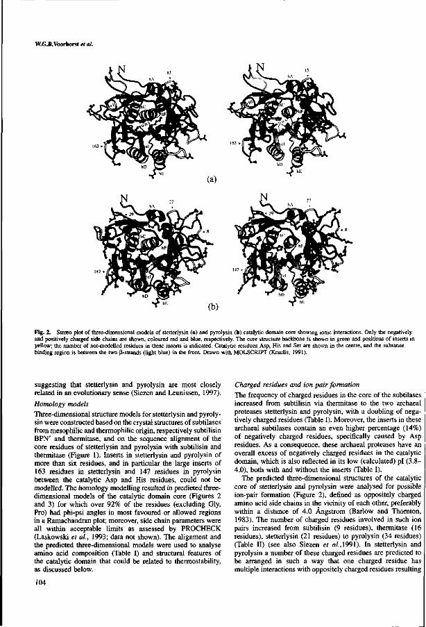
W.G.B.Voorhorst et al.
Fig. 2. Stereo plot of three-dimensional models of stetterlysin (a) and pyrolysin (b) catalytic domain core showing ionic interactions. Only the negatively and positively charged side chains are shown, coloured red and blue, respectively. The core structure backbone is shown in green and positions of inserts in yellow; the number of not-modelled residues in these inserts is indicated. Catalytic residues Asp, His and Ser are shown in the centre, and the substrate binding region is between the two ß-strands (light blue) in the front. Drawn with MOLSCRIPT (Kraulis, 1991).
suggesting that stetterlysin and pyrolysin are most closely related in an evolutionary sense (Siezen and Leunissen, 1997).
Homology models Three-dimensional structure models for stetterlysin and pyroly-sin were constructed based on the crystal structures of subtilases from mesophilic and thermophilic origin, respectively subtilisin BPN' and thermitase, and on the sequence alignment of the core residues of stetterlysin and pyrolysin with subtilisin and thermitase (Figure 1). Inserts in stetterlysin and pyrolysin of more than six residues, and in particular the large inserts of 163 residues in stetterlysin and 147 residues in pyrolysin between the catalytic Asp and His residues, could not be modelled. The homology modelling resulted in predicted three-dimensional models of the catalytic domain core (Figures 2 and 3) for which over 92% of the residues (excluding Gly, Pro) had phi-psi angles in most favoured or allowed regions in a Ramachandran plot; moreover, side chain parameters were all within acceptable limits as assessed by PROCHECK (Laskowski et al, 1993; data not shown). The alignment and the predicted three-dimensional models were used to analyse amino acid composition (Table I) and structural features of the catalytic domain that could be related to thermostability, as discussed below.
104
Charged residues and ion pair formation The frequency of charged residues in the core of the subtilases increased from subtilisin via thermitase to the two archaeal proteases stetterlysin and pyrolysin, with a doubling of negatively charged residues (Table I). Moreover, the inserts in these archaeal subtilases contain an even higher percentage (14%) of negatively charged residues, specifically caused by Asp residues. As a consequence, these archaeal proteases have an overall excess of negatively charged residues in the catalytic domain, which is also reflected in its low (calculated) pi (3.8-4.0), both with and without the inserts (Table I).
The predicted three-dimensional structures of the catalytic core of stetterlysin and pyrolysin were analysed for possible ion-pair formation (Figure 2), defined as oppositely charged amino acid side chains in the vicinity of each other, preferably within a distance of 4.0 Ängstrom (Barlow and Thornton, 1983). The number of charged residues involved in such ion pairs increased from subtilisin (9 residues), thermitase (16 residues), stetterlysin (21 residues) to pyrolysin (34 residues) (Table II) (see also Siezen et al, 1991). In stetterlysin and pyrolysin a number of these charged residues are predicted to be arranged in such a way that one charged residue has multiple interactions with oppositely charged residues resulting

Homology modelling of hyperthermostable subtilases
Fig. 3. Stereo plot of three-dimensional models of stetterlysin (a) and pyrolysin (b) catalytic domain showing aromatic-aromatic interactions. Only the aromatic side chains are shown, in purple. Other details as in Figure 2.
in ion-pair networks. These include for instance K247* in stetterlysin and E143* in pyrolysin (Table II). The predicted ion-pair networks were found to cover the surface of the catalytic core of both enzymes (Figure 2). Only two ion pairs, R247*-D197* and R247*-E251*, are found to be highly conserved. The ion pair R247*-D197* is conserved among subtilisin, thermitase and stetterlysin and subtilisin S41 from psychrophilic origin (Davail et al, 1994), while the pair R247*-E251* is conserved among subtilisin, thermitase, stetterlysin and pyrolysin. Another ionic interaction is conserved among the thermostable thermitase, stetterlysin and pyrolysin, and includes the residues D49*, R94* and D52*; these residues contribute to Ca2+ ion binding in thermitase (Gros et al, 1989). Aromatic residues and interactions
The frequency of aromatic residues in subtilases increased from mesophilic (5.8%) to hyperthermophilic origin (13.8%) (Table I). Moreover, the inserts in the stetterlysin and pyrolysin were found to be extremely rich in aromatic residues, particularly those of pyrolysin that contained 20% aromatic residues. In contrast to what is often found in globular proteins, these aromatic residues were all more or less exposed to the outside of the domain core (Figure 3). The only exception to this was the internal Phe (F149*) in stetterlysin that could be involved in improvement of packing efficiency.
The moderately conserved aromatic residues for all subtilases at position 6, 50, 113 and 189 (Siezen et al, 1991) were
also found to be conserved in pyrolysin and stetterlysin, except for the equivalent of residue 50 in stetterlysin (Figure 1).
The side chain of an aromatic residue can interact with the side chain of another aromatic residue by weak non-bonded forces, leading to so-called aromatic-aromatic interactions as described by Burley and Petsko (1985). The surrounding of the side chain of each aromatic residue in the predicted structure of stetterlysin and pyrolysin was analysed for the possibility to be involved in such aromatic-aromatic interactions (Figure 3). Since nearly all of these aromatic residues were located at the surface of the domain core, they hardly encounter any conformational constraints, and hence it is likely that aromatic residues in the vicinity of each other show an interaction in the native protein. The number of aromatic residues predicted to be involved in such aromatic-aromatic interactions is 7, 14, 14 and 15 for respectively subtilisin, thermitase, stetterlysin and pyrolysin (Table II) (see also Siezen et al, 1991). No aromatic pairs are highly conserved and each enzyme appears to use a different set of aromatic residues for stabilization.
Amino acid composition and replacements The frequency of polar residues in the core of stetterlysin and pyrolysin was found to be decreased by almost a third compared with the less thermostable subtilases, subtilisin and thermitase (Table I). The frequency of Ala and Ser residues decreased about twofold going towards higher thermostability, whereas
105

W.G.B.Voorhorst et al.
Table I. Amino acic
Amino acids Isoelectric point
Charged Acidic Basic Polar Hydrophobic Aromatic
A Ala CCys D Asp E Glu FPhe GGly H His I He KLys L Leu M Met N Asn PPro QGln R Arg S Ser TThr V Val WTrp YTyr
frequencies (%)a in the catalytic
Subtilisin 275 pi = 6.6
12.4 5.5 4.7
28.4 36.7 5.8
13.5 0.0 3.6 1.8 1.1
12.0 2.2 4.7 4.0 5.5 1.8
6.6 5.1 3.6 0.7
13.5 4.7
10.9 1.1 3.6
Thermitase 279 pi = 7.4
12.1 5.4 5.4
31.8 35.1
8.7
15.8 0.4 4.7 0.7 1.1
11.8 1.4
5.0 3.6 3.2 0.4 7.5 4.3 4.3 1.8
10.8 7.9 7.9 2.2 5.4
domain.
Stetterlysin
(core) 273 pi = 4.1
16.2 10.3 3.7
19.4 39.2 9.6
9.5 0.0 7.0 3.3 4.8
12.1 2.2 8.1 2.6 5.9 3.7 4.4 5.5 1.8 1.1 7.0 6.2 9.2 1.8 4.0
(insert) 215 pi = 3.6
18.1 14.0 2.8
24.2 30.1 13.9
3.3 0.0
10.2 3.7 3.7 9.8 1.4 5.6 0.5 9.3 3.3 8.4 4.2 3.3 2.3 2.8 9.8 7.4 1.4 9.8
(total) 488 pi = 3.8
17.0 11.9 3.3
21.5 35.5 12.5
6.8 0.0 8.4 3.5 4.3
11.1 1.8 7.0 1.6 7.4 3.5 6.2 4.9 2.5 1.6 5.1 7.8 8.4 1.6 6.6
Pyrolysin
(core) 273 pi = 4.7
18.3 10.3 5.5
19.4 38.1 8.5
8.8 0.0 5.9 4.4 3.7
14.3 2.6 7.3 3.3 4.8 2.6 3.7 4.7 1.8 2.2 7.0 7.0
11.4 2.2 2.6
(insert) 236 pi = 3.5
17.0 13.6 3.0
22.1 33.1 19.9
4.2 0.0 8.9 4.7 5.1 8.9 0.4 5.1 1.3 7.2 1.3 6.8 5.1 2.1 1.7
5.9 7.2 9.3 2.1
12.7
(total) 509 pi = 4.0
17.7 11.8 4.3
20.6 35.8 13.8
6.7 0.0 7.3 4.5 4.3
11.8 1.6 6.3 2.4 5.9 2.0 5.1 4.9 2.0 2.0 6.5 7.1
10.4 2.2 7.3
1 Charged, RKHDE; acidic, DE; basic, KR; polar, NCQST; hydrophobic, AILFWV; aromatic: FWY.
their frequency in the inserts decreased even further (Table I). Many of the Ala residues present in thermitase were specifically replaced by Leu or Val in stetterlysin and pyrolysin (data not shown).
Specific replacement of residues such as Lys—>Arg, Gly—» Ala, Ser—>Thr and Asp-»Glu have been suggested to be related to thermostability (Argos et ai, 1979; Menéndez-Arias and Argos, 1989). The modelled proteases were analysed for these specific replacements, but none of these was found to occur to any significant extent (data not shown). Moreover, for basic residues only substitutions in the opposite direction, i.e. Arg-»Lys, were observed at three positions from thermitase to stetterlysin. Nevertheless, the overall replacement ratio R/(R+K) increased from subtilisin (0.15) to pyrolysin (0.4), suggesting a preference for Arg residues that are more suitable to form multiple stabilizing ion pairs. Helix stabilization
Compensation of a-helix dipoles can lead to stabilization of a protein (Querol et ai, 1996). Analysis of the helices in stetterlysin and pyrolysin showed that except for helix D (nomenclature Siezen et ai, 1991; Siezen and Leunissen, 1997), all helices appeared to have a negatively charged residue, mainly Asp, near the N-terminus of the helix that may function as N-cap to compensate the helix dipole. At the C-terminus of the helices no preference in amino acid residues could be identified. The compensation of the dipole at the N-terminus has also been observed for the hyperthermostable aerolysin (Völkl et ai, 1994). However, the subtilase S41 from psychrophilic origin also showed N-helix capping (Davail
et ai, 1994). Therefore, the role of N-helix capping in protein thermostabilization is doubtful (Völkl et ai, 1994).
Changing the helix composition was found to be another feature contributing to protein stabilization (Nicholson et ai, 1988; Fersht and Serrano, 1993). Ala residues have been found to be prevalent in a-helices of thermostable enzymes and were also identified as stabilizing this secondary structure (Menendez-Arias and Argos, 1989; Fersht and Serrano, 1993). In the archaeal proteases stetterlysin and pyrolysin no correlation was observed between the number of Ala residues in the helices and thermostability, comparable with what was observed for the citrate synthase from P.furiosus (Muir et ai, 1995). This contrasts with the situation described for the hyperthermostable aerolysin from P.aerophilum where transitions to Ala at the ends of a-helices were suggested to be related to thermostability (Völkl et ai, 1994). However, for stetterlysin and pyrolysin some of the other described stabilizing interactions, ion pairs and aromatic-aromatic interaction were found within some of the helices (Figure 2 and 3). Ca2+ ion binding
The binding of calcium ions in a protein can lead to stabilization by reducing the local flexibility. For the family of subtilases a number of calcium binding sites are known: three of these, Cal, Ca2 and Ca3, are occupied by a Ca2+ ion in thermitase (Gros et ai, 1989; Siezen et ai, 1991). None of these calcium binding sites was found to be present as such in stetterlysin or pyrolysin, since suitable ligands are not present at the equivalent positions in the sequence. However, a few ligands at the Ca2 site were found to be conserved, viz D49* and D52*; these may be involved in calcium binding in combination
106

Homology modelling of hyperthermostable subtilases
Table H. Ionic and aromatic interactions deduced from crystal structures of subtilisin BPN' modelling of stetterlysin (Stet) and pyrolysin (Pyro)
Residues
Ionic: 10-184 42[+7]-42[+5] 4 2 [ + 8 M 6 48-109 87-22 94-49 94-52 97-106 101-99 136-140 141-112 144-143 145-116 170-195 173-140 173-143 181-203 185-258 192-262 194[+1]-197 194[+11-251 194[+11-260 195-1941+3] 239-2391+2] 239-2751+1] 247-1941+5] 247-197 247-251 248-244 248-2751+1] 255-12 267-184 267-255 272-255 272-271 272-275
Subt
K-D K-E
K-E
R-D R-E
Ther
K-D
K-E R-D R-D
K-D
R-D R-E
R-D R-D K-D
Stet
R-D
K-D K-D
R-E
R-D R-D R-E
K-E
K-D K-E
R-E K-D
K-D K-E
Pyro
R-D R-E K-E
R-D R-D R-D R-D
K-E
K-E K-E
R-E R-D
R-E K-D K-E
K-E
R-E K-D
K-E
(Subt) and thermitase (Ther), and predicted from homology
Residues
Aromatic: 4-206 4-214 4 [+ l ] -17 6-206 6-214 21-274 47-113 48-50 48-113 50-113 57-59 91-113 99-101 101-106 136-137 137-144 167-170 167-171 188-189 188-203 189-203 192-262 206-214 211-213 213-214 240-241 261-262 262-263
Subt
Y-W
Y-Y
F-Y Y-Y
Ther
Y-Y Y-Y F-W
W-F W-Y F-Y
Y-Y Y-Y
Y-Y
Y-W
Stet
Y-Y
F-W
F-F F-W F-Y Y-W
F-Y Y-Y Y-Y
Pyro
W-Y W-F Y-W W-Y
F-Y F-W
F-F F-Y F-Y
Y-Y
Numbering of subtilisin BPN' is used as reference throughout; numbers between brackets refer to the inserts (see also Siezen et al, 1991).
with ligands originating from the inserts that are closely located to this site. Moreover, other calcium binding sites in either stetterlysin or pyrolysin may well be present within inserts, since the enzyme activity of pyrolysin is known to be affected by EDTA (Eggen et al, 1990).
Substrate binding and specificity of pyrolysin For pyrolysin the preferential endopeptidase cleavage sites have been determined using the small peptide as l-casein(l-23) and showed a fairly broad specificity with preference for cleavage after large residues, both polar and non-polar (Voorhorst et al, 1996). The substrate binding region was also predicted by homology modelling of the catalytic domain of pyrolysin (Figure 4a). Pyrolysin may activate itself autocataly-tically by removal of its own propeptide, as has been demonstrated for other members of the subtilisin-like serine protease family (Power et ai, 1986; Germain et ai, 1992; Leduc et al, 1992). Therefore, the P4-P2' segment around the pro-peptide cleavage site in precursor pyrolysin was modelled as substrate as well as the previously identified cleavage site at bond 16-17 in aSi-casein(l-23) (Voorhorst et al, 1996) (Figure 4a). The interaction with the pro-peptide is also shown in detail in a stereo view (Figure 4b).
In subtilisin and thermitase the substrate specificity appears to be largely determined by interactions of the substrate P4 and PI residue side chains with the large, hydrophobic S4 and
SI binding subsites (Gron et al, 1992). In pyrolysin these two subsites are predicted to be similar to subtilisin, also suggesting a preference for large hydrophobic PI and P4 side chains such as Phe, Tyr, Leu and Met. In addition, long side chains with a polar end group may also be preferred since they could form H-bonds with N129* and S167* at the bottom of the SI and S4 subsites, respectively. Electrostatic interactions between enzyme and substrate could occur in the S1 subsite where the E156* should lead to a preference for basic PI residues (Lys, Arg), and at the S3 subsite where the R101* should lead to a preference for acidic P3 residues (Glu, Asp). The previously observed substrate specificity with chromogenic substrates is generally in good agreement with this model, although positively charged P3 residues appear to be even more favourable than negatively charged ones (Voorhorst et al, 1996).
The two larger peptide substrates modelled meet the P1-P4 requirements very well (Figure 4). The aSi-casein fragment has large hydrophobic (Leu) and polar residues (Gin) at the PI and P4 positions and an acidic residue (Glu) at P3. In pro-pyrolysin the P4-P2' residues around the putative cleavage site, i.e. Leu-Glu-Pro-LysiMet-Tyr, appear to form very favourable hydrophobic and charge interactions within the substrate-binding region (Figure 4), providing support for autocatalytic activation of pyrolysin. An increased number of electrostatic interactions between pyrolysin and its substrate may explain
107

VV.G.B. Voorhorst et al.
Fig. 4. (a) Schematic representation of the substrate binding region of pyrolysin. Catalytic residues are D32*, H64* and S221*, and N155* is the oxyanion hole residue (large bold). Binding subsite nomenclature S4-S2' according to Schechter and Berger (1967). The two substrates depicted are Gtsi-caseinfD-lS) (Gln-Glu-Val-Leu^Asn-Glu), at the top, and pro-pyrolysin residues - 4 to +2 (Leu-Glu-Pro-LyslMet-Tyr) at the bottom; cleavage site after the PI residue is indicated by a large arrow. Substrate backbone atoms are shown as solid spheres (nitrogen), small open spheres (carbon) and large open spheres (oxygen); potential backbone hydrogen bonds between the substrate and pyrolysin are dotted, (b) Stereo view of the modelled substrate binding region of pyrolysin (thin lines) in complex with pro-pyrolysin residues P6-P2' (thick lines).
the observed higher thermostability of pyrolysin in the presence of substrate (Figure 4b) (data not shown). It is a general feature of enzymes from mesophiles and thermophiles that the presence of substrate confers enhanced stability to the protein. Substrate binding modelling of stetterlysin The substrate binding region predicted for stetterlysin appeared to be somewhat different from that of pyrolysin and had the substitutions H62*Q, D99*E, R101*F, M104T, G166*H and M217*W (compare to Figure 4a). The main difference was the presence of a H166* residue at the end of the SI subsite, suggesting that the enzyme may have preference for cleavage after a negatively charged residue. Cytolysin from Entero-coccus faecalis is the only other subtilase with a His residue at the same position (Siezen and Leunissen, 1997) and it has a very specific substrate specificity with cleavage after a Glu residue (Booth et al, 1996).
The predicted substrate binding region and the postulated autoproteolytic cleavage site (after Lys-Leu-Pro-Asp) of stetterlysin suggest a substrate specificity with preference for a negatively charged residue at the PI position to generate a stabilizing interaction between the substrate and the enzyme.
Discussion Proteins from hyperthermophiles For the identification of features important in protein thermo-stabilization comparative structural data are required for related proteins with differences in thermostability. Numerous conformational characteristics related to thermostability have been identified in this way (reviewed by Daniel et al, 1995; Goldman, 1995; Ó Fâgâin, 1995; Rees and Adams, 1995; Daniel, 1996; Querol et al, 1996; Vieille and Zeikus, 1996). Combined with the hypothesis that the first organisms on this planet were hyperthermophiles (Stetter, 1994), and that therefore the organisms grown at lower temperature are in fact cold adapted, it is essential to include proteins from hyperthermophilic organisms in structural comparisons to identify features related to protein stabilization. To date no subtilases from hyperthermophiles have been crystallized. Therefore, three-dimensional models of the catalytic domain core were predicted by homology modelling using known crystal structures. The homology modelling was initiated with pyrolysin, but for the identification of conserved features related to protein thermostabilization the modelling approach
108

Homology modelling of hyperthermostable subtilases
Table III. Features of the catalytic domain of subtilases that may be related to thermostability
S41 Subtilisin Thermitase Stetterlysin Pyrolysin
Opt. growth temperature host (°C) Half-life values Charge-charge interactions3 (# residues) Aromatic-aromatic interactions3 (# residues) Disulfide bonds Frequency of Asn + Gin (%) Possible N-glycosylation sites in inserts
4 3 min/50°C 4 0 1 9.7
-
30 23 min/60°C 9 7 0
10.2
-
50 1 h/85°C 16 14 0 11.8
-
75
-21 14 0 8.7 7
100 4 h/lOO-C
34 15 0 7.1 8
excluding inserts
was extended with stetterlysin, a pyrolysin homolog from T.stetteri. The predicted three-dimensional models of the catalytic domain core of these two highly thermostable subtilases allowed for a detailed analysis of substrate binding and prediction of the specificity. In the case of pyrolysin this confirmed the experimentally determined autocatalytic cleavage site (Voorhorst et al., 1995). Moreover, the deduced models of stetterlysin and pyrolysin complete the temperature range for subtilases that have an identified or predicted three-dimensional structure, allowing identification of features related to thermostabilization of the core of subtilases as summarized in Table III. The temperature range starts at the lower temperature for life with the subtilisin S41 from psychrophilic origin and via subtilisin BPN' and the more thermostable thermitase to the upper temperature for life with the highly thermostable stetterlysin and hyperthermostable pyrolysin.
Residues sensitive to thermal destruction Disulphide bridges have been identified to contribute to the overall stability of proteins and the introduction of disulphide bridges can enhance thermal stability (Masazumi et al, 1989). However, proteins from hyperthermophiles in general show a strong decrease in the number of cysteine residues, since at higher temperatures cysteines are sensitive to thermal destruction and disulphide bridges tend to scramble (Gupta, 1991 ; Vieille and Zeikus, 1996). Therefore, it is not surprising that stetterlysin and pyrolysin have no cysteine residues.
For other amino acid residues sensitive to thermal destruction, such as Asn and Gin, a reduction in abundance is observed in the catalytic domain core of the highly thermostable proteases stetterlysin and pyrolysin as also has been observed for other proteins from hyperthermophiles (Vieille and Zeikus, 1996). However, the inserts in the catalytic domain of these archaeal proteases have a higher frequency of Asn residues, despite the fact that they are often located at the surface of the protein. Closer analysis of the local amino acid sequence shows that the increased frequency is due to Asn residues that are present in the well known N-glycosylation motif, Asn-X-(Ser/Thr) (see below). Ion pairs Charged residues contributing to ion pairs have been identified as one of the major factors that can influence protein stability (for review see Querole-/ ai, 1996; Vieille and Zeikus, 1996). A correlation has been found with the increased number of ion pairs and a higher thermostability, but also the formation of ion pair networks and thermostability (Tomschy et al, 1994; SzilâgyiandZâvodsky, 1995; Yip et al, 1995).For the glutamate dehydrogenase from P.furiosus a network which included 18 ion pairs was found, with residues having multiple interactions with
oppositely charged residues (Yip et al, 1995). For the proteases compared in our work there is a large increase in the number of residues involved in ion pair and network formation in the core of subtilases from mesophilic to hyperthermophilic origin (Table III). This can be further extended to psychrophiles since subtilisin S41 has even less residues involved in ion pair formation (Davail et al, 1994). Calculation of the number of residues involved in ion pair formation causes an underestimation of the exact number of ion pairs in the native protein, especially since multiple interactions of a charged residue seem to increase towards higher temperatures (Yip et al, 1995). However, the modelling does not allow a further refinement in the exact number of ion-ion interactions.
Simultaneously with this increased number of residues involved in ion-ion interactions towards thermostable enzymes, there is a dramatic decrease down to zero of positively charged residues that are not involved in such interactions. The observed increase of negatively charged residues in the catalytic domain of the studied archaeal subtilases is due to a large increase in Asp residues that are mainly located at the surface of the catalytic domain. This may not be regarded as an adaptation to high temperature, since the subtilase S41 from psychrophilic origin shows a similar frequency of Asp residues (Davail et al., 1994). Modelling studies of a malate dehydrogenase from halophilic origin suggests stabilization by a hydration shell formed by exposed negatively charged residues covering the entire protein surface that may protect against aggregation at high salt concentrations (Dym et al, 1995).
Aromatic interactions
Analysis of the amino acid composition of the different proteases analysed in this study and their (predicted) structures strongly suggests a further positive correlation in thermostability of the enzyme with the presence of aromatic residues that are involved in interactions in the catalytic domain (Table III). Whereas subtilisin S41 from psychrophilic origin has no aromatic-aromatic interactions (Davail etal,\ 994), subtilisin has seven and thermitase has 14 residues that are involved in aromatic-aromatic interactions that are important in the enhanced stability of the structure (Teplyakov et al, 1990; Siezen et al, 1991). The predicted core structures of stetterlysin and pyrolysin contain an equal number of aromatic residues at the surface of the core compared with thermitase and most of them are oriented in such a manner that they may be able to interact or even form clusters as found in thermitase (Figure 3) (Teplyakov et al, 1990; Siezen et al, 1991 ). Additionally, the inserts within the catalytic domain of stetterlysin and pyrolysin, that are in loops on the surface of the core, contain a high percentage of aromatic residues. This suggests that in the total catalytic domain even more aromatic-aromatic interactions may occur to stabilize the structure. That
109

W.CB.Voorhorst et al.
aromatic-aromatic interactions may be important in protein stabilization has also been deduced from the high percentage of aromatic residues in proteins from P.woesei, which has an optimal growth temperature of 100°C (Hensel, 1993). Additionally, the frequency of aromatic residues in thermostable ß-gluco-sidases was also found to be higher than in their mesophilic homologues (Voorhorst etal., unpublished results).
Histidine residues have also been reported to interact with aromatic residues and thereby stabilize the protein (Fersht and Serrano, 1993). For stetterlysin and pyrolysin such interactions seem to be present and possibly extend the size of the aromatic clusters (data not shown).
Stabilization of the external regions
The length of loops connecting elements of regular secondary structure has been suggested to be important for thermostability, such that reducing the length would result in a more compact and therefore more stable enzyme (Rüssel et al, 1994). In contrast, the hyperthermostable proteases pyrolysin and stetterlysin contain large inserts in the catalytic domain. For the other hyperthermostable archaeal subtilases, STABLE and aerolysin, structural core elements were also found to be connected by longer instead of shorter sequences (Volkl et al., 1994; Mayr et al., 1996). At the same time these loops (or inserts) of all these archaeal subtilases contain a relatively high number of Asn residues, which are known to be sensitive to thermal destruction. It is known that deamidation of Asn residues can be delayed or prevented by appropriate hydrogen bonding to the peptide backbone. On the other hand, various of these Asn residues are part of the possible N-glycosylation site Asn-X-(Ser/Thr), suggesting that these Asn residues may be post-translationally modified (Table III). Indeed, all of the highly stable extracellular archaeal proteases have been found to be glycosylated, including the serine proteases pyrolysin and STABLE (Mayr et al, 1996; Voorhorst et al, 1996). This suggests that such modifications may be an additional way to stabilize the structure and prevent (auto)proteolysis.
Acknowledgements We thank Jack Leunissen (CAOS/CAMM Centre, Nijmegen, The Netherlands) and Bernadette Renckens for assistance with homology modelling and design of figures. Part of this work was supported by contracts BIOT-CT93-0274 and BIOT-CT96-0488 of the European Union.
References Argos.R, Rossmann.M.G., Grau.U.M., Zuber.H., Frank.G. and TratschinJ.D.
(1979) Biochemistry. 18, 5698-5703. Barlow.D.J. and Thornton.J.M. (1983) J. Mol.Biol., 168, 867-885. Booth.M.C, Bogie.C.R, Sahl.H., Siezen.R.J., Hatter.K.L. and Gilmore.M.S.
(1996) Mol. Microbiol., 21, 1175-1184. Braxton.S. and Wells.J.A. (1992) Biochemistry, 31, 7796-7801. Brooks.B.R., Bruccoleri,R.E., Olafson.B.D., States.D.J., Swaninathan.S. and
Karplus.M. (1983) J. Comp. Chem.,4, 187-217. Burley.S.K. and Petsko.G.A. (1985) Science, 229, 23-28. Daniel.R.M., Toogood.H.S. and Bergquist.P.L. (1995) Biotechn. Gen. Engng.
Rev., 13,51-102. Daniel.R.M. (1996) Enzyme Microb. Tech., 19, 74-79. Davail.S., Feller.G., Narinx.E. and Gerday.C. (1992) Gene, 116, 143-144. Davail.S., Feller.G., Narinx.E. and Gerday.C. (1994) J. Biol. Chem., 269,
17448-17453. Devereux.J., Haeberli.P. and Smithies.O. (1990) Nucleic Acids Res.. 12, 387-
395. Dym,0., Mevarech.M. and Sussman,J.L. (1995) Science, 267, 1344-1346. Eggen.H.I.L., Geerling.A.C.M., WattsJ. and de Vos.W.M. (1990) FEMS
Microbiol. Lett., 71,17-20. Eggen.R.I.L., Geerling,A.C.M., Jetten.M.S.M. and de Vos.W.M. (1991) J. Biol.
Chem., 266, 6883-6887.
Fersht.A.R. and Serrano,L. (1993) Curr. Opin. Struct. Biol, 3, 75-83. Feller.G. Narinx.E., ArpignyJ.L., Aittaleb.M., Baise.E., Genicot.S. and
Gerday.C. (1996) FEMS Microbiol. Rev., 18, 189-202. Fiala,G. and Stetter.K.O. (1986) Arch, of Microbiol., 145, 56-61. Fontana.A., (1991) In de Presco,G. (ed.), Life under Extreme Conditions:
Biochemical Adaptation. Springer-Verlag, Berlin, Germany, pp. 89-113. Gallagher.T., Bryan.P. and Gilliland,G.L. (1993) Proteins, 16, 205-213. Gallagher.T., Gilliland.G., Wang.L. and Bryan,P. (1995) Structure, 3, 907-914. Germain.D., Dumas.F., Vernet,T., Bourbonnais, Y., Thomas,D. Y. and Boileau.G.
(1992) FEBS Lett., 299, 283-286. Gibson.T.J. (1984) PhD thesis, Cambridge University, Cambridge, UK. Goldman.A. (1995) Structure, 3, 1277-1279. Gron.H., Meldal.M. and Breddam,K. (1992) Biochemistry, 31, 6011-6018. Gros.R, Betzel.C, Dauter.Z., Wilson.K.S. and Hoi,W.G.J. (1989) J. Mol. Biol.,
210, 347-367. Gupta,M.N. (1991) Biotech. Appl. Biochem., 14, 1-11. Heinz.D.W., PriestleJ.R, RahueU, Wilson.K.S. and Griitter,M.G. (1991)
J. Mol. Biol, 217, 353-371. Hensel.R. (1993) In Kates.M., Kushner.D.J. and Matheson.A.T. (eds), The
Biochemistry of Archaea (Archaebacteria). Elsevier, NY, pp. 209-211. HeringaJ., Argos,R, Egmond.R. and de Vlieg,J. (1995) Protein Engng., 8,
21-30. Klingeberg.M., Galunsky.B., Sjoholm.C, Kasche.V. and Antranikian.G. (1995)
Appl. Environ. Microbiol, 61, 3098-3104. Komdorfer.I., Steipe.B., Huber.R., Tomschy.A. and Jaenicke.R. (1995) J. Mol.
Biol, 246, 511-521. Knapp.S., de Vos.W.M., Rice.D. and Ladenstein.R. (1997) J. Mol. Biol, 267,
916-932. Kraulis.P.J. (1991) J. Appl. Crystallogr., 24, 946-950. Laskowski.R.A., MacArthur.M.W., Moss.D.S. and ThorntonJ.M. (1993)
J. Appl. Crystallogr., 26, 283-291. Leduc.R., Molloy.S.S., Thorne.B.A. and Thomas.G. (1992) J. Biol. Chem., 267,
14304-14308. Nichelson.H., Becktel.WJ. and Matthews,B.W. (1988) Nature, 336. 651-656. Masazumi.M., Signor.G. and Matthews.B.W. (19&9) Nature, 342, 291-293. Mayr,J., Lupas,A., KellermannJ., Eckerskorn,C, Baumeister,W. and PetersJ.
(1996) Curr. Biol, 6, 739-749. Menéndez-Arias.L. and Argos.P. (1989) J. Mol. Biol, 206, 397-406. Miroshnichenko.M.L., Bonch-Osmolovskaya,E.A-, Neuner,A.,
Kostrikina,N.A., Chemych.N.A. and Alekseev.V.A. (1989) System. Appl. Microbiol, 12, 257-262.
Muir,J.M.. Russell.R.J.M., Hough.D.W. and Danson.M.J. (1995) Protein Engng., 8, 583-592.
Ó Fâgâin.C. (1995) Biochem. Biophys. Acta, 1252, 1-14. Power.S.D., Adams.R.M. and WellsJ.A. (1986) Proc. Natl Acad. Sei. USA, 83,
3096-3100. Querol.E., Perez-Pons,J.A. and Mozo-Villarias,A. (1996) Protein Engng., 9,
265-271. Rees.D.C. and Adams.M.W.W. (1995) Structure, 3, 251-254. Russell.R.J.M. Hough.D.W., Danson.M.J. and Taylor.G.L. (1984) Structure, 2,
1157-1167. Sali,A. and Blundell.T.L. (1993) Mol. Biol, 234, 779-815. SambrookJ. Fritsch.E.F. and Maniatis.T. (1989), Molecular Cloning: A
Laboratory Manual, 2nd Ed. Cold Spring Harbor Laboratory Press, Cold Spring Harbor, NY.
Schechter.I. and Berger.A. (1967) Biochem. Biophys. Res. Commun., 27, 157-162.
Siezen.R.J., de Vos.W.M., LeunissenJ.A.M. and Dijkstra.B.W. (1991) Protein Engng., 4 ,719-737.
Siezen.R.J. and Leunissen.J.A.M. (1997) Protein Sei., 3, 501-523. Stetter.K.O., Fiala,G., Huber.G., Huber.R. and Segerer.A. (1990) FEMS
Microbiol. Rev., 75, 117-124. Stetter.K.O. (1994) In Bengtson.S. (ed.), Nobel symposium No. 84, Columbia
University Press, NY, pp. 143-151. Szilagyi.A. and Zavodsky.P. (1995) Protein Engng., 8, 779-789. Teplyakov,A.V, Kuranova.I.P-, Harutyanyan,E.H.. Vainshtein.B.K.,
Frömmel.C, Höhne,W.E. and Wilson.K.S. (1990)/ Mol. Biol., 214,261-279. Tomschy.A., Böhrn.G. and Jaenicke.R. (1994) Protein Engng., 7, 1471-1478. Vieille.C. and Zeikus.G.G. (1996) Trends Biotech., 14, 183-190. Volkl.R, Markiewicz.P, Stetter.K.O. and MillerJ.H. (1994) Protein Sei., 3,
1329-1340. Voorhorst.W.G.B., Eggen.R.I.L., Geerling.A.CM., Platteeuw.C, Siezen.R.J.
and de Vos.W.M. (1996) J. Biol. Chem., Ill, 20426-20431. WellsJ.A. and Estell.D.A. (1988) Trends Biochem. Sei., 13, 291-297. Yip.K.S.P, Stillman.T.L., Britton.K.L. Artymiuk.P.J., Baker.P.J.,
Sedelnikova,S.E. Engel.P.C, Pasquo,A., Chiaraluce,R., Consalvi,V, Scandurra.R. and Rice.D.W. (1995) Structure, 3, 1147-1158.
Received February 4, 1997; revised May 8, 1997; accepted May 14, 1997
110

Summary and concluding remarks
Chapter 8
Summary and Concluding Remarks
m

Chapter 8
Hyperthermophiles are recently discovered microorganisms which are able to
grow optimally above 85° C. Most hyperthermophiles belong to the Archaea, the third
domain of life. One of the main interests in hyperthermophiles to deepen the insight in
the way their proteins are stabilized and how to apply this knowledge to improve the
stability of biotechnologically relevant enzymes. In this thesis attention has been
focused on hydrolytic enzymes from hyperthermophilic archaea to provide insight in the
way these microorganisms stabilize their proteins and are able to perform catalysis
around the normal boiling point of water as well as to functionally produce these
enzymes in a mesophilic heterologous host. Additionally, the organization and
expression of a number of genes in a hyperthermophilic archaeon were studied.
Members of two different classes of hydrolytic enzymes, that represent key enzymes in
the metabolism of hyperthermophilic archaea, have been characterized at the molecular
level: (i) glycosyl hydrolases that are required for growth on ß-linked sugars, and (ii)
serine proteases that are involved in the growth on proteins and peptides. The most
extensively studied hyperthermophilic archaeon Pyrococcus furiosus was used as model
organism for the work described in this thesis.
Chapter 1 gives a brief introduction into different aspects of hyperthermophilic
archaea, including an overview of the metabolism of polymeric substrates and the
hydrolytic enzymes involved, a listing of the mechanisms by which hydrolases and
other enzymes from hyperthermophiles are stabilized, and the main characteristics of
their molecular biology. The main part of the thesis deals with the celB locus (Fig. 1) of
P.furiosus and serine proteases of'P.furiosus and the related Thermococcus s tetter i.
Phe tRNA celB adhA adhB lamA birA pgk'
<?< H C = = > [ = = I > E Z = Z > Î [ = = ^ C ]
1 kb
Figure 1. Genetic and transcriptional organization of the P.furiosus celB locus.
112

Summary and concluding remarks
In chapter 2 the isolation and characterization of the extremely thermostable ß-
glucosidase (half-life of 3 days at 100°C) and its celB gene of P.furiosus is described.
The transcriptional organization of the celB gene was analyzed and indicated a single
transcriptional unit, which was sustained by Northern blot analysis (see below) (Fig. 1).
The deduced amino sequence of the ß-glucosidase showed that it is a member of the
glycosyl hydrolase family 1. The pyrococcal celB gene was overexpressed in the
mesophilic host Escherichia coli using the tac promoter, which resulted in a high
production level of ß-glucosidase (up to 20% of the total soluble proteins). The
P.furiosus ß-glucosidase was produced in an active form by E.coli with kinetic and
stability characteristics identical to that of the native pyrococcal enzyme. The
production of the hyperthermostable ß-glucosidase in a mesophilic host allowed for a
simple purification procedure consisting of a heat-treatment of the cell-extracts followed
by a single column chromatography. The high production level of the ß-glucosidase in
the genetically well-accessible host E.coli allowed for protein engineering to gain
insight in structure-stability and function relations. Mutational analysis of the active site
of the ß-glucosidase from P.furiosus showed that the mechanisms for catalysis near the
boiling point of water does not differ from that used by homologous enzymes from the
family 1 of glycosyl hydrolases optimally active at lower temperatures.
Over the last years, the potential of the extremely thermostable P.furiosus ß-
glucosidase as a biocatalyst has been studied. Due to its broad substrate specificity and
high chemical and thermal stability a wide variety of products could be in
glucoconjugation and transglycosylation reactions in water and organic solvents at
temperatures in between 75 to 95°C (Fischer et al, 1996; Trincone et al, 1997). The ß-
glucosidase from P.furiosus has also been analysed for its potential as biocatalyst in the
production of ß-galacto-oligosaccharides from lactose (Jansen et al, 1997).
Furthermore, the celB gene has been developed into a genetic marker to study plant-
bacterium interactions and in competition experiments with differently marked
Rhizobium strains (Sessitsch et al., 1996). The applicability of this system is
exemplified by the development of the CelB Gene Marking Kit (FAO/IAEA).
Due to the high production level of the pyrococcal ß-glucosidase in E.coli and
its simple purification procedure, sufficient pure protein has been generated to initiate
crystalization experiments in collaboration with the group of Prof. G.E. Schulz
(University of Freiburg) and resulted in crystals that diffract to 3.5 Angstrom resolution
(Schulz 1997). Efforts are under way to elucidate a high resolution three-dimensional
structure of ß-glucosidase from P.furiosus guided by the recently determined structure
113

Chapter 8
of the homologous Sulfolobus solfataricus ß-glycosidase LacS in conjunction with a
mutational approach to improve the packaging of the pyrococcal crystals and resolve
uncertain regions.
Analysis of the genomic region preceding the celB gene of P.furiosus, revealed
the presence of two tandem genes, oppositely orientated of celB (Fig. 1). These have
been designated adhA and adhB, since their predicted products showed high homology
to short-chain and iron-containing NADP(H)-dependent alcohol dehydrogenases
(ADHs), respectively. In chapter 3 these two distinct types of ADHs are studied. The
AdhA of P.furiosus showed a high degree of conservation in its primary structure with
bacterial and eucaryal short-chain ADHs, suggesting that a short-chain ADH Was
present in the last common ancestor. The AdhB is a member of the group of iron-
containing ADHs that is characterized by the presence of four conserved histidine
residues. The adhA and adhB genes were overexpressed in E.coli resulting in functional
proteins. AdhB showed NADP(H)-dependent activity with methanol as substrate, but
this activity was rapidly lost preventing a detailed characterization. The purified AdhA
showed a stable NADP(H)-dependent activity towards a broad range of primary and
secondary alcohols. AdhA revealed an optimum activity for substrates with a carbon
chain of 5 residues, with a preference for the oxidation of secondary over primary
alcohols. A higher affinity for aldehydes than for alcohols was observed for AdhA and
the pH optimum of this reaction is near the optimum pH for growth of P.furiosus.
Therefore, it is most likely that the physiological role of AdhA is the reduction of the
aldehyde to the corresponding alcohol thereby removing reduction equivalents and
regenerating the cofactor. Given the localization of the adhA and adhB genes, in the
vicinity of the latnA and celB genes, it tempting to speculate that AdhA and AdhB both
have a function in the metabolism of ß-linked glucose polymers.
In chapter 4 the molecular characterization of an endo-ß-l,3-glucanase and its
lamA gene from P.furiosus is described. The lamA gene was found to be located
downstream of the tandem adhA-adhB genes within the celB locus (Fig. 1). The endo-ß-
1,3-glucanase is the first archaeal member of the glycosyl hydrolases family 16 that is
composed of endo-ß-1,3 and endo-ß-l,3-l,4-glucanases. The lamA gene, without the
coding sequence for its N-terminal leader, was cloned behind the T7-promoter in E.coli.
Using this expression system a functional and extremely stable endo-ß-l,3-glucanase
was produced in E.coli up to 15 % of the total soluble protein. The purified
endoglucanase showed the highest activity on the ß-l,3-glucose polymer laminarin, but
has also activity on the ß-l,3-l,4-glucose polymers lichenan and ß-glucan. The
114
Summary and concluding remarks
pyrococcal endoglucanase showed optimal activity at pH 6-6.5 and the temperature for
maximum activity was 100-105°C, while at 100°C it has a half-life of 19 h. Amino acid
sequence alignment of the glycosyl hydrolases of family 16 showed two subgroups; one
with endo-ß-l,3-l,4-glucanase activity and another with predominantly endo-ß-1,3-
glucanase activity. The latter group is characterized by an additional methionine residue
in the predicted active site. Removal of this methionine in the pyrococcal endo-ß-1,3-
glucanase by protein engineering did not alter its substrate specificity, but only the
catalytic activity.
It was found that P.furiosus was able to grown on laminarin and that the
endoglucanase in vitro hydrolysed laminarin into oligomers and glucose. However, the
hydrolysis proceeded more efficiently in combination with the ß-glucosidase of
P.furiosus. These observations suggest a key role for the extracellular endoglucanase as
well as the intracellular ß-glucosidase in the utilization of ß-l,3-linked glucose polymer
laminarin.
Chapter 5 describes the regulation of transcription of the divergent celB gene
and the adhA-adhB-lamA gene cluster in P.furiosus. Northern blot showed that the
adhA-adhB-lamA gene cluster form a 2.8-kb Operon, designated the lamA operon. This
operon is flanked upstream by the celB gene in opposite orientation and downstream by
the birA gene, with the celB gene being transcribed as monocistronic messengers (Fig.
1). The expression of the enzymes encoded by the celB gene and the lamA operon of
P.furiosus was found to be largely dependent on the carbon source present and was
highest when the pyrococcal cells were grown on the ß-linked glucose polymers
cellobiose and laminarin. The celB gene and the divergently orientated lamA operon
were found to be controlled at the transcriptional level. Moreover, the transcripts were
co-regulated and induced by growth on ß-linked glucose polymers. The transcription
initiation sites of the celB gene and the lamA operon were found to be separated by a
relatively small intergenic spacer of 142 nucleotides that included the back-to-back
promoters, that showed a high degree of conservation, including identical archaeal
TATA-box sequences. Transcriptional analysis using an in vitro transcription system for
P.furiosus revealed that both transcripts are initiated from their in vivo initiation site.
However, the efficiency of transcription initiation was significantly lower than that of
the gdh gene of P.furiosus, suggesting that a positive regulator may contribute to
increase the efficiency of transcription of the divergent celB gene and lamA operon in
P.furiosus. If so, this would be a bacterial type of regulation. A putative regulatory
binding-site could be identified upstream of the promoters of the celB gene and lamA
115

Chapter 8
Operon. These findings open the way use the cell-free transcription system of P.fiiriosus
for the identification of the regulator that is likely to be involved in regulation of the
divergently orientated genes of the celB locus. Alternatively, in vivo analysis of the
transcriptional control of the celB locus may become feasible with the recent
development of a transformation system 'mP.furiosus (Aagaard et ai, 1996).
A combination of the current knowledge and ideas of the utilization of ß-linked
glucose polymers and the transcriptional regulation in the hyperthermophilic archaeon
P.fiiriosus led to the following working model (Fig. 2). Large polymeric substrates
containing ß-l,3-linked glucose residues, such as laminarin are depolymerized to
smaller fragments by the extracellular endo-ß-l,3-glucanase (LamA). After uptake
these fragments can be degraded by the intracellular ß-glucosidase activity of CelB.
The complementary activity of these two hydrolases has been shown in vitro. The co-
regulation of transcription of the genes encoding these hydrolases support their
concerted action in polymère degradation.
©
Lam inaribiose, -triose, -tetraose
1 iilijjgifr;.! Cell membrane
©
Laminaribiose, -those, -tetraose
AMP
Glucose-6-P
I modified Embden-Meyerhof pathway
Figure 2. Model for the utilization of ß-linked glucose polymers, like laminarin. 1, the extracellular endo-ß-l,3-glucanase (LamA); 2, unknown uptake system for oligosaccharides; 3, ß-glucosidase (CelB); 4, ADP-dependent hexokinase.
116

Summary and concluding remarks
The two remaining experimental chapters deal with the characterization and
molecular modelling of proteases involved in the growth on proteinaceous substrates. In
chapter 6 the isolation and characterization of the hyperthermostable serine protease,
pyrolysin, and its gene from P.furiosus are presented. The extracellular pyrolysin was
found to be associated to the cell membrane of P.furiosus. The major purification step
for pyrolysin was obtained via an autoincubation of the membrane fraction in 6 M urea
at 95°C, during which a 100-fold purification was obtained. The purification procedure
resulted in two proteolytically activity fractions, HMW and LMW pyrolysin that were
found to have identical NH2-termini and were glycosylated to a similar extent.
Additionally, autoincubation of the HMW pyrolysin resulted in a proteolytically active
fraction with the size of LMW pyrolysin. Together, these data indicate that the LMW
pyrolysin is generated from the HMW pyrolysin by the autoproteolytic removal of its
COOH-terminal part.
Via reversed genetics the pis gene, coding for the pyrolysin of P.furiosus, was
cloned and characterized. The deduced pyrolysin amino acid sequence consists of 1398
residues and is synthesized as prepro-enzyme, with the mature part of 1249 residues that
showed the highest homology with eucaryal tripeptidyl-peptidases, that form a distinct
subgroup of the subtilisin-like serine proteases (also referred to as subtilases). The NH2-
terminal catalytic domain (approximately 500 residues) showed considerable homology
to subtilisin-like serine protease and contained a large insert of more than 150 residues
between the aspartate and histidine active site residues. The COOH-terminal domain of
pyrolysin together with the large insert in the catalytic domain contains almost all (29 of
32) of the possible N-glycosylation sites present in pyrolysin. Substrate specificity
studies indicated that pyrolysin is a true endopeptidase with a rather broad substrate
specificity and may autocatalytically remove its own propeptide.
The amino acid sequence homology in the catalytic domain of pyrolysin with
other subtilases was sufficiently high to allow for homology modelling to gain insight in
how the structure of the enzyme is stabilized. However, the identification of features as
being important for protein stabilization based on only a single enzyme may lead to
misinterpretations. Therefore, we screened a number of hyperthermophiles for the
presence of subtilases using a PCR approach. Degenerated oligonucleotides were
deduced based on the amino acid sequence of pyrolysin as well as on that surrounding
the active site residues which are highly conserved among most of the subtilisin-like
serine proteases. Chapter 7 describes the results of such a PCR approach that led to the
identification of a DNA fragment in the genome of the extreme thermophilic archaeon
117

Chapter 8
Thermococcus stetteri, which could encode a serine protease, designated stetterlysin.
The deduced sequence of stetterlysin showed high homology with pyrolysin and
contained a similar sized insert between the aspartate and histidine active site residues.
The high homology between the two subtilases, stetterlysin and pyrolysin, and
subtilases with an identified three dimensional structure allowed for homology
modelling. Three-dimensional structure models for stetterlysin and pyrolysin were
constructed based on the crystal structures of subtilases from mesophilic and
thermophilic origin, respectively subtilisin BPN' and thermitase, and on the sequence
alignment of the core residues of stetterlysin and pyrolysin with subtilisin and
thermitase. The predicted model of subtilisin-S41 from psychrophilic origin was also
included in the comparisons. The alignment and the predicted three-dimensional
models were used to the analyze amino acid composition and structural features of the
catalytic domain that could be related to thermostability. The higher thermostability of
the subtilases, especially stetterlysin and pyrolysin, was found to be correlated with an
increased number of residues involved in pairs and networks of charge-charge and
aromatic-aromatic interactions. For stetterlysin and pyrolysin, most of the aromatic
residues were located on the surface of the catalytic core and present in inserts within
this domain, suggesting that for the overall structure of the proteases aromatic-aromatic
interactions may have a even larger impact on the stability of the structure.
Analysis of the location of N-glycosylation sites in highly thermostable
subtilases, including stetterlysin and pyrolysin, showed that most of these sites are
located in surface loops. The modelling of the substrate binding region with known
substrates was in good agreement with the observed broad substrate range for pyrolysin
and the proposed autocatalytic activation (chapter 6).
The PCR approach with deduced oligonucleotides for subtilisin-like serine
proteases also resulted in products with the expected size with DNA from the
hyperthermophilic archaeon Pyrodictium abyssi and the hyperthermophilic bacterium
Fervidobacterium pennavorans, that hybridized with a pyrolysin-derived probe and had
the expected size. The PCR products were cloned and characterized and their deduced
amino acid sequences showed significant homology with the catalytic domain of
subtilases. However, unlike pyrolysin and stetterlysin, they did not contain the large
inserts between the first two active site residues. Using this approach a F.pennavorans
gene for a subtilisin-like serine has been isolated and is currently being characterized.
The hydrolases described in this thesis have been used as models to study
different aspects of enzymes from hyperthermophiles, also referred to as thermozymes.
118

Summary and concluding remarks
Industry has screened hyperthermophiles for enzymes that can be applied as industrial
biocatalysts to replace less stable homologous in current processes or to initiate new
processes. The industrial use of thermozymes is hampered by the cost-ineffective
fermentation properties of the hyperthermophilic organisms, in which they reside.
Therefore, functional overproduction of thermozymes in heterologous hosts may have
considerable impact on the development of these enzymes as biocatalyst. This thesis
describes the functional overproduction of number of thermozymes including a ß-
glucosidase an short-chain alcohol dehydrogenase and an endo-ß-l,3-glucanase.
Moreover, this work has contributed to the insight hydrolytic enzymes from
hyperthermophilic archaea in the relation between their structure-function and structure-
stability.
References:
Aagaard, C , Leviev, I., Aravalli, R.N., Forterre, P., Prieur, D., and Garrett, R.A. (1996) General vectors for archaeal hyperthermophiles: Strategies based on a mobile intron and a plasmid. FEMS Microbiol. Rev. 18: 93-104.
Fischer, L., Bromann, R., Kengen, S.W.M., de Vos, W.M., and Wagner, F. (1996) Catalytic potency of ß-glucosidase from the extremphile Pyrococcus furiosus in glucoconjugate synthesis. Biotechnology 14: 88-93.
Jansen, A and Boon,F. (1997) Unpublished results.
Schulz, G.E. (1997) Unpublished results
Sessitch, A., Wilson, K.J., Akkermans, A.D.L., and de Vos, W.M. (1996) Simultaneous detection of different Rhizobium strains marked with either the Escherichia coli gus A gen or the Pyrococcus furiosus celB gene. Appl. Environm. Microbiol. 62: 4191-4194.
Trincone A, Pagnotta, E., Voorhorst, W.G.B., van der Oost, J., and de Vos, W.M. (1997) Stereochemistry of Glycosyl Transfer Using the ß-glucosidase from the Hyperthermophilic Archaeon Pyrococcus furiosus. (Submitted to Carbohydrates).
119

Chapter 8
120

Samenvatting
Chapter 9
Samenvatting
121
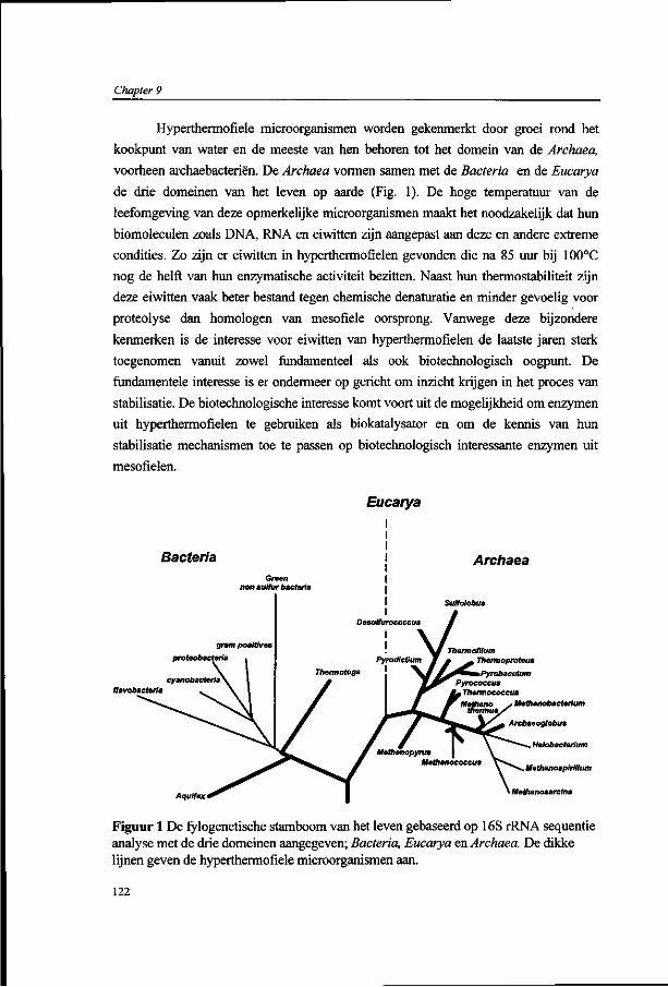
Chapter 9
Hypertheraiofiele microorganismen worden gekenmerkt door groei rond het
kookpunt van water en de meeste van hen behoren tot het domein van de Archaea,
voorheen archaebacteriën. De Archaea vormen samen met de Bacteria en de Eucarya
de drie domeinen van het leven op aarde (Fig. 1). De hoge temperatuur van de
leefomgeving van deze opmerkelijke microorganismen maakt het noodzakelijk dat hun
biomoleculen zoals DNA, RNA en eiwitten zijn aangepast aan deze en andere extreme
condities. Zo zijn er eiwitten in hyperthermofielen gevonden die na 85 uur bij 100°C
nog de helft van hun enzymatische activiteit bezitten. Naast hun thermostabiliteit zijn
deze eiwitten vaak beter bestand tegen chemische denaturatie en minder gevoelig voor
Proteolyse dan homologen van mesofiele oorsprong. Vanwege deze bijzondere
kenmerken is de interesse voor eiwitten van hyperthermofielen de laatste jaren sterk
toegenomen vanuit zowel fundamenteel als ook biotechnologisch oogpunt. De
fundamentele interesse is er ondermeer op gericht om inzicht krijgen in het proces van
stabilisatie. De biotechnologische interesse komt voort uit de mogelijkheid om enzymen
uit hyperthermofielen te gebruiken als biokatalysator en om de kennis van hun
stabilisatie mechanismen toe te passen op biotechnologisch interessante enzymen uit
mesofielen.
Eucarya
Archaea Green
non sulfur bacteria
flavobacteria
Thermofilum Thermoproteus
Aquifex
Melhanobacterium
Archaeoglobus
Halobacterium
Methanospirillum
Methanosarclna
Figuur 1 De fylogenetische stamboom van het leven gebaseerd op 16S rRNA sequentie analyse met de drie domeinen aangegeven; Bacteria, Eucarya en Archaea. De dikke lijnen geven de hyperthermofiele microorganismen aan.
122

Samenvatting
Het onderzoek beschreven in dit proefschrift is ondermeer gericht op het
verkrijgen van inzicht in moleculaire mechanismen van adaptatie van eiwitten van
hyperthermofiele archaea die leiden tot extreme stabiliteit bij hoge temperatuur. Tevens
is getracht enzymen van hyperthermofielen te produceren in een heterologe gastheer
met behoud van hun biochemische karakteristieken, waardoor de toepassings
mogelijkheden van de enzymen uit hyperthermofielen vergroot worden. Om deze
doelstellingen te realiseren werd de aandacht geconcentreerd op twee groepen van
hydrolytische enzymen die een belangrijke plaats in nemen in het metabolisme van
hyperthermofiele archaea: (i) glycosyl hydrolases die betrokken zijn bij de groei op
polymère suikers waarvan de monomeren door middel van ß-verbindingen gekoppeld
zijn, en (ii) serine proteases die een rol vervullen bij de groei op eiwitten en peptiden.
De keuze voor hydrolytische enzymen komt ondermeer voort uit het feit dat homologen
van mesofiele en thermofiele oorsprong goed gekarakteriseerd zijn op biochemisch,
genetisch als ook op structureel niveau. Het organisme dat centraal heeft gestaan in dit
proefschrift is de hyperthermofiele archaeon Pyrococcus furiosus, de meest uitvoerig
bestudeerde vertegenwoordiger van de hyperthermofiele archaea. Regulatie van
transcriptie in dit archaeon is bestudeerd aan de hand van twee divergente transcripten
waarvan de produkten betrokken zijn bij de degradatie van ß-gekoppelde glucose
polymeren. Het onderzoek dat in dit proefschrift beschreven wordt heeft met name
betrekking op het celB locus van het hyperthermofiele archaeon P.fiiriosus en serine
proteases van dit archaeon en van de verwante Thermococcus stetteri.
Phe tRNA celB adhA adhB lamA birA pgk'
1 kb
Figuur 2. Genetische en transcriptionele organisatie van het celB locus van het hyperthermofiele
archaeaon P.fiiriosus.
In hoofdstuk 1 wordt een korte introductie gegeven in de verschillende aspecten
van hyperthermofiele archaea. Dit omvat ondermeer het metabolisme van polymère
substraten en de hydrolytische enzymen die hierbij betrokken zijn, de mechanismen die
123
Chapter 9
een rol spelen bij de stabilisatie van eiwitten in hyperthermofielen, en een aantal
opmerkelijke kenmerken van de moleculaire biologie van Archaea.
In hoofdstuk 2 wordt de isolatie en karakterisatie van het extreem stabiele ß-
glucosidase (halfwaarde tijd van 3 dagen bij 100°C) en het coderende celB gen van
P.fiiriosus beschreven. Dit ß-glucosidase is essentieel voor P.furiosus bij de groei op de
disaccharide cellobiose waar het de ß-l,4-glycoside binding tussen twee glucose
residuen splitst. De transcriptionele organisatie van het celB gen werd geanalyseerd en
suggereerde een aparte transcriptionele unit, hetgeen door Northern blot analyse
ondersteund werd (zie hieronder) (Fig. 2). Het celB gen coderend voor het ß-
glucosidase werd via reversed genetics geisoleerd en de afgeleide aminozuur sequentie
vertoonde de hoogste homologie met glycosyl hydrolases uit familie 1. Het celB van
P.fiiriosus werd tot overexpressie gebracht in de mesofiele gastheer Escherichia coli.
Dit resulteerde in een actief ß-glucosidase dat zowel kinetisch als structureel identiek
bleek te zijn aan het in P.fiiriosus geproduceerde eiwit. De productie van dit
hyperthermostabiel ß-glucosidase (tot 20% van totale oplosbare eiwit fractie) in de
mesofiele gastheer maakt de zuivering relatief eenvoudig door gebruik te maken van
een hitte behandeling gevolgd door een enkele chromatografische stap. Het hoge
productie niveau van het ß-glucosidase in de genetisch goed toegankelijke gastheer
E.coli maakte het mogelijk om door middel van 'protein engineering' inzicht te krijgen
in de structuur-stabiliteit en -functie relatie. Met behulp van plaatsgerichte mutagenese
kon worden aangetoond dat het mechanisme van katalyse voor het hyperthermostabiele
ß-glucosidase van P.furiosus overeenkomt met dat wat gevonden wordt voor een
homoloog uit de family 1 van glycosyl hydrolases dat optimaal actief is bij lage
temperatuur.
Vanwege zijn opmerkelijke kenmerken is in de afgelopen jaren gekeken naar de
mogelijkheden om het hyperthermostabiele ß-glucosidase van P.furiosus te gebruiken
als biokatalysator. Door de brede substraatspecificiteit van het ß-glucosidase was het
mogelijk om diverse produkten te synthetiseren in glucoconjugatie reacties in water en
organische oplosmiddelen bij temperaturen van 75°C tot 95°C. Tevens is gebleken dat
het ß-glucosidase van P.fiiriosus gebruikt kan worden voor de synthese van ß-galacto-
oligosaccharides uit lactose, die een belangrijke rol kunnen spelen in de
voedingsmiddelen industrie vanwege hun potentiële functie als prebiotica (Jansen et al,
1997). Daarnaast is het celB gen van P.furiosus ontwikkeld tot een genetische merker
voor de studie van plant-bacterie interacties en competitie van Rhizobium stammen. De
124
Samenvatting
toepassingsmogelijkheden van dit systeem heeft uiteindelijk geleid tot de ontwikkeling
van de CelB Gene Marking Kit (FAO/IAEA).
Het hoge productie niveau van het pyrococcus ß-glucosidase in E.coli en de
eenvoudige zuiveringsprocedure maakte het mogelijk om voldoende gezuiverd eiwit te
genereren zodat kristallisatie experimenten opgezet konden worden in samenwerking
met de groep van Prof. G. Schulz (Universiteit van Freiburg). Dit heeft geresulteerd in
kristallen die diffracteren met een resolutie van 3,5 Angstrom (Schulz, 1997).
Momenteel wordt geprobeerd om een drie-dimensionale structuur met hoge resolutie
van het ß-glucosidase van P.furiosus op te lossen, gebruik makend van de recent
bepaalde structuur van het homologe LacS van Sulfolobus solfataricus in samenhang
met plaatsgerichte mutagenese om een betere pakking te bewerkstelligen.
Het celB gen van P.furiosus is gelegen naast een in tegenover gestelde richting
georiënteerd gencluster coderend voor twee alcohol dehydrogenasen (adhA, adhE) en
een endoglucanase (lamA) (Fig.2). In hoofdstuk 3 zijn de twee NADP(H)-afhankelijke
alcohol dehydrogenases (ADHs) bestudeerd die elk tot een afzonderlijke groep behoren,
de korte-keten ADH's (AdhA) en de ijzer-bevattende ADH's (AdhB). De primaire
structuur van het AdhA van P.furiosus vertoont een hoge mate van homologie met
korte-keten ADH's van bacteriële en eukaryele oorsprong. Dit kan als een aanwijzing
gezien worden dat er al een korte-keten ADH aanwezig was in de laatste
gemeenschappelijke voorouder van het leven, de progenoot. De primaire structuur van
AdhB van P.furiosus vertoont de hoogste homologie met ijzer-bevattende ADHs, een
relatief kleine groep die gekenmerkt wordt door ondermeer 4 geconserveerde histidine
residuen. De adhA en adhB genen zijn tot expressie gebracht in E.coli hetgeen
resulteerde in functionele eiwitten. Het AdhB, geproduceerd in E.coli, was in staat om
methanol om te zetten in een NADP(H)-afhankelijke reactie, maar deze activiteit ging
snel verloren waardoor er geen gedetailleerde karakterisatie plaats kon vinden. Het
gezuiverde AdhA daarentegen vertoonde een stabiele NADP(H)-afhankelijke activiteit
met een groot aantal primaire en secundaire alcoholen. Het substraat waar AdhA de
hoogste activiteit mee vertoonde bestaat uit een koolstofketen van 5 residuen, met een
voorkeur voor secundaire boven primaire alcoholen. De affiniteit van het P.furiosus
AdhA bleek hoger voor aldehydes dan voor alcoholen en het enzym had een pH
optimum voor deze reductie dat dicht bij de fysiologische pH ligt van P.furiosus. Het is
dan ook het meest aannemelijk dat de fysiologische rol van AdhA bestaat uit de reductie
van de aldehyde tot het corresponderende alcohol waarbij het de reductie-equivalenten
verwijdert en cofactor regenereert. De lokatie van de adhA en adhB genen, in de
125

Chapter 9
nabijheid van het lamA en het celB gen, maakt het verleidelijk te speculeren dat AdhA
en AdhB beiden een rol vervullen in het metabolisme van ß-gekoppelde glucose
polymeren.
In hoofdstuk 4 wordt de moleculaire karakterisatie van een endo-ß-1,3-
glucanase en het hiervoor coderende lamA gen van P.furiosus beschreven. Het lamA
gen ligt stroomafwaarts van de tandem adhA-adhB genen (Fig. 2). Dit endo-ß-1,3-
glucanase van P.furiosus is in het domein van de Archaea de eerste vertegenwoordiger
van de glycosyl hydrolase familie 16, die bestaat uit endo-ß-1,3- en endo-ß-1,3-1,4-
glucanases. Voor de expressie in E. coli werd het lamA gen, zonder het DN A-fragment
coderend voor de N-terminale leader sequentie, achter een T7 promoter geplaatst. Met
dit expressiesysteem werd een functioneel en stabiel endo-ß-l,3-glucanase
geproduceerd, dat 15 % van het totale oplosbare eiwitfractie van E. coli uitmaakte. Het
gezuiverde enzym had de hoogste activiteit met ß-1,3 -glucose polymeren, maar
vertoonde ook activiteit met de ß-l,3-l,4-glucose polymeren lichenan en ß-glucan. Het
endoglucanase vertoonde optimale activiteit bij pH 6-6.5 en een maximale activiteit bij
100-105°C, terwijl het bij 100°C de halfwaarde tijd had van 19 uur. Vergelijking van de
aminozuur volgorde van glycosyl hydrolases van familie 16 geeft een onderverdeling te
zien in een subgroep met endo-ß-l,3-l,4-glucanase activiteit en een tweede subgroep
met voornamelijk endo-ß-l,3-glucanase activiteit. Deze laatste subgroep wordt
gekarakteriseerd door een extra methionine residue in de voorspelde actieve site.
Verwijdering van deze methionine in het endo-ß-l,3-glucanase van P.furiosus door
middel van 'protein engineering' resulteerde niet in een veranderde substraat
specificiteit, maar een verlaagde katalytische activiteit.
Het is gebleken dat P.furiosus in staat is om op het ß-l,3-gekoppelde glucose
polymeer laminarine te groeien. Daarnaast bleek dat het endoglucanase in vitro
laminarine te hydrolyseren tot oligomeren en glucose. Echter, door toevoeging van het
ß-glucosidase van P.furiosus verliep de hydrolyse veel efficiënter. Deze
complementaire activiteiten van het extracellulaire endoglucanase en het intracellulaire
ß-glucosidase suggereren een essentiële rol voor deze enzymen bij groei op laminarine.
In hoofdstuk 5 wordt de regulatie van transcriptie beschreven van het celB gen
en het divergent georiënteerde adhA-adhB-lamA gencluster in P.furiosus. Met behulp
van Northern blot analyse kon worden aangetoond dat de adhA-adhB-lamA genen een
2.8-kb operon vormen, het lamA operon. Dit operon wordt stroomopwaarts geflankeerd
door het celB gen dat een tegenovergestelde oriëntatie bezit en stroomafwaarts door het
birA gen (Fig. 2). Het celB gen bleek overgeschreven te worden als monocistrone
126

Samenvatting
messenger. De aanwezigheid van de enzymen, ß-glucosidase, alcohol dehydrogenase en
endoglucanase, die gecodeerd worden door het celB gen en het lamA operon van
P.furiosus, was in hoge mate afhankelijk van de aanwezige koolstofbron. De hoogste
enzym niveau's werden gevonden in P.furiosus cellen die gegroeid waren op ß-
gekoppelde glucose polymeren, zoals cellobiose en laminarine. Transcriptionele
analyses van het celB gen en het divergent georiënteerde lamA operon gaven aan dat de
controle plaats vindt op transcriptie niveau. Bovendien vindt er co-regulatie van de
transcripten plaats en worden ze geïnduceerd door groei op ß-gekoppelde glucose
polymeren. De transcriptie initiatie startplaatsen van het celB gen en het lamA operon
lagen slechts 142 nucleotiden van elkaar verwijderd. De promotoren van beide genen
vertoonden een hoge mate van conservering, waaronder de identieke archaele TATA-
box. Analyse van transcriptie initiatie met behulp van een in vitro transcriptie systeem
voor P.furiosus toonde aan dat transcriptie van het celB gen en het lamA operon op
dezelfde plaats start als in vivo. Echter, de efficiëntie van transcriptie initiatie van zowel
het celB gen als het lamA operon in dit systeem waren significant lager dan dat van het
efficient overgeschreven gdh gen van P.furiosus. Dit suggereert dat een positieve
regulator een bijdrage zou kunnen leveren aan de efficiëntie van transcriptie van de
divergente transcripten van celB gen en het lamA operon in P.furiosus. Een mogelijke
bindingsplaats voor een dergelijke regulator bevindt zich stroomopwaarts van de
promotoren van het celB gen en het lamA operon. De resultaten van deze
transcriptionele analyses maken het mogelijk om controle van transcriptie te bestuderen
in het in vitro transcriptie systeem van P.furiosus en een regulator te identificeren die
betrokken is bij de regulatie van de divergente genen van het celB locus. Daarnaast
wordt een in vivo analyse van transcriptionele controle van het celB locus wordt
wellicht haalbaar met de recente ontwikkelingen in het transformatie systeem voor
P.furiosus.
Een combinatie van de huidige stand van zaken en inzichten met betrekking tot
het metabolisme van ß-gekoppelde glucose polymeren en de transcriptionele regulatie
in het hyperthermofiele archaeon P.furiosus heeft geleid tot het volgende werkmodel
(Fig. 3). De eerste stap is een depolymerisatie van grote polymère substraten die ß-1,3-
gekoppelde glucose verbindingen bevatte, zoals laminarine, tot kleinere fragmenten
door het extracellulaire endo-ß-l,3-glucanase (LamA). Na opname worden de kleine
fragmenten intracellulair gehydrolyseerd door ß-glucosidase activiteit van CelB. De
complementaire activiteiten van deze twee glycosyl hydrolases kan inderdaad in vitro
worden aangetoond. Ook de co-regulatie van transcriptie van de genen coderend voor
127
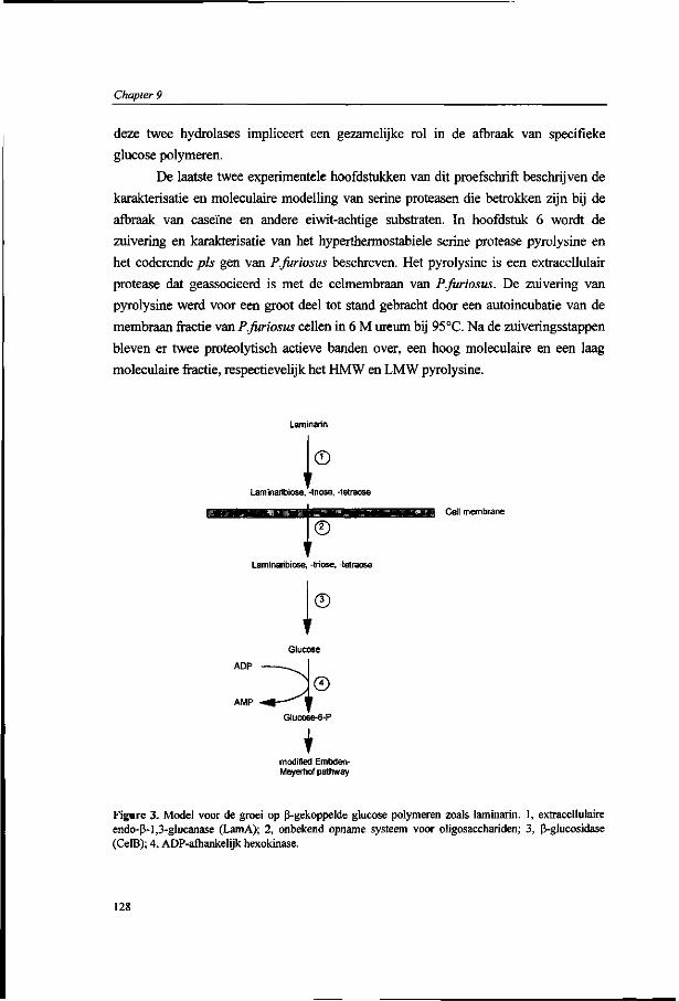
Chapter 9
deze twee hydrolases impliceert een gezamelijke rol in de afbraak van specifieke
glucose polymeren.
De laatste twee experimentele hoofdstukken van dit proefschrift beschrijven de
karakterisatie en moleculaire modelling van serine proteasen die betrokken zijn bij de
afbraak van caseïne en andere eiwit-achtige substraten. In hoofdstuk 6 wordt de
zuivering en karakterisatie van het hyperthermostabiele serine protease pyrolysine en
het coderende pis gen van P.furiosus beschreven. Het pyrolysine is een extracellulair
protease dat geassocieerd is met de celmembraan van P.furiosus. De zuivering van
pyrolysine werd voor een groot deel tot stand gebracht door een autoincubatie van de
membraan fractie van P.furiosus cellen in 6 M ureum bij 95°C. Na de zuiveringsstappen
bleven er twee proteolytisch actieve banden over, een hoog moleculaire en een laag
moleculaire fractie, respectievelijk het HMW en LMW pyrolysine.
© Lamlnaribiose. -triose, -tetraose
© g Cell membrane
Lam in a ri bi ose, -triose, -tetraose
©
ADP
AMP
modified Embden-Meyerhof pathway
Figure 3. Model voor de groei op ß-gekoppelde glucose polymeren zoals laminarin. 1, extracellulaire endo-ß-l,3-glucanase (LamA); 2, onbekend opname systeem voor Oligosacchariden; 3, ß-glucosidase (CelB); 4, ADP-afhankelijk hexokinase.
128
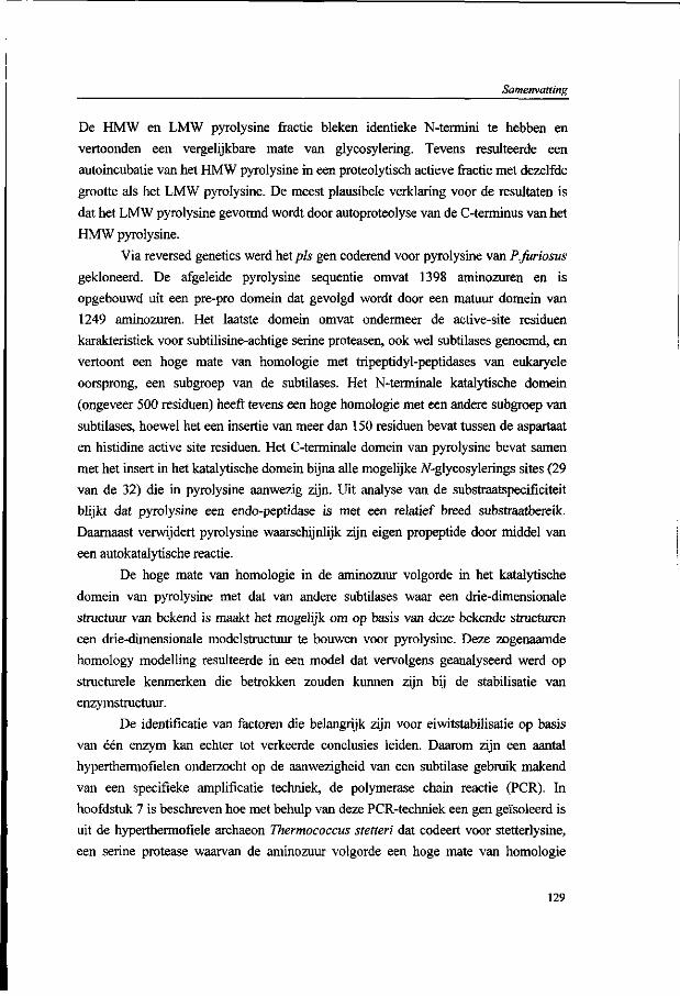
Samenvatting
De HMW en LMW pyrolysine fractie bleken identieke N-termini te hebben en
vertoonden een vergelijkbare mate van glycosylering. Tevens resulteerde een
autoincubatie van het HMW pyrolysine in een proteolytisch actieve fractie met dezelfde
grootte als het LMW pyrolysine. De meest plausibele verklaring voor de resultaten is
dat het LMW pyrolysine gevormd wordt door autoproteolyse van de C-terminus van het
HMW pyrolysine.
Via reversed genetics werd het pis gen coderend voor pyrolysine van P.fitriosus
gekloneerd. De afgeleide pyrolysine sequentie omvat 1398 aminozuren en is
opgebouwd uit een pre-pro domein dat gevolgd wordt door een matuur domein van
1249 aminozuren. Het laatste domein omvat ondermeer de active-site residuen
karakteristiek voor subtilisine-achtige serine proteasen, ook wel subtilases genoemd, en
vertoont een hoge mate van homologie met tripeptidyl-peptidases van eukaryele
oorsprong, een subgroep van de subtilases. Het N-terminale katalytische domein
(ongeveer 500 residuen) heeft tevens een hoge homologie met een andere subgroep van
subtilases, hoewel het een insertie van meer dan 150 residuen bevat tussen de aspartaat
en histidine active site residuen. Het C-terminale domein van pyrolysine bevat samen
met het insert in het katalytische domein bijna alle mogelijke 7V-glycosylerings sites (29
van de 32) die in pyrolysine aanwezig zijn. Uit analyse van de substraatspecificiteit
blijkt dat pyrolysine een endo-peptidase is met een relatief breed substraatbereik.
Daarnaast verwijdert pyrolysine waarschijnlijk zijn eigen propeptide door middel van
een autokatalytische reactie.
De hoge mate van homologie in de aminozuur volgorde in het katalytische
domein van pyrolysine met dat van andere subtilases waar een drie-dimensionale
structuur van bekend is maakt het mogelijk om op basis van deze bekende structuren
een drie-dimensionale modelstructuur te bouwen voor pyrolysine. Deze zogenaamde
homology modelling resulteerde in een model dat vervolgens geanalyseerd werd op
structurele kenmerken die betrokken zouden kunnen zijn bij de stabilisatie van
enzymstructuur.
De identificatie van factoren die belangrijk zijn voor eiwitstabilisatie op basis
van één enzym kan echter tot verkeerde conclusies leiden. Daarom zijn een aantal
hyperthermofielen onderzocht op de aanwezigheid van een subtilase gebruik makend
van een specifieke amplificatie techniek, de polymerase chain reactie (PCR). In
hoofdstuk 7 is beschreven hoe met behulp van deze PCR-techniek een gen geïsoleerd is
uit de hyperthermofiele archaeon Thermococcus stetteri dat codeert voor stetterlysine,
een serine protease waarvan de aminozuur volgorde een hoge mate van homologie
129

Chapter 9
vertoont met die van pyrolysine. De hoge mate van overeenkomst tussen de twee serine
Proteasen uit T.stetteri en P.fiiriosus, en de subtilases waarvan de drie-dimensionale
structuur zijn opgehelderd heeft homology modelling mogelijk gemaakt. Analyse van
de modelstructuren heeft inzicht gegeven in factoren die een bijdrage zouden kunnen
leveren aan de extreme stabiliteit van deze proteasen. Zo werd gevonden dat er een
correlatie is tussen stabiliteit en de toename in het aantal zout-bruggen, zoals dat ook in
ander eiwitten van hyperthermofiele oorsprong is gevonden. Tevens is er in de
stabielere proteasen een significante toename gevonden in het aantal aromatische
residuen, die betrokken kunnen zijn bij de vorming van interacties tussen aromatische
residuen. Zowel in stetterlysine als in pyrolysine bleken de meeste aromatische residuen
gelegen te zijn aan het oppervlak van de catalytische core en in het insert in dit domein.
Dit suggereert dat voor de globale structuur van deze proteasen de interacties tussen
aromatische residuen een belangrijke rol kunnen spelen.
Analyse van de locatie van 7V-glycosylerings sites in thermostabiele subtilases
waaronder stetterlysin en pyrolysin, gaf aan dat de meeste van deze sites zijn
gelokaliseerd in loops aan het oppervlak van de structuur. De modellering van de
substraat bindings regio's met de geïdentificeerde substraten vertoonden een goede
overeenkomst met het voorheen gevonden brede substraatbereik van pyrolysine en de
veronderstelde autokatalytische activering (Hoofdstuk 6).
De PCR benadering die gebruikt werd voor de isolatie van subtilisine-achtige
serine proteasen heeft geresulteerd in DNA fragmenten met de verwachte grootte voor
het hyperthermofiele archeaon Pyrodictium abyssi en de hyperthermofiele bacterium
Fervidobacterium pennavorans. Beide vertoonde hybridisatie met een van pyrolysine
afgeleide probe. De PCR producten werden gekloneerd en gekarakteriseerd en de
afgeleide aminozuur sequentie bleek een significante homologie te hebben met het
catalytische domein van een aantal subtilases. Echter, in tegenstelling tot stetterlysine en
pyrolysine, bevatten ze geen groot insert tussen de eerste twee active site residuen. Het
PCR fragment van F.pennavorans werd vervolgens gebruikt voor de isolatie van
chromosomale DNA fragmenten die het gehele gen bevatten en worden momenteel
gekarakteriseerd.
De hydrolases die beschreven zijn in dit proefschrift zijn gebruikt als model om
verschillende aspecten te bestuderen van enzymen van hyperthermofiele
microorganismen, deze enzymen worden ook wel thermozymen genoemd. De industrie
heeft in hyperthermofielen gezocht naar enzymen de gebruikt zouden kunnen worden
als biokatalysator ter vervanging van minder stabiele homologe enzymen of om nieuwe
130

Samenvatting
processen te initiëren. Echter het gebruik van thermozymen in de industrie wordt
bemoeilijkt door de dure fermentatie processen die nodig zijn om de hyperthermofiele
microorganismen te kweken die de bewuste thermozymen bevatten. Functionele
overproductie van thermozymen in heterologe gastheren zou daarom een sterke bijdrage
kunnen leveren in de ontwikkeling van thermozymen als biokatalysator. Dit proefschrift
beschrijft de functionele overproductie van een aantal thermozymen inclusief een ß-
glucosidase, een korte-keten alcohol dehydrogenase en een endo-ß-l,3-ghicanase.
Tevens heeft het een bijdrage geleverd aan het inzicht in de structuur-stabiliteit en -
structuur functie relatie van hydrolytische enzymen van hyperthermofiele archaea.
Referenties:
Aagaard, C , Leviev, L, Aravalli, R.N., Forterre, P., Prieur, D., and Garrett, R.A. (1996) General vectors for archaeal hyperthermophiles: Strategies based on a mobile intron and a plasmid. FEMS Microbiol. Rev. 18: 93-104.
Fischer, L., Bromann, R., Kengen, S.W.M., de Vos, W.M., and Wagner, F. (1996) Catalytic potency of ß-glucosidase from the extremphile Pyrococcus furiosus in glucoconjugate synthesis. Biotechnology 14: 88-93.
Jansen, A and Boon,F. (1997) Unpublished results.
Schulz, G.E. (1997) Unpublished results
Sessitch, A., Wilson, K.J., Akkermans, A.D.L., and de Vos, W.M. (1996) Simultaneous detection of different Rhizobium strains marked with either the Escherichia coli gus A gen or the Pyrococcus furiosus celB gene. Appl. Environm. Microbiol. 62: 4191-4194.
Trincone A, Pagnotta, E., Voorhorst, W.G.B., van der Oost, J., and de Vos, W.M. (1997) Stereochemistry of Glycosyl Transfer Using the ß-glucosidase from the Hyperthermophilic Archaeon Pyrococcus furiosus. (Submitted to Carbohydrates).
131

Chapter 9
132

Curriculum Vitae
Curriculum Vitae
Willebrordus Gerardus Bernardus (Wilfried) Voorhorst werd geboren op 7 augustus
1967 te Raalte. Na de basisschool is hij begonnen aan de HAVO (1979-1984) gevolgd
door het VWO (1984-1986) op het Florens Radewijns College te Raalte. In 1986 is hij
gestart met de studie Biologie aan de Landbouwuniversiteit Wageningen om na de
propaedeuse de oriëntatie celbiologie te volgen. Het afstudeervak Industriële
Microbiologie (Prof. J.A.M, de Bont) werd gevolgd door Moleculaire Biologie (Prof.
dr. A. van Kammen). Zijn stages vonden plaats bij Shell Research Centre Sittingbourne,
Engeland (Dr. A. Cornish) en de vakgroep Biochemie (Prof. dr. W.R. Hagen). Op 30
augustus 1992 sloot hij zijn universitaire studie af als ingenieur in de Landbouw- en
Milieuwetenschappen. Op 12 augustus 1992 werd hij aangesteld als Assistent-In-
Opleiding (AIO) bij de vakgroep Microbiologie van de Landbouwuniversiteit
Wageningen. Binnen de werkgroep Bacteriële Genetica heeft hij gewerkt aan de
moleculaire karakterisatie van hydrolytische enzymen van hyperthermofiele archaea,
hetgeen uiteindelijk in dit proefschrift heeft geresulteerd.
133

134

List of publications
List of publications:
Sander A. Peters, Wilfried G.B. Voorhorst, Jan Wery, Joan Wellink, and Albert van Kammen (1992) A Regulatory Role for the 32K Protein in Proteolytic Processing of Cowpea Mosaic Virus Polyproteins. Virology, 191, 81-89
Sander A. Peters, Wilfried G.B. Voorhorst, Joan Wellink, and Albert van Kammen (1992) Processing of VPg-Containing Polyproteins Encoded by the B-RNA from Cowpea Mosaic Virus. Virology, 191,90-97
André de Haan, Mark R. Smith, Wilfried G.B. Voorhorst, and Jan A.M. de Bont (1993) Co-factor Regeneration in the Production of 1,2-epoxypropane by Mycobacterium strain E3: The Role of Storage Material. Journal of General Microbiology, 139, 3017-3022
Wilfred R. Hagen, Marc F.J.M. Verhagen, Antinio J. Pierik, Ronnie B.G. Wolbert, Leonard F. Mallée, Wilfried G.B. Voorhorst (1993) Axial Coordination and Reduction Potentials of the 16 Hemes in High-Molecular-Weight Cytochrome c from Desulfovibrio vulgaris. Journal of Inorganic Biochemistry, 51,28
Marc F.J.M. Verhagen, Wilfried G.B. Voorhorst, Joost A. Kolkman, Ronnie B.G. Wolbert, and Wilfred R. Hagen (1993) On the Two Iron Centers of Desulfoferrodoxin. FEBS Letters, 336, 13-18
Mare F.J.M. Verhagen, Antonio J. Pierik, Ronnie B.G. Wolbert, Leonard F. Mallée, Wilfried G.B. Voorhorst, and Wilfred R. Hagen (1994) Axial Coordination and Reduction Potentials of the Sixteen Hemes in High-Molecular-Mass Cytochrome c from Desulfovibrio vulgaris (Hildenborough). European Journal of Biochemistry, 225, 311-319
Rik I.L. Eggen, Ans CM. Geerling, Wilfried G.B. Voorhorst, Remco Kort, and Willem M. de Vos (1994) Molecular and Comparative Analysis of the Hyperthermostable Pyrococcus furiosus Glutamate Dehydrogenase and Its Gene. Biocatalysis, 11, 131-141
Wilfried G.B. Voorhorst, Rik I.L. Eggen, Evert J. Luesink, and Willem M. de Vos (1995) Characterization of the celB Gene Coding for ß-Glucosidase from the Hyperthermophilic Archaeon Pyrococcus furiosus and Its Expression and Site-Directed Mutation in Escherichia coli. Journal of Bacteriology, 177, 7105-7111
Wilfried G.B. Voorhorst, Rik I.L. Eggen, Ans C.M. Geerling, Christ Platteeuw, Roland J. Siezen, and Willem M. de Vos (1996) Isolation and Characterization of the Hyperthermostable Serine Protease, Pyrolysin, and Its Gene from the Hyperthermophilic Archaeon Pyrococcus furiosus. Journal of Biological Chemistry, 271 (34), 20426
Wilfried G.B. Voorhorst, Angela Warner, Willem M. de Vos, and Roland J. Siezen (1997) Homology Modelling of Two Subtilisin-like Serine Proteases from Hyperthermophilic Archaea Pyrococcus furiosus and Thermococcus stetteri. Protein Engineering, 10 (8),905-914
Yannick Gueguen, Wilfried G.B. Voorhorst, John van der Oost, and Willem M. de Vos (1997) Cloning and Sequencing of the lamA gene from the Hyperthermophilic Archaeon Pyrococcus furiosus Coding for an Endo-ß-l,3-glucanase and Its Expression and Site-directed Mutation in Escherichia coli Journal of Biological Chemistry, 272, 31258-64
Wilfried G.B. Voorhorst, Vincent Wittenhorst, John van der Oost, and Willem M. de Vos (1997) Genetic and Biochemical Characterization of a Short-chain and an Iron-containing Alcohol Dehydrogenase from the Hyperthermophilic Archeaon Pyrococcus /wn'asM.s.(submitted for publication)
Wilfried G.B. Voorhorst, Yannick Gueguen, Gerti Schut, Isabell Dahlke, Michael Thomm, John van der Oost, and Willem M. de Vos (1997) Transcriptional Regulation in the Hyperthermophilic Archaeon Pyrococcus furiosus: Coordinated Expression of Divergently Transcribed Genes in Response to ß-linked Glucose Polymers (submitted for publication)
Antionio Trincone, Edoardo Pagnotta, Wilfried G.B. Voorhorst, John van der Oost, and Willem M. de Vos (1997) Stereochemistry of Glycosyl Transfer Using the ß-glucosidase from the Hyperthermophilic Archaeon Pyrococcus furiosus. (Submitted to Carbohydrates)
Willem M. de Vos, Serve Kengen, Wilfried G.B. Voorhorst, and John van der Oost (1998) Sugar Utilization and Its Control in Hyperthermophiles. Extremophiles (In press)
135

136