Molecular Cell Article - Harvard...
15
Molecular Cell Article BID Preferentially Activates BAK while BIM Preferentially Activates BAX, Affecting Chemotherapy Response Kristopher A. Sarosiek, 1,2 Xiaoke Chi, 3 John A. Bachman, 4 Joshua J. Sims, 4 Joan Montero, 1,2 Luv Patel, 1,2 Annabelle Flanagan, 1 David W. Andrews, 3 Peter Sorger, 4 and Anthony Letai 1,2, * 1 Department of Medical Oncology, Dana-Farber Cancer Institute, Boston, MA 02215, USA 2 Harvard Medical School, Boston, MA 02115, USA 3 Department of Chemistry and Chemical Biology, McMaster University, Hamilton, ON L8N 3Z5, Canada 4 Department of Systems Biology, Harvard Medical School, Boston, MA 02115, USA *Correspondence: [email protected] http://dx.doi.org/10.1016/j.molcel.2013.08.048 SUMMARY Apoptosis is a highly regulated form of cell death that controls normal homeostasis as well as the antitumor activity of many chemotherapeutic agents. Commit- ment to death via the mitochondrial apoptotic pathway requires activation of the mitochondrial pore-forming proteins BAK or BAX. Activation can be effected by the activator BH3-only proteins BID or BIM, which have been considered to be function- ally redundant in this role. Herein, we show that sig- nificant activation preferences exist between these proteins: BID preferentially activates BAK while BIM preferentially activates BAX. Furthermore, we find that cells lacking BAK are relatively resistant to agents that require BID activation for maximal induc- tion of apoptosis, including topoisomerase inhibitors and TRAIL. Consequently, patients with tumors that harbor a loss of BAK1 exhibit an inferior response to topoisomerase inhibitor treatment in the clinic. Therefore, BID and BIM have nonoverlapping roles in the induction of apoptosis via BAK and BAX, affecting chemotherapy response. INTRODUCTION Apoptosis is a highly regulated form of cell death that is essential for normal growth and development of an organism and for cull- ing damaged, dysfunctional, or superfluous cells (Jacobson et al., 1997). Apoptosis can be triggered via the extrinsic pathway, which involves activation of cell surface death recep- tors, or the intrinsic pathway, which requires mitochondrial outer membrane permeabilization (MOMP) (Tait and Green, 2010). Activation of either pathway culminates in the activation of the executioner caspases, caspase 3 and caspase 7, for dismantling of the cell (Tait and Green, 2010). Notably, receptor-mediated cell death requires MOMP in most cell types (Tait and Green, 2010). The mitochondrial apoptotic pathway, which contributes to the antitumor effects of many chemotherapies (Johnstone et al., 2002), is controlled by the pro- and antiapoptotic proteins of the BCL-2 family. Members of this family can be divided into three classes based on their sequence homology and function. One class includes the antiapoptotic proteins BCL-2, BCL-w, MCL-1, BFL-1, and BCL-X L , which contain all four BCL-2 homol- ogy domains (BH1-4). The proteins in this class prevent apoptosis by binding and sequestering their proapoptotic coun- terparts. The second class includes the proapoptotic proteins PUMA, BIM, BID, BAD, BIK, NOXA, and BMF, which contain only the BH3 domain (BH3-only). The final class contains BCL- 2-associated X protein (BAX) and BCL-2 antagonist or killer (BAK), which contain domains BH1-3. BAX and BAK, when acti- vated, oligomerize and directly cause MOMP, a critical event during apoptosis. Cytochrome c and other factors are released during MOMP and associate with several cytosolic proteins to activate caspases for cell death. Of the proapoptotic BH3-only proteins, BIM and BID have been shown to directly activate BAX and BAK with a particularly high potency and are consequently termed ‘‘activator’’ BH3-only proteins (Czabotar et al., 2013; Kim et al., 2006; Leshchiner et al., 2013; Letai et al., 2002; Wei et al., 2000). BIM and BID also share similar binding profiles for the antiapoptotic members of the BCL-2 family, since they are both able to bind and inactivate all five of the major antiapoptotic proteins listed above (Certo et al., 2006). Although BIM and BID have been considered to be functionally redundant with regard to BAX and BAK activation, several differ- ences in function and physiological roles have emerged for these two proteins. BIM and BID have nonoverlapping physiological roles in maintenance of homeostasis and distinct expression patterns (Farrow et al., 1995; Kiefer et al., 1995; Krajewski et al., 1996; O’Connor et al., 1998). BIM, but not BID, has been linked to cell death following growth factor withdrawal (Biswas and Greene, 2002) and to the apoptosis that is required for proper regulation of the immune system (Bouillet et al., 1999). In contrast, BID, but not BIM, is cleaved and activated by cas- pases 8 and 10 to trigger cell death in response to activation of cell surface death receptors by Fas, TNF, and TRAIL (Li et al., 1998; Luo et al., 1998). BID has also been implicated in apoptosis following treatment with DNA damaging agents, including topo- isomerase inhibitors (Kamer et al., 2005; Slee et al., 2000; Zinkel et al., 2005). Specifically, topoisomerase I and/or II inhibitors Molecular Cell 51, 751–765, September 26, 2013 ª2013 Elsevier Inc. 751
Transcript of Molecular Cell Article - Harvard...

Molecular Cell
Article
BID Preferentially Activates BAKwhile BIM Preferentially Activates BAX,Affecting Chemotherapy ResponseKristopher A. Sarosiek,1,2 Xiaoke Chi,3 John A. Bachman,4 Joshua J. Sims,4 Joan Montero,1,2 Luv Patel,1,2
Annabelle Flanagan,1 David W. Andrews,3 Peter Sorger,4 and Anthony Letai1,2,*1Department of Medical Oncology, Dana-Farber Cancer Institute, Boston, MA 02215, USA2Harvard Medical School, Boston, MA 02115, USA3Department of Chemistry and Chemical Biology, McMaster University, Hamilton, ON L8N 3Z5, Canada4Department of Systems Biology, Harvard Medical School, Boston, MA 02115, USA
*Correspondence: [email protected]
http://dx.doi.org/10.1016/j.molcel.2013.08.048
SUMMARY
Apoptosis is a highly regulated form of cell death thatcontrols normal homeostasis aswell as the antitumoractivity of many chemotherapeutic agents. Commit-ment to death via the mitochondrial apoptoticpathway requires activation of the mitochondrialpore-forming proteins BAK or BAX. Activation canbe effected by the activator BH3-only proteins BIDor BIM, which have been considered to be function-ally redundant in this role. Herein, we show that sig-nificant activation preferences exist between theseproteins: BID preferentially activates BAK while BIMpreferentially activates BAX. Furthermore, we findthat cells lacking BAK are relatively resistant toagents that require BID activation for maximal induc-tion of apoptosis, including topoisomerase inhibitorsand TRAIL. Consequently, patients with tumors thatharbor a loss of BAK1 exhibit an inferior responseto topoisomerase inhibitor treatment in the clinic.Therefore, BID and BIM have nonoverlapping rolesin the induction of apoptosis via BAK and BAX,affecting chemotherapy response.
INTRODUCTION
Apoptosis is a highly regulated form of cell death that is essential
for normal growth and development of an organism and for cull-
ing damaged, dysfunctional, or superfluous cells (Jacobson
et al., 1997). Apoptosis can be triggered via the extrinsic
pathway, which involves activation of cell surface death recep-
tors, or the intrinsic pathway, which requires mitochondrial outer
membrane permeabilization (MOMP) (Tait and Green, 2010).
Activation of either pathway culminates in the activation of the
executioner caspases, caspase 3 and caspase 7, for dismantling
of the cell (Tait and Green, 2010). Notably, receptor-mediated
cell death requires MOMP in most cell types (Tait and Green,
2010). The mitochondrial apoptotic pathway, which contributes
to the antitumor effects of many chemotherapies (Johnstone
Molecu
et al., 2002), is controlled by the pro- and antiapoptotic proteins
of the BCL-2 family. Members of this family can be divided into
three classes based on their sequence homology and function.
One class includes the antiapoptotic proteins BCL-2, BCL-w,
MCL-1, BFL-1, and BCL-XL, which contain all four BCL-2 homol-
ogy domains (BH1-4). The proteins in this class prevent
apoptosis by binding and sequestering their proapoptotic coun-
terparts. The second class includes the proapoptotic proteins
PUMA, BIM, BID, BAD, BIK, NOXA, and BMF, which contain
only the BH3 domain (BH3-only). The final class contains BCL-
2-associated X protein (BAX) and BCL-2 antagonist or killer
(BAK), which contain domains BH1-3. BAX and BAK, when acti-
vated, oligomerize and directly cause MOMP, a critical event
during apoptosis. Cytochrome c and other factors are released
during MOMP and associate with several cytosolic proteins to
activate caspases for cell death.
Of the proapoptotic BH3-only proteins, BIM and BID have
been shown to directly activate BAX and BAK with a particularly
high potency and are consequently termed ‘‘activator’’ BH3-only
proteins (Czabotar et al., 2013; Kim et al., 2006; Leshchiner et al.,
2013; Letai et al., 2002; Wei et al., 2000). BIM and BID also share
similar binding profiles for the antiapoptotic members of the
BCL-2 family, since they are both able to bind and inactivate
all five of the major antiapoptotic proteins listed above (Certo
et al., 2006).
Although BIM andBID have been considered to be functionally
redundant with regard to BAX and BAK activation, several differ-
ences in function and physiological roles have emerged for these
two proteins. BIM and BID have nonoverlapping physiological
roles in maintenance of homeostasis and distinct expression
patterns (Farrow et al., 1995; Kiefer et al., 1995; Krajewski
et al., 1996; O’Connor et al., 1998). BIM, but not BID, has been
linked to cell death following growth factor withdrawal (Biswas
and Greene, 2002) and to the apoptosis that is required for
proper regulation of the immune system (Bouillet et al., 1999).
In contrast, BID, but not BIM, is cleaved and activated by cas-
pases 8 and 10 to trigger cell death in response to activation of
cell surface death receptors by Fas, TNF, and TRAIL (Li et al.,
1998; Luo et al., 1998). BID has also been implicated in apoptosis
following treatment with DNA damaging agents, including topo-
isomerase inhibitors (Kamer et al., 2005; Slee et al., 2000; Zinkel
et al., 2005). Specifically, topoisomerase I and/or II inhibitors
lar Cell 51, 751–765, September 26, 2013 ª2013 Elsevier Inc. 751
(legend on next page)
Molecular Cell
Activation Preferences among BID, BIM, BAK, and BAX
752 Molecular Cell 51, 751–765, September 26, 2013 ª2013 Elsevier Inc.

Molecular Cell
Activation Preferences among BID, BIM, BAK, and BAX
require BID cleavage to induce maximal apoptosis (Maas et al.,
2011; Slee et al., 2000; Werner et al., 2004). These functional dif-
ferences are generally attributed to differences in stress-induced
regulation of BID and BIM rather than to any difference in activa-
tion of BAK or BAX.
BH3 profiling is an assay that allows detailed study of interac-
tions between BCL-2 family members. In this assay, mitochon-
dria in permeabilized cells are stained with a potential-sensitive
fluorescent dye, JC1, to monitor mitochondrial membrane integ-
rity. Mitochondrial transmembrane potential (DJm), measured
by JC1 fluorescence, is lost following MOMP as a secondary
effect following BAX and BAK activation and oligomerization
(Bossy-Wetzel et al., 1998). BH3 profiling enables monitoring
of mitochondrial integrity following perturbation with different
prodeath stimuli, including BH3 peptides or recombinant pro-
teins.We have previously utilized BH3 profiling to identify cellular
dependence on specific antiapoptotic proteins and to measure
overall mitochondrial apoptotic priming, which is predictive of re-
sponses to chemotherapy in vitro and in patients (Davids et al.,
2012; Deng et al., 2007; Ni Chonghaile et al., 2011; Vo et al.,
2012).
In this study, we show that although both BID and BIM can
activate both BAK and BAX, they exhibit differential activation
preferences: BID preferentially activates BAK, and BIMpreferen-
tially activates BAX. The activation preference of BID for BAK is
generally stronger than that of BIM for BAX, but both preferences
were observed consistently across mouse and human cells in
assays using either recombinant proteins or BH3 domain pep-
tides. These preferences are responsible for the relative protec-
tion of BAK-deficient cells from apoptosis induced by TRAIL or
by topoisomerase inhibitors, both of which require BID activation
for maximal apoptosis. Consequently, BAK deficiency leads to
inferior clinical responses in patients being treated with topo-
isomerase inhibitors.
RESULTS
We had previously observed that the BID BH3 peptide was more
effective than the BIM BH3 peptide at inducing cytochrome
release in mouse liver mitochondria, which contain detectable
levels of BAK, but not BAX (Letai et al., 2002). We therefore
hypothesized that BIM and BID may not be fully redundant in
their ability to activate BAX and BAK. To systematically assess
the abilities of BIM and BID to activate BAX and BAK, we utilized
mouse embryonic fibroblasts (MEFs) from mice that were either
wild-type (WT) or lacking either BAX, or BAK, or both (Figure 1A).
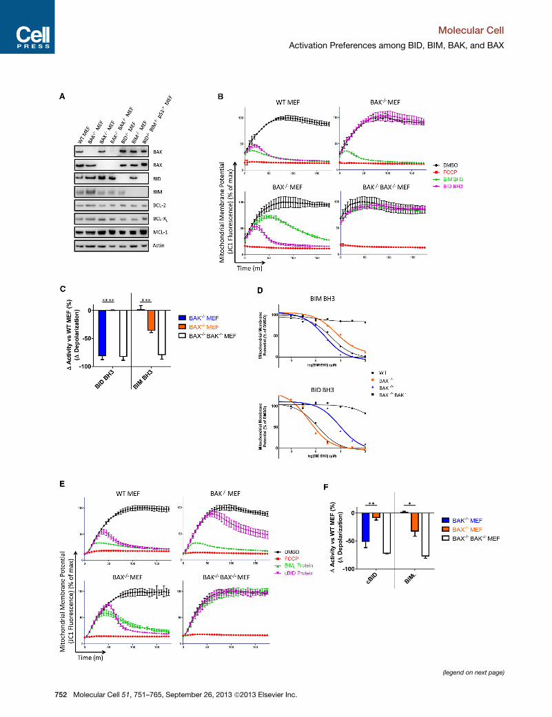
Figure 1. BID and BIM Preferentially Activate BAK and BAX, Respectiv
(A) Western blotting was performed for the indicted proteins. Data shown are rep
(B) Digitonin-permeabilized MEFs were treated with the mitochondrial potential-se
presence of BIM (10 mM) or BID (3 mM)BH3 peptides. DMSO served as a negative c
depolarized mitochondria). Data shown are representative of three independent
(C) The difference in mitochondrial depolarization by peptides was calculated fro
(D) Dose-response curves for BIM and BID BH3 peptide treatment of MEFs f
experiments.
(E) Mitochondrial polarization was monitored in MEFs in the presence of BIML (3
pendent experiments.
(F) The difference in mitochondrial depolarization by proteins was calculated from
Figures S1, S2, and S3 and Table S1.
Molecu
The levels of expression of other major BCL-2 family members in
these MEFs were similar (less than 50% difference) (Figures 1A
and S1A). To accurately identify any differences in the abilities
of BIM and BID peptides to activate BAX and BAK, we used
the BH3 profiling assay, which allowed us to monitor mitochon-
drial potential after peptide treatment. DMSO was used as
vehicle control, and FCCP, a proton ionophore, was used as a
positive control to measure the background fluorescence of
mitochondria that were unable to maintain a potential gradient.
Peptide titration experiments showed that both BIM and BID
BH3 peptides could induce mitochondrial depolarization (indica-
tive of MOMP) in WT MEFs (Figures 1B and S2) with slightly
higher activity of the BID versus the BIM peptide. Similar and
near-complete mitochondrial depolarization was observed with
3 mM BID peptide and 10 mM BIM peptide (Figure 1B, upper
left panel), which prompted us to use these doses to assess
the effect of loss of BAK or BAX. Strikingly, in MEFs lacking
BAK and expressing only BAX, response to BID peptide was
reduced as compared to WT MEFs, while the response to BIM
peptide was unaffected (Figures 1B and 1C). In contrast, the
response of MEFs lacking BAX and expressing only BAK to
BID peptide was comparable to that of WT MEFs, but response
to BIM peptide was significantly attenuated. MEFs lacking both
BAX and BAK exhibited no mitochondrial depolarization in
response to either the BIM or BID peptides. To exclude the pos-
sibility that the activation preferences we observed were due to
differences in the expression of antiapoptotic proteins among
the cell lines instead of direct activation of BAK and BAX, we in-
hibited the major antiapoptotic proteins prior to peptide treat-
ment and continued to observe the same activation preferences
(Figure S3). These data suggest that BID more efficiently acti-
vates BAK while BIM more efficiently activates BAX.
In MEFs, differences in responses to BIM and BID peptides
were evident across a range of peptide concentrations (Fig-
ure 1D) yielding EC50 values that differed almost 3-fold for the
BIM peptide (2.3 mM in BAK�/� and 6.7 mM in BAX�/�) andmore than 10-fold for BID peptide (9.3 mM in BAK�/� and
0.54 mM in BAX�/�) (Table S1). To obtain a more quantitative
understanding of the relative differences among interactions
between BIM, BID, BAX, and BAK, we performed mathematical
modeling of the mitochondrial depolarization observed (see
Experimental Procedures for details). From this modeling, we
obtained comparisons of rate (k) andmaximal effect (Fmax) (Table
S1). The parameters indicate that BID BH3 can activate MOMP
in BAX�/� mitochondria significantly faster than BIM BH3, (k =
0.115 versus 0.036% depolarized mM�1 min�1). For BAK�/�
ely, in Mouse Embryonic Fibroblasts
resentative of three independent experiments.
nsitive dye JC1 and mitochondrial potential was monitored over 180 min in the
ontrol (fully polarizedmitochondria) while FCCP served as positive control (fully
experiments.
m (B) and compared. Mean ± SEM of three independent experiments.
or calculation of EC50. Data shown are representative of three independent
0 nM) or cBID (30 nM) proteins. Data shown are representative of three inde-
(E) and compared. Mean ± SEM of three independent experiments. See also
lar Cell 51, 751–765, September 26, 2013 ª2013 Elsevier Inc. 753

Molecular Cell
Activation Preferences among BID, BIM, BAK, and BAX
mitochondria, BID BH3 is only slightly slower than BIM BH3 (k =
0.021 versus 0.017% depolarized mM�1 min�1) but significantly
less efficient, with an estimated EC50 for the Fmax of 4.5 mM
for BID BH3 and 0.7 mM for BIM BH3. These data confirm a sub-
stantial difference in BIM- and BID-mediated activation of BAX
and BAK.
We next tested if preferential activation would be also
observed for full-length, recombinant BIML and activated,
cleaved BID (cBID) proteins. We observed a close correlation
in the data for peptides and whole proteins (Figures 1E and
1F): BAK loss inhibited cBID-induced MOMP while having no
effect on BIML-induced MOMP. Conversely, loss of BAX had
more of an effect on BIML-induced MOMP than cBID-induced
MOMP, though the distinction was less absolute than for BAK
loss (Figures 1E and 1F). MEFs lacking both BAX and BAK
were again unresponsive to both recombinant BH3-only
proteins.
To determine whether preferential activation of BAK by BID
and BAX by BIM were evident in an alternate cell line of epithelial
origin, we repeated the MEF experiments with baby mouse
kidney (BMK) cells having the relevant genotypes. These cells
were immortalized in parallel using E1A and dominant-negative
p53 (Degenhardt et al., 2002) and ostensibly differ only in their
expression of BAK and BAX (Figures 2A and S1). The activation
preferences evident in the MEFs were observed in the BMKs us-
ing peptides (Figures 2A–2C) or recombinant cBID and BIML and
BIMS proteins (Figures 2D and 2E, BIMS data not shown).
To confirm differential activation of BAX and BAK by BIM and
BID in human cells, we utilized siRNA to knock down expression
of BAX and BAK in HeLa cells (Figure 3A). As in MEFs, the ability
of BIM BH3 peptide to induce mitochondrial depolarization was
most attenuated by loss of BAX, while the ability of BID BH3 to
induce MOMP was most attenuated by loss of BAK (Figures
3B and 3C). HeLa cells treated with recombinant BIML and
cBID gave similar results as the BH3 peptides, though in this
case BAX and BAK loss roughly equally affected cBID-induced
MOMP (Figures 3D and 3E). Overall, the differences in sensitiv-
ities to BIM and BID in the HeLa cells with knockdowns of
BAX and BAK were less categorical than those observed in
MEFs and BMKs, possibly due to far lower concentrations of
BAX and BAK in HeLa cells (Figure S4) or to less than complete
knockdown of BAX and BAK by RNAi. However, we observe the
same trend in all cell types, with loss of BAX preferentially
reducing BIM response, and loss of BAK preferentially reducing
BID response.
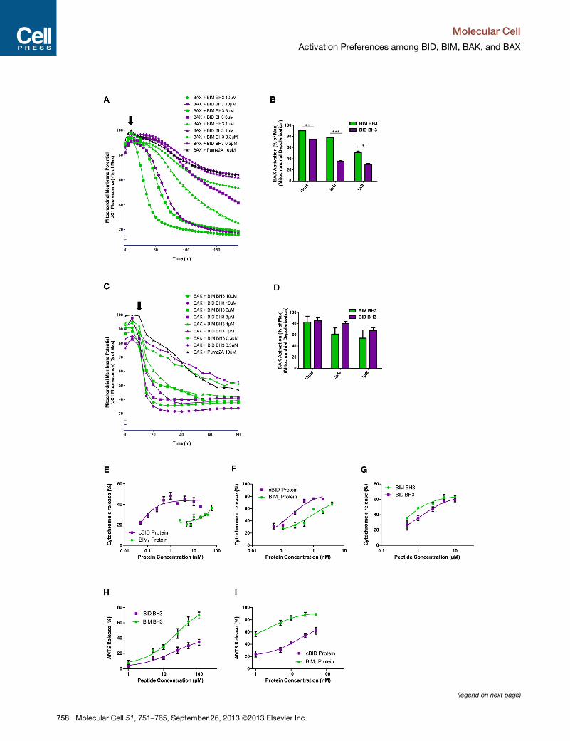
The experiments described above show that BIM and BID
possess distinct activation preferences for endogenously ex-
pressed BAX and BAK. Next we sought to determine whether
such preferences would also be evident with exogenous BAX
and BAK. We therefore incubated digitonin-permeabilized
BAX�/� BAK�/� MEFs with recombinant, full-length BAX protein
for 10min, added BIM or BID BH3 peptides at a range of concen-
trations, and monitored mitochondrial potential (Figures 4A and
4B). BAX, upon activation by BID or BIM, quickly and efficiently
depolarized mitochondria lacking endogenous BAX and BAK.
In agreement with our previous findings, we observed that the
BIM peptide was more efficient at activating BAX protein than
the BID peptide, as evidenced by the faster and more complete
754 Molecular Cell 51, 751–765, September 26, 2013 ª2013 Elsevier
depolarization of mitochondria at several concentrations.
Although the ideal complementary experiment would utilize
bacterially produced and purified BAK, no laboratory to our
knowledge has successfully produced full-length, wild-type
BAK in bacteria. As an alternative, we used in vitro transcription
and translation (IVTT) to produce BAK and then examined its
activation by BID and BIM. Although the results did not reach
statistical significance, BID peptide was more efficient at acti-
vating BAK than BIM peptide (Figures 4C and 4D). To rule out
potential effects of the IVTT buffer on the activity of the peptides,
we tested the ability of BIM and BID peptides to activate recom-
binant BAX in IVTT buffer; the results were consistent with those
shown in Figures 4A and 4B (Figure S5). Thus BID more effi-
ciently activated BAK while BIM more efficiently activated BAX
when all components were recombinant.
Our experiments thus far showed that when treating cells that
were lacking either BAX or BAK with BIM or BID, differences in
preference for activation were evident using an indirect readout
for MOMP (loss of mitochondrial membrane potential). During
apoptosis, release of cytochrome c frommitochondria is a direct
consequence of MOMP, and we therefore confirmed the activa-
tion preferences described above using cytochrome c transloca-
tion as an assay endpoint. Isolation of heavy membranes from
mouse livers yields mitochondria that contain BAK but not BAX
(Letai et al., 2002). It has previously been shown that these
BAK-containing mitochondria release cytochrome c more
readily in response to BID peptide than BIM (Letai et al., 2002),
but the activity of full-length proapoptotic proteins has not
been explored in this setting. We found that cBID was more effi-
cient at inducing cytochrome c release than BIML protein across
a range of concentrations (Figure 4E), consistent with our previ-
ous data. Next, we isolated liver mitochondria that lack detect-
able BAX or BAK from BAK�/� mice (Hsu and Youle, 1997) and
added exogenous, recombinant BAX followed by cBID or
BIML. Mitochondria in this assay released cytochrome c at
much lower concentrations of BIML than mitochondria from
WTmice, but cBID protein was still more efficient than BIML (Fig-
ure 4F). MTCH2 has recently been identified as a powerful facil-
itator of tBID recruitment to the mitochondrial membrane of liver
cells, where it activates BAX or BAK (Zaltsman et al., 2010),
which may explain why cBID was a more efficient activator of
BAX in this model. To test this, we treated these BAX-containing
mitochondria with BH3 peptides instead of full-length proteins
since BID BH3 lacks the MTCH2 binding domain (Katz et al.,
2012). Consistent with our previous data, we observed that
BIM BH3 was more efficient as an activator of BAX than BID
BH3 (Figure 4G). Finally, we used a cell-free, liposome-based
system to assay BAX activation, once again observing that
BIM BH3 and BIML are more efficient activators of recombinant
BAX than the BID peptide or protein (Figures 4H and 4I).
In apoptosis, activation and homo-oligomerization of BAX and
BAK by BIM or BID precedes MOMP and can be assayed
directly. By comparing BAX and BAK oligomerization in the
same experimental sample, we were also able to directly
compare the preference of BID and BIM for activation of BAK
and BAX. We exposed mitochondria to BIM and BID peptides
and monitored BAX and BAK oligomerization by crosslinking
the activated, oligomerized BAX and BAK proteins. We utilized
Inc.

Figure 2. BID and BIM Preferentially Activate BAK and BAX, Respectively, in Baby Mouse Kidney Epithelial Cells
(A) Western blotting was performed for the indicted proteins. Data shown are representative of two independent experiments.
(B) Mitochondrial polarization was monitored in digitonin-permeabilized BMKs in the presence of BIM (100 mM) or BID (100 mM) BH3 peptides. Data shown are
representative of three independent experiments.
(C) The difference in mitochondrial depolarization by peptides was calculated from (B) and compared. Mean ± SEM of three independent experiments.
(D) Mitochondrial polarization was monitored in BMKs of indicated genotype in the presence of BIML (100 nM) or cBID (30 nM) proteins. Data shown are
representative of three independent experiments.
(E) The difference in mitochondrial depolarization by proteins was calculated from (D) and compared. Mean ± SEM of three independent experiments. See also
Figure S1.
Molecular Cell
Activation Preferences among BID, BIM, BAK, and BAX
Molecular Cell 51, 751–765, September 26, 2013 ª2013 Elsevier Inc. 755

Figure 3. BID and BIM Preferentially Activate of BAK and BAX, Respectively, in Human Cancer Cells
(A) HeLa cells were transfected with siRNA that was nontargeting (siNT) or specific for BAX or BAK. Western blotting was used to confirm knockdown of target
proteins. Data shown are representative of three independent experiments.
(B) Mitochondrial polarizationwasmonitored in digitonin-permeabilizedHeLa cells in the presence of BIM (100 mM) or BID (100 mM) BH3 peptides. Data shown are
representative of three independent experiments.
(C) The difference in mitochondrial depolarization by peptides was calculated from (B) and compared. Mean ± SEM of three independent experiments.
(D) Mitochondrial polarization was monitored in HeLa cells in the presence of BIML (100 nM) or cBID (30 nM) proteins. Data shown are representative of three
independent experiments.
(E) The difference in mitochondrial depolarization by proteins was calculated from (D) and compared. Mean ± SEM of three independent experiments. See also
Figure S4.
Molecular Cell
Activation Preferences among BID, BIM, BAK, and BAX
756 Molecular Cell 51, 751–765, September 26, 2013 ª2013 Elsevier Inc.
Molecular Cell
Activation Preferences among BID, BIM, BAK, and BAX
BIM�/�, BID�/�, and p53�/� MEFs for these experiments to
reduce any interference from the endogenously expressed
BIM, BID, or p53 (a regulator of PUMA protein that potentially
activates BAX and/or BAK [Kim et al., 2009]) proteins present
in WT MEFs. As can be seen in Figure 5A, BIM BH3 induces
BAX oligomerization at a lower concentration (0.3 mM) than it
does BAK oligomerization (1.0 mM). Conversely, BID BH3
induced maximal BAK oligomerization at a lower concentration
(3 mM) than it did BAX oligomerization (10 mM). While the selec-
tivity of the interactions is clearly not absolute, the lesson holds
that BIM preferentially induces BAX oligomerization, while BID
preferentially induces BAK oligomerization.
We next tested whether the same preferences would be
observed with cells ectopically expressing BIM and BID from
plasmid vectors. MEFs were transfected with either GFP as a
control, untagged tBID, or untagged BIMEL; cell viability was
measured 24 hr later. We observed similar levels of transfection
efficiency and protein expression in the BAK�/� and BAX�/�
MEFs with the GFP plasmid (Figure S6). As expected, MEFs
lacking BAX had significantly reduced sensitivity to BIMEL over-
expression as compared to MEFs lacking BAK (Figure 5B).
Conversely, loss of BAK reduced sensitivity to tBID overexpres-
sion, while loss of BAX afforded no protection. These results
were consistent with our earlier findings.
The preferences we observed for activation of BAK by BID and
activation of BAX by BIM are more quantitative than absolute,
particularly in the latter case. We asked whether these prefer-
ences nonetheless had biological consequences. We therefore
tested whether differences in BAX and BAK expression might
have a predictable impact on sensitivity to certain classes of
apoptosis-inducing agents. Tumor necrosis factor-related
apoptosis-inducing ligand (TRAIL)-mediated apoptosis requires
BID cleavage to induce MOMP (Yamada et al., 1999), and we
therefore tested the sensitivity of WT, BID�/�, and BIM�/�
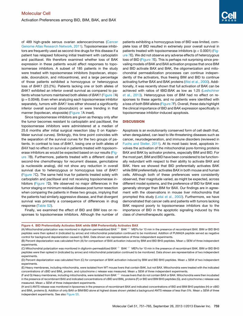
MEFs to TRAIL. As expected, loss of BID protected MEFs from
TRAIL-induced apoptosis to a greater extent than loss of BIM
(Figure 6A). Topoisomerase inhibitors have been used in the
clinic for over 20 years to treat a wide range of malignancies
and are among the most highly prescribed anticancer drugs
(Baldwin and Osheroff, 2005). These inhibitors trigger apoptosis
by inducing DNA double-strand breaks regardless of whether
they target topoisomerase I (topotecan and irinotecan) or topo-
isomerase II (etoposide, mitoxantrone, and doxorubicin) (Bald-
win and Osheroff, 2005; Kaina, 2003; Strumberg et al., 2000).
Efficient induction of apoptosis by topoisomerase II inhibitors re-
quires BID cleavage (Friesen et al., 1996; Fulda et al., 2000; Slee
et al., 2000). It has also been shown that topotecan-induced
apoptosis can be blocked by caspase 8 inhibition (the enzyme
that cleaves Bid to tBid) or by upregulation of BCL-2 and BCL-
XL, indicating that cleavage of BID may be a necessary step in
induction of apoptosis by topoisomerase I inhibitors as well (Fer-
reira et al., 2000).We confirmed that BID is cleaved and activated
in MEFs treated with topoisomerase inhibitors but not cisplatin
(Figure S7A). When we compared the sensitivity of WT, BID�/�,and BIM�/� MEFs to topoisomerase inhibitors, we observed
that BID�/� MEFs were resistant to etoposide, topotecan, and
doxorubicin, in agreement with the previous reports (Figure 5A).
BIM�/�MEFs also exhibited some resistance to this class of che-
Molecu
motherapies, although loss of BIM provided significantly less
resistance than loss of BID. Notably, BID�/�MEFs did not exhibit
any significant differences in sensitivity to other chemotherapies
as compared to BIM�/� MEFs, including the DNA-crosslinking
agent cisplatin, the microtubule inhibitor paclitaxel, or the kinase
inhibitor staurosporine. These data confirm the selective impor-
tance of BID in topoisomerase inhibitor-induced apoptosis.
Since we observed a preference for BID to activate BAK over
BAX, we hypothesized that cells lacking BAK would be less
sensitive to TRAIL- and topoisomerase-induced apoptosis. As
expected, BAK�/� MEFs were less sensitive to these agents
than BAX�/� MEFs while exhibiting similar sensitivity to cisplatin
and paclitaxel (Figure 6B). BAX�/� MEFs exhibited significantly
more resistance to the kinase inhibitor staurosporine than
BAK�/� MEFs, which is interesting considering the higher, but
not significant, resistance of BIM�/� MEFs as compared to
BID�/� MEFs to this agent (Figure 6A). By utilizing Annexin V
and propidium iodide staining and flow cytometric analysis, we
observed that differences observed in the viability assay were
due to the induction of classical apoptosis by chemotherapeutic
agents and not an unrelated effect on cell growth (Figure S8).
Overall, we concluded that there was a correlation between an
agent’s killing beingmore dependent on BAK and its killing being
more dependent on BID (Figure 6C).
We performed a similar analysis of chemosensitivity in BMK
andHeLa cells to determine if the differences in chemosensitivity
would also be present in epithelial and human cancer cells,
respectively. The same agents were utilized with one exception:
HeLa cells are extremely resistant to treatment with the topo-
isomerase II inhibitor etoposide (data not shown), and the topo-
isomerase II inhibitor mitoxantrone was therefore used instead.
As with the MEFs, we observed protection from TRAIL and topo-
isomerase inhibitors in BMK cells lacking BAK as compared to
those lacking BAX (Figure 6D). The protection was not evident
with agents that induced apoptosis via alternate mechanisms
that are less distinctly BID dependent. Finally, we found that
even transient, siRNA-based knockdown of BAK in HeLa cells
prevented much of the apoptosis induced by topoisomerase in-
hibitors and slowed TRAIL-induced caspase activation (Figures
6E and S9), while knockdown of BAX had a significantly smaller
effect. These data are consistent with the fact that BID cleavage
and activation was observed in HeLa cells upon treatment with
topoisomerase inhibitors but not other chemotherapeutic agents
(Figures S7B and S7C). Conversely, BAX knockdown was either
equivalent or superior to BAK knockdown in affording protection
from the other chemotherapies tested (Figure 6E). The chemo-
sensitivity assays performed on MEFs, BMKs, and HeLa cells
showed that topoisomerase inhibitors require BID and BAK for
maximal induction of apoptosis.
The strength of the protection seen in BAK�/� and BID�/� cells
from apoptosis induced by topoisomerase inhibitors suggested
that BAK deficiency in tumors may be particularly detrimental to
patients receiving one of these agents in the clinic. Inhibitors of
topoisomerases I or II are used frequently to treat both hemato-
poietic and solid tumors including lung, ovarian, and cervical
cancers as well as lymphomas, leukemias, and glioblastomas.
Recently, The Cancer Genome Atlas (TCGA) project examined
DNA copy number alterations and clinical outcomes in a series
lar Cell 51, 751–765, September 26, 2013 ª2013 Elsevier Inc. 757
(legend on next page)
Molecular Cell
Activation Preferences among BID, BIM, BAK, and BAX
758 Molecular Cell 51, 751–765, September 26, 2013 ª2013 Elsevier Inc.
Molecular Cell
Activation Preferences among BID, BIM, BAK, and BAX
of 489 high-grade serous ovarian adenocarcinomas (Cancer
Genome Atlas Research Network, 2011). Topoisomerase inhibi-
tors are frequently used as second-line drugs for this disease if a
patient has relapsed following initial treatment with carboplatin
and paclitaxel. We therefore examined whether loss of BAK
expression in these patients would affect responses to topo-
isomerase inhibitors. A subset of 185 patients in the study
were treated with topoisomerase inhibitors (topotecan, etopo-
side, doxorubicin, and mitoxantrone), and a large percentage
of those patients exhibited a homozygous or heterozygous
loss of BAK1 (23.2%). Patients lacking one or both alleles of
BAK1 exhibited an inferior overall survival as compared to pa-
tients whose tumors maintained both alleles of BAK1 (Figure 7A)
(p = 0.0048). Even when analyzing each topoisomerase inhibitor
separately, tumors with BAK1 loss either showed a significantly
inferior overall survival (doxorubicin) or were trending in that
manner (topotecan, etoposide) (Figure 7A inset).
Since topoisomerase inhibitors are given as therapy only after
the tumor becomes resistant to carboplatin and paclitaxel, the
topoisomerase inhibitors were administered at a median of
25.6 months after initial surgical resection (day 0 on Kaplan-
Meier survival curves). Strikingly, this time point coincides with
the separation of the survival curves for the two groups of pa-
tients. In contrast to loss of BAK1, losing one or both alleles of
BAX had no effect on survival in patients treated with topoisom-
erase inhibitors as would be expected based on our results (Fig-
ure 7B). Furthermore, patients treated with a different class of
second-line chemotherapy for recurrent disease, gemcitabine
(a nucleoside analog), did not show any reduction in overall
survival due to heterozygous or homozygous loss of BAK1
(Figure 7C). The same held true for patients treated solely with
carboplatin and paclitaxel and not receiving topoisomerase in-
hibitors (Figure 7D). We observed no major differences in the
tumor staging or minimum residual disease post tumor resection
when comparing the patients in these two groups, implying that
the patients had similarly aggressive disease, and that divergent
survival was primarily a consequence of differences in drug
response (Table S2).
Finally, we examined the effect of BID and BIM loss on re-
sponses to topoisomerase inhibitors. Although the number of
Figure 4. BID Preferentially Activates BAK while BIM Preferentially Ac
(A) Mitochondrial polarization was monitored in digitonin-permeabilized BAX�/� B
peptides were then spiked in (indicated by arrow) and mitochondrial polarization
control for background depolarization caused by BAX. Data shown are represen
(B) Percent depolarization was calculated from (A) for comparison of BAX activat
experiments.
(C) Mitochondrial polarization was monitored in digitonin-permeabilized BAX�/� B
peptides were then spiked in (indicated by arrow) and mitochondrial polarization
experiments.
(D) Percent depolarization was calculated from (C) for comparison of BAK activa
experiments.
(E) Heavy membranes, including mitochondria, were isolated fromWTmouse live
concentrations of cBID and BIML protein, and cytochrome c release was measu
(F andG) Heavymembranes, includingmitochondria, were isolated fromBAK�/�m
in the presence of recombinant BAX and indicated concentrations of cBID and BIM
measured. Mean ± SEM of three independent experiments.
(H and I) ANTS release was monitored in liposomes in the presence of recombina
and BIML proteins (I). Addition of only BAX or BIM/BID alone at highest doses sho
independent experiments. See also Figure S5.
Molecu
patients exhibiting a homozygous loss of BID was limited, com-
plete loss of BID resulted in extremely poor overall survival in
patients treated with topoisomerase inhibitors (p < 0.0001) (Fig-
ure 7E). We did not observe any adverse effects of heterozygous
loss of BID (Figure 7E). This is perhaps not surprising since pre-
vailingmodels of BAK andBAX activation propose that once BIM
and BID activate BAX and BAK, the oligomerization and mito-
chondrial permeabilization processes can continue indepen-
dently of the activators, thus freeing BIM and BID to continue
activating further BAX and BAK proteins (Wei et al., 2000). Addi-
tionally, it was recently shown that full activation of BAK can be
achieved with ratios of tBID:BAK as low as 1:25 (Leshchiner
et al., 2013). Heterozygous loss of BIM had no effect on re-
sponses to these agents, and no patients were identified with
a loss of both BIM alleles (Figure 7F). Overall, these data highlight
the clinical importance of BID and BAK expression specifically in
topoisomerase inhibitor-induced apoptosis.
DISCUSSION
Apoptosis is an evolutionarily conserved form of cell death that,
when deregulated, can lead to life-threatening diseases such as
cancer, neurodegeneration, and immune disorders (reviewed in
Fuchs and Steller, 2011). At its most basic level, apoptosis in-
volves the activation of the mitochondrial pore-forming proteins
BAX and BAK by activator proteins, especially BIM and BID. For
themost part, BIM andBID have been considered to be function-
ally redundant with respect to their ability to activate BAX and
BAK. Here we showed that BID preferentially activates BAK
while BIM preferentially activates BAX in both mouse and human
cells. Although both of these preferences were consistently
observed, their magnitude varied, as might be expected, across
different model systems, and the preference of BID for BAK was
generally stronger than BIM for BAX. Our findings are in agree-
ment with the observations in mouse liver mitochondria that
prompted this study (Letai et al., 2002). Furthermore, we have
demonstrated that cancer cells and patients with tumors lacking
BAK respond poorly to topoisomerase inhibitors due to the
importance of BID in the apoptotic signaling induced by this
class of chemotherapeutic agents.
tivates BAX
AK�/� MEFs for 10 min in the presence of recombinant BAX. BIM or BID BH3
continued to be monitored. Addition of PUMA2A peptide served as negative
tative of three independent experiments.
ion induced by BIM and BID BH3 peptides. Mean ± SEM of three independent
AK�/� MEFs for 10 min in the presence of recombinant BAK. BIM or BID BH3
continued to be monitored. Data shown are representative of two independent
tion induced by BIM and BID BH3 peptides. Mean ± SEM of two independent
rs that contain BAK, but not BAX. Mitochondria were treated with the indicated
red. Mean ± SEM of three independent experiments.
ouse livers that do not contain BAX or BAK.Mitochondria were then incubated
L proteins (F) or BID and BIMBH3 peptides (G), and cytochrome c release was
nt BAX and indicated concentrations of BID and BIM BH3 peptides (H) or cBID
wn yielded a background ANTS release of less than 5%. Mean ± SEM of three
lar Cell 51, 751–765, September 26, 2013 ª2013 Elsevier Inc. 759

Figure 5. BID Preferentially Crosslinks and Activates BAK while BIM Preferentially Crosslinks and Activates BAX(A) Heavy membranes including mitochondria were isolated from MEFs lacking BIM, BID, and p53 and were treated with the indicated concentrations of BH3
peptides. Samples were then treated with the crosslinking agent BMH and analyzed by western blotting for crosslinking of BAX and BAK. *, monomeric BAK and
BAX; **, oligomerized BAK andBAX. Densitometrywas performed in area indicated by dashed rectangle for comparison of oligomerization efficiency. Data shown
are representative of two independent experiments.
(B)MEFs of the indicated genotypewere plated in a 96-well plate and transfected with plasmids (0.1 mg/well) encoding either GFP or untagged tBID or BIMEL in the
pCMV vector. After 24 hr, cell viability was assessed using CellTiterGlo. Mean ± SEM of four independent experiments. See also Figure S6.
Molecular Cell
Activation Preferences among BID, BIM, BAK, and BAX
The preferential activation of BAX and BAK by BIM and BID,
respectively, may shed light on some observations previously re-
ported in the literature. For example, several independent groups
have shown that the apoptosis induced by c-Myc activation is
entirely dependent on BAX but not BAK (Dansen et al., 2006;
Eischen et al., 2001; Sarosiek et al., 2010). It has been also
been reported that c-Myc-induced apoptosis is triggered via up-
regulation of BIM (Egle et al., 2004). Our data showing that BIM
canmore readily activate BAXwould thus provide an explanation
for the BAX dependence of c-Myc-induced cell death. Similarly,
Cartron et al. have reported that BAK-deficient glioblastoma
cells are completely resistant to Fas-mediated cell death (Car-
tron et al., 2003). Because Fas ligand elicits death receptor
signaling and leads to caspase 8 activation and subsequent
BID cleavage and activation (Li et al., 1998), the dependency
of this apoptotic pathway on BAK is potentially explained by
760 Molecular Cell 51, 751–765, September 26, 2013 ª2013 Elsevier
our findings. Additional reports of either BAX or BAK depen-
dence for apoptosis induced by various insults abound and
may potentially be explained by our study (von Haefen et al.,
2004; Handrick et al., 2010; Letai et al., 2002; Zhang et al., 2000).
Previous studies may provide some basis for the evolution of
the differential affinities we observed among BCL-2 family pro-
teins. BIM has been shown to be more important than BID as a
regulator of life and death decisions in the hematopoietic system
since its loss leads to an accumulation of lymphoid and myeloid
cells (Bouillet et al., 1999), a finding that does not hold true for
BID loss (Yin et al., 1999). Interestingly, BAX knockout mice
exhibit a similar hyperplasia of B cells (Knudson et al., 1995)
while BAK knockout mice do not (Lindsten et al., 2000). It is
possible that the increased affinity of BIM for BAX is the product
of coevolution due to both proteins’ vital and delicate roles in
tightly regulating the hematopoietic system.
Inc.
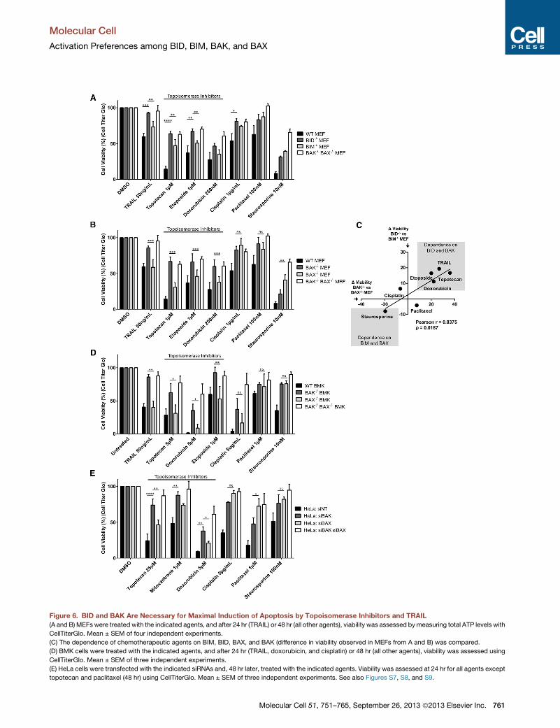
Figure 6. BID and BAK Are Necessary for Maximal Induction of Apoptosis by Topoisomerase Inhibitors and TRAIL
(A and B) MEFs were treated with the indicated agents, and after 24 hr (TRAIL) or 48 hr (all other agents), viability was assessed bymeasuring total ATP levels with
CellTiterGlo. Mean ± SEM of four independent experiments.
(C) The dependence of chemotherapeutic agents on BIM, BID, BAX, and BAK (difference in viability observed in MEFs from A and B) was compared.
(D) BMK cells were treated with the indicated agents, and after 24 hr (TRAIL, doxorubicin, and cisplatin) or 48 hr (all other agents), viability was assessed using
CellTiterGlo. Mean ± SEM of three independent experiments.
(E) HeLa cells were transfected with the indicated siRNAs and, 48 hr later, treated with the indicated agents. Viability was assessed at 24 hr for all agents except
topotecan and paclitaxel (48 hr) using CellTiterGlo. Mean ± SEM of three independent experiments. See also Figures S7, S8, and S9.
Molecular Cell
Activation Preferences among BID, BIM, BAK, and BAX
Molecular Cell 51, 751–765, September 26, 2013 ª2013 Elsevier Inc. 761
A
B
C
D
E F
Figure 7. Patients Treated with Topoisom-
erase Inhibitors Respond Poorly when
Exhibiting a Loss of BAK
(A–D) DNA copy number analysis was performed
on patients with a confirmed diagnosis of high-
grade serous ovarian adenocarcinoma as part of
the TCGA study. (A) Overall survival (OS) was
compared in patients treated with topoisomerase
inhibitors (topotecan, etoposide, mitoxantrone,
and doxorubicin) that exhibited a loss of one or
both alleles of BAK (n = 43 patients) and those that
did not (n = 142). OS was also compared in
patients being treated with each topoisomerase
inhibitor separately (inset): doxorubicin (BAK+/+,
n = 113; BAK+/� or BAK�/�, n = 38), topotecan
(BAK+/+, n = 88; BAK+/� or BAK�/�, n = 32), or
etoposide (BAK+/+ n = 15; BAK+/� or BAK�/�,n = 5). OS was not compared in patients treated
with mitoxantrone due to low number of patients
receiving this therapy (n = 1). (B) OSwas compared
in patients treated with topoisomerase inhibitors
that exhibited a loss of one or both alleles of BAX
(n = 85) and those that had not (n = 100). (C) OS
was compared in patients treated with gemcita-
bine that exhibited a loss of one or both alleles of
BAK (n = 4) and those that had not (n = 30). (D) OS
was compared in patients treated with carboplatin
and paclitaxel, but not topoisomerase inhibitors,
that exhibited a loss of one or both alleles of BAK
(n = 47) and those that had not (n = 281).
(E) OS was compared in patients treated with
topoisomerase inhibitors that exhibited a loss of
one (n = 102) or both (n = 2) alleles of BID and those
that had not (n = 81).
(F) OS was compared in patients treated with
topoisomerase inhibitors that exhibited a loss of
one allele of BIM (n = 29) and those that had not
(n = 156). No patients exhibited a loss of both al-
leles of BIM. See also Table S2.
Molecular Cell
Activation Preferences among BID, BIM, BAK, and BAX
762 Molecular Cell 51, 751–765, September 26, 2013 ª2013 Elsevier Inc.
Molecular Cell
Activation Preferences among BID, BIM, BAK, and BAX
The preferential partnership between BID and BAK has
an equally plausible mode of coevolution. Specifically, it
has been shown that death induced in mice by in vivo admin-
istration of Fas ligand (FasL) is due to severe liver damage
induced by activation of Fas receptor, which is expressed at
high levels on hepatocytes (Hao et al., 2004; Ogasawara
et al., 1993). Furthermore, prodeath FasL signaling is integral
in maintaining liver homeostasis, as evidenced by the devel-
opment of hepatomegaly in Fas knockout mice (Adachi et al.,
1995). FasL, as mentioned previously, induces apoptosis via
activation of caspase 8 and subsequent cleavage and activa-
tion of BID (Luo et al., 1998), which explains how BID�/�
mice are protected from the liver damage induced by in vivo
administration of FasL (Yin et al., 1999). Based on our study,
hepatocytes are particularly well suited to respond to FasL
and BID signaling since their mitochondria contain BAK but
not BAX protein (Korsmeyer et al., 2000; Letai et al., 2002).
In fact, we would expect that BAK�/� but not BAX�/� mice
would be protected from BID-mediated, FasL-induced hepato-
cyte apoptosis, as recently confirmed experimentally (Hikita
et al., 2011). The preferential activation of BAK by BID may
therefore be born of the necessity to regulate liver homeostasis
via the Fas/FasL system with a high degree of specificity and
customization.
Our report focused on the dynamics of BAK and BAX activa-
tion by BID and BIM and its potential functional and clinical rele-
vance, yet the mechanisms responsible for these preferences
remain unknown. Notably, these preferences were observed
with unstructured BH3 domain peptides as well as full-length
proteins, which suggests that the preferences are predominantly
based on the amino acid sequences of the two proteins. There
are many differences between BID and BIM in amino acid
sequence across their roughly 20-mer BH3 domains. Elegant
mutational studies have elucidated key residues within the
BH3 domains of both BID and BIM that are vital for binding
BAK and BAX (Gavathiotis et al., 2008; Leshchiner et al., 2013);
those studies may be extended to identify the components
of the BH3 domains that are responsible for their activation
preferences.
Other factors may also influence the selectivity we
observed. For instance, recent studies have shown that
proteins such as MTCH2 may modulate activation of BAX
and BAK by BID and BIM and thus provide another means
by which to control these effectors in certain cell types (Katz
et al., 2012). Future studies may identify additional modes of
regulation and provide a more complex and nuanced picture
of how cell fate is determined by these critical BCL-2 family
proteins.
The importance of BID for topoisomerase-induced cell
death has been extensively reported (Kamer et al., 2005;
Maas et al., 2011; Slee et al., 2000; Werner et al., 2004; Zinkel
et al., 2005). Our observation that BID preferentially activates
BAK and that, consequently, BAK deficiency protects cells
from apoptosis induced by these agents sheds additional light
on mechanisms of resistance to these often-used chemother-
apies. Our results suggest that guiding chemotherapy deci-
sions based on alterations in BAK and BID may be beneficial
clinically.
Molecu
EXPERIMENTAL PROCEDURES
BH3 Profiling Using Whole Cells
Proliferating cells that were 30%–50% confluent were harvested and counted
for BH3 profiling. We deposited 15 ml of BH3 peptides or recombinant proteins
(see below for peptide sequences) in T-EB (300mMTrehalose, 10mMHEPES-
KOH [pH 7.7], 80 mM KCl, 1 mM EGTA, 1 mM EDTA, 0.1% BSA, and 5 mM
succinate [all Sigma-Aldrich]) into each well in a nontreated black 384-well
plate, one treatment per well, in triplicate, for each independent experiment.
Single-cell suspensions were washed once with T-EB before being resus-
pended at 43 their final density of 6.75 3 105 cells/ml. One volume of the
43 cell suspension was added to one volume of a 43 dye solution containing
4 mM JC1 (Enzo Life Sciences), 40 mg/ml oligomycin (Sigma-Aldrich), 0.02%
digitonin (Sigma-Aldrich), and 20 mM 2-mercaptoethanol (Life Technologies)
in T-EB. The resulting 23 cell/dye solution was kept at room temperature for
5 min to allow cell permeabilization and dye equilibration. Fifteen microliters
of the 23 cell/dye mix was then added to each treatment well of the 384-
well plate and shaken for 15 s inside the plate reader, and the fluorescence
at 590 nMwasmeasured every 5min at room temperature. Peptide treatments
that were used corresponded to the BH3 domains of the BCL-2 family
proteins, and their respective sequences are as follows: BIM: MRPEIWIA
QELRRIGDEFNA; BID: EDIIRNIARHLAQVGDSMDR (New England Peptide,
Gardner, MA). Relative mitochondrial depolarization was defined as the
magnitude of mitochondrial depolarization resulting from BH3 peptide treat-
ment as compared to vehicle DMSO (Sigma-Aldrich) and positive control
FCCP (p-trifluoromethoxy carbonyl cyanide phenyl hydrazone) (Sigma-
Aldrich). The percentage of mitochondrial depolarization was calculated by
comparing the JC1 signal (mitochondrial polarization) in cell lines treated
with each peptide or protein concentration in the following manner:
% Mitochondrial Depolarization=½RðtÞ � FðtÞ�
½RðtÞ � FCCPðtÞ�3100
where R(t) is the fluorescence value in the reference sample (DMSO), F(t) is the
fluorescence value in the test sample (peptide or protein), and FCCP(t) is the
fluorescence value in the positive control sample (FCCP) at a time (t) and aver-
aged over the dynamic portion of depolarization curves.
Western Blotting
Western blotting was performed as previously described (Sarosiek et al.,
2009). Antibodies used are listed in Supplemental Information.
Ovarian Cancer Copy Number Alterations, Treatment History, and
Survival
Treatment history and survival data (clinical follow-up) for all TCGA high-grade
serous ovarian adenocarcinoma cases were obtained from the TCGA data
portal on August 1, 2012. Full procedures are included in Supplemental
Information.
Liver Mitochondria Permeabilization Assays
For mitochondrial assays, mitochondria were isolated from the livers of
wild-type and BAK�/� mice under an approved institutional animal protocol,
and outer membrane permeabilization of mitochondria was assessed
by measuring cytochrome c release as described previously (Shamas-Din
et al., 2013).
SUPPLEMENTAL INFORMATION
Supplemental Information includes nine figures, two tables, and Supplemental
Experimental Procedures and can be found with this article online at http://dx.
doi.org/10.1016/j.molcel.2013.08.048.
ACKNOWLEDGMENTS
We kindly thank Dr. John C. Reed for providing us with the pcDNA3-myc-BAK
plasmid. We gratefully acknowledge funding from the American Cancer Soci-
ety Postdoctoral Fellowship 121360-PF-11-256-01-TBG (K.A.S.), Women’s
lar Cell 51, 751–765, September 26, 2013 ª2013 Elsevier Inc. 763
Molecular Cell
Activation Preferences among BID, BIM, BAK, and BAX
Cancers Program at the Dana-Farber Cancer Institute (K.A.S.), and NIH grants
RO1CA129974 and P01CA139980. A.L. is a Leukemia and Lymphoma Society
Scholar.
Received: February 15, 2013
Revised: July 6, 2013
Accepted: August 22, 2013
Published: September 26, 2013
REFERENCES
Adachi, M., Suematsu, S., Kondo, T., Ogasawara, J., Tanaka, T., Yoshida, N.,
and Nagata, S. (1995). Targeted mutation in the Fas gene causes hyperplasia
in peripheral lymphoid organs and liver. Nat. Genet. 11, 294–300.
Baldwin, E.L., and Osheroff, N. (2005). Etoposide, topoisomerase II and
cancer. Curr. Med. Chem. Anticancer Agents 5, 363–372.
Biswas, S.C., and Greene, L.A. (2002). Nerve growth factor (NGF) down-regu-
lates the Bcl-2 homology 3 (BH3) domain-only protein Bim and suppresses its
proapoptotic activity by phosphorylation. J. Biol. Chem. 277, 49511–49516.
Bossy-Wetzel, E., Newmeyer, D.D., and Green, D.R. (1998). Mitochondrial
cytochrome c release in apoptosis occurs upstream of DEVD-specific cas-
pase activation and independently of mitochondrial transmembrane depolar-
ization. EMBO J. 17, 37–49.
Bouillet, P., Metcalf, D., Huang, D.C., Tarlinton, D.M., Kay, T.W., Kontgen, F.,
Adams, J.M., and Strasser, A. (1999). Proapoptotic Bcl-2 relative Bim required
for certain apoptotic responses, leukocyte homeostasis, and to preclude auto-
immunity. Science 286, 1735–1738.
Cancer Genome Atlas Research Network. (2011). Integrated genomic ana-
lyses of ovarian carcinoma. Nature 474, 609–615.
Cartron, P.F., Juin, P., Oliver, L., Martin, S., Meflah, K., and Vallette, F.M.
(2003). Nonredundant role of Bax and Bak in Bid-mediated apoptosis. Mol.
Cell. Biol. 23, 4701–4712.
Certo, M., Del Gaizo Moore, V., Nishino, M., Wei, G., Korsmeyer, S.,
Armstrong, S.A., and Letai, A. (2006). Mitochondria primed by death signals
determine cellular addiction to antiapoptotic BCL-2 family members. Cancer
Cell 9, 351–365.
Czabotar, P.E., Westphal, D., Dewson, G., Ma, S., Hockings, C., Fairlie, W.D.,
Lee, E.F., Yao, S., Robin, A.Y., Smith, B.J., et al. (2013). Bax crystal structures
reveal how BH3 domains activate Bax and nucleate its oligomerization to
induce apoptosis. Cell 152, 519–531.
Dansen, T.B., Whitfield, J., Rostker, F., Brown-Swigart, L., and Evan, G.I.
(2006). Specific requirement for Bax, not Bak, in Myc-induced apoptosis
and tumor suppression in vivo. J. Biol. Chem. 281, 10890–10895.
Davids, M.S., Deng, J., Wiestner, A., Lannutti, B.J., Wang, L., Wu, C.J., Wilson,
W.H., Brown, J.R., and Letai, A. (2012). Decreased mitochondrial apoptotic
priming underlies stroma-mediated treatment resistance in chronic lympho-
cytic leukemia. Blood 120, 3501–3509.
Degenhardt, K., Sundararajan, R., Lindsten, T., Thompson, C., and White, E.
(2002). Bax and Bak independently promote cytochrome C release from mito-
chondria. J. Biol. Chem. 277, 14127–14134.
Deng, J., Carlson, N., Takeyama, K., Dal Cin, P., Shipp,M., and Letai, A. (2007).
BH3 profiling identifies three distinct classes of apoptotic blocks to predict
response to ABT-737 and conventional chemotherapeutic agents. Cancer
Cell 12, 171–185.
Egle, A., Harris, A.W., Bouillet, P., and Cory, S. (2004). Bim is a suppressor of
Myc-induced mouse B cell leukemia. Proc. Natl. Acad. Sci. USA 101, 6164–
6169.
Eischen, C.M., Roussel, M.F., Korsmeyer, S.J., and Cleveland, J.L. (2001). Bax
loss impairs Myc-induced apoptosis and circumvents the selection of p53
mutations during Myc-mediated lymphomagenesis. Mol. Cell. Biol. 21,
7653–7662.
Farrow, S.N., White, J.H., Martinou, I., Raven, T., Pun, K.T., Grinham, C.J.,
Martinou, J.C., and Brown, R. (1995). Cloning of a bcl-2 homologue by interac-
tion with adenovirus E1B 19K. Nature 374, 731–733.
764 Molecular Cell 51, 751–765, September 26, 2013 ª2013 Elsevier
Ferreira, C.G., Span, S.W., Peters, G.J., Kruyt, F.A., and Giaccone, G. (2000).
Chemotherapy triggers apoptosis in a caspase-8-dependent and mitochon-
dria-controlled manner in the non-small cell lung cancer cell line NCI-H460.
Cancer Res. 60, 7133–7141.
Friesen, C., Herr, I., Krammer, P.H., and Debatin, K.M. (1996). Involvement of
the CD95 (APO-1/FAS) receptor/ligand system in drug-induced apoptosis in
leukemia cells. Nat. Med. 2, 574–577.
Fuchs, Y., and Steller, H. (2011). Programmed cell death in animal develop-
ment and disease. Cell 147, 742–758.
Fulda, S., Strauss, G., Meyer, E., and Debatin, K.M. (2000). Functional CD95
ligand and CD95 death-inducing signaling complex in activation-induced
cell death and doxorubicin-induced apoptosis in leukemic T cells. Blood 95,
301–308.
Gavathiotis, E., Suzuki, M., Davis, M.L., Pitter, K., Bird, G.H., Katz, S.G., Tu,
H.-C., Kim, H., Cheng, E.H.-Y., Tjandra, N., and Walensky, L.D. (2008). BAX
activation is initiated at a novel interaction site. Nature 455, 1076–1081.
Handrick, R., Ontikatze, T., Bauer, K.-D., Freier, F., Rubel, A., Durig, J., Belka,
C., and Jendrossek, V. (2010). Dihydroartemisinin induces apoptosis by a Bak-
dependent intrinsic pathway. Mol. Cancer Ther. 9, 2497–2510.
Hao, C., Song, J.H., Hsi, B., Lewis, J., Song, D.K., Petruk, K.C., Tyrrell, D.L.J.,
and Kneteman, N.M. (2004). TRAIL inhibits tumor growth but is nontoxic to
human hepatocytes in chimeric mice. Cancer Res. 64, 8502–8506.
Hikita, H., Takehara, T., Kodama, T., Shimizu, S., Shigekawa, M., Hosui, A.,
Miyagi, T., Tatsumi, T., Ishida, H., Li, W., et al. (2011). Delayed-onset cas-
pase-dependent massive hepatocyte apoptosis upon Fas activation in Bak/
Bax-deficient mice. Hepatology 54, 240–251.
Hsu, Y.-T., and Youle, R.J. (1997). Nonionic detergents induce dimerization
among members of the Bcl-2 family. J. Biol. Chem. 272, 13829–13834.
Jacobson, M.D., Weil, M., and Raff, M.C. (1997). Programmed cell death in
animal development. Cell 88, 347–354.
Johnstone, R.W., Ruefli, A.A., and Lowe, S.W. (2002). Apoptosis: a link
between cancer genetics and chemotherapy. Cell 108, 153–164.
Kaina, B. (2003). DNA damage-triggered apoptosis: critical role of DNA repair,
double-strand breaks, cell proliferation and signaling. Biochem. Pharmacol.
66, 1547–1554.
Kamer, I., Sarig, R., Zaltsman, Y., Niv, H., Oberkovitz, G., Regev, L.,
Haimovich, G., Lerenthal, Y., Marcellus, R.C., and Gross, A. (2005).
Proapoptotic BID is an ATM effector in the DNA-damage response. Cell 122,
593–603.
Katz, C., Zaltsman-Amir, Y., Mostizky, Y., Kollet, N., Gross, A., and Friedler, A.
(2012). Molecular basis of the interaction between proapoptotic truncated BID
(tBID) protein and mitochondrial carrier homologue 2 (MTCH2) protein: key
players in mitochondrial death pathway. J. Biol. Chem. 287, 15016–15023.
Kiefer, M.C., Brauer, M.J., Powers, V.C., Wu, J.J., Umansky, S.R., Tomei, L.D.,
and Barr, P.J. (1995). Modulation of apoptosis by the widely distributed Bcl-2
homologue Bak. Nature 374, 736–739.
Kim, H., Rafiuddin-Shah, M., Tu, H.-C., Jeffers, J.R., Zambetti, G.P., Hsieh,
J.J.-D., and Cheng, E.H.-Y. (2006). Hierarchical regulation of mitochondrion-
dependent apoptosis by BCL-2 subfamilies. Nat. Cell Biol. 8, 1348–1358.
Kim, H., Tu, H.-C., Ren, D., Takeuchi, O., Jeffers, J.R., Zambetti, G.P., Hsieh,
J.J.-D., and Cheng, E.H.-Y. (2009). Stepwise activation of BAX and BAK by
tBID, BIM, and PUMA initiates mitochondrial apoptosis. Mol. Cell 36, 487–499.
Knudson, C.M., Tung, K.S., Tourtellotte, W.G., Brown, G.A., and Korsmeyer,
S.J. (1995). Bax-deficient mice with lymphoid hyperplasia and male germ
cell death. Science 270, 96–99.
Korsmeyer, S.J., Wei, M.C., Saito, M., Weiler, S., Oh, K.J., and Schlesinger,
P.H. (2000). Pro-apoptotic cascade activates BID, which oligomerizes BAK
or BAX into pores that result in the release of cytochrome c. Cell Death
Differ. 7, 1166–1173.
Krajewski, S., Krajewska, M., and Reed, J.C. (1996). Immunohistochemical
analysis of in vivo patterns of Bak expression, a proapoptotic member of the
Bcl-2 protein family. Cancer Res. 56, 2849–2855.
Inc.
Molecular Cell
Activation Preferences among BID, BIM, BAK, and BAX
Leshchiner, E.S., Braun, C.R., Bird, G.H., and Walensky, L.D. (2013). Direct
activation of full-length proapoptotic BAK. Proc. Natl. Acad. Sci. USA 110,
E986–E995.
Letai, A., Bassik, M.C., Walensky, L.D., Sorcinelli, M.D., Weiler, S., and
Korsmeyer, S.J. (2002). Distinct BH3 domains either sensitize or activate mito-
chondrial apoptosis, serving as prototype cancer therapeutics. Cancer Cell 2,
183–192.
Li, H., Zhu, H., Xu, C.J., and Yuan, J. (1998). Cleavage of BID by caspase 8
mediates the mitochondrial damage in the Fas pathway of apoptosis. Cell
94, 491–501.
Lindsten, T., Ross, A.J., King, A., Zong, W.X., Rathmell, J.C., Shiels, H.A.,
Ulrich, E., Waymire, K.G., Mahar, P., Frauwirth, K., et al. (2000). The combined
functions of proapoptotic Bcl-2 family members bak and bax are essential for
normal development of multiple tissues. Mol. Cell 6, 1389–1399.
Luo, X., Budihardjo, I., Zou, H., Slaughter, C., and Wang, X. (1998). Bid, a Bcl2
interacting protein, mediates cytochrome c release from mitochondria in
response to activation of cell surface death receptors. Cell 94, 481–490.
Maas, C., de Vries, E., Tait, S.W.G., and Borst, J. (2011). Bid can mediate a
pro-apoptotic response to etoposide and ionizing radiation without cleavage
in its unstructured loop and in the absence of p53. Oncogene 30, 3636–3647.
Ni Chonghaile, T., Sarosiek, K.A., Vo, T.T., Ryan, J.A., Tammareddi, A., Moore,
V.D.G., Deng, J., Anderson, K.C., Richardson, P., Tai, Y.T., et al. (2011).
Pretreatment mitochondrial priming correlates with clinical response to cyto-
toxic chemotherapy. Science 334, 1129–1133.
O’Connor, L., Strasser, A., O’Reilly, L.A., Hausmann, G., Adams, J.M., Cory,
S., and Huang, D.C. (1998). Bim: a novel member of the Bcl-2 family that
promotes apoptosis. EMBO J. 17, 384–395.
Ogasawara, J., Watanabe-Fukunaga, R., Adachi, M., Matsuzawa, A., Kasugai,
T., Kitamura, Y., Itoh, N., Suda, T., and Nagata, S. (1993). Lethal effect of the
anti-Fas antibody in mice. Nature 364, 806–809.
Sarosiek, K.A., Nechushtan, H., Lu, X., Rosenblatt, J.D., and Lossos, I.S.
(2009). Interleukin-4 distinctively modifies responses of germinal centre-like
and activated B-cell-like diffuse large B-cell lymphomas to immuno-chemo-
therapy. Br. J. Haematol. 147, 308–318.
Sarosiek, K.A., Malumbres, R., Nechushtan, H., Gentles, A.J., Avisar, E., and
Lossos, I.S. (2010). Novel IL-21 signaling pathway up-regulates c-Myc and
induces apoptosis of diffuse large B-cell lymphomas. Blood 115, 570–580.
Shamas-Din, A., Bindner, S., Zhu, W., Zaltsman, Y., Campbell, C., Gross, A.,
Leber, B., Andrews, D.W., and Fradin, C. (2013). tBid undergoes multiple
conformational changes at the membrane required for Bax activation. J.
Biol. Chem. 288, 22111–22127.
Molecu
Slee, E.A., Keogh, S.A., and Martin, S.J. (2000). Cleavage of BID during cyto-
toxic drug and UV radiation-induced apoptosis occurs downstream of the
point of Bcl-2 action and is catalysed by caspase-3: a potential feedback
loop for amplification of apoptosis-associated mitochondrial cytochrome c
release. Cell Death Differ. 7, 556–565.
Strumberg, D., Pilon, A.A., Smith, M., Hickey, R., Malkas, L., and Pommier, Y.
(2000). Conversion of topoisomerase I cleavage complexes on the leading
strand of ribosomal DNA into 50-phosphorylated DNA double-strand breaks
by replication runoff. Mol. Cell. Biol. 20, 3977–3987.
Tait, S.W.G., and Green, D.R. (2010). Mitochondria and cell death: outer mem-
brane permeabilization and beyond. Nat. Rev. Mol. Cell Biol. 11, 621–632.
Vo, T.-T., Ryan, J., Carrasco, R., Neuberg, D., Rossi, D.J., Stone, R.M.,
Deangelo, D.J., Frattini, M.G., and Letai, A. (2012). Relative mitochondrial
priming of myeloblasts and normal HSCs determines chemotherapeutic suc-
cess in AML. Cell 151, 344–355.
von Haefen, C., Gillissen, B., Hemmati, P.G., Wendt, J., Guner, D., Mrozek, A.,
Belka, C., Dorken, B., and Daniel, P.T. (2004). Multidomain Bcl-2 homolog Bax
but not Bak mediates synergistic induction of apoptosis by TRAIL and 5-FU
through the mitochondrial apoptosis pathway. Oncogene 23, 8320–8332.
Wei, M.C., Lindsten, T., Mootha, V.K., Weiler, S., Gross, A., Ashiya, M.,
Thompson, C.B., and Korsmeyer, S.J. (2000). tBID, a membrane-targeted
death ligand, oligomerizes BAK to release cytochrome c. Genes Dev. 14,
2060–2071.
Werner, A.B., Tait, S.W.G., de Vries, E., Eldering, E., and Borst, J. (2004).
Requirement for aspartate-cleaved bid in apoptosis signaling by DNA-
damaging anti-cancer regimens. J. Biol. Chem. 279, 28771–28780.
Yamada, H., Tada-Oikawa, S., Uchida, A., and Kawanishi, S. (1999). TRAIL
causes cleavage of bid by caspase-8 and loss of mitochondrial membrane
potential resulting in apoptosis in BJAB cells. Biochem. Biophys. Res.
Commun. 265, 130–133.
Yin, X.M., Wang, K., Gross, A., Zhao, Y., Zinkel, S., Klocke, B., Roth, K.A., and
Korsmeyer, S.J. (1999). Bid-deficient mice are resistant to Fas-induced hepa-
tocellular apoptosis. Nature 400, 886–891.
Zaltsman, Y., Shachnai, L., Yivgi-Ohana, N., Schwarz, M., Maryanovich, M.,
Houtkooper, R.H., Vaz, F.M., De Leonardis, F., Fiermonte, G., Palmieri, F.,
et al. (2010). MTCH2/MIMP is a major facilitator of tBID recruitment to mito-
chondria. Nat. Cell Biol. 12, 553–562.
Zhang, L., Yu, J., Park, B.H., Kinzler, K.W., and Vogelstein, B. (2000). Role of
BAX in the apoptotic response to anticancer agents. Science 290, 989–992.
Zinkel, S.S., Hurov, K.E., Ong, C., Abtahi, F.M., Gross, A., and Korsmeyer, S.J.
(2005). A role for proapoptotic BID in the DNA-damage response. Cell 122,
579–591.
lar Cell 51, 751–765, September 26, 2013 ª2013 Elsevier Inc. 765