Molecular Biology Primer – Part 2 Angela Brooks, Raymond Brown, Calvin Chen, Mike Daly, Hoa Dinh,...
56
Molecular Biology Primer – Part 2 Angela Brooks, Raymond Brown, Calvin Chen, Mike Daly, Hoa Dinh, Erinn Hama, Robert Hinman, Julio Ng, Michael Sneddon, Hoa Troung, Jerry Wang, Che Fung Yung Adapted from the above authors’ slides my Mark Fienup
-
date post
22-Dec-2015 -
Category
Documents
-
view
226 -
download
3
Transcript of Molecular Biology Primer – Part 2 Angela Brooks, Raymond Brown, Calvin Chen, Mike Daly, Hoa Dinh,...

Molecular Biology Primer – Part 2
Angela Brooks, Raymond Brown, Calvin Chen, Mike Daly, Hoa Dinh, Erinn Hama, Robert Hinman, Julio Ng, Michael Sneddon, Hoa Troung, Jerry Wang, Che Fung Yung
Adapted from the above authors’ slides my Mark Fienup

2 types of cells: Prokaryotes v.s. Eukaryotes

Prokaryotes v.s. Eukaryotes
Prokaryotes Eukaryotes
Single cell Single or multi cell
No nucleus Nucleus
No organelles Organelles
One piece of circular DNA Chromosomes
No mRNA post transcriptional modification
Exons/Introns splicing
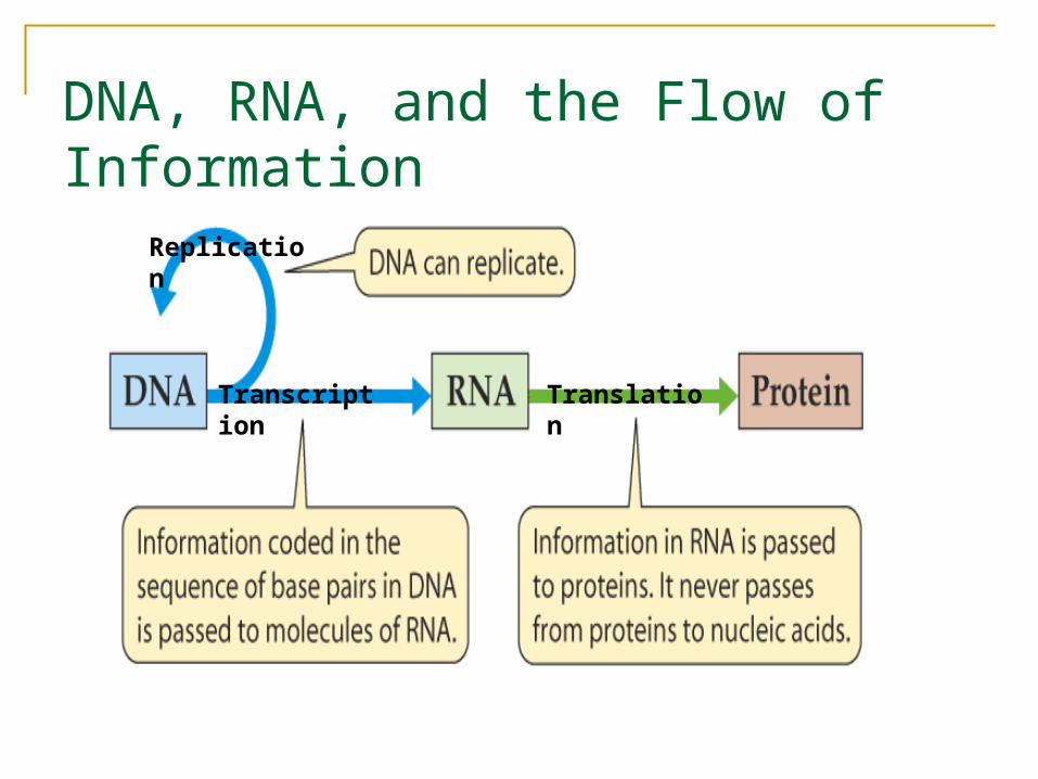
DNA, RNA, and the Flow of Information
TranslationTranscription
Replication
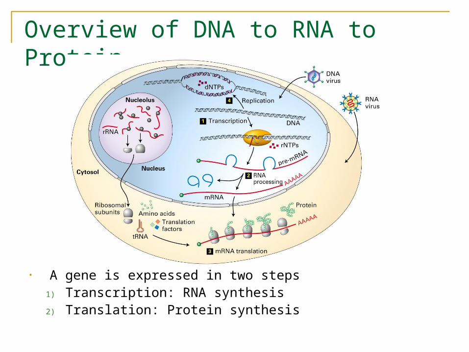
Overview of DNA to RNA to Protein
• A gene is expressed in two steps1) Transcription: RNA synthesis2) Translation: Protein synthesis

Central Dogma Revisited
• Base Pairing Rule: A and T or U is held together by 2 hydrogen bonds and G and C is held together by 3 hydrogen bonds.
• Note: Some mRNA stays as RNA (ie tRNA,rRNA).
DNA hnRNA mRNA
protein
Splicing
Spliceosome
Translation
Transcription
Nucleus
Ribosome in Cytoplasm

Splicing
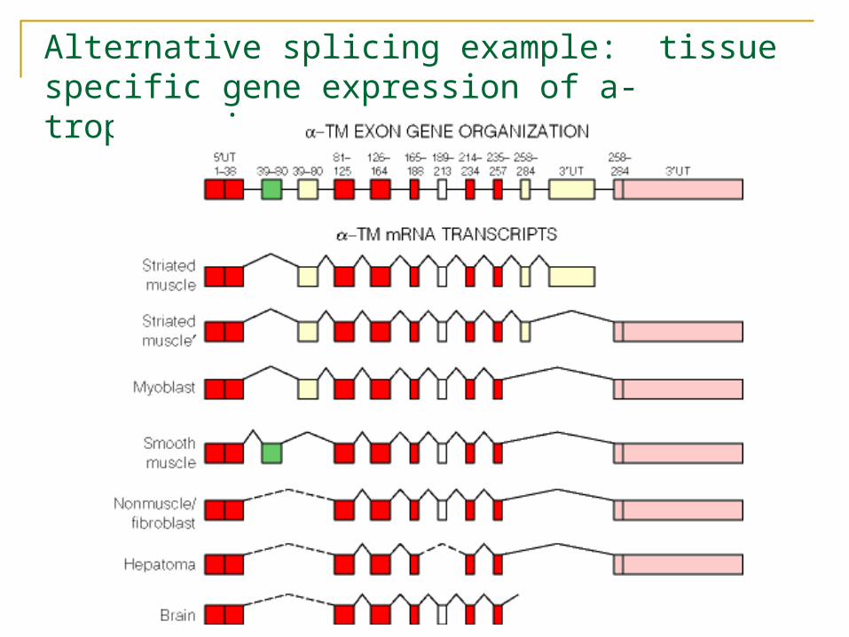
Alternative splicing example: tissue specific gene expression of a-tropomyosin

Translation• The process of going
from RNA to polypeptide/protein.
• Three base pairs of RNA (called a codon) correspond to one amino acid based on a fixed table.
• Always starts with Methionine and ends with a stop codon
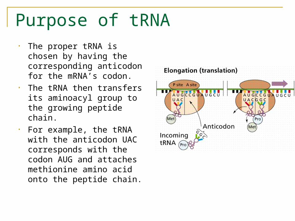
Purpose of tRNA• The proper tRNA is chosen
by having the corresponding anticodon for the mRNA’s codon.
• The tRNA then transfers its aminoacyl group to the growing peptide chain.
• For example, the tRNA with the anticodon UAC corresponds with the codon AUG and attaches methionine amino acid onto the peptide chain.

Translation, continued
• Catalyzed by Ribosome• Using two different
sites, the Ribosome continually binds tRNA, joins the amino acids together and moves to the next location along the mRNA
• ~10 codons/second, but multiple translations can occur simultaneously
http://wong.scripps.edu/PIX/ribosome.jpg
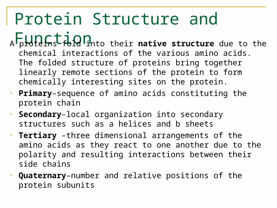
Protein Structure and FunctionA proteins fold into their native structure due to the chemical
interactions of the various amino acids. The folded structure of proteins bring together linearly remote sections of the protein to form chemically interesting sites on the protein.
• Primary–sequence of amino acids constituting the protein chain
• Secondary–local organization into secondary structures such as a helices and b sheets
• Tertiary –three dimensional arrangements of the amino acids as they react to one another due to the polarity and resulting interactions between their side chains
• Quaternary–number and relative positions of the protein subunits
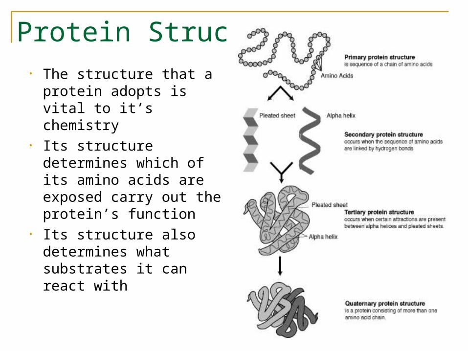
Protein Structure• The structure that a
protein adopts is vital to it’s chemistry
• Its structure determines which of its amino acids are exposed carry out the protein’s function
• Its structure also determines what substrates it can react with

Chemical Bonds ReviewElements - things that cannot be further reduced by chemical reactions
Atom - individual “unit” of an element
Atoms are made up of three stable subatomic particles:
move in orbits around the nucleus at thespeed of light
-1about 1/1,840 as much as a protronelectronnucleus+11.7 x 10⁻²⁴ gramsprotronnucleus none1.7 x 10-24 gramsneutron
“Location”ChargeWeightSubatomicParticle
Electrons and protons are electrically attracted to each other.
Almost all of the mass of an atom is in its nucleus; almost all of the volume of an atom is occupied by electrons.
The number of protons (also known as its atomic number) determines the element. Varying the number of neutronsresults in isotopes. Varying the number of electrons results in ions.
The particles within an atom are bound together by powerful forces. Electrons are easier to add or remove from an atom than a proton or neutron. Chemical reactions largely involve atomsor groups of atoms and the interactions between their electrons.
Chemical ReactivityChemical reactivity – is the degree to which an atom attracts electrons of other atoms.
An atom's attraction for such electrons is determined by the number of electrons in its outer energy level, or outer shell.
Atoms with completely filled outer shells do not attract electrons and are relatively unreactive or inert.
Atoms with incomplete outer shells tend to "share" electrons with other atoms to achieve the electron configurations of the six inert or nobles gases-Helium, Neon, Argon, Krypton, Xenon, and Radon- with 2, 8, 8, 8, 8 and 8 outer shell electrons, respectively.
This sharing of electrons between atoms is called covalent bonding
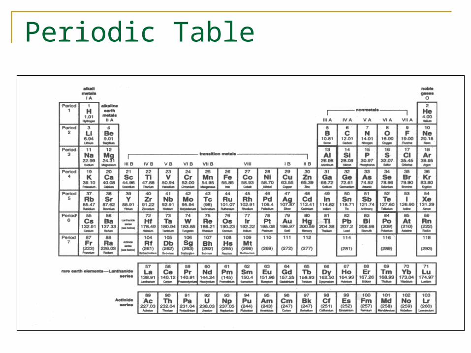
Periodic Table

Electronegativity• Electronegativity is an atoms affinity (“desire”) for electrons.
• An atoms electronegativity is determined by how many electrons it needs to acquire or donate to completely fill or empty its outermost shell of orbitals.
• Hydrogen (1H) and carbon (6C) have outermost shells that are half full so their electronegativity is about equal, so they share electrons equally.
• Oxygen (8O) is more electronegative since it only need to gain 2 (or lose 6) electrons to complete its outermost shell. Therefore, when oxygen bonds with hydrogen (H2O), the electrons spend more time in the vicinity of the oxygen atom causing a slight separation of charge, called a polar bond.
OH H
+ +
-
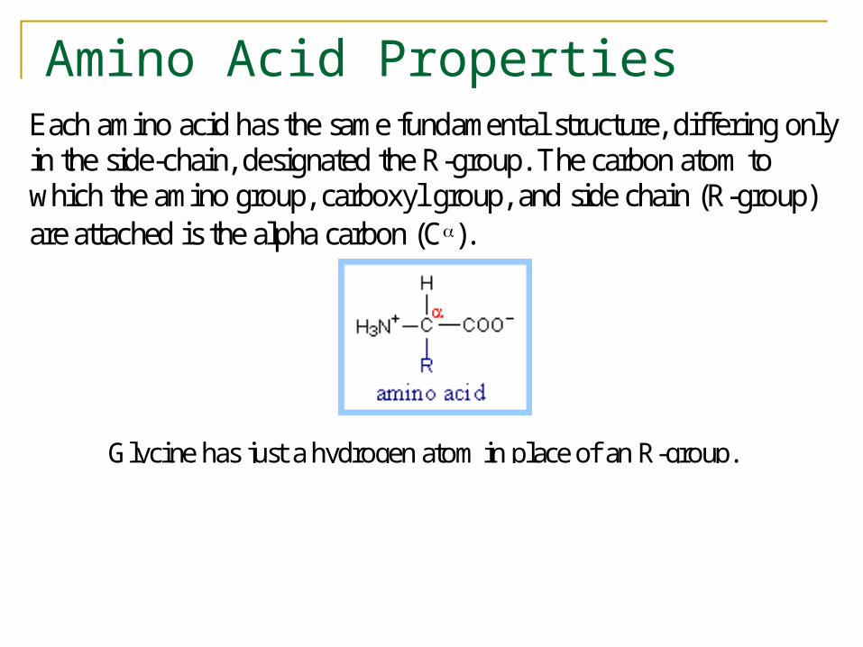
Amino Acid Properties Each amino acid has the same fundamental structure, differing onlyin the side-chain, designated the R-group. The carbon atom towhich the amino group, carboxyl group, and side chain (R-group)are attached is the alpha carbon (C).
Glycine has just a hydrogen atom in place of an R-group.

Nonpolar, hydrophobic R-groups

Polar, hydrophilic R-groups
At physiological pH, some amino acid R-groups are charged, because ofdissociation or association of a proton by a carboxyl or an amino group.
Protein Folding• The amino acids have very different chemical
properties; they interact with each other after the protein is built• This causes the protein to start folding and
adopting it’s functional structure• Proteins may fold in reaction to some ions, and
several separate chains of peptides may join together through their hydrophobic and hydrophilic amino acids to form a polymer

Protein Folding• Proteins tend to fold into the lowest
free energy conformation.• Proteins begin to fold while the
peptide is still being translated.• Proteins bury most of its hydrophobic
residues in an interior core to form an α helix.
• Most proteins take the form of secondary structures α helices and β sheets.
• Molecular chaperones, hsp60 and hsp 70, work with other proteins to help fold newly synthesized proteins.
• Much of the protein modifications and folding occurs in the endoplasmic reticulum and mitochondria.

Protein – Open Problems• A protein is a polypeptide, however to
understand the function of a protein given only the polypeptide sequence is a very difficult problem.
• Protein folding is an open problem. The 3D structure depends on many variables.
• Current approaches often work by looking at the structure of homologous (similar) proteins.
• Improper folding of a protein is believed to be the cause of mad cow disease.
http://www.sanger.ac.uk/Users/sgj/thesis/node2.html for more information on folding
Molecular Biology Tools:
• Copying DNA• Polymerase Chain Reaction• Cloning
• Cutting and Pasting DNA• Restriction Enzymes
• Measuring DNA Length• Electrophoresis• DNA sequencing
• Probing DNA• DNA probes• DNA arrays
Analyzing a Genome
• How to analyze a genome in four “easy” steps.• Cut it
• Use enzymes to cut the DNA in to small fragments.• Copy it
• Copy it many times to make it easier to see and detect.• Read it
• Use special chemical techniques to read the small fragments.• Assemble it
• Take all the fragments and put them back together. This is hard!!!
• Bioinformatics takes over• What can we learn from the sequenced DNA.• Compare interspecies and intraspecies.
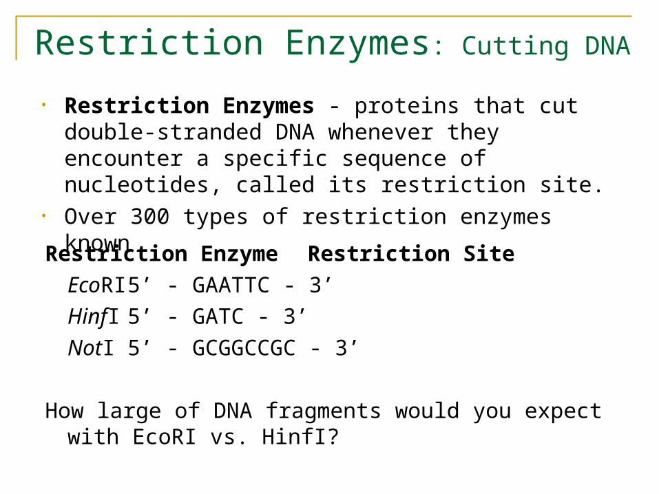
Restriction Enzymes: Cutting DNA
• Restriction Enzymes - proteins that cut double-stranded DNA whenever they encounter a specific sequence of nucleotides, called its restriction site.
• Over 300 types of restriction enzymes known
Restriction Enzyme Restriction Site
EcoRI 5’ - GAATTC - 3’
HinfI 5’ - GATC - 3’
NotI 5’ - GCGGCCGC - 3’
How large of DNA fragments would you expect with EcoRI vs. HinfI?
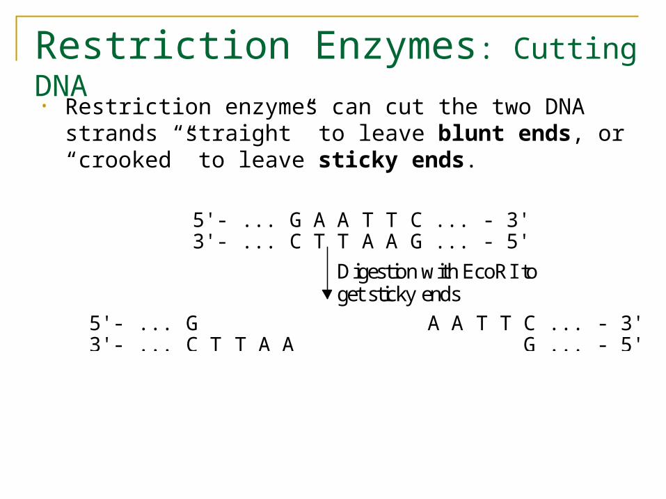
Restriction Enzymes: Cutting DNA
• Restriction enzymes can cut the two DNA strands “straight” to leave blunt ends, or “crooked” to leave sticky ends.
Digestion with EcoRI to
5'- ... G A A T T C ... - 3'
5'- ... G A A T T C ... - 3'
3'- ... C T T A A G ... - 5'
3'- ... C T T A A G ... - 5'
get sticky ends
Why we need so many copies
• Biologists needed to find a way to read DNA codes.• How do you read base pairs that are angstroms in
size?• It is not possible to directly look at it due to DNA’s
small size.• Need to use chemical techniques to detect what you
are looking for.• To read something so small, you need a lot of it, so
that you can actually detect the chemistry.• Need a way to make many copies of the base pairs,
and a method for reading the pairs.
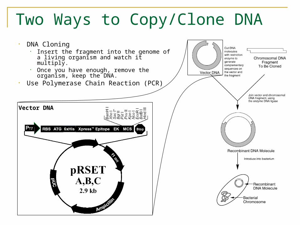
Two Ways to Copy/Clone DNA• DNA Cloning
• Insert the fragment into the genome of a living organism and watch it multiply.
• Once you have enough, remove the organism, keep the DNA.
• Use Polymerase Chain Reaction (PCR)
Vector DNA

Polymerase Chain Reaction• Problem: Modern
instrumentation cannot easily detect single molecules of DNA, making amplification a prerequisite for further analysis
• Solution: PCR doubles the number of DNA fragments at every iteration
1… 2… 4… 8…
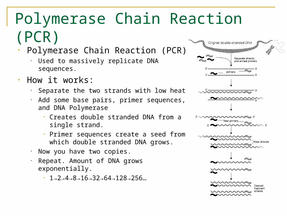
Polymerase Chain Reaction (PCR)
• Polymerase Chain Reaction (PCR)• Used to massively replicate DNA sequences.
• How it works:• Separate the two strands with low heat• Add some base pairs, primer sequences, and
DNA Polymerase• Creates double stranded DNA from a single
strand.• Primer sequences create a seed from which
double stranded DNA grows.• Now you have two copies.• Repeat. Amount of DNA grows exponentially.
• 1→2→4→8→16→32→64→128→256…
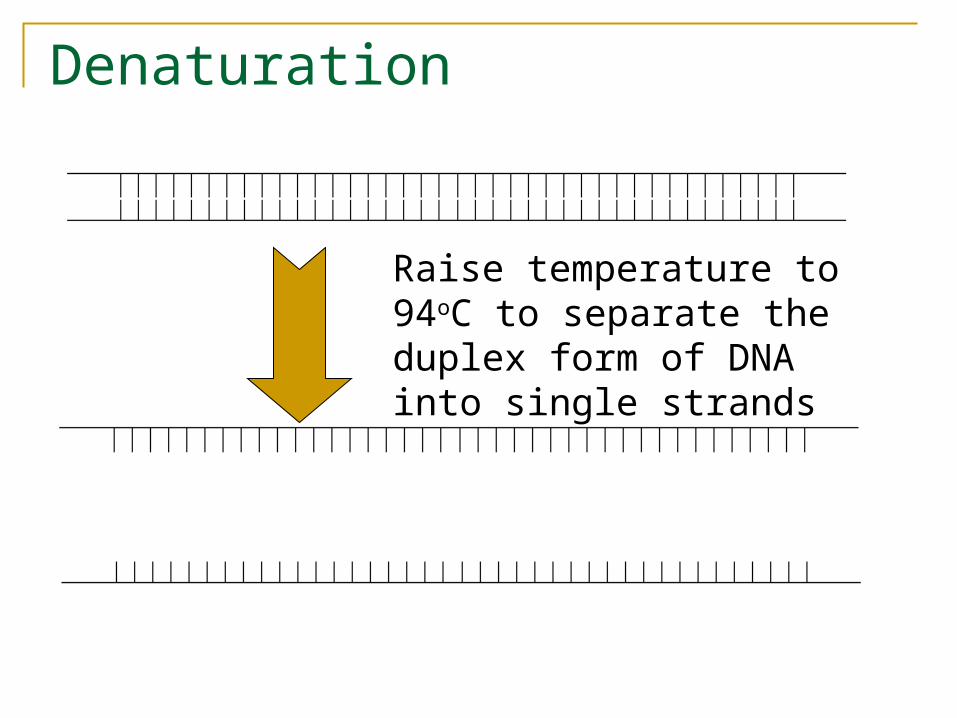
Denaturation
Raise temperature to 94oC to separate the duplex form of DNA into single strands
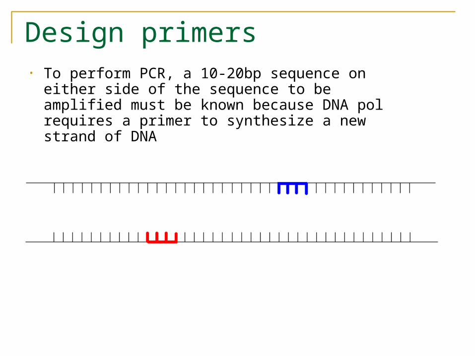
Design primers• To perform PCR, a 10-20bp sequence on either
side of the sequence to be amplified must be known because DNA pol requires a primer to synthesize a new strand of DNA

Annealing• Anneal primers at 50-65oC
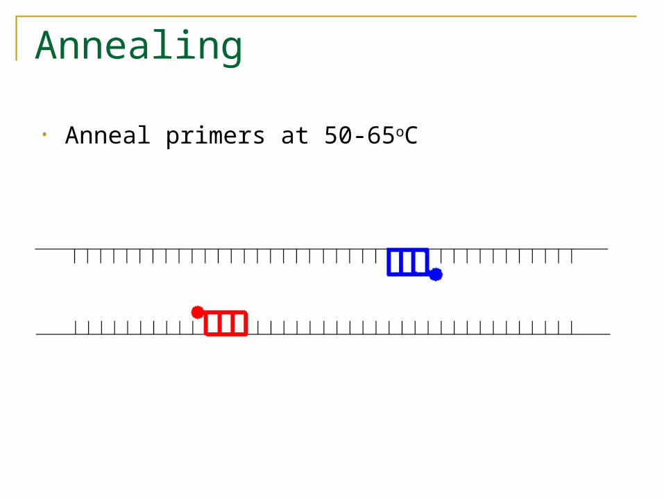
Annealing
• Anneal primers at 50-65oC
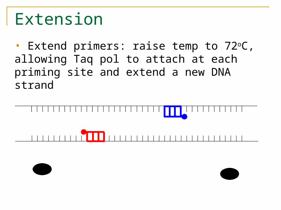
Extension
• Extend primers: raise temp to 72oC, allowing Taq pol to attach at each priming site and extend a new DNA strand
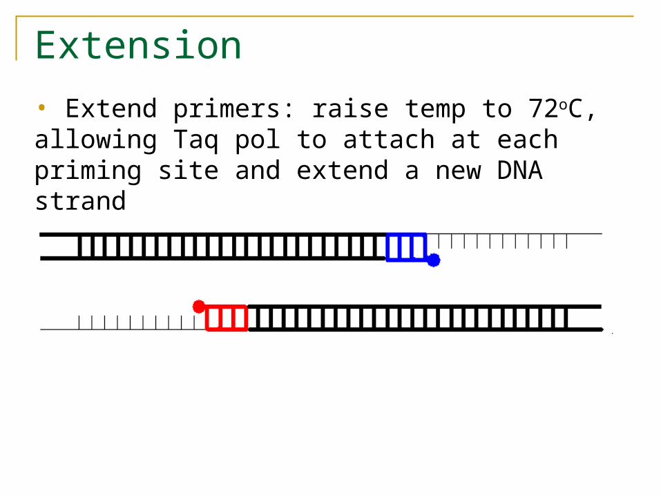
Extension
• Extend primers: raise temp to 72oC, allowing Taq pol to attach at each priming site and extend a new DNA strand

Repeat• Repeat the Denature, Anneal, Extension steps at
their respective temperatures…

Polymerase Chain Reaction

Electrophoresis
• A copolymer of mannose and galactose, agaraose, when melted and recooled, forms a gel with pores sizes dependent upon the concentration of agarose
• The phosphate backbone of DNA is highly negatively charged, therefore DNA will migrate in an electric field
• The size of DNA fragments can then be determined by comparing their migration in the gel to known size standards.

Pasting DNA
• Two pieces of DNA can be fused together by adding chemical bonds
• Hybridization – complementary base-pairing
• Ligation – fixing bonds with single strands
Probes
A probe is single-stranded DNA of 20+ nucleotide which is chosen on the basis of it consisting of the reverse complementary base pairs of the DNA fragment of interest.
Probes can be labeled before hybridization either radioactively or enzymatically (e.g. alkaline phosphatase or horseradish peroxidase), or fluorescently.
Probes are detected by directly exposing the membrane to X-ray film or chemiluminescent methods.

Reading DNA, i.e., DNA Sequencing
Determining the sequence of nucleotides of the DNA strand.
All sequencing methods employ the same basic strategy: generated a complete set of subfragments for a region
being studied whose lengths differ from each other by one nucleotide
label the subfragments ends with different nucleotide specific labels
separate the labeled fragments by size (e.g., using gel eletrophoresis)
used the different labels on the ends of the nucleotide to read the sequence
Automated DNA sequencing using fluorescent primers: (A)
• Labeled DNA fragments are loaded into single lanes of the electrophoresis gel.
• During the electrophoresis run, a laser beam is focused at a specific constant position on the gel.
• As the individual DNA fragments migrate past this position, the laser causes the dyes to fluoresce.
• Maximum fluorescence occurs at different wavelengths for the four dyes, and the information is recorded electronically and the interpreted sequence is stored in a computer database.
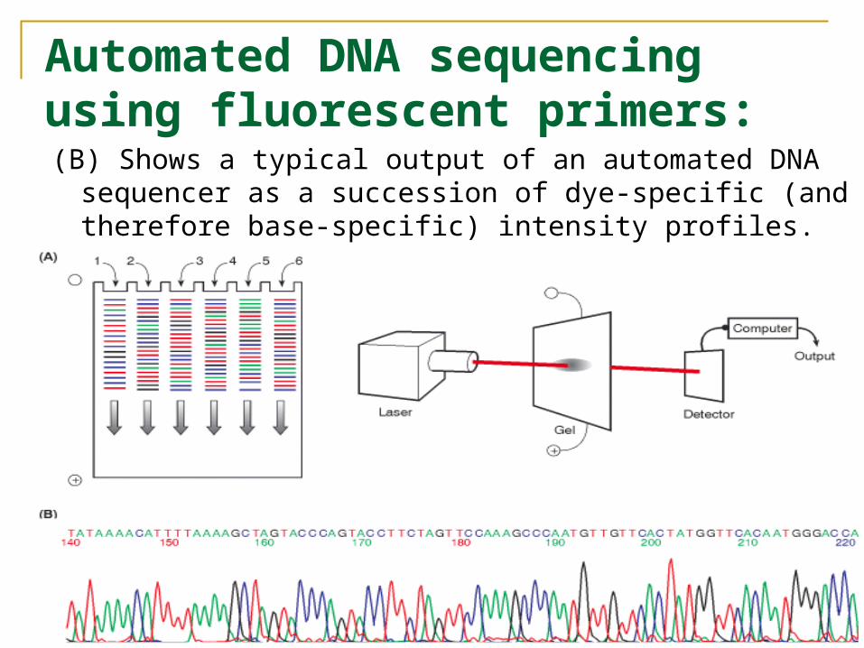
Automated DNA sequencing using fluorescent primers:(B) Shows a typical output of an automated DNA
sequencer as a succession of dye-specific (and therefore base-specific) intensity profiles.

Automated DNA Sequencing Technology: DNA fragments of about 1,000 nucleotides can be
sequenced at one time Each DNA fragment makes up only a small part of
an organism’s genome (eukaryotes have billions of base pairs in their DNA)
Each DNA fragment makes up only a small part of an organism’s gene (eukaryotes genes can be 100,000s base pairs)
After sequencing the DNA fragments, the fragment sequences must be reassembled to form the genome.
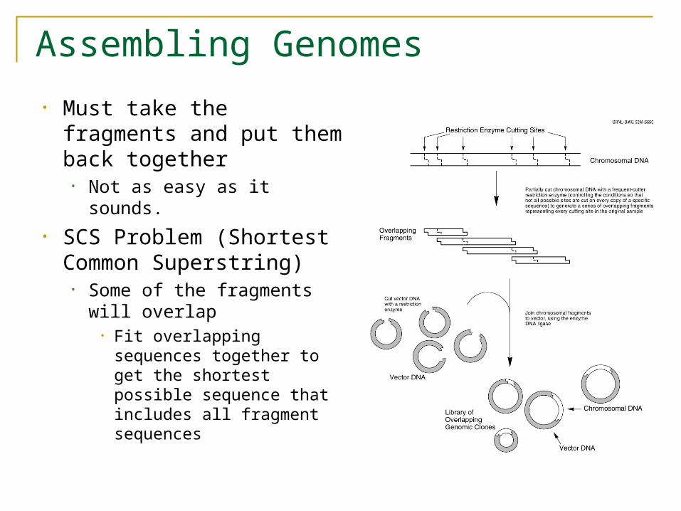
Assembling Genomes
• Must take the fragments and put them back together
• Not as easy as it sounds.
• SCS Problem (Shortest Common Superstring)
• Some of the fragments will overlap
• Fit overlapping sequences together to get the shortest possible sequence that includes all fragment sequences
Assembling Genomes
• DNA fragments contain sequencing errors• Two complements of DNA
• Need to take into account both directions of DNA
• Repeat problem• 50% of human DNA is just repeats• If you have repeating DNA, how do you know where it
goes?
• Assembly is a difficult problem!!!

Microarray (DNA chip)1,000 to 10,000s nucleotide sequences are affixed to individual
positions on the surface of a small glass chip.
Fluorescently labeled copies of the RNA transcripts (cDNA) from an organism are washed over the chip allowing them to hybridize to complementary nucleotides on the chip.
Unbound RNA is washing away.
A laser is used to excite the fluorescent tags and photodetectors quantify the amount of signal associated with each spot of a known sequence.
Application: Determination of relative RNA levels associated with huge numbers of known and predicted genes in a single experiment. DNA chips exist commerically for a variety of organisms.
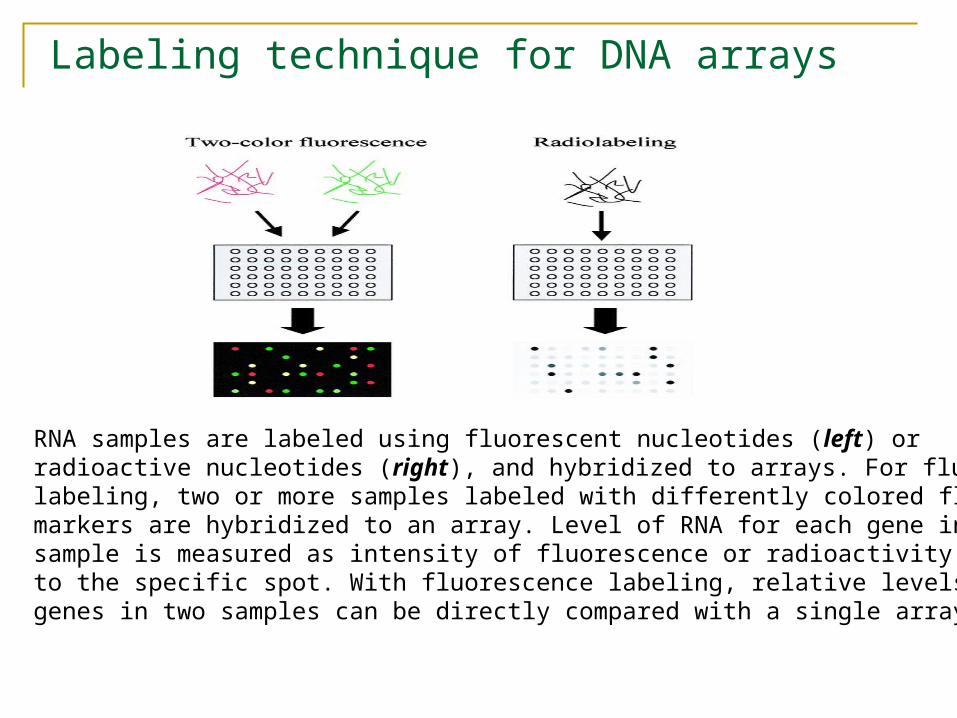
Labeling technique for DNA arrays
RNA samples are labeled using fluorescent nucleotides (left) or radioactive nucleotides (right), and hybridized to arrays. For fluorescentlabeling, two or more samples labeled with differently colored fluorescentmarkers are hybridized to an array. Level of RNA for each gene in the sample is measured as intensity of fluorescence or radioactivity binding to the specific spot. With fluorescence labeling, relative levels of expressedgenes in two samples can be directly compared with a single array.
DNA Arrays--Technical Foundations
• An array works by exploiting the ability of a given mRNA molecule to hybridize to the DNA template.
• Using an array containing many DNA samples in an experiment, the expression levels of hundreds or thousands genes within a cell by measuring the amount of mRNA bound to each site on the array.
• With the aid of a computer, the amount of mRNA bound to the spots on the microarray is precisely measured, generating a profile of gene expression in the cell.
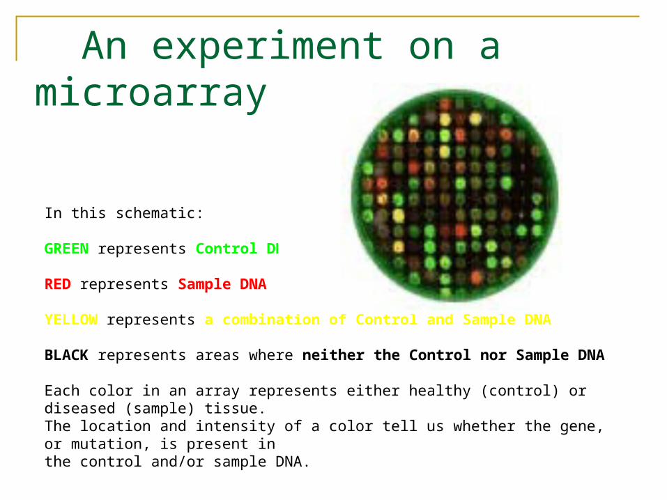
An experiment on a microarray
In this schematic: GREEN represents Control DNA
RED represents Sample DNA YELLOW represents a combination of Control and Sample DNA BLACK represents areas where neither the Control nor Sample DNA Each color in an array represents either healthy (control) or diseased (sample) tissue. The location and intensity of a color tell us whether the gene, or mutation, is present in the control and/or sample DNA.
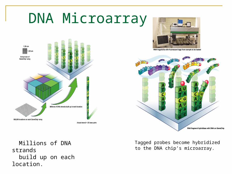
DNA Microarray
Tagged probes become hybridized to the DNA chip’s microarray.
Millions of DNA strands build up on each location.

DNA Microarray
Affymetrix
Each blue spot indicates the location of a PCR product. On a real microarray, each spot is about 100um in diameter.
Microarray is a tool for analyzing gene expression that consists of a glass slide.

Affymetrix GeneChip® Arrays
A combination of photolithography and combinatorial chemistry to manufacture GeneChip® Arrays. With a minimum number of steps, Affymetrix produces arrays with thousands of different probes packed at extremely high density. Enable to obtain high quality, genome-wide data using small sample volumes.
http://www.affymetrix.com/technology/manufacturing/index.affxMay 11,2004 13
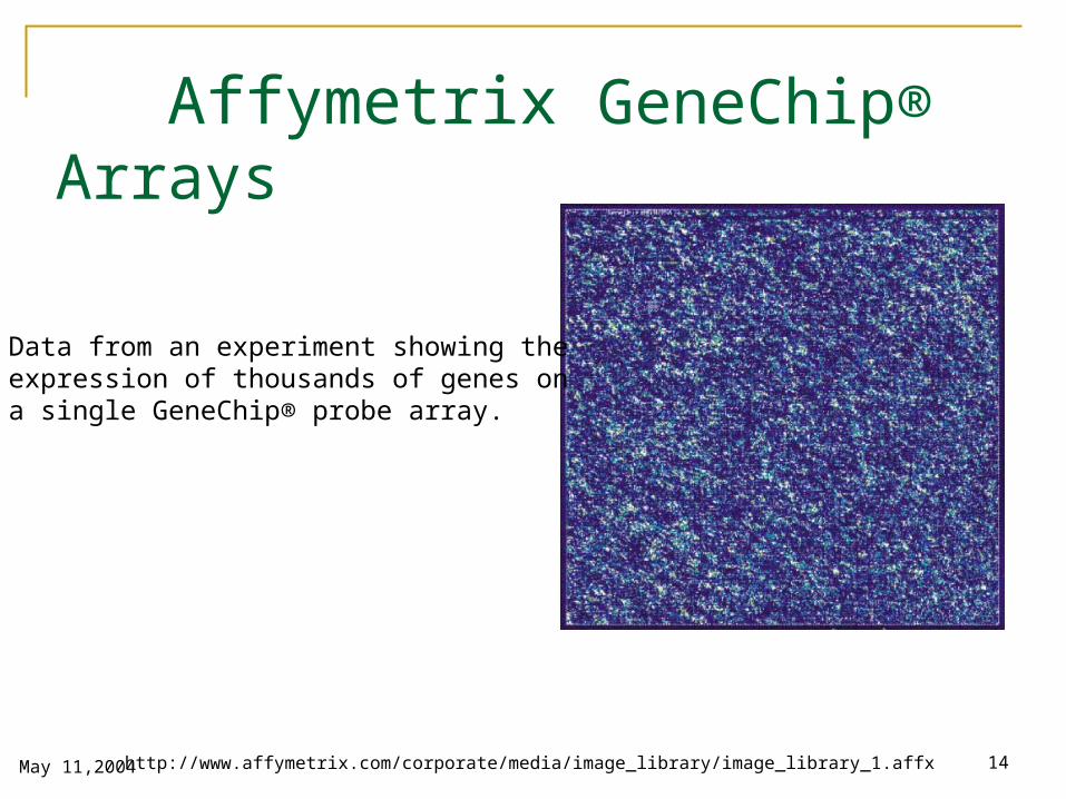
Affymetrix GeneChip® Arrays
Data from an experiment showing the expression of thousands of genes on a single GeneChip® probe array.
http://www.affymetrix.com/corporate/media/image_library/image_library_1.affxMay 11,2004 14