Module VIII Assays - shcollege.ac.in
36
Module VIII Assays
Transcript of Module VIII Assays - shcollege.ac.in

Module VIII Assays

Radio Immuno Assay
• Radioimmunoassay (RIA) is a very sensitive in vitro assay technique used to measure concentrations of antigens (for example, hormone levels in blood) by use of antibodies.
• As such, it can be seen as the inverse of a radio binding assay, which quantifies an antibody by use of corresponding antigens.

• Although the RIA technique is extremely sensitive and extremely specific, requiring specialized equipment, it remains among the least expensive methods to perform such measurements.
• It requires special precautions and licensing, since radioactive substances are used.

Method
• Classically, to perform a radioimmunoassay, a known quantity of an antigen is made radioactive, frequently by labelling it with gamma-radioactive isotopes of iodine, such as 125-I, attached to tyrosine.
• This radiolabeled antigen is then mixed with a known amount of antibody for that antigen, and as a result, the two specifically bind to one another.

• Then, a sample of serum from a patient containing an unknown quantity of that same antigen is added. This causes the unlabelled (or "cold") antigen from the serum to compete with the radiolabeled antigen ("hot") for antibody binding sites.
• As the concentration of "cold" antigen is increased, more of it binds to the antibody, displacing the radiolabeled variant, and reducing the ratio of antibody-bound radiolabeled antigen to free radiolabeled antigen.

• The bound antigens are then separated from the unbound ones, and the radioactivity of the free(unbound) antigen remaining in the supernatant is measured using a gamma counter.
• This method can be used for any biological molecule in principle and is not restricted to serum antigens, nor is it required to use the indirect method of measuring the free antigen instead of directly measuring the captured antigen.

• For example, if it is undesirable or not possible to radiolabel the antigen or target molecule of interest, a RIA can be done if two different antibodies that recognize the target are available and the target is large enough (e.g., a protein) to present multiple epitopes to the antibodies.
• One antibody would be radiolabeled as above while the other would remain unmodified.
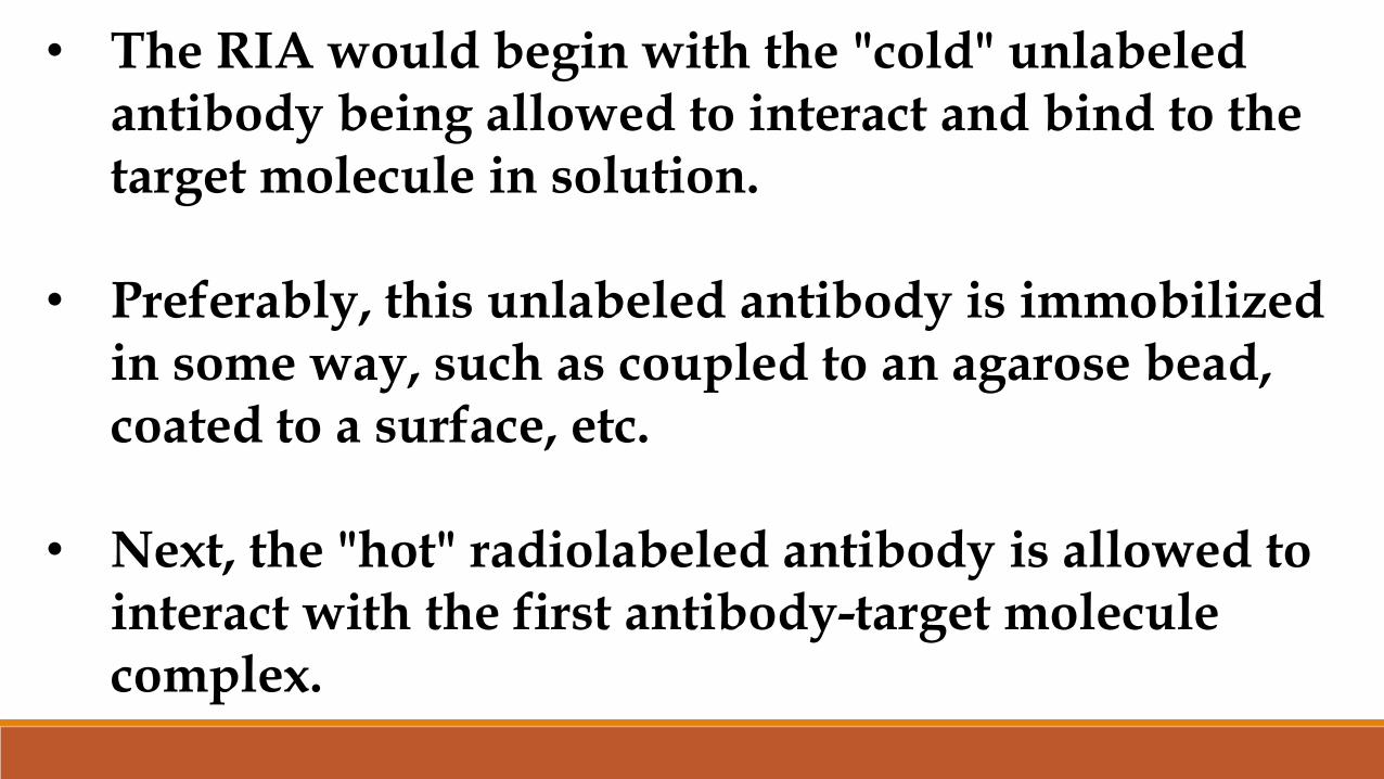
• The RIA would begin with the "cold" unlabeledantibody being allowed to interact and bind to the target molecule in solution.
• Preferably, this unlabeled antibody is immobilized in some way, such as coupled to an agarose bead, coated to a surface, etc.
• Next, the "hot" radiolabeled antibody is allowed to interact with the first antibody-target molecule complex.

• After extensive washing, the direct amount of radioactive antibody bound is measured and the amount of target molecule quantified by comparing it to a reference amount assayed at the same time.
• This method is similar in principle to the non-radioactive sandwich ELISA method.


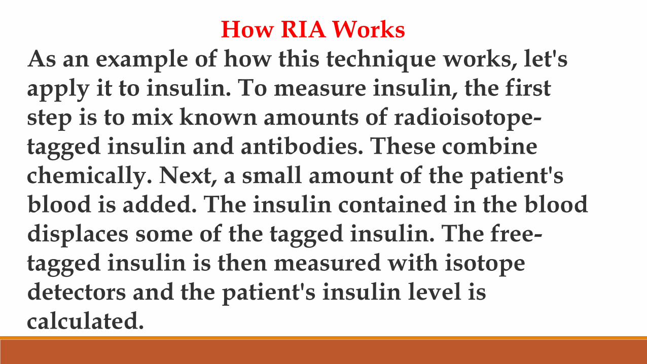
How RIA WorksAs an example of how this technique works, let's apply it to insulin. To measure insulin, the first step is to mix known amounts of radioisotope-tagged insulin and antibodies. These combine chemically. Next, a small amount of the patient's blood is added. The insulin contained in the blood displaces some of the tagged insulin. The free-tagged insulin is then measured with isotope detectors and the patient's insulin level is calculated.

Enzyme Linked Immuno Sorbant Assay (ELISA)
• ELISA assays are similar in principle to RIAs but, instead of using antibodies or antigens conjugated to radioisotopes, they use antibodies or antigens covalently bound to enzymes.
• The conjugated enzymes are selected on the basis of their ability to catalyze the conversion of a substrate into a colored, fluorescent, or chemiluminescent product.

• These assays match the sensitivity of RIAs and have the advantage of being safer and, oft en, less costly.
• A number of variations of the basic ELISA assay have been developed.
• Each type of ELISA can be used qualitatively to detect the presence of antibody or antigen.

• Alternatively, a standard curve based on known concentrations of antibody or antigen can be prepared and used to determine the concentration of a sample.
• Types of ELISA
• Indirect ELISA• Sandwich ELISA• Competitive ELISA
1. Indirect ELISA
• Antibody can be detected, or its concentration determined with an indirect ELISA assay.
• Serum or some other sample containing primary antibody (Ab1) is added to an antigen-coated microtiter well and allowed to react with the antigen attached to the well.

• After any free Ab1 is washed away, the antibody bound to the antigen is detected by adding an enzyme-conjugated secondary antibody (Ab2) that binds to Ab1.
• Any free Ab2 is again washed away, and a substrate for the enzyme is added.

• The amount of coloured, fluorescent, or luminescent reaction product that forms is measured using a specialized plate reader and compared with the amount of product generated when the same set of reactions is performed using a standard curve of known Ab1 concentrations. (A direct ELISA assay would detect the amount of antigen on the plate using enzyme coupled antibodies, and is rarely used.)
• This version of ELISA is the method of choice to detect the presence of serum antibodies against human immunodeficiency virus (HIV), the causative agent of AIDS.
• In this assay, recombinant envelope and core proteins of HIV are adsorbed as solid-phase antigens to microtiter wells.
• Individuals infected with HIV will produce serum antibodies to epitopes on these viral proteins.
• Generally, serum antibodies to HIV can be detected by ELISA within 6 weeks of infection.
2. Sandwich ELISA
• Antigen can be detected or measured by a sandwich ELISA. In this technique, the antibody (rather than the antigen) is immobilized on a microtiter well.
• A sample containing unknown amounts of antigen is allowed to react with the immobilized antibody.
• After the well is washed, a second enzyme-linked antibody specific for a different epitope on the antigen is added and allowed to react with the bound antigen.
• After any free second antibody is removed by washing, substrate is added, and the coloured reaction product is measured.
• A common variant on this assay uses a biotin-linked second antibody and then adds enzyme-linked avidin in an additional step.
• Sandwich ELISAs have proven particularly useful for the measurement of soluble cytokine concentrations in tissue culture supernatants, as well as in serum and body fluids.
• Note that, for this assay to work, the two antibodies used for the antigen immobilization (capture) and detection phases respectively must bind to different determinants (epitopes) on the antigen.
• Sandwich ELISAs therefore routinely use a pair of monoclonal antibodies specific for different regions on the antigen.

Competitive ELISA
• The competitive ELISA provides another extremely sensitive variation for measuring amounts of antigen.
• In this technique, antibody is first incubated in solution with a sample containing antigen.
• The antigen-antibody mixture is then added to an antigen-coated microtiter well.
• The more antigen present in the initial solution-phase sample, the less free antibody will be available to bind to the antigen-coated well.

• After washing off the unbound antibody, an enzyme-conjugated Ab2 specifi c for the isotype of the Ab1 can be added to determine the amount of Ab1 bound to the well.
• In the competitive assay, the higher the concentration of antigen in the original sample, the lower the final signal.
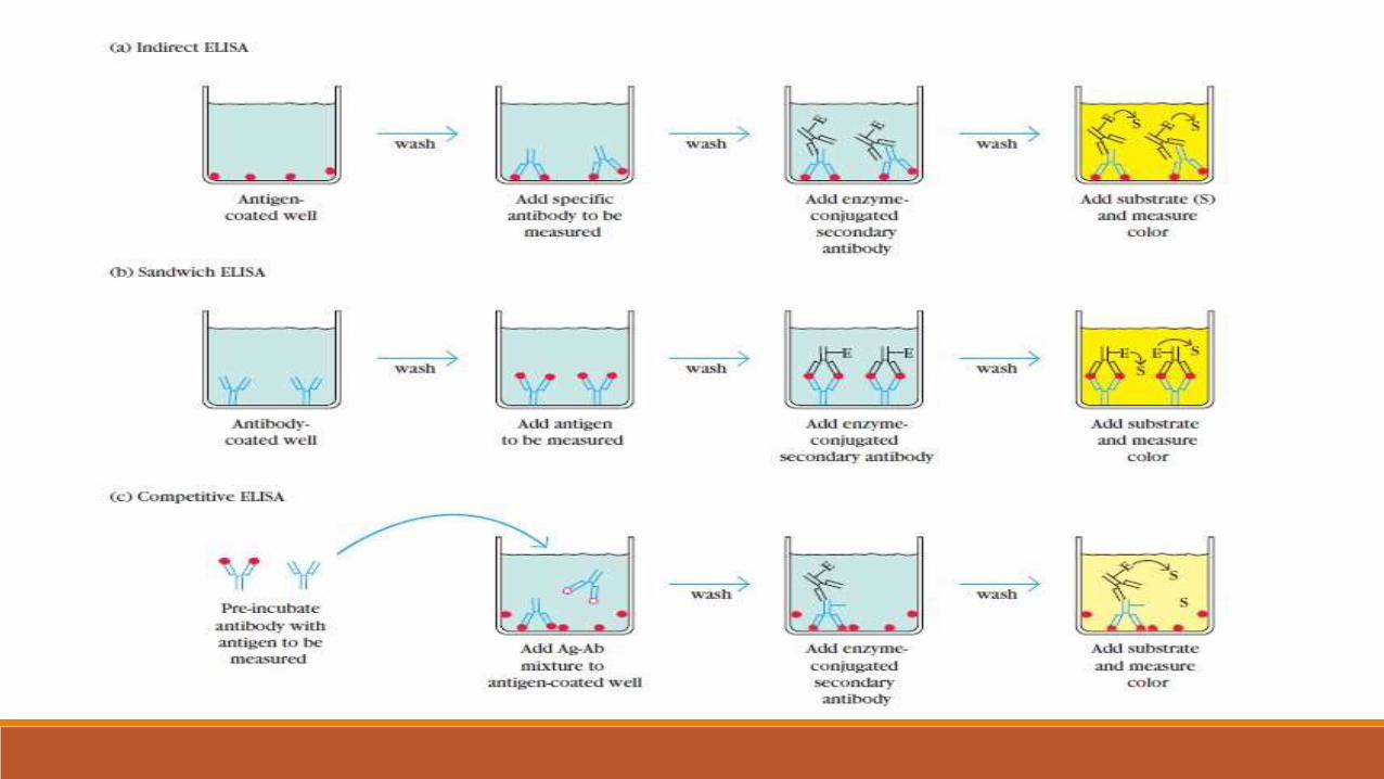
Each assay can be used qualitatively or quantitatively by comparison with standard curves prepared with known concentrations of antibody or antigen. Antibody can be determined with an indirect ELISA (a), whereas antigen can be determined with a sandwich ELISA (b) or competitive ELISA (c). In the competitive ELISA, which is an inhibition-type assay that is identical in principle to the competition RIA described above, the concentration of antigen is inversely proportional to the colour produced.

Available Enzyme Systems for ELISA Assays
• The three enzymes commonly used in ELISA assays are alkaline phosphatase, β-galactosidase, and horseradish peroxidase (HRP), each of which can be used with chromogenic substrates (substrates which are colourless but give rise to a product that absorbs light in the visible range).
• Of these three, β -galactosidase is the least frequently employed.
• The most common substrate for alkaline phosphatase is p-nitrophenyl phosphate (pNPP), which is highly soluble and colorless.
• Alkaline phosphatase cleaves the phosphate group from this substrate to yield p-nitrophenol, which is yellow and absorbs light at 460 nm.
• Both substrate and product are nontoxic and inexpensive, and the substrate can be purchased in tablet form.

• However, the substrate will convert to product if left in solution at room temperature, which somewhat limits the range of the chromogenic ELISA assay.
• Chromogenic alkaline phosphatase assays are therefore extremely useful when a rapid, inexpensive, “yes or no” answer is called for, when the laboratory facilities are limited, and/or for multipurpose teaching laboratories.

• Recently, both fluorescent and luminescent assay kits for alkaline phosphatase have been made available by a variety of companies.
• The lower background level of light emission from such products dramatically enhances the sensitivity of the assays, allowing femtogramlevels of antigens to be detected.