MODULE 3 HONEYBEE DISEASES, PESTS AND ... 3 HONEYBEE DISEASES, PESTS AND POISONING 1 Contents 3.1 a...
48
MODULE 3 HONEYBEE DISEASES, PESTS AND POISONING 1 Contents 3.1 a detailed account of the field diagnosis of American foul brood (AFB) and European foul brood (EFB), including lateral flow devices and a detailed account of the signs of these two diseases; .......... 3 3.2 an account of the life cycle of the causative organisms of AFB and EFB and their development within the larvae;....................................................................................................................................... 5 3.3 a detailed account of the development of AFB and EFB within the colony; ............................................. 6 3.4 a detailed account of the ways in which AFB and EFB are spread from one colony to another; ............ 7 3.5 a detailed account of the authorised treatment of colonies infected with AFB and EFB including methods of destruction of colonies and the sterilisation of equipment; .................................................... 7 3.6 the features that aid recognition of the Asian Hornet (Vespa velutina) and the notifiable pests Small hive beetle (Aethina tumida) and Tropilaelaps mites ............................................................................... 8 3.7 a detailed account of the statutory requirements relating to notifiable diseases and pests and the implementation of these requirements in the United Kingdom, ............................................................. 11 3.8 an account of the statutory requirements relating to the importation of honeybees; .............................12 3.9 a description of the life cycle and natural history of Varroa destructor including its development within the honeybee colony and its spread to other colonies; ................................................................16 3.10 a detailed account of the signs of Varroosis describing methods of detection and ways of monitoring the presence of the varroa mite in honeybee colonies; ........................................................18 3.11 a detailed account of methods of treatment and control of Varroosis, including Integrated Pest Management (IPM) and an outline of the consequences of incorrect administration of chemical treatments, together with a way of determining the resistance of varroa to such treatments; ...............19 3.12 a detailed account of the cause, signs and treatment (if any) of adult bee diseases currently found in the United Kingdom these diseases to include Nosema, Dysentery, Acarine and Amoeba; .............24 3.13 a simple account of the structure and function of the alimentary, excretory and respiratory systems of the adult honeybee and of the life cycle of the causative organisms of adult honeybee diseases; ...27 3.14 a detailed account of the cause, signs and recommended treatment (if any) of the following brood diseases and conditions: chalk brood, sacbrood, chilled brood, bald brood, neglected drone brood and stone brood;.....................................................................................................................................32 3.15 a detailed account of the laboratory methods of diagnosis of Acarine, Nosema and Amoeba diseases in worker honeybees; ..............................................................................................................34 3.16 a detailed description of the fumigation of comb using ethanoic acid (acetic acid), including safety precautions to be taken; .........................................................................................................................35 3.17 a detailed description of procedures by which a colony can be transferred onto clean comb including any precautions that need to be taken and the circumstances which merit such procedures. These procedures to include shook swarm and Bailey comb change; ..............................36 3.18 a description of the effects of chronic bee paralysis (both syndromes), acute bee paralysis virus, black queen cell virus, sacbrood and deformed wing viruses together with an elementary account of the effects of other viruses affecting honeybees including their association with other bee diseases and pests where applicable; ....................................................................................................37 3.19 the scientific names of the causative organisms associated with diseases of honeybees; ...................39 3.20 an outline account of the life cycle of Braula coeca, its effect on the colony and a description of the differences between adult Braula and Varroa; .......................................................................................40 3.21 an outline account of the signs of poisoning by natural substances, pesticides, herbicides and other chemicals to which honeybees may be exposed; ..................................................................................40 3.22 an account of the ways in which honeybees can become exposed to agricultural and pest control chemicals;...............................................................................................................................................41 3.23 a detailed description of the action to take, and practical measures possible, when prior notification of application of toxic chemicals to crops is given; .................................................................................41 3.24 an outline description of a spray liaison scheme operated by a beekeeping association; .....................42 3.25 an account of the action to be taken when spray damage is suspected; ...............................................42 3.26 a description of the damage caused to colonies and equipment by mice, woodpeckers and other pests and ways of preventing this; .........................................................................................................42 3.27 a detailed account of wax moth damage and the life cycle of both the Greater Wax Moth (Galleria mellonella) and the Lesser Wax Moth (Achroia grisella); .......................................................................44 3.28 a detailed account of methods of treating or storing comb with particular reference to preventing wax moth damage; .................................................................................................................................47 Appendix: a detailed account of the authorised treatments for adult bee diseases in the UK; ........................48
Transcript of MODULE 3 HONEYBEE DISEASES, PESTS AND ... 3 HONEYBEE DISEASES, PESTS AND POISONING 1 Contents 3.1 a...
MODULE 3 HONEYBEE DISEASES, PESTS AND POISONING
1
Contents 3.1 a detailed account of the field diagnosis of American foul brood (AFB) and European foul brood
(EFB), including lateral flow devices and a detailed account of the signs of these two diseases; .......... 3 3.2 an account of the life cycle of the causative organisms of AFB and EFB and their development
within the larvae; ....................................................................................................................................... 5 3.3 a detailed account of the development of AFB and EFB within the colony; ............................................. 6 3.4 a detailed account of the ways in which AFB and EFB are spread from one colony to another; ............ 7 3.5 a detailed account of the authorised treatment of colonies infected with AFB and EFB including
methods of destruction of colonies and the sterilisation of equipment; .................................................... 7 3.6 the features that aid recognition of the Asian Hornet (Vespa velutina) and the notifiable pests Small
hive beetle (Aethina tumida) and Tropilaelaps mites ............................................................................... 8 3.7 a detailed account of the statutory requirements relating to notifiable diseases and pests and the
implementation of these requirements in the United Kingdom, ............................................................. 11 3.8 an account of the statutory requirements relating to the importation of honeybees; ............................. 12 3.9 a description of the life cycle and natural history of Varroa destructor including its development
within the honeybee colony and its spread to other colonies; ................................................................ 16 3.10 a detailed account of the signs of Varroosis describing methods of detection and ways of
monitoring the presence of the varroa mite in honeybee colonies; ........................................................ 18 3.11 a detailed account of methods of treatment and control of Varroosis, including Integrated Pest
Management (IPM) and an outline of the consequences of incorrect administration of chemical treatments, together with a way of determining the resistance of varroa to such treatments; ............... 19
3.12 a detailed account of the cause, signs and treatment (if any) of adult bee diseases currently found in the United Kingdom these diseases to include Nosema, Dysentery, Acarine and Amoeba; ............. 24
3.13 a simple account of the structure and function of the alimentary, excretory and respiratory systems of the adult honeybee and of the life cycle of the causative organisms of adult honeybee diseases; ... 27
3.14 a detailed account of the cause, signs and recommended treatment (if any) of the following brood diseases and conditions: chalk brood, sacbrood, chilled brood, bald brood, neglected drone brood and stone brood;..................................................................................................................................... 32
3.15 a detailed account of the laboratory methods of diagnosis of Acarine, Nosema and Amoeba diseases in worker honeybees; .............................................................................................................. 34
3.16 a detailed description of the fumigation of comb using ethanoic acid (acetic acid), including safety precautions to be taken; ......................................................................................................................... 35
3.17 a detailed description of procedures by which a colony can be transferred onto clean comb including any precautions that need to be taken and the circumstances which merit such procedures. These procedures to include shook swarm and Bailey comb change; .............................. 36
3.18 a description of the effects of chronic bee paralysis (both syndromes), acute bee paralysis virus, black queen cell virus, sacbrood and deformed wing viruses together with an elementary account of the effects of other viruses affecting honeybees including their association with other bee diseases and pests where applicable;.................................................................................................... 37
3.19 the scientific names of the causative organisms associated with diseases of honeybees; ................... 39 3.20 an outline account of the life cycle of Braula coeca, its effect on the colony and a description of the
differences between adult Braula and Varroa; ....................................................................................... 40 3.21 an outline account of the signs of poisoning by natural substances, pesticides, herbicides and other
chemicals to which honeybees may be exposed; .................................................................................. 40 3.22 an account of the ways in which honeybees can become exposed to agricultural and pest control
chemicals; ............................................................................................................................................... 41 3.23 a detailed description of the action to take, and practical measures possible, when prior notification
of application of toxic chemicals to crops is given; ................................................................................. 41 3.24 an outline description of a spray liaison scheme operated by a beekeeping association;..................... 42 3.25 an account of the action to be taken when spray damage is suspected;............................................... 42 3.26 a description of the damage caused to colonies and equipment by mice, woodpeckers and other
pests and ways of preventing this; ......................................................................................................... 42 3.27 a detailed account of wax moth damage and the life cycle of both the Greater Wax Moth (Galleria
mellonella) and the Lesser Wax Moth (Achroia grisella); ....................................................................... 44 3.28 a detailed account of methods of treating or storing comb with particular reference to preventing
wax moth damage; ................................................................................................................................. 47 Appendix: a detailed account of the authorised treatments for adult bee diseases in the UK; ........................ 48
MODULE 3 HONEYBEE DISEASES, PESTS AND POISONING
2
General References and Further Reading: http://en.wikipedia.org/wiki/List_of_diseases_of_the_honey_bee http://www.ent.uga.edu/Bees/beekeeping.html http://www.agf.gov.bc.ca/apiculture/factsheets/index.htm Also Celia Davies “The Honey Bee Around and About”
MODULE 3 HONEYBEE DISEASES, PESTS AND POISONING
3
The Candidate shall be able to give:-
3.1 a detailed account of the field diagnosis of American foul brood (AFB) and European foul brood (EFB), including lateral flow devices and a detailed account of the signs of these two diseases;
When in the field:
o Wear full protective clothing and have a smoker well lit.
o Keep the colony subdued with smoke.
o Remove the hive roof and place it on the ground by the hive (to the side of the hive or behind
away from the hive entrance).
o If there are supers on the hive, remove them and place them on the upturned roof, keeping them
covered to prevent robbing.
o Remove any queen excluder and examine the underside for the queen. If she is present return
her to the colony. Place the excluder on the ground next to the roof
o Where two boxes are used for the brood nest, examine the bottom one first.
o Remove the outside comb, which is unlikely to contain brood, and lean it against a front corner of
the hive – you will then have room to work.
o Take each comb in turn, and, holding it by the lugs within the brood chamber, give it a sharp
shake. This will deposit the bees on the bottom of the hive without harming them, the queen or
brood.
o Any bees on a comb may be concealing infected brood from the beekeeper’s view. On combs
free from bees, any abnormality is easily spotted.
o Examine the brood, both sealed and unsealed, quickly but carefully, for any signs of abnormality –
such as discoloured larvae or perforated cappings.
o Look for AFB scales by holding the combs towards the light and scanning the bottom walls of any
open cells.
o Look inside any sealed cells with abnormal looking cappings after opening the cell with a corner of
the hive tool, matchstick or suitable implement.
o To establish the consistency of any dead remains present, probe these with a matchstick.
o Dispose of the used matchstick in the smoker.
o Continue until you have examined all the brood combs; then reassemble the hive.
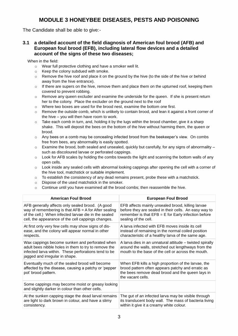
American Foul Brood European Foul Brood
AFB generally affects only sealed brood. (A good way of remembering is that AFB = A for After sealing of the cell.) When infected larvae die in the sealed cell, the appearance of the cell cappings changes.
EFB affects mainly unsealed brood, killing larvae before they are sealed in their cells. An easy way to remember is that EFB = E for Early infection before sealing of the cell.
At first only very few cells may show signs of dis-ease, and the colony will appear normal in other respects.
A larva infected with EFB moves inside its cell instead of remaining in the normal coiled position characteristic of a healthy larva of the same age.
Wax cappings become sunken and perforated when adult bees nibble holes in them to try to remove the infected larva within. These perforations tend to be jagged and irregular in shape.
A larva dies in an unnatural attitude – twisted spirally around the walls, stretched out lengthways from the mouth to the base of the cell or across the mouth.
Eventually much of the sealed brood will become affected by the disease, causing a patchy or ‘pepper pot’ brood pattern.
When EFB kills a high proportion of the larvae, the brood pattern often appears patchy and erratic as the bees remove dead brood and the queen lays in the vacant cells.
Some cappings may become moist or greasy looking and slightly darker in colour than other cells.
At the sunken capping stage the dead larval remains are light to dark brown in colour, and have a slimy consistency.
The gut of an infected larva may be visible through its translucent body wall. The mass of bacteria living within it give it a creamy white colour.

MODULE 3 HONEYBEE DISEASES, PESTS AND POISONING
4
American Foul Brood European Foul Brood
There may then be an unpleasant smell associated with decomposition.
A very unpleasant odour may sometimes accompany severe EFB infection, depending on the presence of certain other species of bacteria in the remains of dead larvae.
Further drying leads to the final stage, which is a very dark brown, rather rough scale lying on the lower side of the cell and extending from just behind the mouth of the cell right back to the base.
As it collapses, a dead larva often looks as though it has melted, turning yellowish-brown and eventually drying up to form a loosely attached brown scale.
To detect scales, hold the comb facing the light: their rough surfaces reflect the light, making them easy to see even when they are almost the same colour as the comb itself.
Conduct the ‘ropiness’ test: insert a matchstick and slowly withdraw it; a brown, mucus-like thread or ‘rope’ 10-30mm long a reliable indicator for AFB.
The ropy condition is followed by a tacky stage as the larval remains in the cell gradually dry up and the colour changes to dark brown.
The proboscis of dead pupae may sometimes remain intact, protruding upwards from the bottom edge of the cell
EFB cannot be reliably identified visually, as the signs of disease can easily be confused with other brood
abnormalities. FERA Bee Inspectors confirm suspect infections in the field by using Lateral Flow Devices
(LFDs). Occasionally sample brood combs (or suspect larvae in plastic tubes) are sent to the NBU
laboratory where
larval gut contents
are examined for the
presence of the
causative bacteria.
To test for foulbrood
using an LFD, put a
sample of suspect
infected larval
material into the
buffer bottle and
shake it for about 20
seconds. Then put
2-3 drops of the
resulting suspension
onto the LFD. The
blue lines at the C
(Control) and T (Test)
lines indicate a
positive result.
MODULE 3 HONEYBEE DISEASES, PESTS AND POISONING
5
Advantages of LFD:
o Available for both AFB and EFB o Can be used in the field o Established, accepted mature technology o Stable – shelf-lives of 12–24 months often without refrigeration o Ease of use: minimal operator-dependent steps and interpretation o Accuracy: LFDs detected Melissococcus plutonius in 96–100% (n = 137) of EFB-infected samples in
laboratory trials. Field validation was equally robust: LFD-testing on site gave correct diagnoses for 96% (n = 184) of samples; false positives were rare (~1%).”
Disadvantages of LFD:
o Indicates only the presence of the disease, not its level o Results must be recorded manually o Based on a specific antibody; test might become ineffective if new strains emerge.
3.2 an account of the life cycle of the causative organisms of AFB and EFB and their development within the larvae;
AFB American foulbrood (AFB) is considered to be the most fatal of honeybee brood diseases. The disease
attacks only the very young larvae; larvae older than 48 hours and adult bees are not susceptible to it.
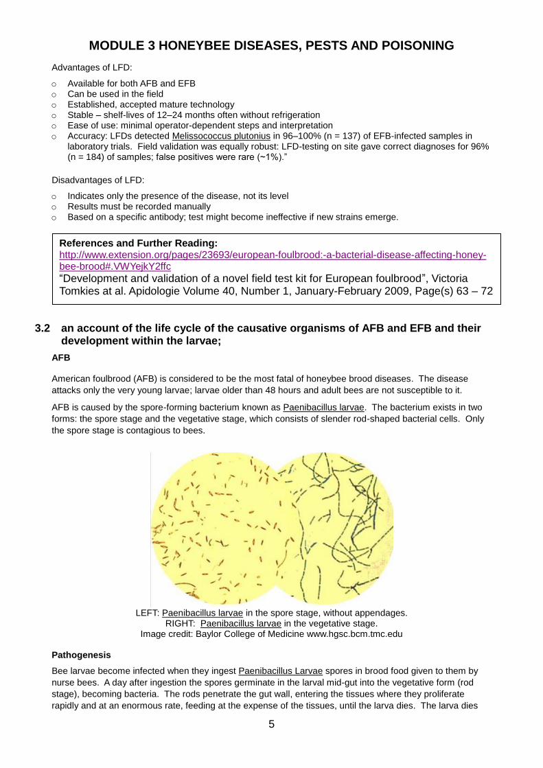
AFB is caused by the spore-forming bacterium known as Paenibacillus larvae. The bacterium exists in two
forms: the spore stage and the vegetative stage, which consists of slender rod-shaped bacterial cells. Only
the spore stage is contagious to bees.
LEFT: Paenibacillus larvae in the spore stage, without appendages.
RIGHT: Paenibacillus larvae in the vegetative stage. Image credit: Baylor College of Medicine www.hgsc.bcm.tmc.edu
Pathogenesis
Bee larvae become infected when they ingest Paenibacillus Larvae spores in brood food given to them by
nurse bees. A day after ingestion the spores germinate in the larval mid-gut into the vegetative form (rod
stage), becoming bacteria. The rods penetrate the gut wall, entering the tissues where they proliferate
rapidly and at an enormous rate, feeding at the expense of the tissues, until the larva dies. The larva dies
References and Further Reading: http://www.extension.org/pages/23693/european-foulbrood:-a-bacterial-disease-affecting-honey-bee-brood#.VWYejkY2ffc
“Development and validation of a novel field test kit for European foulbrood”, Victoria Tomkies at al. Apidologie Volume 40, Number 1, January-February 2009, Page(s) 63 – 72
MODULE 3 HONEYBEE DISEASES, PESTS AND POISONING
6
after its cell has been sealed; sealing the cell stops the supply of nourishment to the bacteria; they cease to
grow and proliferate, and revert to the spore stage.
After death, the normally white larvae turn dark brown and decay into a glue-like mass, which will form a
rope. The decaying mass has a foul smell - hence the name, foulbrood. At the final stage, within a month or
so, a dead larva or pupa dries to a dark brown scale that adheres tightly to the lower side of the cell too
tightly for the bees to remove. Each scale contains millions of infective spores. Once they are inside the
larval gut again, the cycle repeats itself.
EFB The bacterium responsible for causing the symptoms of European Foulbrood (EFB) is probably
Melissococcus plutonius. When it infects a larva, other bacteria move in, causing secondary infections:
• Bacillus alveri and laterosporus
• Bacterium eurydice
• Streptococcus faecalis
The bacteria enter a larva in brood food and multiply in the ventriculus (stomach), feeding on the larval food.
The bacteria lodge between the peritrophic1 membrane and the food in the ventriculus. The bacteria act
essentially as a parasite competing for food, and the larva dies of starvation about 3 or 4 days before the cell
is due to be sealed. During this period the larva contorts itself into unusual positions, twisted spirally or
flattened out lengthways in the cell. Its colour changes from pearly white to cream and then to a yellowy
green. The bacterial mass in the larval stomach causes much of this early colour change.
The supply of food to larvae affects the course of the disease. Because the bacteria compete with the larvae
for food, increasing the supply of food can enable larvae to survive infection.
At the onset of nectar flow in early spring, the number of house bees recruited to forage may increase rapidly
leaving fewer in the hive to feed larvae. Under these conditions, M. plutonius may be able to starve larvae to
death and give rise to symptoms of the disease. When the ratio of nurse bees to larvae stabilises and larvae
receive enough food to survive to pupation, symptoms disappear.
However, EFB can occur throughout a season and will sometimes not abate of its own accord. In severe
cases, it can cause a colony to die. Also, contaminated combs and equipment can cause EFB to recur. The
bacterium that causes EFB does not produce spores, but combs contaminated with it can still re-infect bees
in subsequent years.
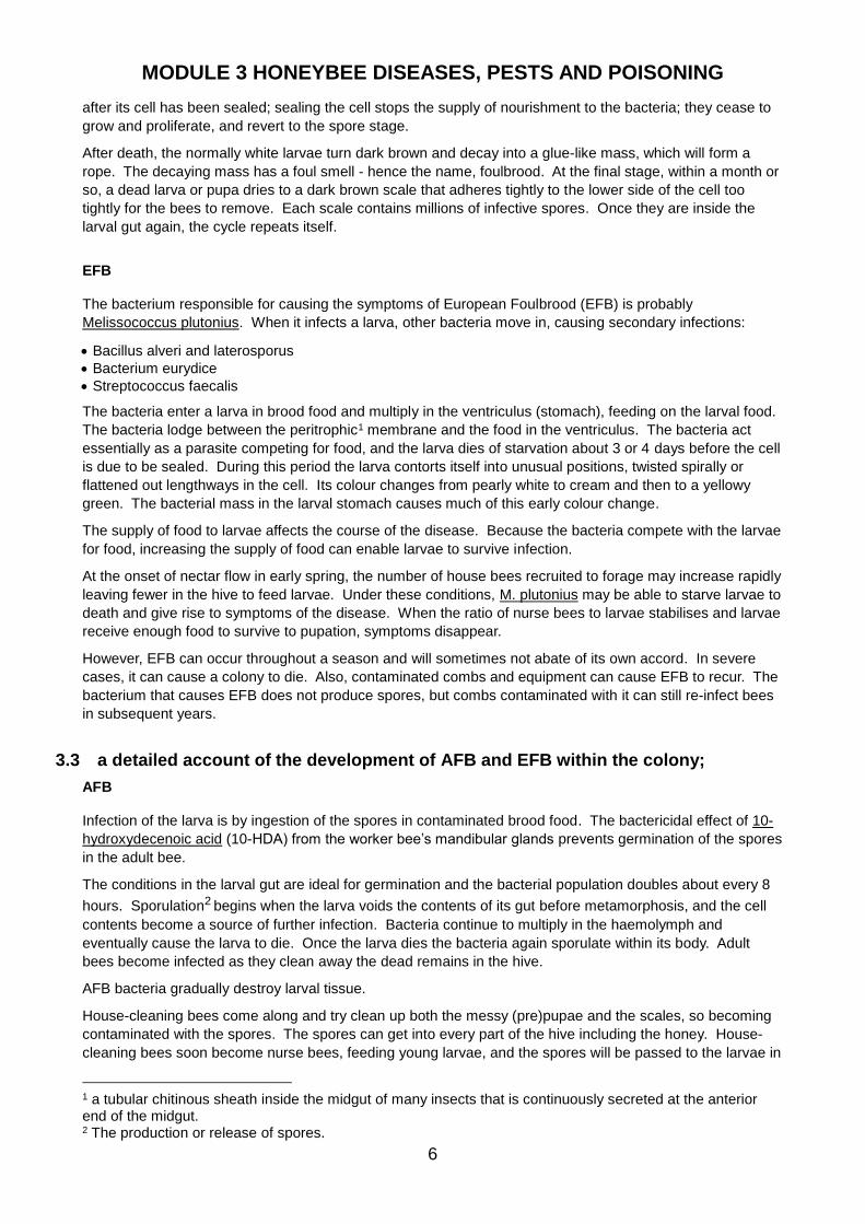
3.3 a detailed account of the development of AFB and EFB within the colony;
AFB Infection of the larva is by ingestion of the spores in contaminated brood food. The bactericidal effect of 10-
hydroxydecenoic acid (10-HDA) from the worker bee’s mandibular glands prevents germination of the spores
in the adult bee.
The conditions in the larval gut are ideal for germination and the bacterial population doubles about every 8
hours. Sporulation2 begins when the larva voids the contents of its gut before metamorphosis, and the cell
contents become a source of further infection. Bacteria continue to multiply in the haemolymph and
eventually cause the larva to die. Once the larva dies the bacteria again sporulate within its body. Adult
bees become infected as they clean away the dead remains in the hive.
AFB bacteria gradually destroy larval tissue.
House-cleaning bees come along and try clean up both the messy (pre)pupae and the scales, so becoming
contaminated with the spores. The spores can get into every part of the hive including the honey. House-
cleaning bees soon become nurse bees, feeding young larvae, and the spores will be passed to the larvae in
1 a tubular chitinous sheath inside the midgut of many insects that is continuously secreted at the anterior end of the midgut. 2 The production or release of spores.
MODULE 3 HONEYBEE DISEASES, PESTS AND POISONING
7
this way. The disease may be quite slow to get going in the beginning; the bees can keep the spread under
control for a time by the removal of diseased larvae in early stages. As the number of young bees declines
the disease takes control and quickly destroys the colony.
EFB There are three important facts involved in the spread of Melissococcus plutonius in a colony:
o M. plutonius never forms spores. The normal vegetative cells are infective and reproduce in huge
numbers in the infected larva.
o The contents of the ventriculus of a larva, and so the bacteria, are “sealed in” until the larva pupates and
the connection between the ventriculus and hindgut opens, when all the waste and bacteria that have
been stored in the larva’s gut pass out into the cell
o Very young adults clean the cells out and later produce food that they feed to larvae.
Taken together these phenomena explain how the disease spreads through the colony. Infected larvae that
survive to pupation discharge the contents of their guts into the cell. House bees pick the bacteria up when
they clean the cell and subsequently feed them to the young larvae in brood food. When a larva spins an
inadequate cocoon, the bacteria are more accessible to the house bees.
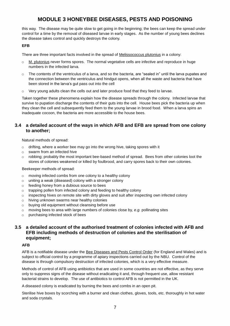
3.4 a detailed account of the ways in which AFB and EFB are spread from one colony to another;
Natural methods of spread:
o drifting, where a worker bee may go into the wrong hive, taking spores with it
o swarm from an infected hive
o robbing; probably the most important bee-based method of spread. Bees from other colonies loot the
stores of colonies weakened or killed by foulbrood, and carry spores back to their own colonies.
Beekeeper methods of spread:
o moving infected combs from one colony to a healthy colony
o uniting a weak (diseased) colony with a stronger colony
o feeding honey from a dubious source to bees
o trapping pollen from infected colony and feeding to healthy colony
o inspecting hives on remote site with dirty gloves and suit after inspecting own infected colony
o hiving unknown swarms near healthy colonies
o buying old equipment without cleansing before use
o moving bees to area with large numbers of colonies close by, e.g. pollinating sites
o purchasing infected stock of bees
3.5 a detailed account of the authorised treatment of colonies infected with AFB and EFB including methods of destruction of colonies and the sterilisation of equipment;
AFB
AFB is a notifiable disease under the Bee Diseases and Pests Control Order (for England and Wales) and is
subject to official control by a programme of apiary inspections carried out by the NBU. Control of the
disease is through compulsory destruction of infected colonies, which is a very effective measure.
Methods of control of AFB using antibiotics that are used in some countries are not effective, as they serve
only to suppress signs of the disease without eradicating it and, through frequent use, allow resistant
bacterial strains to develop. The use of antibiotics to control AFB is not permitted in the UK.
A diseased colony is eradicated by burning the bees and combs in an open pit.
Sterilise hive boxes by scorching with a burner and clean clothes, gloves, tools, etc. thoroughly in hot water
and soda crystals.
MODULE 3 HONEYBEE DISEASES, PESTS AND POISONING
8
EFB
EFB is a notifiable disease under the Bee Diseases and Pests Control Order (for England and Wales) and is
subject to official control by the examination of colonies for signs of disease and compulsory treatment or
destruction of diseased colonies.
There are three options available to the beekeeper in the UK who has colonies infected with EFB:
1. The colonies may be treated with the shook swarm husbandry method. Trials conducted by the National
Bee Unit showed that Shook swarm is more successful than OTC for the control of EFB in England and
Wales: “In the Spring following treatment, shaken colonies were three times less likely to test positive for
M. plutonius. This finding appears logical since OTC treatment does not remove the etiological3 agent
present in the hive. In contrast, the Shook swarm method provides the bees with material free of M.
plutonius. In addition, colonies treated with OTC were five times more likely to show recurrence of EFB
the following year than colonies treated by Shook swarm.”
2. The colonies may be treated with the antibiotic oxytetracycline (OTC, as the formulation Terramycin®);
The Bee Inspector administers Terramycin, mixing it with sugar syrup in a jar with holes in the lid, then shaking the jar over the bees on each frame. It is not put in a feeder on the hive.
3. The colonies may be destroyed, as for AFB. This will be carried out if the colony is too small for other treatment methods, is too heavily infected to respond to treatment, or at the beekeepers request.
However, the range of options available will also depend upon the time of year that the disease is diagnosed
and other factors such as the strength of the colony or the level of infection.
Weak colonies and colonies with a high proportion of diseased brood are destroyed, as with AFB, but lightly
diseased colonies may be treated with antibiotics. Under the Order only an Appointed Officer may carry the
treatment out, using drugs officially dispensed following confirmation of EFB in a disease sample submitted
for diagnosis at an approved laboratory or by LFD. The designated Veterinary Laboratories Agency (VLA)
prescribes the treatment.
3.6 the features that aid recognition of the Asian Hornet (Vespa velutina) and the notifiable pests Small hive beetle (Aethina tumida) and Tropilaelaps mites
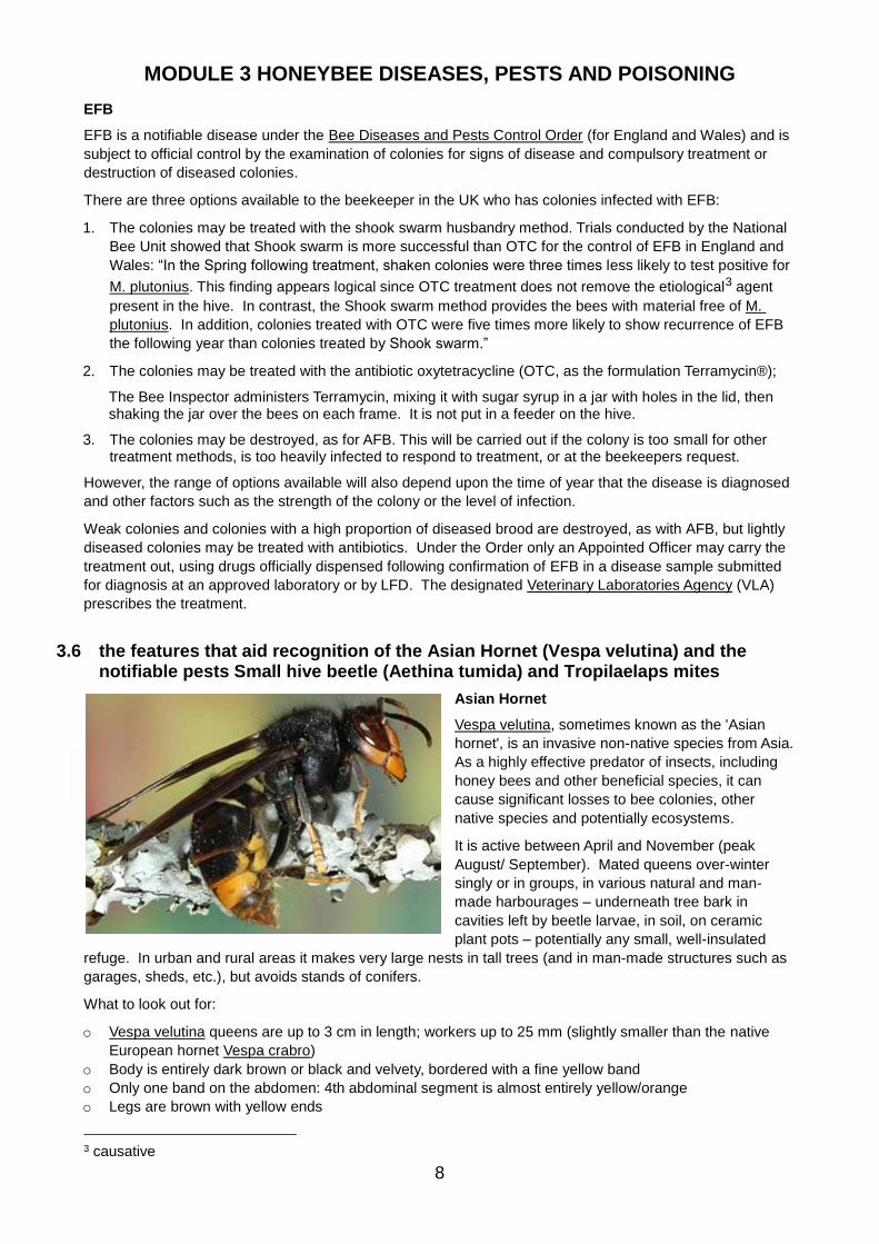
Asian Hornet
Vespa velutina, sometimes known as the 'Asian
hornet', is an invasive non-native species from Asia.
As a highly effective predator of insects, including
honey bees and other beneficial species, it can
cause significant losses to bee colonies, other
native species and potentially ecosystems.
It is active between April and November (peak
August/ September). Mated queens over-winter
singly or in groups, in various natural and man-
made harbourages – underneath tree bark in
cavities left by beetle larvae, in soil, on ceramic
plant pots – potentially any small, well-insulated
refuge. In urban and rural areas it makes very large nests in tall trees (and in man-made structures such as
garages, sheds, etc.), but avoids stands of conifers.
What to look out for:
o Vespa velutina queens are up to 3 cm in length; workers up to 25 mm (slightly smaller than the native
European hornet Vespa crabro)
o Body is entirely dark brown or black and velvety, bordered with a fine yellow band
o Only one band on the abdomen: 4th abdominal segment is almost entirely yellow/orange
o Legs are brown with yellow ends
3 causative

MODULE 3 HONEYBEE DISEASES, PESTS AND POISONING
9
o Head is black with an orange-yellow face
o Unlike the European hornet, Vespa velutina flies only during the day and ceases activity at dusk
Small hive beetle (Aethina tumida)
The small hive beetle is a member of the family of scavengers
or sap beetles, native in Africa. The adult beetle is dark brown
to black in colour and about 5mm in length. It antennae have a
distinctive club shape. Adult beetles can be observed almost
anywhere in a hive, although they are most often found at the
rear of the bottom board.
Females lay irregular masses of eggs in cracks or crevices in a
hive. The eggs hatch in 2–3 days into white larvae that grow to
10–11mm in length. The larvae feed on pollen and honey,
tunnelling through comb with stored honey or pollen, damaging
or destroying cappings and comb. They defecate in honey and
thereby discolour it. The activity of the larvae causes the honey
to ferment; it becomes frothy and develops a characteristic odour of decaying oranges. Damage and
fermentation cause honey to run out of combs.
Larvae mature in about 10–16 days. When they are ready to pupate they leave the hive and burrow into the
soil near it. Pupation may last 3–4 weeks. Adults start to look for honey bee colonies as soon as they
emerge and females generally mate and begin laying eggs about a week after emergence. The adults may
live for up to 6 months. Hive beetles may produce 4–5 generations a year during the warmer seasons.
Heavy infestations cause bees to abscond.
Tropilaelaps mites
There are currently four species of Tropilaelaps mites, but only two of
these (Tropilaelaps clareae, Tropilaelaps mercedesae) are considered
to be serious threats to the Western honey bee, Apis mellifera.
The females of Tropilaelaps clareae are light-reddish brown and about
1.0 mm long x 0.6 mm wide, and the males are almost as large as the
females (about one-third the size of a Varroa mite). The life cycle and
parasitism of Tropilaelaps is similar to that of Varroa destructor.
Tropilaelaps clareae readily infests colonies of Apis mellifera in Asia,
particularly those that
produce brood
continuously.
Adult mites enter cells containing larvae and reproduce within
sealed brood cells, particularly those of drones. Typically, the
female lays three to four eggs on mature bee larvae 48 hours
after the cell is capped, about one day apart.
The eggs hatch after around twelve hours, then the larva
goes through nymphal stages (protonymph, deutonymph)
before reaching the adult stage. Once hatched, all stages of
both female and male mites feed on the haemolymph (blood)
of the developing bee, causing damage through feeding by depriving the developing bee of essential
nourishment required for growth.
Development from egg to adult takes about 6 days, and the adults (including the mother mite) emerge with
the hatching adult bee and then search for new hosts.
Up to 14 adult mites and 10 nymphal stages of mite have been recorded in a single cell.
Mites move rapidly across the brood combs and are therefore easier to spot than Varroa, although they are
much smaller.
Unlike the varroa mite, Tropilaelaps cannot feed on adult bees because its mouthparts are unable to pierce
MODULE 3 HONEYBEE DISEASES, PESTS AND POISONING
10
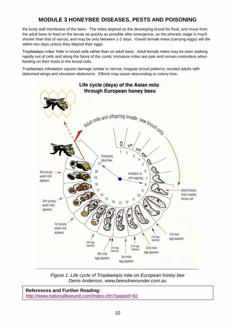
the body wall membrane of the bees. The mites depend on the developing brood for food, and move from
the adult bees to feed on the larvae as quickly as possible after emergence, so the phoretic stage is much
shorter than that of varroa, and may be only between 1-2 days. Gravid female mites (carrying eggs) will die
within two days unless they deposit their eggs.
Tropilaelaps mites ‘hide’ in brood cells rather than on adult bees. Adult female mites may be seen walking
rapidly out of cells and along the faces of the comb; immature mites are pale and remain motionless when
feeding on their hosts in the brood cells.
Tropilaelaps infestation causes damage similar to Varroa: irregular brood patterns; stunted adults with
deformed wings and shrunken abdomens. Effects may cause absconding or colony loss.
Figure 1: Life cycle of Tropilaelaps mite on European honey bee Denis Anderson, www.beesdownunder.com.au
References and Further Reading: http://www.nationalbeeunit.com/index.cfm?pageid=92
MODULE 3 HONEYBEE DISEASES, PESTS AND POISONING
11
3.7 a detailed account of the statutory requirements relating to notifiable diseases and pests and the implementation of these requirements in the United Kingdom,
Relevant Statutory Order is the Bee Diseases and Pest Control (England) Order 2006: SI 2006 No. 342.
Notification
If Beekeeper suspects the presence of a notifiable disease or pest he or she is legally obliged to either
contact the NBU or submit a sample of pest or disease to Fera lab for analysis.
Notifiable Diseases and Pests
Foul brood American Foulbrood European Foulbrood
Pests Small Hive Beetle (SHB) Aethina tumida Tropilaelaps spp mites
Inspections
NBU carries out regular inspections, prefer to involve beekeeper, but has powers to enter premises to
inspect. Beekeeper is responsible for inspecting colonies regularly for signs of notifiable diseases and pests.
If foulbrood or pest suspected:
1. Bee Inspector issues a Standstill Notice
o This prohibits Beekeeper from moving any bees, equipment or hive products from the apiary
o Inspector confirms diagnosis of foulbrood using LFD
2. Bee Inspector sends Apiary Inspection Report (B2) to Fera
o If Foulbrood, report may contain sample
o If pest, report always includes sample
3. Standstill remains in force until statutory control measures have been completed and apiary has been
officially examined and cleared; this is a minimum of 6 weeks
Lab examination
Fera aims to complete an examination and produce a diagnostic report within 1 working day
Report is sent 1st class post to Bee Inspector, who contacts beekeeper and explains procedure
If AFB confirmed
1. Bee Inspector issues Destruction Notice to Beekeeper
2. Beekeeper must:
a. Destroy the infected colony by burning all bees, frames, combs, honey and quilts, usually in a pit
dug for the purpose near the apiary
b. Sterilise the hive bodies using a blowlamp; they may and may be reused
3. All clothing, tools, etc. must be thoroughly cleaned with Soda Crystal solution
4. The Standstill Notice remains in force for minimum of 6 weeks after destruction
5. Bee Inspector will re-inspect Apiary and withdraw the Standstill Notice if no signs of disease are obvious
6. Bee Inspector will usually carry out follow up inspection the following season
If EFB confirmed
1. Bee Inspector will issue either a Treatment Notice or a Destruction Notice
a. Type of notice depends on time of year, level of infection and colony strength
b. Destruction Notice normal if infected Brood Comb >=50% or colony previously infected
c. Treatment Notice will apply if infection light enough to respond to Antibiotics or Shook Swarm,
Beekeeper can decide to destroy colony
2. Shook Swarm Treatment
o Conditional licences offered to remove ripe honey and supers and move colonies to hospital apiary
a. Beekeeper prepares clean hive with either fresh foundation or sterilised drawn comb
b. Burn old brood combs
c. Bee Inspector carries out Shook swarm
d. If no honey flow bees, fed winter feed after 2 days, infected nectar used in comb building
MODULE 3 HONEYBEE DISEASES, PESTS AND POISONING
12
e. Follow up inspection 6 weeks later or start of following season
3. Antibiotic Treatment
o As above a. Bee Inspector applies treatment
b. Honey removed after treatment under licence or after the withdrawal of the Standstill notice must
be stored in sealed containers and is prohibited from sale or consumption for at least 6 months
after the treatment date
If Small Hive Beetle or Tropilaelaps spp. Mites are suspected
1. England and Wales Contingency plan for exotic pests and diseases of honey bees will be invoked
2. NBU will contact Defra and Welsh Assembly Government
3. Defra will notify European Commission
4. NBU will set up a National Disease Control Centre at Fera Lab in York to:
o Coordinate the emergency
o Arrange surveys to assess extent of outbreak
o Procure and deploy necessary resources
o Liaise with beekeeping associations and other interested parties, nationally and locally
o Assess wider impact (e.g. colony losses) on pollination services to agriculture, horticulture and
the environment
o Provide up-to-date information to stakeholders and the media
o A local disease control centre may also be established
5. Statutory Infected Area
o Minimum of 16 km radius around infected colony
▪ Restrictions on movement of bee-related items into and out of area will apply
o If outbreak is isolated and eradication is viable all colonies in affected apiary and surrounding
area will be destroyed. In case of infestation soil 10-20m from hive will be treated if licensed
products exist.
o If outbreak is widespread appropriate control methods and veterinary medicines will be applied
subject to the Veterinary Medicines Directorate
Beekeeper’s responsibilities
1. Follow advice of Bee Inspector
2. Learn to recognise diseases and pests
3. Regularly examine colonies (at least Autumn and Spring)
4. Report suspected foulbrood immediately to local Bee Inspector or NBU
5. Put bees on new comb or foundation after EFB infection
6. Follow hygiene guidelines
7. Keep varroa and other diseases under control, healthy hives have best chance of surviving EFB
8. Be insured
3.8 an account of the statutory requirements relating to the importation of honeybees;
The importation of bees is subject to:
o The Trade in Animals and Related Products Regulations 2011 (‘the TARP Regulations’), which lay down
the controls that apply to imports of Apis mellifera (honey bees) and Bombus spp. (bumble bees) from
other member states and from countries outside the European Union (EU).
o The Bee Diseases and Pests Control (England) Order 2006 (‘the Order’), which lays down the
enforcement provisions for the post-import controls that apply to all imports of bees from countries
outside the EU.
o Commission Regulation (EU) 206/2010, which lists the countries outside the EU from which bees may
be imported, health certification requirements and the post import controls.
References and Further Reading: Statutory Procedures Advice leaflet
MODULE 3 HONEYBEE DISEASES, PESTS AND POISONING
13
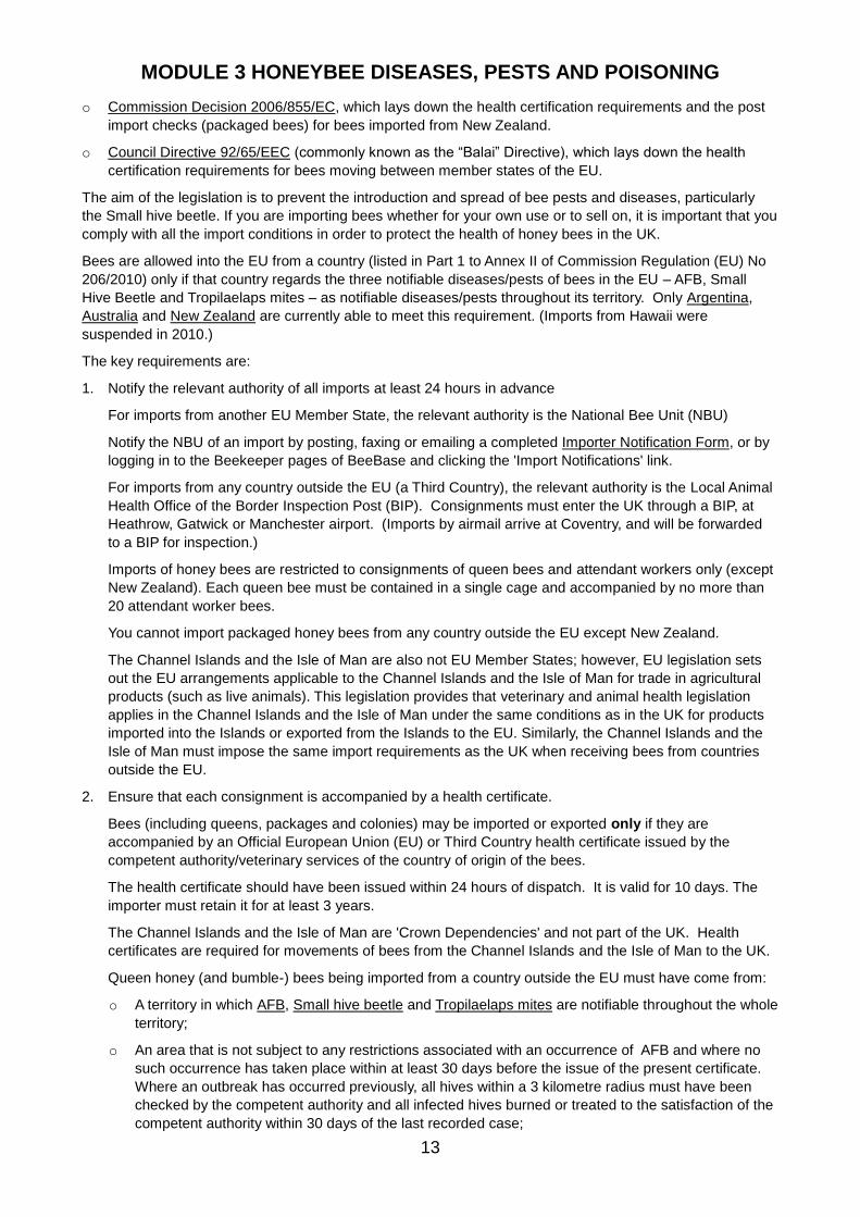
o Commission Decision 2006/855/EC, which lays down the health certification requirements and the post
import checks (packaged bees) for bees imported from New Zealand.
o Council Directive 92/65/EEC (commonly known as the “Balai” Directive), which lays down the health
certification requirements for bees moving between member states of the EU.
The aim of the legislation is to prevent the introduction and spread of bee pests and diseases, particularly
the Small hive beetle. If you are importing bees whether for your own use or to sell on, it is important that you
comply with all the import conditions in order to protect the health of honey bees in the UK.
Bees are allowed into the EU from a country (listed in Part 1 to Annex II of Commission Regulation (EU) No
206/2010) only if that country regards the three notifiable diseases/pests of bees in the EU – AFB, Small
Hive Beetle and Tropilaelaps mites – as notifiable diseases/pests throughout its territory. Only Argentina,
Australia and New Zealand are currently able to meet this requirement. (Imports from Hawaii were
suspended in 2010.)
The key requirements are:
1. Notify the relevant authority of all imports at least 24 hours in advance
For imports from another EU Member State, the relevant authority is the National Bee Unit (NBU)
Notify the NBU of an import by posting, faxing or emailing a completed Importer Notification Form, or by
logging in to the Beekeeper pages of BeeBase and clicking the 'Import Notifications' link.
For imports from any country outside the EU (a Third Country), the relevant authority is the Local Animal
Health Office of the Border Inspection Post (BIP). Consignments must enter the UK through a BIP, at
Heathrow, Gatwick or Manchester airport. (Imports by airmail arrive at Coventry, and will be forwarded
to a BIP for inspection.)
Imports of honey bees are restricted to consignments of queen bees and attendant workers only (except
New Zealand). Each queen bee must be contained in a single cage and accompanied by no more than
20 attendant worker bees.
You cannot import packaged honey bees from any country outside the EU except New Zealand.
The Channel Islands and the Isle of Man are also not EU Member States; however, EU legislation sets
out the EU arrangements applicable to the Channel Islands and the Isle of Man for trade in agricultural
products (such as live animals). This legislation provides that veterinary and animal health legislation
applies in the Channel Islands and the Isle of Man under the same conditions as in the UK for products
imported into the Islands or exported from the Islands to the EU. Similarly, the Channel Islands and the
Isle of Man must impose the same import requirements as the UK when receiving bees from countries
outside the EU.
2. Ensure that each consignment is accompanied by a health certificate.
Bees (including queens, packages and colonies) may be imported or exported only if they are
accompanied by an Official European Union (EU) or Third Country health certificate issued by the
competent authority/veterinary services of the country of origin of the bees.
The health certificate should have been issued within 24 hours of dispatch. It is valid for 10 days. The
importer must retain it for at least 3 years.
The Channel Islands and the Isle of Man are 'Crown Dependencies' and not part of the UK. Health
certificates are required for movements of bees from the Channel Islands and the Isle of Man to the UK.
Queen honey (and bumble-) bees being imported from a country outside the EU must have come from:
o A territory in which AFB, Small hive beetle and Tropilaelaps mites are notifiable throughout the whole
territory;
o An area that is not subject to any restrictions associated with an occurrence of AFB and where no
such occurrence has taken place within at least 30 days before the issue of the present certificate.
Where an outbreak has occurred previously, all hives within a 3 kilometre radius must have been
checked by the competent authority and all infected hives burned or treated to the satisfaction of the
competent authority within 30 days of the last recorded case;
MODULE 3 HONEYBEE DISEASES, PESTS AND POISONING
14
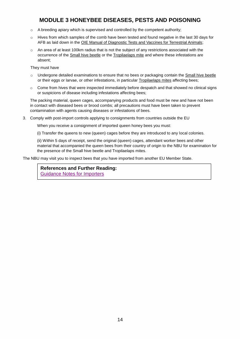
o A breeding apiary which is supervised and controlled by the competent authority;
o Hives from which samples of the comb have been tested and found negative in the last 30 days for
AFB as laid down in the OIE Manual of Diagnostic Tests and Vaccines for Terrestrial Animals;
o An area of at least 100km radius that is not the subject of any restrictions associated with the
occurrence of the Small hive beetle or the Tropilaelaps mite and where these infestations are
absent;
They must have
o Undergone detailed examinations to ensure that no bees or packaging contain the Small hive beetle
or their eggs or larvae, or other infestations, in particular Tropilaelaps mites affecting bees;
o Come from hives that were inspected immediately before despatch and that showed no clinical signs
or suspicions of disease including infestations affecting bees;
The packing material, queen cages, accompanying products and food must be new and have not been
in contact with diseased bees or brood combs; all precautions must have been taken to prevent
contamination with agents causing diseases or infestations of bees.
3. Comply with post-import controls applying to consignments from countries outside the EU
When you receive a consignment of imported queen honey bees you must:
(i) Transfer the queens to new (queen) cages before they are introduced to any local colonies.
(ii) Within 5 days of receipt, send the original (queen) cages, attendant worker bees and other
material that accompanied the queen bees from their country of origin to the NBU for examination for
the presence of the Small hive beetle and Tropilaelaps mites.
The NBU may visit you to inspect bees that you have imported from another EU Member State.
References and Further Reading: Guidance Notes for Importers
MODULE 3 HONEYBEE DISEASES, PESTS AND POISONING
15
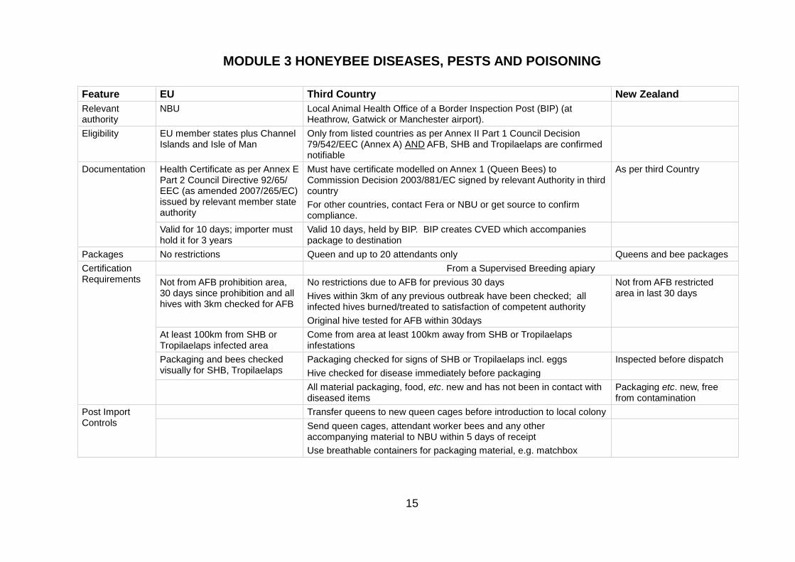
Feature EU Third Country New Zealand
Relevant authority
NBU Local Animal Health Office of a Border Inspection Post (BIP) (at Heathrow, Gatwick or Manchester airport).
Eligibility EU member states plus Channel Islands and Isle of Man
Only from listed countries as per Annex II Part 1 Council Decision 79/542/EEC (Annex A) AND AFB, SHB and Tropilaelaps are confirmed notifiable
Documentation Health Certificate as per Annex E Part 2 Council Directive 92/65/ EEC (as amended 2007/265/EC) issued by relevant member state authority
Must have certificate modelled on Annex 1 (Queen Bees) to Commission Decision 2003/881/EC signed by relevant Authority in third country
For other countries, contact Fera or NBU or get source to confirm compliance.
As per third Country
Valid for 10 days; importer must hold it for 3 years
Valid 10 days, held by BIP. BIP creates CVED which accompanies package to destination
Packages No restrictions Queen and up to 20 attendants only Queens and bee packages
Certification Requirements
From a Supervised Breeding apiary
Not from AFB prohibition area, 30 days since prohibition and all hives with 3km checked for AFB
No restrictions due to AFB for previous 30 days
Hives within 3km of any previous outbreak have been checked; all infected hives burned/treated to satisfaction of competent authority
Original hive tested for AFB within 30days
Not from AFB restricted area in last 30 days
At least 100km from SHB or Tropilaelaps infected area
Come from area at least 100km away from SHB or Tropilaelaps infestations
Packaging and bees checked visually for SHB, Tropilaelaps
Packaging checked for signs of SHB or Tropilaelaps incl. eggs
Hive checked for disease immediately before packaging
Inspected before dispatch
All material packaging, food, etc. new and has not been in contact with diseased items
Packaging etc. new, free from contamination
Post Import Controls
Transfer queens to new queen cages before introduction to local colony
Send queen cages, attendant worker bees and any other accompanying material to NBU within 5 days of receipt
Use breathable containers for packaging material, e.g. matchbox

MODULE 3 HONEYBEE DISEASES, PESTS AND POISONING
16
3.9 a description of the life cycle and natural history of Varroa destructor including its development within the honeybee colony and its spread to other colonies;
What is Varroa?
The varroa mite, Varroa destructor, formerly known as Varroa jacobsoni, is an external parasite of honey
bees. Originally confined to the Asian honey bee, Apis cerana, it has spread in recent decades to the
Western honey bee, Apis mellifera.
Life Cycle
The varroa mite life cycle consists of two distinct phases:
- Phoretic stage where the mature mites move about on adult honey bees
1. During this phase the mite is incapable of laying eggs
- Reproductive phase which occurs within the sealed brood cell
A mite will invade a cell just prior to capping (prefers drone brood) and will hide within the brood food beneath the larva. The larva will consume the brood food within hours of the cell being sealed, releasing the mite.
The mite will establish a feeding site by piercing the cuticle of the immature bee, this feeding site will also be employed by the varroa offspring as their mouthparts are incapable of piercing the larva.
The act of consuming larval haemolymph initiates the creation of eggs within the mite (oogenesis). 60-70 hours after sealing of the cell the first egg is laid, an unfertilised egg that will become a male mite. Each subsequent 30 hours a further fertilised egg will be laid resulting in female mites. 4-5 female eggs will be laid in total.
The mites on hatching will pass through protonymphal and deutonymphal stages on maturing from egg hatching to mature adult, taking 5.8 and 6.6 days for male and female mites respectively.
Male mite mates with mature female mites, on cell opening only the mature females (mother and daughters) leave the cell and will spend a period in the phoretic stage before entering a cell.
Reproduction
The success rate of reproduction (new mature female mites) in worker brood is about 1.7 to 2 but the longer
development period of drone brood increases it to 2-3.
The development and status of a colony affects mite population growth, and depending on circumstances,
mite numbers will increase between 12 and 800 fold.
Life Span
The life expectancy of varroa mites depends on the presence of brood and will vary from 27 days to about 5
months.
During the summer varroa mites live for about 2-3 months during which time they can complete 3-4 breeding
cycles, providing brood is available.
In winter, when brood-rearing is restricted, mites over-winter solely on the bodies of the adult bees within the
cluster, until brood-rearing commences the following spring.
How Varroa Spreads
Varroa mites are mobile and can readily move between bees and within the hive. However, to travel
between colonies they depend upon adult bees for transport – through the natural processes of drifting,
robbing, and swarming. Varroa can spread slowly over long distances in this way.
However, the movement of infested colonies by beekeepers is the principle means of spread over long
distances.

MODULE 3 HONEYBEE DISEASES, PESTS AND POISONING
17
Effects of Varroa
Unlike Apis cerana, Apis mellifera has few natural defences against varroa. The mites feed on both adult
bees and brood, weakening them and spreading harmful pathogens such as bee viruses.
Infested colonies eventually die out unless control measures are regularly applied.
References and Further Reading: A Closer look at Varroa Mite Reproduction Managing Varroa, Beebase
MODULE 3 HONEYBEE DISEASES, PESTS AND POISONING
18
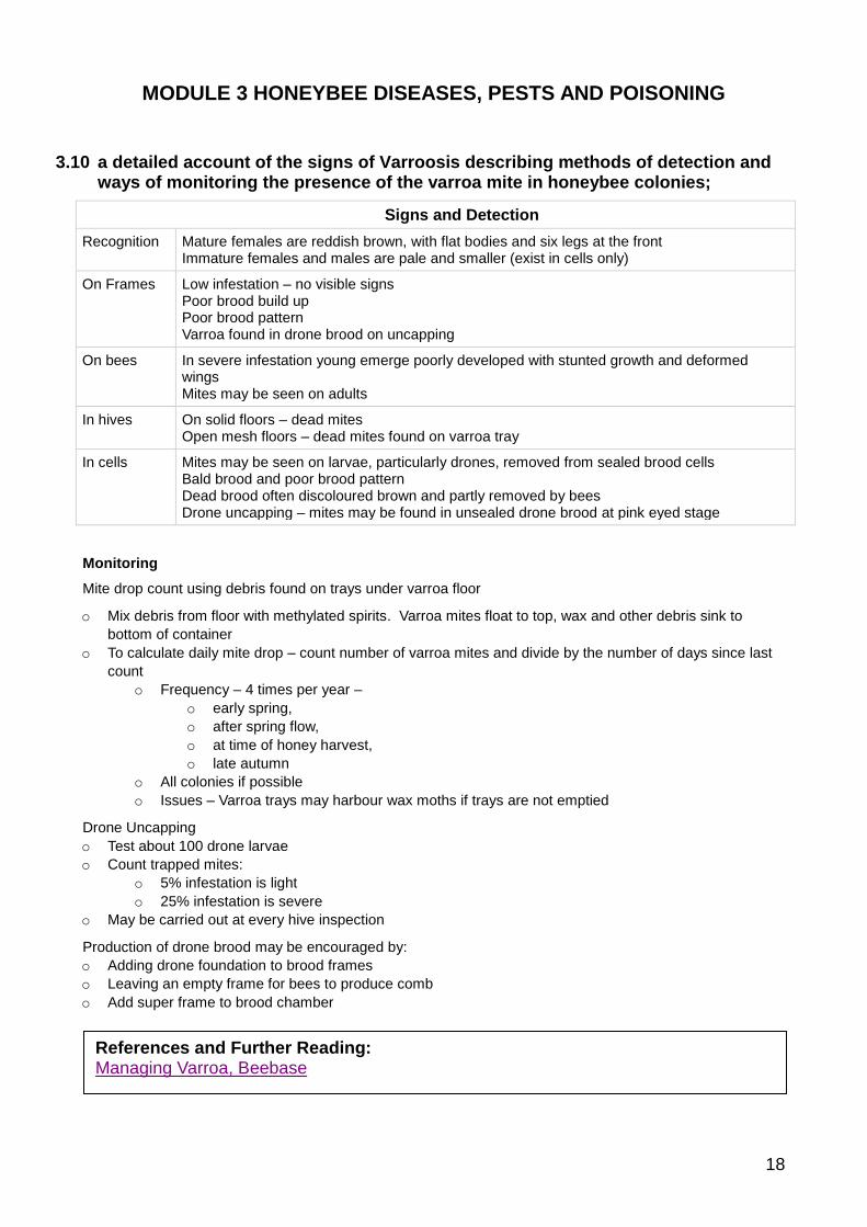
3.10 a detailed account of the signs of Varroosis describing methods of detection and ways of monitoring the presence of the varroa mite in honeybee colonies;
Signs and Detection
Recognition Mature females are reddish brown, with flat bodies and six legs at the front Immature females and males are pale and smaller (exist in cells only)
On Frames Low infestation – no visible signs Poor brood build up Poor brood pattern Varroa found in drone brood on uncapping
On bees In severe infestation young emerge poorly developed with stunted growth and deformed wings Mites may be seen on adults
In hives On solid floors – dead mites Open mesh floors – dead mites found on varroa tray
In cells Mites may be seen on larvae, particularly drones, removed from sealed brood cells Bald brood and poor brood pattern Dead brood often discoloured brown and partly removed by bees Drone uncapping – mites may be found in unsealed drone brood at pink eyed stage
Monitoring
Mite drop count using debris found on trays under varroa floor
o Mix debris from floor with methylated spirits. Varroa mites float to top, wax and other debris sink to
bottom of container
o To calculate daily mite drop – count number of varroa mites and divide by the number of days since last
count
o Frequency – 4 times per year –
o early spring,
o after spring flow,
o at time of honey harvest,
o late autumn
o All colonies if possible
o Issues – Varroa trays may harbour wax moths if trays are not emptied
Drone Uncapping
o Test about 100 drone larvae
o Count trapped mites:
o 5% infestation is light
o 25% infestation is severe
o May be carried out at every hive inspection
Production of drone brood may be encouraged by:
o Adding drone foundation to brood frames
o Leaving an empty frame for bees to produce comb
o Add super frame to brood chamber
References and Further Reading: Managing Varroa, Beebase
MODULE 3 HONEYBEE DISEASES, PESTS AND POISONING
19
3.11 a detailed account of methods of treatment and control of Varroosis, including Integrated Pest Management (IPM) and an outline of the consequences of incorrect administration of chemical treatments, together with a way of determining the resistance of varroa to such treatments;
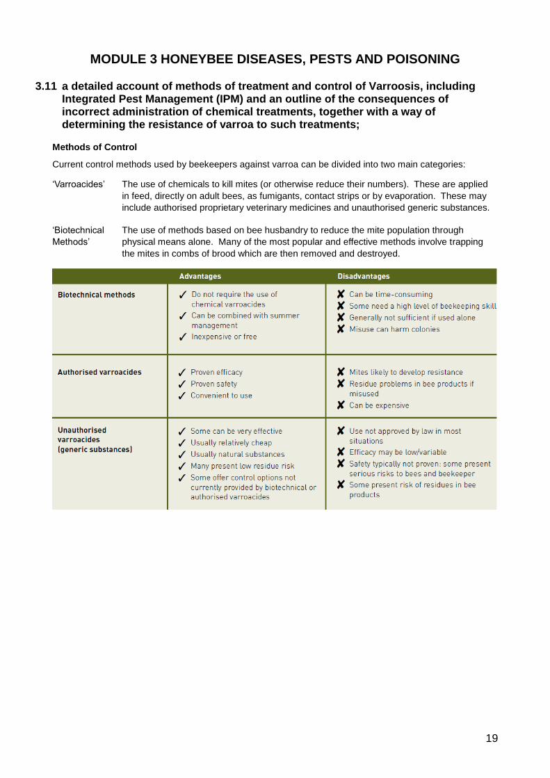
Methods of Control
Current control methods used by beekeepers against varroa can be divided into two main categories:
‘Varroacides’ The use of chemicals to kill mites (or otherwise reduce their numbers). These are applied
in feed, directly on adult bees, as fumigants, contact strips or by evaporation. These may
include authorised proprietary veterinary medicines and unauthorised generic substances.
‘Biotechnical
Methods’
The use of methods based on bee husbandry to reduce the mite population through
physical means alone. Many of the most popular and effective methods involve trapping
the mites in combs of brood which are then removed and destroyed.
MODULE 3 HONEYBEE DISEASES, PESTS AND POISONING
20
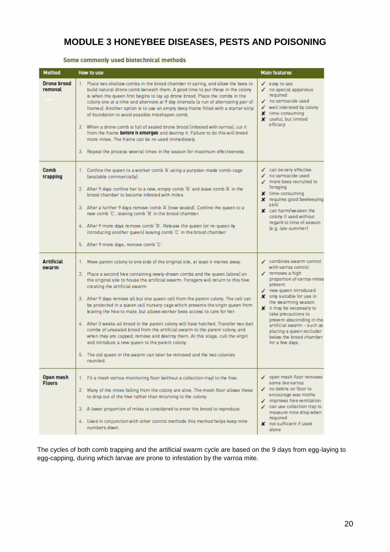
The cycles of both comb trapping and the artificial swarm cycle are based on the 9 days from egg-laying to
egg-capping, during which larvae are prone to infestation by the varroa mite.

MODULE 3 HONEYBEE DISEASES, PESTS AND POISONING
21
Comb trapping: how it works
Comb trapping works by isolating areas of brood – which attract the varroa mite – in frames that are
removed. It deliberately creates broodlessness elsewhere.
Comb A
Comb B
Comb C
0 9 18 27 36
Elapsed time
days
Broodless comb
egg larva pupa
Bee e
merg
es
LEGENDB
ees c
ap
cell
Qu
een
lays e
gg
Mite lays e
gg
s
Mite finds brood cell
Figure 2: Comb Trapping
MODULE 3 HONEYBEE DISEASES, PESTS AND POISONING
22
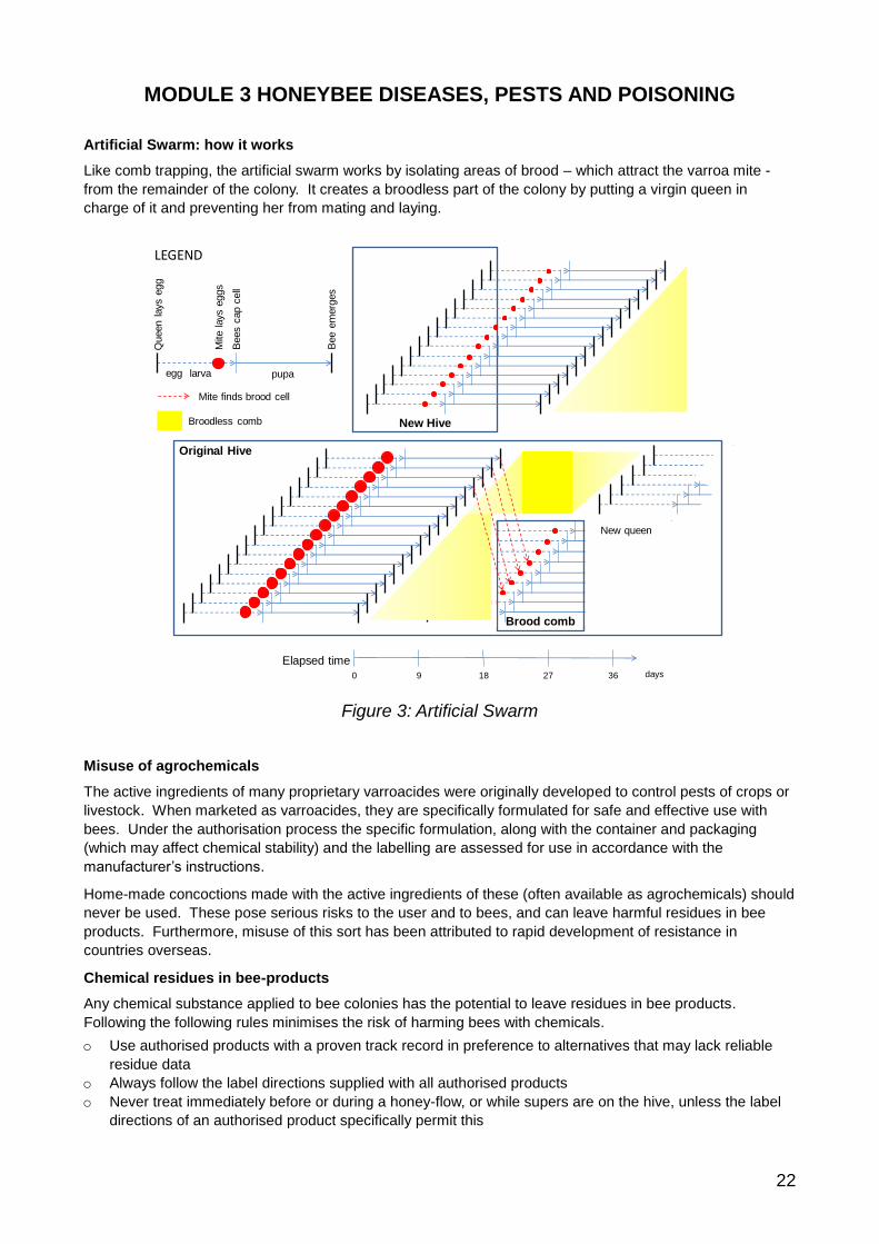
Artificial Swarm: how it works
Like comb trapping, the artificial swarm works by isolating areas of brood – which attract the varroa mite -
from the remainder of the colony. It creates a broodless part of the colony by putting a virgin queen in
charge of it and preventing her from mating and laying.
New Hive
0 9 18 27 36
Elapsed timedays
Brood comb
Broodless comb
egg larva pupa
Bee e
merg
es
LEGEND
Bees c
ap c
ell
Queen l
ays e
gg
Mite lays e
ggs
Mite finds brood cell
Original Hive
New queen
Figure 3: Artificial Swarm
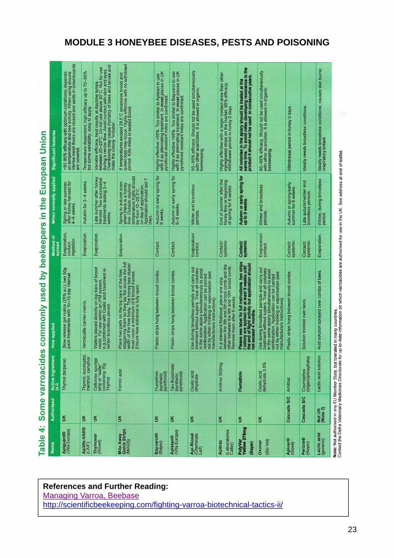
Misuse of agrochemicals
The active ingredients of many proprietary varroacides were originally developed to control pests of crops or
livestock. When marketed as varroacides, they are specifically formulated for safe and effective use with
bees. Under the authorisation process the specific formulation, along with the container and packaging
(which may affect chemical stability) and the labelling are assessed for use in accordance with the
manufacturer’s instructions.
Home-made concoctions made with the active ingredients of these (often available as agrochemicals) should
never be used. These pose serious risks to the user and to bees, and can leave harmful residues in bee
products. Furthermore, misuse of this sort has been attributed to rapid development of resistance in
countries overseas.
Chemical residues in bee-products
Any chemical substance applied to bee colonies has the potential to leave residues in bee products.
Following the following rules minimises the risk of harming bees with chemicals.
o Use authorised products with a proven track record in preference to alternatives that may lack reliable
residue data
o Always follow the label directions supplied with all authorised products
o Never treat immediately before or during a honey-flow, or while supers are on the hive, unless the label
directions of an authorised product specifically permit this
MODULE 3 HONEYBEE DISEASES, PESTS AND POISONING
23
References and Further Reading: Managing Varroa, Beebase http://scientificbeekeeping.com/fighting-varroa-biotechnical-tactics-ii/
MODULE 3 HONEYBEE DISEASES, PESTS AND POISONING
24
3.12 a detailed account of the cause, signs and treatment (if any) of adult bee diseases currently found in the United Kingdom these diseases to include Nosema, Dysentery, Acarine and Amoeba;
Acarine
Acarine is an infestation by the mite Acarapis woodi. The Isle of Wight disease in 1904 – 1920s was
probably acarine.
There are no visible external signs – the signs that beekeeping books usually give - crawling bees,
dislocated wings, etc. - are those of Chronic Bee Paralysis associated with Acarine (although not proved as
a vector).
The mites infest the trachea. Dissection and microscopic examination (20x) of the first thoracic trachea can
confirm diagnosis. Send a sample to a microscopist (in a paper container not plastic).
There has been no approved medicament in the UK since FolbexVA was withdrawn in early 1990 and the
Frow Mixture was banned.
Folbex VA (Bromopropylate impregnated paper strips). The strips were set alight and allowed to
smoulder in the hive, distributing the active ingredient as fumes.
The ‘Frow‘ remedy (named after Richard Watson Frow MBE) contained nitrobenzene, as well as Safrol
oil, Ligroin (petroleum ether), Petrol or Oil of Wintergreen (methyl salicylate). It was highly inflammable
and poisonous to both bees and humans. (Nitrobenzene is highly toxic and possibly carcinogenic.)
Both treatments had a poor therapeutic ratio – i.e. the amount required to kill the mite was too close to
the amount that would harm or even kill the bees.
Even creosote has been used as a treatment
There is some cumulative evidence that essential oils are effective as treatments:
Oil of Wintergreen (Methyl Salicylate) and menthol have been used as treatments.
Grease patties (containing sugar and essential oils such as Oil of Wintergreen) are used in the USA
Frow’s mixture contained an essential oil (Oil of Wintergreen)
“Some beekeepers believe that using thymol for several years has reduced acarine considerably.”
The potential basis of the efficacy of essential oil is that their smell might mask the smell of the young bees
that the female acarine mite uses to identify them as suitable hosts.
Hence, the use of Apiguard or similar anti-varroa treatments containing thymol might help treat acarine.
Acarine shortens the life of an infected bee, but this usually has little effect in the active season. The mite is
spread from old bees to very young bees. A severe winter may cause an infected colony to dwindle in the
spring.
Some strains of bees are more susceptible than others – the ‘tracheal mite’ is a huge problem in the USA
where Italian/NZ crosses are used.
There are external acarine mites: A. exturnus, A. dorsalis and A. vagans – little is known about them.
Nosema
Nosema is the most common disease and is found in seemingly healthy colonies.
References and Further Reading: http://www.nationalbeeunit.com/index.cfm?pageid=191 http://scientificbeekeeping.com/nosema-ceranae-kiss-of-death-or-much-ado-about-nothing/
MODULE 3 HONEYBEE DISEASES, PESTS AND POISONING
25
o Infectious Diseases of the Honey Bee (Dr. Bailey & Brenda Ball) states that 79 of 80 apparently healthy colonies contained Nosema spores.
Two Nosema species have been identified in honey bees in England and Wales: Nosema apis and, more
recently, the Asian species Nosema ceranae.
Both are parasitic microsporidian fungal pathogens.
N. ceranae is a more “generic” parasite than N. apis, and can infect various hosts. It is more closely related
to N. vespula (from yellowjacket wasps) than it is to N. apis.
Different “strains” (haplotypes) of N. ceranae exhibit different degrees of virulence.
Life Cycle
Nosema spp. infect the epithelial cells lining the mid-gut of the bee, where they multiply rapidly.
Within a few days the cells are packed with spores, the resting stage of the parasite.
The protozoa multiply in the ventriculus (30-50 million spores) and impair the digestion of pollen thereby
shortening the life of the bee.
N. ceranae goes on to infect the basal cells, and then spreads throughout the entire alimentary tract,
including the hypopharyngeal and salivary glands, but it infects only 20% of fat bodies and no muscle tissue.
When the host cells rupture, they shed spores into the gut where they are later excreted by the bees.
The spores in excreta can germinate and become active once more, when ingested by another bee
Pathology
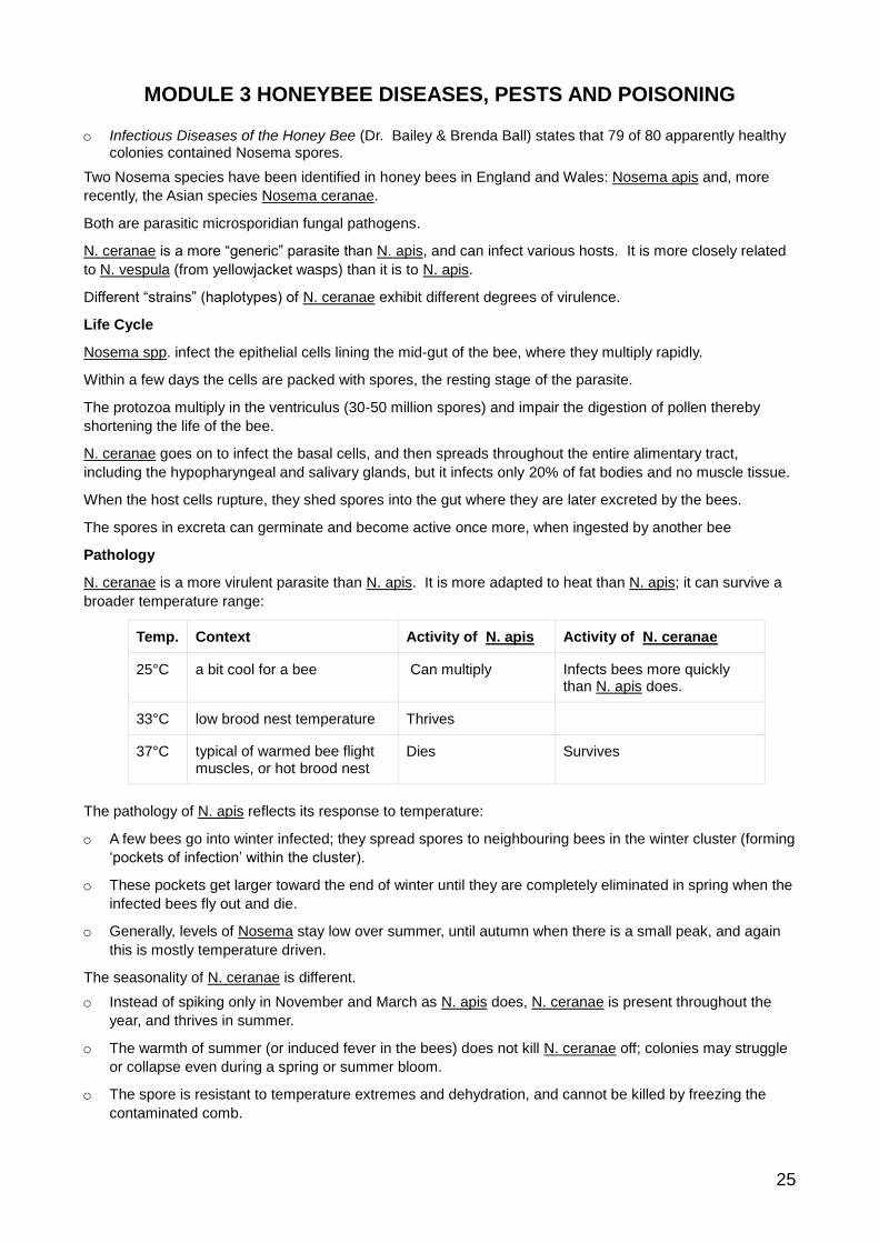
N. ceranae is a more virulent parasite than N. apis. It is more adapted to heat than N. apis; it can survive a
broader temperature range:
Temp. Context Activity of N. apis Activity of N. ceranae
25°C a bit cool for a bee Can multiply Infects bees more quickly than N. apis does.
33°C low brood nest temperature Thrives
37°C typical of warmed bee flight muscles, or hot brood nest
Dies Survives
The pathology of N. apis reflects its response to temperature:
o A few bees go into winter infected; they spread spores to neighbouring bees in the winter cluster (forming
‘pockets of infection’ within the cluster).
o These pockets get larger toward the end of winter until they are completely eliminated in spring when the
infected bees fly out and die.
o Generally, levels of Nosema stay low over summer, until autumn when there is a small peak, and again
this is mostly temperature driven.
The seasonality of N. ceranae is different.
o Instead of spiking only in November and March as N. apis does, N. ceranae is present throughout the
year, and thrives in summer.
o The warmth of summer (or induced fever in the bees) does not kill N. ceranae off; colonies may struggle
or collapse even during a spring or summer bloom.
o The spore is resistant to temperature extremes and dehydration, and cannot be killed by freezing the
contaminated comb.
MODULE 3 HONEYBEE DISEASES, PESTS AND POISONING
26
Effects on Queens
N. apis often causes early supersedure of queens.
o Chilling and stress of shipment or holding at room temperature promotes transmission from attendants to
queen.
o Attendant bees lick up her infected faeces
N. ceranae is not readily transmitted to queens
Symptoms and Effects
There are no obvious signs of Nosema, although Dysentery (q.v.), excreta on combs and hive, frequently accompanies heavy infections.
o Bees normally defecate away from the hive – sometimes the bees defecate in and about the hive because of the excessive build-up of waste matter in their guts.
o House bees become infected by cleaning up the excreta containing spores.
Nosema inhibits the ability of infected bees to digest food.
Bees infected by N. ceranae simply starve to death in the midst of plenty as a result of lack of digestive function.
Bees infected with N. ceranae are hungry, and so attempt to feed more, indulging in risky foraging behaviour,
and depopulating their colonies
They tend to forage at cooler temperatures, or even simply fly off to die.
Foragers infected with N. Ceranae die prematurely, and so inhibit the build-up of the colony.
Infected colonies fail to build up normally in the spring. Dead bees may be seen outside the hive after cleansing flights.
N. ceranae also appears to suppress the bees’ immune functions.
Bees ramp up their immune systems in response to N. apis, but N. ceranae suppresses that system.
In addition, infection by N. ceranae depresses the level of the bee “fountain of youth,” vitellogenin, suggesting that infection may decrease their lifespan by this effect.
Nosema stresses the bees nutritionally and immunologically leaving them prone to viruses.
Nosema breaches a bee’s main barrier to virus infection—the intact gut epithelium.
Diagnosis and Treatment
Confirm Nosema is by microscopic examination (400x): crush 30 bees in water and examine a droplet for
white, rice-shaped bodies.
o Send a sample to a microscopist in a paper container (not plastic).
Crushing bees can release millions of spores; avoid doing it.
Replace and sterilize combs with 80% acetic acid (100 ml./brood box for one week – air before use).
Treatment with the antibiotic Fumidil B (prepared from Aspergillis fumigatus, the causative agent of Stone
Brood!) is no longer permitted in Europe and the UK. (Fumidil B inhibited the reproduction of spores in the
ventriculus, but does not kill them. It also tainted the honey.)
Amoeba
Amoeba is caused by a protozoan amoeba-like parasite Malpighamoeba mellificae.
Cysts are ingested with food and germinate in the rectum. They migrate to the Malpighian tubules (the ‘kidneys’) to create more cysts that then accumulate in the rectum and are excreted.
The infection seems to have no effect on the colony; there are no specific symptoms and no treatment.
Often seen under a microscope when examining a sample for Nosema - grainy circular cysts, larger than the rice-shaped Nosema spores.
Acetic acid destroys the spores.
MODULE 3 HONEYBEE DISEASES, PESTS AND POISONING
27
Braula coeca
Since colonies have been treated for varroa, you are unlikely to see a similar (and harmless) parasite Braula coeca, the bee louse, a wingless fly.
Braula (which has 6 legs, varroa has 8) breeds under cell cappings. Tunnels can spoil appearance of comb honey.
Adults feed on honey taken as queen or workers are feeding.
Viruses
Nosema, acarine, varroa, etc. in themselves do not kill a colony – they weaken it and thereby allow viral
infections to take over.
o It is for this reason that Dr. Bailey considers that it was viral infection (Chronic Bee Paralysis Virus?) and
not acarine that killed so many colonies in the Isle of Wight Disease – the symptoms described such as
crawling bees, trembling wings, etc. are those of CBPV.
It is only in recent years that viruses have been identified using the electron microscope.
There are no cures for viral infection; viruses are immune to any antibiotic treatment. They multiply only in
the living cells of their hosts and any medicament that killed them would kill their hosts.
In practice, most colonies terminally weakened with Nosema or acarine exhibit signs of CBPV, particularly
clustering on top bars and continual trembling.
3.13 a simple account of the structure and function of the alimentary, excretory and respiratory systems of the adult honeybee and of the life cycle of the causative organisms of adult honeybee diseases;
Alimentary System
The alimentary system ingests food, digests it and excretes waste products. The alimentary canal and its
associated glands perform ingestion. The ventriculus digests food into energy and body-building substances
that the haemolymph (blood) circulates. The excretory system collects waste products and removes them
from the insect’s body.
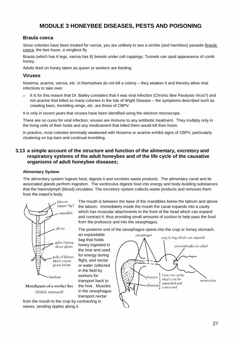
The mouth is between the base of the mandibles below the labrum and above
the labium. Immediately inside the mouth the canal expands into a cavity
which has muscular attachments to the front of the head which can expand
and contract it, thus providing small amounts of suction to help pass the food
from the proboscis and into the oesophagus.
The posterior end of the oesophagus opens into the crop or honey stomach,
an expandable
bag that holds
honey ingested in
the hive and used
for energy during
flight, and nectar
or water collected
in the field by
workers for
transport back to
the hive. Muscles
in the oesophagus
transport nectar
from the mouth to the crop by contracting in
waves, sending ripples along it.
MODULE 3 HONEYBEE DISEASES, PESTS AND POISONING
28
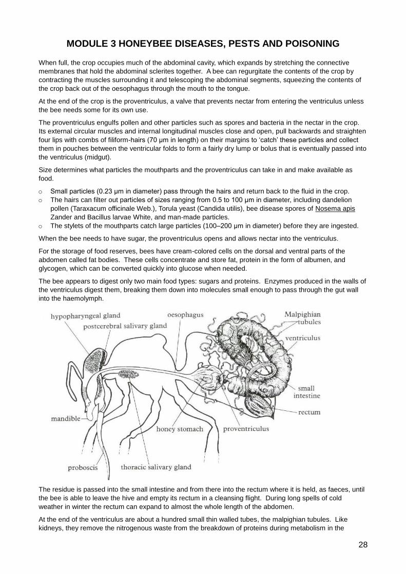
When full, the crop occupies much of the abdominal cavity, which expands by stretching the connective
membranes that hold the abdominal sclerites together. A bee can regurgitate the contents of the crop by
contracting the muscles surrounding it and telescoping the abdominal segments, squeezing the contents of
the crop back out of the oesophagus through the mouth to the tongue.
At the end of the crop is the proventriculus, a valve that prevents nectar from entering the ventriculus unless
the bee needs some for its own use.
The proventriculus engulfs pollen and other particles such as spores and bacteria in the nectar in the crop.
Its external circular muscles and internal longitudinal muscles close and open, pull backwards and straighten
four lips with combs of filiform-hairs (70 μm in length) on their margins to ‘catch’ these particles and collect
them in pouches between the ventricular folds to form a fairly dry lump or bolus that is eventually passed into
the ventriculus (midgut).
Size determines what particles the mouthparts and the proventriculus can take in and make available as
food.
o Small particles (0.23 μm in diameter) pass through the hairs and return back to the fluid in the crop.
o The hairs can filter out particles of sizes ranging from 0.5 to 100 μm in diameter, including dandelion
pollen (Taraxacum officinale Web.), Torula yeast (Candida utilis), bee disease spores of Nosema apis
Zander and Bacillus larvae White, and man-made particles.
o The stylets of the mouthparts catch large particles (100–200 μm in diameter) before they are ingested.
When the bee needs to have sugar, the proventriculus opens and allows nectar into the ventriculus.
For the storage of food reserves, bees have cream-colored cells on the dorsal and ventral parts of the
abdomen called fat bodies. These cells concentrate and store fat, protein in the form of albumen, and
glycogen, which can be converted quickly into glucose when needed.
The bee appears to digest only two main food types: sugars and proteins. Enzymes produced in the walls of
the ventriculus digest them, breaking them down into molecules small enough to pass through the gut wall
into the haemolymph.
The residue is passed into the small intestine and from there into the rectum where it is held, as faeces, until
the bee is able to leave the hive and empty its rectum in a cleansing flight. During long spells of cold
weather in winter the rectum can expand to almost the whole length of the abdomen.
At the end of the ventriculus are about a hundred small thin walled tubes, the malpighian tubules. Like
kidneys, they remove the nitrogenous waste from the breakdown of proteins during metabolism in the
MODULE 3 HONEYBEE DISEASES, PESTS AND POISONING
29
haemolymph. They pass waste products, mainly in the form of uric acid, into the gut to join the faeces in the
rectum.
Excretory System
The excretory system is essentially a sophisticated filtration system that not only removes waste substances
that would otherwise poison cells, but also acts selectively, maintaining the balance of water and salts in the
haemolymph and keeping the osmotic pressure and acidity within narrow limits.
Active cells produce two types of waste:
Type of Waste How produced How removed
Carbon Dioxide respiration respiratory system
Nitrogenous waste
chemical reactions of proteins and other nitrogenous compounds excretory system
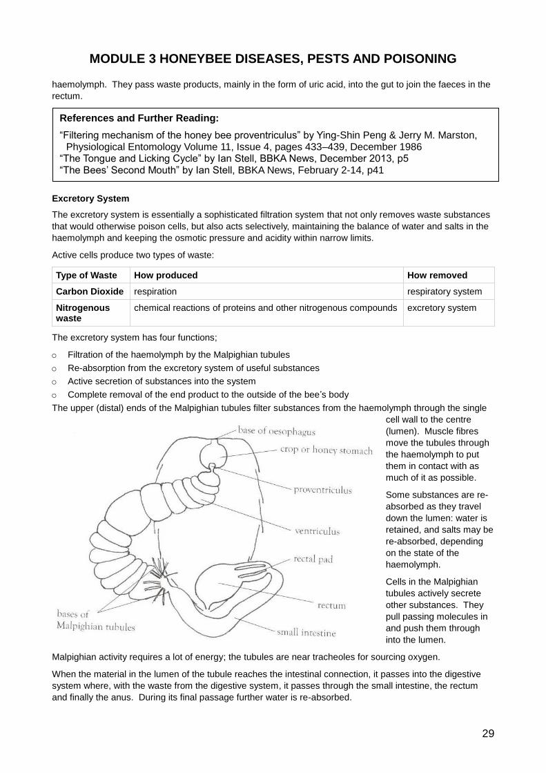
The excretory system has four functions;
o Filtration of the haemolymph by the Malpighian tubules
o Re-absorption from the excretory system of useful substances
o Active secretion of substances into the system
o Complete removal of the end product to the outside of the bee’s body
The upper (distal) ends of the Malpighian tubules filter substances from the haemolymph through the single
cell wall to the centre
(lumen). Muscle fibres
move the tubules through
the haemolymph to put
them in contact with as
much of it as possible.
Some substances are re-
absorbed as they travel
down the lumen: water is
retained, and salts may be
re-absorbed, depending
on the state of the
haemolymph.
Cells in the Malpighian
tubules actively secrete
other substances. They
pull passing molecules in
and push them through
into the lumen.
Malpighian activity requires a lot of energy; the tubules are near tracheoles for sourcing oxygen.
When the material in the lumen of the tubule reaches the intestinal connection, it passes into the digestive
system where, with the waste from the digestive system, it passes through the small intestine, the rectum
and finally the anus. During its final passage further water is re-absorbed.
References and Further Reading:
“Filtering mechanism of the honey bee proventriculus” by Ying-Shin Peng & Jerry M. Marston, Physiological Entomology Volume 11, Issue 4, pages 433–439, December 1986
“The Tongue and Licking Cycle” by Ian Stell, BBKA News, December 2013, p5 “The Bees’ Second Mouth” by Ian Stell, BBKA News, February 2-14, p41
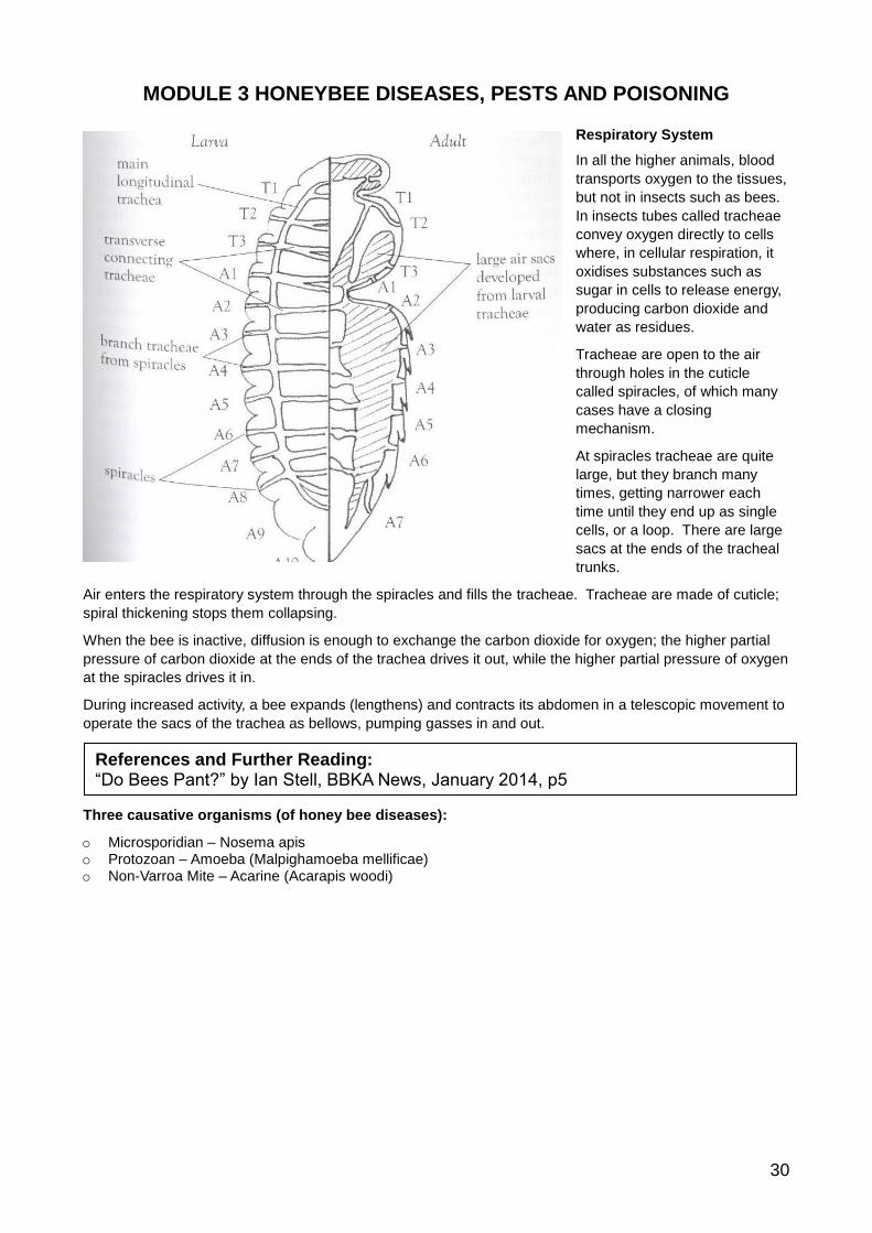
MODULE 3 HONEYBEE DISEASES, PESTS AND POISONING
30
Respiratory System
In all the higher animals, blood
transports oxygen to the tissues,
but not in insects such as bees.
In insects tubes called tracheae
convey oxygen directly to cells
where, in cellular respiration, it
oxidises substances such as
sugar in cells to release energy,
producing carbon dioxide and
water as residues.
Tracheae are open to the air
through holes in the cuticle
called spiracles, of which many
cases have a closing
mechanism.
At spiracles tracheae are quite
large, but they branch many
times, getting narrower each
time until they end up as single
cells, or a loop. There are large
sacs at the ends of the tracheal
trunks.
Air enters the respiratory system through the spiracles and fills the tracheae. Tracheae are made of cuticle;
spiral thickening stops them collapsing.
When the bee is inactive, diffusion is enough to exchange the carbon dioxide for oxygen; the higher partial
pressure of carbon dioxide at the ends of the trachea drives it out, while the higher partial pressure of oxygen
at the spiracles drives it in.
During increased activity, a bee expands (lengthens) and contracts its abdomen in a telescopic movement to
operate the sacs of the trachea as bellows, pumping gasses in and out.
Three causative organisms (of honey bee diseases):
o Microsporidian – Nosema apis o Protozoan – Amoeba (Malpighamoeba mellificae) o Non-Varroa Mite – Acarine (Acarapis woodi)
References and Further Reading: “Do Bees Pant?” by Ian Stell, BBKA News, January 2014, p5
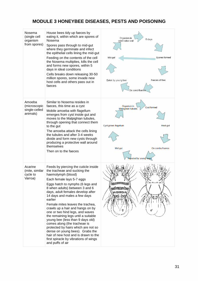
MODULE 3 HONEYBEE DISEASES, PESTS AND POISONING
31
Nosema (single cell organism from spores)
House bees tidy up faeces by eating it, within which are spores of Nosema
Spores pass through to mid-gut where they germinate and infect the epithelial cells lining the mid-gut
Feeding on the contents of the cell the Nosema multiplies, kills the cell and forms new spores, within 5 days in ideal conditions
Cells breaks down releasing 30-50 million spores, some invade new host cells and others pass out in faeces
Amoeba (microscopic single-celled animals)
Similar to Nosema resides in faeces, this time as a cyst
Mobile amoeba with flagellum emerges from cyst inside gut and moves to the Malpighian tubules, through opening that connect them to the gut
The amoeba attack the cells lining the tubules and after 3-4 weeks divide and form new cysts through producing a protective wall around themselves
Then on to the faeces
Acarine (mite, similar cycle to Varroa)
Feeds by piercing the cuticle inside the tracheae and sucking the haemolymph (blood)
Each female lays 5-7 eggs
Eggs hatch to nymphs (6 legs and 8 when adults) between 3 and 6 days, adult females develop after 14 days and males a few days earlier
Female mites leaves the trachea, crawls up a hair and hangs on by one or two hind legs, and waves the remaining legs until a suitable young bee (less than 9 days old) comes along (the tracheae is protected by hairs which are not so dense on young bees). Grabs the hair of new host and is drawn to the first spiracle by vibrations of wings and puffs of air
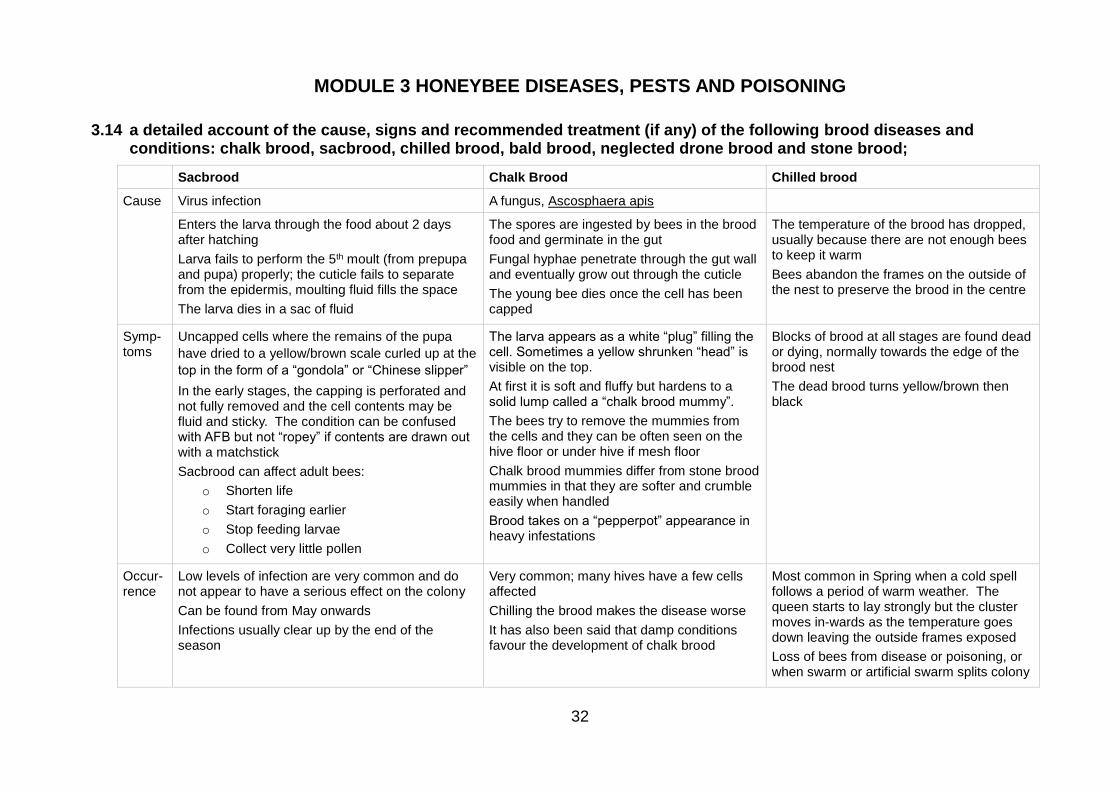
MODULE 3 HONEYBEE DISEASES, PESTS AND POISONING
32
3.14 a detailed account of the cause, signs and recommended treatment (if any) of the following brood diseases and conditions: chalk brood, sacbrood, chilled brood, bald brood, neglected drone brood and stone brood;
Sacbrood Chalk Brood Chilled brood
Cause Virus infection A fungus, Ascosphaera apis
Enters the larva through the food about 2 days after hatching
Larva fails to perform the 5th moult (from prepupa and pupa) properly; the cuticle fails to separate from the epidermis, moulting fluid fills the space
The larva dies in a sac of fluid
The spores are ingested by bees in the brood food and germinate in the gut
Fungal hyphae penetrate through the gut wall and eventually grow out through the cuticle
The young bee dies once the cell has been capped
The temperature of the brood has dropped, usually because there are not enough bees to keep it warm
Bees abandon the frames on the outside of the nest to preserve the brood in the centre
Symp-toms
Uncapped cells where the remains of the pupa
have dried to a yellow/brown scale curled up at the
top in the form of a “gondola” or “Chinese slipper”
In the early stages, the capping is perforated and not fully removed and the cell contents may be fluid and sticky. The condition can be confused with AFB but not “ropey” if contents are drawn out with a matchstick
Sacbrood can affect adult bees:
o Shorten life
o Start foraging earlier
o Stop feeding larvae
o Collect very little pollen
The larva appears as a white “plug” filling the cell. Sometimes a yellow shrunken “head” is visible on the top.
At first it is soft and fluffy but hardens to a solid lump called a “chalk brood mummy”.
The bees try to remove the mummies from the cells and they can be often seen on the hive floor or under hive if mesh floor
Chalk brood mummies differ from stone brood mummies in that they are softer and crumble easily when handled
Brood takes on a “pepperpot” appearance in heavy infestations
Blocks of brood at all stages are found dead or dying, normally towards the edge of the brood nest
The dead brood turns yellow/brown then black
Occur-rence
Low levels of infection are very common and do not appear to have a serious effect on the colony
Can be found from May onwards
Infections usually clear up by the end of the season
Very common; many hives have a few cells affected
Chilling the brood makes the disease worse
It has also been said that damp conditions favour the development of chalk brood
Most common in Spring when a cold spell follows a period of warm weather. The queen starts to lay strongly but the cluster moves in-wards as the temperature goes down leaving the outside frames exposed
Loss of bees from disease or poisoning, or when swarm or artificial swarm splits colony
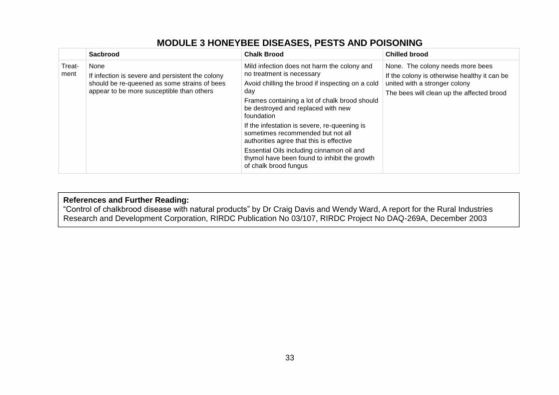
MODULE 3 HONEYBEE DISEASES, PESTS AND POISONING
33
Sacbrood Chalk Brood Chilled brood
Treat-ment
None
If infection is severe and persistent the colony should be re-queened as some strains of bees appear to be more susceptible than others
Mild infection does not harm the colony and no treatment is necessary
Avoid chilling the brood if inspecting on a cold day
Frames containing a lot of chalk brood should be destroyed and replaced with new foundation
If the infestation is severe, re-queening is sometimes recommended but not all authorities agree that this is effective
Essential Oils including cinnamon oil and thymol have been found to inhibit the growth of chalk brood fungus
None. The colony needs more bees
If the colony is otherwise healthy it can be united with a stronger colony
The bees will clean up the affected brood
References and Further Reading: “Control of chalkbrood disease with natural products” by Dr Craig Davis and Wendy Ward, A report for the Rural Industries Research and Development Corporation, RIRDC Publication No 03/107, RIRDC Project No DAQ-269A, December 2003

MODULE 3 HONEYBEE DISEASES, PESTS AND POISONING
34
3.15 a detailed account of the laboratory methods of diagnosis of Acarine, Nosema and Amoeba diseases in worker honeybees;
Acarine
Diagnosis
The disease can easily be diagnosed only by carrying out a dissection and microscopic examination (using a
dissecting microscope with up to x40 magnification) of the primary trachea.
Dissection to expose Acarine infestation
Collect a sample of 50 bees from the suspect colony.
Choose bees crawling and unable to fly, found within
about 3 metres of the front of the hive, rather than
random collection from within the colony. The bees may
be living, dying, or dead. Kill live bees with ethyl alcohol
or by putting them in a deep freeze at -20°C.
Impale each using a double needle placed at an angle
away from the head through the thorax between the
second and third pairs of legs (as shown). The bee
should ventral side up on an angled cork base, the angle
is not critical, but is usually between 45° and 60°.
Using a single edged razor blade, cut the head and first
pair of legs off; make the cut from behind the first pair of
legs to the back of the bee's head, indicated by the red
line on the drawing. Remove the severed head and front
pair of legs with tweezers.
Use fine-tipped tweezers to peel the collar away (shown
red at right) and expose the tracheae more fully. Pull
upwards with a circular motion, following the ring of the
collar. It will peel off easily, usually in one piece. Save
the collar itself for later preparation as a microscope
slide specimen, if required, by immersing in 70%
isopropyl alcohol.
In a healthy or uninfected bee the tracheae have a uniform, creamy-white appearance. In infested bees the
tracheae show patchy discolouration or dark staining, (melanisation, caused by mites feeding). In addition
the eggs, nymphs and adult stages of the mite may also be seen in the trachea.
As mites enter through the spiracle, check the outer end of the trachea first. Light infestations may be
difficult to see, heavy infestations are easily visible as shadows or lumpy dark objects in tracheae that can be
clear to dark brown. Old and/or heavy infestations will render the trachea orange, brown or black.
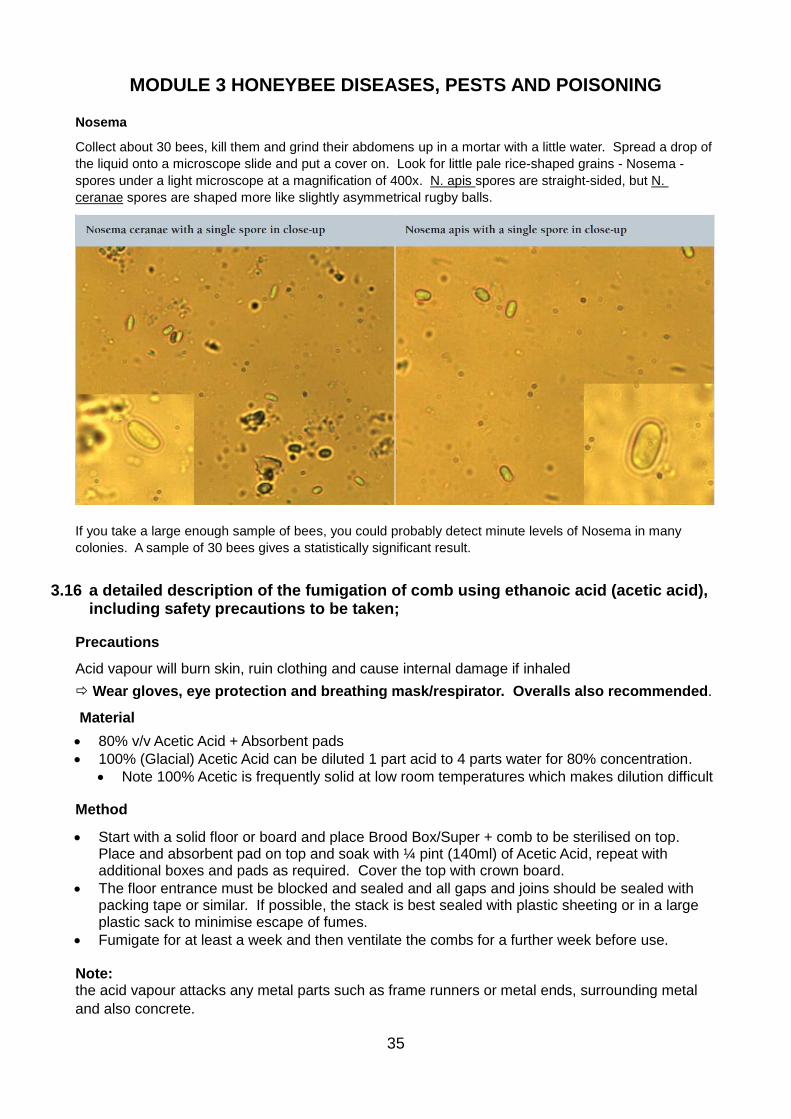
MODULE 3 HONEYBEE DISEASES, PESTS AND POISONING
35
Nosema
Collect about 30 bees, kill them and grind their abdomens up in a mortar with a little water. Spread a drop of
the liquid onto a microscope slide and put a cover on. Look for little pale rice-shaped grains - Nosema -
spores under a light microscope at a magnification of 400x. N. apis spores are straight-sided, but N.
ceranae spores are shaped more like slightly asymmetrical rugby balls.
If you take a large enough sample of bees, you could probably detect minute levels of Nosema in many
colonies. A sample of 30 bees gives a statistically significant result.
3.16 a detailed description of the fumigation of comb using ethanoic acid (acetic acid), including safety precautions to be taken;
Precautions
Acid vapour will burn skin, ruin clothing and cause internal damage if inhaled
Wear gloves, eye protection and breathing mask/respirator. Overalls also recommended.
Material
• 80% v/v Acetic Acid + Absorbent pads
• 100% (Glacial) Acetic Acid can be diluted 1 part acid to 4 parts water for 80% concentration.
• Note 100% Acetic is frequently solid at low room temperatures which makes dilution difficult
Method
• Start with a solid floor or board and place Brood Box/Super + comb to be sterilised on top. Place and absorbent pad on top and soak with ¼ pint (140ml) of Acetic Acid, repeat with additional boxes and pads as required. Cover the top with crown board.
• The floor entrance must be blocked and sealed and all gaps and joins should be sealed with packing tape or similar. If possible, the stack is best sealed with plastic sheeting or in a large plastic sack to minimise escape of fumes.
• Fumigate for at least a week and then ventilate the combs for a further week before use. Note: the acid vapour attacks any metal parts such as frame runners or metal ends, surrounding metal
and also concrete.
MODULE 3 HONEYBEE DISEASES, PESTS AND POISONING
36
3.17 a detailed description of procedures by which a colony can be transferred onto clean comb including any precautions that need to be taken and the circumstances which merit such procedures. These procedures to include shook swarm and Bailey comb change;
Equipment for both Shook Swarm and Bailey Comb Change
A clean brood chamber, containing new frames of foundation
A queen excluder, crown board and floor (all clean).
A contact feeder and heavy sugar syrup. (½ litre of water to 1 kg of sugar)
Shook Swarm
1. Move the hive to one side and assemble a new one in its place, with the queen excluder
between the floor and the brood box.
2. Take the middle four frames out of the new brood box and put them to one side.
3. Find and catch the queen in the old hive and confine her temporarily in a queen cage or
another suitable receptacle.
4. You must now transfer the bees from the old hive to the new one.
5. Take each frame in turn and shake it into the space left by the four frames removed in the
middle of the new brood box. Brush off any reluctant bees off each frame.
6. Once you have cleared all the old frames of bees, shake or brush all the bees still in the old
brood box into the new one.
7. Remove the queen from the queen cage and place her in the centre of the new brood box.
8. Carefully put the four frames missing from the new brood box back in
9. Fit the crown board
10. Feed with heavy sugar syrup - ideally using a contact feeder on the crown board
Bailey Comb Change
1. Place the new brood box over the old one
2. Unless there is a strong nectar flow, feed with heavy Sugar Syrup
3. When the bees have drawn out some of the foundation, find the queen and place her on this
comb
4. About a week later, place a queen excluder over the old brood box and under the new, trapping
the queen in the upper box
5. If possible, make a new hive entrance between the two brood boxes and close off the old
entrance, thus helping to reduce the amount of pollen stored in the old lower combs
6. After three weeks remove the old brood box
7. The brood in the old brood box will have hatched and the comb can then be rendered to
recover the beeswax
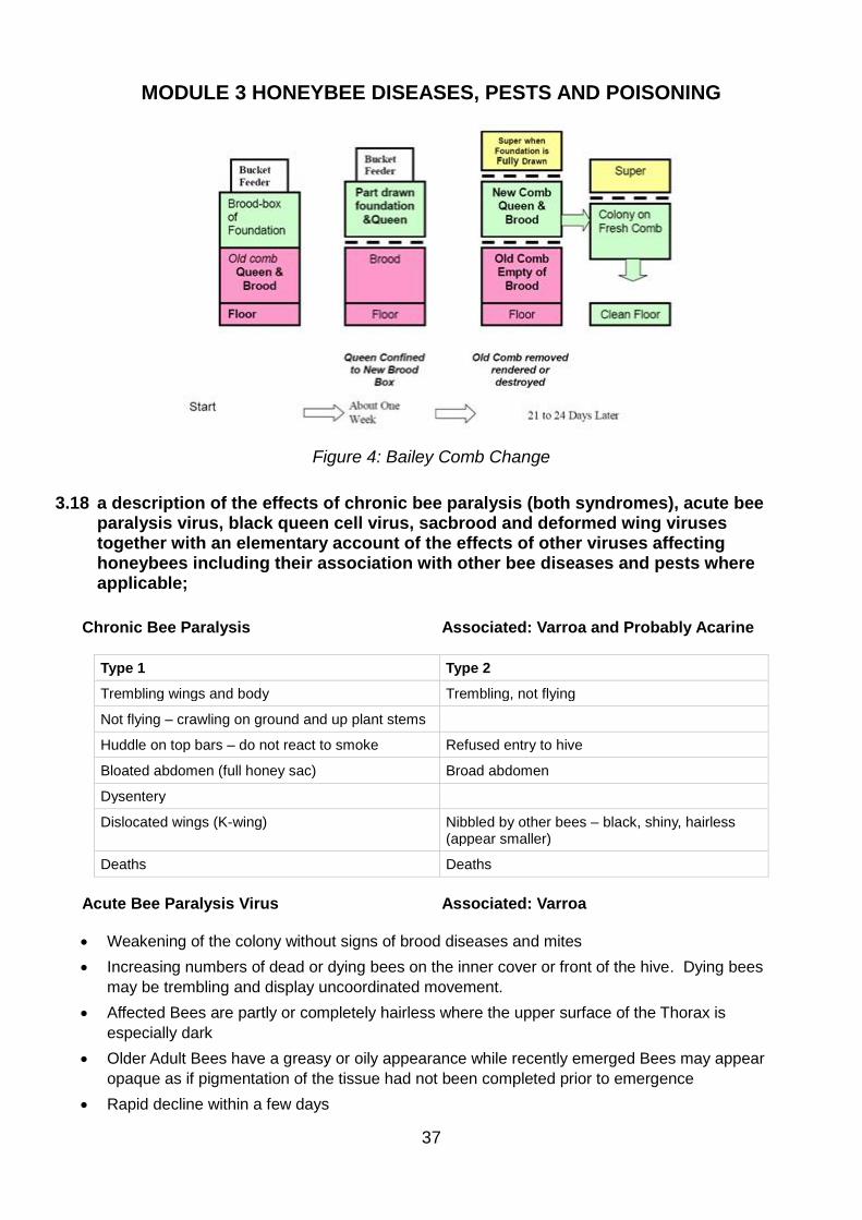
MODULE 3 HONEYBEE DISEASES, PESTS AND POISONING
37
Figure 4: Bailey Comb Change
3.18 a description of the effects of chronic bee paralysis (both syndromes), acute bee paralysis virus, black queen cell virus, sacbrood and deformed wing viruses together with an elementary account of the effects of other viruses affecting honeybees including their association with other bee diseases and pests where applicable;
Chronic Bee Paralysis Associated: Varroa and Probably Acarine
Type 1 Type 2
Trembling wings and body Trembling, not flying
Not flying – crawling on ground and up plant stems
Huddle on top bars – do not react to smoke Refused entry to hive
Bloated abdomen (full honey sac) Broad abdomen
Dysentery
Dislocated wings (K-wing) Nibbled by other bees – black, shiny, hairless (appear smaller)
Deaths Deaths
Acute Bee Paralysis Virus Associated: Varroa
• Weakening of the colony without signs of brood diseases and mites
• Increasing numbers of dead or dying bees on the inner cover or front of the hive. Dying bees
may be trembling and display uncoordinated movement.
• Affected Bees are partly or completely hairless where the upper surface of the Thorax is
especially dark
• Older Adult Bees have a greasy or oily appearance while recently emerged Bees may appear
opaque as if pigmentation of the tissue had not been completed prior to emergence
• Rapid decline within a few days
MODULE 3 HONEYBEE DISEASES, PESTS AND POISONING
38
Black Queen Cell Virus Associated: Nosema
• Turns queen cell black
• Prepupa or pupa is yellow
• Tough skin slightly resembles sacbrood
Sac Brood Associated: Varroa
• The moult at prepupa to pupa goes wrong and the space fills with ectdysial (fluid)
• Moult skin resembles Chinese Slipper
• Changes from yellow to dark brown
• Pupa dies. Can give a short rope – can be confused with AFB
• Adult Bees can be infected when cleaning cell
• Life shortened
• Become foragers earlier
• Stop feeding larvae
• Rarely collect pollen
• Behaviour of adult bees can cause the disease to die out in a colony
Deformed Wing Virus Associated: Varroa and Tropilaelaps
• Damaged appendages, particularly stubby, useless wings
• Shortened, rounded abdomens
• Miscolouring
• Paralysis
• Severely reduced life-span (less than 48 hours)
• Typically expelled from the hive
Other Viruses
Virus Association Symptoms
Slow paralysis virus Varroa Collapse late in the year
Filamentous virus Nosema Haemolymph goes milky
Virus Y Nosema No reported symptoms
Virus X Amoeba Shortens life; Colonies die in spring
Cloudy Wing Virus Wings go cloudy. Bee dies

MODULE 3 HONEYBEE DISEASES, PESTS AND POISONING
39
Summary of Associations
Acarine Chronic Bee Paralysis
Varroa Acute Bee Paralysis Virus
Sacbrood
Deformed Wing VirusTropilaelaps
Slow Paralysis Virus
Black Queen Cell Virus
Nosema
Filamentous Virus
Virus Y
Amoeba Virus X
3.19 the scientific names of the causative organisms associated with diseases of honeybees;
Brood disease
AFB Bacterium Paenibacillus larvae larvae
EFB Bacterium Melissococcus plutonius
Sac brood Virus Morator Aetetulas
Black Queen cell Virus
Chalk brood Fungus Ascosphaera apis
Stone brood Fungus Aspergillus flavus; Aspergillus fumigatus
Adult disease
Nosema Protozoan Nosema apis/ceranae
Amoeba Protozoan Malpighamoeba mellificae
Gregarine Protozoan Gregarinidae
Melanosis Fungus Torulopsis
Viral adult disease
Chronic bee paralysis Virus
Cloudy wing Virus associate
Slow paralysis Virus
Kashmir bee virus Virus
Parasites – mites
Acarine Mite Acarapis woodi
Varroa Mite Varroa jacobsoni/ destructor
Tropilaelaps Beetle
Insects Bee louse Braula coeca
Fly larva Senotainia tricuspis
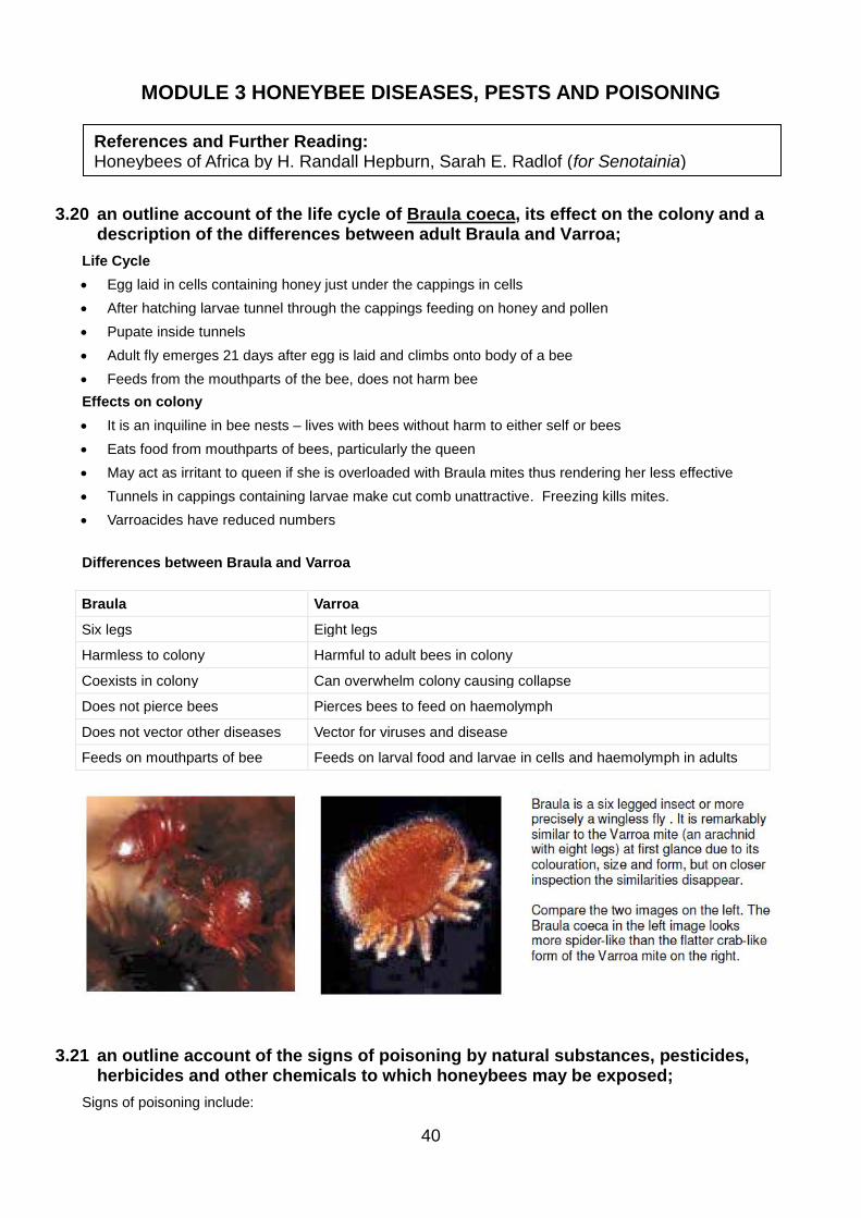
MODULE 3 HONEYBEE DISEASES, PESTS AND POISONING
40
3.20 an outline account of the life cycle of Braula coeca, its effect on the colony and a description of the differences between adult Braula and Varroa;
Life Cycle
• Egg laid in cells containing honey just under the cappings in cells
• After hatching larvae tunnel through the cappings feeding on honey and pollen
• Pupate inside tunnels
• Adult fly emerges 21 days after egg is laid and climbs onto body of a bee
• Feeds from the mouthparts of the bee, does not harm bee
Effects on colony
• It is an inquiline in bee nests – lives with bees without harm to either self or bees
• Eats food from mouthparts of bees, particularly the queen
• May act as irritant to queen if she is overloaded with Braula mites thus rendering her less effective
• Tunnels in cappings containing larvae make cut comb unattractive. Freezing kills mites.
• Varroacides have reduced numbers
Differences between Braula and Varroa
Braula Varroa
Six legs Eight legs
Harmless to colony Harmful to adult bees in colony
Coexists in colony Can overwhelm colony causing collapse
Does not pierce bees Pierces bees to feed on haemolymph
Does not vector other diseases Vector for viruses and disease
Feeds on mouthparts of bee Feeds on larval food and larvae in cells and haemolymph in adults
3.21 an outline account of the signs of poisoning by natural substances, pesticides, herbicides and other chemicals to which honeybees may be exposed;
Signs of poisoning include:
References and Further Reading: Honeybees of Africa by H. Randall Hepburn, Sarah E. Radlof (for Senotainia)
MODULE 3 HONEYBEE DISEASES, PESTS AND POISONING
41
• Large numbers of dead bees at the hive entrance
• Bees returning to apiary spin round on ground before succumbing
• Guards repel affected bees
• Colony becomes upset and bad-tempered
3.22 an account of the ways in which honeybees can become exposed to agricultural and pest control chemicals;
Types of pesticide
Systemic – taken up by the plant through its roots and leaves; e.g. imidacloprid
Specific – attacks one particular species of insect
Crops most likely to be sprayed with chemicals toxic to bees
Oil Seed Rape: Cypermethrin, Deltamethrin, Fenvalerate,Triazopho; imidacloprid
Field Beans: Pyrethroid Hallmark Zeon
Wheat: imidacloprid. Note, bees will not forage on wheat, but might be caught by sprays applied to it.
Oats: imidacloprid
Linseed:
Sugar Beet: imidacloprid
Fruit: apple, pear and cherry
The bee can be caught by sprays:
• When the crop on which it is working is sprayed
• When spray is used on a crop not flowering but contains a lot of flowering weeds
• When a bee is flying over a crop which is being sprayed
• When wind drives spray to hive or bee forage (drift)
3.23 a detailed description of the action to take, and practical measures possible, when prior notification of application of toxic chemicals to crops is given;
If possible move hives at least 3 miles away prior to spraying
Gather as much detail as possible about the spraying:
• What crops and where
• Type of spray
• Time of spraying
• Weather conditions (direction of wind)
Close up hives when spraying is in progress
• Ensure ventilation is maintained
• Provide water supply within hive (contact feeder, wet sponge)
Maintain a clean water supply near the hives after spraying
Shutting the bees inside the hive at any time runs the risk of their dying from overheating, lack of ventilation
or drowning in wax and honey from melted combs. If possible try and provide some shading for hives in full
sun.
Covering the entrance with loose grass may be sufficient to keep the bees in the hive and at the same time
allowing them to draw fresh air into the hive. Remember to remove the grass when the danger has passed.
References and Further Reading: http://www.bbka.org.uk/files/library/bees_and_pesticides-l019_1342859149.pdf
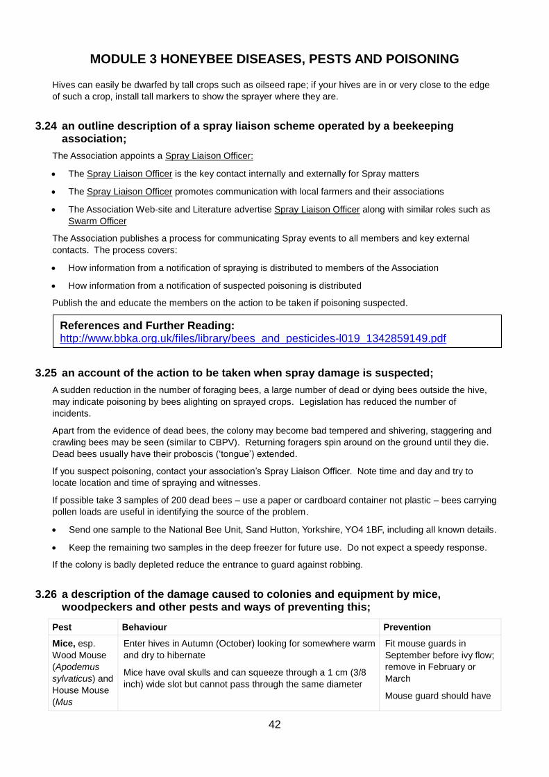
MODULE 3 HONEYBEE DISEASES, PESTS AND POISONING
42
Hives can easily be dwarfed by tall crops such as oilseed rape; if your hives are in or very close to the edge
of such a crop, install tall markers to show the sprayer where they are.
3.24 an outline description of a spray liaison scheme operated by a beekeeping association;
The Association appoints a Spray Liaison Officer:
• The Spray Liaison Officer is the key contact internally and externally for Spray matters
• The Spray Liaison Officer promotes communication with local farmers and their associations
• The Association Web-site and Literature advertise Spray Liaison Officer along with similar roles such as
Swarm Officer
The Association publishes a process for communicating Spray events to all members and key external
contacts. The process covers:
• How information from a notification of spraying is distributed to members of the Association
• How information from a notification of suspected poisoning is distributed
Publish the and educate the members on the action to be taken if poisoning suspected.
3.25 an account of the action to be taken when spray damage is suspected;
A sudden reduction in the number of foraging bees, a large number of dead or dying bees outside the hive,
may indicate poisoning by bees alighting on sprayed crops. Legislation has reduced the number of
incidents.
Apart from the evidence of dead bees, the colony may become bad tempered and shivering, staggering and
crawling bees may be seen (similar to CBPV). Returning foragers spin around on the ground until they die.
Dead bees usually have their proboscis (‘tongue’) extended.
If you suspect poisoning, contact your association’s Spray Liaison Officer. Note time and day and try to
locate location and time of spraying and witnesses.
If possible take 3 samples of 200 dead bees – use a paper or cardboard container not plastic – bees carrying
pollen loads are useful in identifying the source of the problem.
• Send one sample to the National Bee Unit, Sand Hutton, Yorkshire, YO4 1BF, including all known details.
• Keep the remaining two samples in the deep freezer for future use. Do not expect a speedy response.
If the colony is badly depleted reduce the entrance to guard against robbing.
3.26 a description of the damage caused to colonies and equipment by mice, woodpeckers and other pests and ways of preventing this;
Pest Behaviour Prevention
Mice, esp.
Wood Mouse
(Apodemus
sylvaticus) and
House Mouse
(Mus
Enter hives in Autumn (October) looking for somewhere warm
and dry to hibernate
Mice have oval skulls and can squeeze through a 1 cm (3/8
inch) wide slot but cannot pass through the same diameter
Fit mouse guards in
September before ivy flow;
remove in February or
March
Mouse guard should have
References and Further Reading: http://www.bbka.org.uk/files/library/bees_and_pesticides-l019_1342859149.pdf
MODULE 3 HONEYBEE DISEASES, PESTS AND POISONING
43
Pest Behaviour Prevention
domesticus)
Feed on winters stores (pollen, honey) and possibly bees
Smell, urine, faeces disrupt cluster, causing chilling and death
of colony
Nests destroy brood comb, cause mess
Damage comb, frames and hive equipment
a slot less than 8mm high
or holes less than 10mm
diameter
Woodpeckers
esp. Green
Woodpecker
(Picus viridis)
Bores holes in side of hive in very cold weather when they
cannot find forage on hard ground
Can cause chilling and death
Damages hive walls, frames and combs
Loss of bees through eating, woodpecker has long barbed
tongue to extract bee
Cover the hive with wired
netting, leaving space
between netting and hive
but block gaps
Cover with plastic bags but
ensure ventilation is not
affected
Other
mammals and
birds
Shrews, rats, moles, squirrels, hedgehogs, badgers can
disturb colonies during winter
Cattle and horses lean on hives and can overturn them
Swifts, tits, swallows and shrikes can take bees on the wing
(including queens on mating flights).
Sparrows and pheasants sit on hive roofs and take the bees
as they emerge
Common Wasp/ Hornets
Invades nest and can wipe out colony in early August when wasp nests break up and wasps are looking for sugar
Reduce hive entrance at time of threat
Wasp traps – jars containing water and jam (not honey) - outside the entrance, wasps attracted and drown in the jar
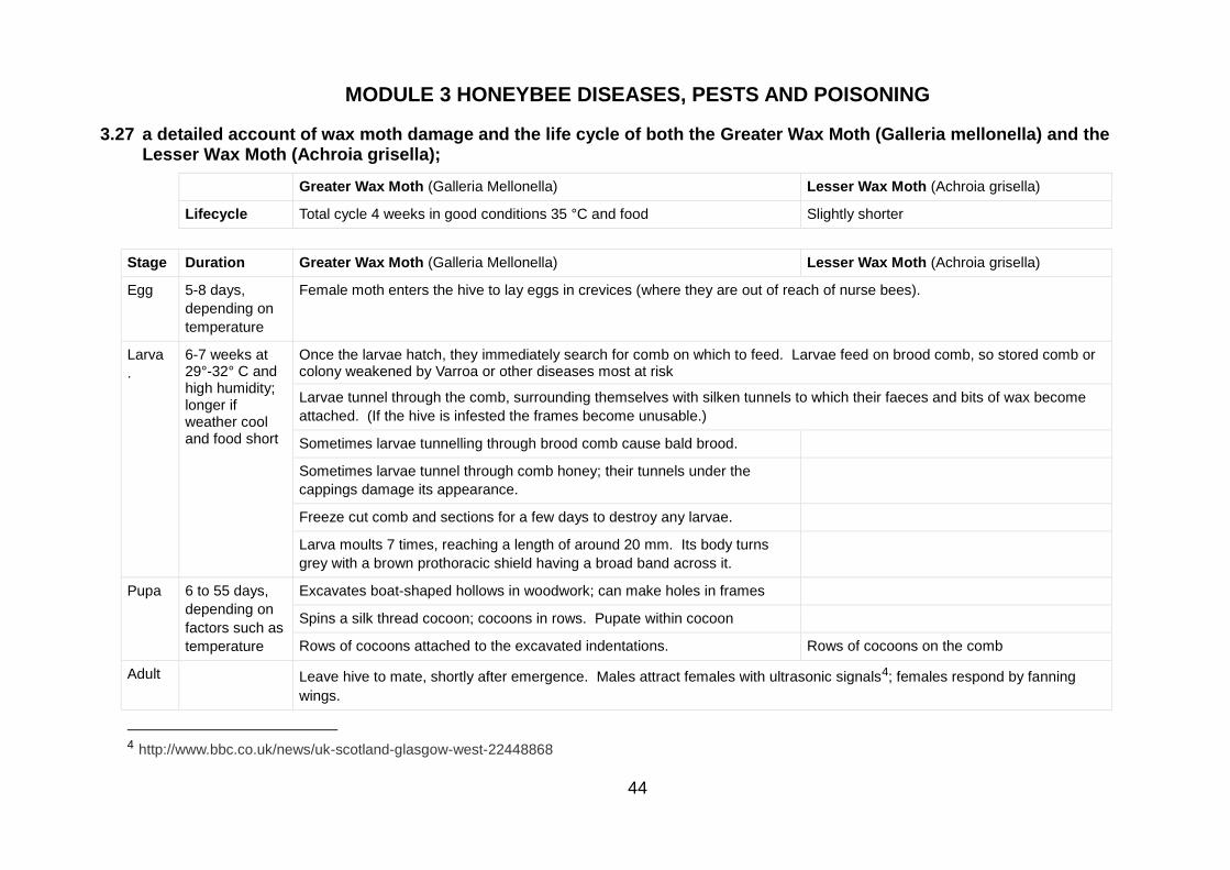
MODULE 3 HONEYBEE DISEASES, PESTS AND POISONING
44
3.27 a detailed account of wax moth damage and the life cycle of both the Greater Wax Moth (Galleria mellonella) and the Lesser Wax Moth (Achroia grisella);
Greater Wax Moth (Galleria Mellonella) Lesser Wax Moth (Achroia grisella)
Lifecycle Total cycle 4 weeks in good conditions 35 °C and food Slightly shorter
Stage Duration Greater Wax Moth (Galleria Mellonella) Lesser Wax Moth (Achroia grisella)
Egg 5-8 days,
depending on
temperature
Female moth enters the hive to lay eggs in crevices (where they are out of reach of nurse bees).
Larva
.
6-7 weeks at 29°-32° C and high humidity; longer if weather cool and food short
Once the larvae hatch, they immediately search for comb on which to feed. Larvae feed on brood comb, so stored comb or colony weakened by Varroa or other diseases most at risk
Larvae tunnel through the comb, surrounding themselves with silken tunnels to which their faeces and bits of wax become
attached. (If the hive is infested the frames become unusable.)
Sometimes larvae tunnelling through brood comb cause bald brood.
Sometimes larvae tunnel through comb honey; their tunnels under the
cappings damage its appearance.
Freeze cut comb and sections for a few days to destroy any larvae.
Larva moults 7 times, reaching a length of around 20 mm. Its body turns
grey with a brown prothoracic shield having a broad band across it.
Pupa
6 to 55 days,
depending on
factors such as
temperature
Excavates boat-shaped hollows in woodwork; can make holes in frames
Spins a silk thread cocoon; cocoons in rows. Pupate within cocoon
Rows of cocoons attached to the excavated indentations. Rows of cocoons on the comb
Adult Leave hive to mate, shortly after emergence. Males attract females with ultrasonic signals4; females respond by fanning
wings.
4 http://www.bbc.co.uk/news/uk-scotland-glasgow-west-22448868
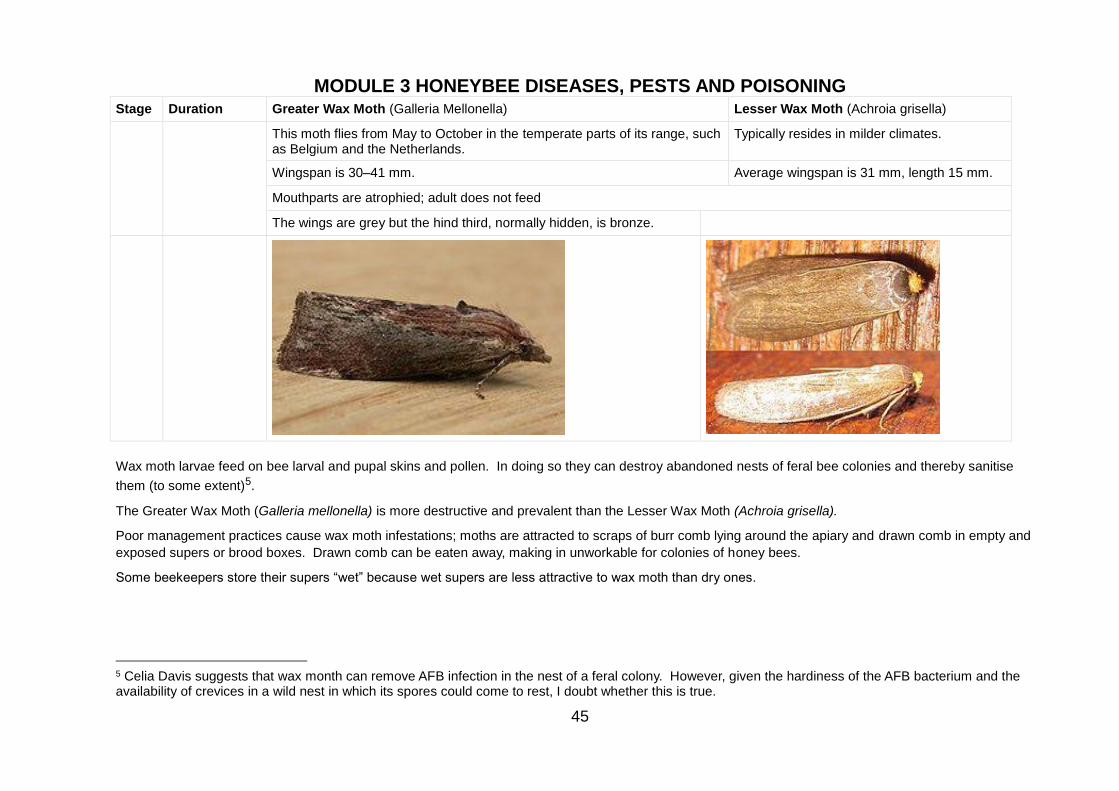
MODULE 3 HONEYBEE DISEASES, PESTS AND POISONING
45
Stage Duration Greater Wax Moth (Galleria Mellonella) Lesser Wax Moth (Achroia grisella)
This moth flies from May to October in the temperate parts of its range, such as Belgium and the Netherlands.
Typically resides in milder climates.
Wingspan is 30–41 mm. Average wingspan is 31 mm, length 15 mm.
Mouthparts are atrophied; adult does not feed
The wings are grey but the hind third, normally hidden, is bronze.
Wax moth larvae feed on bee larval and pupal skins and pollen. In doing so they can destroy abandoned nests of feral bee colonies and thereby sanitise
them (to some extent)5.
The Greater Wax Moth (Galleria mellonella) is more destructive and prevalent than the Lesser Wax Moth (Achroia grisella).
Poor management practices cause wax moth infestations; moths are attracted to scraps of burr comb lying around the apiary and drawn comb in empty and
exposed supers or brood boxes. Drawn comb can be eaten away, making in unworkable for colonies of honey bees.
Some beekeepers store their supers “wet” because wet supers are less attractive to wax moth than dry ones.
5 Celia Davis suggests that wax month can remove AFB infection in the nest of a feral colony. However, given the hardiness of the AFB bacterium and the availability of crevices in a wild nest in which its spores could come to rest, I doubt whether this is true.
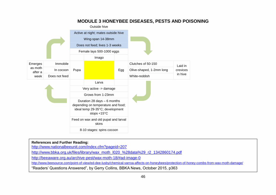
MODULE 3 HONEYBEE DISEASES, PESTS AND POISONING
46
Outside hive
Active at night; mates outside hive
Wing-span 14-38mm
Does not feed; lives 1-3 weeks
Female lays 500-1000 eggs
Imago
Emerges
as moth
after a
week
Immobile
Pupa
Egg
Clutches of 50-150 Laid in
crevices
in hive
In cocoon Olive-shaped, 1-2mm long
Does not feed White-reddish
Larva
Very active -> damage
Grows from 1-23mm
Duration 28 days – 6 months
depending on temperature and food;
ideal temp 29-35°C; development
stops <15°C
Feed on wax and old pupal and larval
skins
8-10 stages: spins cocoon
References and Further Reading:
http://www.nationalbeeunit.com/index.cfm?pageid=207
http://www.bbka.org.uk/files/library/wax_moth_l020_%28data%29_r2_1342860174.pdf
http://beeaware.org.au/archive-pest/wax-moth-18/#ad-image-0
http://www.beesource.com/point-of-view/ed-dee-lusby/chemical-varroa-affects-on-honeybees/protection-of-honey-combs-from-wax-moth-damage/
“Readers’ Questions Answered”, by Gerry Collins, BBKA News, October 2015, p363
MODULE 3 HONEYBEE DISEASES, PESTS AND POISONING
47
3.28 a detailed account of methods of treating or storing comb with particular reference to preventing wax moth damage;
A. Biological methods e.g. Certan
Certan is a suspension of the bacterium Bacillus thuringiensis, which is specific against Lepidoptera larvae. Apply by spraying both sides of the frame. Lasts one season in the comb. Does not taint wax or honey.
B. Flaming
Apply flame torch to used 'woodwork' (floors, roofs, boxes, crown board), concentrating on cracks and joins in the woodwork. The eggs and larvae of the wax moth are tiny and can easily get into these gaps, where they will hide and grow.
For Chemicals, Temperature and Barrier methods, stack equipment so that it is proof against the adult wax
moth and mice. This means:
• use a floor or crown board as a base, raised on bricks off the ground; use entrance blocks and cover
holes to make it moth-tight
• stack supers/brood boxes and frames on this. Use parcel tape to make the joints airtight over winter
o metal grille or queen excluder and empty super if using sulphur (see below)
o well-fitting roof.
C. Chemicals
In all cases, ventilate combs well before re-using them in the hive.
Acetic acid
Effective against wax month. Applied by fumigation. (See Section 3.16 on page 35.)
Sulphur:
Burn paper strips coated with yellow sulphur at the top of the stack; sulphur dioxide gas is heavier than
air and sinks through the stack, killing every life form it encounters. Use a small tin can with holes or a
smoker on its side with the top open as a burner, resting on the queen excluder.
Make sure you don't set the whole thing alight!
DO NOT BREATHE THE FUMES IN! Light upwind and stand well away.
Repeat in three to four weeks.
PDB (Paradichlorobenzene)
PDB now illegal. PDB does NOT kill wax moth at any stage in its lifecycle, but merely deters the adult
from approaching the wax comb and laying the next cycle of eggs. It also taints wax and honey.
D. Temperature
Freezing (to -15°C for at least two hours) is effective against moths and larvae. Safe and non-intrusive. After treatment protect frames, as below.
• A hard frost over several days will kill all stages of the moth in a stack made outdoors.
• Otherwise place in a deep-freeze for 48 hours. Stack as above afterwards.
E. Barrier
As wax moth enters hive through cracks, ensure that hive components fit together well.
Seal equipment for storage, making joints air-tight with packing tape.
Larvae love rolls of corrugated cardboard etc., so do not allow these to accumulate near stored wax.
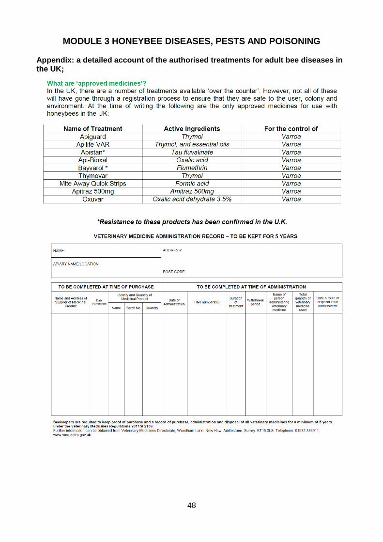
MODULE 3 HONEYBEE DISEASES, PESTS AND POISONING
48
Appendix: a detailed account of the authorised treatments for adult bee diseases in the UK;