Modulation of hippo pathway by alternative splicing
179
HAL Id: tel-02387314 https://tel.archives-ouvertes.fr/tel-02387314 Submitted on 29 Nov 2019 HAL is a multi-disciplinary open access archive for the deposit and dissemination of sci- entific research documents, whether they are pub- lished or not. The documents may come from teaching and research institutions in France or abroad, or from public or private research centers. L’archive ouverte pluridisciplinaire HAL, est destinée au dépôt et à la diffusion de documents scientifiques de niveau recherche, publiés ou non, émanant des établissements d’enseignement et de recherche français ou étrangers, des laboratoires publics ou privés. Modulation of hippo pathway by alternative splicing Diwas Srivastava To cite this version: Diwas Srivastava. Modulation of hippo pathway by alternative splicing. Agricultural sciences. Uni- versité Montpellier, 2019. English. NNT : 2019MONTT015. tel-02387314
Transcript of Modulation of hippo pathway by alternative splicing

HAL Id: tel-02387314https://tel.archives-ouvertes.fr/tel-02387314
Submitted on 29 Nov 2019
HAL is a multi-disciplinary open accessarchive for the deposit and dissemination of sci-entific research documents, whether they are pub-lished or not. The documents may come fromteaching and research institutions in France orabroad, or from public or private research centers.
L’archive ouverte pluridisciplinaire HAL, estdestinée au dépôt et à la diffusion de documentsscientifiques de niveau recherche, publiés ou non,émanant des établissements d’enseignement et derecherche français ou étrangers, des laboratoirespublics ou privés.
Modulation of hippo pathway by alternative splicingDiwas Srivastava
To cite this version:Diwas Srivastava. Modulation of hippo pathway by alternative splicing. Agricultural sciences. Uni-versité Montpellier, 2019. English. �NNT : 2019MONTT015�. �tel-02387314�

THÈSE POUR OBTENIR LE GRADE DE DOCTEUR
DE L’UNIVERSITÉ DE MONTPELLIER
En BIOLOGIE - SANTE
École doctorale- Biologiques pour la Santé (CBS2)
Unité de recherche- Institut de Génétique Moléculaire de Montpellier
Présentée par Diwas SRIVASTAVA
Le 25 Juin 2019
Sous la direction de Dr. François JUGE
et Dr. Jamal TAZI
Devant le jury composé de
Frédérique Peronnet , DR, HDR Institut de Biologie- Paris Seine
Julien Colombani, CR, HDR Université de Copenhague- Danemark
Florence Besse, DR, HDR Institute of Biology Valrose
Anne-Marie Martinez, Pr, HDR Université de Montpellier
Francois Juge, CR,HDR Institut de Génétique Moléculaire de Montpellier
Jamal Tazi, Pr, HDR Université de Montpellier
RAPPORTRICE
RAPPORTEUR
EXAMINATRICE
PRESIDENTE, EXAMINATRICE
DIRECTEUR DE THESE
CO-DIRECTEUR DE THESE
Modulat ion of Hippo Pathway by Alternat ive Spl ic ing


Acknowledgements
First, I would like to express my deepest gratitude to my supervisor, Dr. Francois Juge, who apart
from being a brilliant geneticist, is also a very humble, calm and kind person. I thank you from the
core of my heart for the knowledge and patience that you gave me from the day I joined as a Master
Intern. I have learned a lot of things from, and I hope I will be able to use that to contribute to
society.
I further extend my thanks to Marion, Carine, Christina, Karim, Laurent for being kind and helping
Lab members. I also take an opportunity to extend my thanks to the team of Abivax who were always
kind to me.
I would like to thank my friend and previous Ph.D. student of the lab Mahdi who has been with me
throughout the Ph.D. I would also like to thank Mickael who joined lab last year and has been a like a
brother to me. I will always cherish the numerous games of Jenga that we played at Rebuffy!!
It wouldn’t have been possible to do this Ph.D. without the crucial help of Jamal TAZI, director of our
RNA Metabolism group who not only provided my space in his laboratory and guidance and every
step but also generously gave me extensions for continuing the research after my Ligue funding
period was over. I look up to you as you are a very motivating and inspiring person.
I would take this opportunity to thank all the friends and colleagues from IGMM and Montpellier who
made this journey of Ph.D. colorful.
Sometimes you meet people in your life, and they teach you things just by being who they are, for me
that person is Dr. Pradeep Kumar from Rajeev Gandhi Centre for Biotechnology, Trivandrum-India
who is not only a brilliant scientist but also a wittiest human beings on the planet…What a boss to
work with! I thank you for all the help that you did just before I arrived in France.
I would not miss this opportunity to mention Riccardo Montecchi, Nicole, Mehuli, Ayush, Karim,
Anishesh, Fran, Arunabh and Chandra Mauli my real selfless friends from India and Europe with
whom I have some lovely memories together! I love you guys! And how can I miss Divy from this list,
my dearest buddy from college days. Thank you for always being there for me!
I thank all my four beautiful sisters Ritu Di, Richa Di, Shibbi Di and Nishu and who still pamper me as
they did in my childhood days. I would also like to thank Alok Jiju, Rohit Jiju, Anurag Jiju and Anit Jiju
for loving me and taking care of parents as their own.
Ultimately I dedicate this thesis to the jewel of my family i.e. Nikunj Kumar Srivastava who has
enabled me and motivated me at every step in my life to reach wherever and to do whatever I have
done with my life so far. I may never surpass you for the human being that you are but I can walk on
the path that you have shown me and live for the cause of others because that is the real happiness.
I would love to thank my parents, who made it all possible. Whatever I did and whatever I achieve in
this life will be smaller than the love you have showered upon me. Papa, you left me for heaven just
weeks before my defense and I felt very weak, but I felt your presence around me and the strength
you gave me to be able to defend my thesis. I know that you are always with me. I love you, and I
owe you everything. Love you, Papa & Mummy!

1
TABLE OF CONTENTS:
1.0 INTRODUCTION ......................................................................................................... 15
1.1 The Expansion of proteome: Single gene, multiple mRNA isoforms ....................... 15
1.1.1 Transcription: Where it all begins ...................................................................... 15
1.1.2 A short overview of mRNA processing: .............................................................. 16
1.1.3 Splicing Sites: ...................................................................................................... 17
1.1.4 Spliceosome: The machinery of splicing ........................................................... 19
1.1.5 Two key steps of splicing: ................................................................................... 20
1.1.6 Assembly of Spliceosome and catalysis of splicing reaction: ............................. 21
1.2 Alternative Splicing: Harbinger of complexity! .......................................................... 24
1.2.1 Types of Alternative Splicing: ........................................................................... 24
1.3 Regulators of Alternative Splicing: ............................................................................ 26
1.3.1 Exon and Intron definition: ................................................................................ 26
1.3.2 Cis-acting elements regulating Alternative splicing: .......................................... 27
1.3.3 Trans-acting factors regulating Alternative splicing .......................................... 28
1.3.3.1 Heterogeneous ribonuclear proteins (hnRNPs) ............................................. 29
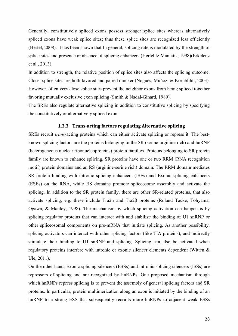
1.3.3.2 SR Protein Family: ........................................................................................... 32
1.3.3.2.1 Techniques to identify bindings of SR protein: .............................................. 33
1.3.3.2.2 Role of SR in constitutive splicing and alternative splicing: ........................... 37
1.3.3.2.3 Role of SR protein besides splicing: ................................................................ 38
1.3.3.2.4 Regulation of SR proteins by Post-translational modification ....................... 40
1.3.3.2.5 Targets of SR proteins:.................................................................................... 40
1.3.3.2.6 SR Protein B52 ............................................................................................... 43
1.3.4 Additional RNA Binding Proteins that regulate slicing: ...................................... 46
1.3.5 Other factors affecting Alternative splicing: ...................................................... 47
1.4 Quality control of splicing: ........................................................................................ 48

2
1.4.1 Nonsense-mediated decay (NMD) ..................................................................... 48
1.4.2 The No-go decay (NGD) .................................................................................... 50
1.4.3 Non-stop decay (NSD) ........................................................................................ 50
1.5 Alternative splicing in diseases and available therapy .............................................. 52
1.5.1 Conclusion: ......................................................................................................... 54
1.6 Hippo pathway and its discovery: ............................................................................. 56
1.7 Hippo Pathway in Drosophila ............................................................................... 58
1.7.1 Upstream regulators of Drosophila Hippo Pathway: ......................................... 61
1.7.1.1 Regulation by Fat: Planar cell polarity ........................................................... 63
1.7.1.2 Regulation by Ex/Mer/Kibra: ......................................................................... 63
1.7.1.3 Regulation by Apical-basal polarity proteins: ................................................ 64
1.7.1.4 Cell-Cell adhesion and junctional proteins ..................................................... 66
1.8 Hippo Pathway in Mammals ..................................................................................... 69
1.8.1 Upstream regulators of Mammalian Hippo Pathway: ........................................ 72
1.8.1.1 Regulation by Fat1-4: ..................................................................................... 72
1.8.1.2 Cellular polarity, Cell-cell contact, and morphology: .................................... 72
1.8.1.3 Mechanotransduction: .................................................................................... 74
1.8.1.4 Soluble factors as upstream signals: ............................................................... 76
1.8.1.5 Components of the cell cycle as upstream regulators: ................................... 76
1.8.1.6 Stress signals as upstream regulators: ............................................................ 77
1.8.1.7 Crosstalk with other pathways: ....................................................................... 77
1.8.1.8 A subtle difference between regulation of the Hippo pathway in Drosophila
and Mammals: ................................................................................................................... 78
1.9 Effectors of the Hippo pathway: Yki/YAP/TAZ ....................................................... 79
1.10 Hippo pathway in diseases: .................................................................................... 83
1.10.1 Implications of the Hippo pathway in cancer: ................................................... 83

3
1.10.2 Hippo pathway in non-cancer diseases: ............................................................. 88
1.11 Hippo pathway as a therapeutic target ....................................................................... 89
1.11.1 YAP/TAZ activators in Regenerative medicine: ............................................... 90
1.11.2 Inhibitors of Hippo signaling as anti-cancer drug candidate: ............................ 91
1.12 Conclusion: ................................................................................................................ 93
2.0 RESULTS ...................................................................................................................... 95
Article in preparation ............................................................................................................... 95
Methods ................................................................................................................................. 102
References .............................................................................................................................. 106
3.0 DISCUSSION AND PERSPECTIVES ................................................................................. 122
4.0 APPENDIX .................................................................................................................... 132
5.0 RERERENCES ................................................................................................................ 141

4
ABBRIVATIONS:
ABCP: apicobasal cell polarity ................................................................................................ 72
AJ: adherens junction .............................................................................................................. 73
ALS: Amyotrophic lateral sclerosis ......................................................................................... 89
AMOT: Angiomotin ................................................................................................................. 68
ANKRD: ankyrin repeat domain-containing protein ............................................................... 72
ANOVA: Analysis of variance .............................................................................................. 105
aPKC: atypical protein kinase C .............................................................................................. 65
AREG: amphiregulin ................................................................................................................ 72
ASOs : Antisense oligonucleotides .......................................................................................... 53
Baz : Bazooka ........................................................................................................................... 65
BPS: Branch Point Sequence ................................................................................................... 18
BRET: bioluminescence resonance energy transfer ................................................................. 31
CAMTA1: calmodulin-binding transcription activator 1 ........................................................ 84
CBC: cap binding complex ...................................................................................................... 17
Clk/Sty : cdc2-like kinase/serine, threonine, and tyrosine kinase ............................................ 40
COSMIC: Catalogue of somatic mutations in cancer .............................................................. 90
Crb: Crumbrs ............................................................................................................................ 64
CRC: Colorectal carcinoma ...................................................................................................... 86
CRISPR: clustered regularly interspaced short palindromic repeats ..................................... 103
CTD: carboxyl-terminal domain .............................................................................................. 16
CTGF: connective tissue growth factor ................................................................................... 72
cycE: cyclin E ........................................................................................................................... 63
Cyr61: cysteine-rich 61 ............................................................................................................ 72
Dchs : Dachous ......................................................................................................................... 62
Dlg : Discs large ....................................................................................................................... 65
DNA : deoxyribonucleic acid ................................................................................................... 15
ECM: extracellular matrix ........................................................................................................ 74
Ed: Echinoid ............................................................................................................................. 66
EJC: Exon Junction Complex .................................................................................................. 49
Ena: Enabled ............................................................................................................................ 66
ESE: exonic splice enhancer .................................................................................................... 26
5
ESS: Exonic Splicing Silencers ................................................................................................ 27
ESTs: expressed sequence tags ................................................................................................ 49
Ex : Expanded .......................................................................................................................... 68
ey-GAL4: eyeless-GAL4 ......................................................................................................... 43
FD: Familial dysautonomia ...................................................................................................... 53
FERM: Band Four-point-one, Ezrin, Radixin and Moesin ...................................................... 63
FoxO1: forkhead box proteins .................................................................................................. 77
GAF: GAGA factor ................................................................................................................ 125
GCs: Gastric cancer .................................................................................................................. 87
HITS-CLIP : high-throughput sequencing of RNA isolated by crosslinking
immunoprecipitation ............................................................................................................ 34
Hpo: Hippo ............................................................................................................................... 59
HSCs : hematopoietic stem cell ............................................................................................... 31
iCLIP : individual nucleotide-resolution cross-linking and immunoprecipitation ................... 32
IKBKAP: i-kappa-B kinase complex-associated protein ......................................................... 53
IRES : Internal Ribosomal Entry Site ...................................................................................... 38
Jub : Ajuba LIM protein ........................................................................................................... 66
KH: K-homology ...................................................................................................................... 29
KS: Kaposi sarcoma ................................................................................................................. 86
LATS : Large tumor suppressor Kinase ................................................................................... 68
lgl: Drosophila lethal giant larvae ........................................................................................... 65
lncRNAs : long noncoding RNAs ............................................................................................ 39
M: Minute gene ...................................................................................................................... 136
MAD: Mothers against DPP .................................................................................................... 68
MAJIQ: Modeling Alternative Junction Inclusion Quantification ......................................... 102
MALAT1: metastasis-associated lung adenocarcinoma transcript 1 ....................................... 39
Mats : MOB as tumor supressors ............................................................................................. 68
Mer: Merlin .............................................................................................................................. 68
MOB1A/MOB1B : Mps one binder kinase activator-like 1A/1B ............................................ 68
MOPD I: Microcephalic osteodysplastic primordial dwarfism type 1 .................................... 54
MPM: Malignant pleural mesothelioma................................................................................... 85
mRNA : messenger RNA ......................................................................................................... 15

6
Ncoa6: Nuclear receptor coactivator 6 .................................................................................... 61
NF2 : Neurofibromin ................................................................................................................ 68
NGD: no-go mRNA decay ....................................................................................................... 48
NMD: nonsense-mediated mRNA decay ................................................................................. 48
NSCLC: Small-cell lung cancer ............................................................................................... 85
NSD: non-stop mRNA decay ................................................................................................... 48
PDAC: Pancreatic ductal adenocarcinoma ............................................................................... 86
PPT: polypyrimidine tract (PPT).............................................................................................. 18
PSI or ................................................................................................ 41
PTC: premature termination codons ........................................................................................ 49
PTPN14: Protein Tyrosine Phosphatase 14.............................................................................. 73
qRRM: quasi-RNA recognition motif ...................................................................................... 29
RBP : RNA binding proteins RBPs.......................................................................................... 20
RCC: renal cell carcinoma ....................................................................................................... 87
RhoA: Ras homolog gene family, member A .......................................................................... 74
RP: Retinitis pigmentosa .......................................................................................................... 54
rRNAs : Ribosomal RNA ......................................................................................................... 16
RUNX: Runt-Related Transcription Factor ............................................................................. 71
Sav: Salvador ............................................................................................................................ 68
SCRA: Sveinsson's chorioretinal atrophy ................................................................................ 88
Scrib : Scribble ......................................................................................................................... 65
Sd: Scalloped ............................................................................................................................ 59
SdBP : Sd-Binding-Protein ...................................................................................................... 60
Sdc: Syndecan .......................................................................................................................... 44
Ser: Serine ................................................................................................................................ 70
SMA : Spinal muscular atrophy ............................................................................................... 52
snRNP : small nuclear ribonucleoproteins ............................................................................... 19
snRNPs : small ribonucleoproteins .......................................................................................... 16
SR: serine-arginine rich ............................................................................................................ 28
SRE: splicing regulatory elements ........................................................................................... 27
SRPK : SR-specific protein kinase........................................................................................... 40
SRSF : serine/arginine-rich protein specific kinase ................................................................. 38

7
ss : splice sites .......................................................................................................................... 18
TAZ : Transcriptional co-activator with PDZ binding motif ................................................... 68
TERT : transcription of telomerase reverse transcriptase ........................................................ 31
Tgi: Tondu-domain-Containing growth inhibitor .................................................................... 68
Trr : Trithorax-related............................................................................................................... 61
Tsh: T shirt ............................................................................................................................... 68
Vam: Vamana ........................................................................................................................... 63
VGLL4: vestigial like family member 4 .................................................................................. 91
VP: Verteporfin ........................................................................................................................ 91
Wnt: Wingless/integrated ......................................................................................................... 77
YAP: Yes-associated protein ................................................................................................... 68
Yki: Yorkie ............................................................................................................................... 59
Zyx: Zyxin ................................................................................................................................ 66

8
LIST OF FIGURES
Figure 2 Conserved sequence elements recognized by the major spliceosome: ...................... 19
Figure 3 Protein composition and snRNA secondary structures of the major human
spliceosomal snRNPs: .............................................................................................................. 20
Figure 4 The two steps of splicing catalysis: ........................................................................... 21
Figure 5 Splicing assembly: ..................................................................................................... 23
Figure 6 Seven types of alternative splicing: ........................................................................... 25
Figure 7 Exon and Intron definition models: ............................................................................ 27
Figure 8 Splicing regulatory elements: .................................................................................... 27
Figure 9 SR proteins and hnRNPs: .......................................................................................... 29
Figure 10 The hnRNP family: .................................................................................................. 30
Figure 11 Domain configuration of human SR proteins: ......................................................... 33
Figure 12 Techniques to identify binding of SR proteins: ....................................................... 35
Figure 13 A simplified schematic of the iCLIP protocol: ........................................................ 36
Figure 14 Multiple roles of SR proteins: .................................................................................. 39
Figure 15 AS affected by individual SR proteins: ................................................................... 42
Figure 16 Bristle phenotype: .................................................................................................... 44
Figure 17 Effect of B52 on growth: ......................................................................................... 45
Figure 18 Rescue of B52: ......................................................................................................... 46
Figure 19 The three primary mRNA surveillance mechanisms: .............................................. 51
Figure 20 SMA and its treatment by ASO: .............................................................................. 53
Figure 21 Implications of the Hippo pathway in cell biology: ............................................... 57
Figure 22 The rise of Hippo pathway: ..................................................................................... 57
Figure 23 Hippo mutant phenotypes in flies: ........................................................................... 58
Figure 24 Schematics of the Hippo pathway in Drosophila melanogaster: ............................ 60
Figure 25 Upstream regulators of Hippo pathway in Drosophila: .......................................... 62
Figure 26 (A) Effect on growth due to MST knockout : .......................................................... 69
Figure 27 Mammalian Hippo Pathway: ................................................................................... 71
Figure 28 Mechanotransduction: .............................................................................................. 74
Figure 29 Localization of YAP/TAZ at lower vs higher cell density: ..................................... 75
Figure 30 Crosstalk: ................................................................................................................. 78

9
Figure 31 Apical versus basal mechanotransduction via Yki or YAP in different animals: ... 79
Figure 32 The eight described isoforms of YAP: .................................................................... 80
Figure 33 Schematic diagram of YAP1 and 2, TAZ, and Yki: ................................................ 82
Figure 34 Cellular function of YAP/TAZ: ............................................................................... 83
Figure 35 : Implications of the Hippo pathway in various cancers and non-cancer diseases: . 84
Figure 36 Schematic demonstration of the small-molecule modulators of YAP: .................... 93
Appendix Figure 37 : CRISPR and screening strategy: ........................................................... 132
Appendix Figure 39 is Yki AS involved in the pro-apoptotic function of Yki upon stress? .... 135
Appendix Figure 40 B52 in cell comptetion: .......................................................................... 137
Appendix Figure 41 B52–/–
clones are partially rescued: ....................................................... 138
Appendix Figure 42 : Rescue of B52 mutants by Yki1 and Yki2: ............................................ 138

10
Abstract
The Hippo pathway is a conserved pathway involved in tissue growth and tumor
suppression. Studies have demonstrated its significance in the development of human cancers.
This cascade controls the activity of the transcription co-activator Yorkie (Yki) in flies and
Yes-associated protein (YAP) in mammals. Due to Alternative Splicing (AS), both Yki and
YAP proteins exist as two isoforms containing one (Yki1/YAP1) or two (Yki2/YAP2) WW
domains. Since WW domains are essential for interaction with specific partners, the
alternative inclusion of this domain in Yki/YAP protein may remodel their interaction
network and therefore their activity. The regulation and functional consequences of AS of
yki/YAP in vivo are unknown.
In this Ph.D. project, we identified that depletion of splicing factor B52 in Drosophila
lowers inclusion of the alternative exon in yki mRNAs and favors the expression of Yki1
isoform at the expense of the Yki2 isoform. B52 depletion in the wing reduces growth and
Yki activity. We demonstrate that Yki1 isoform is an attenuated version of Yki protein that
can compete with Yki2 isoform in the nucleus. To ascertain the role of yki AS in vivo and the
importance of short isoform Yki1, we abrogated this splicing by using CRISPR/Cas9
technology and created flies that can express Yki2 isoform only. yki2only flies are viable but
display a random phenotype of asymmetric wing size. This rise in “fluctuating asymmetry”
that is the consequence of subtle deviation from normal development, suggests that AS of yki
is crucial for the development robustness. Taking together, these results highlight a new layer
of modulation of Hippo pathway via AS of yki.
Alternative inclusion of the second WW domain is a conserved feature between Yki
and YAP. This further supports the idea that Yki1 and YAP1 isoforms have an important
function in vivo and that AS of yki/YAP is a conserved mechanism of control of the Hippo
pathway. This study opens up new perspectives for modulation of the Hippo pathway in
cancer cells by altering YAP AS.

11
Résumé
La voie Hippo est une voie conservée impliquée dans la croissance des tissus et la
suppression de tumeurs. Des études ont démontré son implication dans le développement des
cancers chez l'homme. Cette cascade contrôle l'activité du co-activateur transcriptionnel
Yorkie (Yki) chez la drosophile et de la protéine YAP (Yes Associated Protein) chez les
mammifères. En raison de l'épissage alternatif de leur transcrits, les protéines Yki et YAP
existent sous deux isoformes contenant un domaine WW (Yki1/YAP1) ou deux
(Yki2/YAP2). Puisque les domaines WW sont essentiels pour l’interaction avec des
partenaires spécifiques, l’inclusion alternative de ce domaine dans la protéine Yki/YAP peut
remodeler leur réseau d’interaction et donc leur activité. La régulation et les conséquences
fonctionnelles de l’épissage alternatif de yki / YAP in vivo sont inconnues.
Dans le cadre de ce doctorat, nous avons constaté que la déplétion du facteur
d’épissage B52 chez la drosophile réduit l’inclusion de l’exon alternatif dans l’ARNm de yki
et favorise l’expression de l’isoforme Yki1 aux dépens de l’isoforme Yki2. La déplétion en
B52 dans l'aile réduit la croissance et l'activité de Yki. Nous montrons que l'isoforme Yki1 est
une version atténuée de la protéine Yki qui peut entrer en concurrence avec l'isoforme Yki2
dans le noyau. Pour déterminer le rôle de l’épissage alternatif de yki in vivo et l'importance de
l'isoforme courte Yki1, nous avons abrogé cet épissage en utilisant la technologie
CRISPR/Cas9 et avons créé des mouches capables d'exprimer uniquement l'isoforme Yki2.
Ces mouches yki2only sont viables mais présentent un phénotype aléatoire d’ailes asymétriques.
Cette augmentation de l'«asymétrie fluctuante», qui traduit une déviation par rapport au
développement normal, suggère que l’épissage alternatif de yki est crucial pour la stabilité
développementale. Ces résultats mettent en évidence un nouveau niveau de modulation de la
voie Hippo via l’épissage alternatif de yki.
L'inclusion alternative du deuxième domaine WW est une caractéristique conservée
entre Yki et YAP. Cela conforte l'idée que les isoformes Yki1 et YAP1 ont une fonction
importante in vivo et que l'épissage alternatif de yki/YAP est un mécanisme conservé de
contrôle de la voie Hippo. Cette étude ouvre de nouvelles perspectives pour la modulation de
la voie Hippo dans les cellules cancéreuses en modifiant l’épissage alternatif de YAP.
12
Short Introduction and Objectives :
Over the last decade, the Hippo pathway has emerged as a major player in tissue size control,
regeneration and cancer progression. The pathway is, in particular, regulated by mechanicals
cues and allows to link growth of the cells with their environment. First discovered in
Drosophila, this conserved signaling pathway regulates the activity of a transcription co-
activator called Yorkie (Yki) in flies and YAP (Yes-Associated Protein) in human. The core
of the Hippo pathway is composed by the kinase Hippo (MST1/2 in mammals) which
activates the kinase Warts (LATS1/2 in mammals) that phosphorylates the transcription co-
activator Yki in Drosophila and YAP in mammals. Phosphorylated Yki/YAP is sequestered in
the cytoplasm through interactions with 14-3-3 proteins. Inactivation of the pathway leads to
translocation of unphosphorylated Yki/YAP in the nucleus and activation of its target genes.
Yki/YAP does not bind DNA directly but interacts with several transcription factors, mainly
members of the TEAD family such as Scalloped (Sd) in flies and TEAD1–4 in mammals.
Yki/YAP bound to Sd/TEAD recruits cofactors and chromatin remodeling complexes to
activate transcription of its target genes, which promote cell proliferation and inhibit
apoptosis. Overexpression of Yki/YAP, or inactivation of the Hippo pathway, induces tissue
overgrowth both in Drosophila and mouse models. Studies of mouse models and clinical
samples have now demonstrated the significance of Hippo pathway for the development of
human cancers.
Our lab is interested in the mechanism of Alternative Splicing (AS), that allows
producing multiple mRNAs from a single gene, by modulating inclusion or exclusion of
alternative exons for example, therefore encoding different protein isoforms. By studying the
function of the RNA binding protein B52 in Drosophila, a splicing factor of the SR protein
family, the lab previously showed that overexpression of B52 increases cell growth, whereas
B52 depletion reduces it. By the use of RNAseq data, they identified several AS events
modulated by B52 level in genes involved in cell growth, including several genes linked to
the Hippo pathway and especially yki.
They observed that depletion of the B52 splicing factor favors skipping of yki exon 3,
which encodes one of the two WW domains of Yki protein. Thus, Yki exists as two isoforms
containing one or two WW domain(s), respectively called Yki1 and Yki2. WW domains are

13
protein-protein interaction modules that bind proline-rich motifs (PPxY) present in several
proteins of the pathway such as the kinase Warts, the cytoskeleton-associated protein
Expanded (Ex) and the transcription co-activator Wbp2, as examples. Therefore, modulating
the inclusion/exclusion of one WW domain could change Yki phosphorylation, localization or
co-activator activity. The expression and function of the short Yki1 isoform has never been
described. Remarkably a similar AS event exists in the human YAP homolog: AS of exon 4
gives rise to two isoforms, YAP1 and YAP2, which respectively contain one or two WW
domain(s). Therefore, alternative inclusion of the second WW domain is a conserved feature
between Yki and YAP. Nevertheless, the signaling differences among YAP splicing variants
remain to be elucidated, as is the regulation of YAP AS.
The objectives of my Ph.D. were to investigate and reveal the role of B52-triggered
yki AS and to determine if this AS constitutes a new level of modulation of Hippo pathway in
Drosophila. Our results show that B52 depletion favors expression of the Yki1 isoform
carrying a single WW domain, and reduces growth in part through modulation of yki AS.
Compared to the canonical Yki2 isoform containing two WW domains, Yki1 has reduced
transcriptional and growth-promoting activities, decreased binding to PPxY motifs-containing
proteins, and an inability to bridge two proteins containing PPxY motifs. Nevertheless, Yki1
and Yki2 interact similarly with transcription factors and thus compete in vivo. Flies in which
the yki AS has been abrogated, thereby expressing only the Yki2 isoform, exhibit increased
fluctuating wing asymmetry, a signal of increased developmental noise. These results show
that yki AS represents an additional layer of modulation of Yki activity that unexpectedly
participates in buffering developmental noise, and provide the first experimental evidence that
AS participates in developmental robustness.
I hereby introduce the process of splicing, key components that facilitate splicing and
alternative splicing in general introduction. In a second part I cover in my introduction the
Hippo pathway and its key components in Drosophila and mammals, starting from its
discovery to most recently described key processes that regulate the pathway. The results
obtained during in the thesis are presented in the form of pre manuscript followed by a
discussion

14
INTRODUCTION

15
1.0 INTRODUCTION
1.1 The Expansion of proteome: Single gene, multiple mRNA isoforms
Humans have approximately 21000 protein-coding genes while Caenorhabditis elegans with
much simpler physiology has a genome of 19 000 protein-coding genes and at the same time
Oryza sativa Japonica, commonly known as rice contains approximately 35,825 protein-
coding genes. For decades researchers were puzzled by the fact that the complexity of the
organism does not correlate to the number of protein-coding genes they contain. This
question, however, was answered by the discovery of how common and abundant post-
transcriptional modifications are.
Once messenger RNA (mRNA) starts to be transcribed from DNA, the primary transcript
(pre-mRNA) is generated. To be successfully exported out of the nucleus, this pre-mRNA
would undergo a series of processing events, such as 5' capping, splicing, transcription
termination and 3' polyadenylation (Matlin, Clark, & Smith, 2005). During splicing, some
regions of the pre-mRNA are removed (i.e., introns), and stretches of sequence that contain
the necessary information for protein synthesis (i.e., exons) are ligated together. Alternative
splicing is the phenomenon where different combinations of sequences could be included or
excluded in the final transcript leading to generation of structurally and functionally different
mRNA variants (Nilsen & Graveley, 2010). High frequencies of alternative splicing, post-
translational modifications even leads to increased protein diversity (Wilhelm et al., 2014).
1.1.1 Transcription: Where it all begins
In order to express the encoded information contained in the double-stranded molecule made
of deoxyribonucleic acid (DNA), several key steps have to be completed. The genome
contains this information in genes that code for proteins which in turn control nearly all
functional aspects of a cell. Among the diversity of identified RNA species, messenger RNAs
(mRNAs) are regarded as those that contain the necessary information for the synthesis of
proteins.
During transcription, genes (stretches of DNA) are used as templates for the synthesis of
complementary single-stranded RNA molecules (i.e., transcripts). In eukaryotic cells, based
on the type of gene being targeted, such reaction can be catalyzed by three different enzymes
RNA polymerases (I, II and III). RNA polymerase II is responsible for the synthesis of RNAs
derived from the majority of genes, including those that encode for proteins. While, RNA

16
polymerase I and III are specifically involved in the transcription of ribosomal RNAs
(rRNAs), transfer RNAs (tRNAs) and several small RNAs (Paule & White, 2000).
Transcription by RNA pol II starts with the binding of several transcription factors to a
regulatory region located upstream of the gene, known as the promoter (Fuda, Ardehali, &
Lis, 2009). The transcription factors enable the subsequent assembly of the polymerase and
the formation of the transcription initiation complex. After the assembly steps and further
conformational rearrangements, RNA pol II releases from the large complex and enters the
elongation phase (Kwak & Lis, 2013). During elongation, RNA is synthesized from the
transcription start site (TSS), and nucleotides are incorporated in a complimentary basis in the
5’ to 3’ direction. Eventually, the polymerase transcribes through the cleavage and
polyadenylation signals that mark the end of the gene, and it is released from the DNA
template (Kuehner, Pearson, & Moore, 2011)
1.1.2 A short overview of mRNA processing:
All mRNA molecules undergo several modifications before they are exported to the cytosol,
which includes the addition of a 5’ cap, the polyadenylation of the 3’ end and the removal of
introns via splicing. The carboxyl-terminal domain (CTD) of the RNA polymerase II is
involved in the cap formation by recruiting the capping enzymes (triphosphatase,
guanylyltransferase and methyltransferase). During 5’capping a chemical group is added to
the 5’end of the pre-mRNA. 5’cap is the addition of a modified guanine (5’ guanine-N7 cap)
nucleotide which is subsequently methylated, and it functions to prevent the novel transcript
from degradation (Mandal et al., 2004). Reviewed by (Martinez-Rucobo et al., 2015).
Splicing is a much more complex reaction. During this process, introns are removed, and
exons are ligated together. As a result, a mature mRNA product is obtained. Many proteins
are involved in the splicing process, including small ribonucleoproteins (snRNPs),
heterogeneous nuclear ribonucleoproteins (hnRNPs) and other additional proteins. (Matlin et
al., 2005). To finalize the processing, the 3' end of the pre-mRNA is first cleaved at a specific
site. Sites of cleavage are encoded in the DNA sequence of the gene, and for the vast majority
it is between the highly conserved polyadenylation signal (PAS) AAUAAA and a
downstream sequence element (DSE), usually U or GU-rich (Proudfoot, Furger, & Dye,
2002) and then polyAdenosine (polyA) tail is added, which is typically 200-250 nucleotides
long in mammalian cells. (Figure1).

17
Figure 1 Regulation of eukaryotic gene expression:
mRNA expression starts with the nuclear transcription of genes. Following several processing steps, some of which occur co-transcriptionally, the transcription products are further transformed into mature mRNAs that can then be exported to the cytosol and localized to sub-cellular compartments. mRNA export is linked to strict quality control mechanisms unprocessed RNAs degraded. Once in the cytosol, mRNAs can be recognized by ribosomes and translated into proteins or will be degraded.
Initially, mRNA processing was thought to happen post-transcriptionally. However, in reality,
transcription and processing are not consecutive, but simultaneous and interdependent
(Neugebauer, 2002) and reviewed in (Bentley, 2014). The 3’ poly(A) tail prevents the
degradation by 3’-to-5’ exoribonucleases , facilitates the transfer from the nucleus to the
cytoplasm and promotes mRNA translation. This transfer, however, depends on the correct
processing and successful detection of CBC (cap binding complex) which is bound to 5’cap
of the pre-mRNA. Detection of CBC facilitates mRNAs transfer via nuclear pore (J. D.
Lewis & Izaurralde, 1997).
1.1.3 Splicing Sites:
The process of splicing is complex, in eukaryotes it is carried by a large complex of proteins
and RNAs and together they do the intricate job of removal of introns and stitching of exon
together. This large complex is aptly termed as spliceosome, and it is also one of the most
complicated machinery within the cell (Nilsen, 2003). Recognition of and removal of introns

18
by spliceosome is facilitated by the presence of specific sequence elements within the introns
and at exon-Intron boundaries. The 5’ and 3’ splice sites of an intron are specific and very
crucial recognition sequences that are recognized by the components of splicing machinery.
These sequences extend a few nucleotides into the flanking exon.
There are two types of introns in metazoan pre-mRNA. They are classified as minor introns
also known as U12 type or major class of introns also known as U2 type. The U2 and U12
introns differ by the presence of characteristic splice site sequence, and thus they are spliced
by a different spliceosomal complex.
U2 type introns form the majority of the introns present in metazoans, and they contain the
canonical GT– AG intron boundaries. It must be noted that very few U2 introns also contain
GC-AG boundaries. However, GC-AG introns represent only 0.82% of U2–type introns in
humans or 0.45% of U2 type in Drosophila melanogaster. Overall U2 type introns account
for 99% of all the introns present in humans, Drosophila melanogaster.
Minor class or U12 type introns are so rare that they amount to mere 0.4% of total human
introns (Sheth et al., 2006) while Drosophila melanogaster contains only 19 U12 type introns
(C. F. Lin, Mount, Jarmoowski, & Makaowski, 2010).
In humans, the majority of the Introns are U2 type, in these introns, the 5’splice sites or the
splice donor site is defined by a nine nucleotide (nt) consensus sequence, YAG/GURAGU
(where Y is a pyrimidine, R is a purine. Even though the consensus exists, splice site
sequences are degenerate. The 3' splice site is comprised of three elements, placed within
around 40 nucleotides upstream the intron/exon junction. The 3' splice site itself has a
consensus YAG/N, where Y is a pyrimidine (uracil or cytosine). The spliceosome also
recognizes another sequence motif called Branch Point Sequence (BPS) which is located at
20-40 nt upstream of the 3´ splice site. The BPS YNYURAY (where Y is C or U nucleotide)
contains essential adenosine that is required for the first nucleophilic attack of the splicing
reaction (Reed, 1996). BPS is followed by a polypyrimidine tract (PPT), a pyrimidine-rich
motif which is a string of uracil bases is crucial for efficient BPS utilization and selection —
reviewed in (Valadkhan, 2007a) (Figure 2). On the other hand, U12 type introns so not
contain polypyrimidine tract upstream of 3’ splice site. Reviewed in (Sheth et al., 2006).

19
Figure 2 Conserved sequence elements recognized by the major spliceosome:
This schematic shows four consensus elements required for intron recognition: the 5' splice site, branch point, polypyrimidinetract, and the 3' splice site. R corresponds to purine, Y – to pyrimidine (C or U nucleotide) and N to any nucleotide. Image is adapted from (Wahl, Will, & Lührmann, 2009)
1.1.4 Spliceosome: The machinery of splicing
The spliceosome is a ribonucleoprotein complex that is involved in splicing of nuclear
precursor mRNA (pre-mRNA). There are two types of Spliceosome in eukaryotes: the U2-
dependent spliceosome (Major Spliceosome), which catalyzes the removal of U2-type introns,
and the less abundant U12-dependent spliceosome (Minor Spliceosome), which is present in
only a subset of eukaryotes and splices the rare U12-type class of introns.
The major spliceosome undergoes significant conformational and compositional
rearrangements during the several steps of the splicing reaction. It is composed of five
different small nuclear RNA molecules (snRNAs: U1, U2, U4, U5and U6), as well as around
170 other splicing protein factors. Each of these RNA molecules associates with several
proteins and form complexes called small nuclear ribonucleoproteins (snRNP). Each snRNP
consists of an snRNA (two in the case of U4/U6), a common set of seven Sm proteins (B/B
D3, D2, D1, E, F, and G) and a variable number of particle-specific proteins (Figure 3).

20
Figure 3 Protein composition and snRNA secondary structures of the major human spliceosomal snRNPs:All seven Sm proteins (B/B’, D3, D2, D1, E, F, and G) or LSm proteins (Lsm2-8) are indicated by “Sm” or “LSm” at the top of the boxes showing the proteins associated with each snRNP. The U4/U6.U5 tri-snRNP contains two sets of Sm proteins and one set of LSm proteins Adapted from (Will & Lührmann, 2011)
Such snRNPs form the core of the spliceosome and are directly involved in recognition of
splice sites and branch-point sequences, as well as the catalysis of the splicing reaction. (Will
& Lührmann, 2011). The minor spliceosome is functionally analogous to the major
spliceosome but differs in the use of snRNAs (minor snRNAs are U11, U12, U4atac/U6atac,
and U5). There are many RNA binding proteins (RBPs) in the dynamic structure of the
spliceosome that helps in splicing (Matlin & Moore, 2010).
1.1.5 Two key steps of splicing:
Pre-mRNA splicing is achieved by two consecutive trans-esterification reactions which are
based on nucleophilic attacks between RNA nucleotides. In the very first reaction, 5’ exon is
cleaved from the intron through a nucleophilic attack of the 2
nucleotide in the branch point sequence on the phosphate group of the GU dinucleotide at the
5’ splice site, resulting in the formation of a lariat intermediate. In the second step, the free 3’
hydroxyl group at 5’ss makes the second nucleophilic attack on the phosphodiester bond at
3’ss (3’ exon-intron junction) (Figure 4). This results in ligation of the two exons and release
of excised intron as a lariat. Reviewed in (Valadkhan & Jaladat, 2010) and (Will &
Lührmann, 2011).

21
Figure 4 The two steps of splicing catalysis:
During the first step, the 5’ss and branch adenosine are joined by a 2’-5’ nucleophilic attack, leading to the formation of a lariat structure and a free upstream exon. The second step consists of the 3’-5’ nucleophilic bond between the free exon and the 3’ss. Adapted from (Scotti & Swanson, 2016)
1.1.6 Assembly of Spliceosome and catalysis of splicing reaction:
During the course of the splicing reaction and before the actual intron can be removed, the
active catalytic site of the spliceosome needs to be created, an event that requires many
changes in its composition and conformation. Since spliceosome is both highly dynamic and
flexible, it assembles and dissembles tediously to facilitate each splicing event. It undergoes
both radical structural and compositional changes at every step of its assembly. The assembly
of spliceosome has been studied extensively in vitro. Based on biochemical methods, six
different complexes can be distinguished: the E, A, B, Bact, B*, and C complex. Reviewed in
(Shi, 2017).
RNA helicases from the DExD/H-box family (composed of the DEAD-box, DEAH-box, and
Ski2-like helicases) facilitate the extensive structural and compositional remodeling of
spliceosome assembly and transition to various steps (Cordin, Hahn, & Beggs, 2012).
Cyclophilins, a subfamily of peptidyl-prolyl cis-trans isomerases (PPIases), facilitate
conformational changes within the spliceosome. Reviewed in (Thapar, 2015). Hereby, the
major spliceosome catalyzes the splicing in the following steps (Figure 5).
Step1: The crucial first step in the assembly of the major spliceosome is the association of
U1 snRNP with the 5’ss and U2 snRNP with 3’ ss via formation of protein dimer. This results
in the formation of the commitment complex; also referred to as “E complex.”

22
Step2: U2 snRNP then recognizes branch point sequence followed by interaction with U1
snRNP leading to the formation of pre-spliceosome. This complex made by the interaction
between U1 snRNP and U2 snRNP spans across the exons and brings together the 5’ and
3’splice sites and BPS in close vicinity. Thus this pre-spliceosome Complex A is also referred
to as “Exon definition complex.”
Step3: U4/U6.U5 is a large pre-assembled tri-snRNP spliceosomal complex. It contains U5
snRNA, extensively base-paired U4/U6 snRNAs in addition to over 30 proteins. Recruitment
of this tri-snRNA to complex A results into formation of Complex B.
Step4: The complex B undergoes a series of conformational and compositional
rearrangements resulting in a catalytically active complex called complex Bact or complex B*.
Several remodeling of protein and RNA-RNA interactions facilitate the formation of U2-U6
snRNA structure that brings the 5’splice site and BPS in close proximity and forms the
catalytic core. The activation of complex B leads to unwinding of U4 and U6 snRNA and
expulsion of U4 and U1 from the complex.
Step5: The activated B complex (Bact) catalyzes the first step of splicing. This leads to the
formation of complex C, which contains Intron-lariat intermediate.
Step6: Complex C catalyzes the second step of splicing after it undergoes further
conformational rearrangements.
Step7: After this second transesterification reaction, the ligated exons and a lariat intron are
released. The intron lariat structure is degraded. This is followed by the release of U2, U5 and
U6 snRNP and which are recycled for further rounds of splicing.
However, the mRNA is just not yet free in the nucleus. A complex of proteins is then
deposited to the new exon junction (thus termed as exon junction complex, EJC). This marks
the completion of splicing and allows the mRNP particle to travel to the cytoplasm. Reviewed
in (Black, 2003) (Valadkhan, 2007b) (Wahl et al., 2009) (Will & Lührmann, 2011).

23
Figure 5 Splicing assembly:
Schematic representing the key steps of assembly and activation of the yeast spliceosome and the complete splicing-reaction cycle. Adapted from (Shi, 2017).

24
1.2 Alternative Splicing: Harbinger of complexity!
Constitutive splicing refers to the type of splicing in which introns are systematically
removed, and the exons are ligated together to generate a final mRNA. However, splicing is
also regulated alternatively, i.e., particular exon might be entirely or partially spliced out, or
introns might be retained, or the splicing machinery could make the choice of the different 5'
or 3' splice sites. Such processing of pre mRNA transcript which results in the inclusion of
different part of a transcript into the final mRNA product is called Alternative splicing (AS).
It leads to the formation of alternative mRNA products from a given gene locus. According to
(Q. Pan, Shai, Lee, Frey, & Blencowe, 2008) (E. T. Wang et al., 2008), almost 95% of
multiexonic genes in humans are regulated by produce several RNAs by alternative splicing.
Interestingly, proteins resulting from splicing variants often have distinct molecular functions.
AS can result in the generation of isoforms of proteins with different biological function,
structure, localization and interaction capabilities (Keren, Lev-Maor, & Ast, 2010) (Nilsen &
Graveley, 2010). For example, the two variants of survivin have opposite functions: one has
pro-apoptotic while the other has anti-apoptotic properties (Végran et al., 2007). It has been
estimated that on average, each gene generates around ten mRNA isoforms (Z. Hu et al.,
2015). Around 40% of Drosophila genes contain one or more alternative exon (Q. Pan et al.,
2008)
Alternative Splicing plays a crucial role in the expansion of the coding capacity of eukaryotic
genomes by giving rise to several structurally and functionally different protein isoforms from
a single gene locus. Thus, it fills the gap between the total number of protein-coding genes (<
20,000) (Ezkurdia et al., 2014) compared to the overall number of proteins (> 100,000) and
also imparts to the complexity of the organism (Nilsen & Graveley, 2010).. In unicellular
cells, alternative splicing is absent or very rare, and one gene provides one protein product
(Ast, 2004).
One of the extreme examples of such expansion of protein-coding capacity via alternative
splicing is dscam gene in Drosophila. It can produce a whopping 38,016 different mRNA
isoforms via alternative splicing in four different regions of its pre-mRNA (Black, 2000)
(Graveley, 2001)(Schmucker et al., 2000).
1.2.1 Types of Alternative Splicing:
In eukaryotes, alternative splicing can result via employing one of the following mechanisms:
exon skipping or inclusion, choice of alternative 3’ or 5’ splices site selection, intron
retention, the inclusion of mutually exclusive exon and alternative polyadenylation (J. Chen

25
& Weiss, 2015). The most common type of alternative splicing is a cassette type alternative
exon, i.e., Exon skipping in vertebrates and invertebrates. While in lower metazoans Intron
retention is more frequent (Figure 6). Which splicing event takes place is often determined by
the contributions and activity of the different splicing activators or repressors in the tissue or
during a developmental stage.
Figure 6 Seven types of alternative splicing:
(From Top to bottom) Seven types of alternative splicing. Exon skipping, Mutually exclusive exons, Alternative 3’ splicing, Alternative 5’ splicing, Intron retention, choice of an alternative promoter, Alternative Polyadenylation. Intron retention isthe major alternative splicing event in rice, whereas exon skipping is the most frequent alternative splicing in humans.
Alternative splicing has a central regulatory role in the gene expression pathway. It dictates
several biological functions through the entire lifespan of an organism. It has been shown that
the higher eukaryotes display the higher proportion of alternatively spliced genes indicating
that alternative splicing is an indispensable feature of genomic evolution. Several splicing
events are conserved among different species along with many splicing variants which are

26
species-specific indicating that alternative splicing plays a significant role in species
differentiation along with genomic evolution.
1.3 Regulators of Alternative Splicing:
1.3.1 Exon and Intron definition:
The spliceosome directs both constitutive and alternating splicing with high fidelity. The
average exon size is small in higher eukaryotes (approx 170nt on an average) while Introns
could be up to tens of thousands of nucleotides long and harbor several “cryptic splice” sites.
It is, therefore, a huge task for spliceosome to identify bona fide sites from the pseudo or
cryptic ones. An understanding of how spliceosome accomplishes such task comes from the
following models.
The exon definition model postulates that that exon, rather than introns are the basic unit of
recognition (Exon definition may facilitate splice site selection in RNAs with multiple exons),
and the spliceosome assembles across the exon. In contrast, in lower eukaryotes such as yeast
and fly where the introns are much smaller, early spliceosome assembly is centered around
the introns, which is referred to as “intron definition” (Talerico & Berget, 1994). The intron
definition model postulates that components of the spliceosome assemble and interact across
small introns, where the 5’ and 3’ splice sites are close to each other. Transfection splicing
assays have shown that as intron length rises above 250 nucleotides, splicing becomes quite
inefficient (Fox-Walsh et al., 2005). In intron definition, the spliceosome recognizes the splice
sites across the intron, provided that this intron is not too large (e.g., <200-250 nt) and beyond
this length exon definition prevails (Fox-Walsh et al., 2005) (Figure 7). Furthermore, the
authors of this report found that the inclusion of an RNA cis-element known as an exonic
splice enhancer (ESE) could dramatically increase splicing efficiency of even longer introns.
While, on the other hand, Intron definition mediated splicing is an exception in mammals, it is
a rule in plants, fungi, and invertebrates (Talerico & Berget, 1994) (X. Xiao, Wang, Jang, &
Burge, 2007).

27
Figure 7 Exon and Intron definition models:
The left panel depicts the Intron definition model according to which pairing between the splice sites takes place across an
intron when long exons are separated by short (<250 bp) introns. On the other hand, the right panel shows the Exon
definition model where the splice site communication occurs across exons when they are separated by long (>250 bp)
introns. Adapted from (De Conti, Baralle, & Buratti, 2013)
1.3.2 Cis-acting elements regulating Alternative splicing:
The mammalian junctions that define an exon are weakly conserved and more degenerate
with respect to yeast canonical cis-elements. These elements are necessary but not sufficient
to define exon/intron junctions. Internal exonic sequences far from the 5’ and 3’ splice sites
were essential for exon recognition. These additional elements are called splicing regulatory
elements (SREs), cis-acting elements which are present in the pre-mRNA and varies in terms
of location and effect. They are known as ESE (exonic splicing enhancers), ESS (exonic
splicing silencers), ISE (intronic splicing enhancers) and ISS (intronic splicing silencers).In
general, SREs recruit trans-acting splicing factors that can act as repressor or activators of
splicing. As described earlier, Introns possess several sequence elements required for pre-
mRNA splicing: 5’ and 3’ splice sites, BP and PPT. These splice sites are often termed as
‘‘weak’’ or ‘‘strong’’ splice sites. Splice site strength depends on the complementarity
between splice site sequences and U1 and U2 snRNPs binding to them. The more the degree
of similarity the more the strength of a splice site (Figure 8) (Roca, Sachidanandam, &
Krainer, 2005). (De Conti, Baralle, & Buratti, 2013)
Figure 8 Splicing regulatory elements:SREs can enhance and repress the splice site selection by the spliceosome. Splicing is governed by cis-regulatory sequences in the pre-mRNA; exonic splicing enhancers (ESEs), exonic splicing silencers (ESSs), intronic splicing enhancers (ISEs) and intronic splicing silencers (ISSs)
28
Generally, constitutively spliced exons possess stronger splice sites whereas alternatively
spliced exons have weak splice sites; thus these splice sites are recognized less efficiently
(Hertel, 2008). It has been shown that In general, splicing rate is modulated by the strength of
splice sites and presence or absence of splicing enhancers (Hertel & Maniatis, 1998)(Erkelenz
et al., 2013)
In addition to strength, the relative position of splice sites also affects the splicing outcome.
Closer splice sites are both favored and paired quicker (Nogués, Muñoz, & Kornblihtt, 2003).
However, often very close splice sites prevent the neighbor exons from being spliced together
favoring mutually exclusive exon splicing (Smith & Nadal-Ginard, 1989).
The SREs also regulate alternative splicing in addition to constitutive splicing by specifying
the constitutively or alternatively spliced exon.
1.3.3 Trans-acting factors regulating Alternative splicing
SREs recruit trans-acting proteins which can either activate splicing or repress it. The best-
known splicing factors are the proteins belonging to the SR (serine-arginine rich) and hnRNP
(heterogeneous nuclear ribonucleoproteins) protein families. Proteins belonging to SR protein
family are known to enhance splicing. SR proteins have one or two RRM (RNA recognition
motif) protein domains and an RS (arginine-serine rich) domain. The RRM domain mediates
SR protein binding with intronic splicing enhancers (ISEs) and Exonic splicing enhancers
(ESEs) on the RNA, while RS domains promote spliceosome assembly and activate the
splicing. In addition to the SR protein family, there are other SR-related proteins, that also
activate splici (Roland Tacke, Tohyama,
Ogawa, & Manley, 1998). The mechanism by which splicing activation can happen is by
splicing regulator proteins that can interact with and stabilize the binding of U1 snRNP or
other spliceosomal components on pre-mRNA that initiate splicing. As another possibility,
splicing activators can interact with other splicing factors (like TIA proteins), and indirectly
stimulate their binding to U1 snRNP and splicing. Splicing can also be activated when
regulatory proteins interfere with intronic or exonic silencer elements dependent (Witten &
Ule, 2011).
On the other hand, Exonic splicing silencers (ESSs) and intronic splicing silencers (ISSs) are
repressors of splicing and are recognized by hnRNPs. One proposed mechanism through
which hnRNPs repress splicing is to prevent the assembly of general splicing factors and SR
proteins. In particular, protein multimerization along an exon is initiated by the binding of an
hnRNP to a strong ESS that subsequently recruits more hnRNPs to adjacent weak ESSs

29
(Martinez-Contreras et al., 2007) (Zhu, Mayeda, & Krainer, 2001). Alternatively, hnRNPs can
also repress splicing by looping out entire exons blocking the recruitment snRNPs (Nasim,
Hutchison, Cordeau, & Chabot, 2002) (Damgaard, Tange, & Kjems, 2002). Initially identified
as positive and negative regulators of splicing, respectively, it is now known that their effect
on a particular splicing event is heavily context-dependent (Witten & Ule, 2011).
Figure 9 SR proteins and hnRNPs:These are two major families of alternative splicing regulatory proteins, which are recruited by splicing enhancers and silencers. These regulatory proteins target components of the spliceosome (shown in green) that associate with both the 5and the 3 lice sites flanking the alternative exon and can have either activating or inhibitory effects on the recognition and use of that site. In addition, interactions among components of the spliceosome that are recruited to the 3can mediate exon definition. Adapted from (Kornblihtt et al., 2013)
1.3.3.1 Heterogeneous ribonuclear proteins (hnRNPs)
HnRNPs represent a large family of RBPs that contribute to multiple aspects during mRNA
processing post-transcription. A typical hnRNP usually contain one or more RNA binding
domains(RNA recognition motif, i.e., RRM domain), a qRRM (quasi-RNA recognition
motif), a glycine-rich domain constituting an RGG box (Arg-Gly-Gly repeats) and a K-
homology (KH) domain (Geuens, Bouhy, & Timmerman, 2016).
The hnRNP protein family consists of at least 20 proteins in humans that have been
characterized as components of protein complexes bound to pre-mRNA (hnRNP complexes)
(Dreyfuss, 1993) (Cartegni, Chew, & Krainer, 2002)(Figure 10). The molecular weight of the
hnRNP family ranges from 34 to 120 kDa, and they have been named alphabetically from A
to U. (Piñol-Roma & Dreyfuss, 1992).

30
Figure 10 The hnRNP family:The hnRNPs are named alphabetically from hnRNP A1 to hnRNP U. The members of the hnRNP family are built up of four unique RNA-binding domains (RBDs): RRM RNA recognition motif, qRRM quasi-RNA recognition motif, and KH K-homology domain, RGG RNA-binding domain consisting of Arg-Gly-Gly repeats. Image adapted from : (Geuens et al., 2016)
Although hnRNPs are expressed in all tissues, but the relative amount of different hnRNPs
varies among cell types and exhibit stage-specific expression patterns, for an example, some
hnRNPs are extremely abundant (~100 million copies per nucleus), while others are present in
a lower amount. Together, hnRNPs are similar in abundance to histones in growing cells
(Dreyfuss, 1993) (Kamma, Portman, & Dreyfuss, 1995). The function of hnRNPs depends
upon their localization and upon undergoing PTMs such as methylation, phosphorylation,
ubiquitination and sumoylation they can change their subcellular localization and thus
function (S. P. Han, Tang, & Smith, 2010) (Chaudhury, Chander, & Howe, 2010)
hnRNPs play an essential role in both constitutive and alternative splicing. hnRNPs generally
repress splicing through binding to the silencer sequences (ESSs or ISSs) in the pre-mRNA
and suppress splicing.
Studies have uncovered a variety of ways in which hnRNPs can modulate splicing.
Mechanisms that repress exon usage involve binding that competes with the recruitment of
positive splicing factors (for an example SR protein), as well as the inhibition of protein

31
interactions involved in exon or intron definition. This has been extensively demonstrated for
hnRNPA1, A2/B1, and hnRNP. A multitude of biochemical studies too have demonstrated
that hnRNP binding can promote interactions that facilitate exon inclusion, highlighting their
potential to positively or negatively regulate exon usage (Martinez-Contreras et al., 2007). It
appears that the ability to regulate exon usage both positively and negatively is a general
feature of hnRNPs. (Huelga et al., 2012) reported that hnRNPs from diverse families can
positively and negatively regulate thousands of exons by binding to proximal introns by using
splicing-sensitive microarrays coupled with CLIP. They reported that hnRNPs are similar and
cooperative in the roles that they play in the the regulation of AS and their RNA targets
overlap. The negative of positive regulation of AS could be attributed to the binding of
hnRNP in different positions relative to the exon, a phenomenon already reported for SR and
others RBPs (Witten & Ule, 2011). Position-dependent AS regulation by hnRNPs could be
via looping out the intervening RNA (Blanchette & Chabot, 1999). Indeed it has been shown
that hnRNP A1 and hnRNP H collaborate to create a RNA loop so as to repress the internal
splice sites and while activating external splice sites which are brought in a closer proximity.
The authors used bioluminescence resonance energy transfer (BRET) technology to show the
homotypic and heterotypic interactions between these two hnRNPs in live cells (Fisette,
Toutant, Dugré-Brisson, Desgroseillers, & Chabot, 2010). The binding on opposite sides of an
intron represses exon usage while looping events within an intron can bring together pairs of
splice sites to promote spliceosomal interactions across an intron (Chabot, 2015).
Besides splicing family of hnRNPs have been thoroughly documented to play several roles
such as HnRNP A1 and A2/B1 play a role oligodendrocytic and neuronal mRNA trafficking
(Shan, Munro, Barbarese, Carson, & Smith, 2003). (Villarroya-Beltri et al., 2013) have shown
that hnRNPA2B1 specifically binds miRNA-198 and miRNA-601 and enable their loading in
exosome. HnRNP C too has been shown to play a role in sorting of transcripts according to
their size (McCloskey, Taniguchi, Shinmyozu, & Ohno, 2012). (L. Y. Chen & Lingner, 2012)
showed that hnRNP D plays a role in telomere maintenance via stimulating the transcription
of telomerase reverse transcriptase (TERT) gene. Further, hnRNP I have been shown to
regulate the neonatal immune response in studies conducted in intestinal epithelial cells in
mouse and helps in preventing colitis and onset of colorectal cancer (Z. Jin, Liang, Yang, &
Mei, 2017). Likewise, hnRNP-L has been shown to be critical for hematopoietic stem cell’s
(HSCs) survival and integrity via activation of caspase-dependent death receptor pathways
(Gaudreau et al., 2016). hnRNP-Q is recently implicated in cell proliferation and tumor
initiation in colorectal cancer via enhancing Aurora-A translation (C. H. Lai et al., 2017) By

32
doing individual nucleotide-resolution cross-linking and immunoprecipitation (iCLIP)
studies, (Briese et al., 2018) found around 3500 RNA targets of hnRNP R including
significant interactor, a noncoding RNA 7SK which is essential in regulation of axon
maintenance by hnRNP R.
1.3.3.2 SR Protein Family:
An essential class of RBP involved in splicing is the serine/arginine (SR)-rich SR proteins.
The SR proteins represent a family of splicing factors that are remarkably conserved in
vertebrates and invertebrates and also have been discovered in plants. Members of the SR
family in mammalian cells were discovered in the early 1990s by the identification of factors
associated with purified spliceosomes (X. D. Fu & Maniatis, 1990) (X. D. Fu & Maniatis,
1992). SR proteins are ubiquitous in higher organisms and present in many other diverse
species, such as plants and fungi, reflecting an early emergence in evolutionary history
(Twyffels, Gueydan, & Kruys, 2011). Budding yeast is devoid of “true” SR proteins but has
three SR-like proteins: Npl3, Gbp2 and Hrb1, which likely represents the ancestral basis from
which SR proteins subsequently evolved (Busch & Hertel, 2012).
SR proteins are defined by the presence of one or two RNA recognition motifs and RRM
homolog (RRMH) (a more degenerate pseudo RRM) at the N terminus and an RS domain
(arginine/serine-rich dipeptide domain) at the C terminus. RRM domains are widespread
among RNA binding proteins, and their flexibility permits diverse modes of binding between
proteins (Maris, Dominguez, & Allain, 2005). In humans, Twelve canonical members of the
SR protein family share this characteristic domain structure. RRMs ascertain RNA-binding
specificity, whereas the RS domain functions as a protein-protein interaction module by
recruiting components of the core splicing apparatus to promote splice site pairing (Jane Y.
Wu & Maniatis, 1993). There are 7 homologs of these SR proteins in Drosophila. These
essential splicing factors can bind RNA directly, and their RS and RRM domains act to
promote protein-protein and protein–RNA interactions, facilitating the recruitment and
assembly of the spliceosome (S. H. Xiao & Manley, 1997).
SR-
repetitive characteristic of true SR proteins or an RRM motif, although many of these are able
to bind RNA via different domains and mechanisms (Long & Caceres, 2008). Genome-wide
analyses revealed that RS-domain containing proteins participate in numerous aspects of gene
expression regulation, including those involved in the cell cycle(Boucher, Ouzounis, Enright,
& Blencowe, 2001)
33
The SR family nomenclature is historically diverse but in the beginning of decade an
agreement was reached to have consensus terminology to identify them (Twyffels et al.,
2011) (Manley & Krainer, 2010), thus they have several aliases, however it is not surprising
to find researchers using old style names even now.
Figure 11 Domain configuration of human SR proteins:SRSF1–12 are members of the canonical SR protein splicing family that is defined by N-terminal RRMs followed by a downstream RS domain. The RRM is responsible for RNA binding, while the RS domain mediates protein/protein interactions. Left: Aliases of the SR proteins. Right: Drosophila homologs.
1.3.3.2.1 Techniques to identify bindings of SR protein:
The search for RNA target sequences of SR proteins in genes known to be regulated by SR
proteins first led to the discovery of exonic regulatory sequences of the Drosophila doublesex
gene (M. Tian & Maniatis, 1993), the mouse gene (Watakabe, Tanaka, & Shimura,
1993), the bovine growth hormone gene (Q. Sun, Hampson, & Rottman, 1993) and the
fibronectin gene (Lavigueur, La Branche, Kornblihtt, & Chabot, 1993).

34
The identification of many ESE sequences of individual SR proteins was done via systematic
evolution of ligands through exponential enrichment (SELEX) protocols (Tuerk & Gold,
1990) (Figure 12).
The binding SELEX consists of binding high-affinity RNA targets from a pool of RNAs with
20 random nucleotides between PCR amplifiable sequences to the immobilized SR protein.
RT-PCR then amplifies the bound RNAs. This cycle is repeated at least five times before the
remaining RNAs are sequenced. The idea behind the binding SELEX is that high-affinity
sequences are also strong exonic splicing enhancers. The binding SELEX generally yields one
or two similar high-affinity sequences (Bourgeois, Lejeune, & Stévenin, 2004).
Functional SELEX is an updated and modified version of SELEX that has been developed to
identify the SREs in the cells. Random RNA sequences are added at the site of enhancer
sequences. Exons included after the splicing in vivo or in a cell extract are RT-PCR amplified.
After multiple selection cycles in the presence of one particular SR protein, the amplified
ESEs are sequenced (Bourgeois et al., 2004).
However, despite the advent of SELEX it is challenging to identify the new SRE sequences
and identify the RBPs and their contribution in splicing. Thus, to identify the RBP-RNA map
on a global scale, CLIP (cross-linking and immunoprecipitation) analysis has been developed.
In the CLIP, UV radiation is utilized to cross-link direct RBP-RNA interactions within a very
short distance (~1 Å). Further, a CLIP-Seq also referred to as HITS-CLIP (high-throughput
sequencing of RNA isolated by crosslinking immunoprecipitation) is carried out to identify
the RBP-RNA interactions precisely. In order to increase the efficiency of the cross-linking of
RBP-RNA interaction PAR-CLIP (Photoactivatable Ribonucleoside-Enhanced Crosslinking
and Immunoprecipitation) was developed. Also, iCLIP (individual-nucleotide resolution
CLIP) was developed that allows single-nucleotide resolution of RBP binding sites. Reviewed
in (Jeong, 2017)

35
Figure 12 Techniques to identify binding of SR proteins:
(A) SELEX (systematic evolution of ligands by exponential enrichment) for in vitro identification of SR-binding RNA motifs. A random RNA library was used for the selection of binding RNA sequences. (B) Functional SELEX. Reporter-based in vitro and in vivo identification of splicing regulatory elements. ESEs (Exonic Splicing Enhancers) can be selected in the reporter as shown here. (C) CLIP-Seq (Cross-linking and immunoprecipitation-sequencing) for the global identification of SR-binding motifs in target RNAs
iCLIP is CLIP combined with high-throughput sequencing has been used to identify
transcriptome-wide binding maps of several RNA-binding proteins such as by (Ule et al.,
2003). However the method identification of binding sites with CLIP has some disadvantages;
for an example, it relies on the analysis of overlapping sequence clusters and distances of less
than 30 nucleotides are not resolved. Further, the requirement of reverse transcription to pass
over residual amino acids that remain covalently attached to the RNA at the cross-link site is
also a disadvantage. Primer extension studies have shown that the significant number of
cDNAs prematurely truncate immediately before the ‘cross-link nucleotide’ (Urlaub,
Hartmuth, & Lührmann, 2002). iCLIP, however, employs a different cDNA cloning protocol
which allows identification of the cDNAs that truncate at the cross-link sites therefore, the
position of cDNA truncation allows iCLIP to identify the cross-link sites and provide
nucleotide-resolution precise protein –RNA interaction map (König et al., 2010).
In both CLIP and iCLIP, protein and RNA are crosslinked via UV, and then the protein of
interest is purified, ligate the 3' adaptor, purify the protein-RNA complex, and digest the
protein by proteinase K. However, In CLIP, a 5' adapter is ligated to the RNA before reverse
transcription. Therefore, CLIP can only amplify cDNAs that read through the cross-link site.
However, since the cross-linked nucleotides are covalently bound to the amino acid residue, a
proportion of cDNAs truncate at the cross-link site. In iCLIP, truncated cDNAs are captured

36
by circularization and subsequent linearization. Thus iCLIP is capable of studying a more
extensive repertoire of RBPs (Figure 13).
Figure 13 A simplified schematic of the iCLIP protocol: UV irradiation to covalently cross-link in vivo protein-RNA complexes followed by Cell lysis and partial RNA digestion (step 1). After lysis, the crosslinked RNA is fragmented by the limited concentration of RNase I and RNA fragments are then co-immunoprecipitated with the RBP (step 2), followed by ligation of a 3 -PAGE purification (step 4), the crosslinked RBP is removed through proteinase K digestion and purification of RNA fragments (step 5). Reverse transcription is performed with a primer that includes a barcode (orange) containing both an experimental identifier and a unique molecular identifier (UMI) (step 6). The peptide that is on the crosslink site impairs reverse transcription and commonly leads to truncation of cDNAs at the cross-link site. Therefore, two types of cDNAs are generated: truncated cDNAs and readthrough cDNAs. In iCLIP, the cDNA library is prepared in such a way that both truncated and readthrough cDNAs are amplified (step 7). After PCR amplification and sequencing (step 8), both truncated and readthrough cDNAs are present. Figure adapted and modified from (Zünd, Gruber, Zavolan, & Mühlemann, 2013)
However (Maticzka, Ilik, Aktas, Backofen, & Akhtar, 2018) developed uvCLAP (ultraviolet
cross-linking and affinity purification ) method which they describe as a fast, robust technique
to identify and characterize targets of RBPs in vivo without having to resort to labor-intensive
techniques that use radioactive substances. Their technique uses a tandem affinity purification
tag that consists of two 6 × His tags and an in vivo biotinylation signal peptide, which allowed
tagged RBPs to be sequentially purified by nickel and streptavidin beads. More recently, (Ya
Zhao et al., 2019) have developed a technique called spyCLIP that is a comparatively faster
way to detect RBP-RNA interactions by further eliminating the need of SDS PAGE
separation, membrane transfer and RNA labeling for visualization thus limiting the loss of
RNA during purifications steps. They do so by using a SpyTag (a 13 aa peptide) fused to RBP
and a SpyCatcher system. The covalent binding of SpyTag-Spy-catcher allows harshest
washing steps which could not be done with CLIP, iCLIP or uvCLAP methods. Overall, these
newly developed techniques will further allow understanding the RBP interactions and help to
elucidate their role on a genome-wide scale.

37
1.3.3.2.2 Role of SR in constitutive splicing and alternative splicing:
SR proteins contribute to the dynamic sequential process of spliceosome assembly via
recognition of ESEs. During the 1st step of splicing, i.e., the formation of commitment
complex (E complex) SR protein regulate the recruitment of U1 snRNP at 5’ splice site and
small subunit U2AF at 3’ splice site (S. Cho et al., 2011).
Further, in A complex SR proteins facilitate the binding of U2 snRNP at branchpoint
sequence by neutralizing the negative charge at phosphodiester backbone (H. Shen, Kan, &
Green, 2004)At the next step, SR proteins help in the recruitment of the U4/U6.U5 tri-snRNP
(Roscigno & Garcia-Blanco, 1995). Additionally, RS domains of SR proteins are
hypothesized to associate with the phosphodiester backbone near the 5
U6 binding (H. Shen & Green, 2004). Finally, molecular rearrangements coupled with
dephosphorylation of SR proteins leads to the formation of catalytically active C complex and
eventually results in splicing of introns and ligation of exons together (W. Cao, Jamison, &
Garcia-Blanco, 1997). In addition, SR proteins can mediate splicing of minor U12-introns by
functioning analogously to recruit the machinery of the minor spliceosome through
interactions with the U5 snRNP (H. Shen & Green, 2007).
Besides playing a role in constitutive splicing, SR proteins mediate complex alternative
splicing regulation. SR promotes the inclusion of the alternative exons which have
characteristics short length and weak splice site to be adequately recognized by splicing
machinery. The role of RBPs in AS is defined by the context and location (X. D. Fu & Ares,
2015), for an example, SR proteins associated with exonic sequences mainly act as enhancers,
but SR proteins associated with intronic sequences can act as suppressors (M. Shen & Mattox,
2012). SR proteins have also shown to associate with constitutive exon adjacent to the
alternative exon in order to mediate splicing and. For an example, SRSF1 has been shown to
highlighting the association of SR protein with constitutive exon can affect AS of alternative
exon upstream or downstream (J. Han et al., 2010).
As said earlier, SR proteins can also act to inhibit splicing. SRSF9 has been shown to induce
skipping of exon 7B in hnRNNPA1 pre-mRNA (Simard & Chabot, 2002). Similarly, it has
also been shown that SRSF11 binds to exonic splicing silencer to promote skipping of exon
10 in tau pre-mRNA (J. Y. Wu, Kar, Kuo, Yu, & Havlioglu, 2006). A few years ago (Pandit
et al., 2013) showed that SR proteins not only promote but can also repress the alternative
splicing of cassette exons.

38
1.3.3.2.3 Role of SR protein besides splicing:
Its indicated that mRNA processing events are indeed coupled with transcription (Bentley,
2014). SR proteins co-localize with RNA polymerase II in nuclear speckles, an interaction
mediated by the Pol II C-terminal domain (CTD) in a serine phosphorylation-dependent
manner (Misteli et al., 1998) (Sapra et al., 2009). It has been shown by (De La Mata &
Kornblihtt, 2006) that CDT is absolutely required to recruit SRSF3 for exon exclusion.
Additionally, SRSF1 and SRSF3 have been shown to interact with H3 and associate with
chromatin thus giving an insight into the regulation of SR protein’s function via histone
modification and nucleosome occupancy (Luco, Allo, Schor, Kornblihtt, & Misteli, 2011).
SR proteins can also directly regulate the elongation rate of RNA polymerase II. It has been
shown that depletion of SRSF1 or SRSF2 can induce the accumulation of RNA polymerase
II at gene loci and attenuate elongation (S. Lin, Coutinho-Mansfield, Wang, Pandit, & Fu,
2008). SRSF2 which is an essential non-shuttling SR protein has been shown to be a part of
the 7SK RNP complex that facilitates the release of paused Pol II which often is paused near
gene promoter (Ji et al., 2013).
SR proteins have been shown to shuttle between nucleus and cytoplasm and they play a role
even beyond splicing (Cáceres, Screaton, & Krainer, 1998). Indeed SRSF3 and SRSF7 are
shown to be involved in mRNA export and by recent CLIP analysis some SR proteins
(SRSF1 to SRSF7) are shown to interact with export receptor Nuclear export factor 1
(NXF1/TAP) highlighting a role of SR proteins in export of specific mRNAs from nucleus to
cytoplasm (Hargous et al., 2006) (Müller-McNicoll et al., 2016).
SR protein is also linked with mRNA translation as SRSF1 is detected in polysomal fractions
of HeLa cells cytoplasm (Sanford, Gray, Beckmann, & Cáceres, 2004). Further, (Michlewski,
Sanford, & Cáceres, 2008) have demonstrated that SRSF1 initiates translation via interactions
with components of mTOR pathway. They suggest that SRSF1 recruits mTOR kinase to the
mRNA targets to suppress the activity of 4EBP (a competitive inhibitor of cap-dependent
translation) in a phosphorylation-dependent manner. This allows the release of eIF4E, a
cytoplasmic cap-binding protein and thus initiating the translation. Further, (Maslon, Heras,
Bellora, Eyras, & Cáceres, 2014) have identified more than 500 mRNA translational targets
of SRSF1 via high-throughput deep sequencing. It has also been shown that SRSF3 regulates
Internal Ribosomal Entry Site (IRES)-mediated translation initiation (Bedard, Daijogo, &
Semler, 2007) and SRSF7 is involved in translation of unspliced viral RNA that contains
containing Constitutive Transport Element (CTE) (Swartz, Bor, Misawa, Rekosh, &
Hammarskjold, 2007). Similarly, (Swanson, Sherer, & Malim, 2010) have shown that SRSF5

39
and SRSF6 promote the translation of gag protein. SR protein also plays a role in mRNA
stability for an example; SRSF1 regulates the stability of PKCI- (Lemaire et al.,
2002). (H. Wu et al., 2010) have described an association of SR protein with microRNA and
their role in microRNA biogenesis.
(Bradley, Cook, & Blanchette, 2015) have shown that SR proteins could bind to a diverse set
of RNAs including long ncRNA suggesting that SR proteins can regulate gene expression via
regulating ncRNA. These reports suggest that SR proteins play various functions ranging
from coordination of splicing to transcription and post-transcriptional event (Figure 14).
While on the other hand MALAT1 (metastasis-associated lung adenocarcinoma transcript 1)
which is a long noncoding RNAs (lncRNAs) has been shown to associate with SRSF1,SRSF2
and SRSF3 and its depletion leads to mislocalization of splicing factors in the nuclear
speckles suggesting that SR proteins are regulated by lncRNA (Tripathi et al., 2010)
Figure 14 Multiple roles of SR proteins:During gene expression from the nucleus to the cytoplasm

40
1.3.3.2.4 Regulation of SR proteins by Post-translational modification
Post-translation modifications play an essential role in the regulation of the activity and
localization of SR proteins. SR-specific protein kinase (SRPK) family and other CMGC
kinase family members, such as Clk/Sty (cdc2-like kinase/serine, threonine, and tyrosine
kinase) regulate the phosphorylation of SR proteins by phosphorylating serine residues
throughout the RS domain (Gui, Lane, & Fu, 1994) (Colwill et al., 1996). It is postulated that
phosphorylation of RS domain increases the RNA binding capacity as well as is important for
protein-protein interaction (R. Tacke, Chen, & Manley, 2002) (S. H. Xiao & Manley, 1997)
The localization of SR protein depends upon the phosphorylation as Clk/Sty mediate
phosphorylation facilitates the release of SR proteins from nuclear speckles (Colwill et al.,
1996). After the spliceosomal assembly, some SR proteins are re-phosphorylated so as to
participate in an additional round of mRNA splicing (i.e. they are recycled), and a few
dephosphorylated SR proteins associate with mRNA to signal that mRNA is ready to be
exported to the nucleus. Once the export to nucleus and translation is complete, thereby SR
proteins are re-phosphorylated by SRPK so as to interact with transportin-Sr and be imported
back in the nucleus (Kataoka, Bachorik, & Dreyfuss, 1999) (M. C. Lai, Lin, Huang, Tsai, &
Tarn, 2000)
SR proteins also undergo other post-translational modification such as methylation and
acetylation. It has been shown that blocking methylation can misregulate the localization of
SR proteins and thus, it can affect alternative splicing, translation, and mRNA decay (Bressan
et al., 2009) (Sinha et al., 2010) (Y.-C. Chen et al., 2010) On the other hand acetylation of
SRSF2 occurs in response to genotoxic stress (Edmond et al., 2011). However, the results of
post-translational modification besides phosphorylation are not clearly understood, but they
seem to be essential and contribute to the regulation of SR proteins.
1.3.3.2.5 Targets of SR proteins:
So far, we have seen the wide range of roles that SR proteins play in the process of RNA
metabolism, from export to mRNA decay. In order to further elucidate the implications of SR
in AS and reveal how splicing is regulated it is vital to investigate the targets of RBPs (not
only SR and hnRNPs). Thanks to RNA seq strategies, now it is possible to identify genome-
wide AS events and immunoprecipitation approaches such as iCLIP (Please refer to the
section: 1.2.2.3.2.1 ) to identify targets of RBPs (Huelga et al., 2012) (König et al., 2010)
(Brooks et al., 2011).

41
(Änkö et al., 2012) used iCLIP technique and reported that two SR proteins SRSF3 and
SRSF4 bind to a small subset of targets but their target rarely overlap with each other,
implicating that most of SR proteins targets do not overlap. While (Pandit et al., 2013)
doubted the conclusions and suggested that the sensitivity of the experiments might not have
been enough to capture all the SR proteins bound to the transcripts. Thus, they used CLIP-
seq in combination with splicing-sensitive arrays to reveal that indeed the targets of SRSF1
and SRSF2 extensively overlap and also found that depletion of either of these SR proteins
lead to both inclusion and exclusion of exon, reasserting that SR proteins play a role in both
enhancing and suppression of splicing.
Further, (Bradley et al., 2015) knocked down 8 Drosophila SR proteins (SC35, SF2, SRp54,
XL6, Rbp1, B52, Rsf1, and Rbp1-like) in S2 cells via RNAi (below 10% in each case)
individually and did RNA-seq analysis to in order to reveal the extent to which SR proteins
targets overlap with each other and to what extent SR proteins cooperate with each other to
regulate splicing. They only focused on five simplest types of AS which are cassette exons,
competing donor or acceptor sites, mutually exclusive exons, and intron retention (Figure
15a).
The criteria used for assuming if an AS event is altered they used a “percentage spliced in”
value (PSI or ) 10%. (PSI is generally used to determine the extent of splicing change. The
percent here indicates the efficiency of splicing a specific exon into the transcript population
of a gene. A PSI of 100% indicates constitutive exons that are included in all transcripts and
never removed from expressed isoforms. PSI values below 100% imply reduced inclusion of
alternative exons and denote the percentage of isoforms that contain the exon compared to the
total transcript population) (Schafer et al., 2015).
They detected 561 events (representing 405genes) that were altered by the depletion of 8 SR
proteins, which constitute to around >10% of the simple AS events (5472) that are readily
detected in Drosophila S2 cells. The detected that each SR protein affected a different number
of AS events from B52 affecting 253 while Rbp1 altering just 24 events. Additionally, they
saw that each SR protein affect all types of AS in a similar proportion (Figure 15).

42
Figure 15 AS affected by individual SR proteins:(a.) Types of AS considered in this study. (b.) The total number of AS events regulated by individual SR proteins separated by causing more inclusion (inc.) or exclusion (exc.) of the alternative region represented individually for each AS type. (c.) Pie charts of the distribution of simple AS types differentially regulated by individual SR proteins detected by RNA-Seq. Study by (Bradley et al., 2015)
Their data reflected that SR proteins are both activators and repressors or AS events, they
even found that among the 8 SR proteins 6 repressed more than half of their targets. Further,
they showed that not only multiple SR proteins can affect the same AS event, but they can
also have an antagonistic function in the same region. They also revealed that every AS event
was affected by more than one SR protein with the only exception of B52 which seemed to
regulate most of its targets (74%) individually. Moreover, on the other hand, Rsf1 regulated
almost 82% of its targets in a combinatorial fashion. Finally, they show that all of the 8 SR
proteins regulate the promotor selection of multiple targets and poly(A) site selection thus
revealing much widespread role of SR protein in gene expression by directly affecting
transcription start site and 3’end processing site choices.
Another study which got published a little later in 2015 done by (Brooks et al., 2015)
complements the above-described study by (Bradley et al., 2015) in several ways. Here, the
authors took a total of 56 RBPs including SR and hnRNPs and individually depleted them in
Drosophila S2 DRSC cells by using RNAi. Followed to it, they did RNAseq on the polyA and
RNA isolated from these 56 samples. They looked at three more types of AS events in
addition to 5 that was criteria for study by (Bradley et al., 2015). To confidently assume that
Basing analysis on these
criteria, they detected around 2876 AS events that were affected (out of 23,079AS events) by
one or more of these 56 RNA binding proteins. They found a large number of events affected
by depletion of each SR protein as over 400 events alone were affected by the depletion of SR
protein B52.

43
Similar to the works of (Huelga et al., 2012) who showed that hnRNPs too have overlaps in
their targets & (Bradley et al., 2015) they reasserted the fact that SR proteins and hnRNPs
promote exclusion and skipping and not work each time antagonistically according to the
traditional assumption.
1.3.3.2.6 SR Protein B52
In Drosophila, B52 is ubiquitously expressed throughout all developmental stages, and its
concentration is vital to the viability and development of the organism (Kraus & Lis, 1994).
This 52kDa protein was first characterized because it localized to heat shock puffs upon heat
shock (puffs are the regions of visibly decondensed chromatin) on the polytene chromosome
(Champlin, Frasch, Saumweber, & Lis, 1991).
Homozygous B52 mutants (B5228/B5228) is lethal at the second-instar larval stage (Ring &
Lis, 1994) and overexpression of B52 leads to lethality and various defects such as reduced
salivary glands and curled wings depending on the tissue where B52 is overexpressed (Kraus
& Lis, 1994). In addition, they also determined that the level of B52 suddenly decreases
during the first instar stage, after which it remains only 15%-20% compared to that in the
embryo. From here onwards, the level of B52 remains constant for the rest of the later
development highlighting its role in early development and development throughout the
course of life. They also showed that B52’s expression level is tissue-specific, with the
highest in adult ovaries and lowest expression in the larval intestine while the imaginal disc,
the brain, and ventral ganglion all have significant levels of B52.
(Fic, Juge, Soret, & Tazi, 2007) showed that overexpression of B52 by using eyeless-GAL4
(ey-GAL4) driver in the eye leads to severe defects of eye development while overexpression
of another SR protein, dASF did not prompt any such phenotype.
Further, they also showed that B52 regulates the RNA splicing of eyeless (ey) which is a
master regulator of eye morphogenesis. In another publication (Gabut, Dejardin, Tazi, &
Soret, 2007) showed that expression of GFP-B52 via GMR-Gal4 results in disorganization of
rod and cone cells in eye imaginal discs in the 3rd-instar larvae. These authors have also
identified several B52-interacting mRNAs by performing co-immunoprecipitation of B52
proteins, using GFP overexpression as a control. Some of these targets are in agreement with
the result of genomic SELEX where full-length Baculovirus-expressed B52, is used as a bait
to attract potential RNA targets that have a high affinity for B52 (S. Kim, Shi, Lee, & Lis,
2003).
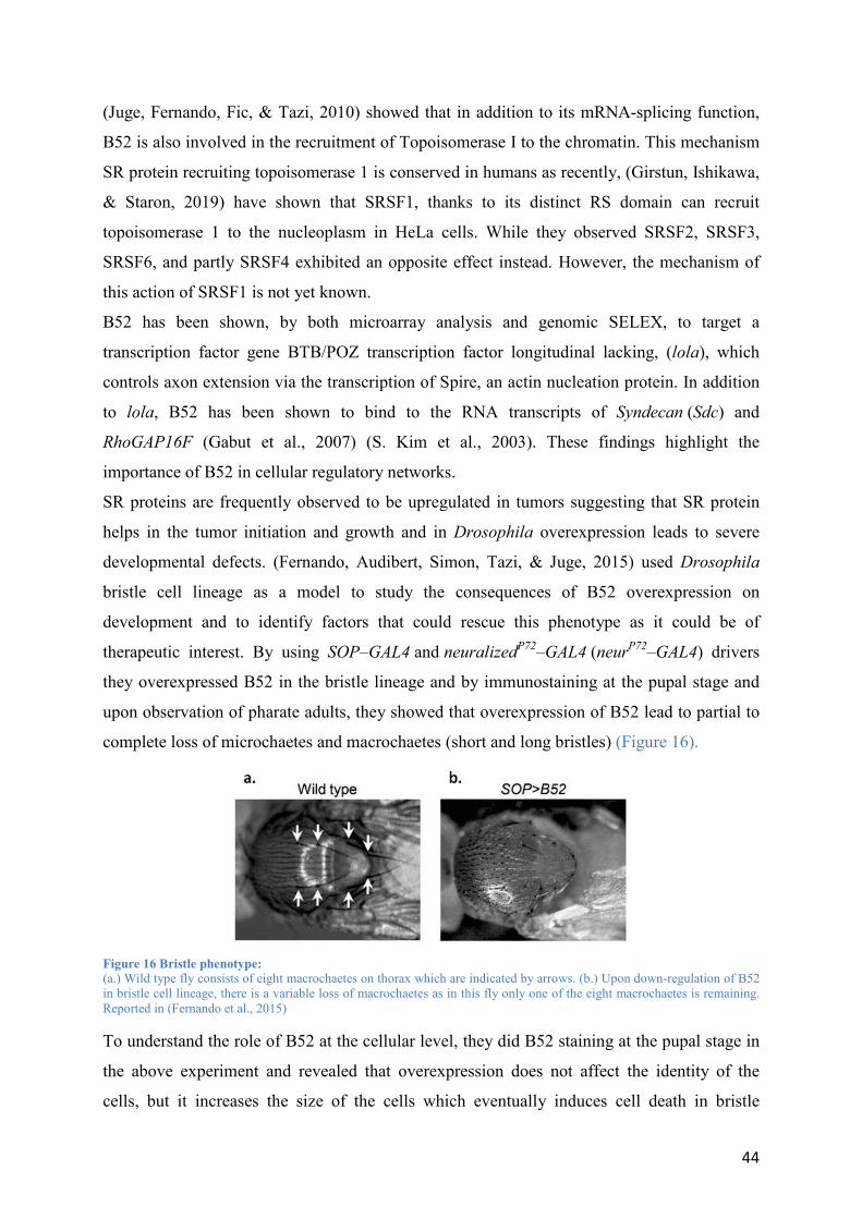
44
(Juge, Fernando, Fic, & Tazi, 2010) showed that in addition to its mRNA-splicing function,
B52 is also involved in the recruitment of Topoisomerase I to the chromatin. This mechanism
SR protein recruiting topoisomerase 1 is conserved in humans as recently, (Girstun, Ishikawa,
& Staron, 2019) have shown that SRSF1, thanks to its distinct RS domain can recruit
topoisomerase 1 to the nucleoplasm in HeLa cells. While they observed SRSF2, SRSF3,
SRSF6, and partly SRSF4 exhibited an opposite effect instead. However, the mechanism of
this action of SRSF1 is not yet known.
B52 has been shown, by both microarray analysis and genomic SELEX, to target a
transcription factor gene BTB/POZ transcription factor longitudinal lacking, (lola), which
controls axon extension via the transcription of Spire, an actin nucleation protein. In addition
to lola, B52 has been shown to bind to the RNA transcripts of Syndecan (Sdc) and
RhoGAP16F (Gabut et al., 2007) (S. Kim et al., 2003). These findings highlight the
importance of B52 in cellular regulatory networks.
SR proteins are frequently observed to be upregulated in tumors suggesting that SR protein
helps in the tumor initiation and growth and in Drosophila overexpression leads to severe
developmental defects. (Fernando, Audibert, Simon, Tazi, & Juge, 2015) used Drosophila
bristle cell lineage as a model to study the consequences of B52 overexpression on
development and to identify factors that could rescue this phenotype as it could be of
therapeutic interest. By using SOP–GAL4 and neuralizedP72
–GAL4 (neurP72
–GAL4) drivers
they overexpressed B52 in the bristle lineage and by immunostaining at the pupal stage and
upon observation of pharate adults, they showed that overexpression of B52 lead to partial to
complete loss of microchaetes and macrochaetes (short and long bristles) (Figure 16).
Figure 16 Bristle phenotype: (a.) Wild type fly consists of eight macrochaetes on thorax which are indicated by arrows. (b.) Upon down-regulation of B52 in bristle cell lineage, there is a variable loss of macrochaetes as in this fly only one of the eight macrochaetes is remaining. Reported in (Fernando et al., 2015)
To understand the role of B52 at the cellular level, they did B52 staining at the pupal stage in
the above experiment and revealed that overexpression does not affect the identity of the
cells, but it increases the size of the cells which eventually induces cell death in bristle

45
lineage. The authors further generated B52 homozygous mutant clones by somatic
recombination and revealed that B52 mutant clones were smaller and eventually gave rise to
smaller bristles in adults (Figure 17 b & c). They also observed that knockdown of B52 lead
to delay in the development of bristle lineage and by Immunostaining, they revealed that B52
knockdown decreases Myc expression (Figure 17 a).
Figure 17 Effect of B52 on growth: (a.) wildtype clone vs Increased cell size of clone overexpressing B52. (b.) Loss-of-function of B52 in the bristle lineage decreases cell growth (c.) bristle arising from wild-type clones vs from B52 homozygous mutants. Reported in (Fernando et al., 2015)
They also reported that B52 overexpression increased dMYC promoter activity measured by
dMYC-lacZ reporter activity and enrichment of phosphorylated Pol II across the dMYC
promoter. Finally, by using a genetic screen they identified several factors that could rescue
the phenotype induced by B52 overexpression including brain tumor (brat) which is a
negative regulator of growth (Frank, Edgar, & Roth, 2002) and lilliputian (lilli) which is a
positive regulator of growth (Wittwer, van der Straten, Keleman, Dickson, & Hafen,
2001)(Figure 18). The authors further showed that mechanistically an antagonism between
B52 and Brat exists as in B52 overexpression clones, overexpression of Brat reduces the
transcription of myc to rescue the overexpression phenotype. Besides revealing the role of
B52 and providing a mechanistic link between B52 and myc transcription this study opens a
new perspective of identifying proteins that could repress the ill-effects of SR protein
overexpression in mammals.

46
Figure 18 Rescue of B52: (a.) Overexpression of B52 in the eye (b.) Co-overexpression of B52 and Brat. Reported in (Fernando et al., 2015)
Recently, (Boyin Liu & Bossing, 2016) have shown the requirement of B52 in the splicing of
Choline acetyltransferase (ChAT
the neurotransmitter acetylcholine. Moreover, B52 has also been shown to regulate the mid-
day siesta in flies by regulating the splicing of period gene suggesting that this SR protein can
participate/contribute in natural sleep behavior as reported in a recent study by (Zhichao
Zhang, Cao, & Edery, 2018). Further studies will be needed to elucidate the complete B52
splicing program and provide an understanding of the role that B52 plays in growth and other
processes.
1.3.4 Additional RNA Binding Proteins that regulate slicing:
In addition to above described RBPs (SR and hnRNPs) that are ubiquitously expressed across
several tissues, several reports have described some tissue-specific RBPs such as RBFOX,
MBNL, PTBP2 (nPTB) and NOVA family proteins are essential regulators of tissue or cell-
specific splicing (X. D. Fu & Ares, 2015). NOVA proteins are the class of RBPs that are
specific to neuronal tissue, and by using CLIPseq techniques, it has been shown that NOVA
regulates AS in brain (Ule et al., 2003). Further CLIP seq analysis revealed the position-
dependent splicing program of NOVA, i.e., binding to downstream intron or upstream intron
promotes exon inclusion or exon skipping. Mechanistically, NOVA binds to an ESS and
blocks binding of U1 snRNP which inhibits the formation of E complex and promotes exon
skipping. While on the other hand, when NOVA binds to ISE downstream of an intron, it
promotes the formation of A complex and complexes that follow and thus it promotes exon
inclusion (Ule et al., 2006). Other RBPs such as RBFOX, MBNL, ESRP1/2 and Quaking too
facilitate a position-dependent splicing program. However, the underlying mechanism of the
position-dependent regulation is mostly unknown.
Additional RBPs such as RBM5 regulate the splicing by inhibiting the formation of B
complex in splicing assembly while RBM17 (or SPF45) inhibits splicing at the second

47
catalytic step during the splicing reaction (Lallena, Chalmers, Llamazares, Lamond, &
Valcárcel, 2002) (Bonnal et al., 2008).
The combinatorial regulation of AS program is also reported, as is the case of splicing of exon
6 of human Fas, which encodes for a type 1 transmembrane protein. TIA-1 binding to ISE in
intron 6 promotes the inclusion of exon 6 via U1 snRNP recruitment and produces an isoform
that promotes apoptosis while binding of another RBP, PTBP1 to an ESS in exon 6 to inhibits
the binding of U2AF65, leads to production of an isoform that lacks exon 6 and does not
promote apoptosis (Izquierdo et al., 2005).
Recent studies have estimated the number of proteins encoded by humans that are capable to
bind directly to RNA to be close to being in thousands (Castello et al., 2012) (Castello et al.,
2016). Thus the repertoire of RBPs that regulate splicing is vastly unmapped.
1.3.5 Other factors affecting Alternative splicing:
Secondary structure of RNA plays an essential role in defining interactive and functioning
properties of the RNA molecule. Even though RNA is a single-stranded molecule, but it can
fold back against itself to produce secondary and tertiary structures (Wan, Kertesz, Spitale,
Segal, & Chang, 2011). RNA secondary structures have been reported to interfere with the
interactions of splicing enhancers or silencers with proteins (Blencowe, 2000) (Smith &
Valcárcel, 2000). Splicing changes occur due to RNA secondary structure formation.
(Shepard & Hertel, 2008) suggested that 4 % of conserved alternative splicing are associated
with secondary structure. For an example, stem-loop structures affect the 5
SMN2’s exon 7 and exon 10 of Tau (N. N. Singh, Singh, & Androphy, 2007) (Hutton et al.,
1998). RNA secondary structures can even bring together distant RNA elements together. For
example, secondary structure in FGFR2 pre-mRNA brings together two elements so as to
activate exon IIIb (Baraniak, Lasda, Wagner, & Garcia-Blanco, 2003). In the Drosophila
Dscam gene, that can form up to 38 000 different isoforms alternative cassette selection
occurs after the formation of secondary structure between docking and selector sequences
surrounding alternative cassette exons. This mechanism ensures that only one of the many
alternative cassette exons is included in the mRNA (Graveley, 2005). In other instances,
intronic secondary structure elements may even function as riboswitches that bind to small
molecule metabolites and activate or repress splicing in response to the concentration of the
metabolite. (Cheah, Wachter, Sudarsan, & Breaker, 2007) Additionally, a substantial amount
of evidence has accumulated demonstrating that transcription rate and chromatin structure can
affect alternative splicing.

48
1.4 Quality control of splicing:
Although “Split gene” architecture in eukaryotes is clearly advantageous such as it expands
the coding capacity, but the same division of genes into exons and introns introduces a
requirement of an intricate splicing regulatory network that is composed of RNA regulatory
sequences, RNA protein complex, and splicing factors.
A ‘‘mistake’’ or ‘‘error’’ can be referred to the generation of any non-canonical molecular
product, which either result in a nonfunctional protein product, or which induces quality
control mechanisms to take action. To maintain the fidelity of gene expression cells evolved
several mRNA surveillance systems (also called as quality control systems) in both the
nucleus and the cytoplasm. These mRNA surveillance pathways detect not only the aberrant
transcripts that may lead to toxic protein but also regulate the wild-type mRNAs (Kervestin &
Jacobson, 2012). In the last decades, much advancement has been made to an understanding
of these mechanisms.
Nuclear mRNA surveillance mechanisms are mainly studied in Saccharomyces cerevisiae
(Garneau, Wilusz, & Wilusz, 2007). Before mRNAs are exported from nucleus to cytoplasm,
the aberrant synthesis of mRNAs (incorrect or defective splicing, cap structure, cleavage or
polyadenylation at 3’ end) or the retention of mRNAs in the nucleus leads to activate different
surveillance mechanisms. The nuclear retention of an mRNA that should be exported to the
cytoplasm leads to decay by nuclear surveillance systems (Vasudevan & Peltz, 2003). One of
the key components of nuclear surveillance is the nuclear exosome. The exosome is
distributed in both nucleus and cytoplasm. To promote RNA degradation, two 3’-5’
exonucleases interact with the core exosome. The nuclear exosome can lead the aberrant
transcripts to accumulate at the site of transcription for degradation (Torchet et al., 2002).
Cytoplasmic mRNA surveillance contains nonsense-mediated mRNA decay (NMD), no-go
mRNA decay (NGD), non-stop mRNA decay (NSD) )(Figure 19)
1.4.1 Nonsense-mediated decay (NMD)
One of the thoroughly-investigated RNA surveillance pathways is the Nonsense-mediated
decay (NMD) pathway. It was identified in S. cerevisiae but has been shown to exist in all
eukaryotes that have been investigated and is evolutionary conserved from yeast to humans
(Y. H. Chen et al., 2008) (Delhi, Queiroz, Inchaustegui, Carrington, & Clayton, 2011)
(Schweingruber, Rufener, Zünd, Yamashita, & Mühlemann, 2013) (He & Jacobson, 2015).
The nonsense-mediated mRNA decay (NMD) pathway is a cytoplasmic surveillance
mechanism that targets and degrades transcripts containing a PTC (premature termination

49
codon) (Leeds, Peltz, Jacobson, & Culbertson, 1991) (Peltz, Brown, & Jacobson, 1993). PTCs
may arise from several sources, including DNA mutations, inaccurate transcription leading to
frameshifts or single nucleotide nonsense mutations, or from alternative splicing that puts stop
codons “in frame” in an inappropriate position.
A multi-subunit complex known as EJC (Exon Junction Complex) is a critical element of
NMD. The EJC is essential for several cellular processes, such as nuclear export and
subcellular localization (Schell, Kulozik, & Hentze, 2002) as well as translation (Nott, Le
Hir, & Moore, 2004) (Wiegand, Lu, & Cullen, 2003). After splicing, the exon-exon junctions
formed are bound by EJC. During the pioneer round of translation, the ribosome displaces
each EJC as it moves along the mRNA. Translation terminates at the natural stop-codon,
which is typically located in the terminal exon and therefore does not have any downstream
bound EJC. However, if an mRNA contains a PTC within the 50 nucleotides or greater
upstream of the last exon-exon junction, the ribosome stalls at the PTC, giving it the chance
to interact with the downstream bound EJC. This triggers the NMD process, which leads to
degradation of the PTC-containing mRNA. When the ribosome recognizes a stop-codon
during translation, it recruits release factors, such as eRF1 and eRF3, which are associated
with the NMD-associated protein UPF1. Recognition of PTC leads to a stable interaction
between UPF1 and UPF2, bridging UPF1 with the EJC. This, in turn, leads to
phosphorylation of UPF1 and recruitment of either SMG6, an endonuclease that cleaves the
NMD targeted RNA, or the SMG5-SMG7 complex that is associated with uncapping and
deadenylation of RNAs. Either of these pathways leads to rapid degradation of the targeted
RNA.
Further, SRSF1 has been shown to stimulate NMD for mRNAs with PTC (Zuo Zhang &
Krainer, 2004) Over-expression of SRSF1 greatly enhanced NMD. This activity of SRSF1 is
independent of its shuttling ability but requires an intact RS domain. Recently the same
laboratory pinpointed the mechanism by which SRSF1 enhances NMD. By tethering SRSF1
to transcript at the various position, they obtained evidence that SRSF1 enhances UPF1
binding to mRNA in nucleus and the increased presence of UPF1 may accelerate the NMD
process (Aznarez et al., 2018).
The link between NMD and AS was first seen in a study of expressed sequence tags (ESTs)
where, (B. P. Lewis, Green, & Brenner, 2003) showed that several alternatively spliced genes
were found to have isoforms that would be predicted to be subject to NMD. These authors
termed this phenomenon regulated unproductive splicing and translations (RUST) and this
mechanism is conserved from humans to yeast (Soergel, Lareau, & Brenner, 2006). Some
50
splicing factors and all SR proteins autoregulate the level of their own expression through
alternative splicing coupled to NMD (AS-NMD or unproductive splicing) (Lareau, Inada,
Green, Wengrod, & Brenner, 2007) (Ni et al., 2007). For example, SRSF1 promotes inclusion
of an intron after the canonical stop codon, which marks it as a premature termination codon
(PTC) to elicit NMD of its own mRNA. These “unproductive splicing” events are highly
conserved between evolutionary kingdoms. For a detailed review of NMD and mechanism it
employs, one could read the following reviews by (Hug, Longman, & Cáceres, 2015),
(Lykke-Andersen & Jensen, 2015) (Brogna, McLeod, & Petric, 2016) and also (Popp &
Maquat, 2018).
The NMD pathway is involved in many biological processes, including cell proliferation and
growth (Weischenfeldt et al., 2008) (Avery et al., 2011) (Lou et al., 2014), embryonic
development and differentiation (Medghalchi, 2002) (Gong, Kim, Woeller, Tang, & Maquat,
2009) (McIlwain et al., 2010) (Bruno et al., 2011), stress response (Gardner, 2010; Sakaki et
al., 2012), innate immunity (Gloggnitzer et al., 2014), and neuronal activity (Colak, Ji, Porse,
& Jaffrey, 2013; Giorgi et al., 2007; He & Jacobson, 2015)
1.4.2 The No-go decay (NGD)
The No-go decay (NGD) is triggered when the elongation complex is stalled during
translation. The stalling could be due to the presence of rare codons or secondary structure or
poly-Lys or poly-Arg tracts, or frameshift sites in mRNA. It detects such mRNA and directs
them for endonucleolytic cleavage, and subsequently, the resulting mRNA fragments are
degraded by the exosome (which is a conserved multiprotein complex that degrades RNAs in
-to- - -
translational mRNA decay following the last translating ribosome) (Doma & Parker, 2006)
(Passos et al., 2009)(Pelechano, Wei, & Steinmetz, 2015).
1.4.3 Non-stop decay (NSD)
Non-stop decay (NSD) is an mRNA surveillance mechanism that identifies transcripts that
does not possess in-frame stop codons. These mRNAs could arise via premature
polyadenylation or point mutations that disrupt stop codon and interestingly are widespread
mistakes in the pathway of gene expression (Frischmeyer et al., 2002) (Vasudevan, Peltz, &
Wilusz, 2002) (Cui & Denis, 2003). Because mRNAs are short of a stop codon, it leads to
ribosomes to cross the poly (A) tail and remove the poly (A)-binding protein (PABP) and stall
at the 3’ end of the mRNA. The deed of the degradation is carried out by cytoplasmic
exosome and Ski complex (Ski2p, Ski3p, and Ski8p) and Ski7p. Ski7p detects the transcript

51
lacking stop codon and recruits the Ski complex and exosome. Once recruited, the ubiquitin-
proteasome pathway degrades the protein product generated from a non-stop transcript.
(Klauer & van Hoof, 2012) (Graille & Séraphin, 2012) (Parker, 2012)
Figure 19 The three primary mRNA surveillance mechanisms:Adapted from (Roy & Jacobson, 2013)

52
1.5 Alternative splicing in diseases and available therapy
Aberrant splicing is implicated in a high number of dysfunction and human diseases (R. K.
Singh & Cooper, 2012). AS has been implicated in nearly all aspects of cancer development,
and therefore, is a central participant in the disease (David & Manley, 2010) (Chabot &
Shkreta, 2016) (Scotti & Swanson, 2016). Alternative splicing is altered in a genome-wide
manner in cancer cells and is now considered a “novel hallmark of cancer” as every common
hallmark of cancer including metastasis is affected by dysregulation of splicing.
In their book, Genetics in medicine, (Nussbaum, Robert L; McInnes, Roderick R; Huntington,
2016) described three groups of splice defects that differ depending on their intronic or exonic
location. The first group consists of splice junction mutations such as mutations at the 5’
donor or 3’ acceptor sites or in the consensus sequences surrounding these donor or acceptor
sites. If the normal acceptor site is inactivated, then other intronic of exonic cryptic splice
sites may elicit a splicing reaction. The second group of splicing defects consists of
mutations in intronic cryptic sites that make it more favorable for splicing in comparison to
the normal splice site. These “activated” cryptic site mutations are often referred to as
“leaky” as the normal splice site is still active and produces normal gene product, thus leading
to a less severe phenotype. The third group of mutations consists of mutations in the ORF
that activate a cryptic splice site in an exon without altering the encoded amino acid (i.e.,
point mutations that become cryptic splice sites). It has been estimated that such cis splicing
mutations constitute 15%–60% of human disease-causing mutations (G. S. Wang & Cooper,
2007). Additionally, mutations disrupting trans-acting splicing regulators cause a wide
spectrum of diseases by globally compromising the splicing of many downstream target genes
(Osborne & Thornton, 2006).
Examples of disease caused by a point mutation in the exonic regulatory element:
SMA (Spinal muscular atrophy) is a neurodegenerative disease that is a leading genetic cause
of mortality in children (Arnold & Fischbeck, 2018). SMA is a result of deletions or
mutations in the survival of motor neuron 1 (SMN1) gene, which results in SMN protein
deficiency. However, humans have a centromeric copy of this gene called SMN2, which is
identical to SMN1 except for a single nucleotide difference in exon 7. Thus, SMN2 cannot
compensate for the loss of SMN1 as most mRNA produced from SMN2 lacks exon 7 (almost
90%). SMN2 thus encodes a truncated protein with reduced functionality. The single
nucleotide mutation on exon 7, generates the binding site for hnRNP A1 which inhibits exon7
inclusion (Figure 20). Reviewed in (Sunghee Cho et al., 2014)

53
Antisense oligonucleotides (ASOs) are a new class of drugs that are short (15–25 nt),
synthetic, single-stranded oligodeoxynucleotides that can bind to complementary target RNA
and thus function to alter, reduce, restore, or modify protein expression through several
distinct mechanisms. They can alter pre-mRNA splicing by sterically blocking splicing
factors, or they can block mRNA translation by preventing ribosome recruitment (C. F.
Bennett & Swayze, 2010) SMA is now treated by Nusinersen which is a 2'-OMe
phosphorothioate ASO that can target exon 7 which in SMN2 pre-mRNA. This facilitates
exon7’s inclusion and leads to the production of functional protein (Wertz & Sahin, 2016).
Finally, in 2016, FDA (U.S. Food and drug administration) approved Nusinersen as an ASO
to be used in SMA patients, and so far it remains as the only FDA approved the medication
for SMA (Aartsma-Rus, 2017).
Figure 20 SMA and its treatment by ASO:
Example of a disease caused by a mutation in the 5
Familial dysautonomia (FD) FD also referred to as Riley–Day syndrome, (hereditary
sensory and autonomic neuropathy type III) is a sporadic, autosomal recessive fatal disorder
caused by loss of function of the i-kappa-B kinase complex-associated protein (IKBKAP)
(Slaugenhaupt et al., 2002) (F. B. Axelrod & Kaufmann, 2014). In virtually all the FD
patients, 5’splice site of exon 20 carries a mutation. This mutation leads to skipping of the
exon 20 thus introducing a PTC. This is detected by mRNA surveillance, and truncated
protein is directed towards NMD for degradation. This further correlates with the neurological
dysfunction in FD patients(Close et al., 2006). Splicing modification therapy via small
molecule Kinetin (6-furfurylaminopurine) has been shown to increase wild-
type IKBKAP mRNA and IKAP protein and thus can be used to treat FD (Felicia B. Axelrod
et al., 2011).
Example of a disease caused by an intronic mutation that activates a cryptic splice site:
It has been shown that mutations in Lamin A lead to Hutchinson–Gilford progeria
syndrome (HGPS), a dominant disorder characterized by the dramatic, rapid appearance of
aging beginning in childhood due to translation of ageing associated protein progerin
(Eriksson et al., 2003)(De Sandre-Giovannoli et al., 2003). Most of the typical Hutchinson–

54
Gilford progeria cases are due to a recurrent, de novo point mutation in LMNA exon 11. This
mutation resides in a splicing enhancer sequence which as a result leads to activation of a
cryptic splicing site resulting in the truncated Lamin A protein.
The use of antisense oligonucleotides to sterically block the aberrant LMNA splicing site in
progerin production has been shown in vitro on human HGPS patient’s cells and in vivo in the
mouse model (Osorio et al., 2011).
Example of disease caused by a mutation in core components of the spliceosome:
Retinitis pigmentosa (RP) is inherited a degenerative retinal disorder that is caused by
germline mutations in core spliceosomal proteins. More than 3,000 mutations in about 80
genes have been identified to be associated with RP Reviewed by (M. M. Liu & Zack, 2013).
Moreover, mutations in RNU4ATAC gene which encodes U4atac, a small nuclear RNA that
is a crucial component of the minor spliceosome leads to Microcephalic osteodysplastic
primordial dwarfism type 1 (MOPD I). Reviewed in (Nagy et al., 2012)
Although there is currently no therapy for the treatment of RP, (Nakamura et al., 2017)
reported the discovery of a small drug-like molecule, called Photoregulin3, which alters the
activity of a transcription factor that regulates rod genes. So far, it has been successfully
tested in the mice model for RP. However, clinical trains in humans are underway.
1.5.1 Conclusion:
Breakthroughs in high-throughput sequencing technologies promise to identify and link many
more human mutations to disease. Moreover, small-molecule compounds, antisense
oligonucleotides, and genome-editing approaches hold promise for correcting the effects of
these mutations common in human genetic variation and disease. ASO especially have a high
potential in future therapy, for an example Eteplirsen, an ASO is now being used to treat
DMD, ASOs to treat ALS are already being investigated and show promising therapeutic
potential (C. A. Stein & Castanotto, 2017). (Le et al., 2019) have described the scope of
ASOs that can target angiogenic factors and act as therapeutic molecules for the treatment of
cancer. Small molecules such as a family of spliceostatins (derived from Pseudomonas) and
herboxidienes and pladienolides (derived from Streptomyces) have been shown to target core
spliceosome machinery and mediate anticancer properties. Reviewed in (J. C. Lin, 2018).
Further, a new strategy referred to as SMaRTTM (Spliceosome-mediated RNA trans-splicing
as a tool for gene therapy)is developed, it is a system used to correct a part of the mutant
transcript instead of full-length cDNA. (Puttaraju, Jamison, Mansfield, Garcia-Blanco, &

55
Mitchell, 1999). Recently (Dooley et al., 2018) have developed a Spliceosome-mediated RNA
trans-splicing strategy to edit and rescue the expression of CEP290 mRNA which is
associated with Leber congenital amaurosis type 10 (LCA10)( The visual impairment) and
Joubert Syndrome (JS) (a rare, autosomal recessive disorder characterized by episodes of
hyperpnea, developmental delays, ataxia, and abnormal eye movements).

56
1.6 Hippo pathway and its discovery:
Researchers around the globe have a long-standing quest to underpin the exact mechanism of
organ size control. Indeed it is quite intriguing to know what precise and complex set of
mechanisms control the cell number during development and regeneration. Many studies have
pointed towards the fact that growth to the right size and patterning to an exact shape is
intrinsic information that is in possession of the organ. For example, In the Salamander, when
eyes or limbs are exchanged between different species with different organ size, they reach a
size characteristic of the donor (Twitty & Schwind, 1931). A similar experiment in
Drosophila showed that immature imaginal discs, when transplanted in a growth-permissive
environment such as the abdomen of an adult female, grow at their normal rate until they
reach their final size (Bryant & Simpson, 1984).
During the course of development, an increase in cell number is required for the growth of the
organ and body size. During the normal function of an organ, processes such as continuous
cell turnover, apoptosis and occasional differentiation of adult stem cells to replace
dysfunctional cells are required to maintain the functionality of the organ. During an injury or
pathology, the process of healing accompanied by cell division and regeneration is required to
compensate for the lost cells and restore the standard functionality. Although the mechanism
of proliferation, apoptosis, regeneration are extensively studied, the coordinated between
these events are poorly understood to this date and mechanisms underlying these molecular
events are yet to be elucidated. However, the discovery and expansion of Hippo pathway have
proven to be an entry point for the researcher in a quest to answering this fundamental
question as Hippo signaling pathway has been shown to play a critical role in controlling
organ size by regulating both cell proliferation and apoptosis. (W. Kim & Jho, 2018).
As of now, the Hippo Pathway has the status of a highly evolutionary conserved regulator of
tissue growth and cell fate. Since its discovery, it has panned out as an integrator of signals
from a vast network of proteins and signaling pathways and serves as an essential regulator of
critical cellular processes such as organ size regulation, cell proliferation, and stem cell
regulation(Harvey, Zhang, & Thomas, 2013) (Figure 21).

57
Figure 21 Implications of the Hippo pathway in cell biology: The Hippo pathway modulates cell proliferation, differentiation, growth, and death. The coordination of different cellular processes by the Hippo pathway may contribute to various physiological and pathological conditions such as development, tissue homeostasis, and tumorigenesis.
Within the last 25 years, the field of the Hippo pathway has rapidly emerged as one of the
most popular pathways due to its widespread roles in processes vital to the development. Thus
it has been on the radar of researches around the globe in a race to identify its components,
regulators and downstream effectors. There are a plethora of reviews with new insights and
advancements in the field every year which are rapidly growing every year (Figure 22)
Figure 22 The rise of Hippo pathway:
The rising number of papers being published every year in the field of the Hippo Pathway.
The Hippo pathway transduces signals from the plasma membrane to the nucleus and
subsequently activates the transcription of genes involved in cell proliferation, anti-apoptosis

58
and cell survival (Chan, Lim, Chen, et al., 2011) (Meng, Moroishi, & Guan, 2016). The
Hippo pathway is activated in response to several upstream signals such as cell-cell contact
and ECM (Extracellular matrix) cytoskeleton etc. which are described in the forthcoming
sections. Both in Drosophila and Mammals, The Hippo pathway functions via three
interlinked parts, i.e. core kinase components, upstream regulatory elements and the
downstream effectors of the pathway.
1.7 Hippo Pathway in Drosophila
Several components of the Hippo pathway were discovered in Drosophila during a genetic
mosaic screening to look for mutants resulting in tissue overgrowth. First component of the
pathway to be found was NDR (Nuclear Dbf2-related) family protein kinase Warts (Wts) in
1995, (LATS1/2) by (Justice, Zilian, Woods, Noll, & Bryant, 1995)(Xu, Wang, Zhang,
Stewart, & Yu, 1995) followed by the WW domain-containing protein Salvador (Sav) in 2002
by (Tapon et al., 2002)(Kango-Singh, 2002). Later in 2003, Hippo (Hpo) (S. Wu, Huang,
Dong, & Pan, 2003) (Harvey, Pfleger, & Hariharan, 2003a) and in 2005 Mob-as tumor
suppressor (Mats) (Z. C. Lai et al., 2005), were identified and found to be essential tumor
suppressors, loss of function of any of these genes result in tissue overgrowth (Figure 23).
Figure 23 Hippo mutant phenotypes in flies:(A,B) Scanning electron micrographs of (A) a wild-type fly and (B) a fly with clones of cells homozygous mutant for hippo
that exhibit overgrowth of the adult cuticle Adapted from (G. Halder & Johnson, 2011)
The name of hippo gene was established based on its mutant adult head phenotype which
mirrors the creased hide of hippopotamus (Udan, Kango-Singh, Nolo, Tao, & Halder, 2003)

59
However, the Hippo Pathway field hit a significant milestone in 2005 when Yorkie (Yki),
was discovered in a yeast two-hybrid screen for Wts binding proteins (J. Huang, Wu, Barrera,
Matthews, & Pan, 2005). Biochemical and genetic studies revealed that Yki is required for
normal tissue growth and its activity is inhibited by Wts-mediated phosphorylation. Yki was
established as a central transcriptional regulator of the Hippo pathway as downregulation of
Yki lead to diminish the overgrowth phenotype caused by deleting upstream kinase Hpo or
Wts. Yki acts as an vital growth promoter, with loss of Yki resulting in a restriction of tissue
growth in Drosophila and overexpression resulted in overgrowth phenotypes that resembled
mutation in hpo, sav, mats, and wts (J. Huang et al., 2005). Although, it was found that CycE
and DIAP1 were transcriptional targets of Yki but the lack of DNA binding domain led to
investigate the transcription factors that could mediate Yki activity. It was 2008 when at least
three groups concluded that Scalloped (Sd) (TEAD family of transcription factors in
Mammals) as Yki DNA binding partner via yeast 2 hybrid experiments (Q. Zhang et al.,
2008)(S. Wu, Liu, Zheng, Dong, & Pan, 2008) (Goulev et al., 2008).
As shown in (Figure 24), In Drosophila When Hippo Pathway is in ON state, i.e., activated,
Hpo forms a complex with Sav. This complex phosphorylates Wts which is in complex with
its co-factor Mats. This phosphorylation activates Wts/Mats kinase which subsequently
phosphorylates Yki as three separate phosphorylation sites, S111, S168 and S250.
Phosphorylation of Yki at S168 site leads to its cytoplasmic retention as it binds to 14-3-3,
therefore, inhibiting its transcriptional activation role (H. Oh & Irvine, 2009)(H. Oh & Irvine,
2008) Recently, Misshapen (Msn) and Ste-20 kinase Happyhour (Hppy) kinases were shown
to act in parallel to Hpo to phosphorylate Wts (Q. Li et al., 2014)(Meng et al., 2015)(Pan et
al., 2015). Two kinases Tao and Par-1 directly regulate the activity of Hpo via
phosphorylation. Tao has been shown to phosphorylate the activation loop of Hpo to promote
its activity (Boggiano, Vanderzalm, & Fehon, 2011) (Poon, Lin, Zhang, & Harvey, 2011).
While on the other hand, polarity kinase Par-1 restricts Hpo by promoting its phosphorylation
at a different site to restrict its activity. Further, Par-1 inhibits the interaction between Hpo
and Sav to promote Sav dephosphorylation and destabilization, further downregulating Hippo
pathway activity (H. L. Huang et al., 2013)
Recently, (X. Sun et al., 2019) showed that deubiquitinase Usp7 positively regulates Yki
transcriptional program by deubiquitinating Yki. Further, they showed that Hpo or warts
inhibit the Usp7-Yki interaction not only to promote Yki’s nuclear accumulation but also
to promote Yki nuclear degradation. This study provides new insight into how the Hippo
pathway negatively regulates the activity of Yki.

60
Figure 24 Schematics of the Hippo pathway in Drosophila melanogaster:
In contrast, when the Hippo pathway is in OFF state, i.e. inactivated, hypophosphorylated
Yki gets translocated to the nucleus, it competes with Tondu domain-containing growth
inhibitor (Tgi) to bind with transcription factor Scalloped (Sd) to regulate their transcriptional
coactivator activity on several target genes (Figure 24). According to (Guo et al., 2013) Sd-
Binding-Protein (SdBP) (SdBP here is Tgi) is a negative regulator of Sd-Yki complex. They
show that Tgi simultaneously binds to Sd through TDU domains and Yki via PPxY motifs to
disrupt the direct Sd-Yki complex and proceeds to form a transcriptionally inactive ternary
complex which results in inhibition of Yki activity. They further show a competition between
Tgi and Yki in binding to Sd. On the other hand, (Koontz et al., 2013) reported that Tgi binds
to Sd in the absence of Yki and repress gene expression. When Yki is in the nucleus, it
displaces the Tgi and converts Sd into a transcriptional activator.
Besides Sd, the transcription factor Homothorax (Hth) a TALE-homeodomain protein (the
homolog of the MEIS homeodomain protein) and zinc finger transcription factor Teashirt
(Tsh) has been shown to be a transcription factor of Yki in the eye disc that regulates
microRNA bantam to promote proliferation and protect eye progenitor cells from apoptosis
(Peng, Slattery, & Mann, 2009). The expression of bantam is also regulated by another
transcriptional complex between Yki and Mad, an effector of Dpp signaling (Hyangyee Oh &

61
Irvine, 2011). Yki interaction with different binding partners might, therefore, be tissue-
dependent, and the availability of Yki partners could be a critical factor in regulating Hippo
pathway activity.
The relationship between transcription and chromatin occurs at two levels. First, the
nucleosome is regulated by ATP-dependent chromatin remodeling complex such as SWI/SNF
family complexes mobilize nucleosomes, facilitating the access of the transcription apparatus
to DNA. Secondly, the histone-modifying enzymes that add or remove chemical groups on
histones leading to compaction of DNA, which prevents or enhances binding of
transcriptional factors to DNA. Indeed Yki, has been shown to interact with GAGA factors
(which influences the chromatin structure and) and Brahma complex (one of the Drosophila
SWI/SNF complexes) and Nuclear receptor coactivator 6 (Ncoa6), a subunit of the Trithorax-
related (Trr) histone H3 lysine 4 (H3K4) methyltransferase complex to regulate the
transcription (Y. Jin et al., 2013)(Hyangyee Oh et al., 2013) (Qing et al., 2014)
Several target genes of Yki are cycE, the inhibitor of apoptosis diap1, myc, miRNA bantam
which collectively drive cell proliferation, inhibition of apoptosis and survival (J. Huang et
al., 2005)(Tapon et al., 2002)(Thompson & Cohen, 2006)(Udan et al., 2003)(Q. Zhang et al.,
2008)(Ziosi et al., 2010). Another interesting example for the last class of genes is the recent
identification of Cactus (Cact),an effector of Toll pathway as a direct target of Yki. Cact is
characterized by the presence of ankyrin repeats and functions to suppress the transcription of
antimicrobial peptides introduced by gram-positive bacterial infection. This discovery further
expands the regulation of which revealed an intriguing link between the Hippo pathway and
innate immunity(Bo Liu et al., 2016).
Therefore, Yki, the effector of the Hippo pathway functions to promote growth and
proliferation whereas the kinase of the pathway acts as tumor suppressors as they function to
suppress the activity of the Yki.
The downstream target genes of the Hippo pathway are essential in the pathway feedback. For
instance, Ex, a cytoskeletal protein for cell morphology, is not only a transcriptional target of
Hippo pathway but also an upstream regulator of the pathway, therefore forming a negative
feedback loop in growth control regulation (Jukam et al., 2013).
1.7.1 Upstream regulators of Drosophila Hippo Pathway:
Core components of the Hippo pathway have been studied extensively to this date. Unlike
typical signaling cascades, the Hippo pathway is not regulated by a defined set of ligand-
receptor interactions, but it is rather modulated by a diverse set of signals from upstream

62
constituents. However, what precise signals activate the kinase cascade to suppress the
activity of transcriptional co-activators is not yet completely understood. Thus, several
researchers were focused on to elucidate the regulators of the Hippo pathway. So far, The
upstream signals that are known to regulate the Hippo pathway such as transmembrane
receptors atypical cadherins Fat(Ft) and Dachous1/2 (Dchs), Kibra-Expanded/Merlin complex
and apicobasal polarity complex, i.e., members of crumbs polarity complex play roles in
maintaining the cell architecture such as cell polarity and cytoskeleton. The expansion of such
knowledge can provide insight into what signals direct cells to adhere to a pattern of growth
and when to inhibit the growth (Figure 25).
Figure 25 Upstream regulators of Hippo pathway in Drosophila:
It is regulated by various factors including the following aPKC, atypical protein kinase C; Crb, Crumbs; D, Dachs; Dlg, Discslarge; Ds, Dachsous; Ed, Echinoid; Ex, Expanded; Ft, Fat; Hth, Homothorax; Jub, Ajuba LIM protein; Lgl, Lethal giant larvae; Mad, Mothers against decapentaplegic (Dpp); Mer, Merlin; Scrib, Scribble; Sd, Scalloped; Tgi, Tondu domain-containing growth inhibitor; Tsh, Teashirt; Zyx, Zyxin. Growth-promoting proteins are depicted in brown and growth repressors in green. These proteins are described in text. AJ, adherens junction; SJ, septate junction. Adapted from (Irvine & Harvey, 2015)

63
1.7.1.1 Regulation by Fat: Planar cell polarity
The Fat-Dachsous signaling pathway is a cell-contact-dependent mechanism that regulates the
Hippo pathway. Fat (Ft) large protocadherin (transmembrane protein) with a molecular
weight of 560kDa (P. A. Mahoney et al., 1991). Ft is the receptor for Hippo Signaling
pathway as it genetically interacts with the pathway (F. C. Bennett & Harvey, 2006) (E. Cho
et al., 2006) (Silva, Tsatskis, Gardano, Tapon, & McNeill, 2006) (Willecke et al., 2006).
Dachsous (Ds) another protocadherin acts as the ligand for Ft (Clark et al., 1995) Fat
signaling pathway has a well-characterized role in regulating planar cell polarity (PCP) in
epithelial tissue (Reddy & Irvine, 2008). Although, PCP itself does not regulate the Hippo
pathway, but loss of Fat leads to overgrowth phenotype and up-regulation of Hippo pathway
target genes such as cycE and diap1, as has been shown for multiple Hippo pathway
components. Mechanistic studies have shown that Fat signaling can positively regulate the
Hippo pathway via localizing Ex, an upstream regulator of the pathway to apical junction
region (F. C. Bennett & Harvey, 2006) (E. Cho et al., 2006) (Silva et al., 2006) (Willecke et
al., 2006). Another mechanism of positive regulation of Hippo signaling by fat signaling is
via inhibition of Wts degradation thus promoting abundance of Wts mediated by the atypical
myosin Dachs (E. Cho et al., 2006) (Rodrigues-Campos & Thompson, 2014) (Rauskolb, Pan,
Reddy, Oh, & Irvine, 2011) (Bosch et al., 2014) In addition, (Vrabioiu & Struhl, 2015)
showed that Fat signaling acts via Dachs to inhibit Wts from acquiring Mats-dependent active
conformation to promote growth in Drosophila wing.
(Misra & Irvine, 2016) identified Vamana (Vam) to influence Dachs membrane localization
and most recently same authors described Early girl as an essential component of Fat
signaling that the levels of localization of Daschs and Vamana (Misra & Irvine, 2019).
1.7.1.2 Regulation by Ex/Mer/Kibra:
The FERM (N-Terminal Globular Domain [Band Four-point-one, Ezrin, Radixin and
Moesin]) domain-containing protein Ex and Merlin (Mer) along with the C2 and WW
domain-containing protein Kibra (Kib) are three important apical membrane proteins of the
Hippo pathway that regulate Yki activity through the core kinase cascade (Baumgartner,
Poernbacher, Buser, Hafen, & Stocker, 2010; Genevet, Wehr, Brain, Thompson, & Tapon,
2010a; Hamaratoglu et al., 2006; McCartney, Kulikauskas, LaJeunesse, & Fehon, 2000; J. Yu
et al., 2010) The general role of FERM domain proteins is to link the F-actin cytoskeleton to
cell membranes and membrane proteins as described by (Mangeat, Roy, & Martin, 1999).

64
Ex and Mer have been shown to physically interact with each other and loss of function of
any of the two genes leads to overgrowth phenotype and deletion of Ex and Mer together
results into even severe overgrowth phenotype due to upregulation of Hippo pathway target
genes as they interact with hippo pathway components (McCartney et al., 2000)
(Hamaratoglu et al., 2006). Similar to ex and mer, kib mutations too leads to overgrowth
phenotype and up-regulation of Hippo pathway targets. Kib protein physically associates with
both Mer and Ex and the three of them can co-localize at the apical junctional region,
suggesting that these three proteins can function together to regulate the Hippo pathway.
Interestingly Ex, Mer, and Kib can each function independently of each other in the regulation
of the Hippo pathway as revealed by the genetic analysis studies (Hamaratoglu et al., 2006);
(Baumgartner et al., 2010) (J. Yu et al., 2010). Studies have revealed that Ex, Mer, and Kib
regulate the Hippo pathway positively by recruiting the pathway components to specific
cortical domains to promote their activation. (Yin et al., 2013) demonstrated that Mer could
also directly recruit Wts to the plasma membrane to activate Hippo signaling, independently
of Hpo kinase activity. Generally, Ex can function as a scaffold that recruits Wts to the apical
junctional region from its inhibitor Jub where it interacts Hpo and gets activated (S. Sun,
Reddy, & Irvine, 2015). (Su, Ludwig, Xu, & Fehon, 2017) have demonstrated that Mer and
Kib can activate Hippo signaling in parallel to Ex at a spatially distinct cellular domain, the
apical cortex. Together, Ex, Mer, and Kibra may activate Hippo pathway at distinct sub-
cellular locations, possibly mediating different inputs of the Hippo pathway. Alternatively, Ex
can also regulate the pathway by physically binding with Yki and sequestering it outside of
the nucleus acting independently of kinase cascade(Badouel et al., 2009). In addition to acting
as upstream regulators of the Hippo pathway, Ex, Mer, and Kib are transcriptional targets of
the Hippo pathway too (Hamaratoglu et al., 2006) (Genevet, Wehr, Brain, Thompson, &
Tapon, 2010b). This suggests the existence of a negative feedback loop in which Yki
activation turns on the expression of positive regulators of the Hippo pathway (Boggiano &
Fehon, 2012) (Irvine & Harvey, 2015)
1.7.1.3 Regulation by Apical-basal polarity proteins:
Apical polarity proteins are located in the apical cell cortex. They have been shown to
regulate the Hippo pathway. Crumbs (Crb) was the first apical polarity protein found to be
involved in Hippo pathway regulation. In Drosophila embryos, Crb is localized at the
subapical plasma membrane and plays a vital role in organizing apical-basal polarity (Tepass,
Theres, & Knust, 1990). Both loss of Crb and constitutive expression leads to Yki activation,

65
up-regulated Hippo target genes, and overgrowth. Crb has been shown to interact with Ex via
its FERM domain and recruits Ex to the apical junctional (AJs) region to promote Hippo
pathway activity (C.-L. Chen et al., 2010) (Ling et al., 2010) (Robinson, Huang, Hong, &
Moberg, 2010) (Grzeschik, Parsons, Allott, Harvey, & Richardson, 2010). On the other hand,
it has also been demonstrated that Crb promotes ubiquitin-mediated degradation of Ex via E3
ubiquitin ligase F- -TrCP (Slmb) suggesting that Crb allows precise tuning
of Hippo pathway activity through its dual regulation of Ex (Ribeiro, Holder, Frith, Snijders,
& Tapon, 2014).
The Par complex consists of atypical protein kinase C (aPKC), its regulatory subunit Par6, the
Rho-GTPase Cdc42, and the scaffolding protein Bazooka (Baz; Drosophila homolog of the C.
elegans and mammalian Par3). aPKC is involved in the regulation of the Hippo pathway.
Expression of a dominant-negative aPKC transgene suppresses Hippo pathway inactivation in
Drosophila lethal giant larvae (lgl) clones (Grzeschik et al., 2010). Overexpression of aPKC
can cause mislocalization of Hpo, activation of Yki, and up-regulation of Hippo pathway
target genes (Grzeschik et al., 2010) (G. Sun & Irvine, 2011)
Lethal giant larvae (Lgl), Discs large (Dlg), and Scribble (Scrib) are the basolateral polarity
protein genes which were first described as “neoplastic” tumor-suppressors as depletion of
any of these genes in resulted in multilayered and invasive tumors in Drosophila imaginal
discs (Bilder, 2004). As studies linked overgrowth phenotype to over-activation of Yki, these
three basolateral polarity proteins are regarded as positive regulators of the Hippo pathway
(Richardson & Portela, 2017)(Enomoto & Igaki, 2011).
Loss of Lgl in the eye of Drosophila does not disrupt cell polarity or activate Jun kinase
signaling but mislocalizes Hpo, suggesting Lgl participates in localization of Hpo
(Bogoyevitch, Ngoei, Zhao, Yeap, & Ng, 2010) (Grzeschik et al., 2010). While on the other
hand, loss of Lgl via RNAi in wing imaginal disc, disrupts cell polarity and causes activation
of JNK signaling leading to Yki activation suggesting the regulation of Yki activity via Lgl is
context dependent (G. Sun & Irvine, 2011). Further, the same authors suggested that JNK
activates the Wts inhibitor Jub via phosphorylation providing a link between basolateral
polarity, JNK signaling and Yki activity (G. Sun & Irvine, 2013).
scrib mutant clones are eliminated by cell competition, a phenomenon of removal of “weak
cells” by surrounding “fit cells” (Grzeschik et al., 2010) (C.-L. Chen, Schroeder, Kango-
Singh, Tao, & Halder, 2012).Further,(Grzeschik et al., 2010) showed that loss of Scrib via
RNAi leads to down-regulation of Hippo pathway target diap1 suggesting that Yki is
inactivated in scrib mutatnt clones. However when (C.-L. Chen et al., 2012), blocked JNK

66
signaling in scrib mutant clones they observed Yki activation and a severe overgrowth
phenotype, suggesting JNK signaling is responsible for repressing Yki activity in scrib clones
and this effect outweighs the activation of Yki by loss of Scrib. Although, the mechanism of
repression of Yki by JNK signaling in this scenario is unknown, (Enomoto & Igaki, 2011)
suggests that JNK can inhibit Yki activity via Wts.
1.7.1.4 Cell-Cell adhesion and junctional proteins
Cell-cell adhesion and junctional proteins such as Ajuba LIM protein (Jub), Zyxin, and
Echinoid regulate the Hippo pathway in Drosophila. Jub is localized at the apical junctions
stabilizes preassembled cadherin complexes (Nola et al., 2011). Down-regulation of Jub
resembles Yki down-regulation phenotypes. For example, it leads to down-regulation of Jub
Hippo pathway target genes such as diap1 and cycE resulting in tissue undergrowth. Also, jub
has been shown to genetically interact with the Hippo pathway components (Das Thakur et
al., 2010). (Rauskolb, Sun, Sun, Pan, & Irvine, 2014) showed that Jub sequesters and inhibits
Wts at the adherens junctions (AJs) to inhibit hippo pathway activity.
Zyxin (Zyx) which is another LIM domain protein, acts as a negative regulator of the Hippo
pathway as it is required for the activity of Yki (Rauskolb et al., 2011) (Gaspar, Holder,
Aerne, Janody, & Tapon, 2015). Depletion of Zyx leads to reduced Yorkie activity and
undergrowth. (Rauskolb et al., 2011) also reported that at apical junctional region Zyx
cooperates with Dachs to promote Wts degradation thus inhibiting Hippo pathway activity
linking Zyx to Fat signaling branch of Hippo pathway regulation. (Gaspar et al., 2015)
demonstrated that Yki activity and growth promoting ability of Zyx depends upon its
interaction with the actin-associated protein Enabled (Ena). More importantly, Ena promotes
F-actin polymerization linking Zyx with F-acitn branch of the Hippo pathway. (Hirata,
Tatsumi, & Sokabe, 2008) showed that Zyx is associated with mechanical-force-dependent
facilitation of actin polymerization at cell adhesive structures; thus this group was the first to
hint at the possibility of mechanical cues regulating the Hippo pathway.
Echinoid (Ed) is an upstream regulator that is required for proper sub-apical localization of
Sav (T. Yue, Tian, & Jiang, 2012). Ed physically interacts with Ex, Yki, Hpo, Kibra, and
Mer. However, Ed only affects Sav localization without affecting Ex or Mer localization.
Depletion of Ed activates Yki, and subsequently upregulating Hippo pathway target gene
expression and tissue overgrowth. Its demonstrated in Drosophila that Ed physically interacts
and stabilizes and therefore inactivates Yki activity (T. Yue et al., 2012). Echinoid (Ed) is a
known receptor of the EGFR pathway (Bai et al., 2001).
67
The upstream regulators of the Hippo pathway include the actin cytoskeleton beneath the
plasma membrane. Indeed, cytoskeleton network F-actin has been demonstrated to be a
negative regulator of the Hippo pathway. Ectopic accumulation of F-actin caused by
Depletion of Capping proteins Capulet (an actin-binding protein that inhibits polymerization),
or constitutive expression of an activated version Diaphanous Dia; induces actin
polymerization resulted in increased Yki activity and quite a substantial overgrowth
(Fernandez et al., 2011) (Sansores-Garcia et al., 2011). It has also been reported that
mutations in Hippo pathway genes ex, mer, hpo, sav, wts, and mats leads to an increased F-
actin accumulation. This could mean that F-actin and Hippo pathway can mutually suppress
each other suggesting that in normal conditions, levels of F-actin levels sustain the Hippo
pathway as the activation of the Hippo pathway will suppress its negative regulator F-actin.
However, regulation of the Hippo pathway by F-actin remain poorly understood to this date
especially in Drosophila. Further, it has been observed that tension regulates the Hippo
pathway in Drosophila.
Spectrin is a vital cytoskeleton protein that supports the integrity of cell
structures. Drosophila - Heavy- -
-Spec is localized along the apical- - H-Spec is
localized at the basolateral and apical membrane, respectively. Spectrin forms hexagonal
networks at the cell cortex in all animal cells and has been reported to have mechanosensory
properties. Spectrin functions in parallel to the Jub-Wts pathway and loss of spectrin result in
Yki activation due to increased tension mediated by activation of the actomyosin network
(Deng et al., 2015) (Fletcher, Lucas, Brain, Tournier, & Thompson, 2012).This indicates that
Spectrin can relay mechanical cues to the Hippo pathway.
In their review (Hariharan, 2015) further postulated on how tension can regulate the pathway.
During growth Drosophila imaginal discs contains high levels of growth promoting
morphogens in the center. One could imagine that due to this, higher growth proliferation
would be focussed at the center but on the contrary uniform growth is observed in the disc,
suggesting that levels of morphogen alone do not dictate the growth in the wings. As the
growth takes place in the center of growing tissue, it also stretches the periphery. The
resulting tension could activate Yki to promote growth. Thus, Yki activation at the periphery
could compensate for non-uniform morphogen levels across tissues and promote uniform
tissue growth (Hariharan, 2015) (Aegerter-Wilmsen, Aegerter, Hafen, & Basler, 2007)
(Aegerter-Wilmsen et al., 2012). Another study showed that artificially increased growth in
clones of wing imaginal discs reduces cytoskeletal tension, which in turn decreases the

68
activation of Yki. This study suggests mechanical feedback in which increased Yki activation
and cell proliferation reduces tension and deactivates Yki. Such feedback could contribute to
evenly distributed growth (Y. Pan, Heemskerk, Ibar, Shraiman, & Irvine, 2016)
A hippo signaling pathway is highly conserved between Drosophila and Humans. In order to
ease further understanding, I am at this moment, giving a table to highlight the core
components of the Hippo pathway in Drosophila and their homologs in Mammalian Pathway.
Table 1 : Conservation of Hpo pathway core components between Drosophila and mammals.
Drosophila Melanogaster proteins Human proteins
Core components Hippo (Hpo) Serine/theonine kinase 4/3 MST1/MST2 or
STK4/STK3Salvador (Sav) Salvador (SAV1 or WW45)
Warts (Wts) Large tumor suppressor Kinase 1/2 (LATS1/2)MOB as tumor supressors (Mats) Mps one binder kinase activator-like 1A/1B
(MOB1A/MOB1B or collectively MOB1)
Yorkie (Yki) Yes-associated protein (YAP), Transcriptional co-activator with PDZ binding motif (TAZ) or (WWTR1)
Upstream ModulatorsFat (Fat) Angiomotin AMOTExpanded (Ex) Ferm domain-containing protein 6 (FRMD6)
Merlin (Mer) Neurofibromin (NF2)Kibra (Kib) Kibra (KBR)
Crumbs (Crb) CRB1–CRB3Ras association family member (Rassf) Ras association domain-containing protein 1–6
(RASSF1-6)
Scrib SCRIBMask MASK1,MASK2
Wbp2 WBP2Dlg DLG1–DLG4Lgl LGL1, LGL2
Jub Ajuba, LIMD1, WTIPDownstream mediatorsScalloped (Sd) TEA domain family member 1/2/3/4
(TEAD1,TEAD2,TEAD3,TEAD4)Tondu-domain-Containing growth inhibitor (Tgi)
Transcription cofactor vestigial-like protein 4 (VGLL4)
Homothorax (Hth) Meis1Tsh --------Mad Mothers against DPP (MAD).

69
1.8 Hippo Pathway in Mammals
Several components of the mammalian Hippo pathway were known prior to the formal
discovery of the Hippo pathway in Drosophila. For example, before MOB1 was recognized
as a tumor suppressor in the eukaryotes, it was known in yeast to be essential for mitosis and
maintenance of the ploidy (Luca & Winey, 1998). Similarly, before the identification of
MST1 as a tumor suppressor, in yeast, it was known to be activated by caspases and mediate
cell death. Mst1 codes for the mammalian Ste20 kinase and its interaction with MAPK, p38,
MKK6, MK7, and SAPK were also reported (Creasy & Chernoff, 1995). The Lats1 kinase
was known as NDR kinase in yeast cells and was known to function in cell division and
morphology maintenance (T. Millward, Cron, & Hemmings, 1995) (T. A. Millward,
Heizmann, Schäfer, & Hammings, 1998). However, the role of these proteins in the Hippo
pathway was unknown. Soon after the Hippo pathway was discovered in Drosophila, studies
in mammals lead to the identification of orthologs of fly Hippo pathway components leading
to the establishment of the mammalian Hippo pathway soon after. In 2003, MST was
identified as a core kinase of the Hippo pathway in mammals (Harvey, Pfleger, & Hariharan,
2003b) (S. Wu et al., 2003). By 2008 when (Hao, Chun, Cheung, Rashidi, & Yang, 2008) (B.
Zhao et al., 2007) showed that LATS1/2 Kinase negatively regulates YAP/TAZ core of Hippo
pathway was considered to be complete in mammals.
Figure 26 (A) Effect on growth due to MST knockout : (A) A mouse liver at two months of age from a wild-type animal and a liver at two months of age from a mouse mutant in which both Mst1/2 have been inactivated. The double null Mst1/2 mutant liver is overgrown owing to an increase in cell numbers. Adapted from (G. Halder & Johnson, 2011)
The mammalian components of the Hippo kinase cascade consist of The MST1 and MST2
kinases, adapter protein SAV1, LATS1/2 and its protein Msp-one-binder (MOB1) and
together they regulate the activity of transcriptional co-activators YAP/TAZ (Yki orthologs).
YAP and TAZ are homologs of the Drosophila Hippo pathway effector Yki. In ON state, i.e.,

70
when the Hippo pathway is active, SAV1 interacts with MST1/2 to form a complex in order
to stabilize MST1/2 which leads to autophosphorylation and thus activation of MST1/2
(Callus, Verhagen, & Vaux, 2006)(Figure 27). LATS kinase which also exists in two isoforms
LATS1 and LATS2 respectively interacts with adapter protein MOB1 to form a complex.
This complex is eventually phosphorylated by phosphorylation of serine 909/872 within the
LATS1/2 activation loop and the hydrophobic motif (LATS1 T1079 and LATS T1041).
LATS1/2-MOB1 further phosphorylates transcriptional coactivator Yes-associated protein
(YAP; homologous to Yki in Drosophila) and TAZ (transcriptional coactivator with PDZ-
binding motif, WWTR1). This phosphorylation takes place on 4 and 5 HXRXXS motifs of
TAZ and YAP respectively. Once phosphorylated, YAP/TAZ interacts with 14- 3-3 protein
gets sequestered in the cytoplasm or degraded by ubiquitination-dependent proteasomal
Phosphorylation of YAP at Ser381 and TAZ at Ser311 has been linked to regulation of
YAP/TAZ protein stability, as it primes subsequent phosphorylation in a phosphodegron by
-TrCP E3
ligase, leading to ubiquitination and degradation (C. Y. Liu et al., 2010) (B. Zhao, Li,
Tumaneng, Wang, & Guan, 2010a) (Schäfer et al., 2005)(Dong et al., 2007)(Hirabayashi et
al., 2008)(Lei et al., 2008)(Oka, Mazack, & Sudol, 2008)(Siew et al., 2008). Many
components of the Hippo pathway have already been shown to be degraded by ubiquitination
such as YAP, LATS1/2; Expanded(Ex) (B. Zhao, Li, Tumaneng, Wang, & Guan, 2010b) (X.
Yang et al., 2011)(Salah, Melino, & Aqeilan, 2011)(B. Ma et al., 2015) (W. Li et al., 2014)
(X. Ma, Richardson, Xue, Xu, & Guo, 2018).
Similar to Yki, YAP/TAZ transcriptional activators lack the DNA binding domain; thus
inside nucleus they interact with transcription factors (Such as TEAD1-4) via its TEAD
binding domain to be bought to the gene promoter(B. Zhao et al., 2008)(H. Zhang et al.,
2009)(Chan et al., 2009). So, In the scenario when Hippo pathway is OFF, the un-
phosphorylated YAP/TAZ gets translocated to the nucleus where it competes with VGLL4
(Vestigial Like Family Member 4; homologous to Tgi in Drosophila) to interact with TEAD
transcription factor so as to induce the transcriptional activation of several sets of gene which
play crucial role in cell growth, differentiation invasion/migration and survival. Reviewed by
(Meng et al., 2016). The C-t -
-helices on one side, while the N-terminal domain of YAP
wraps around TEAD, forming extensive interactions (L. Chen et al., 2010)(Z. Li et al., 2010)
(W. Tian, Tomchick, Luo, Pan, & Yu, 2010)

71
Figure 27 Mammalian Hippo Pathway: Hippo pathway is highly conserved from Drosophila Melanogaster to human, schematic representing ON and OFF state the pathway in mammals
YAP interacts with promoters and distal enhancers to regulate target gene expression, through
mechanisms of modulating chromatin looping and Pol II pausing release(Lian et al., 2010)
(Galli, Carrara, et al., 2015) (Zanconato et al., 2015). YAP/TAZ associate with a plethora of
transcription factors in addition to TEAD1-4 including YAP binding partners SMAD1/2/3/7
(Ferrigno et al., 2002) (Kurisaki, Kose, Yoneda, Heldin, & Moustakas, 2001) (Alarcón et al.,
2009) (Varelas et al., 2008), Runt-Related Transcription Factor (RUNX) (Zaidi et al., 2004)
Erb-B2 Receptor Tyrosine Kinase 4 (ErbB4) (Komuro, Nagai, Navin, & Sudol, 2003), p73
(Strano et al., 2001) and TAZ binding partners RUNX, Peroxisome ProliferatorActivated
(J. H. Hong et al., 2005), Paired Box 3 (Pax3) (Masao Murakami et al.,
2006), T-Box Transcription Factor 5 (TBX5) (M. Murakami, Nakagawa, Olson, &
Nakagawa, 2005) and Thyroid Transcription Factor 1 (TTF-1) (K. S. Park et al., 2004)
Further, It has been shown that the SWI/SNF (a chromatin remodeling protein complex) and
(or) NCOA6 (a histone methyltransferase protein) is recruited by YAP to stimulate TEAD
activity and subsequently act as co-activators of gene expression(Qing et al., 2014a)
(Skibinski et al., 2014).

72
On the other hand, the YAP/TEAD can also act as repressors by engaging deacetylase
complexes (Valencia-Sama et al., 2015) (M. Kim, Kim, Johnson, & Lim, 2015). Multiple
transcriptional downstream target genes of YAP/TAZ has been identified, such as connective
tissue growth factor (CTGF), cysteine-rich 61 (Cyr61),fibroblast growth factor (FGF1),the
EGF family member amphiregulin (AREG), IAP family member BIRC5 , antiapoptotic gene
BCL2L1, ABCB1(ATP Binding Cassette Subfamily B Member 1), ANKRD (ankyrin repeat
domain-containing protein), CAT (catalase), GPATCH4 (G-Patch Domain Containing 4),
LMN-B2 (Lamin-B2), PTGS2 (Prostaglandin-endoperoxide synthase 2), WSB2 (WD repeat
and SOCS box-containing protein 2), and TXN(Thioredoxin) (Koontz et al., 2013)(Rosenbluh
et al., 2012) and described in (Venkataramani et al., 2018). Most of the identified targets of
YAP/TAZ are indispensable to growth, proliferation, control cell migration, cell lineage
commitment viability, tissue specification, morphogenesis and angiogenesis embryonic
development, tissue turnover, and injury repair.
A variety of upstream signals that regulate the YAP/TAZ have been identified as we have
seen in the case of Drosophila Hippo pathway. Also, signals that affect levels of TEAD also
affect the functional output of the Hippo pathway (K. C. Lin et al., 2017)
1.8.1 Upstream regulators of Mammalian Hippo Pathway:
1.8.1.1 Regulation by Fat1-4:
Unlike in Drosophila, The Ft branch of the mammalian Hippo pathway is not well very
understood. Mammals have four Ft orthologs; Fat1-4 and two Dachous orthologs; Hchs1-2.
Ft4 shows maximum homology to Drosophila Ft. Ft4, and Hchs1 mutants in mice show only
planar cell polarity defects but do not show YAP or LATS related defect (Mao, 2006). Ft1
loss in Zebrafish activates YAP through Scrib (Skouloudaki et al., 2009). However, Dachs is
not conserved in mammals.
1.8.1.2 Cellular polarity, Cell-cell contact, and morphology:
Cellular polarity is important in maintaining the structure and function of epithelial cells.
ABCP (apicobasal cell polarity) proteins define the polarity of the cell. Proteins that establish
cell polarity in epithelial cells at cell-cell junctions can activate the core Hippo cascade and
thus play an important regulatory role (Hansen, Moroishi, & Guan, 2015) (F. X. Yu & Guan,
2013). Similarly, proper cellular adhesion is important in maintaining structure and function,
and loss of proper connectivity in either type can cause cellular distress and promote
tumorigenesis. Contact inhibition is also a vital factor in controlling tissue growth and

73
homeostasis. When cells come into contact with neighboring cells, normal proliferating cells
typically undergo contact inhibition and stop proliferating in order to avoid overgrowth
(McClatchey & Yap, 2012) Components of mammalian tight junction, or adherens junction
(AJ) complexes promote mammalian Hippo signaling. For example, loss of Crb3, PALS1
PATJ leads to YAP/TAZ activation in mammalian cells along with polarity defects (Varelas
et al., 2010)
Several cell junction proteins such as AMOT (angiomotin) family proteins, LIN7C -catenin,
PATJ or MPDZ or E cadherin ZO1, ZO2, NPHP4, and LKB1 too have been identified to
interact with core components of the Hippo pathway (Chan et al., 2013) (Chan, Lim, Chong,
et al., 2011)(Dai et al., 2013)(Hirate & Sasaki, 2014)(Michaloglou et al., 2013)(B. Zhao et al.,
2011). -Catenin, an adaptor protein between the membrane and actin cytoskeleton for
adherens junction (AJ) formation, is another known ABCP regulator that influences Hippo
pathway activation. -catenin links the actin cytoskeleton to other adherenes (to external
structures). When the Hippo pathway is ON, -catenin binds with YAP and sequesters it at
the adherens junction, thereby preventing it from moving into the nucleus (F. X. Yu & Guan,
2013) (W. Wang et al., 2012). Interestingly members of the Angiomotin (AMOT) family of
tight junction proteins regulate YAP/TAZ activity either by binding and activating the Hippo
core kinase LATS2 (Paramasivam, Sarkeshik, Yates, Fernandes, & McCollum, 2011), hence
promoting YAP/TAZ phosphorylation, or by directly interacting with YAP/TAZ
independently of Hippo core kinases (Chan et al., 2013) (Chan, Lim, Chong, et al., 2011) (B.
Zhao et al., 2011) (W. Wang, Huang, & Chen, 2011). This mechanism is not conserved in
Drosophila. AMOTs have also been identified as direct substrates of LATS1/2 kinases and
LATS-mediated phosphorylation has been shown to promote stabilization and abundance of
AMOTs, thereby restricting YAP/TAZ activity in a feed-forward mechanism (Adler et al.,
2013). Mutations in E-cadherin, another protein located at the adherens junction, can lead to
the loss of proper cell-cell contact, and promote Epithelial-mesenchymal Transition (EMT)
which is a hallmark of cancer (Hansen et al., 2015)(Harvey et al., 2013). Other adherens
junction proteins such as Protein Tyrosine Phosphatase 14 (PTPN14) are also able to bind to
and sequester YAP in the cytoplasm. The ability of these proteins to sequester phosphorylated
YAP in the cytoplasm prevents transcription of YAP/TAZ target genes, and thus they are
essential regulators of the Hippo pathway (F. X. Yu & Guan, 2013) (W. Wang et al., 2012).
Scribble (SCRIB), a basolateral protein SCRIB responds to changes in cell density by
promoting the assembly of the Crb complex at the apical border of the cell at tight junctions
that promotes YAP/TAZ phosphorylation by LATS1/2 and subsequent cytoplasmic retention.

74
1.8.1.3 Mechanotransduction:
The process by which cells sense external forces and spatial constraints and translate them to
biochemical signals that elicit an adaptive cellular response is called mechanotransduction.
The mechanical forces generated from cell-cell contacts, cell–ECM (extracellular matrix)
interaction, and the microenvironment have been shown to regulate gene expression; thus
they participate in growth, proliferation, and apoptosis. Evidence of YAP/TAZ as two critical
mechanoregulated transcriptional effectors are mounting up. Environmental/physical cues,
such as cell contact, surface texture, and mechanical forces also affect the transcriptional
activities of YAP/TAZ. It has been shown that cells cultured on soft matrices or substrates
with limited surface area lose their actin stress fibers which result in cytoplasmic localization
of YAP/TAZ and transcriptional inactivation (Dupont, Morsut, et al., 2011) (Wada, Itoga,
Okano, Yonemura, & Sasaki, 2011). The key player that relays the information about ECM
stiffness, cell-cell contact, tension, cell shape is RhoGTPase. Mechanistically active RhoA
promotes actin polymerization and stress fiber formation to promote nuclear localization of
YAP/TAZ (Figure 28). Therefore, disruption of actin stress fiber using small molecules that
inhibit the activity of RhoA, ROCK or myosin type II result in cytoplasmic translocation and
inactivation of YAP/TAZ.( (Dupont, Morsut, et al., 2011) (Wada et al., 2011) (B. Zhao et al.,
2012).
Figure 28 Mechanotransduction: Mechanical cues regulate YAP/TAZ activities through Rho GTPase and the Hippo pathway. Cell-cell contact, cell stretching, cell tension, extracellular matrix (ECM) stiffness, and changes in cell geometry modulate activities of Rho GTPases, which in turn lead to actin cytoskeleton remodeling. The actin cytoskeleton can control nuclear-cytoplasmic shuttling and transcriptional activities of YAP/TAZ through both LATS1/2-dependent and LATS1/2-independent mechanisms.
Shear stress can also activate YAP and TAZ, allowing cells to respond to physical injury
directly. Wound healing experiments demonstrate that injured tissues have higher YAP and

75
TAZ activity that drive cell proliferation, cell differentiation, and repopulation of the injured
sites to maintain tissue homeostasis (Zanconato, Cordenonsi, & Piccolo, 2016a)
At low cell densities, YAP/ TAZ are localized in the nucleus and drive the transcription of
target genes responsible for the proliferation, anti-apoptotic roles. While on the other hand at
higher cell densities, the kinase cascade of the pathway phosphorylates the YAP/TAZ leading
the cytoplasmic translocation/retention (H.-L. Zhao et al., 2008) (Figure 29).
Figure 29 Localization of YAP/TAZ at lower vs higher cell density:
Moreover, the rearrangement of actin cytoskeleton and microtubules in response to
mechanical cues were additionally established to influence YAP and TAZ activity. Cells at
low cell density are characterized by F-actin abundance whereas cells were grown at high
density present low to rare levels of this linear microfilament. A positive correlation between
the presence of F-actin stress fibers and the nuclear localization of YAP/TAZ in cells was
observed. Disruption of stress fibers caused cytoplasmic translocation of YAP and TAZ.
Moreover, the microtubule cytoskeleton was revealed to be required for detachment-induced
YAP phosphorylation (Dupont, Morsut, et al., 2011) (Wada et al., 2011) (B. Zhao et al.,
2012)
Further, the spectrin network is also linked as an upstream regulator of the Hippo pathway
mammalian along with Drosophila pathway (Deng et al., 2015)(Fletcher et al., 2015)(Wong
et al., 2015).

76
These findings revealed an essential role for cell architecture and mechanical forces in
regulation of the Hippo pathway signaling. Cell morphology mediated mechanism potentially
co-acts with a cell-cell contact mediated regulation to encourage density-dependent control of
cell proliferation intermediated by the Hippo pathway. This regulation may play a
fundamental role in normal developmental processes as well as in the progression of diseases
such as cancer.
1.8.1.4 Soluble factors as upstream signals:
YAP/TAZ have been shown to be regulated by extracellular soluble factors. This
breakthrough was based on speculation that extracellular molecules such as growth factors or
hormones might contribute to regulating hippo signaling in order to control tissue growth and
homeostasis. Growth factors mitigate the regulation of the pathway via G-protein coupled
receptors (GPCRs) (Miller et al., 2012)(F. X. Yu et al., 2012). These two independent studies
have confirmed that two serum components, lysophosphatidic acid (LPA) and Serum-borne
sphingosine-1-Phosphate (S1P) can inhibit LATS1/2 via activating Rho GTPases and
eventually activating YAP/TAZ activity. (X. Zhou et al., 2015) showed that estrogens could
hyperactivate YAP via activating specific GPCRs. Glucogen, epinephrine has been shown to
inhibit YAP via PKA mediated LATS1/2 activation. The omega-3 polyunsaturated fatty
acids, docosahexaenoic acid (DHA), and eicosapentaenoic acid (EPA) too are found to inhibit
YAP/TAZ via specific GPCRs (K. Zhang et al., 2016). Wnt ligands also have been shown to
regulate YAP activity via GPCRs binding to Frizzled receptors (H. W. Park et al., 2016).
13- -
provides an exciting gateway to manipulate the Hippo pathway signaling with small diffusible
molecules since GPCRs are highly druggable. However, to this date, the mechanism by which
these extracellular signals and cell surface receptors mitigate and regulate the Hippo pathway
remains elusive.
1.8.1.5 Components of the cell cycle as upstream regulators:
LATS1/2 is shown to be phosphorylated in a cell cycle-dependent manner. Although the
precise links are yet to be shed light upon, it has been demonstrated that CDK1 and Aurora A
phosphorylate LATS1/2 during mitosis checkpoint (Toji et al., 2004) (Morisaki et al., 2002)
(Yabuta et al., 2000) (L. Zhang et al., 2015). It was also shown by (Ganem et al., 2014) that
77
the presence of extra centrosome leads to activation of LATS2 and subsequently inhibiting
YAP/TAZ transcriptional activity and stabilization of p53.
Furthermore, (S. Yang et al., 2013) used taxol or nocodazol to arrest the cells in G2/M cycle
and investigated what member of the Hippo pathway are regulated during mitosis. They
reported that YAP is phosphorylated at T119, S289, and S367 by cell cycle kinase CDK1
revealing an additional regulator of YAP mediated activity.
1.8.1.6 Stress signals as upstream regulators:
Cells adapt and overcome to constant internal and external stresses that endanger their
integrity. So it is not surprising to find that several stress signals can modulate Yki/YAP/TAZ
activity in vivo. In 1996 when (Taylor, Wang, & Erikson, 1996) had determined MST1/2 is
activated by a high concentration of arsenite or heat shock, the components of the pathway
were still not yet grouped together; thus a little attention was paid. Later in two decades when
the Hippo pathway was established, it incited new interests in looking for stress signals that
can modulate the kinase cascade of the Hippo Pathway. Further, MST1/2 is shown to be
activated by oxidative stress caused by H2O2 (Lehtinen et al., 2006)(Geng et al., 2015). YAP
interacts with FoxO1 (forkhead box proteins) and activates transcription of manganese
superoxide dismutase (MnSOD) and eventually help in reducing oxidative stress and
ischaemia/reperfusion (I/R)-induced injury in the heart (Shao et al., 2014).
Cells depend upon carbohydrates for metabolism. Recently, it has been demonstrated that
nutritional/energy stress caused by glucose deprivation leads to inhibition of YAP/TAZ
activity via LATS1/2 and AMP-activated protein kinase (AMPK) (W. Wang et al., 2015) (Mo
et al., 2015). (DeRan et al. 2014) showed that this inhibition is enhanced by phosphorylation
of AMOTL1 by AMPK which can regulate LATS1/2 and YAP simultaneously. It was also
demonstrated that this phosphorylation could further interfere with the interaction between
YAP and TEAD and eventually inhibiting TEAD mediated transcription activation program.
Finally, it was also described that during the state of hypoxia, E3 ubiquitin ligase SIAH2
inhibits LATS2 which in turn promotes YAP onco-activity. Loss of SIAH2 in tumor cells
was shown to restore the tumor suppressor function of a Lats2 kinase (B. Ma et al., 2015).
Collectively, these studies established a previously unknown role of the Hippo signaling in
glucose metabolism.
1.8.1.7 Crosstalk with other pathways:
In addition, to the discussed upstream regulators, the Hippo pathway has been shown to cross
talk with Wingless/Ints (Wnt), bone morphogenetic proteins (BMPs), Notch, and Hedgehog

78
(Hh), epidermal growth factor (EGF), as these signals have been shown to control the activity
of YAP/TAZ (Figure 30).
Figure 30 Crosstalk:Crosstalk between the Hippo pathway and Wnt and Notch signaling in the intestine: When the Hippo pathway is activated,
-catenin in the cytoplasm. When the Hippo pathway is inactive, nuclear YAP1 -catenin-induced activation and upregulation of YAP1. Activated YAP1 transcriptionally upregulates expression of Notch receptors, leading to increased activation of Notch signaling and translocation of the NICD to the nucleus. Notch is considered to be a mediator of the Hippo pathway. Adapted from (A. W. Hong, Meng, & Guan, 2016)
These findings established the role of cell architecture and mechanical forces in the regulation
of the Hippo pathway. Cell morphology mediated mechanism potentially coacts with a cell-
cell contact mediated regulation to encourage density-dependent control of cell proliferation
via the Hippo pathway. This regulation may play a fundamental role in normal developmental
processes as well as in the progression of diseases such as cancer.
1.8.1.8 A subtle difference between regulation of the Hippo pathway in
Drosophila and Mammals:
Recently, in studies in Mammals lead to the identification of integrin signaling as an
underlying mechanism for Hippo pathway regulation via mechanical cues such as cell
morphology and cell-ECM contact area (N. G. Kim & Gumbiner, 2015) (Si et al., 2017)
(Elbediwy, Zoé, et al., 2016).
Although Integrin and Src homologs are present in Drosophila, Yki protein does not respond
to integrin stimulation, while in mammals integrin signaling promotes YAP/TAZ activity.
One possible explanation for this different behavior may be that the N-terminus of Yki is
missing a domain necessary to bind PDZ-containing proteins, which is found in its human

79
counterpart YAP and is necessary for the activation of the integrin-Src adhesion branch of the
pathway (Elbediwy & Thompson, 2018).
However, (Elbediwy & Thompson, 2018) also proposed another exciting explanation for this
difference. They draw their conclusions based on a comparative analysis of the Yki protein
and the evolution of the different epithelia: In mammals, the epidermis lacks the apical
domain so it must sense the basal integrin to regulate the cell shape/growth (Refer to the
diagram). While in the case of Drosophila, the epithelial cells are columnar and they can
sense the apical signals, this makes the intigrins unnecessary for the activation of the Hippo
pathway (Figure 31).
Figure 31 Apical versus basal mechanotransduction via Yki or YAP in different animals:
1.9 Effectors of the Hippo pathway: Yki/YAP/TAZ
There are two major classes of isoforms of YAP, namely YAP1(1 and
YAP1 . The isoform with a single WW domain is called often just referred to as
YAP1, and the isoform with two WW domains is referred to as YAP2. These two isoforms
are consequences of Alternative Splicing. In total, there are eight isoforms of YAP which are
named as YAP1- -
Alternatively, in a letter also suggested the nomenclature based on the number of amino acids
by directly mentioning the number of amino acids next to YAP 1 or YAP2 in the format
YAP1-XXX (XXX = number of aa) (Figure 32)

80
Figure 32 The eight described isoforms of YAP:
YAP was initially identified in 1994 by (Fang, Barker, Sudol, & Hanafusa, 1994) as a
mammalian homolog of Yki. It was YAP’s accumulation in the cytoplasm as a complex with
14-3-3 protein led to the assumption that even Yki might interact with 14-3-3. The WW
domain has two highly conserved tryptophans separated by 20-23 amino acids (Salah, 2012),
which mediated the protein-protein interaction at N terminus. The WW domain recognizes a
PPxY motif (proline/proline/any amino acid/tyrosine) that is found in a variety of proteins,
many of which control TAZ/YAP localization and activity (Figure 33).
The YAP proline-rich domain facilitates the interaction with RNA and DNA-binding protein
HNRNPU in the nucleus(Howell, Borchers, & Milgram, 2004). The YAP SH3-binding motif
(PKQPPPLAP) also facilitates interactions with other kinases and adaptor proteins such as
SRC and NCK1/2, respectively (Sudol, 1994). Depending upon the Size of the isoforms, YAP
could range from 450aa to 508aa. YAP also contains a C-terminal PDZ-binding motif, which
mediates interactions with PDZ domains. PDZ domains are 80-90 amino acid protein-
interaction domains that are found in several proteins, many of which are transmembrane or
cytoskeleton-associated (Ye & Zhang, 2013). YAP has been shown to interact with promotors
and distal enhancers in order to promote gene regulation via modulation chromatin looping
and Pol II pausing release(Lian et al., 2010) (Galli, de Laat, et al., 2015)(C. Stein et al.,
2015)(Zanconato, Cordenonsi, & Piccolo, 2016b).
In addition, TAZ is 395aa (50KDa) protein that is one and only paralog of YAP. It shares
close to 50% sequence identity with YAP. The WWTR1 gene encodes the TAZ protein, and it

81
contains a single WW domain along with a transactivation domain and a PDZ binding motif
in its C terminus and a coiled-coil region. TAZ actually resembles the YAP1 as it contains
only one WW domain, This is the reason why YAP/TAZ often have similar function such as
both of these coactivators interact with RUNT family protein RUNX2 (J. H. Hong et al.,
2005) and with well studied TEAD family proteins (TEAD1-4) (Vassilev, Kaneko, Shu,
Zhao, & DePamphilis, 2001)(W. M. Mahoney, Hong, Yaffe, & Farrance, 2005).
However, there are slight differences in the YAP/TAZ such as YAP can interact with the SH3
domain of YES protein and has a proline-rich region while TAZ lacks this feature(Kanai et
al., 2000). TAZ binds SMAD transcriptional modulators via a C-terminal coiled-coil domain,
thereby facilitating TGF signaling by promoting SMAD nuclear accumulation (Varelas et
al., 2008) while YAP interacts with inhibitory Smad7 (Ferrigno et al., 2002) and BMP-
regulated Smad1 (Alarcón et al., 2009). YAP but not TAZ was shown to interact with p73 to
induce pro-apoptotic transcriptional program. Similarly, TAZ but not YAP binds to Pax3,
which is involved in the embryo limb formation (Masao Murakami et al., 2006).
Structurally, Yki (Drosophila homolog of YAP) is closely related to mammalian YAP (Sudol,
1994) (Figure 33). The Yki peptide shares 31% homology with YAP. Similar to YAP, Yki
consist of WW domain that allows it to interact with the PPxY (Where P stands for Proline, X
is any amino acid and T is Tyrosine) motifs of the partners. Notably, the corresponding SH3
binding region is absent in the Drosophila Yki protein.
Although initially restricted to Sd the number of partners of Yki are increasingly growing in
numbers, recently (Bohère et al., 2018) showed that transcription factor Shavenbaby (Svb)
interacts with Yki to activate the expression of Diap1. Thus, the various protein binding
domains present in Yki/YAP/TAZ provide a mechanism for the regulation of their
localization and activity.

82
Figure 33 Schematic diagram of YAP1 and 2, TAZ, and Yki:
Red dot denotes phosphorylation sites.
They include Proline-rich region TEAD-binding domain, Sd (Drosophila homolog of mammalian TEADs)-binding domain, WW: WW domain, C-C: coiled-coil region, TAD: transactivation domain, PDZ BD: PDZ-binding domain. Below is the short description of these domains.
Proline Rich domain: The N terminus of YAP1/2 consists of a proline-rich domain which facilitates the interaction with HNRNPU which is the only known partner to utilize this domain for interaction. TAZ lacks this domain
TEAD Binding domain: This domain is conserved in both YAP and TAZ. YAP/TAZ bind TEAD (TEA domain) transcription factors via TEAD factor-binding domain located at N-terminal region (at amino-terminal region located between amino acid 47-154). Although TEAD4 requires similar TEAD C-terminal sites to interact but it requires different residues on YAP and TAZ.
WW Domains: The WW domain facilitates protein-protein interactions via recognition of proline-rich peptide motifs (PRM) and phosphorylated serine/threonine-proline sites. Aptly named, WW domain consists of 35–40 amino acid residues, including two highly conserved tryptophan (W) residues separated by 20–23 amino acids in the polypeptide chain. WW domain allows YAP/TAZ to bind to PPxY motifs, where P is proline, x is any amino acid and Y is tyrosine.
SH3 domain: YAP was originally identified via a screen for proteins that bind the SH3 (Src homology domain 3) domain of the non-receptor tyrosine kinase YES1. The YAP SH3-binding motif (PKQPPPLAP) facilitates interactions with other kinases and adaptor proteins such as SRC and NCK1/2, respectively. This domain is not present in TAZ.
Coiled-coil domain: This domain is present in C-terminal of both YAP and TAZ but not partners of YAP binding via this domain is known yet. TAZ utilizes this domain in heterodimer formation with TEAD. TAZ binds SMAD transcriptional modulators too via this domain to facilitate TGF-
14-3-3-binding domain: TAZ was identified in a screen for 14-3-3 interactors. YAP and TAZ interact with 14-3-3 when they are phosphorylated at Ser127 and Ser 89 respectively. Interaction with 14-3-3 sequester YAP/TAZ in cytoplasm.
Transcriptional activation domain: The YAP/TAZ C-terminal region is rich in serine, threonine and acidic amino acids. This domain has strong intrinsic transcription stimulation activity.
PDZ-binding domain: Both YAP and TAZ consist of a small PDZ (Postsynaptic density 95/Disc large/Zonula occludens-1) at extreme C terminal region. PDZ binding domain is important for subcellular localization of YAP and TAZ.

83
1.10 Hippo pathway in diseases:
Hippo pathway has vital roles to play in both organism development and progression of the
disease. As we have already seen, Hippo signaling determines organ size, regulates cell
number via proliferation and apoptosis, plays a role in embryogenesis, cell fate specification
after mitosis (Jukam et al., 2013), tissue-specific regeneration of progenitor cells (Yu Wang,
Yu, & Yu, 2017) mechanotransduction (Dupont, Enzo, et al., 2011) (Georg Halder, Dupont,
& Piccolo, 2012) (Morgan, Murphy, & Russell, 2013) and apical and basal signaling
(Elbediwy, Vincent-Mistiaen, & Thompson, 2016). It is not surprising to that altercation or
perturbation of such a complex pathway eventually leads to cell proliferation, inhibition of
apoptosis, misregulation of cellular differentiation which is are critical aspects of cancer
development (Figure 34).
Figure 34 Cellular function of YAP/TAZ:
Along with cancer, perturbed hippo pathway is implicated in several non-cancer disease
processes. I will, however, give a short introduction to this topic. For more extensive
knowledge one can refer to the following reviews by (Plouffe, Hong, & Guan, 2015)
(Zanconato et al., 2016b) (Xiaodong Zhang et al., 2018) (Figure 35).
1.10.1 Implications of the Hippo pathway in cancer:
Due to its complexity and range of upstream signals, the Hippo pathway could be
dysregulated by a range of mechanisms. The core of the pathway plays a tumor suppressor
role and the effector of the pathway, i.e., transcription coactivator is responsible for the
oncogenic events. Mechanisms such as deletion or mutation of an upstream signal such as
GPCR’s, aberrant YAP/TAZ amplification or even by a cross-talk with any of the known
(such as Wnt) or unknown (yet to be linked) signaling pathways could lead to dysregulation
of growth, proliferation, and inhibition of apoptosis. Normal functioning cells depend upon

84
aerobic oxidation of glucose as their primary source of energy, but cancerous cells switch to
anaerobic glycolysis to grow and survive, even in the presence of sufficient oxygen, which is
known as the “Warburg effect” and is a prominent hallmark of cancer. Reviewed in (Gatenby
& Gillies, 2004). Indeed, (Enzo et al., 2015) showed that a critical enzyme of glycolysis,
phosphofructokinase 1 (PFK1) interacts with transcription factor TEAD1 so as to regulate the
activity of YAP/TAZ providing new insight into how YAP/TAZ pro-oncogenic activity be
controlled by glycolysis. Many such mechanisms could lead to the onset of disease and cancer
of which several are identified to this date. A few of these are described below:
Figure 35 : Implications of the Hippo pathway in various cancers and non-cancer diseases:
First described in 1982, Epithelioid hemangioendothelioma (EHE) is a rare malignant
vascular neoplasm. EHEs usually occur in soft tissue, bone, skin and some parenchymatous
organs. Recently, it has been shown that YAP/TAZ chromosome translocations arise in
virtually all EHE cases. This translocation results in a fusion of TAZ to the calmodulin-
binding transcription activator 1 (CAMTA1). Although this translocation occurs in almost all
the EHE cases, the oncogenic mechanism of this fusion is yet to be elucidated, but it is a
strong indication that dysregulated YAP/TAZ fusion proteins may act as cancer drivers
(Tanas et al., 2011) (Flucke et al., 2014).
YAP/TAZ activity has been correlated with an increased danger of metastasis and reduced
survival across all human breast cancer subtypes (Cordenonsi et al., 2011). Studies done on

85
breast cancer tissues have suggested that LATS1/2 mRNA is often poorly expressed and
eventually leads to increased tumor size followed by lymph node metastasis (Visser & Yang,
2010) TAZ has been observed to be highly expressed in invasive breast cancer cell lines and
primary breast cancers. It has been demonstrated that TAZ overexpression is sufficient to
induce cell proliferation, transformation, and epithelial-mesenchymal transition in breast
cancer cell lines (Lei et al., 2008) (Siew et al., 2008). YAP, however, is shown to impart
metastatic abilities to benign tumors in mammary tumor cell lines in the mouse as shown by
(H. Liu et al., 2012). On the other hand, (Q. Chen et al., 2014) demonstrated that
hyperactivating YAP alone leads to differentiation of secretory cells during lactation, but it
cannot induce mammary tumorigenesis in vivo. They speculated that additional mutations
would be required for ultimate tumor-inducing capabilities of YAP. So more investigative
studies are required to decide whether these clashing observations are due to cell type-specific
differences.
Lung cancer, accounting for 12.3% of all tumor prevailing in humans is by far the most
prevalent form of cancers (as of 2018), and it also ranks up in cancer with meager survival
rate. WHO has categorized lung cancer in two categories: non-small cell lung cancer
(NSCLC) (accounting for 85%) and Small-cell lung cancer (NSCLC) (accounting for 15%)
(Oser, Niederst, Sequist, & Engelman, 2015). Lung adenocarcinoma (LAC), Lung squamous
cell carcinoma (LSCC), and Large-cell carcinoma, all belong to the NSCLCs (Yokota &
Kohno, 2004). At the start of this decade, (Yang Wang et al., 2010)and (Z. Zhou et al., 2011)
showed that YAP/TAZ are highly expressed in non-small cell lung cancer (NSCLC), and
either of their knockdown in NSCLC cells is adequate to suppress proliferation, tumor growth
and metastasis in the mouse model. It has also been demonstrated that hyperactivating YAP is
sufficient to drive lung cancer progression in vivo (Lau et al., 2014). Similar to studies in
breast cancer, (Wenjing Zhang et al., 2015) described that YAP/TAZ activation alone might
not be sufficient to drive NSCLC genesis, but they showed that ectopic expression of
YAP/TAZ could transform small adenomas to high-grade LAC in the mouse model.
Malignant pleural mesothelioma (MPM) cancer of the mesothelium is one of the rarest
cancers, but it is an aggressive one which does not respond to current therapy thus survival
rate is lower in patients detected with this form of cancer. It is cancer associated with the
pleura, the lining which covers many of the body’s internal organs. This cancer is associated
with exposure to asbestos, and recently identified germline mutation is also said to predispose
patients to MPM (Takahashi & Landrigan, 2016) (Cheung et al., 2013). Homozygous deletion
or inactivating mutations in NF2, SAV1, or LATS2 are often observed in human malignant

86
mesothelioma tissues and cell lines (H. Murakami et al., 2011). (Sekido et al., 2011) have
shown that nearly 50% of all mesothelioma tumors have inactivation mutation in NF2 gene.
Further, Ajuba LIM protein (AJUBA) can inactivate YAP through signaling via LATS
kinase, and down-regulation of AJUBA led to induction of malignant mesothelioma in MM
line (Tanaka et al., 2015). The downregulation of AJUBA subsequently activates YAP which
regulates the transcription of cell cycle promoting gene CCND1(G1/S-Specific Cyclin-D1)
and FOXM1 (Forkhead Box M1; a transcription factor targeting both G1/S and G2/M
progression regulators) to induce malignant mesothelioma in MM cell lines (Mizuno et al.,
2012). Thus, the Hippo pathway has been strongly implicated in cancers of mesothelioma,
and further research could provide a way to be able to develop a therapy to which this type of
cancer can respond to.
Pancreatic ductal adenocarcinoma (PDAC) is classified as a very aggressive form of
cancer that is often detected only in a very advanced stage as the patient does not experience
any noticeable symptoms until it is too late. PDAC arises from the progression of pancreatic
intraepithelial neoplasias (PanINs) which are precursor lesions in fully grown metastatic
cancer (Hruban, Goggins, Parsons, & Kern, 2000). Mutations in KRAS genes are know to be
one of the initiating factors for PDACs as studies have shown that KRAS mutation is present
in 95% of PDACs (Biankin et al., 2012). Biopsies from patients with PDAC revealed an
increased YAP level and higher nuclear localization. Moreover, YAP knockdown in PDAC
cells resulted in reduced proliferation and reduced anchorage-independent growth, suggesting
YAPs role in PDAC (Weiying Zhang et al., 2014).
Kaposi sarcoma (KS) is cancer that appears on the outer skin, lymph node or some other
organs as cell masses. It is caused by the infection of Kaposi sarcoma-associated herpesvirus
(KSHV) also called as HHV-8 in immunodeficient patients, such as patients with AIDS. (G.
Liu et al., 2015) observed elevated levels of YAP/TAZ as KSHV encodes a vGPCR to inhibit
LATS1/2 and subsequently suppress the Hippo pathway. Although the implementation of
antiretroviral therapy has led to increased patient survival but still this form of cancer remains
to be a killer, and YAP/TAZ can be a future target for therapies (Ledergerber et al., 1999).
Colorectal carcinoma (CRC) is a cancer of the digestive system. Although CRC responds to
the therapy, this cancer is still a morbid one due to the frequency of reoccurrence; thus long
term therapies needs to improve and develop. YAP in often found overexpressed and
YAP/TAZ activity is associated with poor patient outcome (F.-X. Yu, Meng, Plouffe, &
Guan, 2014). Hypermethylation of LATS1 has been shown as a cause of CRC which can
explain the elevated YAP activity (Wierzbicki et al., 2013).

87
YAP has also been implicated in renal cell carcinoma (RCC) which is one of the most
common cancers of the urinary tract. This cancer is distinct from other cancers as it is highly
vascularized and resistant to conventional chemo or radiotherapy. Elevated YAP levels are
associated with RCC too. Recently it was shown that LATS1 promoter is hypermethylated in
RCC and therefore reducing the LATS and activating YAP (K. H. Chen et al., 2014)
(Rybarczyk et al., 2017). (J. J. Cao et al., 2014) supported the role of YAP in RCC by
showing that knockdown of YAP in ccRCC (clear cell Renal Cell Carcinoma) cell lines
inhibits proliferation and initiate apoptosis.
Most of Gastric cancer (GCs) are gastric adenocarcinomas (GAC) that derive from the
glandular epithelium of the stomach. It is the fifth most common cancer and the third leading
cause of deaths worldwide due to cancer (Garattini et al., 2017). The dysregulation of Hippo
signaling is associated with GAC from its initiation to metastasis (Ramos & Camargo, 2012).
Elevated YAP activity and nuclear localization of observed in all kinds of gastric cancers (X.
Hu, Xin, Xiao, & Zhao, 2014). TAZ too is highly expressed and nuclear-localized in human
GAC lines further supporting the YAP/TAZ association in GACs (G. Yue et al., 2014)
Apart from the above-described cancers overexpression of TAZ is observed in high-grade
brain tumors, i.e., Glioblastoma multiforme (T. Tian et al., 2015). Immuno histo-chemistry
studies on patients samples revealed high levels of YAP in both low and high grades of
gliomas. This associate with shorter survival of patients with gliomas (Orr et al., 2011).
Several studies have already described how mutations in RTK-PI3K-PTEN and ARF-MDM2-
p53 INK4a-RB pathways contribute to the strengthening of the oncogenic capacity of
advanced gliomas (Zheng et al., 2008). Recently (Rivas, Antón, & Wandosell, 2018) have
shown an actin cytoskeleton-associated protein WIP (WASP-interacting protein) as a driver
of cancer progression via stabilizing YAP/TAZ.
Ovarian cancer ranks third in the list of most common malignancies in the female
reproductive system (Tworoger, Shafrir, & Hankinson, 2017). 5-8% of all ovarian tumors are
a result of Granulosa cell tumors (GST). In a study published in 2014, (D. Fu et al., 2014)
reported that GST’s show significantly higher levels of YAP when compared to healthy
ovarian tissue. Although at this point the mechanism by which YAP regulates the
proliferation of granulosa cell tumor is unknown.
So far we have seen that YAP/TAZ acts as oncogenes in the majority of cancers. It is worth
mentioning that the Hippo Pathway plays a crucial role in lymphocyte apoptosis and YAP
plays the role of tumor suppressor in hematological cancer such as multiple myeloma,
lymphoma, and leukemia. It has emerged that YAP/TAZ plays a context-dependent role in

88
hematopoietic cancers. It has been observed that YAP if often deleted or down-regulated in
patients samples suffering from early to advanced stages of MM (Cottini et al., 2014). On the
one hand, YAP acts as a tumor suppressor by regulating DNA damage response via
interacting with ABL1 to induce p53-independent apoptosis while on the other hand, (T. S.
Kim et al., 2012) and (Hartmann et al., 2010) have reported the loss of upstream components
of Hippo in leukemia. Recently, TEADs too have been shown to activate the transcription of
the oncogenic program by reprogramming enhancers to drive B cell transformation in
leukemia (Y. Hu et al., 2016). These reports have now cautioned scientist to look at
YAP/TAZ in a context-dependent manner and consider a possibility of a tumor suppressor
role as the current paradigm is that YAP/TAZ are pro-oncogenes in all cancer-related
malignancies.
1.10.2 Hippo pathway in non-cancer diseases:
Given the broad range implications of the Hippo pathway, it is not surprising to find its role in
diseases beyond cancers too. Thus it is essential for current researchers to study the various
mechanistic involvements in such diseases in order to open the possibility of treating such
diseases via therapy based on the input signals or the components of this pathway.
Patients suffering from a rare genetic disorder Neurofibromatosis 2 (NF2) are predisposed to
an additional risk of developing cataracts, and retinal abnormalities and about half of them
proceed to develop them (Parry et al., 1996)(Parry et al., 1994) (Parry et al., 1996). YAP is
required for normal ocular development (J. Y. Kim et al., 2016). Association of Hippo
pathway has been made in ocular abnormalities such as ocular colobomas optic fissure
closure defect (Oatts et al., 2017) (Williamson et al., 2014) , Sveinsson's chorioretinal atrophy
(Kitagawa, 2007) (Fossdal et al., 2004) and retinal degeneration (Hamon et al., 2017).
Sveinsson's chorioretinal atrophy (SCRA) is an autosomal dominant disease linked to the
eye that is described to be caused by the mutation in TEAD1. It is a rare condition
characterized by choroid and retinal degeneration in humans. The study done (Fossdal et al.,
2004) by using genome-wide linkage analysis describes a missense mutation in the TEAD-1
gene that results in the substitution of tyrosine with a histidine (Tyr421) in the C-terminal of
TEAD1 protein in Icelandic patients. This mutation limits the TEAD-YAP complex
formation which results in SCRA.
In a study from 2013, (Enger et al., 2013) have shown evidence that Hippo pathway is
implicated in Sjogren syndrome (SS), a chronic autoimmune disease which causes

89
hyposalivation and ocular dryness in salivary and lacrimal glands respectively. In a study
using non-obese diabetic (NOD) mice (a mouse model for SS), they showed that salivary
glands of NOD mice phenocopies that salivary glands with LATS 2 inhibition. The IHC in
salivary glands of human Sjogren patients exhibits nuclear staining of TAZ along with
upregulation of transcriptional target of TAZ.
Hippo pathway is also associated with cardiac disease. Studies on the sample from patients
with hypertrophic cardiomyopathy and transverse aortic constriction (TAC) in mice, have
shown elevated levels of YAP and decreased phosphorylation of Ser127 suggesting that
nuclear YAP is implicated in the hypertrophic heart (P. Wang et al., 2014). In the same year
(Z. Lin et al., 2014) showed elevated YAP levels and phosphorylated LATS in the heart
samples from patients with ischaemic or nonischemic heart failure suggesting increased hippo
activity.
According to (Basson et al., 1997) a mutation in TBX5 of T-Box family of transcription
factors is implicated in Holt-Oram syndrome. It is an autosomal dominant disorder that
causes the malformation of upper limbs and cardiac abnormalities. It has been demonstrated
by (M. Murakami et al., 2005) that mutation in TBX5 prevents it from binding to TAZ which
subsequently leads to Holt-Oram syndrome.
Apart from the above described mammalian pathologies, components of the Hippo pathway
are also involved in hepatomegaly, neurogenic muscle atrophy, Amyotrophic lateral
sclerosis (ALS), Immunodeficiency, etc. however, a lot of these studies are only done on
mice model, and thus it is always to kept in mind that animal model such as mice tends to
develop different range of disease subtypes. Thus in the coming years, it is necessary to study
the association of these pathway components in a wide range of pathologies in humans.
1.11 Hippo pathway as a therapeutic target
What we have seen so far is that in several of pathologies, deregulation of the Hippo signaling
pathway occurs and eventually elevated YAP/TAZ activity induces expansion of tissue-
specific stem/progenitor cells. This often leads to the development of tumors, and thus for
future cancer therapy, it is essential to shed light on the physiological role of the Hippo
signaling pathway and more specifically YAP/TAZ in tissue homeostasis and cancer stem
cells.
We know that the association between cancer and Hippo pathway is very tissue or cell type-
specific thanks to studies which demonstrated that skin-specific deletion of MST1/2 or
LATS1/2 leads to no abnormalities in the epidermis or MST1/2 or SAV1 deletion does not
90
lead to elevated YAP phosphorylation of Kidney abnormalities (Schlegelmilch et al., 2011)
(Reginensi et al., 2013).On the other hand, we have seen so far how dysregulation of YAP is
observed in numerous forms of tumors and diseases. However, if we look the data presented
in (Harvey et al., 2013) from the COSMIC (Catalogue of somatic mutations in cancer)
database, it shows that so far only NF2 and TAZ are classified at cancer genes out of all the
genes in the pathway. Indeed, they postulate that this information might change in the future
as the volume of the genomic data increases due to the global cancer-sequencing efforts.
According to most current studies, YAP, LAST1/2, GPCRs, NF2, and TAZ are related to the
development of cancer. One possible explanation to the rarity of mutations in genes of Hippo
pathway is so far partially due to the fact that YAP/TAZ is often affected by other pathways
as we have already seen such as Wnt signaling, TGF- -BMP, NOTCH, EFR, and GPCR, etc.
Thus it is critical to investigate and classify all the underlying causes of YAP/TAZ up-
regulation in tumors and paint a much larger picture with the mutations in Hippo genes and
crosstalk with aberrant signaling pathways that can lead to tumor formation. Hereby, I will
discuss the status of targeting hippo pathway components against cancer and the strategies of
the therapy.
1.11.1 YAP/TAZ activators in Regenerative medicine:
Elevated YAP/TAZ incites cell proliferation in tumors, and it is obvious to target inhibition of
YAP/TAZ to control the tumor. However, the cell division and proliferation also corresponds
to the growth of animal tissues, and it is required for tissues to be maintained and repaired.
Thus, it is not surprising to see why scientists emphasize on developing YAP/TAZ activators
in the field of regenerative medicine. Now there are ample amount on studies which shows
that YAP1 activators could be potentially used to promote tissue repairs after injuries. For
example: (Q. Zhou, Li, Zhao, & Guan, 2015) demonstrate that inhibition of the Hippo
pathway improves cardiac function after infarction in adult mice. There are also reports of
YAP1 activation after hepatectomy, and its deletion leads to necrosis. Deletion of YAP1 also
leads to impairment of intestinal regeneration and wound healing. TAZ is also shown to be
activated right after muscle injuries (Hwang et al., 2010). Using the strategy of TAZ
activation, (Hata et al., 2014) put forward a patented compound IBS008738 that activates
TAZ and promotes myogenesis in mouse myoblast cells (C2C12 cells). This compound
facilitates muscle repair after cardiotoxin-induced muscle injury and prevents steroid induced
muscle atrophy in mice. They argue that this compound can be used realistically for old
patients who suffer from sarcopenia and confined to bed. Due to such confinement, they lose

91
skeletal muscle in lower limbs and often become disabled. The TAZ activator, IBS008738
can be administered to such elderly patients to maintaining muscle volume until the patient
can resume exercise. The short term application can further downside the worries of TAZ
activation leading to oncogenesis or inducing indolent tumors.
1.11.2 Inhibitors of Hippo signaling as anti-cancer drug candidate:
As seen in the plethora of studies, YAP/TAZ activity is high in several cancers. This is a
reason why many studies are aiming to target the kinases of the Hippo signaling as it is often
seen that MST1/2 and LATS1/2 activities are lowered in many cancers. Therefore enhancing
kinase activity is an attractive therapeutic target for many involved in this research.
It has been shown that YAP /TAZ confer some degree of chemotherapy resistance to cells.
We know that resistance to RAF and MEK inhibitor therapy is a major challenge in patients
undergoing chemotherapy. Recently, (L. Lin et al., 2015) identified YAP as a culprit that
promotes the resistance to REF and MEK inhibitors. Thus they have opened a possibility of
combined suppression of YAP and RAF or MEK as an improved therapy for enhanced
treatment response and survival of patients.
(Liu-Chittenden et al., 2012) first promoted the idea of targeting YAP-TEAD interactions
using verteporfin (Visudyne) after screening approximately 3000 FDA approved drugs in a
screen to look for small molecules that can inhibit YAP/TEAD complex. Verteporfin (VP) is
a heme analog approved for clinical use as a photosensitizer in patients with neurovascular
macular degeneration. In murine models of hepatocellular carcinoma with YAP-
overexpression and in Yki-overexpressing Drosophila S2 cells, VP was shown to inhibit
YAP-Tead2/Yorkie-Sd complex formation through selective binding to YAP/Yorkie.
- , liver growth was substantially
decreased, and mRNA expression of diap1 was significantly reduced in S2 cells with 200nM
VP treatment. Work by (Jiao et al., 2014) and (Wenjing Zhang et al., 2014) has also suggested
that VGLL4, a transcription co-factor protein, can act as an inhibitor of YAP activity by
competing with YAP for TEAD binding. These studies show VGLL4 expression in-vitro and
in-vivo decreased tumor formation, growth, and cell proliferation in both gastric and lung
cancers. Additionally, (Wei et al., 2017) have shown that verteporfin can suppress YAP
activity in PDAC cells via inhibiting YAP and TEAD interaction and opened a possibility of
using it as an anti-tumor drug in the treatment of pancreatic cancer (Figure36).
Also , Several studies have indicated that studies have shown that knockdown of YAP or
TAZ genetically by RNA interference (RNAi) in various cancer cells could improve immune

92
response to a wide variety of chemotherapeutic drugs such as, cisplatin, anti-tubulin
drugs,EGFR inhibitors ,c-Abl inhibitor, RAF and MEK inhibitors (Lee et al., 2016) (L. Lin et
al., 2015) (D. Lai, Ho, Hao, & Yang, 2011) (Yulei Zhao & Yang, 2015)
(Nishio et al., 2015) found that ivermectin, an antiparasitic drug, and its derivative,
milbemycin D, inhibits YAP1/TAZ activity and suppress tumor growth in MOB1- deficient
mice. Further, researchers wish to find compounds that can allow them to play with the
subcellular localization of YAP/TAZ since the phosphorylated YAP/TAZ and translocated to
the nucleus, and unphosphorylated YAP/TAZ are degraded in cytoplasm. In 2011, (Bao et al.,
2011) described that dobutamine, a class of drug used in patients who recently suffered from a
cardiogenic shock can reabsorb YAP from the nucleus to cytoplasm. This opens a perspective
of investigating and identifying the mechanism of inhibition of YAP-dependent gene
transcription by dobutamine.
Similarly, in 2014 (Sorrentino et al., 2014) and (Z. Wang et al., 2014) described how statin (a
class of drug to treat hypercholesterolemia) indirectly inhibit YAP and TAZ by reducing the
downstream synthesis of geranylgeranyl pyrophosphate, which is required for Rho GTPases
to inhibit LATS kinase activity. They have also shown that statin treatment of breast cancer
cells resulted in lower CSC properties such as self-renewal and inhibited in vitro and in vivo
growth.
Studies performed by (Moroishi et al., 2016) have shown that deletion of LATS1/2 enhances
the antitumor immune response. Via High throughput screen (Fan et al., 2016) have identified
a compound named XMU-MP-1 that can inhibit kinase MST1/2. They have demonstrated that
this molecule can ameliorate chronic liver injury by enhancing tissue repair and regeneration.
However very recently, (Helena et al., 2018)have published results in contrast with (Moroishi
et al., 2016) and affirmed that TAZ promotes human cancer evasion via inducing aberrant
expression of PD-L1 on cancer cells which subsequently evade the antitumor immune
response. The authors speculated that this conflict might arise because Hippo pathway gene
regulation may differ from species to species.

93
Figure 36 Schematic demonstration of the small-molecule modulators of YAP:
These small-molecule modulators of YAP can be classified into three categories: (1) the pink area represents those
regulating the upstream molecules of YAP. (2) the green area represents those modulating the phosphorylation of
YAP and blocking YAP nuclear translocation, such as Dobutamine. (3) The blue area represents those inhibiting YAP
interaction with TEAD1 by directly targeting YAP, such as Verteporfin, VGLL4-mimicking peptide.
1.12Conclusion:
Hippo pathway integrates the signals from a verity of growth pathways, dysregulation of
Hippo pathway components could lead to cancer and other diseases via vast range of
alterations that eventually yield in same molecular result, expression of nuclear localized
YAP/TAZ thus targeting the constitutive YAP/TAZ activation could be of therapeutic benefit
as already shown in cancer model studies. On the other hand, pharmacological inhibition of
Hippo kinases MST1/2 and/or LATS1/2 seems to offer potential therapeutic benefit in an
organ-cell specific setting, especially in patients recovering from myocardial injury as
transient YAP activation in cardiomyocytes could expand the cardiomyocyte cell pool during
therapeutic heart regeneration. However, prolonged inhibition of kinase could also prove to be
harmful to the immune system as increased YAP/TAZ activity result in abnormalities in
various human organs. Thus, it is a must to be extremely cautious while manipulating Hippo
pathway for the therapeutic benefit so that the drug does not significantly alter vital
organs/tissues or pools of progenitors and any undesirable side effects are avoided.
However, right now the future for the therapies does not look so bleak as this is the exciting
time when the Hippo field is expanding tremendously with every passing year. Further
knowledge on the signals that regulate the pathway will prove to be beneficial for the
development of therapies based on modulation of YAP/TAZ and prevention of cancer and
other non-cancer diseases.

94
RESULTS

95
2.0 RESULTS
Article in preparation
Yorkie alternative splicing is required
for developmental robustness
Diwas Srivastava, Marion de Toledo, Laurent Manchon, Jamal Tazi & François Juge
Institut de Génétique Moléculaire de Montpellier, University of Montpellier, CNRS,
Montpellier, France

96
The mechanisms that contribute to developmental stability are barely known1.
Alternative splicing (AS) of mRNA precursors expands transcriptomic and proteomic
diversity and its extent correlates with organism complexity (REFs). Nevertheless its
contribution to developmental robustness has not been documented. Here we show that
AS of yorkie (yki) is required for developmental stability in Drosophila. Yki encodes the
effector of the Hippo pathway that has a central role in controlling organ growth and
regeneration. We identify the splicing factor B52 as necessary for inclusion of yki
alternative exon 3 that encodes one of the two WW domains of Yki protein. Compared
to the canonical Yki2 isoform containing two WW domains, Yki1 isoform with a single
WW domain, has reduced transcriptional and growth-promoting activities, decreased
binding to specific partners, and lacks the ability to bridge two proteins containing
PPxY motifs. Yet, Yki1 and Yki2 interact similarly with transcription factors and can
thus compete in vivo. Flies in which yki AS been abrogated to express only Yki2 isoform
show increased fluctuating asymmetry in the wings giving evidence of increased
developmental noise. Our results provide the first experimental support that AS
participates in developmental robustness and identify yki AS as a new level of control of
Hippo pathway. Remarkably, alternative inclusion of the second WW domain is a
conserved feature between Yki and its human homolog YAP, indicating that modulation
of YAP AS could be a novel strategy to lower YAP activity that is frequently
upregulated in cancer cells.
RNA binding proteins of the SR family are conserved proteins that play a major role
in AS regulation. Drosophila B52 is an essential protein that belongs to this family of splicing
factors. Our previous work identified a positive link between B52 and cell growth2 but the
underlying molecular mechanisms were not characterized. We reasoned that B52’s effect on
growth might be due to AS modulation of one or several genes controlling growth. To
identify AS events that are robustly affected by B52 depletion, we crossed two previously
published RNAseq datasets corresponding to RNAi-mediated depletion of B52 in S2 cells
compared to control cells3,4. We used MAJIQ (Modeling Alternative Junction Inclusion
Quantification)5 to identify Local Splicing Variations (LSV) that vary more that 20% between
control and B52 RNAi conditions, in both datasets (see Methods). This identified 119 AS
events in 119 genes (Suppl. Fig. 1a and Suppl. Table 1). Strikingly, GO term enrichment
analysis of these genes places regulation of growth at the top of the list (Suppl. Fig. 1b).
Interestingly, three genes linked to Hippo pathway were identified: yorkie (yki), WW domain

97
binding protein 2 (Wbp2) and Zyxin (Zyx). RNAseq data indicate that B52 depletion promotes
skipping of exon 3 in yki mRNA, the inclusion of exons 6 and 7 in wbp2 and the retention of
last intron in Zyx (Suppl. Fig. 1c). The functional consequences of these AS events are
unknown.
Hippo signaling is an evolutionary conserved pathway that regulates cell fate and cell
proliferation to control organ growth and regeneration6. The core of the Hippo pathway in
Drosophila consists of two kinases, Hippo (Hpo) and Warts (Wts), and their adaptor proteins,
which phosphorylate the transcriptional coactivator Yorkie (Yki) to sequester it in the
cytoplasm through binding to 14.3.3 proteins. Upon inactivation of the pathway,
unphosphorylated Yki translocates in the nucleus where it binds to transcription factors such
as Scalloped (Sd) and recruits partners such as NcoA6 or Wbp2 to activate transcription of its
target genes.
yki gene contains an alternative exon (exon 3) that encodes one of the two WW
domains of the protein, thus producing two isoforms that we named Yki2 and Yki1 according
to their number of WW domains (Fig. 1a). We confirmed by RT-PCR that RNAi-induced
depletion of B52 in S2R+ cells or in wing discs induces skipping of yki exon 3 and increases
expression of Yki1 at the expense of Yki2 isoform (Fig. 1b,c). To test whether B52 depletion
affects Yki activity, we monitored expression of two reporter genes, ex-lacZ and diap-lacZ,
which are direct targets of Yki. Expression of B52 RNAi in the in the posterior domain of the
wing disc decreases expression of the two reporter genes in this domain reflecting a reduction
of Yki activity (Fig. 1d). This goes along with a reduction of posterior domain size. These
flies are poorly viable at 25° but are viable at 18° and show a net reduction of wing posterior
domain size (Fig. 1e). Using this phenotype, we tested whether B52 interacts genetically with
the Hippo pathway. Overexpression of Yki (UAS-Yki-V5, corresponding to long isoform 7),
as well as depletion of the kinases Hpo or Wts by RNAi, partially rescue the growth defect
induced by B52 depletion (Fig 1f). Therefore increasing Yki activity antagonizes the effect of
B52 depletion. Nevertheless, posterior domain size in these contexts remain smaller than the
corresponding controls (overexpression of Yki or depletion of Wts or Hpo in the absence of
B52 depletion, Fig. 1e and Suppl. Fig. 2). This suggests that B52 depletion affects other genes
or pathways necessary for efficient growth. Moreover, the strong overgrowth induced by Yki
overexpression, or by Hpo or Wts depletion, may compensate a growth defect in B52
depleted cells, unrelated to Hippo pathway. This is unlikely because depletion of Tgi, a
transcriptional repressor interacting with Sd, which induces a very mild increase of posterior
domain size on its own, also rescues the growth defect due to B52 depletion (Fig. 1e).

98
Altogether these results indicate that B52 depletion reduces growth at least in part through the
Hippo pathway and suggest that it lowers Yki activity by modifying alternative splicing of yki
mRNAs. We, therefore, explored the functional differences between the two Yki isoforms.
To compare the activity of Yki isoforms, we created UAS-Yki1 and UAS-Yki2
transgenic flies for GAL4-mediated overexpression, by site-specific integration. Compared to
Yki2, overexpression of Yki1 isoform induces weaker overgrowth phenotypes in the posterior
domain of wings (Fig. 2a) or in the eyes (Suppl. Fig. 3). We analyzed expression of Yki
reporter genes ex-lacZ and diap-lacZ following Yki isoforms overexpression in the posterior
domain of wing discs. Expression of both transgenes is moderately increased by Yki1
overexpression as compared to Yki2 overexpression (Fig. 2b,c). We also analyzed expression
of a bantam miRNA sensor, which expression is inversely correlated to the level of ban
miRNA, a direct target of Yki. Yki1 induces a weaker decrease of bam-sensor expression than
Yki2 isoform (Fig. 2c). Together these results show that Yki1 has a reduced activity
compared to Yki2 in vivo.
Yki1 and Yki2 isoforms differ respectively by the absence or presence of alternative
exon 3, which includes the second WW domain (WW2). Numerous studies have
demonstrated that, in the context of Yki2 isoform, WW domains are required for interaction
with several partners containing PPxY motifs such as Ex, Hpo, Wts, NcoA6, Wbp2 and Tgi8-
15. The absence of one WW domain in Yki1 is supposed to reduce interaction with such
partners, but this assumption has not been experimentally demonstrated. Moreover binding of
proteins that interact with the N-terminal part of Yki, such as Sd or 14.3.3, may also be
affected if the structure of the protein is modified by the absence of the domains encoded by
exon 3. It is therefore important to address these interactions in the context of full-length
proteins and with a quantitative assay. To this end we developed a dual-luciferase co-
Immunoprecipitation (co-IP) method in S2R+ cells inspired by the DULIP method16
described in mammalian cells. Our assay monitors co-IP between a bait protein fused to Flag-
tagged-Firefly luciferase and a prey protein fused to HA-tagged-Renilla luciferase (Fig. 3a).
Quantification of luciferases activities ratio after IP with anti-Flag antibody gives a rapid and
sensitive readout of the interaction between the bait and the prey. Flag-Firefly-Yki1 and Flag-
Firefly-Yki2 were used as baits for interaction with thirteen known Yki partners fused to
Renilla luciferase as preys (Fig. 3b). We used Flag-Firefly as a control for non-specific
interactions. By this approach, we detected significant interactions between Yki isoforms and
all proteins tested, from modest co-IP with GAF, MAD and Cbt, to high and very high
interaction for the other proteins. We found that both isoforms Yki1 and Yki2 interact
99
similarly with the transcription factors Sd, MAD, Cbt and Hth, the chromatin-associated
factors GAF and Mor, and with 14.3.3 protein (Fig. 3c). These results show that interactions
with these partners are not influenced by the domains encoded by yki exon 3. On the other
hand, we saw that interaction with Wts, Ex, NcoA6, Wbp2 and Tgi is reduced two to four
times for Yki1 compared to Yki2 (Fig. 3c). All these proteins contain multiple PPxY motifs.
Interestingly, we did not detect significant difference between Yki isoforms for the binding to
MAD and Hpo which both contain a single PPxY motif. This is in agreement with in vitro
studies reporting cooperative binding between multiple PPxY motifs and tandem WW
domains of YAP and Yki17,18. Altogether, these results suggest that Yki isoforms interact
similarly with TF but that Yki1 cannot efficiently recruit co-activators like NcoA6 or Wbp2
to stimulate transcription.
In addition to increasing the binding of proteins with multiple PPxY motifs, the
presence of two WW domains may also allow Yki2 to interact simultaneously with two
PPxY-containing proteins. To test this hypothesis we compared the ability of Yki isoforms to
bridge different proteins, by monitoring their co-IP using the DULIP assay in the presence or
absence of Yki. Previous reports showed that Yki2 binds to Sd through its N-terminal part
and to Tgi via the WW domains14,15,19,20. Moreover Sd and Tgi were shown to interact
directly14,15. Yki was shown to compete with Tgi for Sd binding14,15, but could also be found
in a trimeric complex with Sd and Tgi14. We, therefore, monitored Sd/Tgi interaction in
absence or presence of Yki isoforms in transfected cells (Fig. 3d). Using Firefly-Sd as bait,
we detected co-IP between Firefly-Sd and Renilla-Tgi in absence of Yki as previously
reported. Both Yki1 and Yki2 isoforms enhanced the co-IP between Sd and Tgi, with Yki2
having a stronger effect. Point mutations in the two WW domains of Yki2 abolish this
interaction. These results are in agreement with the existence of a trimeric complex between
Sd-Tgi and one isoform of Yki.
To further test Yki bridging activity, we analyzed the co-IP between Firefly-Sd and
Renilla-Wbp2 by the same approach. Co-transfection of Yki1 or Yki2 increases the pull-down
between Sd and Wbp2, with Yki2 having a stronger effect. Mutation of both WW domains in
Yki2 abrogates this effect (Fig. 3e). These results indicate that, in interaction with Sd, both
Yki1 and Yki2 isoforms participate in the recruitment of Tgi or Wbp2, with Yki2 being more
efficient owing to its two WW domains. Finally, we tested if the two WW domains of Yki2
can interact with two different proteins carrying PPxY motifs. To this end we analyzed the co-
IP between Tgi and Wbp2 using Firefly-Tgi as bait and Renilla-Wbp2 as prey. Remarkably,

100
Yki2 isoform allowed to pull-down Wbp2 with Tgi, whereas Yki1 isoform did not. As
expected, mutation of the two WW domains in Yki2 abrogates the co-IP (Fig.3f).
Taken together our results show that Yki isoforms interact similarly with TF but differ
by their capacity to bind and bridge PPxY-containing proteins. We propose the following
model (Fig. 4a). Upon repression of Hippo pathway, unphosphorylated Yki isoforms enter the
nucleus. Yki2 being more abundant, joins the transcriptionally repressed Tgi-Sd complex.
Within this trimeric complex, Yki2 may engage only one WW domain thus being able to
recruit through the second WW domain another partner such as Wbp2 or NcoA6, which
finally may or not displace Tgi. This could reconcile the observation that Tgi enhances Sd-
Yki interaction but decreases distance between Sd and Yki as measured by FRET14
suggesting remodeling of the complex. In the B52 depleted situation, Yki1 isoform level is
increased and this isoform substitute to Yki2. In a complex with Sd and Tgi, Yki1 would be
unable to recruit additional partners and activate transcription.
One prediction of this model is that Yki1 should be able to compete with Yki2 for
binding to transcription factors in vivo. To test this hypothesis we used activated forms of
Yki1 and Yki2 carrying the mutation S168A that kills a Wts phosphorylation site and lowers
Yki cytoplasmic retention by 14.3.3 proteins21. We verified that this mutation favors nuclear
accumulation of both Yki1S168A and Yki2S168A isoforms (Suppl. Fig. 4) and increases the
overgrowth phenotypes induced by overexpression of these isoforms in the eye (Suppl. Fig.
3). Of note activated Yki1S168A induces dramatically weaker overgrowth compared to
Yki2S168A confirming its reduced transcriptional activity (Suppl. Fig. 3). Overexpression of
Yki2S168A in the eye induces strong over-growth and remarkably, co-overexpression of
activated Yki1S168A, but not non-activated form Yki1, reduces this phenotype, suggesting that
Yki1 can compete with Yki2 in the nucleus (Fig. 4b). These results are in agreement with a
competition model between a less-active isoform Yki1 and a fully active form Yki2. We
propose that modification of the balance between the two isoforms, such as created by B52
depletion, participate in the regulation of Yki activity.
To evaluate if this balance has a developmental role, we abrogated AS of yki exon3 by
creating flies producing exclusively Yki2 isoform. We edited endogenous yki locus to replace
the central part of yki locus by a portion of yki2 cDNA, therefore eliminating the introns
surrounding exon 3 (See strategy in Suppl. Fig. 5). This allele, denoted yki2only, carries exon2
fused to 4 and do not contain exogenous sequence (Fig. 4c). We confirmed by western
blotting that flies no more produce Yki1 isoform (Fig. 4d). These flies are viable and fertile.
Interestingly we noticed that several flies in the population display asymmetric wings (Fig.

101
4e). The intra-individual variation between the size of right and left wings, called Fluctuating
Asymmetry (FA), reflects a developmental instability. We measured wild type and yki2only
mutant wing areas and quantified the fluctuating asymmetry index FA10 (ref) which takes
into account measurement errors (Suppl. Fig. 6). yki2only flies display higher FA in both males
and females (Fig. 4f), reflecting increased developmental variability upon abrogation of Yki
AS.
We propose that yki AS represents an additional layer of modulation of Yki activity
that participates in buffering developmental noise. Recently it has been shown that, in
addition to its intrinsic effect on tissue growth, Yki is involved in systemic growth by
interacting with ecdysone signaling. Yki is involved in basal expression of ecdysone22,
interacts with the ecdysone receptor coactivator Taiman23 and controls the expression of dilp8
which is involved in inter-organ coordination of growth24. Significantly, genomic deletion of
dilp8 Hippo Responsive Element is sufficient to increases FA in flies24 indicating that Yki’s
control on dilp8 is required to minimize developmental variability. It will be important to
determine to which extent AS of yki is dynamically regulated during normal growth and
regeneration, and which signals are involved.
Our identification of B52 as a modulator of yki AS is a first step in the characterization
of this regulation. Finally it is worth noting that alternative inclusion of the second WW
domain is a conserved feature between Yki and it human homolog YAP. Moreover, YAP1
isoform containing a single WW domain is a weaker transcriptional activator that YAP225.
Therefore modulation of YAP AS could be a new strategy to decrease YAP activity in cancer
cells.

102
Methods
RNAseq analysis
We used MAJIQ (Modeling Alternative Junction Inclusion Quantification) to identify Local
Splicing Variations (LSV) in two previously published datasets from Bradley et al.
(#GSM1552264, # GSM1552267) and Brooks et al. (#GSM627333, #GSM627334 and
#GSM627343). Each dataset contains two replicates of control cells and B52-depelted
RNAseq. The reads were subjected to standard quality control (QC) and filters criteria
according to the following parameters: (1) trimming and cleaning reads that aligned to
primers and/or adaptors, (2) reads with over 50% of low-quality bases (quality value
one read, and (3) reads with over 10% unknown bases (N bases). We have used a software
called Trimmomatic (v0.36) to remove primers and also to remove bad quality reads. After
filtering, we removed short reads (<36bp), the remaining reads are called "clean reads" and
stored as FASTQ format. Reads were aligned to Drosophila melanogaster reference genome
release 6.19 using STAR (Spliced Transcripts Alignment to a Reference). STAR output were
stored in BAM files. BAM files were then submitted to MAJIQ analysis pipeline. LSV
definitions were generated and quantified by MAJIQ. MAJIQ applies several normalization
factors to the raw values before to compute normalized PSI (Percent Selected Index) and
compare them between replicates. To make a selection of best candidate genes we used a
lists for each dataset were compared to select common events between them. This identified
119 AS events in 119 genes that show reproducible change of AS upon B52 depletion. GO
term analysis was performed with PANTHER.
Fly strains and Genetics
Drosophila were maintained on standard cornmeal-yeast medium. Experiments were
performed at 25°C, except for the analysis of wing phenotype of flies expressing B52 RNAi
under the control of HH-Gal4 that were performed at 18°C, as mentioned in figure legends.
Inducible RNAi lines used were UAS-IR-B52 (GD8690), UAS-IR-Wts (GD1563 and
TRiP.HMS00026), UAS-IR Hpo (TRiP.HMS00006), UAS-IR-Tgi (TRiP.HMS00981) and
were previously validated in the literature.
UAS-Yki1 and UAS-Yki1S168A transgenes were constructed from pUAS-Yki-V5-His
and pUAS-YkiS168A-V5-His clones in pUAS-attB vector, kindly provided by K. Irvine (these
clones contain Yki2 isoform cDNA). A XhoI site located between UAS sequences and start

103
codon, flanked by two EcoRI sites, was deleted by EcoRI digestion and re-ligation of these
vectors. This generated UAS-Yki2-V5-His and UAS-Yki2S168A-V5-His. The SfiI–XhoI
fragment containing the C-terminal part of Yki2 was replaced by the corresponding SfiI–XhoI
amplified from a yki1 cDNA obtained by RT-PCR (third instar larvae). This generated UAS-
Yki1-V5-His and UAS-Yki1S168A-V5-His transgenes. The four constructs UAS-Yki2-V5-His,
UAS-Yki2S168A-V5-His, UAS-Yki1-V5-His and UAS-Yki1S168A-V5-His were inserted in
attP2 site. Injections were performed by Bestgene Inc.
yki gene editing
To edit endogenous yki locus by CRISPR/Cas9-mediated Homologous Recombination, a
repair construct corresponding to yki gene without introns 2 and 3, and containing a
piggyback insertion in intron 1, was created by cloning of multiple PCR fragments amplified
with Q5-Taq polymerase (Biolabs). yki gene fragments were amplified from yw flies.
PiggyBac transposon containing the reporter gene 3xP3-DsRed (expresses DsRed in the eye),
was amplified from pHD-3xFLAG-ScarlessDsRed (DGRC #1367) and cloned into a DraI site
present in yki intron 1, thus creating TTAA sequences at both sides of the transposon that are
necessary for its excision. We used two guide RNAs targeting exon 2 and exon 4 of yki,
cloned into pCFD4 (addgene #49411). The plasmid containing the repair contruct was co-
injected with the plasmid encoding the two guides into nos-Cas9 embryos. Injection and
screening for positive DsRED flies in the progeny, were performed by Bestgene Inc. Two
DsRED-positive lines, validated by PCR, were selected for excision of PiggyBac transposon
using a source of transposase (Bloomington #8285). For each line a single excision event was
selected to establish a stock. Two independent yki2only alleles were obtained, called ED1 and
ID3. The entire yki locus was sequenced in these lines. We detected polymorphism in introns
and silent polymorphism in exons. These variations are present between the yw line used to
create the yki2only construct and the nos-Cas9 line in which the injections were made. Both
yki2only lines display enhanced FA.
Molecular biology
cDNAs corresponding to MAD, GAF, Sd, Hth, 14.3.3, Hpo and Wbp2 were amplified from
third instar larvae RNA by RT-PCR, using a forward primer stating at ATG and reverse
primer located just upstream (or sometimes including) the stop codon. Forward primer
contains CACC sequence upstream of ATG to orient cloning in pENTR/D-Topo
(Invitrogen). All cDNAs were entirely sequenced. Other cDNAs were Cabut

104
(DmCD00765693, DNASU), Tgi (DmCD00765105, DNASU), Mor (Addgene #71048), Ex
(kindly provided by N. Tapon). NcoA6 was amplified from a plasmid kindly provided by K.
Irvine.
For DUAL-luciferase co-IP, we developed three destination vectors for the Gateway
system: pAct-Flag-Firefly-RfA, pAct-HA-Renilla-RfA, pAct-RfB-Renilla-HA. The
luciferase-gateway cassettes from mammalian vectors pcDNA-Flag-Firefly-RfA, pcDNA5-
HA-Renilla-RfA and pcDNA5-RfB-Renilla-HA (kindly provided by E. Bertrand) were
cloned into pAFW backbone. cDNAs cloned into pENTRD/Topo were transferred to the
appropriate destination vector by LR recombination (Invitrogen).
Cell culture and co-immunoprecipitations
Drosophila S2R+ cells were maintained in Schneider’s medium (Invitrogen) supplemented
with 10% fet
(Invitrogen) at 27°C. For B52 depletion, S2R+ cells were treated with a mix of two dsRNA
(produced by in vitro transcription) targeting exon 2 and exon 9 of B52, for 72H. RNAs were
extracted with trizol (Sigma). Proteins were prepared in urea buffer. Antibodies used for
western were: rabbit anti-Yki 26, Rabbit anti-B52 27, Mouse anti-actin (DSHB).
For dual-luciferase co-IP, S2R+ cells were transfected with two plasmids (0.15µg
each) in quadruplicate in 24-wells plates (400000 cells/well) using effectene (Qiagen).
Typically, 4 plates were handle at the same time to perform 24 co-IPs in quadruplicate. After
48h of transfection, cells were washed with PBS and lysed with 250µl HNTG buffer (20mM
Hepes pH7.9, 150 mM NaCl, 1mM MgCl2, 1 mM EDTA, 1% triton, 10% glycerol)
supplemented with proteases inhibitors (Halt inhibitor cocktail, Thermo Scientific).
Immunoprecipitation were performed on each lysate in 96-wells Neutravidin plates (Termo
Scientific) previously coated for 2h with biotinylated anti-Flag antibody (Bio-M2, Sigma) in
HNTG (2 µg antibody/well). IP were performed with 100µl of lysate/well and incubated
overnight at 4°. IP were washed 5 times with 200 µl HNTG/well for 5 min at 20° on a
thermomixer (Eppendorf) with intermittent shacking. Firefly and Renilla activities were then
quantified with DUAL luciferase reporter assay (Promega) using 50µl of reagents/well and an
InfiniteF200 reader (TECAN). To quantify input, 10µl of each lysate were transferred in a
white 96 plate and quantified as the same time as IP plate with the same procedure. The level
of co-IP is quantified by calculating the level of co-IP normalized to the efficiency of IP:
(RenillaIP/RenillaInput)/(FireflyIP/FireflyInput).

105
For bridging experiments between a Firefly-tagged bait and a Renilla tagged prey in
absence or presence of Yki isoforms, the same procedure was used with a co-transfection of
0.15µg of Firefly plasmid, 0.15µg Renilla plasmid and 0.3µg pAct-Myc-Yki plasmid (or
without Yki for control). Transfection and IP were performed in quadruplicate as described
above.
Wing measurements
Young flies (1-3 days) of the appropriate genotypes were stored in isopropanol. Wings were
mounted in Euparal (Roth, germany) on glass slide with coverslip and baked overnight at 65°.
For measurements of posterior vs total area (experiments with HH-Gal4 driver), pictures were
acquired on a Leica M80 stereomicroscope equipped with a Leica IC80 HD camera using
LAS software. Quantification was performed with Omero (www.openmicroscopy.org).
For quantification of Fluctuating Asymmetry (FA), left and right wings were mounted
as pairs. Slides were digitalized using Nanozoomer (Hamamatsu). Quantification of wing size
was performed with ImageJ. Each wing was measured twice in two independent sessions, by
one or two persons. In rare case where the variation between replicate measurements was
superior to 0.5% of total wing size, the wing was quantified again to minimize measurement
error. The FA10 index28 was used to estimate FA, i.e. FA corrected for measurement error,
directional asymmetry and inter-individual variation. For all genotypes, the interaction
individual/side was significant, indicating that FA was larger than measurement error.
Conventional two-way mixed model ANOVAs were applied to area data using Prism
Software. These values were used to calculate FA10 index. To compare FA10 values between
genotypes, we used F-test to compare variance of the samples.

106
References
1. Takahashi, K. H. Multiple modes of canalization: Links between genetic, environmental canalizations and developmental stability, and their trait-specificity. Semin Cell Dev Biol 88,
14–20 (2018).2. Fernando, C., Audibert, A., Simon, F., Tazi, J. & Juge, F. A Role for the Serine/Arginine-Rich
(SR) Protein B52/SRSF6 in Cell Growth and Myc Expression in Drosophila. Genetics 199,
1201–1211 (2015).3. Bradley, T., Cook, M. E. & Blanchette, M. SR proteins control a complex network of RNA-
processing events. RNA 21, 75–92 (2015).4. Brooks, A. N. et al. Regulation of alternative splicing in Drosophila by 56 RNA binding
proteins. Genome Res. 25, 1771–1780 (2015).5. Vaquero-Garcia, J. et al. A new view of transcriptome complexity and regulation through the
lens of local splicing variations. Elife 5, e11752 (2016).6. Misra, J. R. & Irvine, K. D. The Hippo Signaling Network and Its Biological Functions. Annu.
Rev. Genet. 52, 65–87 (2018).7. Oh, H. & Irvine, K. D. In vivo analysis of Yorkie phosphorylation sites. Oncogene 28, 1916–
1927 (2009).8. Badouel, C. et al. The FERM-domain protein Expanded regulates Hippo pathway activity via
direct interactions with the transcriptional activator Yorkie. Dev. Cell 16, 411–420 (2009).9. Oh, H., Reddy, B. V. V. G. & Irvine, K. D. Phosphorylation-independent repression of Yorkie
in Fat-Hippo signaling. Dev. Biol. 335, 188–197 (2009).10. Huang, J., Wu, S., Barrera, J., Matthews, K. & Pan, D. The Hippo signaling pathway
coordinately regulates cell proliferation and apoptosis by inactivating Yorkie, the Drosophila Homolog of YAP. Cell 122, 421–434 (2005).
11. Oh, H. et al. Yorkie promotes transcription by recruiting a histone methyltransferase complex. Cell Rep 8, 449–459 (2014).
12. Qing, Y. et al. The Hippo effector Yorkie activates transcription by interacting with a histone methyltransferase complex through Ncoa6. Elife 3, 1260 (2014).
13. Zhang, X., Milton, C. C., Poon, C. L. C., Hong, W. & Harvey, K. F. Wbp2 cooperates with Yorkie to drive tissue growth downstream of the Salvador-Warts-Hippo pathway. Cell Death
Differ. 18, 1346–1355 (2011).14. Guo, T. et al. A novel partner of Scalloped regulates Hippo signaling via antagonizing
Scalloped-Yorkie activity. Cell Res. 23, 1201–1214 (2013).15. Koontz, L. M. et al. The Hippo effector Yorkie controls normal tissue growth by antagonizing
scalloped-mediated default repression. Dev. Cell 25, 388–401 (2013).16. Trepte, P. et al. DULIP: A Dual Luminescence-Based Co-Immunoprecipitation Assay for
Interactome Mapping in Mammalian Cells. J. Mol. Biol. 427, 3375–3388 (2015).17. Webb, C. et al. Structural features and ligand binding properties of tandem WW domains from
YAP and TAZ, nuclear effectors of the Hippo pathway. Biochemistry 50, 3300–3309 (2011).18. Nyarko, A. Differential Binding Affinities and Allosteric Conformational Changes Underlie
Interactions of Yorkie and a Multivalent PPxY Partner. Biochemistry 57, 547–556 (2018).19. Goulev, Y. et al. SCALLOPED interacts with YORKIE, the nuclear effector of the hippo
tumor-suppressor pathway in Drosophila. Curr. Biol. 18, 435–441 (2008).20. Zhang, L. et al. The TEAD/TEF family of transcription factor Scalloped mediates Hippo
signaling in organ size control. Dev. Cell 14, 377–387 (2008).21. Dong, J. et al. Elucidation of a universal size-control mechanism in Drosophila and mammals.
Cell 130, 1120–1133 (2007).22. Moeller, M. E. et al. Warts Signaling Controls Organ and Body Growth through Regulation of
Ecdysone. Curr. Biol. 27, 1652–1659.e4 (2017).23. Zhang, C. et al. The ecdysone receptor coactivator Taiman links Yorkie to transcriptional
control of germline stem cell factors in somatic tissue. Dev. Cell 34, 168–180 (2015).24. Boone, E., Colombani, J., Andersen, D. S. & Léopold, P. The Hippo signalling pathway

107
coordinates organ growth and limits developmental variability by controlling dilp8 expression. Nat Commun 7, 13505 (2016).
25. Komuro, A., Nagai, M., Navin, N. E. & Sudol, M. WW domain-containing protein YAP associates with ErbB-4 and acts as a co-transcriptional activator for the carboxyl-terminal fragment of ErbB-4 that translocates to the nucleus. J. Biol. Chem. 278, 33334–33341 (2003).
26. Oh, H. & Irvine, K. D. In vivo regulation of Yorkie phosphorylation and localization. Development 135, 1081–1088 (2008).
27. Fic, W., Juge, F., Soret, J. & Tazi, J. Eye development under the control of SRp55/B52-mediated alternative splicing of eyeless. PLoS ONE 2, e253 (2007).
28. Palmer, A. R. & Strobeck, C. FLUCTUATING ASYMMETRY: Measurement, Analysis, Patterns. Ann. Rev. Ecol. Syst. 17, 391–421 (1986).

108
FIGURES FOR THE
ARTICLE

109

110

111

112

113
SUPPLEMENTARY
FIGURES

114
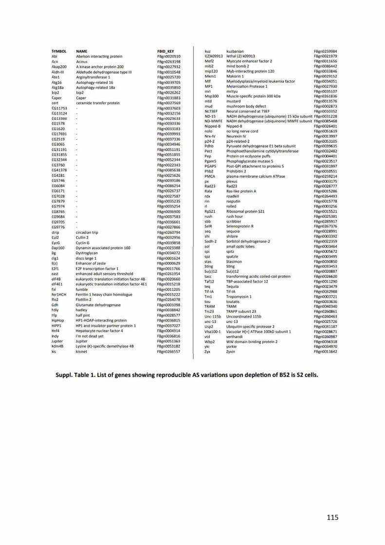
115

116

117

118

119

120

121
Discussion and Perspectives

122
3.0DISCUSSION AND PERSPECTIVES
Alternative splicing (AS) of mRNA precursors is a crucial step in the regulation of the gene
expression pathway. The SR proteins constitute a family of multifunctional RNA binding
proteins that play a significant role in alternative splicing regulation. Several genetic studies
have reported that SR proteins are essential for animal development (Ding et al., 2004) (Feng
et al., 2009) (Jumaa, Wei, & Nielsen, 1999) (H. Y. Wang, Xu, Ding, Bermingham, & Fu,
2001).
In addition to their role in splicing, several SRSF proteins have been shown to regulate
transcription elongation, RNA export, decay, translation (Zhong, Wang, Han, Rosenfeld, &
Fu, 2009) and for B52, transcriptional regulation (Juge et al., 2010).
Our group has been studying the functions and regulations of SR proteins by using
mammalian cells and Drosophila as models, and previously showed that modulation of
protein levels of B52 (which is the closest Drosophila orthologue to human SRSF6) can
affect cell growth in Drosophila (Fernando et al., 2015). The study did not characterize the
underline mechanism by which B52 effects growth. However, they speculated that B52 could
alter the AS of genes involved in the control of growth. Indeed (Fic et al., 2007) and (Gabut
et al., 2007) previously showed that B52 overexpression could alter the AS of genes involved
in development.
Capitalizing on this, in my Ph.D., I aimed to explore the mechanistic link between B52 and
how it can regulate growth.
AS program of B52:
Thanks to the advances in the ability that allows rapid characterization of genome-wide AS
events by RNA sequencing and immunoprecipitation techniques, several groups attempted to
reveal the precise map of several RBPs binding on the transcriptome. Two such studies were
highily interesting for us. In studies by (Brooks et al., 2015) and (Bradley et al., 2015) where,
among other SR proteins, B52 was depleted in Drosophila S2 cells and RNA seq was
performed to check the affected AS events.
We decided to merge the two available datasets order to identify AS events reproducibly
affected by B52 depletion. With our strategy, we identified several AS events modulated by
B52 level in genes involved in cell growth, including three genes linked to the Hippo (Hpo)
pathway. Among these three genes, we also found yorkie (yki), which encodes the central
effector of the Hippo pathway. The second candidate gene was wbp2 that encodes for Wbp2

123
protein which has been shown to bind Yki and enhance its intrinsic transcriptional co-
activator activity (X. Zhang, Milton, Poon, Hong, & Harvey, 2011). The other candidate gene
was zyxin (zyx) that encodes for Zyxin (Zyx) which has been shown to acts as a negative
regulator of the Hippo pathway as it is required for the activity of Yki (Rauskolb et al., 2011)
(Gaspar et al., 2015). RNAseq data indicate that B52 depletion promotes skipping of exon 3
in yki mRNA, the inclusion of exons 6 and 7 in wbp2 and the retention of the last intron in
zyx. The functional consequences of all the identified AS are uncharacterized including that
of yki.
B52 favors Yki1 isoform at the expense of Yki2:
Over the years the Hippo pathway has emerged as a significant player in tissue growth
control, stem cell function, regeneration and tumor suppression. During organogenesis,
optimum levels of Hippo signaling are crucial for maintaining tissue homeostasis. Over-
expression of Hippo signalling results in the formation of smaller organs due to induction of
apoptosis (Harvey et al., 2003b) (Pantalacci, Tapon, & Léopold, 2003) (Udan et al., 2003) (S.
Wu et al., 2003).
With Yki being the central effector of the Hippo pathway in Drosophila where all upstream
signals and kinase cascade converge on Yki to regulate the activity of pathway, finding that
yki AS is altered by depletion of splicing factor B52 was a major reveal for us. Several
studies have shown that the regulation of the transcriptional co-activator Yki is central to the
regulation of the Hippo pathway.
Indeed we confirmed this AS alteration by western, and RT-PCR B52 depleted S2 cells and
Drosophila Wing disc samples. We observed that depletion of B52 induces skipping of yki
alternative exon 3 and increases expression of Yki1 at the expense of Yki2 isoform. Thus,
Yki protein exists as two isoforms containing one or two WW domains thereafter called Yki1
and Yki2 respectively.
Although the canonical isoform of Yki (Yki2) has been characterized and has been described
in plethora of research papers, the presence of short isoform (Yki1) has been largely
neglected since the very first paper described Yki (J. Huang et al., 2005). Thus in this field of
Hippo pathway, it is not known yet if the short isoform of Yki is functional or redundant.
We believed that elucidation of the role of this isoform could better allow us to understand
better how B52 controls growth via the Hippo pathway. To further test the effect of B52 on
the growth we depleted it in the posterior part of Drosophila wing disc and observed that it

124
leads to a reduction of the posterior domain and in discs and in adult wings. In addition, we
saw the depletion of B52 leads to down-regulation of two well-established reporter of Yki
activity, diap-lacZ and Ex-lacZ .
Taking hints from this result, we hypothesized that B52 reduces growth by reducing Yki
activity and if this is true then these phenotypes should be rescued by overexpression of Yki
and by inactivation of the kinases Warts (Wts) or Hippo (Hpo) which phosphorylates to
regulate Yki negatively. Indeed we observed the rescue of the phenotype by overexpressing
Yki and depleting Wts and Hpo, but we did not attain overgrowth as observed by over-
expression of Yki or RNAi Wts/Hpo in the presence of wild type level of B52, suggesting
that B52 is involved in other modulation of other genes and (or) pathways controlling growth.
Given the results that we obtained one could also argue that partial rescue that we see could
correspond to the overcompensatory effect of Yki overexpression of Wts/Hpo depletion of
defect induced by B52 depletion acting independently of Hippo pathway. To this end, we also
depleted transcription factor Tgi that binds to Sd to repress the pathway in normal state and
saw that its depletion alone induces a mild overgrowth, but it too rescues the growth defect
inflicted by depletion of B52. However, we did not attain overgrowth as observed in wild
type lever of B52. Altogether our result allows postulating that although B52 is involved with
other genes and pathway in the regulation of growth, it also regulates growth upto significant
extent via regulating Yki activity by modulating yki AS.
Yki 1 is a weaker isoform compared to Yki2:
At this juncture, we observed that B52 depletion favors the expression of Yki1, decreases Yki
target genes and decreases growth partially through decreased Yki activity. This suggests that
Yki 1 isoform could be less efficient when compared to the canonical Yki2 isoform. Indeed
by overexpression assays, we observed that Yki 1 induces a weaker phenotype when
compared to Yki2 in the eyes of adult Drosophila.
We observed that expression of the Yki reporter transgenes ex-lacZ, diap1-lacZ and bantam
microRNA are weakly activated by Yki1 compared to Yki2. Further, we could extend these
experiments by checking the upregulation of another well-established target of Yki, cell-cycle
regulator CycE (Tapon et al., 2002).
Furthermore we directly compared the ability of Yki1 and Yki2 to activate transcription in
luciferase assays in Drosophila S2 cells. Since Yki does not bind DNA directly, we fused Yki

125
isoforms to the DNA binding domain of GAL4 and tested their activity on a UAS-luciferase
reporter. Our results confirm that Yk1 is a weak transcription activator compared to Yki2.
Lack of second WW domain reduces several interactions of Yki
The only difference between Yki1 and Yki2 is the absence of the second WW domain in
Yki1. Several studies have highlighted that the WW domain is indispensable for Yki’s
interaction with its specific partners that contain PPxY motifs (Hyangyee Oh & Irvine, 2010).
It has been shown that mutation of these domains abolishes Yki activity (H. Oh & Irvine,
2009). It is thus apparent to assume that the lack of other WW domain in Yki1 can directly
reduce its interaction with several above mentioned partners. Moreover, the lack of 2nd WW
domain could hypothetically modify protein structure in such a way that partners of Yki that
interact via its N terminal domain, i.e. WW-PPxY independent interactions could also be
affected thus we decided to investigate this untested assumption.
In addition to kinase Wts/Hpo and transcription factor Sd, Yki has been shown to interact
with Mad (Mothers against Dpp) which is an effector of Dpp signaling (Decapentaplegic) and
Hth (Hemothorax) and Tsh (Teashirt) (Alarcón et al., 2009) (Peng et al., 2009) (Hyangyee
Oh, Reddy, & Irvine, 2009). Yki also interacts with several chromatin-modifying proteins
including GAGA factor (GAF) and a subunit of the Trithorax-related (Trr) histone H3 lysine
4 methyltransferase complex, Nuclear receptor coactivator 6 (Ncoa6) (Qing et al., 2014b)
(Hyangyee Oh et al., 2013).
To ascertain the role of second WW domain and to understand the consequence of its
alternative inclusion on Yki protein’s function, we compared the interaction of each Yki
isoform with several partners, by a semi-quantitative co-immunoprecipitation (Co-IP) after
transfections of luciferase-tagged proteins in S2 cells (Refer to methods). We observed that
indeed Yki 1 interacts weakly with Wts, Ex, NcoA6, Wbp2, and Tgi when compared to Yki2.
All these partners contain PPxY motifs. However, both the isoforms interact similarly with
partners that contain single PPxY motif such as Mad or Hpo. Our results are in agreement
with the in vitro studies performed by (Nyarko, 2018) and (Webb et al., 2011) who have
shown cooperative binding between multiple PPxY motifs and tandem WW domains of YAP
and Yki.
We also observed similar interaction of both isoforms with transcription factors Sd, MAD,
Cbt and Hth, and the chromatin-associated factors GAF Mor and 14-3-3 proteins. We were
expecting such an observation for Sd, Mor and 14-3-3 which have been described to interact
with Yki via its N terminal domain (Goulev et al., 2008) (Hyangyee Oh et al., 2013) (Dong et
al., 2007). However, if Cbt associates with Yki directly or via other transcription factors, is

126
yet to be elucidated (Ruiz-Romero, Blanco, Paricio, Serras, & Corominas, 2015), the authors
also showed the interaction of GAF but were unable to map the binding domain precisely.
Same goes for Hth which is known to physically interacts with Yki but the domain of
interaction is yet to be elucidated (Peng et al., 2009). However, this analysis should be
extended to partners such as Dalao (another component of BRM complex), MED23, MED15,
MED31, MED19, and MED1 (subunits of Mediator complex) which have been shown to
associate with Yki via mass spectroscopy studies (Hyangyee Oh et al., 2013).
These results allowed us to verify our hypothesis that Yki1’s lack of second WW domain
reduces its interactions with several partners, but we saw similar interactions with partners
containing single PPxY motif and transcription factors which bind to Yki via its N terminal
domain. One curious point arose from the observation of these results is the fact that Yki1
isoform interacts weakly even with kinase Wts that functions to retain Yki in the cytoplasm.
If weaker interaction of Yki1 results in lower phosphorylation then it should be accumulated
in nucleus very easily even Yki1 binds weakly with Ex that has been shown to directly
sequester Yki at the apical junction region to prevent it from entering the nucleus, thereby
inhibiting its activation (Badouel et al., 2009). We did not yet realize experiments to
determine the phosphorylation state of the two isoforms of Yki. One counter explanation to
this observation could be the fact that in the normal state the expression level of Yki1
isoform is extremely low.
Further, we speculated that besides strengthening the binding of Yki2 with partners
containing multiple PPxY motifs, the presence of the second domain would allow Yki2 to
interact with another PPxY containing coactivator simultaneously. This would explain the
lower activity of Yki1. Inside the nucleus, Yki2 binds to transcription factor Scalloped (Sd)
via its N terminal domain and via WW domain to repressor Tondu domain-containing growth
inhibitor (Tgi). Studies by (Koontz et al., 2013) and (Guo et al., 2013) have shown an
interaction between Sd and Tgi. However, these two studies slightly differ from each other in
terms of explaining the aftermath of Yki’s recruitment. According to (Koontz et al., 2013)
Yki displaces the complex between Tgi and Sd and converts Sd into a transcription activator
while on the other hand, (Guo et al., 2013) reported that Tgi bind to Sd and Yki
simultaneously to forms an inactive ternary complex that is transcriptionally inactive.
To this end, our Co-IP verified the interaction between Sd and Tgi. In addition, we observed
that both Yki 1 and Yki2 isoforms could enhance this interaction between Sd and Tgi with
Yki2 being more efficient, suggesting that both Yki isoforms can form a trimeric complex
with Sd and Tgi in agreement with the study by (Guo et al., 2013).

127
Studies in mammalian Hippo pathway showed binding of a protein called Wbp2 to interact
with YAP (Mammalian counterpart of Yki). In Drosophila, (X. Zhang et al., 2011) have
described, WW-PPxY dependent association between Yki and Wbp2 and they further
reported that this association enhances Yki’s ability to drive transcription. Taking clues from
this study, one way to explain the weaker activity of Yki1 could due to the fact that it lacks
the second WW domain and thus it cannot recruit other transcriptional activators such as
Wbp2. So we extended our Co-IP experiments between Sd and Wbp2 in the presence of Yki1
or Yki2 and observed that both the isoforms could increase this Co-IP although Yki1
increases it weakly when compared to Yki2. However, in this experiment, only Wbp2
interacts via WW-PPxY motif but the other interaction was not dependent on WW-PPxY
domain.
We suspected that our observations would not be the same if we check Co-IP between Tgi
and Wbp2 in the presence of Yki2 and Yki1 as both of these partners realize interaction with
Yki in WW-PPxY dependent manner. Indeed, we observed that Yki1 could not Co-IP Tgi
and Wbp2 together whereas Yki2 does and point mutations in the two WW domains of Yki
abolish these interactions.
In Drosophila, six isoforms of Wbp2 have been described (source: FlyBase), and indeed from
our screening, we observed that AS of wbp2 is affected by the depletion of B52. It will be
interesting to extend our Co-IP to all these isoforms of Wbp2 and check the interaction of
both isoforms of Yki with individual Wbp2 isoforms because B52 could co-regulate the AS
of yki and wbp2 and more effectively control the regulation of Yki activity.
Model of competition:
Based on our observations so far, Yki isoforms interact similarly with transcription factors
but differ by their capacity to bind and bridge PPxY-containing proteins. We propose that
both the isoforms of Yki might compete with each other for binding to the transcription
factors, when the Hippo pathway is OFF the canonical isoform Yki2 joins the
transcriptionally repressed Tgi-Sd complex via one of its WW domain and could recruit
transcription co-activators with the other WW domain such as PPxY containing Wbp2 or
NcOA6 and finally may or may not displace Tgi. However, in B52 depleted state when there
is more abundant Yki1, it successfully binds to the trimeric complex between Tgi and Sd but
it cannot recruit Wbp2 or NcOA6 and might not displace Tgi so as to initiate the transcription
effectively. This could reconcile with (Guo et al., 2013)’s observation that Tgi enhances Sd-

128
Yki interaction but decreases the distance between Sd and Yki as measured by FRET
suggesting the remodeling of the complex.
To test this hypothesis of competition, we made use of activated forms of Yki which was first
described by (Dong et al., 2007), the activated forms lack the necessary phosphorylation site
that is phosphorylated by Wts, more specifically Ser168 is mutated preventing Yki’s binding
to 14-3-3 proteins and thus it cannot be retained in cytoplasm but accumulated in nucleus.
Overexpression of activated Yki2S168A isoform led to a very strong overgrowth phenotype in
the Drosophila eye and this was expected as it has been shown by (H. Oh & Irvine, 2009). In
addition we observed a weaker overgrowth phenotype induced by activated Yki1S168A
isoform too. To answer if there is a competition between the two isoforms, we co-
overexpressed the Yki1 isoforms and remarkably only activated Yki1S168A could partially
rescue this phenotype suggesting that in the nucleus there is a competition between two
isoforms. This result is in perfect agreement of our proposed model of competition.
Role yki AS in development:
Although the process of AS is vastly studied, and several isoforms that are switched at either
development or in disease are reported but only a very few studies have focussed to dissect
function of gene as isoform level, such is not only the case of yki/YAP AS but implies to vast
number of genes with known isoforms. For example, it is recently reported that the presence
or absence of exon7 in two splicing isoforms of MBNL1 conveys opposite phenotypical
implications of cancer (Tabaglio et al., 2018).
Similarly, in AS is also regulated for development, for an example, ApoER2 which is a
protein encoded by the LRP8 gene also undergoes alternative splicing. The exon 19-
containing domain of ApoER2 is essential for synapse formation while its inclusion is
reduced in the brain of Alzheimer's patients (Hinrich et al., 2016). Thus it is now imperative
to focus on the gene function at isoform level because it could be a key in understanding the
complex regulation like AS and that is why we do not undermine the role of short isoform in
the yki AS.
(Yeo, Holste, Kreiman, & Burge, 2004) highlighted that levels of AS usually differs from one
tissue to another. So it is a possibility that AS of yki can differ from one tissue to another.
This could be checked by doing western on tissues at various stages of development.
However, to ultimately, reveal the role of the yki AS, we aimed to abrogate this in vivo, i.e.
to generate flies expressing exclusively Yki2 (yki2-only) or exclusively Yki1 (yki1-only), by
editing the endogenous yki gene using the CRISPR/Cas9 system. We employed a different

129
strategy at first and failed (Please refer to Appendix Figure 37), but later we employed a
second strategy and managed to create flies expressing Yki2 isoform only. We observed that
flies were homozygous viable, but we noticed a phenotype in the population of these flies.
We noticed an intra variation between the size of the right and left wings. This phenotype of
Fluctuating asymmetry (FA) and it is the consequence of subtle deviation from normal
development. The phenotype of asymmetry cannot be blamed on the environmental or
genetic variations; it arises stochastically during the development. The fact that we observe a
consistent and significant FA in our analysis of Yki2only population compared to control
suggests that indeed the yki AS plays a role in development robustness. Further, we would
like to generate heterozygous yki2only flies so as to check if we rescue the phenotype of FA.
Recently, (Boone, Colombani, Andersen, & Leópold, 2016) have shown that Yki controls the
expression of dilp8 during normal development. They showed that a mutation Hpo-
responsive element (HRE) within the dilp8 promoter is enough to result in increased FA. One
way to test our theory of yki AS a mean to maintain robust development would be via
extending our analysis by testing for genetic interaction between dilp8 mutant and our yki2only
mutant. We expect that the FA will further rise in this population due to the lack of
endogenous Yki1 isoform.
Yki has been implicated in the control of expression of hormone ecdysone (20-
hydroxyecdysone) and couples insulin signaling with ecdysone production. (C. Zhang et al.,
2015) have further shown that Yki interacts with Taiman, ecdysone pathway transcription
factor to control the gene expression of some targets such as piwi, nanos, dilp8. We could
further advance our isoform comparison studies on these targets of Yki2.
One key experiment that is currently underway with our yki2only flies is to investigate the
effect of B52 depletion on the size of the posterior domain.We depleted B52 in the posterior
domain of the wing in our yki2only flies and plan to measure the effect on growth. Upon
comparing with it with the overgrowth induced by B52 depletion in the posterior domain of
wild-type flies if we observe that the overgrowth phenotype exceeds in yki2only flies, then it
would mean that indeed Yki isoform functions to buffer the activity of Yki2 isoform in
development.
Concluding Remarks:
Finally, it is worth noting that this AS is conserved, In mammals, yki homolog, YAP, is
alternatively spliced and is expressed as eight protein isoforms that fall into two categories,
YAP1 and YAP2, which respectively contain one or two WW domain(s)(Sudol, 2013).
130
Therefore alternative inclusion of the second WW domain is a conserved feature between Yki
and YAP. Moreover (Komuro et al., 2003) reported that YAP2 is more efficient isoform than
YAP1 in their studies where they co-precipitated ErbB-4 an epidermal growth receptor. This
further supports the idea that Yki1 and YAP1 isoforms have important function in vivo and
that alternative splicing of Yki/YAP is a conserved mechanism of control of the Hippo
pathway.
There a are plethora of reports showing YAP is amplified and overexpressed in various
tumors types where YAP protein relocalizes in the nucleus. YAP knockdown in many human
cancer cell lines, such as prostate cancer or malignant mesothelioma cell lines, suppresses
their growth and ability to trigger tumor formation. As of now, the focus of therapeutic
research studies is towards targeting YAP, which indeed is a promising way to hamper tumor
cell growth. Recently, molecules targeting YAP, such as verteporfin that inhibits the
formation of the YAP-TEAD complex, have been described to block tumor growth in mice
(Liu-Chittenden et al., 2012). Identification of such new modulators of this pathway can,
therefore, open new therapeutic opportunities for cancer treatment. However, to this date,
none of the studies differentiate between the various isoforms of YAP. We wonder if the AS
of YAP in cancer cells allows the production of more YAP2 instead of YAP1 which could
even be a hallmark of tumorigenesis. One amazing future perspective of this study would be
to study the extent of YAP splicing and the ration of isoforms in various tumor samples.
Further investigation could be directed towards finding small molecules that manipulate the
alternative splicing of YAP as a way to modulate YAP activity in cancer cells. Thus our
research should open new perspectives for modulation of the Hippo pathway in cancer cells
by altering YAP alternative splicing.
The regulation of the Hippo pathway has evolved into a complex network over the past two
decades, with new players added at an unprecedented pace. Our study shall add even more
complexity into this regulatory network.

131
APPENDIX

132
4.0APPENDIX
first strategy employed to generate the yki1only
& yki2only
flies: To be able to generate two
versions of Yki gene that can encode only Yki1 or only Yki2, we targeted a Double-Stranded
Break (DSB) in exon 2 and exon 4 of Yki with two guide RNAs and repair the gap with the
portion of cDNAs encoding Yki1 or Yki2. The template to repair is provided as ssODN
(single strand oligo donor nucleotide).
Appendix Figure 37 : CRISPR and screening strategy:
(a.) Our premier strategy to create yki1-only
and yki2-only
flies. (b.) screening
We injected the plasmid coding for two guide RNAs and repair template in the embryos
expressing Cas9. Since the injections are done in the germline, it was necessary to cross the
injected individuals with wild type flies before doing a non-lethal genetic screen on the
progeny of this cross. The screening was done by performing a PCR on each wing of an adult
progeny fly to determine the flies which are either yki1only & yki
2only (Figure 37).
At first, we succeeded to establish Yki1 isoform expressing flies. We determined that the
homozygous (yki1only
/ yki1only ) were lethal at the embryonic stage and we even determined by
using the null allele of yki, i.e. ykiB5 that the flies were lethal due to the mutation that we

133
created. However for an unknown reason we do not detect expression of neither Yki1 protein
nor yki1 mRNA in these flies. Therefore this allele is not a true yki1only allele but a null.
After another attempt we were able to generate Yki2 isoform expressing flies and here to we
found out that it contains a small deletion of 4 amino acid residues at the repair site in exon 4,
thus we named the allele as yki2-only and nevertheless we decided to characterize these
flies and in the meantime we worked on new strategy to create good yki2-only flies without. We
reasoned that such a small mutation might not change the behavior of this isoform
completely. We discovered that homozygous yki2-only / yki
2-only flies were viable
without any visible phenotype. What was surprising for us to find that yki2-only / yki
2-
only flies depicted 50% lethality which we did not expect given the flies had no visible
phenotype (Figure 38). Could it be due to the lack of Yki1 which is absolutely required at the
early stage of development? Alternatively, could it be an issue of parental fertility which is
affected? Or is it merely due to the modification of the Yki2 peptide due to the minor
mutation?
Appendix Figure 38 Characterization of yki1-only
and yki2-only
flies:
Schematic representing the embryo hatching. Parents were kept on medium at 25°C. Embryos were carefully counted. In
general 5% lethality in wild-type flies is expected. 25% lethality in next two populations is expected because CyO/CyO is
lethal. An additional 25% lethality in yki1-only
/CyO-Tb suggests that homozygous is lethal. 50% lethality is observed in yki2-
only population. n= number of embryos counted.
Another argument that we tried to investigate was if the lack of phenotype in adult flies was
is simply because in normal developmental conditions the Yki1 isoform has less activity, but
in case of stress, it increases to counteract the severity of Yki2’s transcriptional output.
Indeed (Di Cara et al., 2015) showed the loss of Hippo pathway component during stress

134
induced by chemical causes the Hippo pathway to switch its role from being anti-apoptotic
to apoptotic. They showed that upon stress stimulation by caffeine, Yki interacts with the
stress-responsive transcription factor p53 in flies with loss of Hippo components such as Wts
or Hpo (Figure 39 a & b). We asked how our heterozygous yki1only
& homozygous yki2only
would respond to caffeine treatment and what will be the effect of overexpression of Yki1
or Yki2 activated and non activated isoforms? Unfortunately, we were unable to reproduce
the pro-apoptotic function in our experiments. It could be due to the technical problem with
the medium. At this point, we decide to focus on generating the yki2only
flies without any
mutation and resume the characterization studies when we obtain it (Figure 39 c).

135
Appendix Figure 39 is Yki AS involved in the pro-apoptotic function of Yki upon stress?
(a. & b.) (Di Cara et al., 2015) Genetic interactions between Hippo pathway components in no caffeine and caffeine
medium. C. (Bottom right): We see no difference in the effect on the phenotype in wild type , hetrogygous yki1only and
yki2only flies raised on regular and caffeine medium. (Top right) we see no effect on the phenotype with ey>UAS-wts-
or ey>UAS-Hpo- between flies raised on regular and caffeine medium. GMR>UAS-wts- -Hpo-
too did not result in any visible difference in phenotype in eye or elsewhere in the two medium. (Right middle and bottom)
Finally overexpression of activated and non activated Yki isoforms too ey>UAS-Yki1, ,GMR>UAS-Yki1, ey>UAS-Yki1S168A
GMR>UAS-Yki1S168A
GMR>UAS-Yki2, ,GMR>UAS-Yki2, ey>UAS-Yki2S168A
GMR>UAS-Yki2S168A
did not yield any different
phenotype.

136
Role of B52 in cell competition:
To further explore the link between B52 and cell competition, we generated the B52
mutant clones by mitotic recombination in wing imaginal disc. Interestingly we observed
that B52-/-
clones are eliminated over time via apoptosis. This could be due to the
phenomenon of cell competition (Appendix-Figure 40 a). This phenomenon was first
described by the analysis of a dominant mutation in a Drosophila Minute (M) gene that
affects ribosomal biogenesis and therefore slows down translation and protein synthesis.
While homozygous mutations (M/M) of these genes are cell lethal, heterozygous (M/+)
adults are viable and fertile, although they suffer a developmental delay.
Interestingly, clones of heterozygous cells (M/+) growing in a wild type wing disc were not
underrepresented in the adult wing, as expected, but completely eliminated (Morata &
Ripoll, 1975). The eliminated M/+ cells are referred to as “looser cells,” and surrounding
wild type cells are referred to as “winner cells” (Appendix-Figure 40b). The elimination
induces the compensatory proliferation of winner cells, and thus the organ retains the
correct size. We induced, B52-/-
mutant clones in disc where the surrounding cells have a
mutation in the minute gene (M/-) and we observed no elimination suggesting that indeed
the elimination was due to cell competition. Thus cell competition is a context-dependent
phenomenon since the looser cells survive when surrounded by looser cells and gets
eliminated when surround by winner cells (Appendix Figure 40c).
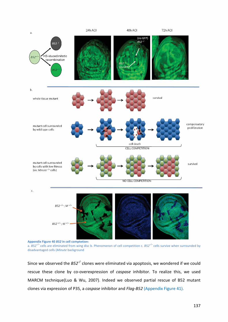
137
Appendix Figure 40 B52 in cell comptetion:
a. B52–/–
cells are eliminated from wing disc b. Phenomenon of cell competition c. B52–/–
cells survive when surrounded by
disadvantaged cells (Minute background
Since we observed the B52-/-
clones were eliminated via apoptosis, we wondered if we could
rescue these clone by co-overexpression of caspase inhibitor. To realize this, we used
MARCM technique(Luo & Wu, 2007). Indeed we observed partial rescue of B52 mutant
clones via expression of P35, a caspase inhibitor and Flag-B52 (Appendix Figure 41).

138
Appendix Figure 41 B52–/–
clones are partially rescued:
B52–/–
clones are partially rescued by expression of the caspase inhibitor P35 in the clone (MARCM)
According to literature several pathways are involved in cell competition including Hippo
pathway (Tyler, Li, Zhuo, Pellock, & Baker, 2007), cell polarity (Agrawal, Kango, Mishra, &
Sinha, 1995) (Brumby & Richardson, 2003)(Woods & Bryant, 1991), JAK/STAT (Rodrigues et
al., 2012) Dpp signaling (Moreno, Basler, & Morata, 2002). In one of our primary analysis of
RNAseq data by (Bradley et al., 2015) (Brooks et al., 2015) we saw that indeed B52 alters the
splicing of the yki, dlg and STAT92E.
First, we checked the overexpression of Yki2 in B52 mutant clones via MARCM technique,
the idea was to be able to rescue the B52 mutant clones from getting eliminated because if
B52 depletion leads to more production of Yki1 isoform the maybe overexpressing would
prevent B52 mutant clones from elimination. However, to this end, we did not see any
rescue (Appendix Figure 42).
Appendix Figure 42 : Rescue of B52 mutants by Yki1 and Yki2:
Just by expressing Yki1 or Yki2 the elimination of B52 mutant clones cannot be rescued efficiently.

139
As of now we have tools to analyze the expression of the two isoforms of dlg and STAT92E
in B52 mutant clones hopefully we will be able to show if that it is B52 that affects the
splicing of these genes, and thus it plays a role in cell competition.

140
REFERENCES

141
5.0RERERENCES
Aartsma-Rus, A. (2017). FDA Approval of Nusinersen for Spinal Muscular Atrophy Makes 2016 the Year of Splice Modulating Oligonucleotides. Nucleic Acid Therapeutics.https://doi.org/10.1089/nat.2017.0665
Adler, J. J., Johnson, D. E., Heller, B. L., Bringman, L. R., Ranahan, W. P., Conwell, M. D., … Wells, C. D. (2013). Serum deprivation inhibits the transcriptional co-activator YAP and cell growth via phosphorylation of the 130-kDa isoform of Angiomotin by the LATS1/2 protein kinases. Proceedings of the National Academy of Sciences.https://doi.org/10.1073/pnas.1308236110
Aegerter-Wilmsen, T., Aegerter, C. M., Hafen, E., & Basler, K. (2007). Model for the regulation of size in the wing imaginal disc of Drosophila. Mechanisms of Development.https://doi.org/10.1016/j.mod.2006.12.005
Aegerter-Wilmsen, T., Heimlicher, M. B., Smith, A. C., de Reuille, P. B., Smith, R. S., Aegerter, C. M., & Basler, K. (2012). Integrating force-sensing and signaling pathways in a model for the regulation of wing imaginal disc size. Journal of Cell Science.https://doi.org/10.1242/jcs.120758
Agrawal, N., Kango, M., Mishra, A., & Sinha, P. (1995). Neoplastic transformation and aberrant cell-cell interactions in genetic mosaics of lethal(2)giant larvae (lgl), a tumor suppressor gene of Drosophila. Developmental Biology. https://doi.org/10.1006/dbio.1995.0017
Alarcón, C., Zaromytidou, A. I., Xi, Q., Gao, S., Yu, J., Fujisawa, S., … Massagué, J. (2009). Nuclear CDKs Drive Smad Transcriptional Activation and Turnover in BMP and TGF- Cell.https://doi.org/10.1016/j.cell.2009.09.035
Änkö, M. L., Müller-McNicoll, M., Brandl, H., Curk, T., Gorup, C., Henry, I., … Neugebauer, K. M. (2012). The RNA-binding landscapes of two SR proteins reveal unique functions and binding to diverse RNA classes. Genome Biology. https://doi.org/10.1186/gb-2012-13-3-r17
Arnold, E. S., & Fischbeck, K. H. (2018). Spinal muscular atrophy. In Handbook of Clinical
Neurology. https://doi.org/10.1016/B978-0-444-64076-5.00038-7
Ast, G. (2004). How did alternative splicing evolve? Nature Reviews Genetics.https://doi.org/10.1038/nrg1451
Avery, P., Vicente-Crespo, M., Francis, D., Nashchekina, O., Alonso, C. R., & Palacios, I. M. (2011). Drosophila Upf1 and Upf2 loss of function inhibits cell growth and causes animal death in a Upf3-independent manner. RNA. https://doi.org/10.1261/rna.2404211
Axelrod, F. B., & Kaufmann, H. (2014). Familial Dysautonomia. In Encyclopedia of the Neurological
Sciences. https://doi.org/10.1016/B978-0-12-385157-4.01038-1
Axelrod, F. B., Liebes, L., Simson, G. G. Von, Mendoza, S., Mull, J., Leyne, M., … Slaugenhaupt, S. A. (2011). Kinetin improves IKBKAP mRNA splicing in patients with familial dysautonomia. Pediatric Research. https://doi.org/10.1203/PDR.0b013e31822e1825
Aznarez, I., Nomakuchi, T. T., Tetenbaum-Novatt, J., Rahman, M. A., Fregoso, O., Rees, H., & Krainer, A. R. (2018). Mechanism of Nonsense-Mediated mRNA Decay Stimulation by Splicing Factor SRSF1. Cell Reports. https://doi.org/10.1016/j.celrep.2018.04.039
Badouel, C., Gardano, L., Amin, N., Garg, A., Rosenfeld, R., Le Bihan, T., & McNeill, H. (2009). The FERM-Domain Protein Expanded Regulates Hippo Pathway Activity via Direct Interactions with the Transcriptional Activator Yorkie. Developmental Cell.https://doi.org/10.1016/j.devcel.2009.01.010
Bai, J., Chiu, W., Wang, J., Tzeng, T., Perrimon, N., & Hsu, J. (2001). The cell adhesion molecule Echinoid defines a new pathway that antagonizes the Drosophila EGF receptor signaling

142
pathway. Development (Cambridge, England).
Bao, Y., Nakagawa, K., Yang, Z., Ikeda, M., Withanage, K., Ishigami-Yuasa, M., … Hata, Y. (2011). A cell-based assay to screen stimulators of the Hippo pathway reveals the inhibitory effect of dobutamine on the YAP-dependent gene transcription. Journal of Biochemistry.https://doi.org/10.1093/jb/mvr063
Baraniak, A. P., Lasda, E. L., Wagner, E. J., & Garcia-Blanco, M. A. (2003). A Stem Structure in Fibroblast Growth Factor Receptor 2 Transcripts Mediates Cell-Type-Specific Splicing by Approximating Intronic Control Elements. Molecular and Cellular Biology.https://doi.org/10.1128/mcb.23.24.9327-9337.2003
Basson, C. T., Bachinsky, D. R., Lin, R. C., Levi, T., Elkins, J. A., Soults, J., … Seidman, C. E. (1997). Mutations in human cause limb and cardiac malformation in Holt-Oram syndrome. Nature Genetics. https://doi.org/10.1038/ng0197-30
Baumgartner, R., Poernbacher, I., Buser, N., Hafen, E., & Stocker, H. (2010). The WW Domain Protein Kibra Acts Upstream of Hippo in Drosophila. Developmental Cell, 18(2), 309–316. https://doi.org/10.1016/j.devcel.2009.12.013
Bedard, K. M., Daijogo, S., & Semler, B. L. (2007). A nucleo-cytoplasmic SR protein functions in viral IRES-mediated translation initiation. EMBO Journal.https://doi.org/10.1038/sj.emboj.7601494
Bennett, C. F., & Swayze, E. E. (2010). RNA Targeting Therapeutics: Molecular Mechanisms of Antisense Oligonucleotides as a Therapeutic Platform. Annual Review of Pharmacology and
Toxicology. https://doi.org/10.1146/annurev.pharmtox.010909.105654
Bennett, F. C., & Harvey, K. F. (2006). Fat Cadherin Modulates Organ Size in Drosophila via the Salvador/Warts/Hippo Signaling Pathway. Current Biology.https://doi.org/10.1016/j.cub.2006.09.045
Bentley, D. L. (2014). Coupling mRNA processing with transcription in time and space. Nature
Reviews Genetics. https://doi.org/10.1038/nrg3662
Biankin, A. V., Waddell, N., Kassahn, K. S., Gingras, M. C., Muthuswamy, L. B., Johns, A. L., … Grimmond, S. M. (2012). Pancreatic cancer genomes reveal aberrations in axon guidance pathway genes. Nature. https://doi.org/10.1038/nature11547
Bilder, D. (2004). Epithelial polarity and proliferation control: Links from the Drosophilaneoplastictumor suppressors. Genes and Development. https://doi.org/10.1101/gad.1211604
Black, D. L. (2000). Protein diversity from alternative splicing: A challenge for bioinformatics and post-genome biology. Cell. https://doi.org/10.1016/S0092-8674(00)00128-8
Black, D. L. (2003). Mechanisms of Alternative Pre-Messenger RNA Splicing. Annual Review of
Biochemistry. https://doi.org/10.1146/annurev.biochem.72.121801.161720
Blanchette, M., & Chabot, B. (1999). Modulation of exon skipping by high-affinity hnRNP A1-binding sites and by intron elements that repress splice site utilization. In The EMBO Journal.
Blencowe, B. J. (2000). Exonic splicing enhancers: Mechanism of action, diversity and role in human genetic diseases. Trends in Biochemical Sciences. https://doi.org/10.1016/S0968-0004(00)01549-8
Boggiano, J. C., & Fehon, R. G. (2012). Growth Control by Committee: Intercellular Junctions, Cell Polarity, and the Cytoskeleton Regulate Hippo Signaling. Developmental Cell.https://doi.org/10.1016/j.devcel.2012.03.013
Boggiano, J. C., Vanderzalm, P. J., & Fehon, R. G. (2011). Tao-1 phosphorylates Hippo/MST kinases to regulate the Hippo-Salvador-Warts tumor suppressor pathway. Developmental Cell.https://doi.org/10.1016/j.devcel.2011.08.028

143
Bogoyevitch, M. A., Ngoei, K. R. W., Zhao, T. T., Yeap, Y. Y. C., & Ng, D. C. H. (2010). c-Jun N-terminal kinase (JNK) signaling: Recent advances and challenges. Biochimica et Biophysica
Acta - Proteins and Proteomics. https://doi.org/10.1016/j.bbapap.2009.11.002
Bohère, J., Mancheno-Ferris, A., Al Hayek, S., Zanet, J., Valenti, P., Akino, K., … Payre, F. (2018). Shavenbaby and Yorkie mediate Hippo signaling to protect adult stem cells from apoptosis. Nature Communications. https://doi.org/10.1038/s41467-018-07569-0
Bonnal, S., Martínez, C., Förch, P., Bachi, A., Wilm, M., & Valcárcel, J. (2008). RBM5/Luca-15/H37 Regulates Fas Alternative Splice Site Pairing after Exon Definition. Molecular Cell.https://doi.org/10.1016/j.molcel.2008.08.008
Boone, E., Colombani, J., Andersen, D. S., & Leópold, P. (2016). The Hippo signalling pathway coordinates organ growth and limits developmental variability by controlling dilp8 expression. Nature Communications. https://doi.org/10.1038/ncomms13505
Bosch, J. A., Sumabat, T. M., Hafezi, Y., Pellock, B. J., Gandhi, K. D., & Hariharan, I. K. (2014). The Drosophila F-box protein Fbxl7 binds to the protocadherin Fat and regulates Dachs localization and Hippo signaling. ELife. https://doi.org/10.7554/elife.03383
Boucher, L., Ouzounis, C. A., Enright, A. J., & Blencowe, B. J. (2001). A genome-wide survey of RS domain proteins. RNA (New York, N.Y.).
Bourgeois, C. F., Lejeune, F., & Stévenin, J. (2004). Broad Specificity of SR (Serine{plus 45 degree rule}Arginine) Proteins in the Regulation of Alternative Splicing of Pre-Messenger RNA. Progress in Nucleic Acid Research and Molecular Biology. https://doi.org/10.1016/S0079-6603(04)78002-2
Bradley, T., Cook, M. E., & Blanchette, M. (2015). SR proteins control a complex network of RNA-processing events. RNA. https://doi.org/10.1261/rna.043893.113
Bressan, G. C., Moraes, E. C., Manfiolli, A. O., Kuniyoshi, T. M., Passos, D. O., Gomes, M. D., & Kobarg, J. (2009). Arginine methylation analysis of the splicing-associated SR protein SFRS9/SRp30C. Cellular and Molecular Biology Letters. https://doi.org/10.2478/s11658-009-0024-2
Briese, M., Saal-Bauernschubert, L., Ji, C., Moradi, M., Ghanawi, H., Uhl, M., … Sendtner, M. (2018). hnRNP R and its main interactor, the noncoding RNA 7SK, coregulate the axonal transcriptome of motoneurons. Proceedings of the National Academy of Sciences.https://doi.org/10.1073/pnas.1721670115
Brogna, S., McLeod, T., & Petric, M. (2016). The Meaning of NMD: Translate or Perish. Trends in
Genetics. https://doi.org/10.1016/j.tig.2016.04.007
Brooks, A. N., Duff, M. O., May, G., Yang, L., Bolisetty, M., Landolin, J., … Brenner, S. E. (2015). Regulation of alternative splicing in Drosophila by 56 RNA binding proteins. Genome Research.https://doi.org/10.1101/gr.192518.115
Brooks, A. N., Yang, L., Duff, M. O., Hansen, K. D., Park, J. W., Dudoit, S., … Graveley, B. R. (2011). Conservation of an RNA regulatory map between Drosophila and mammals. Genome
Research. https://doi.org/10.1101/gr.108662.110
Brumby, A. M., & Richardson, H. E. (2003). scribble mutants cooperate with oncogenic Ras or Notch to cause neoplastic overgrowth in Drosophila. EMBO Journal.https://doi.org/10.1093/emboj/cdg548
Bruno, I. G., Karam, R., Huang, L., Bhardwaj, A., Lou, C. H., Shum, E. Y., … Wilkinson, M. F. (2011). Identification of a MicroRNA that Activates Gene Expression by Repressing Nonsense-Mediated RNA Decay. Molecular Cell. https://doi.org/10.1016/j.molcel.2011.04.018
Bryant, P. J., & Simpson, P. (1984). of B JOLOGY REVIEW. THE QUARTERLY REVIEW of B

144
JOLOGY, 71(2), 181–220.
Busch, A., & Hertel, K. J. (2012). Evolution of SR protein and hnRNP splicing regulatory factors. Wiley Interdisciplinary Reviews: RNA. https://doi.org/10.1002/wrna.100
Cáceres, J. F., Screaton, G. R., & Krainer, A. R. (1998). A specific subset of SR proteins shuttles continuously between the nucleus and the cytoplasm. Genes and Development.https://doi.org/10.1101/gad.12.1.55
Callus, B. A., Verhagen, A. M., & Vaux, D. L. (2006). Association of mammalian sterile twenty kinases, Mst1 and Mst2, with hSalvador via C-terminal coiled-coil domains, leads to its stabilization and phosphorylation. FEBS Journal, 273(18), 4264–4276. https://doi.org/10.1111/j.1742-4658.2006.05427.x
Cao, J. J., Zhao, X. M., Wang, D. L., Chen, K. H., Sheng, X., Li, W. Bin, … He, J. (2014). YAP is overexpressed in clear cell renal cell carcinoma and its knockdown reduces cell proliferation and induces cell cycle arrest and apoptosis. Oncology Reports. https://doi.org/10.3892/or.2014.3349
Cao, W., Jamison, S. F., & Garcia-Blanco, M. A. (1997). Both phosphorylation and dephosphorylation of ASF/SF2 are required for pre-mRNA splicing in vitro. RNA.
Cartegni, L., Chew, S. L., & Krainer, A. R. (2002). Listening to silence and understanding nonsense: exonic mutations that affect splicing. Nature Reviews. Genetics. https://doi.org/10.1038/nrg775
Castello, A., Fischer, B., Eichelbaum, K., Horos, R., Beckmann, B. M., Strein, C., … Hentze, M. W. (2012). Insights into RNA Biology from an Atlas of Mammalian mRNA-Binding Proteins. Cell.https://doi.org/10.1016/j.cell.2012.04.031
Castello, A., Fischer, B., Frese, C. K., Horos, R., Alleaume, A. M., Foehr, S., … Hentze, M. W. (2016). Comprehensive Identification of RNA-Binding Domains in Human Cells. Molecular
Cell. https://doi.org/10.1016/j.molcel.2016.06.029
Chabot, B. (2015). My road to alternative splicing control: from simple paths to loops and interconnections. Biochemistry and Cell Biology. https://doi.org/10.1139/bcb-2014-0161
Chabot, B., & Shkreta, L. (2016). Defective control of pre-messenger RNA splicing in human disease. Journal of Cell Biology. https://doi.org/10.1083/jcb.201510032
Champlin, D. T., Frasch, M., Saumweber, H., & Lis, J. T. (1991). Characterization of a Drosophila protein associated with boundaries of transcriptionally active chromatin. Genes and
Development. https://doi.org/10.1101/gad.5.9.1611
Chan, S. W., Lim, C. J., Chen, L., Chong, Y. F., Huang, C., Song, H., & Hong, W. (2011). The hippo pathway in biological control and cancer development. Journal of Cellular Physiology, 226(4), 928–939. https://doi.org/10.1002/jcp.22435
Chan, S. W., Lim, C. J., Chong, Y. F., Pobbati, A. V., Huang, C., & Hong, W. (2011). Hippo pathway-independent restriction of TAZ and YAP by angiomotin. Journal of Biological
Chemistry. https://doi.org/10.1074/jbc.C110.212621
Chan, S. W., Lim, C. J., Guo, F., Tan, I., Leung, T., & Hong, W. (2013). Actin-binding and cell proliferation activities of angiomotin family members are regulated by Hippo pathway-mediated phosphorylation. Journal of Biological Chemistry. https://doi.org/10.1074/jbc.M113.527598
Chan, S. W., Lim, C. J., Loo, L. S., Chong, Y. F., Huang, C., & Hong, W. (2009). TEADs mediate nuclear retention of TAZ to promote oncogenic transformation. Journal of Biological Chemistry.https://doi.org/10.1074/jbc.M901568200
Chaudhury, A., Chander, P., & Howe, P. H. (2010). Heterogeneous nuclear ribonucleoproteins(hnRNPs) in cellular processes: Focus on hnRNP E1’s multifunctional regulatory roles. RNA.https://doi.org/10.1261/rna.2254110

145
Cheah, M. T., Wachter, A., Sudarsan, N., & Breaker, R. R. (2007). Control of alternative RNA splicing and gene expression by eukaryotic riboswitches. Nature.https://doi.org/10.1038/nature05769
Chen, C.-L., Sansores-Garcia, L., Tao, C., Gajewski, K. M., Halder, G., & Bossuyt, W. (2010). The apical-basal cell polarity determinant Crumbs regulates Hippo signaling in Drosophila. Proceedings of the National Academy of Sciences. https://doi.org/10.1073/pnas.1004060107
Chen, C.-L., Schroeder, M. C., Kango-Singh, M., Tao, C., & Halder, G. (2012). Tumor suppression by cell competition through regulation of the Hippo pathway. Proceedings of the National
Academy of Sciences. https://doi.org/10.1073/pnas.1113882109
Chen, J., & Weiss, W. A. (2015). Alternative splicing in cancer: Implications for biology and therapy. Oncogene. https://doi.org/10.1038/onc.2013.570
Chen, K. H., He, J., Wang, D. L., Cao, J. J., Li, M. C., Zhao, X. M., … Liu, W. J. (2014). Methylation-associated inactivation of LATS1 and its effect on demethylation or overexpression on YAP and cell biological function in human renal cell carcinoma. International Journal of
Oncology. https://doi.org/10.3892/ijo.2014.2687
Chen, L., Chan, S. W., Zhang, X. Q., Walsh, M., Lim, C. J., Hong, W., & Song, H. (2010). Structural basis of YAP recognition by TEAD4 in the Hippo pathway. Genes and Development.https://doi.org/10.1101/gad.1865310
Chen, L. Y., & Lingner, J. (2012). AUF1/HnRNP D RNA Binding Protein Functions in Telomere Maintenance. Molecular Cell. https://doi.org/10.1016/j.molcel.2012.06.031
Chen, Q., Zhang, N., Gray, R. S., Li, H., Ewald, A. J., Zahnow, C. A., & Pan, D. (2014). A temporal requirement for Hippo signaling in mammary gland differentiation, growth, and tumorigenesis. Genes and Development. https://doi.org/10.1101/gad.233676.113
Chen, Y.-C., Milliman, E. J., Goulet, I., Cote, J., Jackson, C. A., Vollbracht, J. A., & Yu, M. C. (2010). Protein Arginine Methylation Facilitates Cotranscriptional Recruitment of Pre-mRNA Splicing Factors. Molecular and Cellular Biology. https://doi.org/10.1128/MCB.00359-10
Chen, Y. H., Su, L. H., Huang, Y. C., Wang, Y. T., Kao, Y. Y., & Sun, C. H. (2008). UPF1, a conserved nonsense-mmediated mRNA decay factor, regulates cyst wall protein transcripts in Giardia lamblia. PLoS ONE. https://doi.org/10.1371/journal.pone.0003609
Cheung, M., Talarchek, J., Schindeler, K., Saraiva, E., Penney, L. S., Ludman, M., & Testa, J. R. (2013). Further evidence for germline BAP1 mutations predisposing to melanoma and malignant mesothelioma. Cancer Genetics. https://doi.org/10.1016/j.cancergen.2013.05.018
Cho, E., Feng, Y., Rauskolb, C., Maitra, S., Fehon, R., & Irvine, K. D. (2006). Delineation of a Fat tumor suppressor pathway. Nature Genetics. https://doi.org/10.1038/ng1887
Cho, S., Hoang, A., Sinha, R., Zhong, X.-Y., Fu, X.-D., Krainer, A. R., & Ghosh, G. (2011). Interaction between the RNA binding domains of Ser-Arg splicing factor 1 and U1-70K snRNP protein determines early spliceosome assembly. Proceedings of the National Academy of
Sciences. https://doi.org/10.1073/pnas.1017700108
Cho, S., Moon, H., Loh, T. J., Oh, H. K., Kim, H.-R., Shin, M.-G., … Shen, H. (2014). 3Sequences of Spinal Muscular Atrophy Related SMN2 Pre-mRNA Include Enhancers for Nearby Exons. The Scientific World Journal. https://doi.org/10.1155/2014/617842
Clark, H. F., Brentrup, D., Schneitz, K., Bieber, A., Goodman, C., & Noll, M. (1995). Dachsous encodes a member of the cadherin superfamily that controls imaginai disc morphogenesis in Drosophila. Genes and Development. https://doi.org/10.1101/gad.9.12.1530
Close, P., Hawkes, N., Cornez, I., Creppe, C., Lambert, C. A., Rogister, B., … Chariot, A. (2006). Transcription Impairment and Cell Migration Defects in Elongator-Depleted Cells: Implication

146
for Familial Dysautonomia. Molecular Cell. https://doi.org/10.1016/j.molcel.2006.04.017
Colak, D., Ji, S. J., Porse, B. T., & Jaffrey, S. R. (2013). XRegulation of axon guidance by compartmentalized nonsense-mediated mRNA decay. Cell.https://doi.org/10.1016/j.cell.2013.04.056
Colwill, K., Pawson, T., Andrews, B., Prasad, J., Manley, J. L., Bell, J. C., & Duncan, P. I. (1996). The Clk/Sty protein kinase phosphorylates SR splicing factors and regulates their intranuclear distribution. The EMBO Journal.
Cordenonsi, M., Zanconato, F., Azzolin, L., Forcato, M., Rosato, A., Frasson, C., … Piccolo, S. (2011). The hippo transducer TAZ confers cancer stem cell-related traits on breast cancer cells. Cell. https://doi.org/10.1016/j.cell.2011.09.048
Cordin, O., Hahn, D., & Beggs, J. D. (2012). Structure, function and regulation of spliceosomal RNA helicases. Current Opinion in Cell Biology. https://doi.org/10.1016/j.ceb.2012.03.004
Cottini, F., Hideshima, T., Xu, C., Sattler, M., Dori, M., Agnelli, L., … Tonon, G. (2014). Rescue of Hippo coactivator YAP1 triggers DNA damage-induced apoptosis in hematological cancers. Nature Medicine. https://doi.org/10.1038/nm.3562
Creasy, C. L., & Chernoff, J. (1995). Cloning and characterization of a human protein kinase with homology to Ste20. Journal of Biological Chemistry, 270(37), 21695–21700.https://doi.org/10.1074/jbc.270.37.21695
Cui, Y., & Denis, C. L. (2003). In Vivo Evidence that Defects in the Transcriptional Elongation Factors RPB2, TFIIS, and SPT5 Enhance Upstream Poly(A) Site Utilization. Molecular and
Cellular Biology. https://doi.org/10.1128/mcb.23.21.7887-7901.2003
Dai, X., She, P., Chi, F., Feng, Y., Liu, H., Jin, D., … Zhao, B. (2013). Phosphorylation of angiomotin by Lats1/2 kinases inhibits F-actin binding, cell migration, and angiogenesis. Journal of
Biological Chemistry, 288(47), 34041–34051. https://doi.org/10.1074/jbc.M113.518019
Damgaard, C. K., Tange, T. Ø., & Kjems, J. (2002). HnRNP A1 controls HIV-1 mRNA splicing through cooperative binding to intron and exon splicing silencers in the context of a conserved secondary structure. RNA. https://doi.org/10.1017/S1355838202023075
Das Thakur, M., Feng, Y., Jagannathan, R., Seppa, M. J., Skeath, J. B., & Longmore, G. D. (2010). Ajuba LIM Proteins Are Negative Regulators of the Hippo Signaling Pathway. Current Biology.https://doi.org/10.1016/j.cub.2010.02.035
David, C. J., & Manley, J. L. (2010). Alternative pre-mRNA splicing regulation in cancer: Pathways and programs unhinged. Genes and Development. https://doi.org/10.1101/gad.1973010
De Conti, L., Baralle, M., & Buratti, E. (2013). Exon and intron definition in pre-mRNA splicing. Wiley Interdisciplinary Reviews: RNA. https://doi.org/10.1002/wrna.1140
De La Mata, M., & Kornblihtt, A. R. (2006). RNA polymerase II C-terminal domain mediates regulation of alternative splicing by SRp20. Nature Structural and Molecular Biology.https://doi.org/10.1038/nsmb1155
De Sandre-Giovannoli, A., Bernard, R., Cau, P., Navarro, C., Amiel, J., Boccaccio, I., … Lévy, N. (2003). Lamin A truncation in Hutchinson-Gilford progeria. Science.https://doi.org/10.1126/science.1084125
Delhi, P., Queiroz, R., Inchaustegui, D., Carrington, M., & Clayton, C. (2011). Is there a classical nonsense-mediated decay pathway in trypanosomes? PLoS ONE.https://doi.org/10.1371/journal.pone.0025112
Deng, H., Wang, W., Yu, J., Zheng, Y., Qing, Y., & Pan, D. (2015). Spectrin regulates Hippo signaling by modulating cortical actomyosin activity. ELife. https://doi.org/10.7554/eLife.06567

147
deRan, M., Yang, J., Shen, C. H., Peters, E. C., Fitamant, J., Chan, P., … Wu, X. (2014). Energy stress regulates Hippo-YAP signaling involving AMPK-mediated regulation of angiomotin-like 1 protein. Cell Reports. https://doi.org/10.1016/j.celrep.2014.09.036
Di Cara, F., Maile, T. M., Parsons, B. D., Magico, A., Basu, S., Tapon, N., & King-Jones, K. (2015). The Hippo pathway promotes cell survival in response to chemical stress. Cell Death and
Differentiation. https://doi.org/10.1038/cdd.2015.10
Ding, J. H., Xu, X., Yang, D., Chu, P. H., Dalton, N. D., Ye, Z., … Fu, X. D. (2004). Dilated cardiomyopathy caused by tissue-specific ablation of SC35 in the heart. EMBO Journal.https://doi.org/10.1038/sj.emboj.7600054
Doma, M. K., & Parker, R. (2006). Endonucleolytic cleavage of eukaryotic mRNAs with stalls in translation elongation. Nature. https://doi.org/10.1038/nature04530
Dong, J., Feldmann, G., Huang, J., Wu, S., Zhang, N., Comerford, S. A., … Pan, D. (2007). Elucidation of a Universal Size-Control Mechanism in Drosophila and Mammals. Cell, 130(6), 1120–1133. https://doi.org/10.1016/j.cell.2007.07.019
Dooley, S. J., McDougald, D. S., Fisher, K. J., Bennicelli, J. L., Mitchell, L. G., & Bennett, J. (2018). Spliceosome-Mediated Pre-mRNA trans-Splicing Can Repair CEP290 mRNA. Molecular
Therapy - Nucleic Acids. https://doi.org/10.1016/j.omtn.2018.05.014
Dreyfuss, G. (1993). hnRNP Proteins and the Biogenesis of mRNA. Annual Review of Biochemistry,62(1), 289–321. https://doi.org/10.1146/annurev.biochem.62.1.289
Dupont, S., Enzo, E., Cordenonsi, M., Zanconato, F., Piccolo, S., Forcato, M., … Giulitti, S. (2011). Role of YAP/TAZ in mechanotransduction. Nature, 474(7350), 179–183. https://doi.org/10.1038/nature10137
Dupont, S., Morsut, L., Aragona, M., Enzo, E., Giulitti, S., Cordenonsi, M., … Piccolo, S. (2011). Role of YAP/TAZ in mechanotransduction. Nature. https://doi.org/10.1038/nature10137
Edmond, V., Moysan, E., Khochbin, S., Matthias, P., Brambilla, C., Brambilla, E., … Eymin, B. (2011). Acetylation and phosphorylation of SRSF2 control cell fate decision in response to cisplatin. EMBO Journal. https://doi.org/10.1038/emboj.2010.333
Elbediwy, A., & Thompson, B. J. (2018). Evolution of mechanotransduction via YAP/TAZ in animal epithelia. Current Opinion in Cell Biology. https://doi.org/10.1016/j.ceb.2018.02.003
Elbediwy, A., Vincent-Mistiaen, Z. I., & Thompson, B. J. (2016). YAP and TAZ in epithelial stem cells: A sensor for cell polarity, mechanical forces and tissue damage. BioEssays.https://doi.org/10.1002/bies.201600037
Elbediwy, A., Zoé, Vincent-Mistiaen, I., Wculek, S. K., Sahai, E., Vincent-Mistiaen, Z. I., … Gerhardt, H. (2016). Integrin signalling regulates YAP and TAZ to control skin homeostasis. Journal of Cell Science, 129(11), e1.1-e1.1. https://doi.org/10.1242/jcs.192518
Enger, T. B., Samad-Zadeh, A., Bouchie, M. P., Skarstein, K., Galtung, H. K., Mera, T., … A Kukuruzinska, M. (2013). The hippo signaling pathway is required for salivary gland development and its dysregulation is associated with sjogren’s syndrome. Laboratory
Investigation. https://doi.org/10.1038/labinvest.2013.114
Enomoto, M., & Igaki, T. (2011). Deciphering tumor-suppressor signaling in flies: Genetic link between Scribble/Dlg/Lgl and the Hippo pathways. Journal of Genetics and Genomics.https://doi.org/10.1016/j.jgg.2011.09.005
Enzo, E., Santinon, G., Pocaterra, A., Aragona, M., Bresolin, S., Forcato, M., … Dupont, S. (2015). Aerobic glycolysis tunes YAP/TAZ transcriptional activity. The EMBO Journal.https://doi.org/10.15252/embj.201490379
Eriksson, M., Brown, W. T., Gordon, L. B., Glynn, M. W., Singer, J., Scott, L., … Collins, F. S.
148
(2003). Recurrent de novo point mutations in lamin A cause Hutchinson-Gilford progeria syndrome. Nature. https://doi.org/10.1038/nature01629
Erkelenz, S., Mueller, W. F., Evans, M. S., Busch, A., Schöneweis, K., Hertel, K. J., & Schaal, H. (2013). Position-dependent splicing activation and repression by SR and hnRNP proteins rely on common mechanisms. RNA. https://doi.org/10.1261/rna.037044.112
Ezkurdia, I., Juan, D., Rodriguez, J. M., Frankish, A., Diekhans, M., Harrow, J., … Tress, M. L. (2014). Multiple evidence strands suggest that theremay be as few as 19 000 human protein-coding genes. Human Molecular Genetics. https://doi.org/10.1093/hmg/ddu309
Fan, F., He, Z., Kong, L. L., Chen, Q., Yuan, Q., Zhang, S., … Zhou, D. (2016). Pharmacological targeting of kinases MST1 and MST2 augments tissue repair and regeneration. Science
Translational Medicine. https://doi.org/10.1126/scitranslmed.aaf2304
Fang, K. S., Barker, K., Sudol, M., & Hanafusa, H. (1994). A transmembrane protein-tyrosine phosphatase contains spectrin-like repeats in its extracellular domain. Journal of Biological
Chemistry, 269(19), 14056–14063.
Feng, Y., Valley, M. T., Lazar, J., Yang, A. L., Bronson, R. T., Firestein, S., … Manley, J. L. (2009). SRp38 Regulates Alternative Splicing and Is Required for Ca2+ Handling in the Embryonic Heart. Developmental Cell. https://doi.org/10.1016/j.devcel.2009.02.009
Fernandez, B. G., Janody, F., Gaspar, P., Jezowska, B., Rebelo, S. R., & Bras-Pereira, C. (2011). Actin-Capping Protein and the Hippo pathway regulate F-actin and tissue growth in Drosophila. Journal of Cell Science. https://doi.org/10.1242/jcs.092866
Fernando, C., Audibert, A., Simon, F., Tazi, J., & Juge, F. (2015). A role for the serine/arginine-rich (SR) protein B52/SRSF6 in cell growth and myc expression in Drosophila. Genetics.https://doi.org/10.1534/genetics.115.174391
Ferrigno, O., Lallemand, F., Verrecchia, F., L’hoste, S., Camonis, J., Atfi, A., & Mauviel, A. (2002). Yes-associated protein (YAP65) interacts with Smad7 and potentiates its inhibitory activity against TGF- Oncogene. https://doi.org/10.1038/sj.onc.1205623
Fic, W., Juge, F., Soret, J., & Tazi, J. (2007). Eye development under the control of SRp55/B52-mediated alternative splicing of eyeless. PLoS ONE.https://doi.org/10.1371/journal.pone.0000253
Fisette, J. F., Toutant, J., Dugré-Brisson, S., Desgroseillers, L., & Chabot, B. (2010). hnRNP A1 and hnRNP H can collaborate to modulate 5 RNA.https://doi.org/10.1261/rna.1890310
Fletcher, G. C., Elbediwy, A., Khanal, I., Ribeiro, P. S., Tapon, N., & Thompson, B. J. (2015). The Spectrin cytoskeleton regulates the Hippo signalling pathway. The EMBO Journal.https://doi.org/10.15252/embj.201489642
Fletcher, G. C., Lucas, E. P., Brain, R., Tournier, A., & Thompson, B. J. (2012). Positive feedback and mutual antagonism combine to polarize crumbs in the drosophila follicle cell epithelium.Current Biology. https://doi.org/10.1016/j.cub.2012.04.020
Flucke, U., Vogels, R. J. C., de Saint Aubain Somerhausen, N., Creytens, D. H., Riedl, R. G., van Gorp, J. M., … Mentzel, T. (2014). Epithelioid Hemangioendothelioma: Clinicopathologic, immunhistochemical, and molecular genetic analysis of 39 cases. Diagnostic Pathology.https://doi.org/10.1186/1746-1596-9-131
Fossdal, R., Jonasson, F., Kristjansdottir, G. T., Kong, A., Stefansson, H., Gosh, S., … Stefansson, K. (2004). A novel TEAD1 mutation is the causative allele in Sveinsson’s chorioretinal atrophy (helicoid peripapillary chorioretinal degeneration). Human Molecular Genetics.https://doi.org/10.1093/hmg/ddh106

149
Fox-Walsh, K. L., Dou, Y., Lam, B. J., Hung, S. -p., Baldi, P. F., & Hertel, K. J. (2005). The architecture of pre-mRNAs affects mechanisms of splice-site pairing. Proceedings of the
National Academy of Sciences. https://doi.org/10.1073/pnas.0508489102
Frank, D. J., Edgar, B. A., & Roth, M. B. (2002). The Drosophila melanogaster gene brain tumor negatively regulates cell growth and ribosomal RNA synthesis. Development (Cambridge,
England).
Frischmeyer, P. A., Van Hoof, A., O’Donnell, K., Guerrerio, A. L., Parker, R., & Dietz, H. C. (2002). An mRNA surveillance mechanism that eliminates transcripts lacking termination codons. Science. https://doi.org/10.1126/science.1067338
Fu, D., Lv, X., Hua, G., He, C., Dong, J., Lele, S. M., … Wang, C. (2014). YAP regulates cell proliferation, migration, and steroidogenesis in adult granulosa cell tumors. Endocrine-Related
Cancer. https://doi.org/10.1530/ERC-13-0339
Fu, X. D., & Ares, M. (2015). Context-dependent control of alternative splicing by RNA-binding proteins. Nature Reviews Genetics. https://doi.org/10.1038/nrg3778
Fu, X. D., & Maniatis, T. (1990). Factor required for mammalian spliceosome assembly is localized to discrete regions in the nucleus. Nature. https://doi.org/10.1038/343437a0
Fu, X. D., & Maniatis, T. (1992). Isolation of a complementary DNA that encodes the mammalian splicing factor SC35. Science. https://doi.org/10.1126/science.1373910
Fuda, N. J., Ardehali, M. B., & Lis, J. T. (2009). Defining mechanisms that regulate RNA polymerase II transcription in vivo. Nature. https://doi.org/10.1038/nature08449
Gabut, M., Dejardin, J., Tazi, J., & Soret, J. (2007). The SR Family Proteins B52 and dASF/SF2 Modulate Development of the Drosophila Visual System by Regulating Specific RNA Targets. Molecular and Cellular Biology. https://doi.org/10.1128/mcb.01876-06
Galli, G. G., Carrara, M., Yuan, W. C., Valdes-Quezada, C., Gurung, B., Pepe-Mooney, B., … Camargo, F. D. (2015). YAP Drives Growth by Controlling Transcriptional Pause Release from Dynamic Enhancers. Molecular Cell. https://doi.org/10.1016/j.molcel.2015.09.001
Galli, G. G., de Laat, W., Carrara, M., Zhang, T., Camargo, F. D., Geeven, G., … Gurung, B. (2015). YAP Drives Growth by Controlling Transcriptional Pause Release from Dynamic Enhancers. Molecular Cell, 60(2), 328–337. https://doi.org/10.1016/j.molcel.2015.09.001
Ganem, N. J., Cornils, H., Chiu, S. Y., O’Rourke, K. P., Arnaud, J., Yimlamai, D., … Pellman, D. (2014). Cytokinesis failure triggers hippo tumor suppressor pathway activation. Cell.https://doi.org/10.1016/j.cell.2014.06.029
Garattini, S. K., Basile, D., Cattaneo, M., Fanotto, V., Ongaro, E., Bonotto, M., … Aprile, G. (2017). Molecular classifications of gastric cancers: Novel insights and possible future applications. World Journal of Gastrointestinal Oncology. https://doi.org/10.4251/wjgo.v9.i5.194
Gardner, L. B. (2010). Nonsense-Mediated RNA Decay Regulation by Cellular Stress: Implications for Tumorigenesis. Molecular Cancer Research. https://doi.org/10.1158/1541-7786.mcr-09-0502
Garneau, N. L., Wilusz, J., & Wilusz, C. J. (2007). The highways and byways of mRNA decay. Nature Reviews Molecular Cell Biology. https://doi.org/10.1038/nrm2104
Gaspar, P., Holder, M. V., Aerne, B. L., Janody, F., & Tapon, N. (2015). Zyxin antagonizes the FERM protein expanded to couple f-actin and yorkie-dependent organ growth. Current Biology.https://doi.org/10.1016/j.cub.2015.01.010
Gatenby, R. A., & Gillies, R. J. (2004). Why do cancers have high aerobic glycolysis? Nature
Reviews Cancer. https://doi.org/10.1038/nrc1478

150
Gaudreau, M. C., Grapton, D., Helness, A., Vadnais, C., Fraszczak, J., Shooshtarizadeh, P., … Möröy, T. (2016). Heterogeneous Nuclear Ribonucleoprotein L is required for the survival and functional integrity of murine hematopoietic stem cells. Scientific Reports.https://doi.org/10.1038/srep27379
Genevet, A., Wehr, M. C., Brain, R., Thompson, B. J., & Tapon, N. (2010a). Kibra Is a Regulator of the Salvador/Warts/Hippo Signaling Network. Developmental Cell.https://doi.org/10.1016/j.devcel.2009.12.011
Genevet, A., Wehr, M. C., Brain, R., Thompson, B. J., & Tapon, N. (2010b). Kibra Is a Regulator of the Salvador/Warts/Hippo Signaling Network. Developmental Cell, 18(2), 300–308. https://doi.org/10.1016/j.devcel.2009.12.011
Geng, J., Sun, X., Wang, P., Zhang, S., Wang, X., Wu, H., … Zhou, D. (2015). Kinases Mst1 and Mst2 positively regulate phagocytic induction of reactive oxygen species and bactericidal activity. Nature Immunology. https://doi.org/10.1038/ni.3268
Geuens, T., Bouhy, D., & Timmerman, V. (2016). The hnRNP family: insights into their role in health and disease. Human Genetics. https://doi.org/10.1007/s00439-016-1683-5
Giorgi, C., Yeo, G. W., Stone, M. E., Katz, D. B., Burge, C., Turrigiano, G., & Moore, M. J. (2007). The EJC Factor eIF4AIII Modulates Synaptic Strength and Neuronal Protein Expression. Cell.https://doi.org/10.1016/j.cell.2007.05.028
Girstun, A., Ishikawa, T., & Staron, K. (2019). Effects of SRSF1 on subnuclear localization of topoisomerase I. Journal of Cellular Biochemistry. https://doi.org/10.1002/jcb.28459
Gloggnitzer, J., Akimcheva, S., Srinivasan, A., Kusenda, B., Riehs, N., Stampfl, H., … Riha, K. (2014). Nonsense-mediated mRNA decay modulates immune receptor levels to regulate plant antibacterial defense. Cell Host and Microbe. https://doi.org/10.1016/j.chom.2014.08.010
Gong, C., Kim, Y. K., Woeller, C. F., Tang, Y., & Maquat, L. E. (2009). SMD and NMD are competitive pathways that contribute to myogenesis: Effects on PAX3 and myogenin mRNAs. Genes and Development. https://doi.org/10.1101/gad.1717309
Goulev, Y., Flagiello, D., Silber, J., Zider, A., Gonzalez-Marti, B., & Fauny, J. D. (2008). SCALLOPED Interacts with YORKIE, the Nuclear Effector of the Hippo Tumor-Suppressor Pathway in Drosophila. Current Biology, 18(6), 435–441. https://doi.org/10.1016/j.cub.2008.02.034
Graille, M., & Séraphin, B. (2012). Surveillance pathways rescuing eukaryotic ribosomes lost in translation. Nature Reviews Molecular Cell Biology. https://doi.org/10.1038/nrm3457
Graveley, B. R. (2001). Alternative splicing: Increasing diversity in the proteomic world. Trends in
Genetics. https://doi.org/10.1016/S0168-9525(00)02176-4
Graveley, B. R. (2005). Mutually exclusive splicing of the insect Dscam Pre-mRNA directed by competing intronic RNA secondary structures. Cell. https://doi.org/10.1016/j.cell.2005.07.028
Grzeschik, N. A., Parsons, L. M., Allott, M. L., Harvey, K. F., & Richardson, H. E. (2010). Lgl, aPKC, and Crumbs Regulate the Salvador/Warts/Hippo Pathway through Two Distinct Mechanisms. Current Biology, 20(7), 573–581. https://doi.org/10.1016/j.cub.2010.01.055
Gui, J. F., Lane, W. S., & Fu, X. D. (1994). A serine kinase regulates intracellular localization of splicing factors in the cell cycle. Nature. https://doi.org/10.1038/369678a0
Guo, T., Lu, Y., Li, P., Yin, M. X., Lv, D., Zhang, W., … Zhang, L. (2013). A novel partner of Scalloped regulates Hippo signaling via antagonizing Scalloped-Yorkie activity. Cell Research.https://doi.org/10.1038/cr.2013.120
Halder, G., Dupont, S., & Piccolo, S. (2012). Transduction of mechanical and cytoskeletal cues by YAP and TAZ. Nature Reviews Molecular Cell Biology. https://doi.org/10.1038/nrm3416

151
Halder, G., & Johnson, R. L. (2011). Hippo signaling: growth control and beyond. Development.https://doi.org/10.1242/dev.045500
Hamaratoglu, F., Willecke, M., Kango-Singh, M., Nolo, R., Hyun, E., Tao, C., … Halder, G. (2006). The tumour-suppressor genes NF2/Merlin and Expanded act through Hippo signalling to regulate cell proliferation and apoptosis. Nature Cell Biology. https://doi.org/10.1038/ncb1339
Hamon, A., Masson, C., Bitard, J., Gieser, L., Roger, J. E., & Perron, M. (2017). Retinal degeneration triggers the activation of YAP/TEAD in reactive Müller cells. Investigative Ophthalmology and
Visual Science. https://doi.org/10.1167/iovs.16-21366
Han, J., Ding, J.-H., Byeon, C. W., Kim, J. H., Hertel, K. J., Jeong, S., & Fu, X.-D. (2010). SR Proteins Induce Alternative Exon Skipping through Their Activities on the Flanking Constitutive Exons. Molecular and Cellular Biology. https://doi.org/10.1128/mcb.01117-10
Han, S. P., Tang, Y. H., & Smith, R. (2010). Functional diversity of the hnRNPs: past, present and perspectives. Biochemical Journal. https://doi.org/10.1042/bj20100396
Hansen, C. G., Moroishi, T., & Guan, K. L. (2015). YAP and TAZ: A nexus for Hippo signaling and beyond. Trends in Cell Biology. https://doi.org/10.1016/j.tcb.2015.05.002
Hao, Y., Chun, A., Cheung, K., Rashidi, B., & Yang, X. (2008). Tumor suppressor LATS1 is a negative regulator of oncogene YAP. Journal of Biological Chemistry.https://doi.org/10.1074/jbc.M709037200
Hargous, Y., Hautbergue, G. M., Tintaru, A. M., Skrisovska, L., Golovanov, A. P., Stevenin, J., … Allain, F. H. T. (2006). Molecular basis of RNA recognition and TAP binding by the SR proteins SRp20 and 9G8. EMBO Journal. https://doi.org/10.1038/sj.emboj.7601385
Hariharan, I. K. (2015). Organ Size Control: Lessons from Drosophila. Developmental Cell.https://doi.org/10.1016/j.devcel.2015.07.012
Hartmann, E. M., Campo, E., Wright, G., Lenz, G., Salaverria, I., Jares, P., … Rosenwald, A. (2010). Pathway discovery in mantle cell lymphoma by integrated analysis of high-resolution gene expression and copy number profiling. Blood. https://doi.org/10.1182/blood-2010-01-263806
Harvey, K. F., Pfleger, C. M., & Hariharan, I. K. (2003a). Harvey_Cell_2003. 114, 457–467. https://doi.org/10.1016/S0092-8674(03)00557-9
Harvey, K. F., Pfleger, C. M., & Hariharan, I. K. (2003b). The Drosophila Mst ortholog, hippo, restricts growth and cell proliferation and promotes apoptosis. Cell.https://doi.org/10.1016/S0092-8674(03)00557-9
Harvey, K. F., Zhang, X., & Thomas, D. M. (2013). The Hippo pathway and human cancer. Nature
Reviews Cancer, 13(4), 246–257. https://doi.org/10.1038/nrc3458
Hata, Y., Abe, S., Sarkar, A., Yang, Z., Iwasa, H., Ishigami-Yuasa, M., … Maruyama, J. (2014). Screening with a Novel Cell-Based Assay for TAZ Activators Identifies a Compound That Enhances Myogenesis in C2C12 Cells and Facilitates Muscle Repair in a Muscle Injury Model.Molecular and Cellular Biology. https://doi.org/10.1128/mcb.01346-13
He, F., & Jacobson, A. (2015). Nonsense-Mediated mRNA Decay: Degradation of Defective Transcripts Is Only Part of the Story. Annual Review of Genetics.https://doi.org/10.1146/annurev-genet-112414-054639
Helena, J. J. van R., Azad, T., Ling, M., Hao, Y., Snetsinger, B., Khanal, P., … Yang, X. (2018). The hippo pathway component taz promotes immune evasion in human cancer through PD-L1. Cancer Research. https://doi.org/10.1158/0008-5472.CAN-17-3139
Hertel, K. J. (2008). Combinatorial control of exon recognition. Journal of Biological Chemistry.https://doi.org/10.1074/jbc.R700035200

152
Hertel, K. J., & Maniatis, T. (1998). The Function of Multisite Splicing Enhancers. Molecular Cell.https://doi.org/10.1016/S1097-2765(00)80045-3
Hinrich, A. J., Jodelka, F. M., Chang, J. L., Brutman, D., Bruno, A. M., Briggs, C. A., … Hastings, M. L. (2016). Therapeutic correction of ApoER2 splicing in Alzheimer’s disease mice using antisense oligonucleotides. EMBO Molecular Medicine.https://doi.org/10.15252/emmm.201505846
Hirabayashi, S., Nakagawa, K., Sumita, K., Hidaka, S., Kawai, T., Ikeda, M., … Hata, Y. (2008). Threonine 74 of MOB1 is a putative key phosphorylation site by MST2 to form the scaffold to activate nuclear Dbf2-related kinase 1. Oncogene. https://doi.org/10.1038/onc.2008.66
Hirata, H., Tatsumi, H., & Sokabe, M. (2008). Mechanical forces facilitate actin polymerization at focal adhesions in a zyxin-dependent manner. Journal of Cell Science.https://doi.org/10.1242/jcs.030320
Hirate, Y., & Sasaki, H. (2014). The role of angiomotin phosphorylation in the Hippo pathway during preimplantation mouse development. Tissue Barriers. https://doi.org/10.4161/tisb.28127
Hong, A. W., Meng, Z., & Guan, K. L. (2016). The Hippo pathway in intestinal regeneration and disease. Nature Reviews Gastroenterology and Hepatology.https://doi.org/10.1038/nrgastro.2016.59
Hong, J. H., Hwang, E. S., McManus, M. T., Amsterdam, A., Tian, Y., Kalmukova, R., … Yaffe, M. B. (2005). TAZ, a transcriptional modulator of mesenchymal stem cell differentiation. Science.https://doi.org/10.1126/science.1110955
Howell, M., Borchers, C., & Milgram, S. L. (2004). Heterogeneous nuclear ribonuclear protein U associates with YAP and regulates its co-activation of bax transcription. Journal of Biological
Chemistry. https://doi.org/10.1074/jbc.M401070200
Hruban, R. H., Goggins, M., Parsons, J., & Kern, S. E. (2000). Progression model for pancreatic cancer. Clinical Cancer Research.
Hu, X., Xin, Y., Xiao, Y., & Zhao, J. (2014). Overexpression of YAP1 is Correlated with Progression, Metastasis and Poor Prognosis in Patients with Gastric Carcinoma. Pathology and Oncology
Research. https://doi.org/10.1007/s12253-014-9757-y
Hu, Y., Zhang, Z., Kashiwagi, M., Yoshida, T., Joshi, I., Jena, N., … Georgopoulos, K. (2016). Superenhancer reprogramming drives a B-cell-epithelial transition and high-risk leukemia. Genes and Development. https://doi.org/10.1101/gad.283762.116
Hu, Z., Scott, H. S., Qin, G., Zheng, G., Chu, X., Xie, L., … Wei, C. (2015). Revealing Missing Human Protein Isoforms Based on Ab Initio Prediction, RNA-seq and Proteomics. Scientific
Reports. https://doi.org/10.1038/srep10940
Huang, H. L., Wang, S., Yin, M. X., Dong, L., Wang, C., Wu, W., … Zhang, L. (2013). Par-1Regulates Tissue Growth by Influencing Hippo Phosphorylation Status and Hippo-Salvador Association. PLoS Biology. https://doi.org/10.1371/journal.pbio.1001620
Huang, J., Wu, S., Barrera, J., Matthews, K., & Pan, D. (2005). The Hippo signaling pathway coordinately regulates cell proliferation and apoptosis by inactivating Yorkie, the Drosophila homolog of YAP. Cell, 122(3), 421–434. https://doi.org/10.1016/j.cell.2005.06.007
Huelga, S. C., Vu, A. Q., Arnold, J. D., Liang, T. D., Liu, P. P., Yan, B. Y., … Yeo, G. W. (2012). Integrative Genome-wide Analysis Reveals Cooperative Regulation of Alternative Splicing by hnRNP Proteins. Cell Reports. https://doi.org/10.1016/j.celrep.2012.02.001
Hug, N., Longman, D., & Cáceres, J. F. (2015). Mechanism and regulation of the nonsense-mediated decay pathway. Nucleic Acids Research. https://doi.org/10.1093/nar/gkw010
Hutton, M., Lendon, C. L., Rizzu, P., Baker, M., Froelich, S., Houlden, H. H., … Heutink, P. (1998).

153
Association of missense and 5’-splice-site mutations in tau with the inherited dementia FTDP-17. Nature. https://doi.org/10.1038/31508
Hwang, J.-H., Jeong, H., Hong, J.-H., Byun, M. R., Yaffe, M. B., Bae, S., … Hwang, E. S. (2010).TAZ as a novel enhancer of MyoD-mediated myogenic differentiation. The FASEB Journal.https://doi.org/10.1096/fj.09-151324
Irvine, K. D., & Harvey, K. F. (2015). Control of organ growth by patterning and hippo signaling in drosophila. Cold Spring Harbor Perspectives in Biology.https://doi.org/10.1101/cshperspect.a019224
Izquierdo, J. M., Majós, N., Bonnal, S., Martínez, C., Castelo, R., Guigó, R., … Valcárcel, J. (2005). Regulation of fas alternative splicing by antagonistic effects of TIA-1 and PTB on exon definition. Molecular Cell. https://doi.org/10.1016/j.molcel.2005.06.015
Jeong, S. (2017). SR Proteins: Binders, Regulators, and Connectors of RNA. Molecules and Cells.https://doi.org/10.14348/molcells.2017.2319
Ji, X., Zhou, Y., Pandit, S., Huang, J., Li, H., Lin, C. Y., … Fu, X. D. (2013). SR proteins collaborate with 7SK and promoter-associated nascent RNA to release paused polymerase. Cell.https://doi.org/10.1016/j.cell.2013.04.028
Jiao, S., Wang, H., Shi, Z., Dong, A., Zhang, W., Song, X., … Zhou, Z. (2014). A Peptide Mimicking VGLL4 Function Acts as a YAP Antagonist Therapy against Gastric Cancer. Cancer Cell.https://doi.org/10.1016/j.ccr.2014.01.010
Jin, Y., Xu, J., Yin, M. X., Lu, Y., Hu, L., Li, P., … Zhang, L. (2013). Brahma is essential forDrosophila intestinal stem cell proliferation and regulated by hippo signaling. ELife.https://doi.org/10.7554/eLife.00999.001
Jin, Z., Liang, F., Yang, J., & Mei, W. (2017). hnRNP I regulates neonatal immune adaptation and prevents colitis and colorectal cancer. PLoS Genetics.https://doi.org/10.1371/journal.pgen.1006672
Juge, F., Fernando, C., Fic, W., & Tazi, J. (2010). The SR Protein B52/SRp55 Is Required for DNA Topoisomerase I Recruitment to Chromatin, mRNA Release and Transcription Shutdown. PLoS
Genetics. https://doi.org/10.1371/journal.pgen.1001124
Jukam, D., Xie, B., Rister, J., Terrell, D., Charlton-Perkins, M., Pistillo, D., … Cook, T. (2013). Opposite feedbacks in the Hippo pathway for growth control and neural fate. Science.https://doi.org/10.1126/science.1238016
Jumaa, H., Wei, G., & Nielsen, P. J. (1999). Blastocyst formation is blocked in mouse embryos lacking the splicing factor SRp20. Current Biology. https://doi.org/10.1016/S0960-9822(99)80394-7
Justice, R. W., Zilian, O., Woods, D. F., Noll, M., & Bryant, P. J. (1995). The Drosophila tumor suppressor gene warts encodes a homolog of human myotonic dystrophy kinase and is required for the control of cell shape and proliferation. Genes and Development, 9(5), 534–546. https://doi.org/10.1101/gad.9.5.534
Kamma, H., Portman, D. S., & Dreyfuss, G. (1995). Cell Type-Specific Expression of hnRNP Proteins. Experimental Cell Research. https://doi.org/10.1006/excr.1995.1366
Kanai, F., Marignani, P. A., Sarbassova, D., Yagi, R., Hall, R. A., Donowitz, M., … Yaffe, M. B. (2000). TAZ: A novel transcriptional co-activator regulated by interactions with 14-3-3 and PDZ domain proteins. EMBO Journal. https://doi.org/10.1093/emboj/19.24.6778
Kango-Singh, M. (2002). Shar-pei mediates cell proliferation arrest during imaginal disc growth in Drosophila. Development, 129(24), 5719–5730. https://doi.org/10.1242/dev.00168
Kataoka, N., Bachorik, J. L., & Dreyfuss, G. (1999). Transportin-SR, a nuclear import receptor for SR

154
proteins. Journal of Cell Biology. https://doi.org/10.1083/jcb.145.6.1145
Keren, H., Lev-Maor, G., & Ast, G. (2010). Alternative splicing and evolution: Diversification, exon definition and function. Nature Reviews Genetics. https://doi.org/10.1038/nrg2776
Kervestin, S., & Jacobson, A. (2012). NMD: A multifaceted response to premature translational termination. Nature Reviews Molecular Cell Biology. https://doi.org/10.1038/nrm3454
Kim, J. Y., Park, R., Lee, J. H. J., Shin, J., Nickas, J., Kim, S., & Cho, S. H. (2016). Yap is essential for retinal progenitor cell cycle progression and RPE cell fate acquisition in the developing mouse eye. Developmental Biology. https://doi.org/10.1016/j.ydbio.2016.09.001
Kim, M., Kim, T., Johnson, R. L., & Lim, D. S. (2015). Transcriptional co-repressor function of the hippo pathway transducers YAP and TAZ. Cell Reports.https://doi.org/10.1016/j.celrep.2015.03.015
Kim, N. G., & Gumbiner, B. M. (2015). Adhesion to fibronectin regulates Hippo signaling via the FAK-Src-PI3K pathway. Journal of Cell Biology. https://doi.org/10.1083/jcb.201501025
Kim, S., Shi, H., Lee, D. K., & Lis, J. T. (2003). Specific SR protein-dependent splicing substrates identified through genomic SELEX. Nucleic Acids Research. https://doi.org/10.1093/nar/gkg286
Kim, T. S., Lee, D. H., Kim, S. K., Shin, S. Y., Seo, E. J., & Lim, D. S. (2012). Mammalian sterile 20-like kinase 1 suppresses lymphoma development by promoting faithful chromosome segregation. Cancer Research. https://doi.org/10.1158/0008-5472.CAN-11-3956
Kim, W., & Jho, E. H. (2018). The history and regulatory mechanism of the Hippo pathway. BMB
Reports, 51(3), 106–118. https://doi.org/10.5483/BMBRep.2018.51.3.022
Kitagawa, M. (2007). A Sveinsson’s chorioretinal atrophy-associated missense mutation in mouse Tead1 affects its interaction with the co-factors YAP and TAZ. Biochemical and Biophysical
Research Communications. https://doi.org/10.1016/j.bbrc.2007.07.129
Klauer, A. A., & van Hoof, A. (2012). Degradation of mRNAs that lack a stop codon: A decade of nonstop progress. Wiley Interdisciplinary Reviews: RNA. https://doi.org/10.1002/wrna.1124
Komuro, A., Nagai, M., Navin, N. E., & Sudol, M. (2003). WW domain-containing protein YAP associates with ErbB-4 and acts as a co-transcriptional activator for the carboxyl-terminal fragment of ErbB-4 that translocates to the nucleus. Journal of Biological Chemistry.https://doi.org/10.1074/jbc.M305597200
König, J., Zarnack, K., Rot, G., Curk, T., Kayikci, M., Zupan, B., … Ule, J. (2010). ICLIP reveals the function of hnRNP particles in splicing at individual nucleotide resolution. Nature Structural
and Molecular Biology. https://doi.org/10.1038/nsmb.1838
Koontz, L. M., Liu-Chittenden, Y., Yin, F., Zheng, Y., Yu, J., Huang, B., … Pan, D. (2013). The Hippo Effector Yorkie Controls Normal Tissue Growth by Antagonizing Scalloped-Mediated Default Repression. Developmental Cell. https://doi.org/10.1016/j.devcel.2013.04.021
Kornblihtt, A. R., Schor, I. E., Alló, M., Dujardin, G., Petrillo, E., & Muñoz, M. J. (2013). Alternative splicing: A pivotal step between eukaryotic transcription and translation. Nature Reviews
Molecular Cell Biology. https://doi.org/10.1038/nrm3525
Kraus, M. E., & Lis, J. T. (1994). The concentration of B52, an essential splicing factor and regulator of splice site choice in vitro, is critical for Drosophila development. Molecular and Cellular
Biology. https://doi.org/10.1128/mcb.14.8.5360
Kuehner, J. N., Pearson, E. L., & Moore, C. (2011). Unravelling the means to an end: RNA polymerase II transcription termination. Nature Reviews Molecular Cell Biology.https://doi.org/10.1038/nrm3098
Kurisaki, A., Kose, S., Yoneda, Y., Heldin, C.-H., & Moustakas, A. (2001). Transforming Growth

155
Factor- Induces Nuclear Import of Smad3 in an Importin- 1 and Ran-dependent Manner. Molecular Biology of the Cell. https://doi.org/10.1091/mbc.12.4.1079
Kwak, H., & Lis, J. T. (2013). Control of Transcriptional Elongation. Annual Review of Genetics.https://doi.org/10.1146/annurev-genet-110711-155440
Lai, C. H., Huang, Y. C., Lee, J. C., Tseng, J. T. C., Chang, K. C., Chen, Y. J., … Hung, L. Y. (2017). Translational upregulation of Aurora-A by hnRNP Q1 contributes to cell proliferation and tumorigenesis in colorectal cancer. Cell Death and Disease.https://doi.org/10.1038/cddis.2016.479
Lai, D., Ho, K. C., Hao, Y., & Yang, X. (2011). Taxol resistance in breast cancer cells is mediated by the hippo pathway component TAZ and its downstream transcriptional targets Cyr61 and CTGF. Cancer Research. https://doi.org/10.1158/0008-5472.CAN-10-2711
Lai, M. C., Lin, R. I., Huang, S. Y., Tsai, C. W., & Tarn, W. Y. (2000). A human importin-protein, transportin-SR2, interacts with the phosphorylated RS domain of SR proteins. Journal
of Biological Chemistry. https://doi.org/10.1074/jbc.275.11.7950
Lai, Z. C., Wei, X., Shimizu, T., Ramos, E., Rohrbaugh, M., Nikolaidis, N., … Li, Y. (2005). Control of cell proliferation and apoptosis by mob as tumor suppressor, mats. Cell, 120(5), 675–685. https://doi.org/10.1016/j.cell.2004.12.036
Lallena, M. J., Chalmers, K. J., Llamazares, S., Lamond, A. I., & Valcárcel, J. (2002). Splicing regulation at the second catalytic step by Sex-lethal involves 3Cell. https://doi.org/10.1016/S0092-8674(02)00730-4
Lareau, L. F., Inada, M., Green, R. E., Wengrod, J. C., & Brenner, S. E. (2007). Unproductive splicing of SR genes associated with highly conserved and ultraconserved DNA elements. Nature. https://doi.org/10.1038/nature05676
Lau, A. N., Curtis, S. J., Fillmore, C. M., Rowbotham, S. P., Mohseni, M., Wagner, D. E., … Kim, C. F. (2014). Tumor-propagating cells and Yap/Taz activity contribute to lung tumor progressionand metastasis. EMBO Journal. https://doi.org/10.1002/embj.201386082
Lavigueur, A., La Branche, H., Kornblihtt, A. R., & Chabot, B. (1993). A splicing enhancer in the human fibronectin alternate ED1 exon interacts with SR proteins and stimulates U2 snRNP binding. Genes and Development. https://doi.org/10.1101/gad.7.12a.2405
Le, B. T., Raguraman, P., Kosbar, T. R., Fletcher, S., Wilton, S. D., & Veedu, R. N. (2019). Antisense Oligonucleotides Targeting Angiogenic Factors as Potential Cancer Therapeutics. Molecular
Therapy - Nucleic Acids, 14(March), 142–157. https://doi.org/10.1016/j.omtn.2018.11.007
Ledergerber, B., Egger, M., Erard, V., Weber, R., Hirschel, B., Furrer, H., … Telenti, A. (1999). AIDS-related opportunistic illnesses occurring after initiation of potent antiretroviral therapy: The Swiss HIV cohort study. Journal of the American Medical Association.https://doi.org/10.1001/jama.282.23.2220
Lee, J. E., Park, H. S., Lee, D., Yoo, G., Kim, T., Jeon, H., … Chung, C. (2016). Hippo pathway effector YAP inhibition restores the sensitivity of EGFR-TKI in lung adenocarcinoma having primary or acquired EGFR-TKI resistance. Biochemical and Biophysical Research
Communications. https://doi.org/10.1016/j.bbrc.2016.04.089
Leeds, P., Peltz, S. W., Jacobson, A., & Culbertson, M. R. (1991). The product of the yeast UPF1 gene is required for rapid turnover of mRNAs containing a premature translational termination codon. Genes and Development. https://doi.org/10.1101/gad.5.12a.2303
Lehtinen, M. K., Yuan, Z., Boag, P. R., Yang, Y., Villén, J., Becker, E. B. E., … Bonni, A. (2006). A Conserved MST-FOXO Signaling Pathway Mediates Oxidative-Stress Responses and Extends Life Span. Cell. https://doi.org/10.1016/j.cell.2006.03.046

156
Lei, Q. Y., Zhang, H., Zhao, B., Zha, Z. Y., Bai, F., Pei, X. H., … Guan, K. L. (2008). TAZ promotes cell proliferation and epithelial-mesenchymal transition and is inhibited by the hippo pathway. Molecular and Cellular Biology.
Lemaire, R., Prasad, J., Kashima, T., Gustafson, J., Manley, J. L., & Lafyatis, R. (2002). Stability of a PKCI-1-related mRNA is controlled by the splicing factor ASF/SF2: A novel function for SR proteins. Genes and Development. https://doi.org/10.1101/gad.939502
Lewis, B. P., Green, R. E., & Brenner, S. E. (2003). Evidence for the widespread coupling of alternative splicing and nonsense-mediated mRNA decay in humans. Proceedings of the
National Academy of Sciences. https://doi.org/10.1073/pnas.0136770100
Lewis, J. D., & Izaurralde, E. (1997). The role of the cap structure in RNA processing and nuclear export. European Journal of Biochemistry. https://doi.org/10.1111/j.1432-1033.1997.00461.x
Li, Q., Li, S., Mana-Capelli, S., RothFlach, R. J., Danai, L. V., Amcheslavsky, A., … Ip, Y. T. (2014). The conserved misshapen-warts-yorkie pathway acts in enteroblasts to regulate intestinal stem cells in drosophila. Developmental Cell, 31(3), 291–304. https://doi.org/10.1016/j.devcel.2014.09.012
Li, W., Cooper, J., Zhou, L., Yang, C., Erdjument-Bromage, H., Zagzag, D., … Giancotti, F. G. (2014). Merlin/NF2 Loss-Driven Tumorigenesis Linked to CRL4DCAF1-Mediated Inhibition of the Hippo Pathway Kinases Lats1 and 2 in the Nucleus. Cancer Cell.https://doi.org/10.1016/j.ccr.2014.05.001
Li, Z., Zhao, B., Wang, P., Chen, F., Dong, Z., Yang, H., … Xu, Y. (2010). Structural insights into the YAP and TEAD complex. Genes and Development. https://doi.org/10.1101/gad.1865810
Lian, I., Kim, J., Okazawa, H., Zhao, J., Zhao, B., Yu, J., … Guan, K. L. (2010). The role of YAP transcription coactivator in regulating stem cell self-renewal and differentiation. Genes and
Development. https://doi.org/10.1101/gad.1903310
Lin, C. F., Mount, S. M., Jarmoowski, A., & Makaowski, W. (2010). Evolutionary dynamics of U12-type spliceosomal introns. BMC Evolutionary Biology. https://doi.org/10.1186/1471-2148-10-47
Lin, J. C. (2018). Therapeutic applications of targeted alternative splicing to cancer treatment. International Journal of Molecular Sciences. https://doi.org/10.3390/ijms19010075
Lin, K. C., Moroishi, T., Meng, Z., Jeong, H. S., Plouffe, S. W., Sekido, Y., … Guan, K. L. (2017). Regulation of Hippo pathway transcription factor TEAD by p38 MAPK-induced cytoplasmic translocation. Nature Cell Biology. https://doi.org/10.1038/ncb3581
Lin, L., Sabnis, A. J., Chan, E., Olivas, V., Cade, L., Pazarentzos, E., … Bivona, T. G. (2015). The Hippo effector YAP promotes resistance to RAF- and MEK-targeted cancer therapies. Nature
Genetics. https://doi.org/10.1038/ng.3218
Lin, S., Coutinho-Mansfield, G., Wang, D., Pandit, S., & Fu, X. D. (2008). The splicing factor SC35 has an active role in transcriptional elongation. Nature Structural and Molecular Biology.https://doi.org/10.1038/nsmb.1461
Lin, Z., Von Gise, A., Zhou, P., Gu, F., Ma, Q., Jiang, J., … Pu, W. T. (2014). Cardiac-specific YAP activation improves cardiac function and survival in an experimental murine MI model. Circulation Research. https://doi.org/10.1161/CIRCRESAHA.115.303632
Ling, C., Zheng, Y., Yin, F., Yu, J., Huang, J., Hong, Y., … Pan, D. (2010). The apical transmembrane protein Crumbs functions as a tumor suppressor that regulates Hippo signaling by binding to Expanded. Proceedings of the National Academy of Sciences of the United States
of America. https://doi.org/10.1073/pnas.1004279107
Liu-Chittenden, Y., Huang, B., Shim, J. S., Chen, Q., Lee, S. J., Anders, R. A., … Pan, D. (2012). Genetic and pharmacological disruption of the TEAD-YAP complex suppresses the oncogenic

157
activity of YAP. Genes and Development. https://doi.org/10.1101/gad.192856.112
Liu, B., & Bossing, T. (2016). Single neuron transcriptomics identify SRSF/SR protein B52 as a regulator of axon growth and Choline acetyltransferase splicing. Scientific Reports.https://doi.org/10.1038/srep34952
Liu, B., Zheng, Y., Yin, F., Yu, J., Silverman, N., & Pan, D. (2016). Toll Receptor-Mediated Hippo Signaling Controls Innate Immunity in Drosophila. Cell, 164(3), 406–419. https://doi.org/10.1016/j.cell.2015.12.029
Liu, C. Y., Zha, Z. Y., Zhou, X., Zhang, H., Huang, W., Zhao, D., … Guan, K. L. (2010). The hippo tumor pathway promotes TAZ degradation by phosphorylating a phosphodegron and recruiting
-TrCP E3 ligase. Journal of Biological Chemistry.https://doi.org/10.1074/jbc.M110.152942
Liu, G., Yu, F. X., Kim, Y. C., Meng, Z., Naipauer, J., Looney, D. J., … Guan, K. L. (2015). Kaposi sarcoma-associated herpesvirus promotes tumorigenesis by modulating the Hippo pathway. Oncogene. https://doi.org/10.1038/onc.2014.281
Liu, H., Jiang, Z.-G., Schindler, J. W., Lamar, J. M., Hynes, R. O., & Stern, P. (2012). The Hippo pathway target, YAP, promotes metastasis through its TEAD-interaction domain. Proceedings
of the National Academy of Sciences, 109(37), E2441–E2450. https://doi.org/10.1073/pnas.1212021109
Liu, M. M., & Zack, D. J. (2013). Alternative splicing and retinal degeneration. Clinical Genetics.https://doi.org/10.1111/cge.12181
Long, J. C., & Caceres, J. F. (2008). The SR protein family of splicing factors: master regulators of gene expression. Biochemical Journal. https://doi.org/10.1042/bj20081501
Lou, C. H., Shao, A., Shum, E. Y., Espinoza, J. L., Huang, L., Karam, R., & Wilkinson, M. F. (2014). Posttranscriptional Control of the Stem Cell and Neurogenic Programs by the Nonsense-Mediated RNA Decay Pathway. Cell Reports. https://doi.org/10.1016/j.celrep.2014.01.028
Luca, F. C., & Winey, M. (1998). MOB1, an Essential Yeast Gene Required for Completion of Mitosis and Maintenance of Ploidy. Molecular Biology of the Cell.https://doi.org/10.1091/mbc.9.1.29
Luco, R. F., Allo, M., Schor, I. E., Kornblihtt, A. R., & Misteli, T. (2011). Epigenetics in alternative pre-mRNA splicing. Cell. https://doi.org/10.1016/j.cell.2010.11.056
Luo, L., & Wu, J. S. (2007). A protocol for mosaic analysis with a repressible cell marker (Marcm) in drosophila. Nature Protocols. https://doi.org/10.1038/nprot.2006.320
Lykke-Andersen, S., & Jensen, T. H. (2015). Nonsense-mediated mRNA decay: An intricate machinery that shapes transcriptomes. Nature Reviews Molecular Cell Biology.https://doi.org/10.1038/nrm4063
Ma, B., Chen, Y., Chen, L., Cheng, H., Mu, C., Li, J., … Wu, S. (2015). Hypoxia regulates Hippo signalling through the SIAH2 ubiquitin E3 ligase. Nature Cell Biology.https://doi.org/10.1038/ncb3073
Ma, X., Richardson, H. E., Xue, L., Xu, T., & Guo, X. (2018). POSH regulates Hippo signaling through ubiquitin-mediated expanded degradation. Proceedings of the National Academy of
Sciences. https://doi.org/10.1073/pnas.1715165115
Mahoney, P. A., Weber, U., Onofrechuk, P., Biessmann, H., Bryant, P. J., & Goodman, C. S. (1991). The fat tumor suppressor gene in Drosophila encodes a novel member of the cadherin gene superfamily. Cell. https://doi.org/10.1016/0092-8674(91)90359-7
Mahoney, W. M., Hong, J.-H., Yaffe, M. B., & Farrance, I. K. G. (2005). The transcriptional co-activator TAZ interacts differentially with transcriptional enhancer factor-1 (TEF-1) family

158
members. Biochemical Journal. https://doi.org/10.1042/bj20041434
Mandal, S. S., Chu, C., Wada, T., Handa, H., Shatkin, A. J., & Reinberg, D. (2004). Functional interactions of RNA-capping enzyme with factors that positively and negatively regulate promoter escape by RNA polymerase II. Proceedings of the National Academy of Sciences.https://doi.org/10.1073/pnas.0401493101
Mangeat, P., Roy, C., & Martin, M. (1999). ERM proteins in cell adhesion and membrane dynamics. Trends in Cell Biology. https://doi.org/10.1016/S0962-8924(99)01544-5
Manley, J. L., & Krainer, A. R. (2010). A rational nomenclature for serine/arginine-rich protein splicing factors (SR proteins). Genes and Development. https://doi.org/10.1101/gad.1934910
Mao, Y. (2006). Dachs: an unconventional myosin that functions downstream of Fat to regulate growth, affinity and gene expression in Drosophila. Development.https://doi.org/10.1242/dev.02427
Maris, C., Dominguez, C., & Allain, F. H. T. (2005). The RNA recognition motif, a plastic RNA-binding platform to regulate post-transcriptional gene expression. FEBS Journal.https://doi.org/10.1111/j.1742-4658.2005.04653.x
Martinez-Contreras, R., Cloutier, P., Shkreta, L., Fisette, J.-F., Revil, T., & Chabot, B. (2007). hnRNP proteins and splicing control. Advances in Experimental Medicine and Biology.
Martinez-Rucobo, F. W., Kohler, R., van de Waterbeemd, M., Heck, A. J. R., Hemann, M., Herzog, F., … Cramer, P. (2015). Molecular Basis of Transcription-Coupled Pre-mRNA Capping. Molecular Cell. https://doi.org/10.1016/j.molcel.2015.04.004
Maslon, M. M., Heras, S. R., Bellora, N., Eyras, E., & Cáceres, J. F. (2014). The translational landscape of the splicing factor SRSF1 and its role in mitosis. ELife.https://doi.org/10.7554/elife.02028
Maticzka, D., Ilik, I. A., Aktas, T., Backofen, R., & Akhtar, A. (2018). UvCLAP is a fast and non-radioactive method to identify in vivo targets of RNA-binding proteins. Nature
Communications. https://doi.org/10.1038/s41467-018-03575-4
Matlin, A. J., Clark, F., & Smith, C. W. J. (2005). Understanding alternative splicing: Towards a cellular code. Nature Reviews Molecular Cell Biology. https://doi.org/10.1038/nrm1645
Matlin, A. J., & Moore, M. J. (2010). Spliceosome Assembly and Composition.https://doi.org/10.1007/978-0-387-77374-2_2
McCartney, B. M., Kulikauskas, R. M., LaJeunesse, D. R., & Fehon, R. G. (2000). The neurofibromatosis-2 homologue, Merlin, and the tumor suppressor expanded function together in Drosophila to regulate cell proliferation and differentiation. Development (Cambridge,
England).
McClatchey, A. I., & Yap, A. S. (2012). Contact inhibition (of proliferation) redux. Current Opinion
in Cell Biology. https://doi.org/10.1016/j.ceb.2012.06.009
McCloskey, A., Taniguchi, I., Shinmyozu, K., & Ohno, M. (2012). hnRNP C tetramer measures RNA length to classify RNA polymerase II transcripts for export. Science.https://doi.org/10.1126/science.1218469
McIlwain, D. R., Pan, Q., Reilly, P. T., Elia, A. J., McCracken, S., Wakeham, A. C., … Mak, T. W. (2010). Smg1 is required for embryogenesis and regulates diverse genes via alternative splicing coupled to nonsense-mediated mRNA decay. Proceedings of the National Academy of Sciences.https://doi.org/10.1073/pnas.1007336107
Medghalchi, S. M. (2002). Rent1, a trans-effector of nonsense-mediated mRNA decay, is essential for mammalian embryonic viability. Human Molecular Genetics.https://doi.org/10.1093/hmg/10.2.99

159
Meng, Z., Moroishi, T., & Guan, K. L. (2016). Mechanisms of Hippo pathway regulation. Genes and
Development, 30(1), 1–17. https://doi.org/10.1101/gad.274027.115
Meng, Z., Moroishi, T., Mottier-Pavie, V., Plouffe, S. W., Hansen, C. G., Hong, A. W., … Guan, K. L. (2015). MAP4K family kinases act in parallel to MST1/2 to activate LATS1/2 in the Hippo pathway. Nature Communications, 6, 1–13. https://doi.org/10.1038/ncomms9357
Michaloglou, C., Lehmann, W., Martin, T., Delaunay, C., Hueber, A., Barys, L., … Schmelzle, T. (2013). The Tyrosine Phosphatase PTPN14 Is a Negative Regulator of YAP Activity. PLoS
ONE. https://doi.org/10.1371/journal.pone.0061916
Michlewski, G., Sanford, J. R., & Cáceres, J. F. (2008). The Splicing Factor SF2/ASF RegulatesTranslation Initiation by Enhancing Phosphorylation of 4E-BP1. Molecular Cell.https://doi.org/10.1016/j.molcel.2008.03.013
Miller, E., Yang, J., Deran, M., Wu, C., Su, A. I., Bonamy, G. M. C., … Wu, X. (2012). Identification of serum-derived sphingosine-1-phosphate as a small molecule regulator of YAP. Chemistry and
Biology. https://doi.org/10.1016/j.chembiol.2012.07.005
Millward, T. A., Heizmann, C. W., Schäfer, B. W., & Hammings, B. A. (1998). Calcium regulation of NDR protein kinase mediated by S100 calcium-binding proteins. EMBO Journal.https://doi.org/10.1093/emboj/17.20.5913
Millward, T., Cron, P., & Hemmings, B. A. (1995). Molecular cloning and characterization of a conserved nuclear serine(threonine) protein kinase. Proceedings of the National Academy of
Sciences of the United States of America.
Misra, J. R., & Irvine, K. D. (2016). Vamana Couples Fat Signaling to the Hippo Pathway. Developmental Cell. https://doi.org/10.1016/j.devcel.2016.09.017
Misra, J. R., & Irvine, K. D. (2019). Early girl is a novel component of the Fat signaling pathway. PLoS Genetics. https://doi.org/10.1371/journal.pgen.1007955
Misteli, T., Cáceres, J. F., Clement, J. Q., Krainer, A. R., Wilkinson, M. F., & Spector, D. L. (1998). Serine phosphorylation of SR proteins is required for their recruitment to sites of transcription in vivo. Journal of Cell Biology. https://doi.org/10.1083/jcb.143.2.297
Mizuno, T., Murakami, H., Fujii, M., Ishiguro, F., Tanaka, I., Kondo, Y., … Sekido, Y. (2012). YAP induces malignant mesothelioma cell proliferation by upregulating transcription of cell cycle-promoting genes. Oncogene. https://doi.org/10.1038/onc.2012.5
Mo, J. S., Meng, Z., Kim, Y. C., Park, H. W., Hansen, C. G., Kim, S., … Guan, K. L. (2015). Cellular energy stress induces AMPK-mediated regulation of YAP and the Hippo pathway. Nature Cell
Biology. https://doi.org/10.1038/ncb3111
Morata, G., & Ripoll, P. (1975). Minutes: Mutants of Drosophila autonomously affecting cell division rate. Developmental Biology. https://doi.org/10.1016/0012-1606(75)90330-9
Moreno, E., Basler, K., & Morata, G. (2002). Cells compete for decapentaplegic survival factor to prevent apoptosis in Drosophila wing development. Nature. https://doi.org/10.1038/416755a
Morgan, J. T., Murphy, C. J., & Russell, P. (2013). What do mechanotransduction, Hippo, Wnt, and
disease. Experimental Eye Research. https://doi.org/10.1016/j.exer.2013.06.012
Morisaki, T., Hirota, T., Iida, S. ichi, Marumoto, T., Hara, T., Nishiyama, Y., … Saya, H. (2002). WARTS tumor suppressor is phosphorylated by Cdc2/cyclin B at spindle poles during mitosis. FEBS Letters, 529(2–3), 319–324. https://doi.org/10.1016/S0014-5793(02)03360-4
Moroishi, T., Hayashi, T., Pan, W. W., Fujita, Y., Holt, M. V., Qin, J., … Guan, K. L. (2016). The Hippo Pathway Kinases LATS1/2 Suppress Cancer Immunity. Cell.https://doi.org/10.1016/j.cell.2016.11.005

160
Müller-McNicoll, M., Botti, V., de Jesus Domingues, A. M., Brandl, H., Schwich, O. D., Steiner, M. C., … Neugebauer, K. M. (2016). SR proteins are NXF1 adaptors that link alternative RNA processing to mRNA export. Genes and Development. https://doi.org/10.1101/gad.276477.115
Murakami, H., Mizuno, T., Taniguchi, T., Fujii, M., Ishiguro, F., Fukui, T., … Sekido, Y. (2011). LATS2 is a tumor suppressor gene of malignant mesothelioma. Cancer Research.https://doi.org/10.1158/0008-5472.CAN-10-2164
Murakami, M., Nakagawa, M., Olson, E. N., & Nakagawa, O. (2005). A WW domain protein TAZ is a critical coactivator for TBX5, a transcription factor implicated in Holt-Oram syndrome. Proceedings of the National Academy of Sciences. https://doi.org/10.1073/pnas.0509109102
Murakami, M., Tominaga, J., Makita, R., Uchijima, Y., Kurihara, Y., Nakagawa, O., … Kurihara, H. (2006). Transcriptional activity of Pax3 is co-activated by TAZ. Biochemical and Biophysical
Research Communications. https://doi.org/10.1016/j.bbrc.2005.10.214
Nagy, R., Wang, H., Albrecht, B., Wieczorek, D., Gillessen-Kaesbach, G., Haan, E., … Westman, J. A. (2012). Microcephalic osteodysplastic primordial dwarfism type I with biallelic mutations in the RNU4ATAC gene. Clinical Genetics. https://doi.org/10.1111/j.1399-0004.2011.01756.x
Nakamura, P. A., Shimchuk, A. A., Tang, S., Wang, Z., DeGolier, K., Ding, S., & Reh, T. A. (2017). Small molecule Photoregulin3 prevents retinal degeneration in the RhoP23H mouse model of retinitis pigmentosa. ELife. https://doi.org/10.7554/elife.30577
Nasim, F. U. H., Hutchison, S., Cordeau, M., & Chabot, B. (2002). High-affinity hnRNP A1 binding sites and duplex-forming inverted repeats have similar effects on 5support of a common looping out and repression mechanism. RNA.https://doi.org/10.1017/S1355838202024056
Neugebauer, K. M. (2002). On the importance of being co-transcriptional. Journal of Cell Science.https://doi.org/10.1242/jcs.00073
Ni, J. Z., Grate, L., Donohue, J. P., Preston, C., Nobida, N., O’Brien, G., … Ares, M. (2007). Ultraconserved elements are associated with homeostatic control of splicing regulators by alternative splicing and nonsense-mediated decay. Genes and Development.https://doi.org/10.1101/gad.1525507
Nilsen, T. W. (2003). The spliceosome: The most complex macromolecular machine in the cell? BioEssays. https://doi.org/10.1002/bies.10394
Nilsen, T. W., & Graveley, B. R. (2010). Expansion of the eukaryotic proteome by alternative splicing. Nature. https://doi.org/10.1038/nature08909
Nishio, M., Takano, Y., Suzuki, A., Sekido, Y., Itoh, T., Morikawa, T., … Nakano, T. (2015). Dysregulated YAP1/TAZ and TGF- -deficient mice. Proceedings of the National Academy of Sciences.https://doi.org/10.1073/pnas.1517188113
Nogués, G., Muñoz, M. J., & Kornblihtt, A. R. (2003). Influence of Polymerase II Processivity on Alternative Splicing Depends on Splice Site Strength. Journal of Biological Chemistry.https://doi.org/10.1074/jbc.M309156200
Nola, S., Daigaku, R., Smolarczyk, K., Carstens, M., Martin-Martin, B., Longmore, G., … Braga, V. M. M. (2011). Ajuba is required for Rac activation and maintenance of E-cadherin adhesion. Journal of Cell Biology. https://doi.org/10.1083/jcb.201107162
Nott, A., Le Hir, H., & Moore, M. J. (2004). Splicing enhances translation in mammalian cells: An additional function of the exon junction complex. Genes and Development.https://doi.org/10.1101/gad.1163204
Nussbaum, Robert L; McInnes, Roderick R; Huntington, F. W. (2016). Thompson & Thompson

161
Genetics in Medicine 8eTh. In Elsevier. https://doi.org/10.1001/jama.1992.03480150121052
Nyarko, A. (2018). Differential Binding Affinities and Allosteric Conformational Changes Underlie Interactions of Yorkie and a Multivalent PPxY Partner. Biochemistry.https://doi.org/10.1021/acs.biochem.7b00973
Oatts, J. T., Hull, S., Michaelides, M., Arno, G., Webster, A. R., & Moore, A. T. (2017). Novel heterozygous mutation in YAP1 in a family with isolated ocular colobomas. Ophthalmic
Genetics. https://doi.org/10.1080/13816810.2016.1188122
Oh, H., & Irvine, K. D. (2008). In vivo regulation of Yorkie phosphorylation and localization. Development, 135(6), 1081–1088. https://doi.org/10.1242/dev.015255
Oh, H., & Irvine, K. D. (2009). In vivo analysis of Yorkie phosphorylation sites. Oncogene, 28(17), 1916–1927. https://doi.org/10.1038/onc.2009.43
Oh, H., & Irvine, K. D. (2010). Yorkie: the final destination of Hippo signaling. Trends in Cell
Biology. https://doi.org/10.1016/j.tcb.2010.04.005
Oh, H., & Irvine, K. D. (2011). Cooperative Regulation of Growth by Yorkie and Mad through bantam. Developmental Cell. https://doi.org/10.1016/j.devcel.2010.12.002
Oh, H., Reddy, B. V. V. G., & Irvine, K. D. (2009). Phosphorylation-independent repression of Yorkie in Fat-Hippo signaling. Developmental Biology.https://doi.org/10.1016/j.ydbio.2009.08.026
Oh, H., Slattery, M., Ma, L., Crofts, A., White, K. P., Mann, R. S., & Irvine, K. D. (2013). Genome-wide Association of Yorkie with Chromatin and Chromatin-Remodeling Complexes. Cell
Reports. https://doi.org/10.1016/j.celrep.2013.01.008
Oka, T., Mazack, V., & Sudol, M. (2008). Mst2 and Lats kinases regulate apoptotic function of Yes kinase-associated protein (YAP). Journal of Biological Chemistry.https://doi.org/10.1074/jbc.M804380200
Orr, B. A., Bai, H., Odia, Y., Jain, D., Anders, R. A., & Eberhart, C. G. (2011). Yes-associated protein 1 is widely expressed in human brain tumors and promotes glioblastoma growth. Journal of
Neuropathology and Experimental Neurology. https://doi.org/10.1097/NEN.0b013e31821ff8d8
Osborne, R. J. R. J., & Thornton, C. A. (2006). RNA-dominant diseases. Human Molecular Genetics.https://doi.org/10.1093/hmg/ddl181
Oser, M. G., Niederst, M. J., Sequist, L. V., & Engelman, J. A. (2015). Transformation from non-small-cell lung cancer to small-cell lung cancer: Molecular drivers and cells of origin. The
Lancet Oncology. https://doi.org/10.1016/S1470-2045(14)71180-5
Osorio, F. G., Navarro, C. L., Cadinanos, J., Lopez-Mejia, I. C., Quiros, P. M., Bartoli, C., … Lopez-Otin, C. (2011). Splicing-Directed Therapy in a New Mouse Model of Human Accelerated Aging. Science Translational Medicine. https://doi.org/10.1126/scitranslmed.3002847
Pan, D., Liu, B., Wang, W., Zheng, Y., Uster, E., & Deng, H. (2015). Identification of Happyhour/MAP4K as Alternative Hpo/Mst-like Kinases in the Hippo Kinase Cascade. Developmental Cell, 34(6), 642–655. https://doi.org/10.1016/j.devcel.2015.08.014
Pan, Q., Shai, O., Lee, L. J., Frey, B. J., & Blencowe, B. J. (2008). Deep surveying of alternative splicing complexity in the human transcriptome by high-throughput sequencing. Nature
Genetics. https://doi.org/10.1038/ng.259
Pan, Y., Heemskerk, I., Ibar, C., Shraiman, B. I., & Irvine, K. D. (2016). Differential growth triggers mechanical feedback that elevates Hippo signaling. Proceedings of the National Academy of
Sciences. https://doi.org/10.1073/pnas.1615012113
Pandit, S., Zhou, Y., Shiue, L., Coutinho-Mansfield, G., Li, H., Qiu, J., … Fu, X. D. (2013). Genome-
162
wide Analysis Reveals SR Protein Cooperation and Competition in Regulated Splicing. Molecular Cell. https://doi.org/10.1016/j.molcel.2013.03.001
Pantalacci, S., Tapon, N., & Léopold, P. (2003). The salvador partner Hippo promotes apoptosis and cell-cycle exit in Drosophila. Nature Cell Biology, 5(10), 921–927. https://doi.org/10.1038/ncb1051
Paramasivam, M., Sarkeshik, A., Yates, J. R., Fernandes, M. J. G., & McCollum, D. (2011). Angiomotin family proteins are novel activators of the LATS2 kinase tumor suppressor. Molecular Biology of the Cell. https://doi.org/10.1091/mbc.e11-04-0300
Park, H. W., Kim, Y. C., Yu, B., Moroishi, T., Mo, J.-S., Steven, W., … Guan, K.-L. (2016). HHS Public Access. Cell, 162(4), 780–794. https://doi.org/10.1016/j.cell.2015.07.013.Alternative
Park, K. S., Whitsett, J. A., Di Palma, T., Hong, J. H., Yaffe, M. B., & Zannini, M. (2004). TAZ Interacts with TTF-1 and Regulates Expression of Surfactant Protein-C. Journal of Biological
Chemistry. https://doi.org/10.1074/jbc.M312569200
Parker, R. (2012). RNA degradation in Saccharomyces cerevisae. Genetics.https://doi.org/10.1534/genetics.111.137265
Parry, D. M., Eldridge, R., Kaiser-Kupfer, M. I., Bouzas, E. A., Pikus, A., & Patronas, N. (1994). Neurofibromatosis 2 (NF2): Clinical characteristics of 63 affected individuals and clinical evidence for heterogeneity. American Journal of Medical Genetics.https://doi.org/10.1002/ajmg.1320520411
Parry, D. M., MacCollin, M. M., Kaiser-Kupfer, M. I., Pulaski, K., Nicholson, H. S., Bolesta, M., … Gusella, J. F. (1996). Germ-line mutations in the neurofibromatosis 2 gene: correlations with disease severity and retinal abnormalities. American Journal of Human Genetics.
Passos, D. O., Doma, M. K., Shoemaker, C. J., Muhlrad, D., Green, R., Weissman, J., … Parker, R. (2009). Analysis of Dom34 and Its Function in No-Go Decay. Molecular Biology of the Cell.https://doi.org/10.1091/mbc.e09-01-0028
Paule, M. R., & White, R. J. (2000). Transcription by RNA polymerases I and III. Nucleic Acids
Research. https://doi.org/10.1093/nar/28.6.1283
Pelechano, V., Wei, W., & Steinmetz, L. M. (2015). Widespread co-translational RNA decay reveals ribosome dynamics. Cell. https://doi.org/10.1016/j.cell.2015.05.008
Peltz, S. W., Brown, A. H., & Jacobson, A. (1993). mRNA destabilization triggered by premature translational termination depends on at least three cis-acting sequence elements and one trans-acting factor. Genes and Development. https://doi.org/10.1101/gad.7.9.1737
Peng, H. W., Slattery, M., & Mann, R. S. (2009). Transcription factor choice in the Hippo signaling pathway: Homothorax and yorkie regulation of the microRNA bantam in the progenitor domain of the Drosophila eye imaginal disc. Genes and Development.https://doi.org/10.1101/gad.1820009
Piñol-Roma, S., & Dreyfuss, G. (1992). Shuttling of pre-mRNA binding proteins between nucleus and cytoplasm. Nature. https://doi.org/10.1038/355730a0
Plouffe, S. W., Hong, A. W., & Guan, K. L. (2015). Disease implications of the Hippo/YAP pathway. Trends in Molecular Medicine. https://doi.org/10.1016/j.molmed.2015.01.003
Poon, C. L. C., Lin, J. I., Zhang, X., & Harvey, K. F. (2011). The sterile 20-like kinase Tao-1 controls tissue growth by regulating the Salvador-Warts-Hippo pathway. Developmental Cell.https://doi.org/10.1016/j.devcel.2011.09.012
Popp, M. W., & Maquat, L. E. (2018). Nonsense-mediated mRNA Decay and Cancer. Current
Opinion in Genetics and Development. https://doi.org/10.1016/j.gde.2017.10.007

163
Proudfoot, N. J., Furger, A., & Dye, M. J. (2002). Integrating mRNA processing with transcription. Cell. https://doi.org/10.1016/S0092-8674(02)00617-7
Puttaraju, M., Jamison, S. F., Mansfield, S. G., Garcia-Blanco, M. A., & Mitchell, L. G. (1999). Spliceosome-mediated RNA trans-splicing as a tool for gene therapy. Nature Biotechnology.https://doi.org/10.1038/6986
Qing, Y., Yin, F., Wang, W., Zheng, Y., Guo, P., Schozer, F., … Pan, D. (2014a). The Hippo effector Yorkie activates transcription by interacting with a histone methyltransferase complex through Ncoa6. ELife. https://doi.org/10.7554/eLife.02564
Qing, Y., Yin, F., Wang, W., Zheng, Y., Guo, P., Schozer, F., … Pan, D. (2014b). The Hippo effector Yorkie activates transcription by interacting with a histone methyltransferase complex through Ncoa6. ELife, 3(July2014), 1–13. https://doi.org/10.7554/eLife.02564
Ramos, A., & Camargo, F. D. (2012). The Hippo signaling pathway and stem cell biology. Trends in
Cell Biology. https://doi.org/10.1016/j.tcb.2012.04.006
Rauskolb, C., Pan, G., Reddy, B. V. V. G., Oh, H., & Irvine, K. D. (2011). Zyxin links fat signaling to the hippo pathway. PLoS Biology. https://doi.org/10.1371/journal.pbio.1000624
Rauskolb, C., Sun, S., Sun, G., Pan, Y., & Irvine, K. D. (2014). Cytoskeletal tension inhibits Hippo signaling through an Ajuba-Warts complex. Cell. https://doi.org/10.1016/j.cell.2014.05.035
Reddy, B. V. V. G., & Irvine, K. D. (2008). The Fat and Warts signaling pathways: new insights into their regulation, mechanism and conservation. Development. https://doi.org/10.1242/dev.020974
Reed, R. (1996). Initial splice-site recognition and pairing during pre-mRNA splicing. Current
Opinion in Genetics and Development. https://doi.org/10.1016/S0959-437X(96)80053-0
Reginensi, A., Scott, R. P., Gregorieff, A., Bagherie-Lachidan, M., Chung, C., Lim, D. S., … McNeill, H. (2013). Yap- and Cdc42-Dependent Nephrogenesis and Morphogenesis during Mouse Kidney Development. PLoS Genetics. https://doi.org/10.1371/journal.pgen.1003380
Ribeiro, P., Holder, M., Frith, D., Snijders, A. P., & Tapon, N. (2014). Crumbs promotes expanded recognition and degradation by the SCFSlimb/ -TrCP ubiquitin ligase. Proceedings of the
National Academy of Sciences. https://doi.org/10.1073/pnas.1315508111
Richardson, H. E., & Portela, M. (2017). Tissue growth and tumorigenesis in Drosophila: cell polarity and the Hippo pathway. Current Opinion in Cell Biology.https://doi.org/10.1016/j.ceb.2017.03.006
Ring, H. Z., & Lis, J. T. (1994). The SR protein B52/SRp55 is essential for Drosophila development. Molecular and Cellular Biology. https://doi.org/10.1128/mcb.14.11.7499
Rivas, S., Antón, I. M., & Wandosell, F. (2018). WIP-YAP/TAZ as a new pro-oncogenic pathway in glioma. Cancers, 10(6), 1–10. https://doi.org/10.3390/cancers10060191
Robinson, B. S., Huang, J., Hong, Y., & Moberg, K. H. (2010). Crumbs Regulates Salvador/Warts/Hippo Signaling in Drosophila via the FERM-Domain Protein Expanded. Current Biology. https://doi.org/10.1016/j.cub.2010.03.019
Roca, X., Sachidanandam, R., & Krainer, A. R. (2005). Determinants of the inherent strength of human 5 RNA. https://doi.org/10.1261/rna.2040605
Rodrigues-Campos, M., & Thompson, B. J. (2014). The ubiquitin ligase FbxL7 regulates the Dachsous-Fat-Dachs system in Drosophila. Development. https://doi.org/10.1242/dev.113498
Rodrigues, A. B., Zoranovic, T., Ayala-Camargo, A., Grewal, S., Reyes-Robles, T., Krasny, M., … Bach, E. A. (2012). Activated STAT regulates growth and induces competitive interactions independently of Myc, Yorkie, Wingless and ribosome biogenesis. Development.https://doi.org/10.1242/dev.076760

164
Roscigno, R. F., & Garcia-Blanco, M. A. (1995). SR proteins escort the U4/U6•U5 tri-snRNP to the spliceosome. RNA.
-Catenin-driven cancers require a YAP1 transcriptional complex for survival and tumorigenesis. Cell. https://doi.org/10.1016/j.cell.2012.11.026
Roy, B., & Jacobson, A. (2013). The intimate relationships of mRNA decay and translation. Trends in
Genetics. https://doi.org/10.1016/j.tig.2013.09.002
Ruiz-Romero, M., Blanco, E., Paricio, N., Serras, F., & Corominas, M. (2015). Cabut/dTIEG associates with the transcription factor Yorkie for growth control. EMBO Reports.https://doi.org/10.15252/embr.201439193
Rybarczyk, A., Klacz, J., Wronska, A., Matuszewski, M., Kmiec, Z., & Wierzbicki, P. M. (2017). Overexpression of the YAP1 oncogene in clear cell renal cell carcinoma is associated with poor outcome. Oncology Reports, 38(1), 427–439. https://doi.org/10.3892/or.2017.5642
Sakaki, K., Yoshina, S., Shen, X., Han, J., DeSantis, M. R., Xiong, M., … Kaufman, R. J. (2012). RNA surveillance is required for endoplasmic reticulum homeostasis. Proceedings of the
National Academy of Sciences. https://doi.org/10.1073/pnas.1110589109
Salah, Z. (2012). WW domain-containing proteins: Retrospectives and the future. Frontiers in
Bioscience. https://doi.org/10.2741/3930
Salah, Z., Melino, G., & Aqeilan, R. I. (2011). Negative regulation of the Hippo pathway by E3 ubiquitin ligase ITCH is sufficient to promote tumorigenicity. Cancer Research.https://doi.org/10.1158/0008-5472.CAN-10-3516
Sanford, J. R., Gray, N. K., Beckmann, K., & Cáceres, J. F. (2004). A novel role for shuttling SR proteins in mRNA translation. Genes and Development. https://doi.org/10.1101/gad.286404
Sansores-Garcia, L., Bossuyt, W., Wada, K. I., Yonemura, S., Tao, C., Sasaki, H., & Halder, G. (2011). Modulating F-actin organization induces organ growth by affecting the Hippo pathway. EMBO Journal. https://doi.org/10.1038/emboj.2011.157
Sapra, A. K., Änkö, M. L., Grishina, I., Lorenz, M., Pabis, M., Poser, I., … Neugebauer, K. M. (2009). SR Protein Family Members Display Diverse Activities in the Formation of Nascent and Mature mRNPs In Vivo. Molecular Cell. https://doi.org/10.1016/j.molcel.2009.02.031
Schäfer, A., Nousiainen, M., Chan, E. H. Y., Chalamalasetty, R. B., Silljé, H. H. W., & Nigg, E. A. (2005). The Ste20-like kinase Mst2 activates the human large tumor suppressor kinase Lats1. Oncogene, 24(12), 2076–2086. https://doi.org/10.1038/sj.onc.1208445
Schafer, S., Miao, K., Benson, C. C., Heinig, M., Cook, S. A., & Hubner, N. (2015). Alternative Splicing Signatures in RNA-seq Data: Percent Spliced in (PSI). Current Protocols in Human
Genetics / Editorial Board, Jonathan L. Haines ... [et Al.].https://doi.org/10.1002/0471142905.hg1116s87
Schell, T., Kulozik, A. E., & Hentze, M. W. (2002). Minireview Integration of splicing , transport and translation to achieve mRNA quality control by the nonsense-mediated decay pathway. Genome
Biology.
Schlegelmilch, K., Mohseni, M., Kirak, O., Pruszak, J., Rodriguez, J. R., Zhou, D., … Camargo, F. D. -catenin to control epidermal proliferation. Cell.
https://doi.org/10.1016/j.cell.2011.02.031
Schmucker, D., Clemens, J. C., Shu, H., Worby, C. A., Xiao, J., Muda, M., … Zipursky, S. L. (2000). Drosophila Dscam is an axon guidance receptor exhibiting extraordinary molecular diversity. Cell. https://doi.org/10.1016/S0092-8674(00)80878-8
Schweingruber, C., Rufener, S. C., Zünd, D., Yamashita, A., & Mühlemann, O. (2013). Nonsense-

165
mediated mRNA decay - Mechanisms of substrate mRNA recognition and degradation in mammalian cells. Biochimica et Biophysica Acta - Gene Regulatory Mechanisms.https://doi.org/10.1016/j.bbagrm.2013.02.005
Scotti, M. M., & Swanson, M. S. (2016). RNA mis-splicing in disease. Nature Reviews Genetics.https://doi.org/10.1038/nrg.2015.3
Sekido, Y., Pass, H., Bader, S., Mew, D., Christman, M., Gazdar, A., … Sekido, Y. (2011). Neurofibromatosis Type 2 (NF2) Gene Is Somatically Mutated in Mesothelioma but not in Lung Cancer. Cancer Research. https://doi.org/10.1158/0008-5472.can-10-2164
Shan, J., Munro, T. P., Barbarese, E., Carson, J. H., & Smith, R. (2003). A molecular mechanism for mRNA trafficking in neuronal dendrites. The Journal of Neuroscience
the Society for Neuroscience.
Shao, D., Zhai, P., Del Re, D. P., Sciarretta, S., Yabuta, N., Nojima, H., … Sadoshima, J. (2014). A functional interaction between Hippo-YAP signalling and FoxO1 mediates the oxidative stress response. Nature Communications. https://doi.org/10.1038/ncomms4315
Shen, H., & Green, M. R. (2004). A pathway of sequential Arginine-Serine-Rich Domain-Splicing signal interactions during mammalian spliceosome assembly. Molecular Cell.https://doi.org/10.1016/j.molcel.2004.10.021
Shen, H., & Green, M. R. (2007). RS domain-splicing signal interactions in splicing of U12-type and U2-type introns. Nature Structural and Molecular Biology. https://doi.org/10.1038/nsmb1263
Shen, H., Kan, J. L. C., & Green, M. R. (2004). Arginine-Serine-Rich Domains Bound at Splicing Enhancers Contact the Branchpoint to Promote Prespliceosome Assembly. Molecular Cell.https://doi.org/10.1016/S1097-2765(04)00025-5
Shen, M., & Mattox, W. (2012). Activation and repression functions of an SR splicing regulator depend on exonic versus intronic-binding position. Nucleic Acids Research.https://doi.org/10.1093/nar/gkr713
Shepard, P. J., & Hertel, K. J. (2008). Conserved RNA secondary structures promote alternative splicing. RNA. https://doi.org/10.1261/rna.1069408
Sheth, N., Roca, X., Hastings, M. L., Roeder, T., Krainer, A. R., & Sachidanandam, R. (2006). Comprehensive splice-site analysis using comparative genomics. Nucleic Acids Research.https://doi.org/10.1093/nar/gkl556
Shi, Y. (2017). Mechanistic insights into precursor messenger RNA splicing by the spliceosome. Nature Reviews Molecular Cell Biology. https://doi.org/10.1038/nrm.2017.86
Si, Y., Ji, X., Cao, X., Dai, X., Xu, L., Zhao, H., … Zhao, B. (2017). Src Inhibits the hippo tumor suppressor pathway through tyrosine phosphorylation of Lats1. Cancer Research.https://doi.org/10.1158/0008-5472.CAN-17-0391
Siew, W. C., Chun, J. L., Guo, K., Chee, P. N., Lee, I., Hunziker, W., … Hong, W. (2008). A role for TAZ in migration, invasion, and tumorigenesis of breast cancer cells. Cancer Research, 68(8), 2592–2598. https://doi.org/10.1158/0008-5472.CAN-07-2696
Silva, E., Tsatskis, Y., Gardano, L., Tapon, N., & McNeill, H. (2006). The Tumor-Suppressor Gene fat Controls Tissue Growth Upstream of Expanded in the Hippo Signaling Pathway. Current
Biology. https://doi.org/10.1016/j.cub.2006.09.004
Simard, M. J., & Chabot, B. (2002). SRp30c Is a Repressor of 3’ Splice Site Utilization. Molecular
and Cellular Biology. https://doi.org/10.1128/mcb.22.12.4001-4010.2002
Singh, N. N., Singh, R. N., & Androphy, E. J. (2007). Modulating role of RNA structure in alternative splicing of a critical exon in the spinal muscular atrophy genes. Nucleic Acids Research.https://doi.org/10.1093/nar/gkl1050

166
Singh, R. K., & Cooper, T. A. (2012). Pre-mRNA splicing in disease and therapeutics. Trends in
Molecular Medicine. https://doi.org/10.1016/j.molmed.2012.06.006
Sinha, R., Allemand, E., Zhang, Z., Karni, R., Myers, M. P., & Krainer, A. R. (2010). Arginine Methylation Controls the Subcellular Localization and Functions of the Oncoprotein Splicing Factor SF2/ASF. Molecular and Cellular Biology. https://doi.org/10.1128/mcb.01270-09
Skibinski, A., Breindel, J. L., Prat, A., Galván, P., Smith, E., Rolfs, A., … Kuperwasser, C. (2014). The Hippo Transducer TAZ Interacts with the SWI/SNF Complex to Regulate Breast Epithelial Lineage Commitment. Cell Reports. https://doi.org/10.1016/j.celrep.2014.02.038
Skouloudaki, K., Puetz, M., Simons, M., Courbard, J.-R., Boehlke, C., Hartleben, B., … Walz, G. (2009). Scribble participates in Hippo signaling and is required for normal zebrafish pronephros development. Proceedings of the National Academy of Sciences.https://doi.org/10.1073/pnas.0811691106
Slaugenhaupt, S. A., Blumenfeld, A., Gill, S. P., Leyne, M., Mull, J., Cuajungco, M. P., … Gusella, J. F. (2002). Tissue-Specific Expression of a Splicing Mutation in the Gene Causes Familial Dysautonomia. The American Journal of Human Genetics. https://doi.org/10.1086/318810
Smith, C. W. J., & Nadal- -tropomyosin exons enforced by an unusual lariat branch point location: Implications for constitutive splicing. Cell.https://doi.org/10.1016/0092-8674(89)90678-8
Smith, C. W. J., & Valcárcel, J. (2000). Alternative pre-mRNA splicing: the logic of combinatorial control. Trends in Biochemical Sciences. https://doi.org/10.1016/S0968-0004(00)01604-2
Soergel, D. A. W., Lareau, L. F., & Brenner, S. E. (2006). Regulation of Gene Expression by Coupling of Alternative Splicing and NMD. In Nonsense-mediated mRNA Decay.
Sorrentino, G., Ruggeri, N., Specchia, V., Cordenonsi, M., Mano, M., Dupont, S., … Del Sal, G. (2014). Metabolic control of YAP and TAZ by the mevalonate pathway. Nature Cell Biology.https://doi.org/10.1038/ncb2936
Stein, C. A., & Castanotto, D. (2017). FDA-Approved Oligonucleotide Therapies in 2017. Molecular
Therapy. https://doi.org/10.1016/j.ymthe.2017.03.023
Stein, C., Bardet, A. F., Roma, G., Bergling, S., Clay, I., Ruchti, A., … Bauer, A. (2015). YAP1 Exerts Its Transcriptional Control via TEAD-Mediated Activation of Enhancers. PLoS Genetics.https://doi.org/10.1371/journal.pgen.1005465
Strano, S., Rossi, M., Fontemaggi, G., Munarriz, E., Soddu, S., Sacchi, A., & Blandino, G. (2001). From p63 to p53 across p73. FEBS Letters. https://doi.org/10.1016/S0014-5793(01)02119-6
Su, T., Ludwig, M. Z., Xu, J., & Fehon, R. G. (2017). Kibra and Merlin Activate the Hippo Pathway Spatially Distinct from and Independent of Expanded. Developmental Cell, 40(5), 478–490.e3. https://doi.org/10.1016/j.devcel.2017.02.004
Sudol, M. (1994). Yes-associated protein (YAP65) is a proline-rich phosphoprotein that binds to the SH3 domain of the Yes proto-oncogene product. Oncogene.
Sudol, M. (2013). YAP1 oncogene and its eight isoforms. Oncogene.https://doi.org/10.1038/onc.2012.520
Sun, G., & Irvine, K. D. (2011). Regulation of Hippo signaling by Jun kinase signaling during compensatory cell proliferation and regeneration, and in neoplastic tumors. Developmental
Biology. https://doi.org/10.1016/j.ydbio.2010.11.036
Sun, G., & Irvine, K. D. (2013). Ajuba family proteins link JNK to hippo signaling. Science
Signaling. https://doi.org/10.1126/scisignal.2004324
Sun, Q., Hampson, R. K., & Rottman, F. M. (1993). In vitro analysis of bovine growth hormone pre-

167
mRNA alternative splicing. Involvement of exon sequences and trans-acting factor(s). Journal
of Biological Chemistry.
Sun, S., Reddy, B. V. V. G., & Irvine, K. D. (2015). Localization of Hippo signalling complexes and Warts activation in vivo. Nature Communications. https://doi.org/10.1038/ncomms9402
Sun, X., Ding, Y., Zhan, M., Li, Y., Gao, D., Wang, G., … Zhou, Z. (2019). Usp7 regulates Hippo pathway through deubiquitinating the transcriptional coactivator Yorkie. Nature
Communications. https://doi.org/10.1038/s41467-019-08334-7
Swanson, C. M., Sherer, N. M., & Malim, M. H. (2010). SRp40 and SRp55 Promote the Translation of Unspliced Human Immunodeficiency Virus Type 1 RNA. Journal of Virology.https://doi.org/10.1128/jvi.02526-09
Swartz, J. E., Bor, Y. C., Misawa, Y., Rekosh, D., & Hammarskjold, M. L. (2007). The shuttling SR protein 9G8 plays a role in translation of unspliced mRNA containing a constitutive transport element. Journal of Biological Chemistry. https://doi.org/10.1074/jbc.M701660200
Tabaglio, T., Low, D. H., Teo, W. K. L., Goy, P. A., Cywoniuk, P., Wollmann, H., … Guccione, E. (2018). MBNL1 alternative splicing isoforms play opposing roles in cancer. Life Science
Alliance. https://doi.org/10.26508/lsa.201800157
Tacke, R., Chen, Y., & Manley, J. L. (2002). Sequence-specific RNA binding by an SR protein requires RS domain phosphorylation: Creation of an SRp40-specific splicing enhancer. Proceedings of the National Academy of Sciences. https://doi.org/10.1073/pnas.94.4.1148
Tacke, R., Tohyama, M., Ogawa, S., & Manley, J. L. (1998). Human Tra2 proteins are sequence-specific activators of pre-mRNA splicing. Cell. https://doi.org/10.1016/S0092-8674(00)81153-8
Takahashi, K., & Landrigan, P. J. (2016). The Global Health Dimensions of Asbestos and Asbestos-Related Diseases. Annals of Global Health. https://doi.org/10.1016/j.aogh.2016.01.019
Talerico, M., & Berget, S. M. (1994). Intron definition in splicing of small Drosophila introns. Molecular and Cellular Biology. https://doi.org/10.1128/mcb.14.5.3434
Tanaka, I., Osada, H., Fujii, M., Fukatsu, A., Hida, T., Horio, Y., … Sekido, Y. (2015). LIM-domain protein AJUBA suppresses malignant mesothelioma cell proliferation via Hippo signaling cascade. Oncogene. https://doi.org/10.1038/onc.2013.528
Tanas, M. R., Sboner, A., Oliveira, A. M., Erickson-Johnson, M. R., Hespelt, J., Hanwright, P. J., … Rubin, B. P. (2011). Identification of a disease-defining gene fusion in epithelioid hemangioendothelioma. Science Translational Medicine.https://doi.org/10.1126/scitranslmed.3002409
Tapon, N., Harvey, K. F., Bell, D. W., Wahrer, D. C. R., Schiripo, T. A., Haber, D. A., & Hariharan, I. K. (2002). salvador promotes both cell cycle exit and apoptosis in Drosophila and is mutated in human cancer cell lines. Cell, 110(4), 467–478. https://doi.org/10.1016/S0092-8674(02)00824-3
Taylor, L. K., Wang, H. C., & Erikson, R. L. (1996). Newly identified stress-responsive protein kinases, Krs-1 and Krs-2. Proceedings of the National Academy of Sciences of the United States
of America.
Tepass, U., Theres, C., & Knust, E. (1990). crumbs encodes an EGF-like protein expressed on apical membranes of Drosophila epithelial cells and required for organization of epithelia. Cell.https://doi.org/10.1016/0092-8674(90)90189-L
Thapar, R. (2015). Roles of prolyl isomerases in RNA-mediated gene expression. Biomolecules.https://doi.org/10.3390/biom5020974
Thompson, B. J., & Cohen, S. M. (2006). The Hippo Pathway Regulates the bantam microRNA to Control Cell Proliferation and Apoptosis in Drosophila. Cell, 126(4), 767–774.

168
https://doi.org/10.1016/j.cell.2006.07.013
Tian, M., & Maniatis, T. (1993). A splicing enhancer complex controls alternative splicing of doublesex pre-mRNA. Cell. https://doi.org/10.1016/0092-8674(93)90298-5
Tian, T., Li, A., Lu, H., Luo, R., Zhang, M., & Li, Z. (2015). TAZ promotes temozolomide resistance by upregulating MCL-1 in human glioma cells. Biochemical and Biophysical Research
Communications. https://doi.org/10.1016/j.bbrc.2015.05.115
Tian, W., Tomchick, D. R., Luo, X., Pan, D., & Yu, J. (2010). Structural and functional analysis of the YAP-binding domain of human TEAD2. Proceedings of the National Academy of Sciences.https://doi.org/10.1073/pnas.1000293107
Toji, S., Hosomi, T., Nojima, H., Yabuta, N., Suzuki, S., Tamai, K., … Kobayashi, T. (2004). The centrosomal protein Lats2 is a phosphorylation target of Aurora-A kinase. Genes to Cells, 9(5), 383–397. https://doi.org/10.1111/j.1356-9597.2004.00732.x
Torchet, C., Bousquet-Antonelli, C., Milligan, L., Thompson, E., Kufel, J., & Tollervey, D. (2002). Processing of 3 -extended read-through transcripts by the exosome can generate functional mRNAs. Molecular Cell. https://doi.org/10.1016/S1097-2765(02)00544-0
Tripathi, V., Ellis, J. D., Shen, Z., Song, D. Y., Pan, Q., Watt, A. T., … Prasanth, K. V. (2010). The nuclear-retained noncoding RNA MALAT1 regulates alternative splicing by modulating SR splicing factor phosphorylation. Molecular Cell. https://doi.org/10.1016/j.molcel.2010.08.011
Tuerk, C., & Gold, L. (1990). Systematic evolution of ligands by exponential enrichment: RNA ligands to bacteriophage T4 DNA polymerase. Science. https://doi.org/10.1126/science.2200121
Twitty, V. C., & Schwind, J. L. (1931). The growth of eyes and limbs transplanted heteroplastically between two species of Amblystoma. Journal of Experimental Zoology, 59(1), 61–86. https://doi.org/10.1002/jez.1400590105
Tworoger, S. S., Shafrir, A. L., & Hankinson, S. E. (2017). Ovarian cancer. In Schottenfeld and
Fraumeni Cancer Epidemiology and Prevention, Fourth Edition.https://doi.org/10.1093/oso/9780190238667.003.0046
Twyffels, L., Gueydan, C., & Kruys, V. (2011). Shuttling SR proteins: More than splicing factors. FEBS Journal. https://doi.org/10.1111/j.1742-4658.2011.08274.x
Tyler, D. M., Li, W., Zhuo, N., Pellock, B., & Baker, N. E. (2007). Genes affecting cell competition in drosophila. Genetics. https://doi.org/10.1534/genetics.106.061929
Udan, R. S., Kango-Singh, M., Nolo, R., Tao, C., & Halder, G. (2003). Hippo promotes proliferation arrest and apoptosis in the Salvador/Warts pathway. Nature Cell Biology, 5(10), 914–920. https://doi.org/10.1038/ncb1050
Ule, J., Jensen, K. B., Ruggiu, M., Mele, A., Ule, A., & Darnell, R. B. (2003). CLIP Identifies Nova-Regulated RNA Networks in the Brain. Science. https://doi.org/10.1126/science.1090095
Ule, J., Stefani, G., Mele, A., Ruggiu, M., Wang, X., Taneri, B., … Darnell, R. B. (2006). An RNA map predicting Nova-dependent splicing regulation. Nature. https://doi.org/10.1038/nature05304
Urlaub, H., Hartmuth, K., & Lührmann, R. (2002). A two-tracked approach to analyze RNA-protein crosslinking sites in native, nonlabeled small nuclear ribonucleoprotein particles. Methods.https://doi.org/10.1016/S1046-2023(02)00020-8
Valadkhan, S. (2007a). The spliceosome: A ribozyme at heart? Biological Chemistry.https://doi.org/10.1515/BC.2007.080
Valadkhan, S. (2007b). The spliceosome: caught in a web of shifting interactions. Current Opinion in
Structural Biology. https://doi.org/10.1016/j.sbi.2007.05.001
Valadkhan, S., & Jaladat, Y. (2010). The spliceosomal proteome: At the heart of the largest cellular

169
ribonucleoprotein machine. Proteomics. https://doi.org/10.1002/pmic.201000354
Valencia-Sama, I., Zhao, Y., Lai, D., Van Rensburg, H. J. J., Hao, Y., & Yang, X. (2015). Hippo component TAZ functions as a co-through TEA domain (TEAD) transcription factor. Journal of Biological Chemistry.https://doi.org/10.1074/jbc.M115.642363
Varelas, X., Sakuma, R., Samavarchi-Tehrani, P., Peerani, R., Rao, B. M., Dembowy, J., … Wrana, J. L. (2008). TAZ controls Smad nucleocytoplasmic shuttling and regulates human embryonic stem-cell self-renewal. Nature Cell Biology. https://doi.org/10.1038/ncb1748
Varelas, X., Samavarchi-Tehrani, P., Narimatsu, M., Weiss, A., Cockburn, K., Larsen, B. G., … Wrana, J. L. (2010). The Crumbs Complex Couples Cell Density Sensing to Hippo-Dependent Control of the TGF- -SMAD Pathway. Developmental Cell.https://doi.org/10.1016/j.devcel.2010.11.012
Vassilev, A., Kaneko, K. J., Shu, H., Zhao, Y., & DePamphilis, M. L. (2001). TEAD/TEF transcription factors utilize the activation domain of YAP65, a Src/Yes-associated protein localized in the cytoplasm. Genes and Development, 15(10), 1229–1241. https://doi.org/10.1101/gad.888601
Vasudevan, S., & Peltz, S. W. (2003). Nuclear mRNA surveillance. Current Opinion in Cell Biology.https://doi.org/10.1016/S0955-0674(03)00051-6
Vasudevan, S., Peltz, S. W., & Wilusz, C. J. (2002). Non-stop decay - A new mRNA surveillance pathway. BioEssays. https://doi.org/10.1002/bies.10153
Végran, F., Boidot, R., Oudin, C., Defrain, C., Rebucci, M., & Lizard-Nacol, S. (2007). Association of p53 gene alterations with the expression of antiapoptotic survivin splice variants in breast cancer. Oncogene. https://doi.org/10.1038/sj.onc.1209784
Venkataramani, V., Kuffer, S., Cheung, K. C. P., Jiang, X., Trumper, L., Wulf, G. G., & Strobel, P. (2018). CD31 expression determines redox status and chemoresistance in human angiosarcomas. Clinical Cancer Research. https://doi.org/10.1158/1078-0432.CCR-17-1778
Villarroya-Beltri, C., Gutiérrez-Vázquez, C., Sánchez-Cabo, F., Pérez-Hernández, D., Vázquez, J., Martin-Cofreces, N., … Sánchez-Madrid, F. (2013). Sumoylated hnRNPA2B1 controls the sorting of miRNAs into exosomes through binding to specific motifs. Nature Communications.https://doi.org/10.1038/ncomms3980
Visser, S., & Yang, X. (2010). LATS tumor suppressor: A new governor of cellular homeostasis. Cell
Cycle. https://doi.org/10.4161/cc.9.19.13386
Vrabioiu, A. M., & Struhl, G. (2015). Fat/Dachsous Signaling Promotes Drosophila Wing Growth by Regulating the Conformational State of the NDR Kinase Warts. Developmental Cell.https://doi.org/10.1016/j.devcel.2015.11.027
Wada, K.-I., Itoga, K., Okano, T., Yonemura, S., & Sasaki, H. (2011). Hippo pathway regulation by cell morphology and stress fibers. Development. https://doi.org/10.1242/dev.070987
Wahl, M. C., Will, C. L., & Lührmann, R. (2009). The Spliceosome: Design Principles of a Dynamic RNP Machine. Cell. https://doi.org/10.1016/j.cell.2009.02.009
Wan, Y., Kertesz, M., Spitale, R. C., Segal, E., & Chang, H. Y. (2011). Understanding the transcriptome through RNA structure. Nature Reviews Genetics. https://doi.org/10.1038/nrg3049
Wang, E. T., Sandberg, R., Luo, S., Khrebtukova, I., Zhang, L., Mayr, C., … Burge, C. B. (2008). Alternative isoform regulation in human tissue transcriptomes. Nature.https://doi.org/10.1038/nature07509
Wang, G. S., & Cooper, T. A. (2007). Splicing in disease: Disruption of the splicing code and the decoding machinery. Nature Reviews Genetics. https://doi.org/10.1038/nrg2164

170
Wang, H. Y., Xu, X., Ding, J. H., Bermingham, J. R., & Fu, X. D. (2001). SC35 plays a role in T cell development and alternative splicing of CD45. Molecular Cell. https://doi.org/10.1016/S1097-2765(01)00181-2
Wang, P., Mao, B., Luo, W., Wei, B., Jiang, W., Liu, D., … Yuan, Z. (2014). The alteration of Hippo/YAP signaling in the development of hypertrophic cardiomyopathy. Basic Research in
Cardiology. https://doi.org/10.1007/s00395-014-0435-8
Wang, W., Huang, J., & Chen, J. (2011). Angiomotin-like proteins associate with and negatively regulate YAP1. Journal of Biological Chemistry. https://doi.org/10.1074/jbc.C110.205401
Wang, W., Huang, J., Wang, X., Yuan, J., Li, X., Feng, L., … Chen, J. (2012). PTPN14 is required for the density-dependent control of YAP1. Genes and Development.https://doi.org/10.1101/gad.192955.112
Wang, W., Xiao, Z. D., Li, X., Aziz, K. E., Gan, B., Johnson, R. L., & Chen, J. (2015). AMPK modulates Hippo pathway activity to regulate energy homeostasis. Nature Cell Biology.https://doi.org/10.1038/ncb3113
Wang, Y., Dong, Q., Zhang, Q., Li, Z., Wang, E., & Qiu, X. (2010). Overexpression of yes-associated protein contributes to progression and poor prognosis of non-small-cell lung cancer. Cancer
Science. https://doi.org/10.1111/j.1349-7006.2010.01511.x
Wang, Y., Yu, A., & Yu, F. X. (2017). The Hippo pathway in tissue homeostasis and regeneration. Protein and Cell. https://doi.org/10.1007/s13238-017-0371-0
Wang, Z., Wu, Y., Wang, H., Zhang, Y., Mei, L., Fang, X., … Huang, L. (2014). Interplay of mevalonate and Hippo pathways regulates RHAMM transcription via YAP to modulate breast cancer cell motility. Proc Natl Acad Sci U S A. https://doi.org/10.1073/pnas.1319190110
Watakabe, A., Tanaka, K., & Shimura, Y. (1993). The role of exon sequences in splice site selection. Genes and Development. https://doi.org/10.1101/gad.7.3.407
Webb, C., Upadhyay, A., Giuntini, F., Eggleston, I., Furutani-Seiki, M., Ishima, R., & Bagby, S. (2011). Structural features and ligand binding properties of tandem WW domains from YAP and TAZ, nuclear effectors of the hippo pathway. Biochemistry. https://doi.org/10.1021/bi2001888
Wei, H., Wang, F., Wang, Y., Li, T., Xiu, P., Zhong, J., … Li, J. (2017). Verteporfin suppresses cell survival, angiogenesis and vasculogenic mimicry of pancreatic ductal adenocarcinoma via disrupting the YAP-TEAD complex. Cancer Science. https://doi.org/10.1111/cas.13138
Weischenfeldt, J., Damgaard, I., Bryder, D., Theilgaard-Mönch, K., Thoren, L. A., Nielsen, F. C., … Porse, B. T. (2008). NMD is essential for hematopoietic stem and progenitor cells and for eliminating by-products of programmed DNA rearrangements. Genes and Development.https://doi.org/10.1101/gad.468808
Wertz, M. H., & Sahin, M. (2016). Developing therapies for spinal muscular atrophy. Annals of the
New York Academy of Sciences. https://doi.org/10.1111/nyas.12813
Wiegand, H. L., Lu, S., & Cullen, B. R. (2003). Exon junction complexes mediate the enhancing effect of splicing on mRNA expression. Proceedings of the National Academy of Sciences of the
United States of America. https://doi.org/10.1073/pnas.1934877100
Wierzbicki, P. M., Adrych, K., Kartanowicz, D., Stanislawowski, M., Kowalczyk, A., Godlewski, J., … Kmiec, Z. (2013). Underexpression of LATS1 TSG in colorectal cancer is associated with promoter hypermethylation. World Journal of Gastroenterology.https://doi.org/10.3748/wjg.v19.i27.4363
Wilhelm, M., Schlegl, J., Hahne, H., Gholami, A. M., Lieberenz, M., Savitski, M. M., … Kuster, B. (2014). Mass-spectrometry-based draft of the human proteome. Nature.https://doi.org/10.1038/nature13319

171
Will, C. L., & Lührmann, R. (2011). Spliceosome structure and function. Cold Spring Harbor
Perspectives in Biology. https://doi.org/10.1101/cshperspect.a003707
Willecke, M., Hamaratoglu, F., Kango-Singh, M., Udan, R., Chen, C. lin, Tao, C., … Halder, G. (2006). The Fat Cadherin Acts through the Hippo Tumor-Suppressor Pathway to Regulate Tissue Size. Current Biology. https://doi.org/10.1016/j.cub.2006.09.005
Williamson, K. A., Rainger, J., Floyd, J. A. B., Ansari, M., Meynert, A., Aldridge, K. V., … Fitzpatrick, D. R. (2014). Heterozygous loss-of-function mutations in YAP1 cause both isolated and syndromic optic fissure closure defects. American Journal of Human Genetics.https://doi.org/10.1016/j.ajhg.2014.01.001
Witten, J. T., & Ule, J. (2011). Understanding splicing regulation through RNA splicing maps. Trends
in Genetics. https://doi.org/10.1016/j.tig.2010.12.001
Wittwer, F., van der Straten, A., Keleman, K., Dickson, B. J., & Hafen, E. (2001). Lilliputian: an AF4/FMR2-related protein that controls cell identity and cell growth. Development (Cambridge,
England).
-spectrin regulates the hippo signaling pathway and modulates the basal actin network. Journal of Biological
Chemistry. https://doi.org/10.1074/jbc.M114.629493
Woods, D. F., & Bryant, P. J. (1991). The discs-large tumor suppressor gene of Drosophila encodes a guanylate kinase homolog localized at septate junctions. Cell. https://doi.org/10.1016/0092-8674(81)90009-X
Wu, H., Sun, S., Tu, K., Gao, Y., Xie, B., Krainer, A. R., & Zhu, J. (2010). A Splicing-Independent Function of SF2/ASF in MicroRNA Processing. Molecular Cell.https://doi.org/10.1016/j.molcel.2010.02.021
Wu, J. Y., Kar, A., Kuo, D., Yu, B., & Havlioglu, N. (2006). SRp54 (SFRS11), a Regulator for tau Exon 10 Alternative Splicing Identified by an Expression Cloning Strategy. Molecular and
Cellular Biology. https://doi.org/10.1128/mcb.00739-06
Wu, J. Y., & Maniatis, T. (1993). Specific interactions between proteins implicated in splice site selection and regulated alternative splicing. Cell. https://doi.org/10.1016/0092-8674(93)90316-I
Wu, S., Huang, J., Dong, J., & Pan, D. (2003). hippo encodes a Ste-20 family protein kinase that restricts cell proliferation and promotes apoptosis in conjunction with salvador and warts. Cell,114(4), 445–456. https://doi.org/10.1016/S0092-8674(03)00549-X
Wu, S., Liu, Y., Zheng, Y., Dong, J., & Pan, D. (2008). The TEAD/TEF family protein Scalloped mediates transcriptional output of the Hippo growth-regulatory pathway. Developmental Cell,14(3), 388–398. https://doi.org/10.1016/j.devcel.2008.01.007
Xiao, S. H., & Manley, J. L. (1997). Phosphorylation of the ASF/SF2 RS domain affects both protein-protein and protein-RNA interactions and is necessary for splicing. Genes and Development.https://doi.org/10.1101/gad.11.3.334
Xiao, X., Wang, Z., Jang, M., & Burge, C. B. (2007). Coevolutionary networks of splicing cis-regulatory elements. Proceedings of the National Academy of Sciences.https://doi.org/10.1073/pnas.0707349104
Xu, T., Wang, W., Zhang, S., Stewart, R., & Yu, W. (1995). Identifying tumor suppressors in genetic mosaics: the Drosophila lats gene encodes a putative protein kinase. Development, 121(4), 1053–1063. Retrieved from http://www.ncbi.nlm.nih.gov/entrez/query.fcgi?cmd=Retrieve&db=PubMed&dopt=Citation&list_uids=7743921
Yabuta, N., Fujii, T., Copeland, N. G., Gilbert, D. J., Jenkins, N. A., Nishiguchi, H., … Nojima, H.

172
(2000). Structure, expression, and chromosome mapping of LATS2, a mammalian homologue of the Drosophila tumor suppressor gene lats/warts. Genomics, 63(2), 263–270. https://doi.org/10.1006/geno.1999.6065
Yang, S., Zhang, L., Liu, M., Chong, R., Ding, S. J., Chen, Y., & Dong, J. (2013). CDK1 phosphorylation of YAP promotes mitotic defects and cell motility and is essential for neoplastic transformation. Cancer Research. https://doi.org/10.1158/0008-5472.CAN-13-2049
Yang, X., Ho, K. C., Zhou, Z., Cyr, T. D., Chun, A., & She, Y.-M. (2011). Itch E3 ubiquitin ligase regulates large tumor suppressor 1 stability. Proceedings of the National Academy of Sciences.https://doi.org/10.1073/pnas.1101273108
Ye, F., & Zhang, M. (2013). Structures and target recognition modes of PDZ domains: recurring themes and emerging pictures. Biochemical Journal. https://doi.org/10.1042/bj20130783
Yeo, G., Holste, D., Kreiman, G., & Burge, C. B. (2004). Variation in alternative splicing across human tissues. Genome Biology, 5(10), R74. https://doi.org/10.1186/gb-2004-5-10-r74
Yin, F., Yu, J., Zheng, Y., Chen, Q., Zhang, N., & Pan, D. (2013). XSpatial organization of hippo signaling at the plasma membrane mediated by the tumor suppressor merlin/NF2. Cell.https://doi.org/10.1016/j.cell.2013.08.025
Yokota, J., & Kohno, T. (2004). Molecular footprints of human lung cancer progression. Cancer
Science. https://doi.org/10.1111/j.1349-7006.2004.tb02203.x
Yu, F.-X., Meng, Z., Plouffe, S. W., & Guan, K.-L. (2014). Hippo Pathway Regulation of Gastrointestinal Tissues. Annual Review of Physiology. https://doi.org/10.1146/annurev-physiol-021014-071733
Yu, F. X., & Guan, K. L. (2013). The Hippo pathway: Regulators and regulations. Genes and
Development. https://doi.org/10.1101/gad.210773.112
Yu, F. X., Zhao, B., Panupinthu, N., Jewell, J. L., Lian, I., Wang, L. H., … Guan, K. L. (2012). Regulation of the Hippo-YAP pathway by G-protein-coupled receptor signaling. Cell.https://doi.org/10.1016/j.cell.2012.06.037
Yu, J., Zheng, Y., Dong, J., Klusza, S., Deng, W. M., & Pan, D. (2010). Kibra Functions as a Tumor Suppressor Protein that Regulates Hippo Signaling in Conjunction with Merlin and Expanded. Developmental Cell, 18(2), 288–299. https://doi.org/10.1016/j.devcel.2009.12.012
Yue, G., Sun, X., Gimenez-Capitan, A., Shen, J., Yu, L., Teixido, C., … Wei, J. (2014). TAZ is highly expressed in gastric signet ring cell carcinoma. BioMed Research International.https://doi.org/10.1155/2014/393064
Yue, T., Tian, A., & Jiang, J. (2012). The Cell Adhesion Molecule Echinoid Functions as a Tumor Suppressor and Upstream Regulator of the Hippo Signaling Pathway. Developmental Cell.https://doi.org/10.1016/j.devcel.2011.12.011
Zaidi, S. K., Sullivan, A. J., Medina, R., Ito, Y., Van Wijnen, A. J., Stein, J. L., … Stein, G. S. (2004). Tyrosine phosphorylation controls Runx2-mediated subnuclear targeting of YAP to repress transcription. EMBO Journal. https://doi.org/10.1038/sj.emboj.7600073
Zanconato, F., Cordenonsi, M., & Piccolo, S. (2016a). YAP/TAZ at the Roots of Cancer. Cancer Cell.https://doi.org/10.1016/j.ccell.2016.05.005
Zanconato, F., Cordenonsi, M., & Piccolo, S. (2016b). YAP/TAZ at the Roots of Cancer. Cancer
Cell, 29(6), 783–803. https://doi.org/10.1016/j.ccell.2016.05.005
Zanconato, F., Forcato, M., Battilana, G., Azzolin, L., Quaranta, E., Bodega, B., … Piccolo, S. (2015). Genome-wide association between YAP/TAZ/TEAD and AP-1 at enhancers drives oncogenic growth. Nature Cell Biology. https://doi.org/10.1038/ncb3216

173
Zhang, C., Robinson, B. S., Xu, W., Yang, L., Yao, B., Zhao, H., … Moberg, K. H. (2015). The Ecdysone Receptor Coactivator Taiman Links Yorkie to Transcriptional Control of Germline Stem Cell Factors in Somatic Tissue. Developmental Cell.https://doi.org/10.1016/j.devcel.2015.05.010
Zhang, H., Liu, C. Y., Zha, Z. Y., Zhao, B., Yao, J., Zhao, S., … Guan, K. L. (2009). TEAD transcription factors mediate the function of TAZ in cell growth and epithelial-mesenchymal transition. Journal of Biological Chemistry. https://doi.org/10.1074/jbc.M900843200
Zhang, K., Hu, Z., Qi, H., Shi, Z., Chang, Y., Yao, Q., … Hong, W. (2016). G-protein-coupled receptors mediate omega-3 PUFAs-inhibited colorectal cancer by activating the Hippo pathway. Oncotarget. https://doi.org/10.18632/oncotarget.11089
Zhang, L., Chen, X., Stauffer, S., Yang, S., Chen, Y., & Dong, J. (2015). CDK1 phosphorylation of TAZ in mitosis inhibits its oncogenic activity. Oncotarget.https://doi.org/10.18632/oncotarget.5189
Zhang, Q., Wang, B., Chen, Y., Zhang, L., Jiang, J., & Ren, F. (2008). The TEAD/TEF Family of Transcription Factor Scalloped Mediates Hippo Signaling in Organ Size Control. Developmental
Cell, 14(3), 377–387. https://doi.org/10.1016/j.devcel.2008.01.006
Zhang, W., Gao, Y., Li, F., Tong, X., Ren, Y., Han, X., … Ji, H. (2015). YAP promotes malignant progression of Lkb1-deficient lung adenocarcinoma through downstream regulation of survivin. Cancer Research. https://doi.org/10.1158/0008-5472.CAN-14-3396
Zhang, W., Gao, Y., Li, P., Shi, Z., Guo, T., Li, F., … Ji, H. (2014). VGLL4 functions as a new tumor suppressor in lung cancer by negatively regulating the YAP-TEAD transcriptional complex. Cell
Research. https://doi.org/10.1038/cr.2014.10
Zhang, W., Nandakumar, N., Shi, Y., Manzano, M., Smith, A., Graham, G., … Yi, C. (2014). Downstream of mutant KRAS, the transcription regulator YAP is essential for neoplastic progression to pancreatic ductal adenocarcinoma. Science Signaling.https://doi.org/10.1126/scisignal.2005049
Zhang, X., Milton, C. C., Poon, C. L. C., Hong, W., & Harvey, K. F. (2011). Wbp2 cooperates with Yorkie to drive tissue growth downstream of the Salvador-Warts-Hippo pathway. Cell Death
and Differentiation. https://doi.org/10.1038/cdd.2011.6
Zhang, X., Zhao, H., Li, Y., Xia, D., Yang, L., Ma, Y., & Li, H. (2018). The role of YAP/TAZ activity in cancer metabolic reprogramming. Molecular Cancer. https://doi.org/10.1186/s12943-018-0882-1
Zhang, Z., Cao, W., & Edery, I. (2018). The SR protein B52/SRp55 regulates splicing of the period thermosensitive intron and mid-day siesta in Drosophila. Scientific Reports.https://doi.org/10.1038/s41598-017-18167-3
Zhang, Z., & Krainer, A. R. (2004). Involvement of SR proteins in mRNA surveillance. Molecular
Cell. https://doi.org/10.1016/j.molcel.2004.10.031
Zhao, B., Li, L., Lu, Q., Wang, L. H., Liu, C. Y., Lei, Q., & Guan, K. L. (2011). Angiomotin is a novel Hippo pathway component that inhibits YAP oncoprotein. Genes and Development.https://doi.org/10.1101/gad.2000111
Zhao, B., Li, L., Tumaneng, K., Wang, C. Y., & Guan, K. L. (2010a). A coordinated phosphorylation -TRCP. Genes and Development.
https://doi.org/10.1101/gad.1843810
Zhao, B., Li, L., Tumaneng, K., Wang, C. Y., & Guan, K. L. (2010b). A coordinated phosphorylation -TRCP. Genes and Development.
https://doi.org/10.1101/gad.1843810

174
Zhao, B., Li, L., Wang, L., Wang, C. Y., Yu, J., & Guan, K. L. (2012). Cell detachment activates the Hippo pathway via cytoskeleton reorganization to induce anoikis. Genes and Development.https://doi.org/10.1101/gad.173435.111
Zhao, B., Wei, X., Li, W., Udan, R. S., Yang, Q., Kim, J., … Guan, K. L. (2007). Inactivation of YAP oncoprotein by the Hippo pathway is involved in cell contact inhibition and tissue growth control. Genes and Development. https://doi.org/10.1101/gad.1602907
Zhao, B., Ye, X., Yu, J., Li, L., Li, W., Li, S., … Guan, K. L. (2008). TEAD mediates YAP-dependent gene induction and growth control. Genes and Development, 22(14), 1962–1971. https://doi.org/10.1101/gad.1664408
Zhao, H.-L., Rowlands, D. K., Chan, J. C. N., Lai, F. M. M., Gu, X.-M., Xu, G., … Tong, P. C. Y. (2008). Topographical associations between islet endocrine cells and duct epithelial cells in the adult human pancreas. Clinical Endocrinology. https://doi.org/10.1111/j.1365-2265.2008.03190.x
Zhao, Y., & Yang, X. (2015). The Hippo pathway in chemotherapeutic drug resistance. International
Journal of Cancer. https://doi.org/10.1002/ijc.29293
Zhao, Y., Zhang, Y., Teng, Y., Liu, K., Liu, Y., Li, W., & Wu, L. (2019). SpyCLIP: an easy-to-use and high-throughput compatible CLIP platform for the characterization of protein–RNA interactions with high accuracy. Nucleic Acids Research. https://doi.org/10.1093/nar/gkz049
Zheng, H., Ying, H., Yan, H., Kimmelman, A. C., Hiller, D. J., Chen, A. J., … DePinho, R. A. (2008). p53 and Pten control neural and glioma stem/progenitor cell renewal and differentiation. Nature.https://doi.org/10.1038/nature07443
Zhong, X. Y., Wang, P., Han, J., Rosenfeld, M. G., & Fu, X. D. (2009). SR Proteins in Vertical Integration of Gene Expression from Transcription to RNA Processing to Translation. Molecular
Cell. https://doi.org/10.1016/j.molcel.2009.06.016
Zhou, Q., Li, L., Zhao, B., & Guan, K. L. (2015). The hippo pathway in heart development, regeneration, and diseases. Circulation Research.https://doi.org/10.1161/CIRCRESAHA.116.303311
Zhou, X., Wang, S., Wang, Z., Feng, X., Liu, P., Lv, X. B., … Guan, K. L. (2015). Estrogen regulates Hippo signaling via GPER in breast cancer. Journal of Clinical Investigation.https://doi.org/10.1172/JCI79573
Zhou, Z., Hao, Y., Liu, N., Raptis, L., Tsao, M. S., & Yang, X. (2011). TAZ is a novel oncogene in non-small cell lung cancer. Oncogene. https://doi.org/10.1038/onc.2010.606
Zhu, J., Mayeda, A., & Krainer, A. R. (2001). Exon identity established through differential antagonism between exonic splicing silencer-bound hnRNP A1 and enhancer-bound SR proteins. Molecular Cell. https://doi.org/10.1016/S1097-2765(01)00409-9
Ziosi, M., Baena-López, L. A., Grifoni, D., Froldi, F., Pession, A., Garoia, F., … Pession, A. (2010). dMyc functions downstream of yorkie to promote the supercompetitive behavior of hippo pathway mutant Cells. PLoS Genetics, 6(9). https://doi.org/10.1371/journal.pgen.1001140
Zünd, D., Gruber, A. R., Zavolan, M., & Mühlemann, O. (2013). Translation-dependent displacement of UPF1 from coding sequences causes its enrichment in 3 Nature Structural and
Molecular Biology. https://doi.org/10.1038/nsmb.2635
Abstract:
The Hippo pathway is a conserved pathway involved in tissue growth and tumor suppression.
Studies have demonstrated its significance in the development of human cancers. This cascade
controls the activity of the transcription co-activator Yorkie (Yki) in flies and Yes-associated protein
(YAP) in mammals. Due to Alternative Splicing (AS), both Yki and YAP proteins exist as two isoforms
containing one (Yki1/YAP1) or two (Yki2/YAP2) WW domains. Since WW domains are essential for
interaction with specific partners, the alternative inclusion of this domain in Yki/YAP protein may
remodel their interaction network and therefore their activity. The regulation and functional
consequences of AS of yki/YAP in vivo are unknown.
In this Ph.D. project, we identified that depletion of splicing factor B52 in Drosophila lowers inclusion
of the alternative exon in yki mRNAs and favors the expression of Yki1 isoform at the expense of the
Yki2 isoform. B52 depletion in the wing reduces growth and Yki activity. We demonstrate that Yki1
isoform is an attenuated version of Yki protein that can compete with Yki2 isoform in the nucleus. To
ascertain the role of yki AS in vivo and the importance of short isoform Yki1, we abrogated this
splicing by using CRISPR/Cas9 technology and created flies that can express Yki2 isoform only. yki2only
flies are viable but display a random phenotype of asymmetric wing size. This rise in “fluctuating
asymmetry” that is the consequence of subtle deviation from normal development, suggests that AS
of yki is crucial for the development robustness. Taking together, these results highlight a new layer
of modulation of Hippo pathway via AS of yki.
Alternative inclusion of the second WW domain is a conserved feature between Yki and YAP. This
further supports the idea that Yki1 and YAP1 isoforms have an important function in vivo and that AS
of yki/YAP is a conserved mechanism of control of the Hippo pathway. This study opens up new
perspectives for modulation of the Hippo pathway in cancer cells by altering YAP AS.
Résumé :
La voie Hippo est une voie conservée impliquée dans la croissance des tissus et la suppression de
tumeurs. Des études ont démontré son implication dans le développement des cancers chez
l'homme. Cette cascade contrôle l'activité du co-activateur transcriptionnel Yorkie (Yki) chez la
drosophile et de la protéine YAP (Yes Associated Protein) chez les mammifères. En raison de
l'épissage alternatif de leur transcrits, les protéines Yki et YAP existent sous deux isoformes
contenant un domaine WW (Yki1/YAP1) ou deux (Yki2/YAP2). Puisque les domaines WW sont
essentiels pour l’interaction avec des partenaires spécifiques, l’inclusion alternative de ce domaine
dans la protéine Yki/YAP peut remodeler leur réseau d’interaction et donc leur activité. La régulation
et les conséquences fonctionnelles de l’épissage alternatif de yki / YAP in vivo sont inconnues.
Dans le cadre de ce doctorat, nous avons constaté que la déplétion du facteur d’épissage B52 chez la
drosophile réduit l’inclusion de l’exon alternatif dans l’ARNm de yki et favorise l’expression de
l’isoforme Yki1 aux dépens de l’isoforme Yki2. La déplétion en B52 dans l'aile réduit la croissance et
l'activité de Yki. Nous montrons que l'isoforme Yki1 est une version atténuée de la protéine Yki qui
peut entrer en concurrence avec l'isoforme Yki2 dans le noyau. Pour déterminer le rôle de l’épissage
alternatif de yki in vivo et l'importance de l'isoforme courte Yki1, nous avons abrogé cet épissage en
utilisant la technologie CRISPR/Cas9 et avons créé des mouches capables d'exprimer uniquement
l'isoforme Yki2. Ces mouches yki2only
sont viables mais présentent un phénotype aléatoire d’ailes
asymétriques. Cette augmentation de l'«asymétrie fluctuante», qui traduit une déviation par rapport
au développement normal, suggère que l’épissage alternatif de yki est crucial pour la stabilité
développementale. Ces résultats mettent en évidence un nouveau niveau de modulation de la voie
Hippo via l’épissage alternatif de yki.
L'inclusion alternative du deuxième domaine WW est une caractéristique conservée entre Yki et
YAP. Cela conforte l'idée que les isoformes Yki1 et YAP1 ont une fonction importante in vivo et que
l'épissage alternatif de yki/YAP est un mécanisme conservé de contrôle de la voie Hippo. Cette étude
ouvre de nouvelles perspectives pour la modulation de la voie Hippo dans les cellules cancéreuses
en modifiant l’épissage alternatif de YAP.