Modulation of Fluorescence Resonance Energy Transfer Efficiency for White Light Emission from a...
12
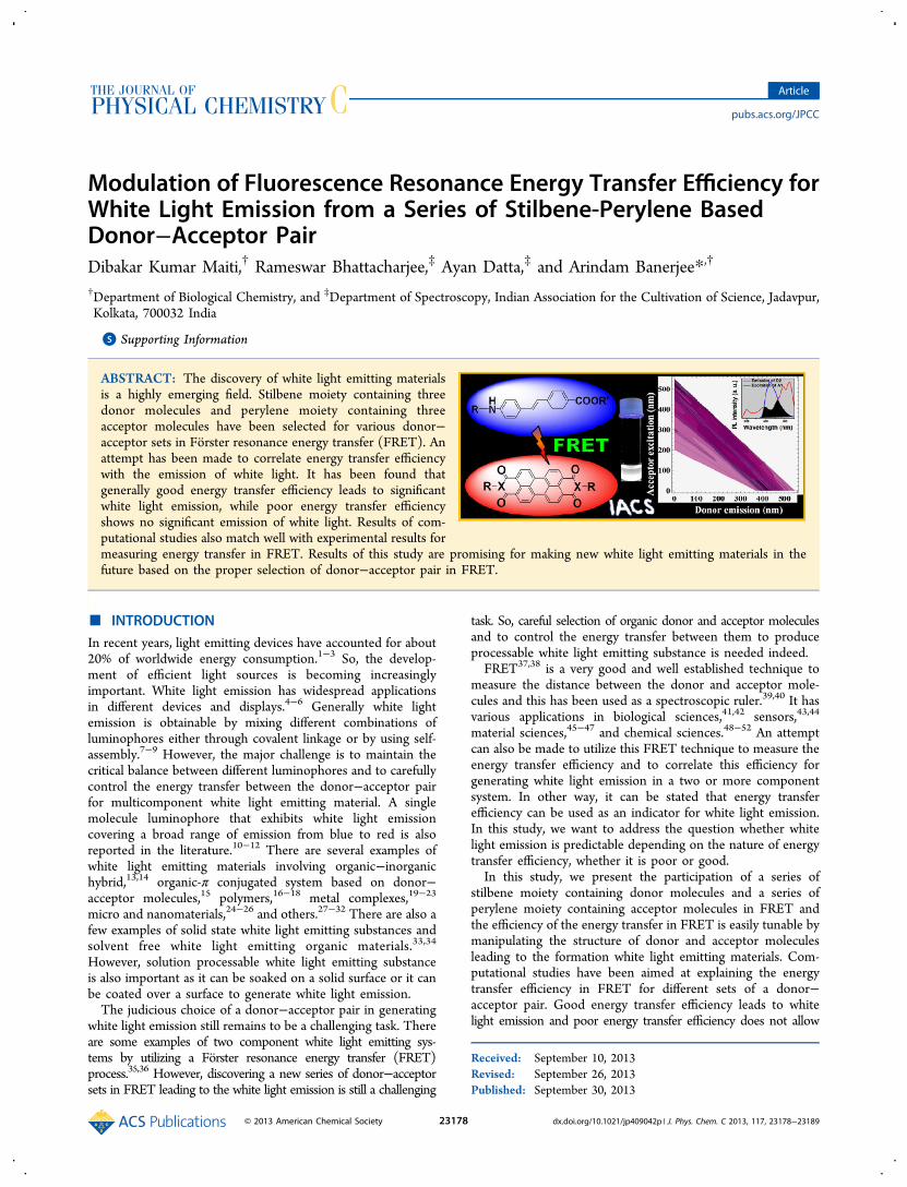
Modulation of Fluorescence Resonance Energy Transfer Efficiency for White Light Emission from a Series of Stilbene-Perylene Based Donor−Acceptor Pair Dibakar Kumar Maiti, † Rameswar Bhattacharjee, ‡ Ayan Datta, ‡ and Arindam Banerjee* ,† † Department of Biological Chemistry, and ‡ Department of Spectroscopy, Indian Association for the Cultivation of Science, Jadavpur, Kolkata, 700032 India * S Supporting Information ABSTRACT: The discovery of white light emitting materials is a highly emerging field. Stilbene moiety containing three donor molecules and perylene moiety containing three acceptor molecules have been selected for various donor− acceptor sets in Fö rster resonance energy transfer (FRET). An attempt has been made to correlate energy transfer efficiency with the emission of white light. It has been found that generally good energy transfer efficiency leads to significant white light emission, while poor energy transfer efficiency shows no significant emission of white light. Results of com- putational studies also match well with experimental results for measuring energy transfer in FRET. Results of this study are promising for making new white light emitting materials in the future based on the proper selection of donor−acceptor pair in FRET. ■ INTRODUCTION In recent years, light emitting devices have accounted for about 20% of worldwide energy consumption. 1−3 So, the develop- ment of efficient light sources is becoming increasingly important. White light emission has widespread applications in different devices and displays. 4−6 Generally white light emission is obtainable by mixing different combinations of luminophores either through covalent linkage or by using self- assembly. 7−9 However, the major challenge is to maintain the critical balance between different luminophores and to carefully control the energy transfer between the donor−acceptor pair for multicomponent white light emitting material. A single molecule luminophore that exhibits white light emission covering a broad range of emission from blue to red is also reported in the literature. 10−12 There are several examples of white light emitting materials involving organic−inorganic hybrid, 13,14 organic-π conjugated system based on donor− acceptor molecules, 15 polymers, 16−18 metal complexes, 19−23 micro and nanomaterials, 24−26 and others. 27−32 There are also a few examples of solid state white light emitting substances and solvent free white light emitting organic materials. 33,34 However, solution processable white light emitting substance is also important as it can be soaked on a solid surface or it can be coated over a surface to generate white light emission. The judicious choice of a donor−acceptor pair in generating white light emission still remains to be a challenging task. There are some examples of two component white light emitting sys- tems by utilizing a Fö rster resonance energy transfer (FRET) process. 35,36 However, discovering a new series of donor−acceptor sets in FRET leading to the white light emission is still a challenging task. So, careful selection of organic donor and acceptor molecules and to control the energy transfer between them to produce processable white light emitting substance is needed indeed. FRET 37,38 is a very good and well established technique to measure the distance between the donor and acceptor mole- cules and this has been used as a spectroscopic ruler. 39,40 It has various applications in biological sciences, 41,42 sensors, 43,44 material sciences, 45−47 and chemical sciences. 48−52 An attempt can also be made to utilize this FRET technique to measure the energy transfer efficiency and to correlate this efficiency for generating white light emission in a two or more component system. In other way, it can be stated that energy transfer efficiency can be used as an indicator for white light emission. In this study, we want to address the question whether white light emission is predictable depending on the nature of energy transfer efficiency, whether it is poor or good. In this study, we present the participation of a series of stilbene moiety containing donor molecules and a series of perylene moiety containing acceptor molecules in FRET and the efficiency of the energy transfer in FRET is easily tunable by manipulating the structure of donor and acceptor molecules leading to the formation white light emitting materials. Com- putational studies have been aimed at explaining the energy transfer efficiency in FRET for different sets of a donor− acceptor pair. Good energy transfer efficiency leads to white light emission and poor energy transfer efficiency does not allow Received: September 10, 2013 Revised: September 26, 2013 Published: September 30, 2013 Article pubs.acs.org/JPCC © 2013 American Chemical Society 23178 dx.doi.org/10.1021/jp409042p | J. Phys. Chem. C 2013, 117, 23178−23189
Transcript of Modulation of Fluorescence Resonance Energy Transfer Efficiency for White Light Emission from a...
Modulation of Fluorescence Resonance Energy Transfer Efficiency forWhite Light Emission from a Series of Stilbene-Perylene BasedDonor−Acceptor PairDibakar Kumar Maiti,† Rameswar Bhattacharjee,‡ Ayan Datta,‡ and Arindam Banerjee*,†
†Department of Biological Chemistry, and ‡Department of Spectroscopy, Indian Association for the Cultivation of Science, Jadavpur,Kolkata, 700032 India
*S Supporting Information
ABSTRACT: The discovery of white light emitting materialsis a highly emerging field. Stilbene moiety containing threedonor molecules and perylene moiety containing threeacceptor molecules have been selected for various donor−acceptor sets in Forster resonance energy transfer (FRET). Anattempt has been made to correlate energy transfer efficiencywith the emission of white light. It has been found thatgenerally good energy transfer efficiency leads to significantwhite light emission, while poor energy transfer efficiencyshows no significant emission of white light. Results of com-putational studies also match well with experimental results formeasuring energy transfer in FRET. Results of this study are promising for making new white light emitting materials in thefuture based on the proper selection of donor−acceptor pair in FRET.
■ INTRODUCTIONIn recent years, light emitting devices have accounted for about20% of worldwide energy consumption.1−3 So, the develop-ment of efficient light sources is becoming increasinglyimportant. White light emission has widespread applicationsin different devices and displays.4−6 Generally white lightemission is obtainable by mixing different combinations ofluminophores either through covalent linkage or by using self-assembly.7−9 However, the major challenge is to maintain thecritical balance between different luminophores and to carefullycontrol the energy transfer between the donor−acceptor pairfor multicomponent white light emitting material. A singlemolecule luminophore that exhibits white light emissioncovering a broad range of emission from blue to red is alsoreported in the literature.10−12 There are several examples ofwhite light emitting materials involving organic−inorganichybrid,13,14 organic-π conjugated system based on donor−acceptor molecules,15 polymers,16−18 metal complexes,19−23
micro and nanomaterials,24−26 and others.27−32 There are also afew examples of solid state white light emitting substances andsolvent free white light emitting organic materials.33,34
However, solution processable white light emitting substanceis also important as it can be soaked on a solid surface or it canbe coated over a surface to generate white light emission.The judicious choice of a donor−acceptor pair in generating
white light emission still remains to be a challenging task. Thereare some examples of two component white light emitting sys-tems by utilizing a Forster resonance energy transfer (FRET)process.35,36 However, discovering a new series of donor−acceptorsets in FRET leading to the white light emission is still a challenging
task. So, careful selection of organic donor and acceptor moleculesand to control the energy transfer between them to produceprocessable white light emitting substance is needed indeed.FRET37,38 is a very good and well established technique to
measure the distance between the donor and acceptor mole-cules and this has been used as a spectroscopic ruler.39,40 It hasvarious applications in biological sciences,41,42 sensors,43,44
material sciences,45−47 and chemical sciences.48−52 An attemptcan also be made to utilize this FRET technique to measure theenergy transfer efficiency and to correlate this efficiency forgenerating white light emission in a two or more componentsystem. In other way, it can be stated that energy transferefficiency can be used as an indicator for white light emission.In this study, we want to address the question whether whitelight emission is predictable depending on the nature of energytransfer efficiency, whether it is poor or good.In this study, we present the participation of a series of
stilbene moiety containing donor molecules and a series ofperylene moiety containing acceptor molecules in FRET andthe efficiency of the energy transfer in FRET is easily tunable bymanipulating the structure of donor and acceptor moleculesleading to the formation white light emitting materials. Com-putational studies have been aimed at explaining the energytransfer efficiency in FRET for different sets of a donor−acceptor pair. Good energy transfer efficiency leads to whitelight emission and poor energy transfer efficiency does not allow
Received: September 10, 2013Revised: September 26, 2013Published: September 30, 2013
Article
pubs.acs.org/JPCC
© 2013 American Chemical Society 23178 dx.doi.org/10.1021/jp409042p | J. Phys. Chem. C 2013, 117, 23178−23189
any emission of white light with only one exception out of ninedonor−acceptor pair. Moreover, most of these donor−acceptorsets (seven out of nine) under suitable conditions emit white light.
■ RESULTS AND DISCUSSION
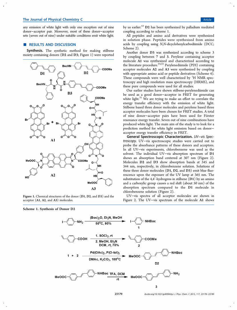
Synthesis. The synthetic method for making stilbenemoiety containing donors (D2 and D3; Figure 1) were reported
by us earlier.53 D2 has been synthesized by palladium mediatedcoupling according to scheme 1.All peptides and amino acid derivatives were synthesized
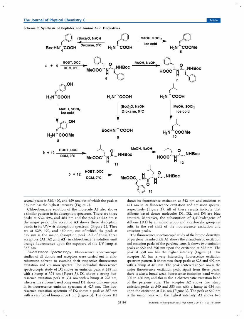
in solution phase. Peptides were synthesized from aminoacids by coupling using N,N-dicyclohexylcarbodiimide (DCC;Scheme 2).Another donor D3 was synthesized according to scheme 3
by coupling between 7 and 3. Perylene containing acceptormolecule A1 was synthesized and characterized according tothe literature procedure.54,55 Perylenediimide (PDI) containingacceptor molecules A2 and A3 were synthesized by couplingwith appropriate amino acid or peptide derivatives (Scheme 4).These compounds were well characterized by 1H NMR spec-troscopy and high resolution mass spectroscopy (HRMS), andthese pure compounds were used for all studies.Our earlier studies have shown stilbene-perylenediimide can
be used as a good donor−acceptor in FRET for generatingwhite light.31 We are trying to make an effort to correlate theenergy transfer efficiency with the emission of white light.Stilbene based three donor molecules and perylene based threeacceptor molecules have been chosen for FRET studies. A totalof nine donor−acceptor pairs have been used for Forsterresonance energy transfer. Seven out of nine combinations haveproduced white light. The main aim of the study is to look for aprediction method for white light emission based on donor−acceptor energy transfer efficiency in FRET.
General Spectroscopic Characterization. UV−vis Spec-troscopy. UV−vis spectroscopic studies were carried out toprobe the absorbance patterns of these donors and acceptors.In all UV−vis experiments, chlorobenzene was used as thesolvent. The individual UV−vis absorption spectrum of D1shows an absorption band centered at 307 nm (Figure 2).Molecules D2 and D3 show absorption bands at 345 and344 nm, respectively, in chlorobenzene solution. Solutions ofthese three donor molecules (D1, D2, and D3) emit blue fluo-rescence upon the exposure of the UV lamp at 365 nm. Thesubstitution of the 4,4′-hydrogens in stilbene (D1) by an aminoand a carboxylic group causes a red shift (about 50 nm) of theabsorption spectrum compared to the D1 molecule inchlorobenzene solution (Figure 2).UV−vis spectra of all acceptor molecules are shown in
Figure 2. The UV−vis spectrum of the molecule A1 showsFigure 1. Chemical structures of the donor (D1, D2, and D3) and theacceptor (A1, A2, and A3) molecules.
Scheme 1. Synthesis of Donor D2
The Journal of Physical Chemistry C Article
dx.doi.org/10.1021/jp409042p | J. Phys. Chem. C 2013, 117, 23178−2318923179
several peaks at 525, 490, and 459 nm, out of which the peak at525 nm has the highest intensity (Figure 2).Chlorobenzene solution of the molecule A2 also shows
a similar pattern in its absorption spectrum. There are threepeaks at 532, 495, and 464 nm and the peak at 532 nm isthe major peak. The acceptor A3 shows three absorptionbands in its UV−vis absorption spectrum (Figure 2). Theyare at 529, 490, and 460 nm, out of which the peak at529 nm is the major absorption peak. All of these threeacceptors (A1, A2 ,and A3) in chlorobenzene solution emitorange fluorescence upon the exposure of the UV lamp at365 nm.Fluorescence Spectroscopy. Fluorescence spectroscopic
studies of all donors and acceptors were carried out in chlo-robenzene solvent to examine their respective fluorescenceexcitation and emission spectra. The individual fluorescencespectroscopic study of D1 shows an emission peak at 358 nmwith a hump at 374 nm (Figure 3). D1 shows a strong fluo-rescence excitation peak at 331 nm with a hump at 296 nm,whereas the stilbene based compound D2 shows only one peakin its fluorescence emission spectrum at 423 nm. The fluo-rescence excitation spectrum of D2 shows a peak at 367 nmwith a very broad hump at 321 nm (Figure 3). The donor D3
shows its fluorescence excitation at 342 nm and emission at421 nm in its fluorescence excitation and emission spectra,respectively (Figure 3). All of these results indicate thatstilbene based donor molecules D1, D2, and D3 are blueemitters. Moreover, the substitution of 4,4′-hydrogens ofstilbene (D1) by an amino group and a carboxylic group re-sults in the red shift of the fluorescence excitation andemission peaks.The fluorescence spectroscopic study of the bromo derivative
of perylene bisanhydride A1 shows the characteristic excitationand emission peaks of the perylene core. It shows two emissionpeaks at 550 and 590 nm upon the excitation at 528 nm. Thepeak at 550 nm has the higher intensity (Figure 3). Thisacceptor A1 has a very interesting fluorescence excitationspectrum pattern. It shows two sharp peaks at 528 and 492 nmwith a hump at 461 nm. The peak centered at 528 nm is themajor fluorescence excitation peak. Apart from these peaks,there is also a broad weak fluorescence excitation band within300 to 450 nm, and this is also a characteristic excitation bandof the perylene core. The acceptor A2 shows two sharpemission peaks at 540 and 583 nm with a hump at 634 nmupon the excitation at 534 nm (Figure 3). The peak at 540 nmis the major peak with the highest intensity. A2 shows two
Scheme 2. Synthesis of Peptides and Amino Acid Derivatives
The Journal of Physical Chemistry C Article
dx.doi.org/10.1021/jp409042p | J. Phys. Chem. C 2013, 117, 23178−2318923180
major fluorescence excitation peaks at 534 and 495 nm with ahump at 464 nm. In this case, a weak broad fluorescence excita-tion is also observable from 300 to 400 nm. The acceptor A3exhibits two major emission peaks at 538 and 583 nm with abroad hump at 635 nm (Figure 3). The fluorescence excitationspectrum shows two major peaks at 529 and 490 nm with asmall hump at 460 nm. A3 also has a weak fluorescence excita-tion within 300 to 400 nm. The above-mentioned fluorescencespectroscopic study suggests that these three acceptors A1, A2,and A3 are orange fluorescence emitting materials in chloro-benzene solution. This is also supported by the observation ofbright orange fluorescence obtained for all these three acceptors(A1, A2, and A3) upon the exposure of the UV lamp at 365 nm(Figure 2). Quantum yields of each of these compounds aredetermined. These values are as follows: D1, 5.43%; D2, 5.34%;D3, 6.72%; A1, 39.60%; A2, 32.17%; and A3, 33.06%.Energy Transfer and White Light Processing Studies.
UV−vis Spectroscopy. UV−vis spectroscopic studies of allcombinations of these donors and acceptors were carried out toexamine spectral characteristic features of each of these donor−acceptor pair. Each combination of donor−acceptor pairremarkably shows a wide range of absorption in their respectiveUV−vis spectrum. In case of two donor−acceptor pair D1-A2and D1-A3, no white light emission is obtained upon theexposure of UV lamp (365 nm). Interestingly, for all other
donor−acceptor pair (D1-A2, D2-A1, D2-A2, D2-A3, D3-A1,D3-A2, and D3-A3), the respective absorption spectrum showsa wide range of absorption from 350 to 600 nm (Figure 4).A certain ratio of donor and acceptor component in each ofthese seven combinations emits white light in chlorobenzenesolution upon the exposure to a UV-lamp of 365 nm. UV−visspectra of all of these white light emitting solutions arepresented in Figure 4.
Fluorescence Spectroscopy. The fluorescence spectroscopyis a very informative tool to demonstrate the energy transfer be-tween a donor−acceptor pair (Figure 5). Efficient fluorescenceresonance energy transfer (FRET) occurred for six donor−acceptor pair (D2-A1, D2-A2, D2-A3, D3-A1, D3-A2, andD3-A3) and for other three pair (D1-A1, D1-A2m and D1-A3)no fluorescence energy transfer was taken place. Interestingly,only a certain ratio of donor and acceptor exhibits a white lightemission for each of these six pair. In this study, the D2-A1 pairis discussed in details as a representative example. The chloro-benzene solution of D2 (2.8 × 10⁻⁵ M) shows the fluorescenceemission peak at 423 nm upon excitation at its fluorescenceexcitation wavelength 367 nm. The gradual addition of A1 (7 ×10⁻⁶ to 4.2 × 10⁻⁵ M) causes a decrease in intensity of the peakat 423 nm (upon excitation at 367 nm) and peaks at 550 and590 nm are steadily increased in intensity. Thus, the fluo-rescence emission peak intensity at 423 nm decreases due tothe fluorescence resonance energy transfer from D2 to A1. Thistype of energy transfer observation demonstrates that D2 andD3 act as donor and A1, A2, A3 act as acceptor. For each com-bination of donor−acceptor pair (D2-A1, D2-A2, D2-A3,D3-A1, D3-A2, and D3-A3), the gradual addition of the acceptorto the donor is associated with a steady decrease in intensity ofthe donor’s fluorescence emission peak with the steady increaseof the intensity of acceptor’s emission peak (Figure 5). It isinteresting to observe that combinations with D1 is notsuccessful for energy transfer due to the poor overlaps of thedonor’s emission spectrum and acceptor’s excitation spectrum(Figure 5). The substitution of 4,4′-hydrogens in stilbene (D1)by an amino group and a carboxylic group in D2 and D3 results
Scheme 3. Synthesis Scheme of Donor D3
Scheme 4. Synthesis Scheme of Acceptors A1, A2, and A3
Figure 2. UV−vis absorption spectra of donors D1 (a), D2 (b), and D3 (c) and acceptors A1 (d), A2 (e), and A3 (f) in chlorobenzene solution.
The Journal of Physical Chemistry C Article
dx.doi.org/10.1021/jp409042p | J. Phys. Chem. C 2013, 117, 23178−2318923181

in a red shift of the corresponding fluorescence emission peakof donors D2 and D3. This is actually responsible for the betteroverlap between the fluorescence excitation peaks of theacceptor (A1, A2, and A3) molecules with each of these donor(D2 and D3) molecules’ emission. So, it can be stated thatefficient energy transfers occur involving those donor−acceptorpair, which contains either D2 or D3 as the donor molecule.
Comparison between the UV−vis Spectra andFluorescence Excitation Spectra. Comparison betweenthe UV−vis absorption spectra and fluorescence excitationspectra for three donor and acceptor containing solutionswere performed. All these experiments were carried outwith a common acceptor molecule A2 and three differentdonor molecules (D1, D2, and D3). The UV−vis spectrum
Figure 3. Fluorescence excitation and emission spectra of donors D1 (a), D2 (b), and D3 (c) and acceptors A1 (d), A2 (e), and A3 (f) inchlorobenzene solution. Excitation and emission spectra are indicated within corresponding figures.
Figure 4. UV−vis absorption spectra of white light emitting solutions of different pair of donors and acceptors: D2-A1 (a), D2-A3 (b), D2-A2 (c),D3-A1 (d), D3-A3 (e), D3-A2 (f), and D1-A1 (g). (h) White light emitting solutions exposed to the UV lamp at 365 nm.
The Journal of Physical Chemistry C Article
dx.doi.org/10.1021/jp409042p | J. Phys. Chem. C 2013, 117, 23178−2318923182

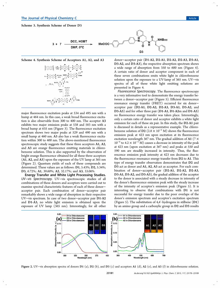
of D1-A2 pair contains all peaks corresponding to D1 aswell as A2.However, the fluorescence excitation of the same solution
exhibits only those peaks which are corresponding to theacceptor’s (A2) excitation wavelength (Figure 6) for emissionat 547 nm. This indicates that practically there is no energytransfer between the D1-A2 pair. Interestingly, for the donor−acceptor pair D2-A2 the fluorescence excitation spectrumshows the major peak at 370 nm corresponding to the excita-tion wavelength of the donor D2. This convincingly suggeststhat FRET is occurring between D2 and A2 leading to thesignificant contribution of the donor D2 in the emissionspectrum of acceptor A2 at 540 nm. Similar observation is alsofound for the D3-A2 donor−acceptor pair. These observationsclearly suggest that significant energy transfer is occurringinvolving donors D2 or D3 and A2, whereas no energy transferis apparent involving the donor D1 and acceptor A2. To ex-plain these phenomenon the overlap integrals, Forster distancesand maximum energy transfer efficiencies were calculatedexperimentally for each donor−acceptor pair.Overlap Integrals, Forster Distances, and Maximum
Energy Transfer Efficiency. There are overlaps between thefluorescence excitation spectrum of acceptor molecule and fluo-rescence emission spectrum of the donor molecule (Figure 7),
and this is very important for the occurrence of a successful re-sonance energy transfer. The FRET parameters overlap integral(J), Forster distances (R0), and intermolecular distances (r) aredetermined by using equations S1, S2 and S3 mentioned in theSupporting Information. The energy transfer efficiencies weredetermined by using the following equation:37,38
= −EFF
1 DA
D (1)
In this case for each donor−acceptor the fluorescenceemission spectrum of the donor and the fluorescence excitationspectrum of the acceptor were plotted in the same graph atnormalized condition (Figure 7) and the overlap integrals werecalculated. It is evident from figure 7 that the spectral over-laps are quite sufficient for the combinations D2-A1, D2-A2,D2-A3, D3-A1, D3-A2, and D3-A3 that leads to the efficientenergy transfer from donor to acceptor. However, spectraloverlaps are not sufficient for D1-A1, D1-A2, and D1-A3 pairleading no significant transfer to occur. Table 1 demonstratesthe efficiency of energy transfer among each pair of donor andacceptor.The energy transfer efficiency calculations from experimental
values has confirmed that the efficiencies of energy transferfrom donor to acceptor are decreasing with an increase in the
Figure 5. Changes in fluorescence spectra of donors upon the gradual addition of acceptor to the respective donors: D1-A1 (a), D1-A2 (b), D1-A3(c), D2-A1 (d), D2-A2 (e), D2-A3 (f), D3-A1 (g), D3-A2 (h), and D3-A3 (i). Added concentration of the corresponding acceptors are indicated inrespective figures.
The Journal of Physical Chemistry C Article
dx.doi.org/10.1021/jp409042p | J. Phys. Chem. C 2013, 117, 23178−2318923183
intermolecular distances (r) between the donor and acceptormolecule. Moreover, the plot of energy transfer efficiencyversus intermolecular distance show the low RET between D1and other acceptors (A1, A2, and A3; Figure 8). This plot(Figure 8) is potentially important for measuring distance of20−40 Å, that indicates the donor−acceptor set can be used asa spectroscopic ruler.Time Correlated Single Photon Count (TCSPC) Study.
Average fluorescence lifetimes were determined by performingthe TCSPC experiments. In this study, chlorobenzene was alsotaken as the solvent to carry out all experiments. Only D1, D2,A1, and A2 were examined for this purpose. For measuring the
lifetime of D1 and D2, samples were excited at 340 and 375 nmrespectively nearer to their excitation wavelength. The averagelifetime of D1 and D2 were 0.18 ns (recorded at 358 nm) and0.21 ns (recorded at 423 nm) respectively. To measure theaverage fluorescence lifetime of acceptors the samples wereexcited at 440 nm and the emissions were recorded at 550 nm.The average lifetime of A1 and A2 are estimated to be 5.36 and5.38 ns, respectively (Figure 9).However, in presence of the acceptor the average fluo-
rescence lifetimes of donors are changed. It is obtained fromthe data that the average fluorescence lifetime of the donor wasdecreased in presence of the acceptor (Figure 10).Moreover, the energy transfer efficiencies for each combina-
tion were determined by using the following equation:37,38
ττ
= −E 1 DA
D (2)
where E is the energy transfer efficiency, τDA is the donor’slifetime in presence of the acceptor, and τD is the donor’slifetime in absence of the acceptor.The results are given in Table2.It is evident from Table 2 that the donor−acceptor consisting
of D2 are very efficient for energy transfer, whereas the D1containing donor−acceptor pair are less efficient for the energytransfer. These results are matched quite well with the energytransfer efficiency data obtained by using integral areacalculation method in eq 1.
Computational Insights. For a microscopic understandingof the mechanism of RET in our systems, DFT calculationswere performed at the M06-2X/6-31+g(d,p) level of theoryusing the Gaussian 09 suite of programs.56 The ability of theM06-2X functional to account for dispersion interactions inπ-stacked systems has been well-established.57−59 Time-dependent DFT (TDDFT) calculations were performed tomodel the emission and absorption process of the donor andacceptor by considering 20 excited states each. For making thecalculations computationally tractable, −BOC and −CH3groups in D2 were substituted by hydrogens, whereasperylenebisanhydride and perylenediimide were chosen asmodels for A1 and A2, respectively. Table 3 shows theimportant electronic transitions for D1, D2, A1, and A2alongwith their emission/absorption wavelengths and oscillatorstrengths. For both absorption and emission, electronictransitions are composed of entirely HOMO → LUMO andLUMO → HOMO levels, respectively. Figure S11 shows theHOMO and LUMO plots for D1, D2, A1, and A2.Figure 11 shows the donor emission and acceptor excitation
profiles for the D1-A1, D1-A2, D2-A1, and D2-A2 pair. Fromboth the contour plots as well as the spectral plots, goodoverlap for the D2−A1 and D2−A2 pair are evident, whereasthe overlap is poor for D1-A1 and D1-A2. Agreement betweenthe experimental and the theoretical plots are excellent whichsuggests that the TDDFT calculations on the model systemscapture the essential excitation profile for our systems. A pooroverlap of the donor emission and the acceptor absorption forthe D1-A1 and D1-A2 pair explains their low resonance energytransfer (RET).So, as to understand the local interactions between the
“effective” FRET pair namely D2-A1 and D2-A2, structuresfor the molecular dimers were optimized. As it is seen fromFigure 12, donors and acceptors are arranged in an almostperfectly parallel π-stacked fashion where a possibility of inter-molecular N−H···O hydrogen bonding is present. Hydrogen
Figure 6. Comparison between PL excitation spectrum and UV−visabsorption spectrum of various donors with the acceptor A2: D1-A2(a), D2-A2 (b), and D3-A2 (c).
The Journal of Physical Chemistry C Article
dx.doi.org/10.1021/jp409042p | J. Phys. Chem. C 2013, 117, 23178−2318923184

bonding interaction between donor and acceptor moleculescan play an important role in enhancing the efficiency of theFRET.60−62 The average distance between the pair are ∼3.3and 3.5 Å for D2-A1 and D2-A2, respectively. The bindingenergies (BSSE corrected) for D2-A1 and D2-A2 are calculatedto be −19.9 and −22.1 kcal/mol, respectively. Hence, sub-stantial stabilization of the donor and acceptor pair by van derWaals interactions due to close proximity between the donorand the acceptor molecules facilitates highly efficient RET.Transition charge densities between the ground and excitedstates for D2 and A2 are calculated within the TDDFT method.The Coulombic coupling matrix elements between D2 and A2are calculated using the transition density cube (TDC)formalism63 for which grid size of 20 × 20 × 20 was used.For understanding the variation of the Coulomb coupling(VDA) between the donor (D2) and the acceptor (A2) withincrease in separation between them, the distance between D2and A2 (rDA) was increased from an initial 3.5 Å to 100 Å in theparallel π-stacked configuration. Within the TDC method the
overlap between the molecular orbital of the donor andacceptor is explicitly considered.VDA is essentially an electrostatic coupling term between
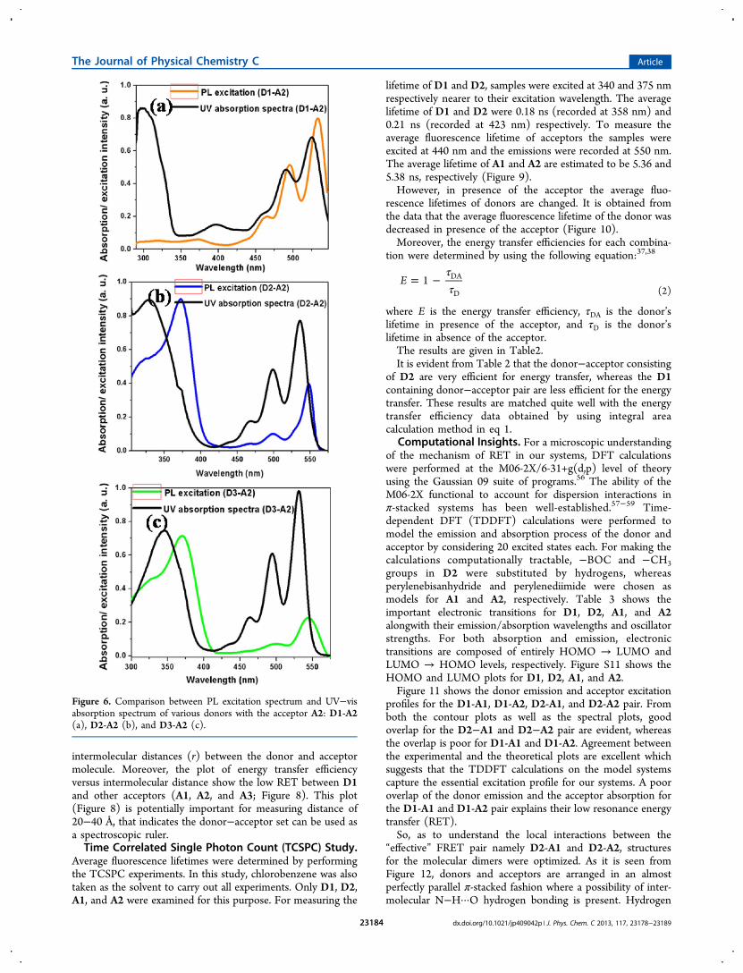
transition charges densities scaled by Coulomb forces, andtherefore, VDA has a monotonically decaying behavior.64,65 Forcomparison with the experimental data, the percentage ofenergy transfer between D2 and A2 was theoretically estimatedas: % of RET at distance r = VDA(r) × 100/VDA (r = 3.5 Å).Since, the equilibrium intermolecular distance between themonomers is calculated as 3.5 Å (structure shown in Figure 12),maximum RET (100%) must occur at r = 3.5 Å.Figure 13 shows the variation in the experimental and
theoretical percentage of energy transfer for the D2-A2 pair
Figure 7. Overlaps between various emission spectra of donors and excitation spectra of acceptors.
Table 1. Energy Transfer Efficiency (E) and Forster Distance(R0) Calculation Data for Each Pair of Donor and Acceptora
donor acceptor J(λ) in (M−1 cm−1 nm4) R0 in Å max E
D1 A1 1.36 × 10¹⁴ 20.74 0.18D1 A2 3.27 × 10¹³ 16.36 0.27D1 A3 3.64 × 10¹³ 16.65 0.27D2 A1 4.42 × 10¹⁴ 25.16 0.48D2 A2 2.99 × 10¹⁴ 23.57 0.60D2 A3 2.60 × 10¹⁴ 23.04 0.65D3 A1 6.24 × 10¹⁴ 27.71 0.44D3 A2 1.23 × 10¹⁴ 21.14 0.46D3 A3 2.21 × 10¹⁴ 23.32 0.59
aκ2 = 2/3, η = 1.5248.
Figure 8. Energy transfer efficiency vs intermolecular distance plots fordifferent donor−acceptor pairs.
The Journal of Physical Chemistry C Article
dx.doi.org/10.1021/jp409042p | J. Phys. Chem. C 2013, 117, 23178−2318923185

with rDA. For both the cases, the data are fitted to a power lawof the form: arb. For the experimental percentages of RET, a =3.24 and b = −3.52 ± 0.10, whereas for the theoretical data, a =350 ± 7.31 and b = −1.00 ± 0.01. Clearly, our results stronglydeviate from the well-known r−6 behavior expected from asimple dipole−dipole interaction model within the Forstermodel.37,38 Higher RET efficiencies for the experimental pairand its faster decay rate with rDA compared to the theoretical
Figure 9. Time correlated single photon count (TCSPC) decay profileof different donors and acceptors recorded for individual samples.
Figure 10. Changes in fluorescence decay profile of different pair ofdonor−acceptor in chlorobenzene.
Table 2. Lifetimes, Energy Transfer Efficiencies, andDonor’s Lifetime for Different Donor−Acceptor Pairs
pair τD (ns) τDA (ns) E = 1 − (τDA/τD) % of E
D1-A1 0.181 0.145 0.199 19.9D1-A2 0.181 0.132 0.2707 27.07D2-A1 0.21 0.101 0.519 51.9D2-A2 0.21 0.0995 0.526 52.6
Table 3. Electronic States Involved in Absorption/Emission,Oscillator Strength ( f), and Wavelength for Transition(in nm)
molecule electronic transitions oscillator strength ( f) λmax (nm)
D1 LUMO → HOMO (100%) 0.90 356.43D2 LUMO → HOMO (100%) 1.23 391.86A1 HOMO → LUMO (100%) 0.71 441.76A2 HOMO → LUMO (100%) 0.73 446.23
Figure 11. Contour plots and spectral overlap (experimental andtheoretical) for the donor emission and acceptor excitations for (a)D1−A1, (b) D1−A2, (c) D2−A1, and (d) D2−A2 pairs.
Figure 12. Minimum energy π-stacked structures for D2-A1 andD2-A2 dimers.
The Journal of Physical Chemistry C Article
dx.doi.org/10.1021/jp409042p | J. Phys. Chem. C 2013, 117, 23178−2318923186
values may arise due to the possibility of formation of multipleD2 − A2 pair due to aggregation. Such an aggregation oftenleads to the rapid energy transfer via a funnel like structure as isknown for the photosynthetic proteins.66 Computationalstudies match quite well with our experimental results for themeasurement of the efficiency of the energy transfer. Bothresults suggest that D1-A1 and D1-A2 are very poor com-binations for FRET, whereas D2-A1 and D2-A2 are excellentdonor−acceptor pairs for FRET indicating the role of D1 as apoor donor and D2 as an excellent donor.CIE Coordinates. The fluorescence resonance energy trans-
fer between donor and acceptor for D2-A1, D2-A2, D2-A3,D3-A1, D3-A2, and D3-A3 leads to the generation of a whitelight emission. Each of these six combinations was able toproduce bright white light. The CIE (1931) coordinates foreach pair with certain concentration was given in Table 4, andthe CIE coordinate figures were given in the SupportingInformation as Figure S12. Combination involving the donorD1, only the D1-A1 produces an emission of white light thougha very poor energy transfer occurred between the donor andacceptor. This may be due to the combination of twocomplementary colors. However, the other two combinationsinvolving D1 like D1-A2 and D1-A3, do not produce any whitelight emission under similar conditions.
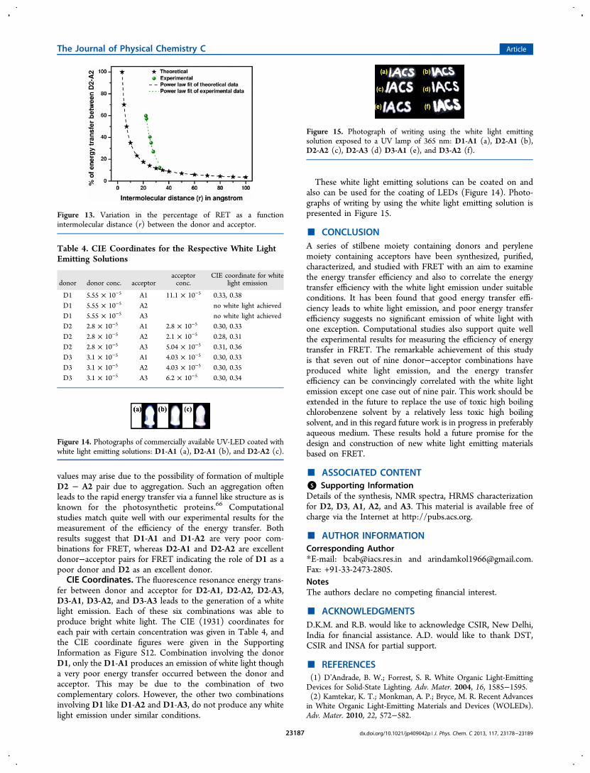
These white light emitting solutions can be coated on andalso can be used for the coating of LEDs (Figure 14). Photo-graphs of writing by using the white light emitting solution ispresented in Figure 15.
■ CONCLUSIONA series of stilbene moiety containing donors and perylenemoiety containing acceptors have been synthesized, purified,characterized, and studied with FRET with an aim to examinethe energy transfer efficiency and also to correlate the energytransfer efficiency with the white light emission under suitableconditions. It has been found that good energy transfer effi-ciency leads to white light emission, and poor energy transferefficiency suggests no significant emission of white light withone exception. Computational studies also support quite wellthe experimental results for measuring the efficiency of energytransfer in FRET. The remarkable achievement of this studyis that seven out of nine donor−acceptor combinations haveproduced white light emission, and the energy transferefficiency can be convincingly correlated with the white lightemission except one case out of nine pair. This work should beextended in the future to replace the use of toxic high boilingchlorobenzene solvent by a relatively less toxic high boilingsolvent, and in this regard future work is in progress in preferablyaqueous medium. These results hold a future promise for thedesign and construction of new white light emitting materialsbased on FRET.
■ ASSOCIATED CONTENT*S Supporting InformationDetails of the synthesis, NMR spectra, HRMS characterizationfor D2, D3, A1, A2, and A3. This material is available free ofcharge via the Internet at http://pubs.acs.org.
■ AUTHOR INFORMATIONCorresponding Author*E-mail: [email protected] and [email protected]: +91-33-2473-2805.NotesThe authors declare no competing financial interest.
■ ACKNOWLEDGMENTSD.K.M. and R.B. would like to acknowledge CSIR, New Delhi,India for financial assistance. A.D. would like to thank DST,CSIR and INSA for partial support.
■ REFERENCES(1) D’Andrade, B. W.; Forrest, S. R. White Organic Light-EmittingDevices for Solid-State Lighting. Adv. Mater. 2004, 16, 1585−1595.(2) Kamtekar, K. T.; Monkman, A. P.; Bryce, M. R. Recent Advancesin White Organic Light-Emitting Materials and Devices (WOLEDs).Adv. Mater. 2010, 22, 572−582.
Figure 13. Variation in the percentage of RET as a functionintermolecular distance (r) between the donor and acceptor.
Table 4. CIE Coordinates for the Respective White LightEmitting Solutions
donor donor conc. acceptoracceptorconc.
CIE coordinate for whitelight emission
D1 5.55 × 10⁻⁵ A1 11.1 × 10⁻⁵ 0.33, 0.38D1 5.55 × 10⁻⁵ A2 no white light achievedD1 5.55 × 10⁻⁵ A3 no white light achievedD2 2.8 × 10⁻⁵ A1 2.8 × 10⁻⁵ 0.30, 0.33D2 2.8 × 10⁻⁵ A2 2.1 × 10⁻⁵ 0.28, 0.31D2 2.8 × 10⁻⁵ A3 5.04 × 10⁻⁵ 0.31, 0.36D3 3.1 × 10⁻⁵ A1 4.03 × 10⁻⁵ 0.30, 0.33D3 3.1 × 10⁻⁵ A2 4.03 × 10⁻⁵ 0.30, 0.35D3 3.1 × 10⁻⁵ A3 6.2 × 10⁻⁵ 0.30, 0.34
Figure 14. Photographs of commercially available UV-LED coated withwhite light emitting solutions: D1-A1 (a), D2-A1 (b), and D2-A2 (c).
Figure 15. Photograph of writing using the white light emittingsolution exposed to a UV lamp of 365 nm: D1-A1 (a), D2-A1 (b),D2-A2 (c), D2-A3 (d) D3-A1 (e), and D3-A2 (f).
The Journal of Physical Chemistry C Article
dx.doi.org/10.1021/jp409042p | J. Phys. Chem. C 2013, 117, 23178−2318923187

(3) Costa, R. D.; Ortι, E.; Bolink, H. J.; Monti, F.; Accorsi, G.;Armaroli, N. Luminescent Ionic Transition-Metal Complexes forLight-Emitting Electrochemical Cells. Angew. Chem., Int. Ed. 2012, 51,8178−8121.(4) Kido, J.; Kimura, M.; Nagai, K. Multilayer White Light-EmittingOrganic Electroluminescent Device. Science 1995, 267, 1332−1334.(5) Gather, M. C.; Kohnen, A.; Meerholz, M. White Organic Light-Emitting Diodes. Adv. Mater. 2011, 23, 233−248.(6) Sun, Y.; Giebink, N. C.; Kanno, H.; Ma, B.; Thompson, M. E.;Forrest, S. R. Management of singlet and triplet excitons for efficientwhite organic light-emitting devices. Nature 2006, 440, 908−912.(7) Vijayakumar, C.; Praveen, V. K.; Ajayaghosh, A. RGB Emissionthrough Controlled Donor Self-Assembly and Modulation ofExcitation Energy Transfer: A Novel Strategy to White-Light-EmittingOrganogels. Adv. Mater. 2009, 21, 2059−2063.(8) Abbel, R.; Weegen, R. v. d.; Pisula, W.; Surin, M.; Leclere, P.;Lazzaroni, R.; Meijer, E. W.; Schenning, A. P. H. J. Multicolour Self-Assembled Fluorene Co-Oligomers: From Molecules to the SolidState via White-Light-Emitting Organogels. Chem.Eur. J. 2009, 15,9737−9746.(9) Giansante, C.; Raffy, G.; Schafer, C.; Rahma, H.; Kao, M.-T.;Olive, A. G. L.; Guerzo, A. D. White-Light-Emitting Self-AssembledNanoFibers and Their Evidence by Microspectroscopy of IndividualObjects. J. Am. Chem. Soc. 2011, 133, 316−325.(10) Shelton, A. H.; Sazanovich, I. V.; Weinstein, J. A.; Ward, M. D.Controllable three-component luminescence from a 1,8-naprhthali-mide/Eu(III) complex: white light emission from a single molecule.Chem. Commun. 2012, 48, 2749−2751.(11) Liu, Y.; Nishiura, M.; Wang, Y.; Hou, Z. π-Conjugated AromaticEnynes as a Single-Emitting Component for White Electrolumines-cence. J. Am. Chem. Soc. 2006, 128, 5592−5593.(12) Huynh, H. V.; He, X.; Baumgartner, T. Halochromic generationof white light emission using a single dithienophosphole luminophore.Chem. Commun. 2013, 49, 4899−4901.(13) Ki, W.; Li, J.; Eda, G.; Chhowalla, M. Direct white light emissionfrom inorganic−organic hybrid semiconductor bulk materials. J. Mater.Chem. 2010, 20, 10676−10679.(14) Huyal, I. O.; Koldemir, U.; Ozel, T.; Demir, H. V.; Tuncel, D.On the origin of high quality white light emission from a hybridorganic/inorganic light emitting diode using azide functionalizedpolyfluorene. J. Mater. Chem. 2008, 18, 3568−3574.(15) Abbel, R.; Grenier, C.; Pouderoijen, M. J.; Stouwdam, J. W.;Leclere, P. E. L. G.; Sijbesma, R. P.; Meijer, E. W.; Schenning, A. P. H.J. White-Light Emitting Hydrogen-Bonded Supramolecular Copoly-mers Based on π-Conjugated Oligomers. J. Am. Chem. Soc. 2009, 131,833−843.(16) Tu, G.; Mei, C.; Zhou, Q.; Cheng, Y.; Geng, Y.; Wang, L.; Ma,D.; Jing, X.; Wang, F. Highly Efficient Pure-White-Light-EmittingDiodes from a Single Polymer: Polyfluorene with NaphthalimideMoieties. Adv. Funct. Mater. 2006, 16, 101−106.(17) Liu, J.; Guo, X.; Bu, L.; Xie, Z.; Cheng, Y.; Geng, Y.; Wang, L.;Jing, X.; Wang, F. White Electroluminescence from a Single-PolymerSystem with Simultaneous Two-Color Emission: Polyfluorene as BlueHost and 2,1,3-Benzothiadiazole Derivatives as Orange Dopants onthe Side Chain. Adv. Funct. Mater. 2007, 17, 1917−1925.(18) Shih, H.-M.; Wu, R.-C.; Shih, P.-I.; Wang, C.-L.; Hsu, C.-S.Synthesis of Fluorene-based Hyperbranched Polymers for Solution-Processable Blue, Green, Red, and White Light-Emitting Devices. J.Polym. Sci., Part A: Polym. Chem. 2012, 50, 696−710.(19) He, G.; Guo, D.; He, C.; Zhang, X.; Zhao, X.; Duan, C. A Color-Tunable Europium Complex Emitting Three Primary Colors andWhite Light. Angew. Chem. 2009, 121, 6248−6251.(20) Kim, T.-H.; Lee, H. K.; Park, O. O.; Chin, B. D.; Lee, S.-H.;Kim, J. K. White-Light-Emitting Diodes Based on Iridium Complexesvia Efficient Energy Transfer from a Conjugated Polymer. Adv. Funct.Mater. 2006, 16, 611−617.(21) Ho, C.-L.; Wong, W.-Y.; Wang, Q.; Ma, D.; Wang, L.; Lin, Z. AMultifunctional Iridium-Carbazolyl Orange Phosphor for High-Performance Two-Element WOLED Exploiting Exciton-Managed
Fluorescence/Phosphorescence. Adv. Funct. Mater. 2008, 18, 928−937.(22) Sykes, D.; Tidmarsh, I. S.; Barbieri, A.; Sazanovich, I. V.;Weinstein, J. A.; Ward, M. D. d-f Energy Transfer in a Series of IrIII/EuIII Dyads: Energy-Transfer Mechanisms and White-Light Emission.Inorg. Chem. 2011, 50, 11323−11339.(23) Hou, L.; Duan, L.; Qiao, J.; Zhang, D.; Wang, L.; Cao, Y.; Qiu,Y. Efficient solution-processed phosphor-sensitized single-emitting-layer white organic light-emitting devices: fabrication, characteristics,and transient analysis of energy transfer. J. Mater. Chem. 2011, 21,5312−5318.(24) Lei, Y.; Liao, Q.; Fu, H.; Yao, J. Orange-Blue-Orange TriblockOne-Dimensional Heterostructures of Organic Microrods for White-Light Emission. J. Am. Chem. Soc. 2010, 132, 1742−1743.(25) Zhao, Y. S.; Fu, H.; Hu, F.; Peng, A.; Yang, W.; Yao, J. TunableEmission from Binary Organic One-Dimensional Nanomaterials: AnAlternative Approach to White-Light Emission. Adv. Mater. 2008, 20,79−83.(26) Sun, H.; Zhang, H.; Zhang, J.; Wei, H.; Ju, J.; Li, M.; Yang, B.White-light emission nanofibers obtained from assembling aqueoussingle-colored CdTe NCs into a PPV precursor and PVA matrix. J.Mater. Chem. 2009, 19, 6740−6744.(27) Ki, W.; Li, J. A Semiconductor Bulk Material That Emits DirectWhite Light. J. Am. Chem. Soc. 2008, 130, 8114−8115.(28) Rao, K. V.; Datta, K. K. R.; Eswaramoorthy, M.; George, S. J.Highly Pure Solid-State White-Light Emission from Solution-Processable Soft-Hybrids. Adv. Mater. 2013, 25, 1713−1718.(29) Vijayakumar, C.; Sugiyasu, K.; Takeuchi, M. Oligofluorene-based electrophoretic nanoparticles in aqueous medium as a donorscaffold for fluorescence resonance energy transfer and white-lightemission. Chem. Sci. 2011, 2, 291−294.(30) Roushan, M.; Zhang, X.; Li, J. Solution-Processable White-Light-Emitting Hybrid Semiconductor Bulk Materials with HighPhotoluminescence Quantum Efficiency. Angew. Chem., Int. Ed. 2012,51, 436−439.(31) Maiti, D. K.; Banerjee, A. A peptide based two component whitelight emitting system. Chem. Commun. 2013, 49, 6909−6911.(32) Fang, X.; Roushan, M.; Zhang, R.; Peng, J.; Zeng, H.; Li, J.Tuning and Enhancing White Light Emission of II−VI BasedInorganic−Organic Hybrid Semiconductors as Single-Phased Phos-phors. Chem. Mater. 2012, 24, 1710−1717.(33) Babu, S. S.; Aimi, J.; Ozawa, H.; Shirahata, N.; Saeki, A.; Seki, S.;Ajayaghosh, A.; Mohwald, H.; Nakanishi, T. Solvent-Free LuminescentOrganic Liquids. Angew. Chem., Int. Ed. 2012, 51, 3391−3395.(34) Liu, S.; Li, F.; Diao, Q.; Ma, Y. Aggregation-induced enhancedemission materials for efficient white organic light-emitting devices.Org. Electron. 2010, 11, 613−617.(35) Park, S.; Kwon, J. E.; Kim, S. H.; Seo, J.; Chung, K.; Park, S.-Y.;Jang, D.-J.; Medina, B. M.; Gierschner, J.; Park, S. Y. A White-Light-Emitting Molecule: Frustrated Energy Transfer between ConstituentEmitting Centers. J. Am. Chem. Soc. 2009, 131, 14043−14049.(36) Wang, R.; Peng, J.; Qiu, F.; Yang, Y. Enhanced white-lightemission from multiple fluorophores encapsulated in a single layer ofdiblock copolymer micelles. Chem. Commun. 2011, 47, 2787−2789.(37) Forster, T. Transfer Mechanisms of Electronic Excitation.Discuss. Faraday Soc. 1959, 27, 7−17.(38) Lakowicz, J. R. Principles of Fluorescence Spectroscopy, 2nd ed.;Kluwer/Plenum: New York, 1999.(39) Stryer, L. Fluorescence Energy Transfer as a SpectroscopicRuler. Annu. Rev. Biochem. 1978, 47, 819−846.(40) Sahoo, H. Forster resonance energy transfer − A spectroscopicnanoruler: Principle and applications. J. Photochem. Photobiol. C 2011,12, 20−30.(41) Yuan, L.; Lin, W.; Zheng, K.; Zhu, S. FRET-Based Small-Molecule Fluorescent Probes: Rational Design and BioimagingApplications. Acc. Chem. Res. 2013, 46, 1462−1473.(42) Lu, S.; Wang, Y.; Huang, H.; Pan, Y.; Chaney, E. J.; Boppart, S.A.; Ozer, H.; Strongin, A. Y.; Wang, Y. Quantitative FRET Imaging to
The Journal of Physical Chemistry C Article
dx.doi.org/10.1021/jp409042p | J. Phys. Chem. C 2013, 117, 23178−2318923188

Visualize the Invasiveness of Live Breast Cancer Cells. PLOS One2013, 8, e58569(1−9).(43) Yuan, L.; Lin, W.; Chen, B.; Xie, Y. Development of FRET-Based Ratiometric Fluorescent Cu2+ Chemodosimeters and theApplications for Living Cell Imaging. Org. Lett. 2012, 14, 432−435.(44) Suzuki, M.; Husimi, Y.; Komatsu, H.; Suzuki, K.; Douglas, K. T.Quantum Dot FRET Biosensors that Respond to pH, to Proteolytic orNucleolytic Cleavage, to DNA Synthesis, or to a MultiplexingCombination. J. Am. Chem. Soc. 2008, 130, 5720−5725.(45) Huebsch, N. D.; Mooney, D. J. Fluorescent Resonance EnergyTransfer: A Tool for Probing Molecular Cell-Biomaterial Interactionsin Three Dimensions. Biomaterials 2007, 28, 2424−2437.(46) Sahoo, H.; Nau, W. M. Phosphorylation-Induced Conforma-tional Changes in Short Peptides Probed by Fluorescence ResonanceEnergy Transfer in the 10 Å Domain. ChemBioChem 2007, 8, 567−573.(47) Sahoo, H.; Roccatano, D.; Hennig, A.; Nau, W. M. A 10-ÅSpectroscopic Ruler Applied to Short Polyprolines. J. Am. Chem. Soc.2007, 129, 9762−9772.(48) Ward, M. D. Photo-induced electron and energy transfer in non-covalently bonded supramolecular assemblies. Chem. Soc. Rev. 1997,26, 365−375.(49) Praveen, V. K.; George, S. J.; Varghese, R.; Vijayakumar, C.;Ajayaghosh, A. Self-Assembled π-Nanotapes as Donor Scaffolds forSelective and Thermally Gated Fluorescence Resonance EnergyTransfer (FRET). J. Am. Chem. Soc. 2006, 128, 7542−7550.(50) Guerzo, A. D.; Olive, A. G. L.; Reichwagen, J.; Hopf, H.;Desvergne, J.-P. Energy Transfer in Self-Assembled [n]-Acene FibersInvolving ≥100 Donors Per Acceptor. J. Am. Chem. Soc. 2005, 127,17984−17985.(51) Hippius, C.; van Stokkum, I. H. M.; Gsanger, M.; Groeneveld,M. M.; Williams, R. M.; Wurthner, F. Sequential FRET Processes inCalix[4]arene-Linked Orange-Red-Green Perylene Bisimide DyeZigzag Arrays. J. Phys. Chem. C 2008, 112, 2476−2486.(52) Samanta, S. K.; Bhattacharya, S. Wide-Range Light-HarvestingDonor−Acceptor Assemblies through Specific Intergelator Interac-tions via Self-Assembly. Chem.Eur. J. 2012, 18, 15875−15885.(53) Maiti, D. K.; Banerjee, A. A Synthetic Amino Acid ResidueContaining A New Oligopeptide-Based Photosensitive Fluorescent.Organogel. Chem. Asian J. 2013, 8, 113−120.(54) Wurthner, F.; Stepanenko, V.; Chen, Z.; Saha-Moller, C. R.;Kocher, N.; Stalke, D. Preparation and Characterization ofRegioisomerically Pure 1,7-Disubstituted Perylene Bisimide Dyes. J.Org. Chem. 2004, 69, 7933−7939.(55) Dubey, R. K.; Niemi, M.; Kaunisto, K.; Efimov, A.; Tkachenko,N. V.; Lemmetyinen, H. Direct Evidence of Significantly DifferentChemical Behavior and Excited-State Dynamics of 1,7- and 1,6-Regioisomers of Pyrrolidinyl-Substituted Perylene Diimide. Chem.Eur. J. 2013, 19, 6791−6806.(56) Frisch, M. J.; Trucks, G. W.; Schlegel, H. B.; Scuseria, G. E.;Robb, M. A.; Cheeseman, J. R.; Scalmani, G.; Barone, V.; Mennucci,B.; Petersson, G. A.; Nakatsuji, H.; Caricato, M.; Li, X.; Hratchian, H.P.; Izmaylov, A. F.; Bloino, J.; Zheng, G.; Sonnenberg, J. L.; Hada, M.;Ehara, M.; Toyota, K.; Fukuda, R.; Hasegawa, J.; Ishida, M.; Nakajima,T.; Honda, Y.; Kitao, O.; Nakai, H.; Vreven, T.; Montgomery, Jr., J. A.;Peralta, J. E.; Ogliaro, F.; Bearpark, M.; Heyd, J. J.; Brothers, E.; Kudin,K. N.; Staroverov, V. N.; Kobayashi, R.; Normand, J.; Raghavachari, K.;Rendell, A.; Burant, J. C.; Iyengar, S. S.; Tomasi, J.; Cossi, M.; Rega,N.; Millam, J. M.; Klene, M.; Knox, J. E.; Cross, J. B.; Bakken, V.;Adamo, C.; Jaramillo, J.; Gomperts, R.; Stratmann, R. E.; Yazyev, O.;Austin, A. J.; Cammi, R.; Pomelli, C.; Ochterski, J. W.; Martin, R. L.;Morokuma, K.; Zakrzewski, V. G.; Voth, G. A.; Salvador, P.;Dannenberg, J. J.; Dapprich, S.; Daniels, A. D.; Farkas, O.;Foresman, J. B.; Ortiz, J. V.; Cioslowski, J.; Fox, D. J. Gaussian 09,revision A.1; Gaussian, Inc.: Wallingford, CT, 2009.(57) Zhao, Y.; Truhlar, D. G. The M06 suite of density functionalsfor main group thermochemistry, thermochemical kinetics, non-covalent interactions, excited states, and transition elements: two new
functionals and systematic testing of four M06-class functionals and 12other functionals. Theor. Chem. Acc. 2008, 120, 215−241.(58) Jissy, A. K.; Datta, A. Effect of External Electric Field on H-Bonding and π-Stacking Interactions in Guanine Aggregates.ChemPhysChem 2012, 13, 4163−4172.(59) Jose, D.; Datta, A. Role of Multicentered Bonding in ControllingMagnetic Interactions in π-Stacked Bis-dithiazolyl Radical. Cryst.Growth Design 2011, 11, 3137−3140.(60) Zhao, G.-J.; Liu, J.-Y.; Zhou, L.-C.; Han, K.-L. Site-SelectivePhotoinduced Electron Transfer from Alcoholic Solvents to theChromophore Facilitated by Hydrogen Bonding: A New FluorescenceQuenching Mechanism. J. Phys. Chem. B 2007, 111, 8940−8945.(61) Zhao, G.-J.; Han, K.-L. Hydrogen Bonding in the ElectronicExcited State. Acc. Chem. Res. 2012, 45, 404−413.(62) Zhang, M.-X.; Zhao, G.-J. Modification of n-Type OrganicSemiconductor Performance of Perylene Diimides by Substitution inDifferent Positions: Two-Dimensional π-Stacking and HydrogenBonding. ChemSusChem 2012, 5, 879−887.(63) Kruger, B. P.; Scholes, G. D.; Fleming, G. R. Calculation ofCouplings and Energy-Transfer Pathways between the Pigments ofLH2 by the ab Initio Transition Density Cube Method. J. Phys. Chem.B 1998, 102, 5378−5386.(64) Sissa, C.; Manna, A. K.; Terenziani, F.; Painelli, A.; Pati, S. K.Beyond the Fo rster formulation for resonance energy transfer: therole of dark states. Phys. Chem. Chem. Phys. 2011, 13, 12734−12744.(65) Sissa, C.; Terenziani, F.; Painelli, A.; Manna, A. K.; Pati, S. K.Resonance energy transfer between polar charge-transfer dyes: A focuson the limits of the dipolar approximation. Chem. Phys. 2012, 404, 9−15.(66) Ritz, T.; Damjanovic, A.; Schulten, K. The Quantum Physics ofPhotosynthesis. ChemPhysChem 2002, 3, 243−248.
The Journal of Physical Chemistry C Article
dx.doi.org/10.1021/jp409042p | J. Phys. Chem. C 2013, 117, 23178−2318923189