
Modulation of Cell Wall Structure and Antimicrobial ... · Kinase and Phosphatase ... (STP) in S....
11
INFECTION AND IMMUNITY, Apr. 2009, p. 1406–1416 Vol. 77, No. 4 0019-9567/09/$08.000 doi:10.1128/IAI.01499-08 Copyright © 2009, American Society for Microbiology. All Rights Reserved. Modulation of Cell Wall Structure and Antimicrobial Susceptibility by a Staphylococcus aureus Eukaryote-Like Serine/Threonine Kinase and Phosphatase Amanda M. Beltramini, 1,3 Chitrangada D. Mukhopadhyay, 2,3 and Vijay Pancholi 2,3 * Integrated Biomedical Graduate Program, 1 Department of Pathology, 2 and Center for Microbial Interface Biology, 3 The Ohio State University, Columbus, Ohio 43210 Received 9 December 2008/Returned for modification 19 January 2009/Accepted 22 January 2009 It is well established that prokaryotes and eukaryotes alike utilize phosphotransfer to regulate cellular functions. One method by which this occurs is via eukaryote-like serine/threonine kinase (ESTK)- and phosphatase (ESTP)-regulated pathways. The role of these enzymes in Staphylococcus aureus has not yet been examined. This resilient organism is a common cause of hospital-acquired and community-associated infec- tions, infecting immunocompromised and immunocompetent hosts alike. In this study, we have characterized a major functional ESTK (STK) and ESTP (STP) in S. aureus and found them to be critical modulators of cell wall structure and susceptibility to cell wall-acting -lactam antibiotics. By utilizing gene knockout strategies, we created S. aureus N315 mutants lacking STP and/or STK. The strain lacking both STP and STK displayed notable cell division defects, including multiple and incomplete septa, bulging, and irregular cell size, as observed by transmission electron microscopy. Mutants lacking STP alone displayed thickened cell walls and increased resistance to the peptidoglycan-targeting glycylglycine endopeptidase lysostaphin, compared to the wild type. Additionally, mutant strains lacking STK or both STK and STP displayed increased sensitivity to cell wall-acting cephalosporin and carbapenem antibiotics. Together, these results indicate that S. aureus STK- and STP-mediated reversible phosphorylation reactions play a critical role in proper cell wall architecture, and thus the modulation of antimicrobial resistance, in S. aureus. Staphylococcus aureus constitutes a major public health threat, as it is the most common hospital-associated pathogen in the world and its prevalence in community-acquired infec- tions is on the rise (18). This gram-positive coccus is armed with a wide variety of virulence factors that contribute to dis- eases ranging from mild food poisoning, skin lesions, and boils to severe and often fatal endocarditis, osteomyelitis, pneumo- nia, and toxic shock syndrome (28). Staphylococci are known for their evolving mechanisms of antimicrobial resistance, which have resulted in the spread of methicillin-resistant and even vancomycin-resistant S. aureus, severely limiting treat- ment options for those infected (41). The signaling cascades which enable the staphylococcus to evolve such resistance mechanisms and cause infection remain a major field of study. A recent comparative analysis of several prokaryotic ge- nomes suggests that one-component regulatory systems (in contrast to the conventional paradigm of two-component reg- ulatory systems) are, in fact, the most abundant signaling sys- tems in prokaryotes (43). These one-component systems in- clude eukaryote-like serine/threonine kinases (ESTKs) and phosphatases (ESTPs), which have emerged as critical signal- ing molecules in prokaryotes over the past decade (5). Since the first characterization of an ESTK in soil bacteria (Myxo- coccus xanthus [34]), similar ESTKs have been reported in several gram-negative and -positive bacteria, including Pseudo- monas aeruginosa (32, 33), Streptococcus agalactiae (39), S. pneumoniae (14, 35), S. pyogenes (22), Bacillus subtilis (30), and S. mutans (21), as well as Mycobacterium tuberculosis (reviewed in reference 3). These enzymes have been implicated in various steps of bacterial pathogenesis. More specifically, ESTKs are critical for colonization of the host and establishment of infec- tion, as they have been shown to affect the abilities of bacteria to replicate (23, 42), adhere to host cells (22), form biofilms (21), and cause disease (14, 39, 47). Currently, no information is available on the role of ESTK- mediated signaling in S. aureus. In the present investigation, based on a genome-wide BlastP search, we have identified the presence of a major putative ESTK (designated STK) and its cotranscribing ESTP (designated STP) in methicillin-resistant S. aureus strain N315. We have further characterized the bio- chemical properties of STK and STP and found them to be critical modulators of cell wall structure and hence resistance to specific cell wall-acting -lactam antibiotics. MATERIALS AND METHODS Bacterial strains and growth conditions. The bacterial strains and plasmids used in this study are shown in Table 1. Wild-type S. aureus N315 and derived mutant strains were grown in tryptic soy broth (TSB) or on tryptic soy agar (TSA) (BD Diagnostic Systems). The media were supplemented with 10 g/ml chlor- amphenicol, 12 g/ml tetracycline, 5 g/ml erythromycin, 250 g/ml X-Gal (5-bromo-4-chloro-3-indolyl--D-galactopyranoside), and/or 1.5 g/ml anhy- drotetracycline (Clontech) where indicated. Escherichia coli strains DH5, XL- 1-Blue, and BL21(DE3)pLysS were all grown in Luria-Bertani broth or agar (BD Diagnostic Systems) supplemented with 50 g/ml carbenicillin or 100 g/ml ampicillin where indicated. All bacterial strains were grown at 37°C unless otherwise stated. Cotranscription assay. Total RNA was isolated from mid-log phase (optical density at 600 nm 0.6) cultures of strain N315 grown in TSB using Qiagen’s * Corresponding author. Mailing address: Department of Pathology, The Ohio State University, 288 Tzagournis Medical Research Facility, 420 West 12th Avenue, Columbus, OH 43210. Phone: (614) 688-8053. Fax: (614) 688-3192. E-mail: [email protected]. Published ahead of print on 2 February 2009. 1406 on June 27, 2020 by guest http://iai.asm.org/ Downloaded from
Transcript of Modulation of Cell Wall Structure and Antimicrobial ... · Kinase and Phosphatase ... (STP) in S....
INFECTION AND IMMUNITY, Apr. 2009, p. 1406–1416 Vol. 77, No. 40019-9567/09/$08.00�0 doi:10.1128/IAI.01499-08Copyright © 2009, American Society for Microbiology. All Rights Reserved.
Modulation of Cell Wall Structure and Antimicrobial Susceptibility bya Staphylococcus aureus Eukaryote-Like Serine/Threonine
Kinase and Phosphatase�
Amanda M. Beltramini,1,3 Chitrangada D. Mukhopadhyay,2,3 and Vijay Pancholi2,3*Integrated Biomedical Graduate Program,1 Department of Pathology,2 and Center for Microbial Interface Biology,3
The Ohio State University, Columbus, Ohio 43210
Received 9 December 2008/Returned for modification 19 January 2009/Accepted 22 January 2009
It is well established that prokaryotes and eukaryotes alike utilize phosphotransfer to regulate cellularfunctions. One method by which this occurs is via eukaryote-like serine/threonine kinase (ESTK)- andphosphatase (ESTP)-regulated pathways. The role of these enzymes in Staphylococcus aureus has not yet beenexamined. This resilient organism is a common cause of hospital-acquired and community-associated infec-tions, infecting immunocompromised and immunocompetent hosts alike. In this study, we have characterizeda major functional ESTK (STK) and ESTP (STP) in S. aureus and found them to be critical modulators of cellwall structure and susceptibility to cell wall-acting �-lactam antibiotics. By utilizing gene knockout strategies,we created S. aureus N315 mutants lacking STP and/or STK. The strain lacking both STP and STK displayednotable cell division defects, including multiple and incomplete septa, bulging, and irregular cell size, asobserved by transmission electron microscopy. Mutants lacking STP alone displayed thickened cell walls andincreased resistance to the peptidoglycan-targeting glycylglycine endopeptidase lysostaphin, compared to thewild type. Additionally, mutant strains lacking STK or both STK and STP displayed increased sensitivity to cellwall-acting cephalosporin and carbapenem antibiotics. Together, these results indicate that S. aureus STK- andSTP-mediated reversible phosphorylation reactions play a critical role in proper cell wall architecture, andthus the modulation of antimicrobial resistance, in S. aureus.
Staphylococcus aureus constitutes a major public healththreat, as it is the most common hospital-associated pathogenin the world and its prevalence in community-acquired infec-tions is on the rise (18). This gram-positive coccus is armedwith a wide variety of virulence factors that contribute to dis-eases ranging from mild food poisoning, skin lesions, and boilsto severe and often fatal endocarditis, osteomyelitis, pneumo-nia, and toxic shock syndrome (28). Staphylococci are knownfor their evolving mechanisms of antimicrobial resistance,which have resulted in the spread of methicillin-resistant andeven vancomycin-resistant S. aureus, severely limiting treat-ment options for those infected (41). The signaling cascadeswhich enable the staphylococcus to evolve such resistancemechanisms and cause infection remain a major field of study.
A recent comparative analysis of several prokaryotic ge-nomes suggests that one-component regulatory systems (incontrast to the conventional paradigm of two-component reg-ulatory systems) are, in fact, the most abundant signaling sys-tems in prokaryotes (43). These one-component systems in-clude eukaryote-like serine/threonine kinases (ESTKs) andphosphatases (ESTPs), which have emerged as critical signal-ing molecules in prokaryotes over the past decade (5). Sincethe first characterization of an ESTK in soil bacteria (Myxo-coccus xanthus [34]), similar ESTKs have been reported inseveral gram-negative and -positive bacteria, including Pseudo-
monas aeruginosa (32, 33), Streptococcus agalactiae (39), S.pneumoniae (14, 35), S. pyogenes (22), Bacillus subtilis (30), andS. mutans (21), as well as Mycobacterium tuberculosis (reviewedin reference 3). These enzymes have been implicated in varioussteps of bacterial pathogenesis. More specifically, ESTKs arecritical for colonization of the host and establishment of infec-tion, as they have been shown to affect the abilities of bacteriato replicate (23, 42), adhere to host cells (22), form biofilms(21), and cause disease (14, 39, 47).
Currently, no information is available on the role of ESTK-mediated signaling in S. aureus. In the present investigation,based on a genome-wide BlastP search, we have identified thepresence of a major putative ESTK (designated STK) and itscotranscribing ESTP (designated STP) in methicillin-resistantS. aureus strain N315. We have further characterized the bio-chemical properties of STK and STP and found them to becritical modulators of cell wall structure and hence resistanceto specific cell wall-acting �-lactam antibiotics.
MATERIALS AND METHODS
Bacterial strains and growth conditions. The bacterial strains and plasmidsused in this study are shown in Table 1. Wild-type S. aureus N315 and derivedmutant strains were grown in tryptic soy broth (TSB) or on tryptic soy agar (TSA)(BD Diagnostic Systems). The media were supplemented with 10 �g/ml chlor-amphenicol, 12 �g/ml tetracycline, 5 �g/ml erythromycin, 250 �g/ml X-Gal(5-bromo-4-chloro-3-indolyl-�-D-galactopyranoside), and/or 1.5 �g/ml anhy-drotetracycline (Clontech) where indicated. Escherichia coli strains DH5�, XL-1-Blue, and BL21(DE3)pLysS were all grown in Luria-Bertani broth or agar (BDDiagnostic Systems) supplemented with 50 �g/ml carbenicillin or 100 �g/mlampicillin where indicated. All bacterial strains were grown at 37°C unlessotherwise stated.
Cotranscription assay. Total RNA was isolated from mid-log phase (opticaldensity at 600 nm � 0.6) cultures of strain N315 grown in TSB using Qiagen’s
* Corresponding author. Mailing address: Department of Pathology,The Ohio State University, 288 Tzagournis Medical Research Facility,420 West 12th Avenue, Columbus, OH 43210. Phone: (614) 688-8053.Fax: (614) 688-3192. E-mail: [email protected].
� Published ahead of print on 2 February 2009.
1406
on June 27, 2020 by guesthttp://iai.asm
.org/D
ownloaded from

RNeasy Mini Kit and subjected to on-column DNA digestion using Qiagen’sRNase-free DNase set. A 2.5-�g sample of this RNA was used to create cDNAusing Superscript RTII (Invitrogen) in accordance with the manufacturer’s in-structions. An identical reaction was performed without reverse transcriptase(RT) as a negative control. cDNA with or without RT and genomic DNA(gDNA) were used as templates in PCRs using primer sets specific for theoverlapping (#108/#109) and outermost regions of stp and stk (#44/#107 and#110/#46), as shown in Fig. 1A.
Production of rSTP and rSTK. Primer sets STP-F/STP-R and STK-F/STK-Rwere used to create 744-bp and 1,995-bp fragments, respectively, encodingSA1062 (stp) and SA1063 (stk) from S. aureus N315 gDNA. These fragmentswere inserted separately into the multiple cloning site of His-tagged expressionvector pET14B (Novagen) to create pET14B-STP and pET14B-STK. Expressionof recombinant His-tagged STK and STP was induced in E. coli BL21 for 4 h bythe addition of 1 mM isopropyl-�-D-thiogalactopyranoside (IPTG). Recombi-nant STK (rSTK) and rSTP were purified from the respective cell lysates bydenaturing and nondenaturing methods, respectively, using Ni-nitrilotriaceticacid column chromatography as recommended in the manufacturer’s instructions(Qiagen). rSTK was further purified by fast protein liquid chromatography usinga Superdex 200 HR 10/30 column. Eluted proteins were dialyzed overnightagainst 25 mM Tris/HCl buffer, pH 8.
In vitro kinase assays. Briefly, 0.5 �g rSTK and/or 1 �g myelin basic protein(MBP) were incubated separately (control reactions) and in the indicated com-binations with 1 �Ci [�-32P]ATP in 40 �l of kinase buffer (50 mM Tris/HCl [pH7.5], 1 mM dithiothreitol, 10 mM MgCl2 or MnCl2) in the absence or presenceof 1 �g rSTP for up to 30 min at room temperature, as described previously (22).Phosphorylated and dephosphorylated proteins were resolved by 12% sodiumdodecyl sulfate-polyacrylamide gel electrophoresis (SDS-PAGE) and identifiedby autoradiography.
Thin-layer chromatography. Sites of STK-mediated autophosphorylation andphosphorylation of MBP were measured essentially as described previously (22).
Briefly, phosphorylated protein bands resolved by SDS-PAGE were excised andsubjected to 6 M HCl hydrolysis at 110°C for 6 h, followed by two-dimensionalthin-layer chromatography using isobutyric acid-NH4OH for the 12-h first-di-mension run and 2-propanol–HCl–H2O for the 8-h second-dimension run. Phos-phoamino acid standards were run along with the hydrolyzed proteins. Radio-
TABLE 1. Plasmids and strains used in this study
Plasmid or strain Description Source or reference
PlasmidspET14B N-terminally His-tagged expression vector NovagenpET14B-STP pET14B containing entire SA1062 gene This studypET14B-STK pET14B containing entire SA1063 gene This studypMAD E. coli-S. aureus shuttle vector for gene inactivation 2pDC123 Source of chloramphenicol acetyltransferase resistance gene (chl) 8pMAD�STKchl pMAD containing up- and downstream regions of SA1063 flanking chl This studypKOR1 E. coli-S. aureus shuttle vector with ccdB selection and lambda recombination capabilities 4pKOR1�STP pKOR1 containing up- and downstream regions of SA1062 This studypKOR1�STP/STK pKOR1 containing regions upstream of SA1062 and downstream of SA1063 This studypCN36 Source of tet(M) gene for tetracycline resistance 9pCN40 E. coli-S. aureus shuttle vector 9pCN40tet pCN40 with ermC gene replaced with tet(M) from pCN36 This studypCN40tet-STP pCN40tet containing entire SA1062 gene and putative promoter upstream This studypCN40tet-STK pCN40tet containing entire SA1063 gene and putative promoter upstream of SA1062 This studypCN40tet-STPSTK pCN40tet containing overlapping SA1062 and SA1063 genes and putative promoter
upstream of SA1062This study
E. coli strainsXL-1-Blue endA1 gyrA96(Nalr) thi-1 recA1 relA1 lac glnV44 �F::Tn10 proAB� lacIq �(lacZ)M15
hsdR17(rK� mK
�)Stratagene
DH5� Subcloning efficiency F� �80dlacZ�M15 (lacZYA-argF)U169 recA1 endA1 hsdR17(rK
� mK�) phoA supE44 � thi-1 gyrA96 relA1
Invitrogen
BL21(DE3)pLysS F� ompT hsdSB(rB� mB
�) gal dcm(DE3)pLysS (Camr) Invitrogen
S. aureus strainsRN4220 Restriction-deficient derivative of NCTC 8325-4 24N315 Methicillin-resistant S. aureus parent strain 26N315�STP N315 lacking SA1062 This studyN315�STK N315 lacking SA1063 This studyN315�STP/STK N315 lacking both SA1062 and SA1063 This studyN315pCN40tet N315 containing pCN40tet (vector-only control) This studyN315�STP�STP Complemented N315�STP strain containing pCN40tet-STP This studyN315�STK�STK Complemented N315�STK strain containing pCN40tet-STK This studyN315�STP/STK�STP/STK Complemented N315�STP/STK strain containing pCN40tet-STPSTK This study
FIG. 1. stp and stk are cotranscribed. (A) Schematic of overlapping pat-tern of SA1062 (stp) and SA1063 (stk) and primer annealing sites. Thefour-nucleotide overlap is in bold. The stop codon for SA1062 is underlined.Primer binding sites are indicated by arrows. (B) Results of PCRs usingcDNA, gDNA, or cDNA reaction mixtures lacking RT as templates. Lanes 1,4, and 7 were performed using primer set 44/107; lanes 2, 5, and 8 utilized108/109; and lanes 3, 6, and 9 utilized 110/46, annealing on flanking oroverlapping regions of stp/stk as indicated in panel A.
VOL. 77, 2009 S. AUREUS STK/STP-MEDIATED REVERSIBLE PHOSPHORYLATION REACTIONS 1407
on June 27, 2020 by guesthttp://iai.asm
.org/D
ownloaded from

active spots of the hydrolysates were visualized by autoradiography, whilephosphoamino acid standards were visualized by ninhydrin staining. Specificphosphorylated amino acids were identified by aligning the autoradiograph withthe stained standards.
Construction of N315�STK. Primer sets AF/AR and BF/BR were used toamplify approximately 600 bp upstream and 750 bp downstream of SA1063 (stk).These primers were designed to keep the cotranscribing SA1062 sequence intact,including the stop codon-containing four overlapping nucleotides at its 3 end.Similarly, primer set catF/catR was used to amplify the 743-bp chloramphenicolacetyltransferase (cat) gene and its promoter from pDC123 (8). These fragmentswere digested with the appropriate enzymes (Table 2) and sequentially insertedinto the multiple cloning site of the temperature-sensitive vector pMAD (2) tocreate pMAD�STKchl such that the cat gene was flanked by upstream anddownstream regions of SA1063. This plasmid was passed through S. aureus strainRN4220 and electroporated into S. aureus strain N315 to create S. aureus strainN315�SA1063 as described previously (2). The resultant mutant was selected onTSA containing chloramphenicol and confirmed by PCR, gDNA sequencing, andWestern blotting.
Construction of N315�STP and N315�STP/STK. Both mutant strainsN315�STP and N315�STP/STK were created by using plasmid pKOR1 (4).Primer sets #33/#34, #58/#59, and #35/#36 were used to amplify approxi-mately 1 kb upstream of SA1062, 1 kb downstream of SA1062, and 1 kb down-stream of SA1063, respectively. The resulting gene products were subjected toSacII (New England BioLabs) digestion and ligated (Epicentre BiotechnologiesFast-Link DNA ligation kit) such that two fragments containing ligated up- anddownstream regions of SA1062 and SA1062-SA1063 were created. The ligationproducts were used as templates in two PCRs using primer sets #33/#59 and#33/#36. Respective PCR products were then inserted separately into temper-ature-sensitive plasmid pKOR1 by using BP Clonase (Invitrogen Gateway Clo-
nase System). These plasmids were then transformed into E. coli DH5�, verifiedby DNA sequencing, passed through S. aureus strain RN4220, and ultimatelyelectroporated separately into strain N315 for allelic replacement as describedpreviously (4). Final selection of colonies was made on TSA containing anhy-drotetracycline. A single chloramphenicol-sensitive transformant was identifiedfor each strain, and the appropriate deletions were confirmed by PCR, gDNAsequencing, and Western blotting.
Complementation of mutants with SA1062 and/or SA1063. To enable selectionin erythromycin-resistant S. aureus strain N315, the erythromycin resistancecassette (ermC) from pCN40 was replaced with the tetracycline resistance cas-sette [tet(M)] from pCN36 (9) to create pCN40tet. The sequence and location ofthe endogenous promoter which facilitates stp and stk transcription in S. aureusare not known. pCN40 contains the endogenous S. aureus �-lactamase promotermodule immediately upstream of its multiple cloning site (9). In addition to this,a 53-bp DNA fragment (nucleotides 1201589 to 1201642, located directly up-stream of cotranscribing SA1062-SA1063 [stp-stk]) was included in frame andupstream of stp and/or stk to include the endogenous ribosomal binding site in allcomplementation constructs (20). To create the appropriate constructs, primerset #73/#65 was used to amplify SA1062, including the upstream region, tocreate pCN40tet-STP. In the same manner, primer set #73/#66 was used tocreate pCN40tetSTP/STK, again including the upstream region. Similarly,primer sets #73/#87 and #88/#66 were used to amplify SA1063 and its upstreamregion. These fragments were ligated together after digestion with NdeI andinserted into the complementation vector to create pCN40tet-STK. All comple-mentation plasmids were passed through S. aureus strain RN4220 before beinginserted into their respective mutants and verified by sequencing and Westernblotting.
Western blotting. Proteins in total cell lysates of N315, N315�STP,N315�STK, and N315�STP/STK and their corresponding complemented strains
TABLE 2. Primers used in this study
Use and name Sequence (5 3 3)a Restriction site
Cotranscription assay#44 GCCATGTTAACTTAATTCCTGTCAACC#107 CGCAACCAATTTTCAGCTTGATG#108 ACAGATAAACGTGTGAGTCCAGA#109 CTTCTTCATCAACATCGATCATACTTAC#110 GAATCGGTAGATGTACCATACACTG#46 TCGACCTGTCTTCAAAATGGCA
Recombinant proteinsSTP-F CTACGGCCATATGCTAGAGGCACAATTTTTTACTGATACTGGAC NdeISTP-R CCGCGGATCCTCATACTTTATCACCTTCAATAGCCG BamHISTK-F CTACGGCCATATGATAGGTAAAATAATAAATGAACG NdeISTK-R CCGCGGATCCTTAAACATCATCATAGCTGACTTCTTTTTC BamHI
Deletion strainsAF GCGGATCCCATAAAGCAGGAGAAGTTGCAAG BamHIAR CTAGCTAGCTCATACTTTATCACCTTCAATAGC NheIBF CTAGCTAGCAATTGAAGTAAATGTACCGAGG NheIBR TGCCATGGCACTTACCGACACTGATTGACCAC NcoIcatF CGGCTAGCCGTTGACTTTTAAAAAAGGATTGATTC NheIcatR CGGCTAGCCTTATAAAAGCCAGTCATTAGGC NheI#33 GGGGACCACTTTGTACAAGAAAGCTGGGTGAAACAGATTTTTGAATGGTT
ATATCAA#34 GTTTCACCGCGGGTTTCTACTTGTCGTTCCTTTGC SacII#58 GTTTCACCGCGGAGTATGATAGGTAAAATAATAAATG SacII#59 GGGGACAAGTTTGTACAAAAAAGCAGGCTCTGGCTTTTGGAATTGCTGATG
ATGAG#35 GTTTCACCGCGGATAATTGAAGTAAATGTACCGAG SacII#36 GGGGACAAGTTTGTACAAAAAAGCAGGCTCCATATCGATTTAATTCAAGAA
Complementation#73 CGCGGATCCCTTGTGGTCAATTAAGAGCA BamHI#65 CCGGAATTCTCATACTTTATCACCTTCAATAG EcoRI#66 CCGGAATTCTTAAATATCATCATAGCTGACTTC EcoRI#87 CTACGGCCATATGTTGTCTTTACCTCGTTTCTACTTGTCG NdeI#88 CTACGGCCATATGATGATAGGTAAAATAATAAATGAACG NdeI
a Restriction sites are in bold, and att recombination sites are in italics.
1408 BELTRAMINI ET AL. INFECT. IMMUN.
on June 27, 2020 by guesthttp://iai.asm
.org/D
ownloaded from

were resolved by 12% SDS-PAGE under reducing conditions and transferred topolyvinylidene difluoride membranes (Bio-Rad). Antisera against rSTP andrSTK were custom made by Lampire Biologicals using a 50-day express-linerabbit protocol. These antisera were used to reveal the presence or absence ofSTK and STP in the appropriate lysates. As a control, N315 total lysates werealso probed with prebleed serum obtained prior to immunization of the rabbitswith rSTP or rSTK.
Microscopy. Stationary-phase cultures of S. aureus N315 (control) and eachmutant strain grown in TSB were fixed overnight at 4°C in 0.1 M cacodylatebuffer (pH 7.4) containing 2.5% glutaraldehyde and 4% paraformaldehyde.Fixed cells were washed and embedded in 2% low-temperature-gelling agarose,cut into blocks, and postfixed in 1% osmium tetroxide. Blocks were then rinsedin water and stained in 1% uranyl acetate before being subjected to multiwashdehydration using increasing (50 to 100%) concentrations of ethanol. This wasfollowed by propylene oxide, 1:1 propylene oxide-Eponate 12 resin, 1:2 pro-pylene oxide-resin, and 100% resin washes. Blocks were then sectioned, stainedin 2% uranyl acetate and Reynolds lead citrate, and viewed with an FEI TecnaiG2 Spirit transmission electron microscope (TEM). All procedures subsequentto fixing were performed at the Campus Microscopy and Imaging Facility at TheOhio State University.
Lysostaphin susceptibility assays. Lysostaphin susceptibility assays were per-formed similarly to those described previously (16). Overnight cultures of S.aureus N315 (control), N315�STP, N315�STK, N315�STP/STK, and the corre-sponding complemented strains were pelleted, washed once in lysostaphin buffer(20 mM Tris/HCl [pH 7], 150 mM NaCl, 1 mM EDTA), and resuspended in freshbuffer to the original culture volume. Lysostaphin (Sigma) was added to eachculture at a final concentration of 0.5 �g/ml. The optical density at 600 nm ofeach culture was read (BMG POLARstar Galaxy) at the indicated time pointsand plotted as a percentage of the initial reading, which was set at 100% and 0min. As a negative control and to measure autolysis, lysostaphin buffer alone(without lysostaphin) was used in identical assays. P values were determined byusing Student’s unpaired t test, and significance was defined as P � 0.05.
Antibiotic susceptibility assays. The MICs, in micrograms per milliliter, ofvarious antimicrobial agents for each S. aureus isolate were initially determinedby using the MicroScan WalkAway system (Siemens Healthcare Diagnostics).MicroScan is an automated, commercially available system for rapid identifica-tion and susceptibility testing of gram-negative and -positive bacteria. Identifi-cation to the species level was performed by using the Gram-Positive ID/ASTCombo PC29 panel. The MIC determination and quality control protocols usedwere in accordance with standards established by the Clinical and LaboratoryStandards Institute (formerly the National Committee for Clinical LaboratoryStandards) (10). MicroScan was performed at the Clinical Microbiology Facilityat The Ohio State University Medical Center. Where indicated, MICs for thewild-type, mutant, and corresponding complemented mutant strains were deter-mined by using Etest strips (AB Biodisk) on triplicate samples in accordance withthe manufacturer’s instructions.
RESULTS
Presence of cotranscribing ESTP and ESTK in the S. aureusgenome. Genome analysis (BlastP; NCBI) of methicillin-resis-tant and vancomycin-sensitive S. aureus strain N315 revealedthe presence of SA1062 and SA1063, which encode a putative744-bp, 247-amino-acid ESTP (possessing protein phosphatase2C-specific motifs I to XI [6]) and a 1,995-bp, 664-amino-acidESTK (possessing a complete set of STK-specific Hanks motifsI to XI [19]), respectively. These genes are predicted to becotranscribed based on four overlapping nucleotides (ATGA)at the junction of their 3 and 5 ends (Fig. 1A). This overlap-ping pattern and the sequences of both genes are 100% con-served in all of the methicillin-sensitive and -resistant staphy-lococcal genomes we examined (Mu50, MW2, N315, RF122,COL, MRSA252, MSSA476, USA300, and NCTC8325) andare similar to those of many other gram-positive pathogens(see the supplemental material to reference 22). Further se-quence analysis also supported the putative cotranscribing na-ture of these genes by revealing the presence a typical 13-nucleotide
staphylococcal stem and 6-nucleotide loop transcriptional termina-tor (7, 13) downstream of SA1063 but not of SA1062.
To investigate the cotranscribing nature of these two genes,we performed RT-PCR assays using cDNA or gDNA tem-plates and stp-stk overlapping-region (#108/#109)-specific andflanking-region (#44/#107 and #110/#46)-specific primer sets(annealing sites in Fig. 1A). Flanking region-specific primersonly produced amplicons when gDNA (Fig. 1B, lanes 4 and 6),but not cDNA (Fig. 1B, lanes 1 and 3), was used as a template,while overlapping-region-specific primers produced ampliconswhen both cDNA and gDNA were used as a template (Fig. 1B,lanes 2 and 5). This, together with the absence of PCR prod-ucts in reactions lacking RT and cDNA as a template (Fig. 1B,lanes 7 to 9) confirmed that SA1062 and SA1063 are encodedon a common RNA transcript.
STK and STP are functional proteins. To determine bio-chemical and biological functions of S. aureus STP and STK,we cloned SA1062 and SA1063, produced purified recombi-nant proteins (rSTP and rSTK), and examined their roles in invitro phosphorylation reactions.
rSTK autophosphorylated and catalyzed phosphotransfer toa nonspecific model substrate, MBP (Fig. 2A). Addition ofrSTP to the kinase reaction mixture resulted in dephosphory-lation of both autophosphorylated STK and STK-phosphory-lated MBP, indicating that both rSTK and rSTP are function-ally active. STK autophosphorylation occurred in time- anddose-dependent manners (Fig. 2B) and occurred most effi-ciently when Mg2� was replaced with Mn2� as the sole cation,suggesting that S. aureus STK-mediated phosphorylation fa-vors manganese as a cofactor. To further determine phosphor-ylation specificity, we performed two-dimensional thin-layerchromatography with acid hydrolysates of autophosphorylatedrSTK and phosphorylated MBP. Alignment of radioactivephosphorylated amino acid spots that migrated from the hy-drolysates of autophosphorylated STK (Fig. 2C) and phosphor-ylated MBP (data not shown) with the ninhydrin-stained phos-phothreonine standard (Fig. 2C) revealed that S. aureus STKspecifically targets threonine residues in its substrate(s).
Creation and confirmation of N315�STP, N315�STK, andN315�STP/STK. To study the contributions of STK-mediatedkinase and STP-mediated phosphatase activities in staphy-lococcal biology, we created three mutant strains lackingstp (N315�STP), stk (N315�STK), or both stp and stk(N315�STP/STK). We also validated their functional proper-ties by using corresponding complemented strains (N315pCN40tet,N315�STP�STP, N315�STK�STK, and N315�STP/STK�STP/STK). As shown in Fig. 3A, PCR analysis using primers specificfor stp (STP-F/STP-R) and stk (STK-F/STK-R) confirmed theabsence of the appropriate genes in each deletion strain. Ad-ditionally, Western blot analysis of total bacterial lysates fromeach strain probed with anti-rSTP (Fig. 3B, top) or anti-rSTKserum (Fig. 3B, bottom) revealed distinct antibody-reactingbands corresponding to STP (�32 kDa) and STK (�77 kDa)in the wild-type strain, all complemented strains, and the ap-propriate mutants. To express the missing gene products in themutant strains, pCN40 vectors containing an S. aureus �-lac-tamase promoter (PblaZ) and the appropriate wild-type genewere used. Thus, the relatively lower expression level of STP inthe corresponding complemented strain compared to the wildtype is likely due to the use of PblaZ instead of its native
VOL. 77, 2009 S. AUREUS STK/STP-MEDIATED REVERSIBLE PHOSPHORYLATION REACTIONS 1409
on June 27, 2020 by guesthttp://iai.asm
.org/D
ownloaded from

promoter, which has not been identified. As described below,the amount of STK and/or STP expressed in the comple-mented strains seemed to be sufficient to regain the function-ality of the wild-type strain. Importantly, the presence of anSTP-reacting band in N315�STK (Fig. 3B, lane 3) and anSTK-reacting band in N315�STP (Fig. 3B, lane 2) indicatedthat the deletion strategies used left the stp transcript stable inthe absence of stk and had no polar effects on the stk transcriptin the absence of stp.
To determine the function of STK and STP in S. aureusgrowth, the isogenic mutant strains were grown on TSA bloodagar plates and their colony morphology and hemolytic pat-terns were compared to those of the parent N315 strain. Wedid not observe any obvious differences among the four strainsin these assays. Additionally, the growth curves of all mutantsand their complemented strains were similar to that of thewild-type strain (N315) in TSB (data not shown), prompting usto investigate other effects of the deletions of stp and/or stk inS. aureus.
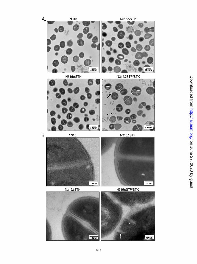
Role of STP/STK in S. aureus cell wall morphology. To moreclosely examine any morphological changes between mutants,we subjected stationary-phase cultures of the three deletionstrains (N315�STP, N315�STK, and N315�STP/STK) and thewild-type strain (N315) to TEM. Results revealed clear defectsin cell division and cell septum formation when both the stpand stk genes were deleted (Fig. 4A and B, white arrows).N315�STP/STK cells also displayed a noticeable increase inoverall cell size (�25% compared to N315). Although thesechanges were not apparent to a significant extent in N315�STP
or N315�STK, the cell wall thickness of N315�STP mutantswas nearly double that of the other three strains (Fig. 4B).Further examination of cell wall and membrane regions ofN315�STK and N315�STP/STK cells at higher resolution(Fig. 4B) revealed characteristically weak, electron-dense,wavy, and often interrupted membranes with a fragile appear-ance, as opposed to the thickly stained, distinctly visible, anduninterrupted cell walls seen in N315 and N315�STP cells.Additional notable observations in the N315�STP/STK TEMimages included the apparent “peeling” of cell wall or mem-brane-like material away from the bacterium (Fig. 4A, blackarrows). Notably, deletion of both stk and stp resulted in amore conspicuous phenotype than deletion of stk or stp alone,suggesting coordinated roles for STK and STP in the determi-nation of cell wall structure.
N315�STP mutants display alterations in lysostaphin sus-ceptibility. Based on the results obtained by TEM, we sus-pected that deletion of stp and/or stk may directly affect cellwall structure. We therefore determined the susceptibility ofeach mutant and its corresponding complemented strain to thepeptidoglycan-targeting glycylglycine endopeptidase lyso-staphin by measuring the decline in cell density (16). Upon theaddition of lysostaphin, the wild-type (N315) and double-mu-tant (N315�STP/STK) strains and all complemented strains(triangles) reached 50% lysis by approximately 20 min (Fig. 5),while N315�STP took a significantly longer incubation time(35 min, P � 0.002 compared to the wild type; Fig. 5B). No-tably, N315�STK reached 50% lysis by approximately 25 min(Fig. 5C), lysing more slowly than N315 but much less signif-
FIG. 2. Autoradiographs of in vitro kinase reactions. (A) Purified rSTK was incubated in kinase buffer with [�-32P]ATP and Mn2� alone andin the presence of MBP and rSTP as indicated. Proteins were resolved by SDS-PAGE and visualized by autoradiography. Shown are representativeautoradiographs. (B) Increasing amounts of purified rSTK were added to separate kinase reaction mixtures to observe the dose-dependent effect.One microgram of rSTK was used for the time course and cation dependency experiments, where minutes and cations are shown above theautoradiographs. (C) Thin-layer chromatography results showing phosphoamino acid standards stained with ninhydrin (left) and autophospho-rylated rSTK hydrolysis products revealed by autoradiography (right). The first and second dimensions are indicated by arrows.
1410 BELTRAMINI ET AL. INFECT. IMMUN.
on June 27, 2020 by guesthttp://iai.asm
.org/D
ownloaded from

icantly than N315�STP (P � 0.05 at 20 and 25 min comparedto the wild-type strain). The slopes and standard deviations ofthe N315, N315�STP, N315�STK, and N315�STP/STK lyso-staphin digestion curves determined by a linear regressionanalysis of the data points between 5 and 25 min (the timeperiod in which most of the lysis occurred) were �2.35 � 0.11,�1.48 � 0.031, �1.90 � 0.06, and �2.17 � 0.15, respectively.Control reactions carried out in the absence of lysostaphinshowed no significant changes in kinetics, indicating that theeffects observed at the given incubation times were due tolysostaphin and not autolysis. Based on these results, we con-cluded that N315�STP bacteria are more resistant to the ef-fects of lysostaphin than is the parent strain N315 as a directresult of the deletion of stp.
N315�STK and N315�STP/STK are more susceptible tocell wall-acting antibiotics. Conspicuous changes in cell divi-sion and wall morphology (Fig. 4) and altered lysostaphin
susceptibilities (Fig. 5) in STP- and/or STK-specific mutantssuggested that S. aureus N315 STP and STK may play animportant role in cell division and/or peptidoglycan structure.Changes in the cell wall of S. aureus have been shown to affectstaphylococcal susceptibility to certain antibiotics, particularlythose which target cell wall structure or synthesis (1, 31).Hence, we determined the susceptibilities of N315 and itsisogenic mutant strains to 22 antibiotics with various knowntargets. N315�STP did not show any change in susceptibilitycompared to the wild-type strain (Table 3). However, theN315�STK and, to a greater degree, N315�STP/STK mutantstrains displayed increased susceptibility to multiple antibioticstested (Table 3, bold values). Most appreciably, N315�STP/STK bacteria displayed susceptibility increases ranging from2-fold in the case of cefazolin to more than 50-fold in the caseof ertapenem, compared to N315. The affected antibiotics forboth N315�STK and N315�STP/STK included cephalosporins(cefazolin, ceftriaxone, cefotaxime) and carbapenems (ertap-enem, imipenem, meropenem), all of which act by interferingwith bacterial cell wall synthesis (12, 36, 37). To verify theseeffects, all of the strains were subjected to antibiotic sensitivitytesting with Etest strips containing gradients of representativeantibiotics from each of these groups, specifically, those inwhich the MICs changed for both N315�STK and N315�STP/STK (cefotaxime, ceftriaxone, and ertapenem). The MICs forthe wild-type and deletion strains were identical to those ob-tained by MicroScan (data not shown), while those forN315�STK�STK and N315�STP/STK�STP-STK returned tothose for the wild-type strain (Table 3), validating the Micro-Scan data. The increased selective susceptibility of N315�STKand N315�STP/STK to cell wall-acting antibiotics confirmed arole for STK in staphylococcal cell wall biosynthesis and struc-ture.
DISCUSSION
In this study, we report for the first time functional roles foran ESTK and an ESTP in S. aureus. ESTKs and ESTPs arecharacterized in a variety of bacteria with different geneticbackgrounds and pathogenic potentials; thus, deletion of oneor more of these enzymes results in different phenotypes basedon the bacteria being studied. For example, deletion of ESTKsresults in extremely long chain formation in Streptococcus aga-lactiae (39), aggregation in group A Streptococcus (22), and nosignificant changes in Enterococcus faecalis grown in nutrition-ally enriched medium (25). In the present investigation, dele-tion of both stk and stp in S. aureus resulted in dysregulation ofcell wall components and division machinery, as revealedby TEM and susceptibility testing to cell wall-acting antibiotics.
Like many other gram-positive ESTKs (22, 40), the extra-cellular domain of S. aureus STK contains a domain containingthree PASTA (penicillin-binding protein- and serine/threoninekinase-associated) repeats. PASTA domains contain one tofive PASTA repeats (25, 48), bind peptidoglycan (40), and arebelieved to recruit penicillin binding proteins (PBPs) to sites ofcell growth (48). Thus, it is possible that S. aureus STK alsouses PASTA repeats as an external sensor to recognize thelevel of unlinked peptidoglycan and mediate appropriate sig-naling cascades intracellularly via its cytoplasmic N-terminalkinase domain. Future experimental verification is required to
FIG. 3. Confirmation of mutant strains. (A) PCR analysis ofgDNA isolated from the indicated strains with primers specific forstp and stk. (B) Western blot assays of total cell lysates probedseparately with (top) anti-rSTP and (bottom) anti-rSTK productionsera (right) and corresponding prebleed (PB, left) sera. Lanes: 1,N315; 2, N315�STP; 3, N315�STK; 4, N315�STP/STK; 5,N315pCN40tet; 6, N315�STP�STP; 7, N315�STK�STK; 8,N315�STP/STK�STP/STK. Arrows indicate the locations of STKand STP within the lysates.
VOL. 77, 2009 S. AUREUS STK/STP-MEDIATED REVERSIBLE PHOSPHORYLATION REACTIONS 1411
on June 27, 2020 by guesthttp://iai.asm
.org/D
ownloaded from

determine whether this signaling cascade indeed facilitates thetranscription of one or more cell wall synthesis genes or in-volves direct phosphorylation of PBPs, as has been shown inmycobacteria (11).
Although dephosphorylation is an essential step in nearly allsignaling networks, information regarding the regulatoryrole(s) of ESTPs is limited. This may be due in part to theirrequirement for bacterial survival, as ESTP-specific mutationshave proven lethal in S. pyogenes (22). Mutants and variants ofS. agalactiae (39) and S. pneumoniae (35) have been reported,although detailed characteristics of these mutants are notavailable. In the present study, we were able to create both stpand stp/stk deletion mutants, indicating that in S. aureus N315,STP is not essential for survival. We speculatively attribute thisto the unique presence of other putative and nonconservedESTPs in the N315 genome (SA0140, SA1662, and SA2225).Notably, out of eight other S. aureus genomes we examined,homologs of SA2225, but not SA0140 and SA1662, were found
in only five (Mu50, MW2, RF122, MRSA252, and MSSA476,missing from USA300, NCTC8325, and COL) (http://genomics.ornl.gov/mist; 44). The presence of an additional noncotrans-cribing putative STK (SA0077) (44) may play a role in com-pensating for the loss of stk in the appropriate deletion strains.
Staphylococcal cell wall peptidoglycan is composed ofstrands of alternating N-acetylglucosamine and N-acetyl-muramic acid sugars joined together by amino acid side peptideswhich are cross-linked by pentaglycine chains (46). These pen-taglycine chains serve as a substrate for lysostaphin-mediatedcleavage. Changes in this structure directly affect lysostaphinbinding and cleavage activities in S. aureus (17). The mostnotable effects of the stp deletion alone were thicker cells wallscompared to those of wild-type S. aureus (Fig. 4B) and theperhaps consequential increased resistance to lysostaphin (Fig.5B). This suggests an alteration in peptidoglycan compositionand/or structure, specifically, pentapeptide bridge compositionand/or cross-linking frequency. Deletion of stk had a minor
FIG. 4. Analysis of cell morphology by TEM. Shown are representative fields of TSB-grown stationary-phase wild-type (control) and mutantS. aureus strains, as indicated. White arrows indicate abnormal septation; black arrows indicate evidence of apparent wall or membrane shedding.Magnifications: A, �30,500; B, �120,000.
FIG. 5. Lysostaphin sensitivity assays. Results indicate the susceptibilities of (A) N315; (B) N315�STP, (C) N315�STK, and (D) N315�STP/STK; and their appropriate complemented strains (triangles) to lysostaphin. Autolysis (no lysostaphin) control results are shown as closed squares.Data shown are the averages of three independent experiments. Error bars indicate standard deviations. Data are presented as percentages of theoriginal culture turbidity. OD620, optical density at 620 nm.
VOL. 77, 2009 S. AUREUS STK/STP-MEDIATED REVERSIBLE PHOSPHORYLATION REACTIONS 1413
on June 27, 2020 by guesthttp://iai.asm
.org/D
ownloaded from

effect on lysostaphin susceptibility, while deletion of both stpand stk did not affect lysostaphin susceptibility (Fig. 5B). Thiscould be due to compensatory mechanisms (such as the afore-mentioned noncotranscribing STK and STPs) being activatedin the absence of this primary STP-STK pair, as it is notuncommon for STKs to share substrates (23). Additionally, theabsence of the extracellular, environment-sensing PASTA do-main upon deletion of stk or stp/stk may result in mutants withdefects in the ability to sense unlinked peptidoglycan (40) andregulate pentaglycine chain formation by way of PBP recruit-ment (48). The synchronized role of STK and STP may there-fore be maintaining equilibrium in cross-linking. Increased re-sistance to lysostaphin digestion in the absence of stp couldthus be due to an absence of substrate or to uncontrolledrecruitment of PBPs and resultant increases in cross-linking.While our ongoing experiments analyzing cell wall structures inthese mutants may reveal alterations, increased lysostaphinresistance in the absence of stp is consistent with the increasedcell wall thickness observed (Fig. 4B).
Deletion of stk alone resulted in increased susceptibility tocell wall-acting �-lactam antibiotics (Table 3), as has beenreported for E. faecalis ESTK PrkC deletion strains (25), andincreased cell wall fragility (Fig. 4). Importantly, deletion ofboth stp and stk significantly enhanced these phenotypes, re-sulting in further selective antibiotic susceptibility (Table 3)and pronounced defects in cell division (Fig. 4). These additiveeffects, combined with the ability of rSTP to reverse STK-mediated phosphorylation (Fig. 2A) and the cotranscribingnature of stp and stk (Fig. 1B), suggest that STK and STP areindeed cognate proteins. Thus, we believe that although STPand STK, on their own, may contribute to the regulation of
certain cell wall synthesis steps, it is probably their coordinatedreversible phosphorylation which is most critical.
It is important to note that only MICs of �-lactam antibioticswhich specifically target cell wall synthesis were affected by thedeletion of stk or stk and stp. PBPs, which have transpeptidase,transglycosylase, and/or carboxypeptidase activities to facilitatepeptidoglycan synthesis (29), are often the primary target of�-lactam antibiotics. Meropenems, and specifically ertapenem,bind a variety of PBPs to inhibit cell wall synthesis, particularlyPBP2 and -3 in E. coli (36). Cephalosporins also directly in-teract with PBPs. Ceftriaxone has been shown to bind tightly toS. aureus PBP1 (12), and a more potent cefotaxime binds S.aureus PBP1 and -2 with a higher affinity than other PBPs (15,37). Based on the ceftriaxone- and cefotaxime-sensitive phe-notypes observed in N315�STK and N315�STP/STK in thisstudy, it is reasonable to believe that STK and coordinatedSTP/STK signaling may, in some way, modulate the function-ality of PBP1 and/or -2. PBPs have also been implicated in avariety of functions, including cell division and septum forma-tion, by directly interacting with cell division proteins, includ-ing FtsZ (29, 38, 45), and modulating peptidoglycan synthesisin S. aureus specifically (27). Thus, the alterations in cell wallstructure and division and increased antibiotic susceptibility ofS. aureus in the absence of STP and/or STK in the presentstudy are likely attributable to the altered recruitment andregulation of S. aureus PBPs and other necessary cell wallsynthesis and division proteins.
Taken together, the results shown in the present studyclearly indicate that S. aureus STP and STK and their coordi-nated phosphorylation play an important role in cell wall struc-ture, cell division, and �-lactam antibiotic susceptibility. In the
TABLE 3. MICs of antibiotics tested
Antibiotic(s)
MIC(s) (�g/ml) for strain:
N315 N315�STP N315�STK N315�STK�STK N315�STP/STK N315�STP/STK�STP/STK
Most affectedCefazolin �16 �16 �16 <8Cefotaxime �256 �256 43 �256a 12 �256Ceftriaxone �32 32 <8 �32 <8 �32Ertapenem �32 �32 18 �32 0.6 �32Imipenem �8 8 8 <4
UnaffectedAmoxicillin-clavulanate �4/2 �4/2 �4/2 �4/2Ampicillin-sulbactam �8/4 �8/4 �8/4 �8/4Ampicillin �8 �8 �8 �8Clindamycin �4 �4 �4 �4Erythromycin �4 �4 �4 �4Gentamicin �4 �4 �4 �4Levofloxacin �2 �2 �2 �2Linezolid 2 2 2 2Moxifloxacin �2 �2 �2 �2Nitrofurantoin �32 �32 �32 �32Oxacillin �2 �2 �2 �2Penicillin �8 �8 �8 �8Rifampin �1 �1 �1 �1Synercid �0.5 �0.5 �0.5 �0.5Tetracycline �4 �4 �4 �4Trimethoprim-sulfamethoxazole �0.5/9.5 �0.5/9.5 �0.5/9.5 �0.5/9.5
a The complementation MICs shown are averages of three independent experiments with Etest strips. The antibiotic gradients on the Etest strips were 0.016 to 256�g/ml (cefotaxime and ceftriaxone) and 0.002 to 32 �g/ml (ertapenem).
1414 BELTRAMINI ET AL. INFECT. IMMUN.
on June 27, 2020 by guesthttp://iai.asm
.org/D
ownloaded from

context of the dramatic rise in antimicrobial resistance, as wellas the continued prevalence of staphylococci in hospital envi-ronments, the present study provides an important insight toconsider, as these enzymes have the potential to serve as noveltargets for alternative antibiotic therapy.
ACKNOWLEDGMENTS
We thank Olaf Schneewind and Taeok Bae for pKOR1, Emman-uelle Charpentier for pCN36 and pCN40, Hong Jin for thin-layerchromatography, and Preeti Pancholi for antibiotic susceptibility test-ing by MicroScan.
A.B. is a recipient of two subsequent training grant awards from theNational Institutes of Health (T32GM068412, a Systems in IntegrativeBiology fellowship administered through the Integrative BiomedicalGraduate Program, and T32AI065411, an NRSA training grant ad-ministered by the Center for Microbial Interface Biology). This workwas also supported by NIH grant AI064912 and an OSUMC StrategicInitiative grant (V.P.).
REFERENCES
1. Antignac, A., K. Sieradzki, and A. Tomasz. 2007. Perturbation of cell wallsynthesis suppresses autolysis in Staphylococcus aureus: evidence for coregula-tion of cell wall synthetic and hydrolytic enzymes. J. Bacteriol. 189:7573–7580.
2. Arnaud, M., A. Chastanet, and M. Debarbouille. 2004. New vector forefficient allelic replacement in naturally nontransformable, low-GC-content,gram-positive bacteria. Appl. Environ. Microbiol. 70:6887–6891.
3. Av-Gay, Y., and M. Everett. 2000. The eukaryotic-like Ser/Thr protein ki-nases of Mycobacterium tuberculosis. Trends Microbiol. 8:238–244.
4. Bae, T., and O. Schneewind. 2006. Allelic replacement in Staphylococcusaureus with inducible counter-selection. Plasmid 55:58–63.
5. Bakal, C. J., and J. E. Davies. 2000. No longer an exclusive club: eukaryoticsignaling domains in bacteria. Trends Cell Biol. 10:32–37.
6. Bork, P., N. P. Brown, H. Hegyi, and J. Schultz. 1996. The protein phos-phatase 2C (PP2C) superfamily: detection of bacterial homologues. ProteinSci. 5:1421–1425.
7. Brodsky, L. I., V. V. Ivanov, Y. L. Kalaydzidis, A. M. Leontovich, V. K. Nikolaev,S. I. Feranchuk, and V. A. Drachev. 1995. GeneBee-NET: internet-based serverfor analyzing biopolymers structure. Biochemistry 60:923–928.
8. Chaffin, D. O., and C. E. Rubens. 1998. Blue/white screening of recombinantplasmids in gram-positive bacteria by interruption of alkaline phosphatasegene (phoZ) expression. Gene 219:91–99.
9. Charpentier, E., A. I. Anton, P. Barry, B. Alfonso, Y. Fang, and R. P. Novick.2004. Novel cassette-based shuttle vector system for gram-positive bacteria.Appl. Environ. Microbiol. 70:6076–6085.
10. Clinical and Laboratory Standards Institute. 2008. Performance standardsfor antimicrobial susceptibility testing; 18th informational supplement. Clin-ical and Laboratory Standards Institute, Wayne, PA.
11. Dasgupta, A., P. Datta, M. Kundu, and J. Basu. 2006. The serine threoninekinase PknB of Mycobacterium tuberculosis phosphorylates PBPA, a penicil-lin-binding protein required for cell division. Microbiology 152:493–504.
12. Davies, T. A., M. G. P. Page, W. Shang, T. Andrew, M. Kania, and K. Bush.2007. Binding of ceftobiprole and comparators to the penicillin-binding proteinsof Escherichia coli, Pseudomonas aeruginosa, Staphylococcus aureus, and Strep-tococcus pneumoniae. Antimicrob. Agents Chemother. 51:2621–2624.
13. de Hoon, M. J. L., Y. Makita, K. Nakai, and M. Miyano. 2005. Prediction oftranscriptional terminators in Bacillus subtilis and related species. PLoSComput. Biol. 1:e25.
14. Echenique, J., A. Kadioglu, S. Romao, P. W. Andrew, and M. C. Trombe.2004. Protein serine/threonine kinase StkP positively controls virulence andcompetence in Streptococcus pneumoniae. Infect. Immun. 72:2434–2437.
15. Georgopapadakou, N. H., B. A. Dix, and Y. R. Mauriz. 1986. Possible phys-iological functions of penicillin-binding proteins in Staphylococcus aureus.Antimicrob. Agents Chemother. 29:333–336.
16. Grundling, A., D. M. Missiakas, and O. Schneewind. 2006. Staphylococcus aureusmutants with increased lysostaphin resistance. J. Bacteriol. 188:6286–6297.
17. Grundling, A., and O. Schneewind. 2006. Cross-linked peptidoglycan medi-ates lysostaphin binding to the cell wall envelope of Staphylococcus aureus. J.Bacteriol. 188:2463–2472.
18. Grundmann, H., M. Aires-de-Sousa, J. Boyce, and E. Tiemersma. 2006.Emergence and resurgence of meticillin-resistant Staphylococcus aureus as apublic-health threat. Lancet 368:874–885.
19. Hanks, S. K., A. M. Quinn, and T. Hunter. 1988. The protein kinase family:conserved features and deduced phylogeny of the catalytic domains. Science241:42–52.
20. Haugen, S. P., W. Ross, and R. L. Gourse. 2008. Advances in bacterialpromoter recognition and its control by factors that do not bind DNA. Nat.Rev. Microbiol. 6:507–519.
21. Hussain, H., P. Branny, and E. Allan. 2006. A eukaryotic-type serine/threo-nine protein kinase is required for biofilm formation, genetic competence,and acid resistance in Streptococcus mutans. J. Bacteriol. 188:1628–1632.
22. Jin, H., and V. Pancholi. 2006. Identification and biochemical characteriza-tion of a eukaryotic-type serine/threonine kinase and its cognate phos-phatase in Streptococcus pyogenes: their biological functions and substrateidentification. J. Mol. Biol. 357:1351–1372.
23. Kang, C. M., D. A. Abbott, S. T. Park, C. C. Dascher, L. C. Cantley, and R. N.Husson. 2005. The Mycobacterium tuberculosis serine/threonine kinase PknAand PknB: substrate identification and regulation of cell shape. Genes Dev.19:1692–1704.
24. Kreiswirth, B. N., S. Lofdahl, M. J. Betley, M. O’Reilly, P. M. Schlievert,M. S. Bergdoll, and R. P. Novick. 1983. The toxic shock syndrome exotoxinstructural gene is not detectably transmitted by a prophage. Nature 305:709–712.
25. Kristich, C. J., C. L. Wells, and G. M. Dunny. 2007. A eukaryotic-type ser/thrkinase in Enterococcus faecalis mediates antimicrobial resistance and intes-tinal persistence. Proc. Natl. Acad. Sci. USA 104:3508–3513.
26. Kuroda, M., T. Ohta, I. Uchiyama, T. Baba, H. Yuzawa, I. Kobayashi, L. Cui,A. Oguchi, K. Aoki, Y. Nagai, J. Lian, T. Ito, M. Kanamori, H. Matsumaru,A. Maruyama, H. Murakami, A. Hosoyama, Y. Mizutani-Ui, N. K. Taka-hashi, T. Sawano, R. Inoue, C. Kaito, K. Sekimizu, H. Hirakawa, S. Kuhara,S. Goto, J. Yabuzaki, M. Kanehisa, A. Yamashita, K. Oshima, K. Furuya, C.Yoshino, T. Shiba, M. Hattori, N. Ogasawara, H. Hayashi, and K. Hira-matsu. 2001. Whole genome sequencing of methicillin-resistant Staphylococ-cus aureus. Lancet 357:1225–1240.
27. Łeski, T. A., and A. Tomasz. 2005. Role of penicillin-binding protein 2(PBP2) in the antibiotic susceptibility and cell wall cross-linking of Staphy-lococcus aureus: evidence for the cooperative functioning of PBP2, PBP4,and PBP2A. J. Bacteriol. 187:1815–1824.
28. Lowy, F. D. 1998. Staphylococcus aureus infections. N. Engl. J. Med. 339:520–532.
29. Macheboeuf, P., C. Contreras-Martel, V. Job, D. Otto, and A. Dessen. 2006.Penicillin binding proteins: key players in bacterial cell cycle and drug resis-tance processes. FEMS Microbiol. Rev. 30:673–691.
30. Madec, E., A. Laszkiewicz, A. Iwanicki, M. Obuchowski, and S. Seror. 2002.Characterization of a membrane-linked Ser/Thr protein kinase in Bacillussubtilis, implicated in developmental processes. Mol. Microbiol. 46:571–586.
31. Mainardi, J. L., R. Villet, T. D. Bugg, C. Mayer, and M. Arthur. 2008.Evolution of peptidoglycan biosynthesis under the selective pressure of an-tibiotics in gram-positive bacteria. FEMS Microbiol. Rev. 32:386–408.
32. Motley, S. T., and S. Lory. 1999. Functional characterization of a serine/threonine protein kinase of Pseudomonas aeruginosa. Infect. Immun. 67:5386–5394.
33. Mukhopadhyay, S., V. Kapatral, W. Xu, and A. M. Chakrabarty. 1999.Characterization of a Hank’s type serine/threonine kinase and serine/threo-nine phosphoprotein phosphatase in Pseudomonas aeruginosa. J. Bacteriol.181:6615–6622.
34. Munoz-Dorado, J., S. Inouye, and M. Inouye. 1991. A gene encoding aprotein serine/threonine kinase is required for normal development of M.xanthus, a gram-negative bacterium. Cell 67:995–1006.
35. Novakova, L., L. Saskova, P. Pallova, J. Janeeek, J. Novotna, A. Ulrych, J.Echenique, M.-C. Trombe, and P. Branny. 2005. Characterization of a eu-karyotic type serine/threonine protein kinase and protein phosphatase ofStreptococcus pneumoniae and identification of kinase substrates. FEBS Lett.272:1243–1254.
36. Odenholt, I. 2001. Ertapenem: a new carbapenem. Expert Opin. Investig.Drugs 10:1157–1166.
37. Palmer, S. M., S. L. Kang, D. M. Cappelletty, and M. J. Rybak. 1995.Bactericidal killing activities of cefepime, ceftazidime, cefotaxime, and ceftri-axone against Staphylococcus aureus and �-lactamase-producing strains ofEnterobacter aerogenes and Klebsiella pneumoniae in an in vitro infectionmodel. Antimicrob. Agents Chemother. 39:1764–1771.
38. Pereira, S. F. F., A. O. Henriques, M. G. Pinho, H. de Lencastre, and A.Tomasz. 2007. Role of PBP1 in cell division of Staphylococcus aureus. J.Bacteriol. 189:3525–3531.
39. Rajagopal, L., A. Clancy, and C. E. Rubens. 2003. A eukaryotic type serine/threonine kinase and phosphatase in Streptococcus agalactiae reversiblyphosphorylate an inorganic pyrophosphatase and affect growth, cell segre-gation, and virulence. J. Biol. Chem. 278:14429–14441.
40. Shah, I. M., M.-H. Laaberki, D. Popham, and J. Dworkin. 2008. A eukary-otic-like Ser/Thr kinase signals bacteria to exit dormancy in response topeptidoglycan fragments. Cell 135:486–496.
41. Siegel, J. D., E. Rhinehart, M. Jackson, and L. Chiarello. 2006. Managementof multidrug-resistant organisms in healthcare settings. Centers for DiseaseControl and Prevention, Atlanta, GA.
42. Thakur, M., and P. K. Chakraborti. 2006. GTPase activity of mycobacterialFtsZ is impaired due to its transphosphorylation by the eukaryotic typeser/thr kinase, PknA. J. Biol. Chem. 281:40107–40113.
43. Ulrich, L. E., E. V. Koonin, and I. B. Zhulin. 2005. One-component systemsdominate signal transduction in prokaryotes. Trends Microbiol. 13:52–56.
VOL. 77, 2009 S. AUREUS STK/STP-MEDIATED REVERSIBLE PHOSPHORYLATION REACTIONS 1415
on June 27, 2020 by guesthttp://iai.asm
.org/D
ownloaded from

44. Ulrich, L. E., and I. B. Zhulin. 2007. MiST: a microbial signal transductiondatabase. Nucleic Acids Res. 35:D386–D390.
45. Varma, A., and K. D. Young. 2004. FtsZ collaborates with penicillin binding proteinsto generate bacterial cell shape in Escherichia coli. J. Bacteriol. 186:6768–6774.
46. Vollmer, W., D. Blanot, and M. A. de Pedro. 2008. Peptidoglycan structureand architecture. FEMS Microbiol. Rev. 32:149–167.
47. Wang, J., C. Li, H. Yang, A. Mushegian, and S. Jin. 1998. A novel serine/threonine protein kinase homologue of Pseudomonas aeruginosa is specifi-cally inducible within the host infection site and is required for full virulencein neutropenic mice. J. Bacteriol. 180:6764–6768.
48. Yeats, C., R. D. Finn, and A. Bateman. 2002. The PASTA domain: a �-lac-tam-binding protein. Trends Biochem. Sci. 27:438–440.
Editor: A. Camilli
1416 BELTRAMINI ET AL. INFECT. IMMUN.
on June 27, 2020 by guesthttp://iai.asm
.org/D
ownloaded from