![Clinical Study High Intensity Focused Ultrasound versus ... · Clinical Study High Intensity Focused Ultrasound versus ... Nazareno Suardi ... ca-tion system [ ].](https://static.fdocuments.us/doc/165x107/5af97c097f8b9aff288d3dc0/clinical-study-high-intensity-focused-ultrasound-versus-study-high-intensity.jpg)
Modeling of Thermal Damage from Focused Ultrasound ...
26
Modeling of Thermal Damage from Focused Ultrasound Exposures for Heterogeneous Tissues Adam C. Waspe, PhD [email protected] Aug. 11, 2014 * Some content adapted from lecture notes from Rajiv Chopra and Charles Mougenot and cited references
Transcript of Modeling of Thermal Damage from Focused Ultrasound ...
Modeling of Thermal Damage from
Focused Ultrasound Exposures for
Heterogeneous Tissues
Adam C. Waspe, [email protected]
Aug. 11, 2014
* Some content adapted from lecture notes from Rajiv Chopra and
Charles Mougenot and cited references

What is Focused Ultrasound?
• Focused ultrasound is a noninvasive technique to enhance biological therapies by exposing tissues to acoustic energy:
– Spatial / temporal control over temperature
– Localized drug delivery (thermal, mechanical)
– Functional / structural modification of tissues
• Clinical adoption of FUS has expanded rapidly in recent years due to an active research community, strong commercial support, and better visualization/thermometry tools
• Paediatric/foetal applications are starting to be explored, due to the potential to delivery a non-ionizing energy based therapy, in a noninvasive manner

Focused Ultrasound Principles
• Ultrasound generates 2 types of waves when interacting with tissue– Longitudinal (fluids, soft tissue and bone), and shear waves (bone only)
– Pressure is positive during compression and negative during rarefaction of the wave
• As waves traverse a lossy medium, attenuation (absorption, scattering and mode conversion) reduces the energy delivery
• Waves are focused geometrically, mechanically, or electronically to aim all the energy emitted from the transducer into a small target
• Acoustic intensity (power focused over a small area) determines the amount of thermal energy deposited at the focus
f
c=λ

How is Acoustic Energy Described
• Electrical Power: delivered to the transducer by the RF amplifier [W]
• Acoustic Power: electrical power de-rated by the measured transducer efficiency (η) [W]
• Acoustic Intensity (I): Majority of the acoustic power traverses through the FWHM of the focus [W/cm2]
• Acoustic Pressure (P): Peak positive (compressional) and peak negative (rarefactional) pressure of the longitudinal ultrasound wave [MPa]
DiaT
FFWHM
λ22.1=
c
PI
ρ2
2
=
∫∫∞
= dxdyyxIPower ),(
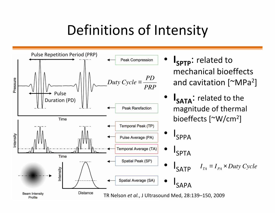
Definitions of Intensity
• ISPTP: related to
mechanical bioeffects
and cavitation [~MPa2]
• ISATA: related to the
magnitude of thermal
bioeffects [~W/cm2]
• ISPPA
• ISPTA
• ISATP
• ISAPA5
Pulse Repetition Period (PRP)
CycleDutyII PATA ×=
PRP
PDCycleDuty =
Pulse
Duration (PD)
TR Nelson et al., J Ultrasound Med, 28:139–150, 2009
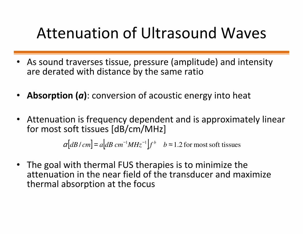
Attenuation of Ultrasound Waves
• As sound traverses tissue, pressure (amplitude) and intensity are derated with distance by the same ratio
• Absorption (a): conversion of acoustic energy into heat
• Attenuation is frequency dependent and is approximately linear for most soft tissues [dB/cm/MHz]
• The goal with thermal FUS therapies is to minimize the attenuation in the near field of the transducer and maximize thermal absorption at the focus
[ ] [ ] essoft tissumost for 2.1/ 11 ≈= −−bfMHzcmdBacmdB
bα

Attenuation of Ultrasound Waves
• Attenuation at a depth (d) is modeled as an
exponential decay of the wave amplitude
(basee)
• Attenuation is reported using dB (base10)
• The Neper [Np] is a basee logarithmic ratio
[ ] ( ) [ ]
[ ]
−==
=→≈=
dP
PdB
dBNpcmNpecmdB
o
d
aa
7.8explog20log20Pressure Relative
7.81/7.8log20/
1010
10
αµµα
d
odaePdP
µ−=)(
)(dB levelintensity Relativelog10log20)(dB level pressure Relative 1010 =≡=o
d
o
d
I
I
P
P
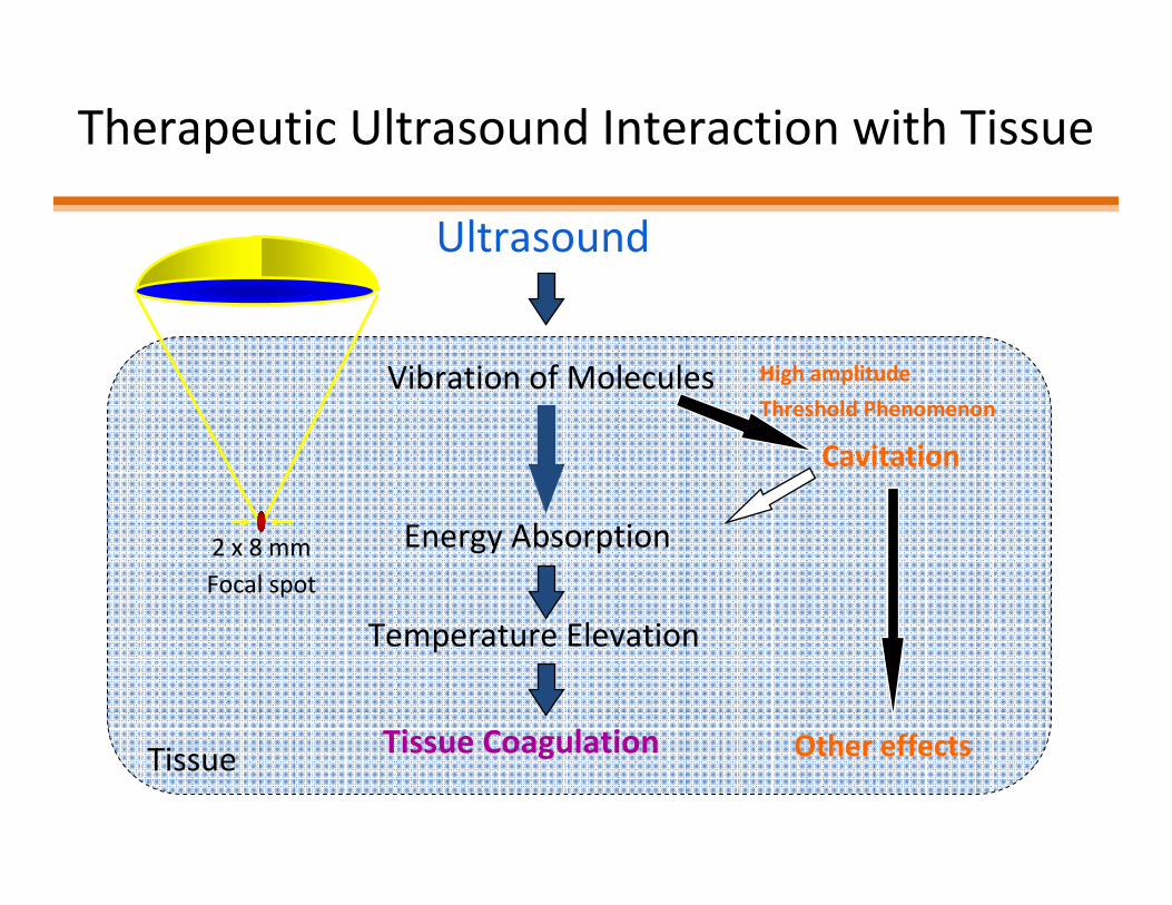
Therapeutic Ultrasound Interaction with Tissue
Ultrasound
Tissue Coagulation
Energy Absorption
Temperature Elevation
Vibration of Molecules
Cavitation
Other effectsTissue
High amplitude
Threshold Phenomenon
2 x 8 mm
Focal spot

Ultrasound Treatment Techniques
• Cavitation: high-power pulsed-wave (PW) exposures (100-500W, 0.1-10% duty cycle, 1um to 100 ms burst durations) to mechanically break up tissues
• Ablation: high-power continuous-wave (CW) exposures (10-200W, 5-60s exposures) to thermally coagulate tissues
• Hyperthermia: low-power CW exposures to locally control temperatue without coagulation
• Sonoporation: low-power PW exposures (usually combined with a microbubble contrast agent) to mechanically weaken cell membranes, open tight-junctions between cells, etc.

HIFU Treatment of Bone Tumours
Gd-T1-w MRI of an 18-year-old woman
who underwent HIFU ablation for tibia
osteosarcoma. (a) Before HIFU treatment
shows a hypervascular lesion (arrow) in
the tibia. Images obtained (b) 2 weeks
and (c) 12, (d) 24, and (e) 36 months after
HIFU show no evidence of enhancement
in treated tumor region (arrow).
[1] Primary Bone Malignancy: Effective Treatment with High-Intensity
Focused Ultrasound Ablation Chen W., et al., Radiol., 2010; 255(3):967-78.
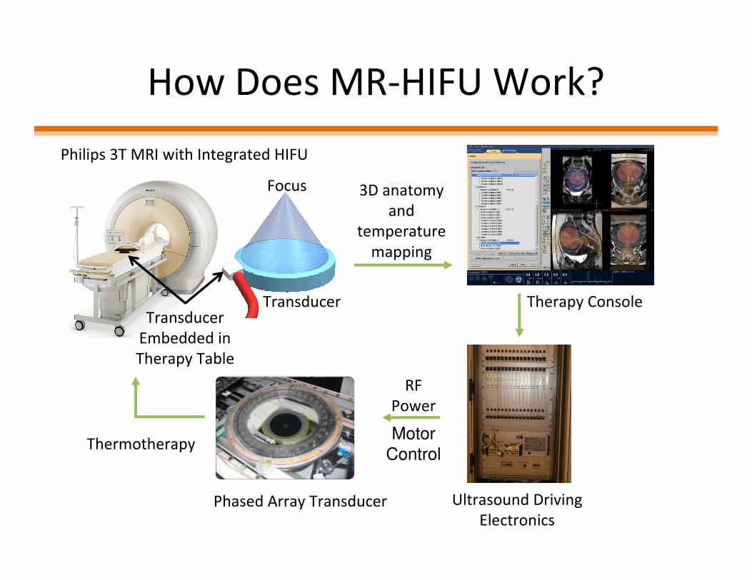
How Does MR-HIFU Work?
3D anatomy
and
temperature
mapping
Phased Array Transducer
Philips 3T MRI with Integrated HIFU
Therapy Console
Thermotherapy
RF
Power
Motor
Control
Ultrasound Driving
Electronics
Focus
TransducerTransducer
Embedded in
Therapy Table

System Setup
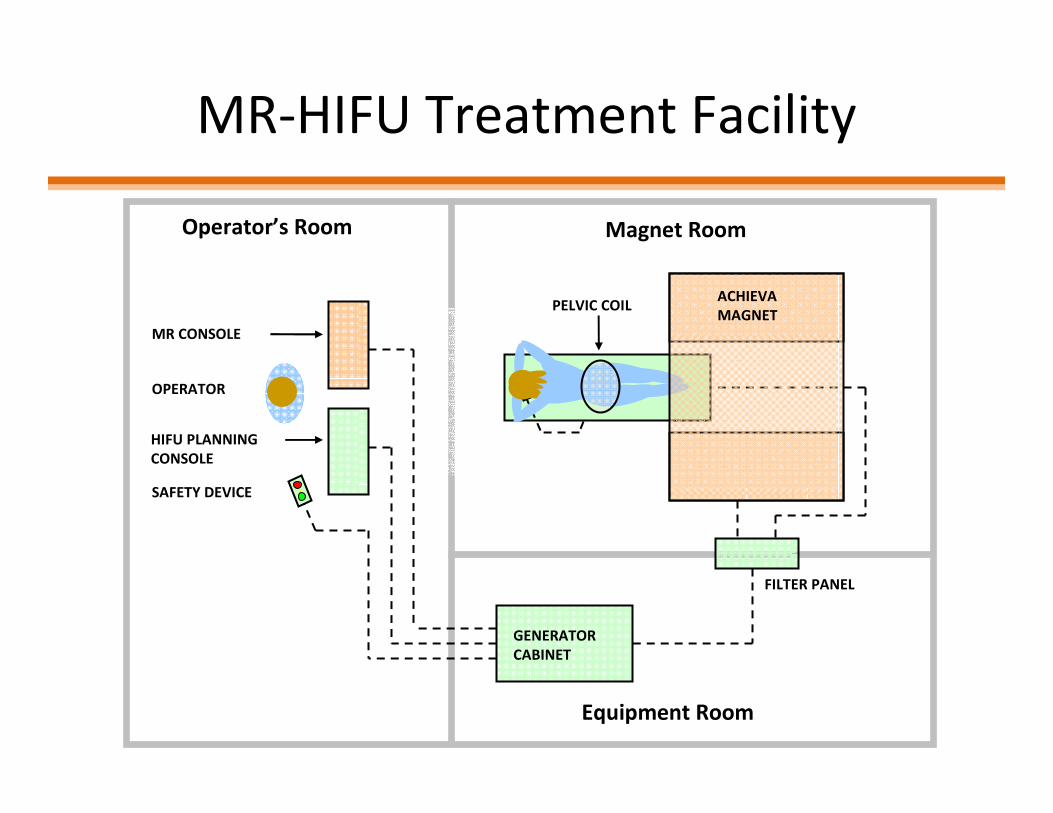
MR-HIFU Treatment Facility
OPERATOR
MR CONSOLE
HIFU PLANNING
CONSOLE
.SAFETY DEVICE
FILTER PANEL
GENERATOR
CABINET
Operator’s Room Magnet Room
Equipment Room
PELVIC COILACHIEVA
MAGNET
MRI Thermometry
• Temperature measurement is based on the water proton resonance frequency (PRF) shift induced phase differences between dynamic frames
• Temperature in bone and fat tissue can not be measured with the PRF method
• From MR dynamic phase images a relative frequency shift is be calculated
• The phase of a MR image is sensitive to disturbances such as transducer movement and magnetic field drift and to patient movement
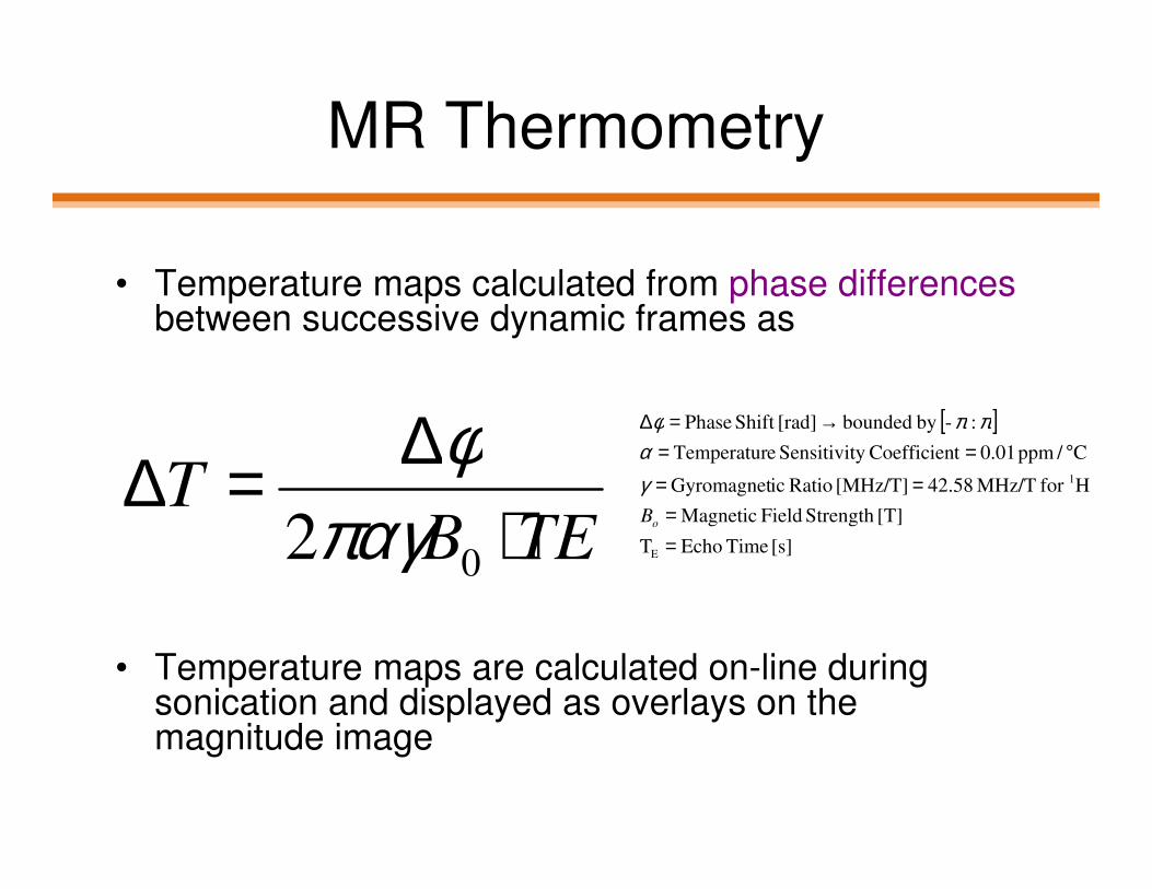
MR Thermometry
• Temperature maps calculated from phase differencesbetween successive dynamic frames as
• Temperature maps are calculated on-line during sonication and displayed as overlays on the magnitude image
TEBT
⋅∆=∆
02παγφ [ ]
[s] Time EchoT
[T]Strength Field Magnetic
Hfor MHz/T 42.58[MHz/T] Ratio icGyromagnet
C/ppm 0.01tCoefficieny Sensitivit eTemperatur
:-by bounded [rad]Shift Phase
E
1
==
==°==
→=∆
oB
γα
ππφ

Bone MR Thermometry Example
Heating signal is strong at bone surface but non-existant in the
cortical bone and fatty bone marrow.

Thermal Dose Model
• Thermal dose is calculated on a voxel-by-voxel basis as a time integral as temperature is measured throughout treatment
• 240 EM (equivalent minutes) at 43°C is sufficient to cause thermal necrosis in “soft tissue”
• Caution: a 1 second exposure at 57°C can produce thermal necrosis (273 EM)
∫∫∫∫−−−−====
t
tTdtrtTD
0
))(43()( )43(50.0
)43(25.0
CTr
CTr
°°°°>>>>====°°°°<<<<====

Advantages with this Model
• The increase in the rate of cell killing with temperature is relatively constant (for T>43, T<43)– For every degree above 43°C the required time to
coagulate the tissue halves (120 minutes @ 44°C, 60 minutes @ 45°C)
• Formulation relates all time-temperature curves back to a single temperature, chosen arbitrarily as 43°C – trend seems to be conserved across multiple cell types, even though sensitivity to heat will differ
• Model valid for high temperatures seen in HIFU
• Valid for tissues with different thermal sensitivity but threshold for thermal dose required for cell death changes

Problems with this Model
• Different tissues have varying thermal sensitivity and will ablate at different thermal doses:– “soft tissue” will become necrotic at 240 EM
– Nerve tissue may damage at much lower doses
– Bone may require much higher dose for ablation
• Non-linear response between temperature and cell death à higher probability of dying with increasing temperature and time of exposure
• Measuring dose does not directly predict damage
• Model primarily validated for cell cultures so ambiguity between calculated thermal dose and ablation volumes from imaging/pathology

Pennes Bioheat Transfer Model
• Proposed in 1940s for modeling heat transfer
in the body due to an externally applied
heating/cooling sources
• Harry Pennes (a Neurologist at Columbia Univ)
experimented on patients by inserting
thermocouples into patients’ forearms
• Model accounts the thermal conductivity,
specific heat capacity, and blood perfusion of
specific tissue types (muscle, organs, skin, etc)
20

Pennes Bioheat Transfer Equation
• Validated heating model (does not predict
dose or thermal damage) that has “stood the
test of time” against other models and is
applicable to many different heating source
types (ultrasonic, RF, laser, etc.)
• Highly dependent on “good”
tissue properties
ρc = ∂T
∂t= ∇ ⋅ k∇T − ωcb T − Tb( ) + Q
ρ = Tissue Density [kg/m3]
c = Specific Heat Capacity [J/kg/°C]
k = Thermal Conductivity [W/m/°C]
ω = Blood Perfusion [kg/m3 /s]
Q = Heat Deposition from Ultrasound [W/m3]
T = Temperature [°C]
Tb = Arterial Blood Temperature [37°C]
t = Time [s]

Modeling Questions
1. Can a tissue type dependant thermal damage model (similar to the Pennes Bioheat model for tissue heating) be derived that takes into account the thermal dose, thermal conductivity, thermal diffusion, specific heat, and perfusion of the tissue of interest and surrounding structures?
1. Can a spatially dependant (and perhaps a patient specific) thermal damage model be derived which can work directly with intraoperative temperature measurements to better predict the volume of ablated tissue during a MR-HIFU treatment?
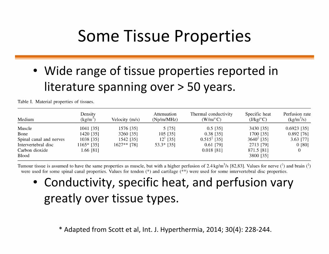
Some Tissue Properties
• Wide range of tissue properties reported in
literature spanning over > 50 years.
• Conductivity, specific heat, and perfusion vary
greatly over tissue types.
* Adapted from Scott et al, Int. J. Hyperthermia, 2014; 30(4): 228-244.
* Adapted from Dewhirst et al, Int. J.
Hyperthermia, 2003; 19(3): 267-294.
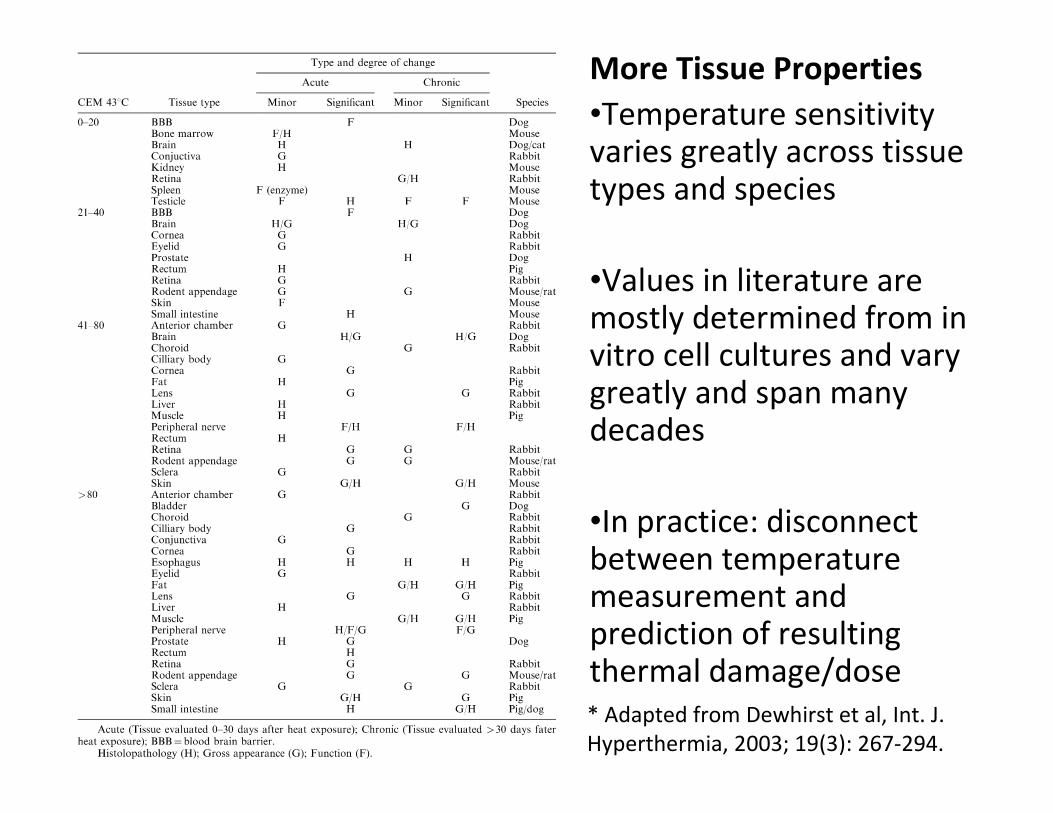
More Tissue Properties
•Temperature sensitivity varies greatly across tissue types and species
•Values in literature are mostly determined from in vitro cell cultures and vary greatly and span many decades
•In practice: disconnect between temperature measurement and prediction of resulting thermal damage/dose

Summary of HIFU
• MR-HIFU is a flexible energy-based treatment modality
• Focusing the ultrasound beam enhances the treatment over a small target area by a factor of ~1000X
• Attenuation is the primary contributor to both losses on the way to the target AND is the mechanism for thermal treatment at the target
• MR thermometry “closes the loop” to monitor and control treatment temperatures in real time
• Caution: thermal dose builds up cumulatively and at an increasing rate with temperature à Current models do not account for differences in tissue types (may under/ over dose thermo-resistive/sensitive tissues)

References
[1] Sapareto S.A. and Dewey W.C., Thermal dose determination in cancer therapy. Int J Radiat Oncol Biol Phys, 10(6): p. 787-800 (1984).
[2] Sassaroli E., Li K.C.P., O’Neill B.E., Modeling focused ultrasound exposure for the optimal control of thermal dose distribution. The ScientificWorld Journal, Article ID 252741, 11 pages, (2012).
[3] Dewey W.C., Arrhenius relationships from molecule and cell to the clinic. Int J Hyperthermia, 10: p.457-83 (1994).
[4] Pennes H.H., Analysis of tissue and arterial blood temperatures in the resting human forearm. J Appl Physiol, 1: p. 93-122 (1948).
[5] Scott S.J., et al, Interstitial ultrasound ablation of vertebral and paraspinal tumours: Parametric and patient-specific simulations. Int J Hyperthermia, 30(4): p. 228-244 (2014).
[6] Dewhirst M.W., et al, Basic principles of thermal dosimetry and thermal thresholds for tissue damage from hyperthermia. Int J Hyperthermia, 19(3): p. 267-294 (2003).
[7] Roemer R.B., Engineering aspects of hyperthermia therapy, AnnuRev Biomed Eng, 1: p. 347-376 (1999).

![High Intensity Focused Ultrasound Facial Skin Lifting · HIFU Mechanism HIFU [High Intensity Focused Ultra-sound] • Non-invasive treatment with thermal energy that focused ultrasound](https://static.fdocuments.us/doc/165x107/5fd8347ce1f6a6277a18bb54/high-intensity-focused-ultrasound-facial-skin-hifu-mechanism-hifu-high-intensity.jpg)